Article

Mechanism of Motion Direction Detection Based on Barlow’s Retina Inhibitory Scheme in Direction-Selective Ganglion Cells

1
Faculty of Electrical and Computer Engineering, Kanazawa University, Kakuma-machi, Kanazawa 920-1192, Japan
2
Department of Intelligence Information Systems, University of Toyama, 3190 Gofuku, Toyama 930-8555, Japan
*
Authors to whom correspondence should be addressed.
Electronics 2021, 10(14), 1663; https://doi.org/10.3390/electronics10141663
Submission received: 12 May 2021 / Revised: 9 July 2021 / Accepted: 10 July 2021 / Published: 13 July 2021
(This article belongs to the Section Bioelectronics)

Abstract

:
Previous studies have reported that directionally selective ganglion cells respond strongly in their preferred direction, but are only weakly excited by stimuli moving in the opposite null direction. Various studies have attempted to elucidate the mechanisms underlying direction selectivity with cellular basis. However, these studies have not elucidated the mechanism underlying motion direction detection. In this study, we propose the mechanism based on Barlow’s inhibitory scheme for motion direction detection. We described the local motion-sensing direction-selective neurons. Next, this model was used to construct the two-dimensional multi-directional detection neurons which detect the local motion directions. The information of local motion directions was finally used to infer the global motion direction. To verify the validity of the proposed mechanism, we conducted a series of experiments involving a dataset with a number of images. The proposed mechanism exhibited good performance in all experiments with high detection accuracy. Furthermore, we compare the performance of our proposed system and traditional Convolution Neural Network (CNN) on motion direction prediction. It is found that the performance of our system is much better than that of CNN in terms of accuracy, calculation speed and cost.

1. Introduction

Neurons that process the visual signals are pervasive in the retina of various organisms. The elucidation of the visual signal processing mechanism in the neurons is critical for both neurobiology and computer science. The motion direction detection by the retinal ganglion cells in the cerebral cortex of cats was reported more than 60 years ago [1]. In 1965, Barlow and Levick discovered direction-selective units in the rabbit retina system [2]. Further studies demonstrated that motion detection signals can be generated in the output neurons of the rabbit retina within two synapses of the photoreceptors [3]. Oyster and Barlow demonstrated the preferred directions of 102 direction-selective ganglion cells in the rabbit retina [4]. Additionally, Barlow, Hill, and Levick demonstrated that the direction-selective ganglion cells were responsive when the object moved in the preferred direction [2,3]. In addition to the retina of rabbits [5,6,7], direction-selective responses were detected in several other species, including mice [8,9,10], flies [11,12,13,14], and cats [15,16,17,18].
The amacrine cells were identified as the key source of the inhibitory directional response in the ganglion cells [19,20,21,22]. In 2002, Fried reported that the direction-selective ganglion cells received inhibitory input when the objects moved in the null direction [23]. In 2012, Vaney demonstrated that the starburst amacrine cells played an important role in the detection of motion direction [24]. Later in 2015, Morrie reported that the probability of neurotransmitter release in all inhibitory starburst amacrine cell synapses on the direction-selective ganglion cells was identical [25]. Hanson demonstrated the precise spatiotemporal patterns of inhibition that determine directional responses in the ganglion cells arising from the distinct specializations of the starburst network [26].
Then, four alpha ganglion cell types in mouse retina are researched in 2017 [27]. In 2020, researchers found gap junction connections function in the development of the nervous system [28]. Recent, researches describes the inhibitory in the presence of visual noise by starburst amacrine cells [29]. In 2021, researches are carried on the optic flow patterns in the cortex [30].
However, the researches mentioned above have all focused on the single-cell level, and failed to explain how these neurons function and cooperate to detect the overall motion direction [31]. Therefore, even now little is known about the mechanism of motion direction detection and the visual nervous system [21,32]. In this study, we proposed the mechanism to explain motion direction detection in the visual nervous system. We hypothesize that the local motion-sensing direction-selective neurons are present in the visual nervous system, which receives inputs from the photoreceptors and responds when the motion is in the preferred direction and reject the simulate from null direction by veto gate. We implemented the local motion directionally detective neuron with Barlow retina inhibitory mechanism and extend it to multi motion directionally detective neurons. We assume that there are multi motion-sensing direction-selective neuron arrays in the visual nervous system and these neuron arrays obtained the local motion direction of every region. Based on this local motion direction information, global motion direction inferred. To prove the effectiveness of the proposed mechanism, we performed a series of experiments with a dataset of 17,800 images with various shapes, sizes, and positions moving in different motion directions. In actual situations, neural selection cells will be affected by noise, and we also simulated the appearance of noise. The computer simulation showed that the proposed mechanism was consistent with most biological experiments.

2. Materials and Methods

2.1. Barlow Directionally Selective Ganglion Cells

Barlow and Hill suggested that the neurons in the rabbit retina system exhibit direction selectivity and proposed two schemes of direction selectivity: An excitatory scheme and an inhibitory scheme. In this study, Barlow’s inhibitory scheme is discussed. Figure 1 shows the inhibitory scheme for direction detection in a null direction from up to down, which will reject the null stimulus by veto. In this case, the corresponding veto gate of A receives its photoreceptor input and a delayed inhibitory input from B. The photoreceptors A, B, and C, which are activated due to enhanced illumination in their corresponding region, might be the basic unit of human vision corresponding to one or a small pixel area (for example, 2 × 2 = 4 or 4 × 4 = 16 pixels) in an image. The spot of light traveling through the receptive field in the null direction will activate the receptor in the sequence C-B-A. The enhanced illumination-induced activity elicited in region B will pass the horizontal cells, which causes a delay. The response will reach the ’Veto Gate’ at the same time as the activity elicited in A and veto the response of A. Thus, the response from receptors will inhibit the next response in the null direction. However, if the spot of light travels in the preferred direction (A-B-C), the response of A will arrive at the veto gate before the inhibitory response of B. Similarly, A cannot veto the responses of B and C. Thus, each response will reach and activate the corresponding ’Veto Gate’ without inhibition.

2.2. Local Directionally Detective Neurons

In this section, we introduce the operating principle of the proposed mechanism through describing a local motion-sensing direction-selective neuron that is based on Barlow’s inhibitory direction-selective scheme. In Figure 1, a light spot is traveling across the receptive field. The preferred direction is from A to C, while the null direction is from C to A. The response from receptor B is delayed before it laterally passes to A and inhibits the response of A. In Figure 2a, the motion is from C to B (null direction) and the response from C will inhibit that of B. Hence, the neurons will not fire. In Figure 2b, the motion is from A to B (preferred direction) and the response of B is not inhibited, which results in the firing of the neuron. The mechanism of Barlow’s inhibitory direction-selective neurons has been elucidated. These neurons can detect the motion direction in one dimension. Of course, we can extend it to two-dimensional eight-direction-selective neurons easily.

2.3. Global Directionally Detective Neurons

In this section, we introduce a more complex situation. Consider a two-dimensional receptive field that is divided into M × N regions. Each region corresponds to the basic unit of human vision. The light illuminating the receptive field will be converted into an electrical signal by the photoreceptors. Next, the light is transferred to the ganglion cells directly or through bipolar cells to stimulate the ganglion cells. In this study, we assume that the light illuminating a region of the two-dimensional receptive field is transmitted to the ganglion cells directly through a photoreceptor for simplicity. The inputs can be defined as the function R(i, j, t) (where i = 1, 2, , M and j = 1, 2, , N denote positions in the two-dimensional receptive field and t denotes time). The function R(i, j, t) is activated to 1 if the light illuminates Region (i, j) at time t. We define the positions in the center of the pattern at time t and t − Δ t as R(i, j, t) and R(i, j, t − Δ t), respectively. The other part of the pattern is defined as follows: R(i, j, t); R(i − 1, j, t); R(i − 1, j − 1, t); R(i, j, t − Δ t); R(i − 1, j, t − Δ t); R(i − 1, j − 1, t − Δ t); All pixels in these patterns are processed by the corresponding neurons. For example, a photoreceptor at Region (i, j) processes the signal using the local motion-sensitive direction-selective neurons and considering eight regions adjacent to Region (i, j).
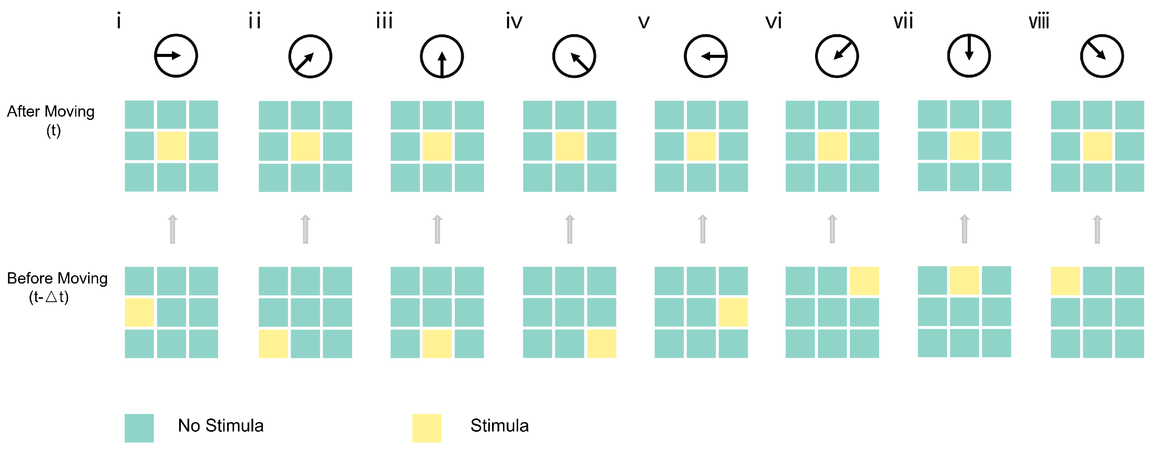
We identified the following eight direction-selective neurons by selectively screening a particular region in the preferred direction:
(i) RIGHTWARD neuron (0°) with interaction between R(i, j, t) and R(i − 1, j, t − Δ t), (ii) UPPER RIGHTWARD neuron (45°) with interaction between R(i, j, t) and R(i−1, j−1, t− Δ t), (iii) UPWARD neuron (90°) with interaction between R(i, j, t) and R(i, j+1, t − Δ t), (iv) UPPER LEFTWARD neuron (135°) with interaction between R(i, j, t) and R(i+1, j − 1, t − Δ t), (v) LEFTWARD neuron (180°) with interaction between R(i, j, t) and R(i+1, j, t − Δ t), (vi) LOWER LEFTWARD neuron (−135°) with interaction between R(i, j, t) and R(i+1, j+1, t− Δ t), (vii) DOWNWARD neuron (−90°) with interaction between R(i, j, t) and R(i, j − 1, t − Δ t), (viii) LOWER RIGHTWARD neuron (−45°) with interaction between R(i, j, t) and R(i − 1, j + 1, t − Δ t). Thus, we can detect eight directions of motion. Local motion-sensing direction-selective neurons are summarized in Figure 3. It contains eight basic inhibitory direction-selective neurons.
Several methods can be used to extract the information on local motion direction. In this study, we use eight direction-selective neurons to scan every region of the two-dimonsional receptive field to extract the information of local motion directions. We used a simple two-dimensional (5 × 5) moving image of a 7-type pattern as shown in Figure 4 to explain the mechanism. Each input comprises the following two successive images of the moving pattern: One at time t (after-moving state) and the other at t − Δ t (before-moving state). The 7-type pattern is shifted LOWER LEFTWARD (45°) with −1 unit along the x-direction and −1 unit along the y-direction. In the after-moving pattern, each neuron from (1, 1) to (5, 5) over the two-dimensional receptive field (5 × 5) at time t will detect its region at time t and the corresponding region at time t − Δ t. This indicated that no neuron fires at regions (1, 1), (1, 2), , and (5, 5). Only the LOWER LEFTWARD (−45°) neuron fires at (3, 2). The DOWNWARD (−90°) neuron, the LOWER LEFTWARD (−45°) neuron, and the LEFTWARD (−45°) neuron fire at (3, 3). The LOWER LEFTWARD (−45°) neuron and the LEFTWARD (−45°) neuron fire at (5, 3). Thus, there are four detection directions for LOWER LEFTWARD (−45°) neuron, two detection directions for LEFTWARD (180°) neuron, one detection direction for DOWNWARD (−90°) neuron, and one detection direction for UPPER LEFTWARD (90°) neuron. Thus, a global LOWER LEFTWARD (45°) motion direction can be inferred.

3. Results and Discussion

Several computer simulations were performed to validate the proposed mechanism. We generated a dataset of images, each comprising 32 × 32 pixels. The light spots in these images are from 1 pixel to 32 pixels and move randomly in one of the following eight directions: (1) RIGHTWARD, (2) UPPER RIGHTWARD, (3) UPWARD, (4) UPPER LEFTWARD, (5) LEFTWARD, (6) LOWER LEFTWARD, (7) DOWNWARD, (8) LOWER LEFTWARD. The input to the simulation program comprises the following two states of the moving pattern: After-moving state (at time t) and before-moving state (at t − Δ t).
In the proposed inhibitory mechanism, each pixel of the image contains eight local motion-sensing direction-selective neurons. We extracted the information on local motion direction of every pixel, recorded the activities of these neurons, and inferred global motion direction from the local motion direction information. The direction corresponding to the most activated neuron is considered the direction of motion. Figure 5 shows an example of a 1-pixel pattern. The activities of the eight local motion-sensing direction-selective retinal ganglion neurons were recorded in the right part. The number on the far right represents the number of activations of neurons in each direction. The DOWNWARD neuron was activated 1 time, which was the highest among these neurons. Thus, we can concluded that 1 pixel pattern were displaced −1 unit along the y-direction (a DOWNWARD movement). The same results can be obtained for other multi-pixel patterns, including a randomly generated 32-pixel pattern. Figure 6 shows the 32-pixel pattern. The UPWARD neuron was activated 15 times, which is the highest among these neurons. Thus, the 32-pixel pattern can be considered as to displace 1 unit along the y-axis (an UPWARD movement). Figure 7 shows the measurements of the random-dot patterns. Two images of the patterns were used as shown above. In this example, 30% of light spots were selected randomly and displaced 1 unit along the x-direction. The yellow pixels were displaced −1 unit along the x-direction and 1 unit along the y-direction (an UPPER RIGHTWARD movement). The UPPER RIGHTWARD neuron was activated 76 times, which is the highest among these neurons. From these figures, we obtain 98.3% detection accuracy in our experiment. These findings indicated that the proposed mechanism can detect motion direction with high accuracy in a wide range of applications.
Furthermore, we compared the performance of our proposed mechanism and traditional convolution neural network (CNN) on motion direction prediction. We used a three layer convolution neural network which contains (1) a convolutional layer with convolution kernel (3 × 3) and 16 feature map as the first layer; (2) a convolutional layer with convolution kernel (3 × 3) and 32 feature map as the second layer; (3) a full connect layer with inputs (8 × 8 × 32) and outputs as the third layer. We generated a dataset with 32 × 32 pixel images, 70% of which is regarded as the training set, and the remaining 30% as the test set. Each image contains (1) an object from 2 pixels to 32 pixels, and (2) 8 pixel static background distributed in the picture in the form of random-dot. Learning of CNN was performed with back-propagation under Adam optimizer with a learning rate of 0.005.
The result is summarized in Table 1. We can see that both the proposed mechanism and CNN learned the orientation detection very well and reached a high identification accuracy. While we continue to add noise in it, CNN has a poor performance. The accuracy dropped to 55.775% as the noise is 300%.
As mentioned above, we conducted various experiments using the proposed neurons to detect the direction of the moving object. We changed the number of pixels from 1 to 32 and obtained high accuracy in all experiments. These results demonstrate that the proposed mechanism can efficiently detect the motion direction of the object and has better performance in the presence of noise, which is very important in practical applications. Furthermore, our model is more similar to the biological model. Finally, we compare it with CNN and other neural networks. It is found that our model shows very high efficiency. Using 1660Ti GPU, neural networks need hours of time to train and a lot of training data. The proposed mechanism only takes a few seconds to get the results and does not require pre-training.
Compared with previous research mentioned above [12,25,27], these researches are all focused on the single-cell level. Researches failed to explain how these neurons function and cooperate to detect the overall motion direction [31]. Until now, the mechanism of direction selection and the visual nervous system is still unclear, our mechanism successfully explained motion direction detection in the visual nervous system and achieve good results in a series of experiments.

4. Conclusions

In this study, we proposed the mechanism to discribe the cooperate function of neurons in the retina system. In the rabbit retina system, some nerve cells only receive the information transmitted by the photoreceptor cells from a certain area. We presented a novel retina inhibitory mechanism to explain the cooperation among these neurons in the retina system. We extended it to multi motion-sensing direction-selective neurons and used them to extract the information on motion direction by scanning over the two-dimonsional receptive field. From the information of local motion directions in every region. We can infer the motion direction of objects with multi-spots in a two-dimensional field by calculating the activation of all neurons. The proposed mechanism appears to be part of the human motion direction detection system and can be applied to motion direction detection in various sensing systems. In actual situations, neural selection cells will be affected by noise. We take noise into consideration and compare the mechanism with traditional CNN and found our mechanism has a better performance in the case of high signal-to-noise ratio. At the same time, compared with CNN or other neural networks, our model has better interpretability (More similar to biological models) and very high efficiency. Taking 1660Ti GPU as an example, the neural network needs hours of training and tens of thousands of samples. The proposed neurons can get accurate prediction results in a few seconds. Similarly, this mechanism can be regarded as a guide for understanding other basic phenomena in motion perception, including the speed, direction, and binocular perception of movement. In addition, this mechanism provides structure at the level of the primary visual cortex and provides a systematic explanation for the mechanisms involved in neurons processing information from receptors. This mechanism in the primary visual cortex can help understand other mammalian sensory systems, such as those used for taste and touch.

Author Contributions

Y.T. and Z.T. designed the study. M.H. performed the experiments. Y.T., Z.T. and M.H. analyzed the data and wrote the paper. All authors have read and agreed to the published version of the manuscript.

Funding

This work was funded by JSPS KAKENHI Grant No. 19K12136.

Data Availability Statement

The code presented in this study are openly available in https://github.com/MZH1991/Inhibitory_Code (access on 12 July 2021).

Conflicts of Interest

The authors declare no competing interest.

References

  1. Hubel, D.H. Single unit activity in striate cortex of unrestrained cats. J. Physiol. 1959, 148, 226–238. [Google Scholar] [CrossRef]
  2. Barlow, H.B.; Hill, R.M. Selective sensitivity to direction of movement in ganglion cells of the rabbit retina. Science 1963, 139, 412. [Google Scholar] [CrossRef] [Green Version]
  3. Barlow, H.B.; Hill, R.M.; Levick, W.R. Retinal ganglion cells responding selectively to direction and speed of image motion in the rabbit. J. Physiol. 1964, 173, 377–407. [Google Scholar] [CrossRef]
  4. Oyster, C.W.; Barlow, H.B. Direction-selective units in rabbit retina: Distribution of preferred directions. Science 1967, 155, 841–842. [Google Scholar] [CrossRef] [Green Version]
  5. Oyster, C.W.; Takahashi, E.; Collewijn, H. Direction-selective retinal ganglion cells and control of optokinetic nystagmus in the rabbit. Vis. Res. 1972, 12, 183–193. [Google Scholar] [CrossRef]
  6. Semm, P. Antidromically activated direction selective ganglion cells of the rabbit. Neurosci. Lett. 1978, 9, 207–211. [Google Scholar] [CrossRef]
  7. Oyster, C.W.; Simpson, J.I.; Takahashi, E.S.; Soodak, R.E. Retinal ganglion cells projecting to the rabbit accessory optic system. J. Comp. Neurol. 1980, 190, 49–61. [Google Scholar] [CrossRef] [PubMed]
  8. Mimura, K.; Dräger, U.C. Receptive fields of single cells and topography in mouse visual cortex. J. Comp. Neurol. 1975, 160, 269–289. [Google Scholar]
  9. Jain, V.; Murphy-Baum, B.L.; deRosenroll, G.; Sethuramanujam, S.; Delsey, M.; Delaney, K.R.; Awatramani, G.B. The functional organization of excitation and inhibition in the dendrites of mouse direction-selective ganglion cells. eLife 2020, 9, e52949. [Google Scholar] [CrossRef]
  10. Ran, Y.; Huang, Z.; Baden, T.; Schubert, T.; Baayen, H.; Berens, P.; Franke, K.; Euler, T. Type-specific dendritic integration in mouse retinal ganglion cells. Nat. Commun. 2020, 11, 2101. [Google Scholar] [CrossRef]
  11. Mimura, K. Neural mechanisms, subserving directional selectivity of movement in the optic lobe of the fly. J. Comp. Physiol. 1972, 80, 409–437. [Google Scholar] [CrossRef]
  12. Haag, J.; Arenz, A.; Serbe, E.; Gabbiani, F.; Borst, A. Complementary mechanisms create direction selectivity in the fly. eLife 2016, 5, e17421. [Google Scholar] [CrossRef]
  13. Mauss, A.S.; Vlasits, A.; Borst, A.; Feller, M. Visual circuits for direction selectivity. Annu. Rev. Neurosci. 2017, 40, 211–230. [Google Scholar] [CrossRef] [PubMed]
  14. Borst, A. A biophysical mechanism for preferred direction enhancement in fly motion vision. PLoS Comput. Biol. 2018, 14, e1006240. [Google Scholar] [CrossRef] [PubMed]
  15. Rowe, M.H.; Stone, J. Properties of ganglion cells in the visual streak of the cat’s retina. J. Comp. Neurol. 1976, 169, 99–125. [Google Scholar] [CrossRef]
  16. Nelson, R.; Famiglietti, E.V., Jr.; Kolb, H. Intracellular staining reveals different levels of stratification for on-and off-center ganglion cells in cat retina. J. Neurophysiol. 1978, 41, 472–483. [Google Scholar] [CrossRef]
  17. Cleland, B.G.; Levick, W.R. Properties of rarely encountered types of ganglion cells in the cat’s retina and on overall classification. J. Physiol. 1974, 240, 457–492. [Google Scholar] [CrossRef]
  18. Levick, W.R.; Thibos, L.N.; Morstyn, R. Retinal ganglion cells and optic decussation of white cats. Vis. Res. 1980, 20, 1001–1006. [Google Scholar] [CrossRef]
  19. Vaney, D.I. The mosaic of amacrine cells in the mammalian retina. Prog. Retin. Res. 1990, 9, 49–100. [Google Scholar] [CrossRef]
  20. Brecha, N.; Johnson, D.; Peichl, L.; Wässle, H. Cholinergic amacrine cells of the rabbit retina contain glutamate decarboxylase and gamma-aminobutyrate immunoreactivity. Proc. Natl. Acad. Sci. USA 1988, 85, 6187–6191. [Google Scholar] [CrossRef] [PubMed] [Green Version]
  21. He, S.; Masland, R.H. Retinal direction selectivity after targeted laser ablation of starburst amacrine cells. Nature 1997, 389, 378–382. [Google Scholar] [CrossRef]
  22. Kim, T.; Kerschensteiner, D. Inhibitory control of feature selectivity in an object motion sensitive circuit of the retina. Cell Rep. 2017, 19, 1343–1350. [Google Scholar] [CrossRef] [Green Version]
  23. Fried, S.I.; Münch, T.A.; Werblin, F.S. Mechanisms and circuitry underlying directional selectivity in the retina. Nature 2002, 420, 411–414. [Google Scholar] [CrossRef] [PubMed]
  24. Vaney, D.I.; Sivyer, B.; Taylor, W.R. Direction selectivity in the retina: Symmetry and asymmetry in structure and function. Nat. Rev. Neurosci. 2012, 13, 194–208. [Google Scholar] [CrossRef]
  25. Morrie, R.D.; Feller, M.B. An asymmetric increase in inhibitory synapse number underlies the development of a direction selective circuit in the retina. J. Neurosci. 2015, 35, 9281–9286. [Google Scholar] [CrossRef] [PubMed] [Green Version]
  26. Hanson, L.; Sethuramanujam, S.; deRosenroll, G.; Jain, V.; Awatramani, G.B. Retinal direction selectivity in the absence of asymmetric starburst amacrine cell responses. eLife 2019, 8, e42392. [Google Scholar] [CrossRef] [PubMed]
  27. Krieger, B.; Qiao, M.; Rousso, D.L.; Sanes, J.R.; Meister, M. Four alpha ganglion cell types in mouse retina: Function, structure, and molecular signatures. PLoS ONE 2017, 12, e0180091. [Google Scholar] [CrossRef] [PubMed] [Green Version]
  28. Zhang, L.; Wu, Q.; Zhang, Y. Early visual motion experience shapes the gap junction connections among direction selective ganglion cell. PLoS Biol. 2020, 18, e3000692. [Google Scholar] [CrossRef]
  29. Chen, Q.; Smith, R.G.; Huang, X.; Wei, W. Preserving inhibition with a disinhibitory microcircuit in the retina. eLife 2020, 9, e62618. [Google Scholar] [CrossRef]
  30. Rasmussen, R.N.; Matsumoto, A.; Arvin, S.; Yonehara, K. Binocular integration of retinal motion information underlies optic flow processing by the cortex. Curr. Biol. 2021, 31, 1165–1174. [Google Scholar] [CrossRef]
  31. Fukushima, K.; Miyake, S. Neocognitron: A self-organizing neural network model for a mechanism of visual pattern recognition. In Competition and Cooperation in Neural Nets; Springer: Berlin/Heidelberg, Germany, 1982; pp. 267–285. [Google Scholar]
  32. Taylor, W.R.; He, S.; Levick, W.R.; Vaney, D.I. Dendritic computation of direction selectivity by retinal ganglion cells. Science 2000, 289, 2347–2350. [Google Scholar] [CrossRef] [PubMed]
Figure 1. Barlow’s inhibitory scheme for the directional selectivity of ganglion cells. In the left part of the pattern, a light spot (yellow) is traveling across the receptive field (blue). A, B and C are receptors. Response will be delayed when it passes horizontal cells. The veto gate offers Vote operations.
Electronics 10 01663 g001
Figure 2. Local directionally detective neurons. In the left part of the pattern, a light spot (yellow) is traveling across the receptive field (blue). A, B and C are receptors. Response will be delayed when it passes horizontal cells. The veto gate offers Vote operations. (a) object moves in the null direction, (b) object moves in the preferred direction.
Electronics 10 01663 g002
Figure 3. The local directionally detective neurons (i) Rightward, (ii) Upper Rightward, (iii) Upward, (iv) Upper Leftward, (v) Leftward, (vi) Lower Leftward, (vii) Downward, (viii) Lower Leftward.
Electronics 10 01663 g003
Figure 4. The mechanism of global motion direction detection, local motion direction detection neurons are arrayed in 5 × 5 regions to detect the two-dimensional receptive field.
Electronics 10 01663 g004
Figure 5. Computer simulation of machanism for detecting the moving direction of a one-pixel pattern being moved at −1 unit along the y direction (a DOWNWARD movement) in two dimensions. (LEFT) images before and after moving, (RIGHT) responses of neurons recorded.
Electronics 10 01663 g005
Figure 6. Computer simulation of machanism for detecting the moving direction of a one-pixel pattern being moved at 1 unit along the y direction (an UPWARD movement) in two dimensions. (LEFT) images before and after moving, (RIGHT) responses of neurons recorded.
Electronics 10 01663 g006
Figure 7. Computer simulation of machanism for detecting the moving direction of a random-dot pattern in two dimensions; 30% patterns were selected randomly and were displaced. (LEFT) images before and after moving, (RIGHT) responses of neurons recorded.
Electronics 10 01663 g007
Table 1. Comparison of identification accuracy between CNN and inhibitory mechanism.
NOISE0%50%100%150%200%300%
NOISENOISENOISENOISENOISENOISE
CNN99.35%88.2%78.125%70.475%65.525%55.775%
Proposed Method98.275%94.25%87.725%82.45%78.075%70.125%
Publisher’s Note: MDPI stays neutral with regard to jurisdictional claims in published maps and institutional affiliations.