
Concurrent Validation of 3D Joint Angles during Gymnastics Techniques Using Inertial Measurement Units

 ,
,  ,
, 
 , and
, and
Abstract
1. Introduction
2. Materials and Methods
3. Results
4. Discussion and Conclusions
Author Contributions
Funding
Institutional Review Board Statement
Informed Consent Statement
Data Availability Statement
Conflicts of Interest
Appendix A

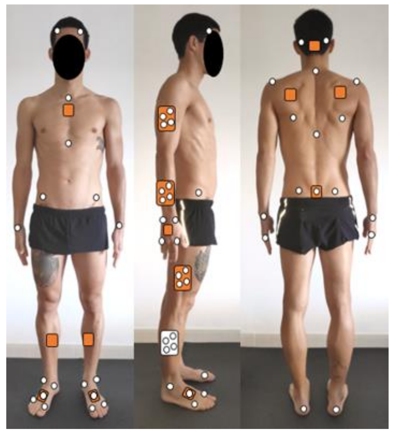
| Virtual Markers | Anatomical Tracking Markers | Coordinate Systems Definitions | |
|---|---|---|---|
| Global | XSens_lab_O—created as a projection onto the floor of the midpoint between the right and left hip joint centers (calculated through regression (Bell et al., 1990)) XSens_lab_Z—created below XSens_lab_O as an offset of 20 cm along the first global coordinate system vertical axis XSens_lab_Ant—created in front of XSens_lab_O, at the same height, as an offset of 20 cm in the direction of the line from the midpoint between the posterior superior iliac spines and the midpoint between the anterior superior iliac spines | - | Origin—XSens_lab_O Longitudinal axis (Z)—unit vector from XSens_lab_Z to XSens_lab_O, pointing up Medial-lateral axis (Y)—unit vector perpendicular to the plane containing the XSens_lab_O, XSens_lab_Z and XSens_lab_Ant, pointing left Anterior-Posterior axis (X)—unit vector perpendicular to Z and Y, pointing backwards |
| Pelvis | HipMid—created above the Global origin at the height of the right greater trochanter * L5—created above HipMid as a vertical offset (along the Global vertical axis) equal to the length of the Pelvis ** RHIP/LHIP—created above HipMid as an offset to the right/left along the Global medio-lateral axis, equal to 36% of the distance between the anterior superior iliac spines | RASIS/LASIS—Right/left anterior superior iliac spine RPSIS/LPSIS—Right/left posterior superior iliac spine SFZ—Sacrum flat zone | Origin—midpoint between RHIP and LHIP Longitudinal axis (Z)—unit vector from L5 to Origin, pointing up Anterior-Posterior axis (X)—unit vector perpendicular to the plane containing the Origin, RHIP and LHIP, pointing forward Medial-lateral axis (Y)—unit vector perpendicular to Z and X, pointing left |
| Thighs | RKneeC/LKneeC—created below RHIP/LHIP as an offset, along the Global vertical axis, equal to the length of the right thigh * | RTH1-4—rigid cluster of 4 non-colinear markers attached to the lateral aspect of the thigh | Origin—RHIP/LHIP Longitudinal axis (Z)—unit vector from RKneeC/LkneeC to RHIP/LHIP, pointing up Anterior-Posterior axis (X)—unit vector perpendicular to the plane containing RHIP, LHIP and RKneeC/LKneeC, pointing forward Medial-lateral axis (Y)—unit vector perpendicular to Z and X, pointing left |
| Shanks | RAnkleC/LankleC—created below RKneeC/LKneeC as an offset, along the Global vertical axis, equal to the length of the right shank * | RSK1-4—rigid cluster of 4 non-colinear markers attached to the lateral aspect of the shank | Origin—RKneeC/LKneeC Longitudinal axis (Z)—unit vector from RAnkleC/LAnkleC to RKneeC/LKneeC, pointing up Anterior-Posterior axis (X)—unit vector perpendicular to the plane containing RKneeC, LKneeC and RAnkleC/LAnkleC, pointing forward Medial-lateral axis (Y)—unit vector perpendicular to Z and X, pointing left |
| Feet | RdistalFoot/LdistalFoot—created in front of RAnkleC/LAnkleC as an offset, along the Global anterior axis, equal to the length of the right foot * RToebase/LToebase—created bellow RdistalFoot/LdistalFoot as an offset of 5 cm, along the Global vertical axis | R_FM1/L_FM1—distal head of the 1st metatarsus R_FM2/L_FM2—proximal head of the 2nd metatarsus R_FM5/L_FM5—distal head of the 5th metatarsus R_FAL/L_FAL—apex of the lateral malleolus R_TAM/L_TAM—apex of the medial malleolus R_FCC/L_FCC—posterior surface of the calcaneus | Origin—RAnkleC/LAnkleC Anterior-Posterior axis (X)—unit vector from RAnkleC/LAnkleC to RdistalFoot/LdistalFoot, pointing forward Medial-lateral axis (Y)—unit vector perpendicular to the plane containing RAnkleC/LAnkleC, RdistalFoot/LdistalFoot and RToebase/LToebase, pointing left Longitudinal axis (Z)—unit vector perpendicular to X and Y, pointing up |
| Head | Head_Mid—created above C7 as an offset, along the Global vertical axis, equal to half the head’s length ** | R_F_head/L_F_head—right/left aspect of the forehead R_B_head/L_B_head—right/left aspect of the back of the head | Origin—C7 Longitudinal axis (Z)—unit vector from C7 to Head_Mid, pointing up Medial-lateral axis (Y)—unit vector perpendicular to the plane containing C7, Head_Mid and Posterior, pointing left Anterior-Posterior axis (X)—unit vector perpendicular to Y and Z, pointing forward |
| Thorax | C7—created above L5 as an offset, along the Global vertical axis, equal to the length of trunk * Posterior—created as an offset of 10 cm from the Global origin, along the Global posterior axis and at the height of the right greater trochanter * | AXIF—xiphisternal joint CLAV—jugular notch C7—spinous process of 7th cervical vertebra T10—spinous process of the 10th thoracic vertebra R_SIA/L_SIA—inferior angle of the scapula R_SAE/L_SAE—scapula acromial edge | Origin—L5 Longitudinal axis (Z)—unit vector from L5 to C7, pointing up Medial-lateral axis (Y)—unit vector perpendicular to the plane containing L5, C7 and Posterior, pointing left Anterior-Posterior axis (X)—unit vector perpendicular to Y and Z, pointing forward |
| Upper arms | RClavOrigin/LClavOrigin—created as an upward and lateral offset from L5, respectively along the Global vertical and medial-lateral axis, to the origin of the clavicle ** RShoulderC/LShoulderC—created as an offset to the right/left of RClavOrigin, along the Global medial-lateral axis, equal to the length of the right clavicle** RElbowC/LElbowC—created below RShoulderC/LShoulderC as an offset, along the Global vertical axis, equal to the length of the right upper arm ** | RUA1-4—rigid cluster of 4 non-colinear markers attached to the lateral aspect of the upper arm | Origin—RShoulderC/LShoulderC Longitudinal axis (Z)—unit vector from RElbowC/LElbowC to RShoulderC/LShoulderC, pointing up Anterior-Posterior axis (X)—unit vector perpendicular to the plane containing RShoulderC/LShoulderC, RElbowC/LElbowC and C7, pointing forward Medial-lateral axis (Y)—unit vector perpendicular to Z and X, pointing left |
| Forearms | RWristC/LWristC—created below RElbowC/LElbowC as an offset, along the Global vertical axis, equal to the length of the right forearm ** | RFA1-4—rigid cluster of 4 non-colinear markers attached to the lateral aspect of the forearm | Origin—RElbowC/LElbowC Longitudinal axis (Z)—unit vector from RWristC/LWristC to RElbowC/LElbowC, pointing up Medial-lateral axis (Y)—unit vector perpendicular to the plane containing RElbowC/LElbowC, RWristC/LWristC and C7, pointing backward/forward Anterior-Posterior axis (X)—unit vector perpendicular to Z and X, pointing medially |
| Hands | RHandTip/LHandTip—created below RWristC/LWristC as an offset, along the Global vertical axis, equal to 40% of the length of the right hand ** | R/L_USP: Ulna styloid process R/L_RSP: radius styloid process R/L_HM2: head of 2nd metacarpus R/L_HM5: head of 5th metacarpus | Origin—RWristC/LWristC Longitudinal axis (Z)—unit vector from RHandTip/LHandTip to RWristC/LWristC, pointing up Medial-lateral axis (Y)—unit vector perpendicular to the plane containing RHandTip/LHandTip, RWristC/LWristC and C7, pointing backward/forward Anterior-Posterior axis (X)—unit vector perpendicular to Z and X, pointing medially |
| Execution Goal | Rotation of 360° on Transversal Axis and 180° on Longitudinal Axis, Passing through the Handstand Position (with Legs Separated) | |||||
|---|---|---|---|---|---|---|
| Result Goal | Change Direction of Movement (from Forward Direction to Backward Direction) | |||||
| Phases of Movement | Placement phase | Main phase | Finalization phase | |||
| Technical Description [32] | Initial hop Raise arms up and forward Forward rotation of the body Dominant leg steps forward | Dominant hand place in front of the gymnast on the floor carpet Place non-dominant hand further in centerline facing backward The bent dominant leg pushes and rear leg swings overhead Accentuated push from the shoulders Body turns during hands placement and pushing stage from both hands to feet | Feet touch the floor carpet with body in a concave shape Shoulders rise rapidly with the arms in front of the shoulders | |||
| Mechanical Actions | Increase in horizontal and angular velocities | Increase in angular and vertical velocities | Increase in angular velocity | |||
| Joint Angles | Head (F/E, A/A, I/E), Shoulder (F/E, A/A; I/E), Elbow (F/E), Thorax-thigh (F/E, A/A, I/E), Knee (F/E), Foot (F/E) | |||||
| Frames of Interest | (a) Contact (0% time) and (b) impulsion of dominant foot with floor carpet | (c) 1st hand contact (11.65% time), (d) 2nd hand contact (22.06% time), (e) impulsion of both hands with floor carpet | (f) End of round-off (47.04% time) | |||
| (a) | (b) | (c) | (d) | (e) | (f) | |
 |  |  |  |  |  | |
| Execution Goal | Rotation of 360° on Transversal Axis, Passing through the Handstand Position (Legs Together) | ||||
|---|---|---|---|---|---|
| Result Goal | Increase the Horizontal Velocity to Perform More Elements in Sequence | ||||
| Phases of Movement | Placement phase | Main phase | Finalization phase | ||
| Technical Description [32] | Jump backward Raise arms overhead Extend hips for an arched body posture | Hands contact the floor carpet Elbows may be slightly flexed Snapped down of the legs Push down both hands on the floor carpet for taking off | Body in a slightly inclined position Arms in front Knees and hips slightly flexed | ||
| Mechanical Actions | Increase in horizontal and angular velocities | Increase in angular velocity | Decrease in horizontal and angular velocities | ||
| Joint Angles | Head (F/E, A/A, I/E), Shoulder (F/E, A/A; I/E), Elbow (F/E), Thorax-thigh (F/E, A/A, I/E), Knee (F/E), Foot (F/E) | ||||
| Frames of Interest | (g) and (h) Impulsion of both feet contact with floor carpet | (i) Handstand position (75.52%) and (j) impulsion of both hands with floor carpet | (k) End of back handspring (100%) | ||
| (g) | (h) | (i) | (j) | (k) | |
 |  |  |  |  | |

References
- Kruk, E.; Reijne, M. Accuracy of human motion capture systems for sport applications; state-of-the-art review. Eur. J. Sport Sci. 2018, 18, 806–819. [Google Scholar] [CrossRef] [PubMed]
- Horenstein, R.; Lewis, C.; Yan, S.; Halverstadt, A.; Shefelbine, S. Validation of magneto-inertial measuring units for measuring hip joint angles. J. Biomech. 2019, 91, 170–174. [Google Scholar] [CrossRef]
- Koska, D.; Gaudel, J.; Hein, T.; Maiwald, C. Validation of an inertial measurement unit for the quantification of rearfoot kinematics during running. Gait Posture 2018, 64, 135–140. [Google Scholar] [CrossRef]
- Roetenberg, D.; Luinge, H.; Slycke, P. Xsens MVN: Full 6DOF Human Motion Tracking Using Miniature Inertial Sensors. Available online: https://www.researchgate.net/profile/Per-Slycke/publication/239920367_Xsens_MVN_Full_6DOF_human_motion_tracking_using_miniature_inertial_sensors/links/0f31752f1f60c20b18000000/Xsens-MVN-Full-6DOF-human-motion-tracking-using-miniature-inertial-sensors.pdf (accessed on 6 December 2019).
- Chambers, R.; Gabbett, T.J.; Cole, M.H.; Beard, A. The Use of Wearable Microsensors to Quantify Sport-Specific Movements. Sport Med. 2015, 45, 1065–1081. [Google Scholar] [CrossRef]
- Camomilla, V.; Bergamini, E.; Fantozzi, S.; Vannozzi, G. Trends supporting the in-field use of wearable inertial sensors for sport performance evaluation: A systematic review. Sensors 2018, 18, 873. [Google Scholar] [CrossRef]
- Robert-Lachaine, X.; Mecheri, H.; Larue, C.; Plamondon, A. Validation of inertial measurement units with an optoelectronic system for whole-body motion analysis. Med. Biol. Eng. Comput. 2017, 55, 609–619. [Google Scholar] [CrossRef]
- Lebel, K.; Boissy, P.; Nguyen, H.; Duval, C. Inertial measurement systems for segments and joints kinematics assessment: Towards an understanding of the variations in sensors accuracy. Biomed. Eng. Online 2017, 16, 1–16. [Google Scholar] [CrossRef]
- Kim, S.; Nussbaum, M.A. Performance evaluation of a wearable inertial motion capture system for capturing physical exposures during manual material handling tasks. Ergonomics 2013, 56, 314–326. [Google Scholar] [CrossRef]
- Schepers, M.; Giuberti, M.; Bellusci, G. Xsens MVN: Consistent Tracking of Human Motion Using Inertial Sensing; Technical Report; Xsens Technologies B.V: Enschede, The Netherlands, March 2018; pp. 1–8. Available online: https://www.researchgate.net/profile/Martin-Schepers/publication/324007368_Xsens_MVN_Consistent_Tracking_of_Human_Motion_Using_Inertial_Sensing/links/5ab8be2f0f7e9b68ef51f7ba/Xsens-MVN-Consistent-Tracking-of-Human-Motion-Using-Inertial-Sensing.pdf (accessed on 14 May 2021).
- Miller, E.; Kaufman, K. Cross-sectional validation of inertial measurement units for estimating trunk flexion kinematics during treadmill disturbances. Med. Eng. Phys. 2019, 70, 51–54. [Google Scholar] [CrossRef]
- Fantozzi, S.; Giovanardi, A.; Magalhães, F.A.; Di Michele, R.; Cortesi, M.; Gatta, G. Assessment of three-dimensional joint kinematics of the upper limb during simulated swimming using wearable inertial-magnetic measurement units. J. Sports Sci. 2016, 34, 1073–1080. [Google Scholar] [CrossRef]
- Cappozzo, A.; Cappello, A.; Croce, U.D.; Pensalfini, F. Surface-marker cluster design criteria for 3-d bone movement reconstruction. IEEE Trans. Biomed. Eng. 1997, 44, 1165–1174. [Google Scholar] [CrossRef] [PubMed]
- Blair, S.; Duthie, G.; Robertson, S.; Hopkins, W.; Ball, K. Concurrent validation of an inertial measurement system to quantify kicking biomechanics in four football codes. J. Biomech. 2018, 73, 24–32. [Google Scholar] [CrossRef] [PubMed]
- Mo, S.; Chow, D.H.K. Accuracy of three methods in gait event detection during overground running. Gait Posture 2018, 59, 93–98. [Google Scholar] [CrossRef] [PubMed]
- Grood, E.S.; Suntay, W.J. A Joint Coordinate System for the Clinical Description of Thre-Dimensional Motions: Application to the Knee. J. Biomech. 1983, 105, 136–144. [Google Scholar] [CrossRef]
- Sjostrand, P.; Lemmetty, H.; Hughes, K.; Gryga, P.; Jónsdóttir, S. 2017–2021 Code of Points Seniors and Juniors Teamgym. 2019. Available online: https://gymtranet.dk/media/4720/ueg-teamgym-cop-2017-revision-a-may-2018.pdf (accessed on 14 May 2021).
- Pataky, T. One-dimensional statistical parametric mapping in Python. Comput. Methods Biomech. Biomed. Engin. 2012, 15, 295–301. [Google Scholar] [CrossRef]
- Pataky, T.; Vanrenterghem, J.; Robinson, M. The probability of false positives in zero-dimensional analyses of one-dimensional kinematic, force and EMG trajectories. J. Biomech. 2016, 49, 1468–1476. [Google Scholar] [CrossRef]
- Cuesta-Vargas, A.I.; Galán-Mercant, A.; Williams, J.M. The use of inertial sensors system for human motion analysis. Phys. Ther. Rev. 2010, 15, 462–473. [Google Scholar] [CrossRef]
- Mavor, M.P.; Ross, G.B.; Clouthier, A.L.; Karakolis, T.; Graham, R.B. Validation of an IMU suit for military-based tasks. Sensors 2020, 20, 4280. [Google Scholar] [CrossRef]
- Nüesch, C.; Roos, E.; Pagenstert, G.; Mündermann, A. Measuring joint kinematics of treadmill walking and running: Comparison between an inertial sensor based system and a camera-based system. J. Biomech. 2017, 57, 32–38. [Google Scholar] [CrossRef]
- Tadano, S.; Takeda, R.; Miyagawa, H. Three dimensional gait analysis using wearable acceleration and gyro sensors based on quaternion calculations. Sensors 2013, 13, 9321–9343. [Google Scholar] [CrossRef]
- Duc, C.; Salvia, P.; Lubansu, A.; Feipel, V.; Aminian, K. A wearable inertial system to assess the cervical spine mobility: Comparison with an optoelectronic-based motion capture evaluation. Med. Eng. Phys. 2014, 36, 49–56. [Google Scholar] [CrossRef]
- Teufl, W.; Miezal, M.; Taetz, B.; Frohlichi, M.; Bleser, G. Validity of inertial sensor based 3D joint kinematics of static and dynamic sport and physiotherapy specific movements. PLoS ONE 2019, 14, e0213064. [Google Scholar] [CrossRef]
- Pedro, B.; Cabral, S.; Veloso, A.P. Concurrent validity of an inertial measurement system in tennis forehand drive. J. Biomech. 2021, 121, 110410. [Google Scholar] [CrossRef]
- Poitras, I.; Dupuis, F.; Bielmann, M.; Campeau-Lecours, A.; Mercier, C.; Bouyer, L.J.; Roy, J.S. Validity and reliability of wearable sensors for joint angle estimation: A systematic review. Sensors 2019, 19, 1555. [Google Scholar] [CrossRef]
- Bessone, V.; Höschele, N.; Schwirtz, A.; Seiberl, W. Validation of a new inertial measurement unit system based on different dynamic movements for future in-field applications. Sport Biomech. 2019, 1–16. [Google Scholar] [CrossRef] [PubMed]
- Zhang, J.; Novak, A.; Brouwer, B.; Li, Q. Concurrent validation of Xsens MVN measurement of lower limb joint angular kinematics. Physiol. Meas. 2013, 34, N63–N69. [Google Scholar] [CrossRef]
- De Vries, W.; Veeger, H.; Baten, C.; van der Helm, F. Magnetic distortion in motion labs, implications for validating inertial magnetic sensors. Gait Posture 2009, 29, 535–541. [Google Scholar] [CrossRef]
- Mecheri, H.; Robert-Lachaine, X.; Larue, C.; Plamondon, A. Evaluation of Eight Methods for Aligning Orientation of Two Coordinate Systems. J. Biomech. Eng. 2016, 138, 2–5. [Google Scholar] [CrossRef]
- Mack, M.; Federbusch, S.; Ferber, M.; Heinen, T. Movement prototypes and their relationship in the performance of a gymnastics floor routine. J. Hum. Sport Exerc. 2019, 1–16. [Google Scholar] [CrossRef]


| Joint and Plane | CMC | CMC Std Dev. | RMSE (°) | RMSE Std Dev. |
|---|---|---|---|---|
| Neck A/A | 0.54 * | 0.14 | 5.79 | 2.89 |
| Neck F/E | 0.98 | 0.02 | 7.21 | 3.70 |
| Neck I/E | 0.85 | 0.18 | 7.48 | 4.00 |
| Shoulder A/A | 0.88 | 0.12 | 9.86 | 3.90 |
| Shoulder F/E | 0.99 | 0.00 | 12.57 * | 3.42 |
| Shoulder I/E | 0.98 | 0.01 | 8.46 | 4.59 |
| Elbow F/E | 0.96 | 0.04 | 4.20 | 1.38 |
| Wrist F/E | 0.99 | 0.00 | 6.91 | 2.00 |
| Thorax–thigh A/A | 0.96 | 0.02 | 8.19 | 2.37 |
| Thorax–thigh F/E | 0.97 | 0.03 | 21.42 * | 10.31 |
| Thorax–thigh I/E | 0.90 | 0.06 | 16.31 * | 4.62 |
| Knee F/E | 0.98 | 0.01 | 8.36 | 3.42 |
| Ankle F/E | 0.95 | 0.02 | 11.07 * | 1.82 |
Publisher’s Note: MDPI stays neutral with regard to jurisdictional claims in published maps and institutional affiliations. |
© 2021 by the authors. Licensee MDPI, Basel, Switzerland. This article is an open access article distributed under the terms and conditions of the Creative Commons Attribution (CC BY) license (https://creativecommons.org/licenses/by/4.0/).
Share and Cite
Barreto, J.; Peixoto, C.; Cabral, S.; Williams, A.M.; Casanova, F.; Pedro, B.; Veloso, A.P. Concurrent Validation of 3D Joint Angles during Gymnastics Techniques Using Inertial Measurement Units. Electronics 2021, 10, 1251. https://doi.org/10.3390/electronics10111251
Barreto J, Peixoto C, Cabral S, Williams AM, Casanova F, Pedro B, Veloso AP. Concurrent Validation of 3D Joint Angles during Gymnastics Techniques Using Inertial Measurement Units. Electronics. 2021; 10(11):1251. https://doi.org/10.3390/electronics10111251
Chicago/Turabian StyleBarreto, Joana, César Peixoto, Sílvia Cabral, Andrew Mark Williams, Filipe Casanova, Bruno Pedro, and António P. Veloso. 2021. "Concurrent Validation of 3D Joint Angles during Gymnastics Techniques Using Inertial Measurement Units" Electronics 10, no. 11: 1251. https://doi.org/10.3390/electronics10111251
APA StyleBarreto, J., Peixoto, C., Cabral, S., Williams, A. M., Casanova, F., Pedro, B., & Veloso, A. P. (2021). Concurrent Validation of 3D Joint Angles during Gymnastics Techniques Using Inertial Measurement Units. Electronics, 10(11), 1251. https://doi.org/10.3390/electronics10111251