Article

Inhibition of Melanogenesis via Passive Immune Targeted Alpha-MSH Binder Polypeptide

1
Research Institute of Life Science, College of Veterinary Medicine, Gyeongsang National University, 501 Jinju-daero, Jinju 52828, Republic of Korea
2
Department of Animal Science, College of Life Science, Sangji University, Wonju 26339, Republic of Korea
*
Author to whom correspondence should be addressed.
These authors contributed equally to this work.
Cosmetics 2025, 12(1), 12; https://doi.org/10.3390/cosmetics12010012
Submission received: 6 December 2024 / Revised: 6 January 2025 / Accepted: 8 January 2025 / Published: 17 January 2025
(This article belongs to the Special Issue 10th Anniversary of Cosmetics—Recent Advances and Perspectives)

Abstract

Alpha-melanocyte stimulating hormone (α-MSH) is a hormone that stimulates the formation of melanin, which is responsible for protecting the skin from UV rays. However, excessive production of melanin causes pigmentation, leading to skin disorders, such as melasma and freckles. Using phage display technology, we screened a modified hagfish VLRB (α-MSH target binding polypeptide) library for polypeptides that recognize α-MSH. This was expressed in E. coli to produce binding proteins that specifically bind to α-MSH. In this study, we investigated the effect of α-MSH binder protein on the inhibition of melanogenesis in B16F10 cells stimulated with α-MSH and the mechanism of inhibition. The α-MSH-induced inhibition of intracellular and extracellular melanogenesis was accompanied by the downregulation of TRP1 and TRP2, and melanogenesis-related proteins, such as tyrosinase and MITF, were significantly downregulated. These results suggest that the α-MSH binder polypeptide regulates melanogenesis inhibition and its associated mechanisms.

1. Introduction

Alpha-melanocyte stimulating hormone (α-MSH) is a peptide hormone that belongs to the melanocortin family of proopiomelanocortin hormone (POMC), a precursor protein produced by the pituitary gland [1]. α-MSH binds to the melanocortin 1 receptor (MC1R), which is present in melanocytes after UV exposure, activating adenylyl cyclase, which then activates protein kinase A via intracellular cAMP, which subsequently phosphorylates the cAMP response element binding protein (CREB), leading to the expression of microphthalmia-associated transcription factor (MITF), a transcription factor essential for melanogenesis [2,3]. MITF then upregulates tyrosinase and tyrosinase-related protein (TRP), which promote melanogenesis. There are two main types of melanin: Tyrosine is degraded by tyrosinase to 3,4-dihydroxy-L-phenylalanine (L-DOPA), which is subsequently converted to DOPAquinone, which is converted to DOPAchrome and decarboxylated to dihydroxyindole (DHI) by TRP2 and dihydroxyindole carboxylic acid (DHICA) by TRP1 to form eumelanins, which are black or brown in color [4]. In contrast, under cysteine-rich conditions, cysteinylDOPA is formed, resulting in the formation of yellow or reddish pheomelanins [5]. Currently, the mechanisms that inhibit melanin production include blocking the activity of tyrosinase with natural compounds, such as hydroquinone or licorice extract, which inhibits the conversion of tyrosine to L-dopa, and preventing the oxidation of tyrosine with antioxidants such as vitamin C [6,7,8]. There are also methods that inhibit the migration of melanin into keratinocytes, such as niacinamide, and bleaching agents, that can be used to lighten skin [9,10]. However, there are problems with the lack of effectiveness and reproducibility due to the low bioavailability of natural products, and various compounds based on them are associated with dermatological diseases and various side effects [11,12,13]. Therefore, for the development of substances with low cytotoxicity, various attempts at the use of biological agents are needed.
The adaptive immune system of jawless vertebrates (Agnatha), such as lampreys and hagfish, is mediated by variable lymphocyte receptors (VLRs) that recognize and bind antigens, where the molecules possess leucine-rich repeats (LRRs) with a 20–29 residue sequence motif that differs from the immunoglobulins of jawed vertebrates (gnathostomes) [14]. Of the various known VLRs, lymphocytes expressing VLRA and VLRC are expressed on the surface of T cells, while VLRB is similar to the B-cell receptor (BCR), which recognizes specific antigens with high affinity and binds to protein and carbohydrate antigens. In addition, in the case of VLRB, a gene segment/cassette modeled on the LRR sequence is assembled by insertion of an incomplete germline VLR locus independent of the recombination activating gene [14,15,16]. The VLRB genes encode a single polypeptide containing an antigen-binding domain and multiple LRR motifs forming a globule surface, a single gene encoding an N-terminal LRR cap (LRRNT), the first LRR (LRR1), one to eight variable LRRs (LRRV), a terminal or end LRRV (LRRVe), a peptide (CP), and a C-terminal variable loop/cap (LRRCT) [17,18]. The stability and versatility of VLRBs under different conditions make them promising candidates for biological components [19]. Recent work conducted on VLRB from hagfish demonstrates its potential to be used as an alternative to traditional immunoglobulin antibodies [20].
In this study, blood was collected from medium-sized stabilized inshore hagfish (Eptatretus burgeri) immunized by subcutaneous injection of antigen (α-MSH); lymphocytes were isolated and the variable lymphocyte receptor (VLR) gene was isolated to establish a cDNA library. We combined the antigen recognition site genes from our previously established VLR cDNA library with an SLIME backbone and used the resulting binder library to identify protein genes that specifically bind to α-MSH as a phage display [21]. After insertion of the discovered gene into the expressing strain, Gram-negative bacteria (Escherichia coli), the bacteria were cultured for 3 days, and the precipitated bacteria and medium were separated using centrifugation to discard the bacteria, and the binder protein in the supernatant was purified by FPLC to identify the protein that specifically binds to α-MSH.
Recently, interest in skin whitening has increased, and many studies have been conducted to prevent skin diseases, such as melasma and pigmentation. In particular, various natural materials and synthetic chemicals have been used in whitening research, and various attempts have been made to inhibit melanin formation, but research on biological agents is still lacking [22,23]. In this study, we selected a target called α-MSH by using a protein that specifically binds to α-MSH, produced an antibody-like binder protein (Called α-MSH target binding polypeptide) that reacts to only one antigen, and then confirmed the inhibitory effect of α-MSH on melanogenesis. The anti-whitening effect was then confirmed in B16F10 cells, which will serve as a basis for future whitening effects through passive immunity.

2. Materials and Methods

2.1. Reagents, Chemicals and Standards

3-(4,5-Dimethylthiazol-2-yl)-2,5-diphenyltetrazolium bromide (MTT) was purchased from Duchefa Biochemie (Haarlem, The Netherlands). Cell Counting Kit 8 (WST-8/CCK8) was purchased from Abcam (cat. no. Ab228554), (Waltham, MA, USA). α-MSH (M4135) was purchased from Sigma-Aldrich (St. Louis, MO, USA). MITF antibody (cat. no. sc-515925), TRP1 antibody (cat. no. sc-166857), TRP2/DCT antibody (cat. no. sc-74439), and tyrosinase antibody (cat. no. sc-20035) were purchased from Santa Cruz biotechnology Inc. (Dallas, TX, USA), and β-actin (cat. no. 4970S) was purchased from Cell Signaling Technology (Danvers, MA, USA). Horseradish peroxidase (HRP)-conjugated secondary antibodies to anti mouse (cat. no. A90-116P) were obtained from Bethyl Laboratories, Inc. (Montgomery, AL, USA).

2.2. Configuring the Phage Display Variant Hagfish VLRB Library

2.2.1. Screening for VLRB (α-MSH Target Binding Polypeptide)

The phage display system for screening α-MSH-recognizing polypeptides was prepared following the method of Sirimanapong et al. (2024) [20]. The target protein, α-MSH, was immobilized by coating it on immunotubes (SPL Life Sciences, Pocheon-si, Republic of Korea, cat. No. 43015) at a concentration of 20 μg/mL and blocked with a blocking buffer composed of 1% bovine serum albumin (BSA) in 1x PBS containing 0.05% (v/v) Tween 20 (PBS-T). The tubes were washed twice with PBS-T after blocking and incubated with the phage library containing approximately 1012 PFU for 1 h. The unbound phages were removed by washing with excess PBS-T and a final wash with PBS. Bound phage was eluted with 0.2 M glycine-HCl (pH 2.5) and transferred to tubes containing 1 M Tris-HCl (pH 9.0) for neutralization. Collected bacteriophages were reproduced by incubating with log-phage E. coli TG1 at 37 °C for 1 h. To estimate cell titer, 100 μL of the bacterial culture supernatant was set aside for dilution and plating, while the remaining culture was centrifuged and plated subsequently on 2x YT agar containing 1% glucose and 100 μg/mL ampicillin. After overnight incubation at 30 °C, infected bacterial cells were harvested and grown in 2x YT media (1% glucose and 100 μg/mL ampicillin) from OD600 of 0.1 to OD600 of 0.5. Bacterial cells were then incubated with M13KO7 helper phage at 37 °C for 1 h, collected by centrifugation, and cultured overnight in 2x YT media (1% glucose, 100 μg/mL ampicillin, 50 μg/mL kanamycin, and 0.5 mM IPTG) at 30 °C. The bacterial culture supernatant containing enriched bacteriophage displaying target protein binders was then collected by precipitation with 20% PEG in 2.5 M NaCl and resuspended in PBS. Phage concentration was estimated by measuring the A260. This constitutes the new phage library used for the next round of panning.

2.2.2. Phage ELISA

The sixth round of phage panning was conducted, 96 colonies were randomly selected on the titer plate, and phage ELISA was performed using the method of Sirimanapong et al. (2024) [20].

2.2.3. Expression and Purification of α-MSH Target Binding Polypeptide

The clones selected in the phage ELISA were used as templates for PCR amplification using specific primers and the NEB Next Ultra II Q5 Master Mix (New England Biolabs, Ipswich, MA, USA). α-MSH-binding polypeptide expression was carried out following the method of Sirimanapong et al. (2024) [20], and the polypeptide was isolated by affinity purification using HisPur Ni-NTA Magnetic Beads and elution with 1 M imidazole in 1x PBS. The polypeptide solution was clarified and subjected to further purification using fast protein liquid chromatography (FPLC) using SeperdexTM 75 Increase 10/300 GL column using 1x PBS as elution buffer. FPLC fractions were run on SDS-PAGE using 12% polyacrylamide gel. Fractions containing the putative polypeptide were pooled and further analyzed.

2.3. Anti-Whitening Effect of α-MSH-Specific Targeted Protein

2.3.1. Cell Culture and Morphological Change

HaCat cells were obtained from ATCC, and B16F10 cells were obtained from the Korean Cell Line Bank and were maintained in 10% fetal bovine serum (FBS, Gibco BRL, Waltham, MA, USA); Dulbecco’s modified Eagle’s medium (DMEM) supplemented with 0.2% sodium bicarbonate (Sigma-Aldrich, St. Louis, MO, USA) was used as the medium. The cells were incubated at 37 °C in 5% CO2. The cells were grown to 90% confluence before the experiments were performed. Then, B16F10 cells were treated with 1 µM α-MSH, 500 µM arbutin, and 10 and 25 µg/mL α-MSH target-binding polypeptides. They were treated with the same composition in a new medium at 48 h intervals for 9 days and the cell morphology was observed on days 3, 6, and 9.

2.3.2. Cell Viability Assay

HaCaT cells were seeded at a density of 1 × 10⁴ cells per well in 96 well plates and then cultured, and then treated with various concentrations (low and high) of α-MSH targeted binder protein (0, 0.01, 0.05, 0.1, 0.25, 0.75, 1, 5, and 10 µg/mL) and (0, 1, 2.5, 5, 7.5, 10, 25, 50, 75, and 100 µg/mL) at 37 °C for 24 h. B16F10 cells were seeded at a density of 1 × 10⁴ cells per well in 96 well plates and then cultured, and treated with arbutin (0, 1, 2.5, 5, 10, 50, 100, 500, and 1000 µM) and with (low and high) α-MSH targeted binder protein (0, 0.01, 0.05, 0.1, 0.25, 0.75, 1, 5, and 10 µg/mL) and (0, 1, 2.5, 5, 7.5, 10, 25, 50, 75, and 100 µg/mL) at 37 °C for 24 h. After incubation, 10 μL of CCK-8 solution was added and MTT solution (10 µL; 5 mg/mL) was added to the plate and incubated at 37 °C for ~2 h. The insoluble formazan crystals were then dissolved in DMSO after the growth media was completely washed away. The absorbance of the converted dye was measured at a wavelength of 460 nm and 560 nm using a Multiskan FC microplate reader (Thermo Scientific, Rockford, IL, USA).

2.3.3. In Vitro Tyrosinase Inhibition Assay

The modified Macrini method was used to determine the inhibitory effect of α-MSH-targeted polypeptides on tyrosinase activity [24]. L-ascorbic acid and α-MSH target binding polypeptides were added at concentrations of 1, 10, 50, 100, and 200 µg/mL. A total of 220 μL of 0.1 M sodium phosphate buffer (pH 6.5) was added to the test tube with 20 μL of α-MSH target-binding polypeptide and L-ascorbic acid prepared at a concentration of 10–200 μg/mL. Then, 40 μL of 1.5 mM L-tyrosine (Sigma-Aldrich, St. Louis, MO, USA) was added. Mushroom tyrosinase (Sigma-Aldrich, St. Louis, MO, USA) 20 μL was prepared in 1500 units and incubated at 37 °C for 15 min. DOPAchrome, which is formed from 3,4-dihydroxyphenylalanine (DOPA) by tyrosinase, was detected at the end of the reaction. The amount of DOPAchrome formed from 3,4-dihydroxyphenylalanine (DOPA) by tyrosinase was measured at 490 nm.

2.3.4. Intracellular and Extracellular Melanin Content

Intracellular and extracellular melanin measurements were made using a modification of the Hosoi method [25]. B16F10 cells were seeded in 60 mm plates at a density of 1 × 10⁵ cells/well for 24 h at 37 °C. After that, it was treated with α-MSH, 10 and 25 µg/mL α-MSH target binding polypeptide for 48 h at 37 °C. The medium supernatant was then collected and the extracellular melanin content was measured at 490 nm, and 2 × 10 6 cells for each group were lysed with lysis buffer (RIPA buffer). The cell precipitate obtained by centrifugation was washed with alcohol and then lysed with 1 N NaOH solution supplemented with 10% DMSO for 1 h at 90 °C, and the absorbance was measured at 490 nm.

2.3.5. Western Blot Analysis

B16F10 cells were seeded at a density of 1 × 10⁶ cells/well in 60 mm plates and treated with α-MSH; 10 and 25 µg/mL α-MSH target-binding polypeptides were treated for 48 h at 37 °C. Cells were then lysed in ice-cold RIPA buffer (50 mM Tris-HCL (pH 8.0), 0.5% sodium deoxycholate, 1 mM EDTA, 150 mM NaCl, 0.1 SDS, and 1% NP 40). Protein concentrations were determined using the PierceTM BCA Protein Assay (Thermo Scientific, Rockford, IL, USA) according to the manufacturer’s instructions. Equal amounts of protein (10 μg) were separated by SDS-PAGE on 10% gels and transferred to PVDF membranes using JP/WSE-4040 HorizeBLOT 4M-R WSE-4045 (ATTO Blotting System, USA). The blots were then blocked with EzBlockChemi (ATTO Blotting System, Japan) for 1 h at room temperature. Membranes were further incubated with 1:1000 dilutions of primary antibodies overnight at 4 °C. The membranes were washed three times for 10 min with TBS-T and probed with a second antibody for 2 h at room temperature. The second antibody was diluted 1:5000. Blots were visualized using ClarityTM ECL substrate reagent (Bio Rad Laboratories, Inc., Hercules, CA, USA) and quantified by densitometry using Image J 1.54k software (National Institutes of Health) with β-Actin as a loading control. Experiments were performed in triplicate.

2.4. Statistical Analysis

Test measurements were expressed as mean ± standard deviation (M ± SD) in triplicate. Statistical analysis was performed using SPSS version 12.0 (SPSS Inc., Chicago, IL, USA) and one-way factorial analysis of variance (ANOVA). Statistical significance was analyzed by Duncan’s multiple range and Student’s t-test at p < 0.05 level after a one-way analysis of variance. (# p < 0.05, ## p < 0.01, ### p < 0.001 versus untreated, positive control group; and * p < 0.05, ** p < 0.01, *** p < 0.001 versus arbutin-treated, negative control group).

3. Results

3.1. Phage Library Screening for α-MSH Antigen

The purified α-MSH protein was utilized as an antigen in phage display library panning and ELISA. We produced the α-MSH toxin-specific VLRB antibodies from phage-display VLRB libraries after iteratively biopanning against the purified α-MSH protein. The colonies’ recovery following the initial panning cycle indicated that the phages were selectively binding to the antigen. The recovered antigen-binding phage was further concentrated in later biopanning cycles. A significant quantity of positive antigen-binding phages was selected, as evidenced by an increase in colonies from the recovered phage. Following six iterations of panning against recombinant α-MSH, 96 separate colonies were chosen. The three colonies with the strongest signal-to-noise ratio at 450 nm were isolated and sequenced, and the three sequences identified from the most concentrated sequences were selected for expression and purification.

3.2. Expression, Purification, and Antigen Binding of α-MSH Target Binding Polypeptide

The α-MSH target binding polypeptide was expressed in a significant amount in BL21 cells and purified using Ni-NTA magnetic beads and FPLC and after purification through affinity complex and FPLC (Table 1). A single band of the same size (approximately 32.26 kDa) as the expected α-MSH target binding polypeptide was observed through SDS-PAGE, confirming the high purity of the VLRB protein (Figure 1A). Subsequently, the reaction to the α-MSH antigen was confirmed by ELISA, and the VLRB-like structure α-MSH target binding polypeptide (Sample ID: MSH2-C5-1) showed a higher positive reaction than No VLRB, indicating a high affinity for the α-MSH antigen (Figure 1B). In addition, from Figure 1C, the receptor-ligand binding kinetics (Kd) of the α-MSH antigen and α-MSH target binding polypeptide was 0.0001 M, confirming a very strong binding between the two molecules.

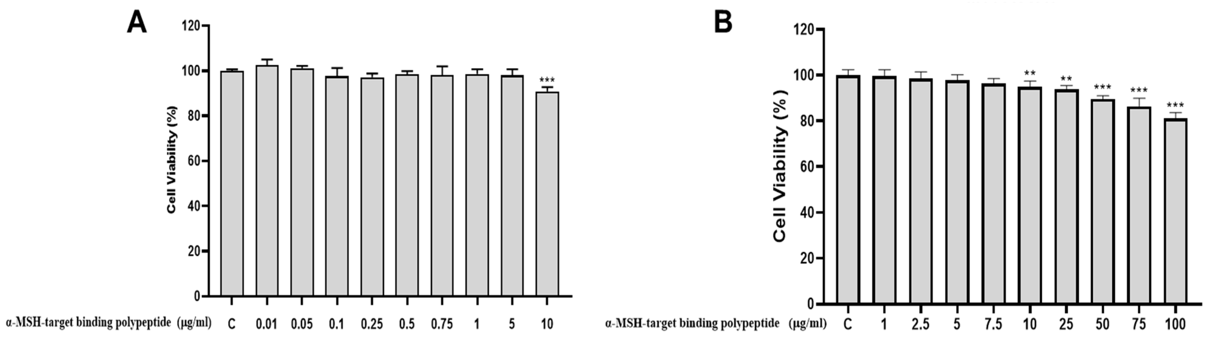
3.3. The Cytotoxicity of α-MSH Target Binding Polypeptide on the HaCaT Cell

For the α-MSH target binding polypeptide, the 3-(3,4-dimethyl-thiazolyl-2)-2,5-diphenyl tetrazolium bromide (MTT) assay was carried out in HaCaT cells to determine the cytotoxicity (Figure 2A,B). The α-MSH target binding polypeptide was treated with HaCaT cells at concentrations of low and high (0, 0.01, 0.05, 0.1, 0.25, 0.75, 1, 5, and 10 µg/mL) and (0, 1, 2.5, 5, 7.5, 10, 25, 50, 75, and 100 µg/mL) for 24 h. We found that the α-MSH target binding polypeptide was non-toxic at 100 μg/mL (Figure 2B). The results showed no cytotoxicity in normal dermal keratinocytes up to 100 µg/mL, which is a conservative concentration for further experiments.

3.4. The Cytotoxicity of Arbutin on the B16F10

To confirm the cytotoxicity of B16F10 against arbutin, the cells were treated with arbutin (0, 1, 2.5, 5, 10, 50, 100, 500, 1000 μM) using the 3-(3,4-dimethyl-thiazolyl-2)-2,5-diphenyl tetrazolium bromide (MTT) assay (Figure 3). As a result, 500 μM was selected for further experimentation.

3.5. The Cytotoxicity of α-MSH-Target Binding Polypeptide on the B16F10

To confirm the cytotoxicity of α-MSH-target binding polypeptide in B16F10, α-MSH-targeted binding polypeptide was tested by the WST-1 (CCK) assay and 3-(3,4-dimethyl-thiazolyl-2)-2,5-diphenyl tetrazolium bromide (MTT) assay at (0, 0.1, 0.25, 0.5, 0.75, 1, 3, 5, 10, and 25 μg/mL) and (0, 1, 2.5, 5, 7.5, 10, 25, 50, 75, and 100 μg/mL) (Figure 4). Relatively, no cytotoxicity to B16F10 was observed and 10 and 25 μg/mL were selected for further experimentation (Figure 4C).

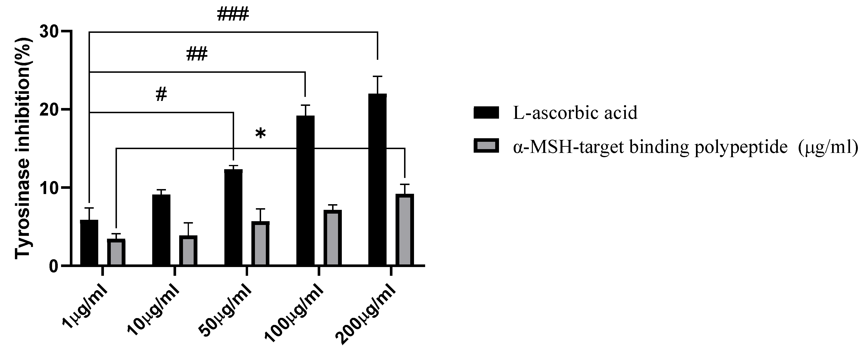
3.6. Tyrosinase Inhibition Activity

To determine whether the α-MSH-target binding polypeptide has a direct effect of tyrosinase on melanin biosynthesis, the activity of mushroom tyrosinase was measured (Figure 5). Ascorbic acid was used as a positive control, and the α-MSH-targeted binding polypeptide was treated at the same concentrations (1, 10, 50, 100, and 200 μg/mL). Ascorbic acid showed a relatively high tyrosinase inhibitory activity of 10% at 50 μg/mL, while the α-MSH-targeted polypeptide did not show direct tyrosinase inhibitory activity with an inhibitory effect of 10% at 200 μg/mL.

3.7. Determination of Intracellular and Extracellular Melanin Content

To measure the intracellular and extracellular melanin content of the α-MSH-target binding polypeptide, melanin was induced in B16F10 cells stimulated with α-MSH to determine the extracellularly secreted and intracellularly present melanin content (Figure 6). Using arbutin as a positive control versus α-MSH-stimulated melanocytes, extracellular melanin decreased at a concentration of 25 μg/mL α-MSH-target binding polypeptide but was not significantly different from the positive control (Figure 6A). However, the intracellular melanin content was reduced by 40% at 25 μg/mL compared to the α-MSH treatment group, confirming that the α-MSH-target binding polypeptide binds to α-MSH in the cells and reduces melanogenesis.

3.8. Cell Morphological Change

B16F10 cells were treated with α-MSH to induce melanin synthesis and then treated with arbutin (500 μM) and α-MSH-target binding polypeptide (10 and 25 μg/mL) for 9 days to observe cell morphological changes (Figure 7). In the arbutin-treated group, the cells became progressively worse, with reduced melanogenesis on days 6 and 9, while in the α-MSH-target binding polypeptide-treated group, there was no significant change in melanogenesis until day 6, followed by a relative decrease in melanin produced at 25 μg/mL on day 9.

3.9. Effect of α-MSH Target Binding Polypeptide on the Expression of Melanogenic Proteins in B16F10 Cells

In B16F10 cells where melanin synthesis was induced by α-MSH, α-MSH target binding polypeptide treatment dose-dependently inhibited TRP1, TRP2, MITF, and tyrosinase (Figure 8). TRP1 and TRP2 were downregulated at 25 μg/mL, but not significantly (Figure 8C,D). MITF, however, showed a significant downregulation at 25 μg/mL. These results showed that the α-MSH target binding polypeptide inhibits α-MSH-mediated melanogenesis by down-regulating melanogenesis-related proteins TRP1, TRP2, tyrosinase, and especially MITF.

4. Discussion

Melanin is a general term for the black or brown pigment found in the tissues of the skin and eyes of various animals. Pigmentation is one of the changes in skin color that occurs when the amount of melanin in the skin tissue increases excessively. The skin produces alpha-MSH as a response to stress caused by the sun, which then signals the skin cells [26]. Afterward, alpha-MSH forms melanosomes in the melanocytes present in the dermal cells, which form melanin and deliver it to the keratinocytes [27].
Passive immunity is the opposite of active immunity and is a system that borrows antibodies that have already been formed in other people or animals [28]. Based on this principle, antibody therapeutics have a wide variety of targets that each component inhibits and exhibits various effects. They are biopharmaceuticals that use monoclonal antibodies that bind to specific antigens as therapeutics [29,30], typically used for anti-cancer, treatment of infectious diseases, inflammation suppression, immune regulation, and skin diseases [30,31]. This passive immunity, also called monoclonal antibodies, targets specific antigens only, reducing drug-drug interactions and maximizing efficacy, resulting in lower side effects and risks than other chemicals that have random effects [32].
This study confirmed the anti-melanogenic effect of an antibody-like protein that specifically targets α-MSH on melanin formation induced by α-MSH. The mechanism of inhibiting melanin formation by initially blocking alpha-MSH with the produced polypeptide was confirmed (Figure 9).
In particular, abundant cytotoxicity studies were conducted in this experiment. This is partly because the safety of the binder protein is not fully guaranteed. The multiple MTT assays conducted in this study were conducted to provide a more in-depth toxicity assessment, as most biological agents may contain toxins that are not expected, and further research is needed to identify unknown side effects and conduct more relevant studies [33,34]. As shown in Figure 5, the α-MSH target-binding polypeptide does not have a significant effect on the direct inhibition of tyrosine kinase. However, as shown in Figure 6, melanin synthesis is reduced at both the intracellular and extracellular levels. In addition, as shown in Figure 8, binding of α-MSH to keratinocytes results in a significant downregulation of MITF and TRP2, which are involved in the regulation of melanin synthesis, and the cells become unresponsive to the MC1R receptor of melanocytes, resulting in downregulation of intracellular melanin synthesis signaling. This suggests that it can inhibit melanin synthesis in cells and has a potential whitening effect. In addition, in this study, the molecular weight of the α-MSH target binding polypeptide actually produced is 32.26 kDa, which makes it difficult to regulate the biological pathway that occurs within the cell membrane, as shown in Table 1. This is also the reason why α-MSH, a regulator of the cell membrane outside of the antigen involved in melanin formation, was selected.

5. Conclusions

This study examined the effects of biological agents on skin whitening, and the study period was considerably shorter than that of other chemical agents. However, this study will help in the development of biological agents that focus on a single target, namely α-MSH, which is a major player in melanin formation, and we plan to conduct research using programs such as molecular docking simulations to predict how well α-MSH-targeted polypeptides bind to α-MSH targets. In addition, the development of passive immunity agents using this method of screening specific antigens is expected to have a significant impact on the cosmetics industry through its results, as it can target and focus on various cosmetic diseases such as acne and hair loss.

Author Contributions

Conceptualization, S.-H.J. and H.-H.K.; methodology, S.-H.J. and A.J.D.R.-A.; formal analysis, S.-H.J. and H.-H.K.; writing-original draft preparation, S.-H.J. and H.-H.K.; writing-review and editing, P.B.B. and M.R.N.A.; investigation, S.-H.J., E.-H.K. and M.-J.A.; validation, S.-H.J.; project administration, S.-H.J. and H.-H.K.; supervision, T.-S.J. and G.-S.K. All authors have read and agreed to the published version of the manuscript.

Funding

This research received no external funding.

Institutional Review Board Statement

Not applicable.

Informed Consent Statement

Not applicable.

Data Availability Statement

The datasets used and/or analysed during the current study available from the corresponding author on reasonable request.

Acknowledgments

This study was supported by the National Research Foundation of Korea, funded by the Ministry of Science and ICT (grant nos. RS-2023-0024337661382, RS-2023-00235511, and RS-2024-00411709) and Jinju Bioindustry Foundation, Bio 21 Center.

Conflicts of Interest

The authors declare no conflicts of interest.

References

  1. Dall’Olmo, L.; Papa, N.; Surdo, N.C.; Marigo, I.; Mocellin, S. Alpha-melanocyte stimulating hormone (α-MSH): Biology, clinical relevance and implication in melanoma. J. Transl. Med. 2023, 21, 562. [Google Scholar] [CrossRef]
  2. Thody, A.J. Alpha-MSH and the regulation of melanocyte function. Ann. N. Y. Acad. Sci. 1999, 885, 217–229. [Google Scholar] [CrossRef] [PubMed]
  3. Li, G.-H.; Xu, L.; Xiong, M.; Qu, M. Role of α-MSH-MC1R-cAMP Signaling Pathway in Regulating the Melanin Synthesis in Silky Fowl. J. Cell. Signal. 2018, 3, 195. [Google Scholar] [CrossRef]
  4. Adhikari, M.; Ali, A.; Kaushik, N.; Choi, E. Perspective in Pigmentation Disorders. In Comprehensive Clinical Plasma Medicine: Cold Physical Plasma for Medical Application; Springer: Cham, Switzerland, 2018; pp. 363–400. [Google Scholar]
  5. Snyman, M.; Walsdorf, R.E.; Wix, S.N.; Gill, J.G. The metabolism of melanin synthesis—From melanocytes to melanoma. Pigment. Cell Melanoma Res. 2024, 37, 438–452. [Google Scholar] [CrossRef]
  6. Chandorkar, N.; Tambe, S.; Amin, P.; Madankar, C. Alpha Arbutin as a Skin Lightening Agent: A Review. Int. J. Pharm. Res. 2021, 13, 3502–3510. [Google Scholar] [CrossRef]
  7. Kim, H.J.; Seo, S.H.; Lee, B.G.; Lee, Y.S. Identification of tyrosinase inhibitors from Glycyrrhiza uralensis. Planta Medica 2005, 71, 785–787. [Google Scholar] [CrossRef]
  8. Sanadi, R.M.; Deshmukh, R.S. The effect of Vitamin C on melanin pigmentation—A systematic review. J. Oral Maxillofac. Pathol. JOMFP 2020, 24, 374–382. [Google Scholar] [CrossRef] [PubMed]
  9. Hakozaki, T.; Minwalla, L.; Zhuang, J.; Chhoa, M.; Matsubara, A.; Miyamoto, K.; Greatens, A.; Hillebrand, G.G.; Bissett, D.L.; Boissy, R.E. The effect of niacinamide on reducing cutaneous pigmentation and suppression of melanosome transfer. Br. J. Dermatol. 2002, 147, 20–31. [Google Scholar] [CrossRef]
  10. Chang, N.-F.; Chen, Y.-S.; Lin, Y.-J.; Tai, T.-H.; Chen, A.-N.; Huang, C.-H.; Lin, C.-C. Study of Hydroquinone Mediated Cytotoxicity and Hypopigmentation Effects from UVB-Irradiated Arbutin and DeoxyArbutin. Int. J. Mol. Sci. 2017, 18, 969. [Google Scholar] [CrossRef]
  11. Zhu, Y.; Ouyang, Z.; Du, H.; Wang, M.; Wang, J.; Sun, H.; Kong, L.; Xu, Q.; Ma, H.; Sun, Y. New opportunities and challenges of natural products research: When target identification meets single-cell multiomics. Acta Pharm. Sin. B 2022, 12, 4011–4039. [Google Scholar] [CrossRef]
  12. Shaivi, L.; Turabi, K.; Aich, J.; Devarajan, S.; Unni, D.; Garse, S. In Silico Approaches in the Repurposing of Bioactive Natural Products for Drug Discovery; Elsevier: Amsterdam, The Netherlands, 2023; pp. 125–147. [Google Scholar]
  13. Majtan, J.; Bucekova, M.; Jesenak, M. Natural Products and Skin Diseases. Molecules 2021, 26, 4489. [Google Scholar] [CrossRef]
  14. Boehm, T.; McCurley, N.; Sutoh, Y.; Schorpp, M.; Kasahara, M.; Cooper, M.D. VLR-Based Adaptive Immunity. Annu. Rev. Immunol. 2012, 30, 203–220. [Google Scholar] [CrossRef]
  15. Das, S.; Rast, J.P.; Li, J.; Kadota, M.; Donald, J.A.; Kuraku, S.; Hirano, M.; Cooper, M.D. Evolution of variable lymphocyte receptor B antibody loci in jawless vertebrates. Proc. Natl. Acad. Sci. USA 2021, 118, e2116522118. [Google Scholar] [CrossRef] [PubMed]
  16. Boehm, T.; Hirano, M.; Holland, S.J.; Das, S.; Schorpp, M.; Cooper, M.D. Evolution of Alternative Adaptive Immune Systems in Vertebrates. Annu. Rev. Immunol. 2018, 36, 19–42. [Google Scholar] [CrossRef] [PubMed]
  17. Herrin, B.R.; Alder, M.N.; Roux, K.H.; Sina, C.; Ehrhardt, G.R.A.; Boydston, J.A.; Turnbough, C.L.; Cooper, M.D. Structure and specificity of lamprey monoclonal antibodies. Proc. Natl. Acad. Sci. USA 2008, 105, 2040–2045. [Google Scholar] [CrossRef] [PubMed]
  18. Alder, M.N.; Rogozin, I.B.; Iyer, L.M.; Glazko, G.V.; Cooper, M.D.; Pancer, Z. Diversity and Function of Adaptive Immune Receptors in a Jawless Vertebrate. Science 2005, 310, 1970–1973. [Google Scholar] [CrossRef]
  19. Waters, E.A.; Shusta, E.V. The variable lymphocyte receptor as an antibody alternative. Curr. Opin. Biotechnol. 2018, 52, 74–79. [Google Scholar] [CrossRef]
  20. Sirimanapong, W.; Thaijongrak, P.; Sudpraseart, C.; Bela-ong, D.B.; Rodelas-Angelia, A.J.D.; Angelia, M.R.N.; Hong, S.; Kim, J.; Thompson, K.D.; Jung, T.S. Passive immunoprophylaxis with Ccombodies against Vibrio parahaemolyticus in Pacific white shrimp (Penaeus vannamei). Fish Shellfish Immunol. 2024, 154, 109973. [Google Scholar] [CrossRef]
  21. Hassan, K.M.A.; Hansen, J.D.; Herrin, B.R.; Amemiya, C.T. Generation of Lamprey Monoclonal Antibodies (Lampribodies) Using the Phage Display System. Biomolecules 2019, 9, 868. [Google Scholar] [CrossRef]
  22. Jeong, S.-H. A Review of Current Research on Natural Skin Whitening Products. Asian J. Beauty Cosmetol. 2018, 16, 599–607. [Google Scholar] [CrossRef]
  23. Gillbro, J.M.; Olsson, M.J. The melanogenesis and mechanisms of skin-lightening agents—Existing and new approaches. Int. J. Cosmet. Sci. 2011, 33, 210–221. [Google Scholar] [CrossRef] [PubMed]
  24. Macrini, D.; Suffredini, I.; Varella, A.; Younes, R.; Ohara, M. Extracts from Amazonian plants have inhibitory activity against tyrosinase: An in vitro evaluation. Braz. J. Pharm. Sci. 2009, 45, 715–721. [Google Scholar] [CrossRef]
  25. Hosoi, J.; Abe, E.; Suda, T.; Kuroki, T. Regulation of melanin synthesis of B16 mouse melanoma cells by 1 alpha, 25-dihydroxyvitamin D3 and retinoic acid. Cancer Res. 1985, 45, 1474–1478. [Google Scholar] [PubMed]
  26. Maranduca, M.A.; Branisteanu, D.; Serban, D.N.; Branisteanu, D.C.; Stoleriu, G.; Manolache, N.; Serban, I.L. Synthesis and physiological implications of melanic pigments. Oncol. Lett. 2019, 17, 4183–4187. [Google Scholar] [CrossRef]
  27. Videira, I.F.; Moura, D.F.; Magina, S. Mechanisms regulating melanogenesis. An. Bras. De Dermatol. 2013, 88, 76–83. [Google Scholar] [CrossRef]
  28. Baxter, Active and passive immunity, vaccine types, excipients and licensing. Occup. Med. 2007, 57, 552–556. [CrossRef]
  29. Yao, S.; Zhu, Y.; Chen, L. Advances in targeting cell surface signalling molecules for immune modulation. Nat. Rev. Drug Discov. 2013, 12, 130–146. [Google Scholar] [CrossRef]
  30. Lu, R.-M.; Hwang, Y.-C.; Liu, I.J.; Lee, C.-C.; Tsai, H.-Z.; Li, H.-J.; Wu, H.-C. Development of therapeutic antibodies for the treatment of diseases. J. Biomed. Sci. 2020, 27, 1. [Google Scholar] [CrossRef]
  31. Sockolov, M.E.; Alikhan, A.; Zargari, O. Non-psoriatic dermatologic uses of monoclonal antibody therapy. J. Dermatol. Treat. 2009, 20, 319–327. [Google Scholar] [CrossRef]
  32. Castelli, M.S.; McGonigle, P.; Hornby, P.J. The pharmacology and therapeutic applications of monoclonal antibodies. Pharmacol. Res. Perspect. 2019, 7, e00535. [Google Scholar] [CrossRef]
  33. Pichler, W.J. Adverse side-effects to biological agents. Allergy 2006, 61, 912–920. [Google Scholar] [CrossRef] [PubMed]
  34. Clarke, J.B. Mechanisms of adverse drug reactions to biologics. Handb. Exp. Pharmacol. 2010, 196, 453–474. [Google Scholar]
Figure 1. Expression, purification, and antigen-binding ability of α-MSH target binding polypeptide. (A) FPLC purification of α-MSH target binding polypeptide complex by SuperdexTM 75 10/300GL (Cytiva, Marlborough, MA, USA) and followed by 15% SDS-PAGE. (B) ELISA result of absorbance read at 450 nm, antigen concentration at 200 ng/well. (C) α-MSH target binding polypeptide for Kd determination.
Cosmetics 12 00012 g001
Figure 2. The effects of α-MSH-targeted binding polypeptide on HaCaT keratinocytes cell viability. Cytotoxicity of α-MSH target binding polypeptide was measured for 24 h after treatment with (A) low concentrations (0, 0.01, 0.05, 0.25, 0.5, 0.75, 1, 5, and 10 μg/mL) and (B) high concentrations (0, 1, 2.5, 5, 7.5, 10, 25, 50, 75, and 100 μg/mL) of α-MSH target binding polypeptide. Comparison of α-MSH target binding polypeptide with control ** p < 0.01, *** p < 0.001.
Cosmetics 12 00012 g002
Figure 3. The effects of arbutin on B16F10 mouse melanoma cell viability. Cytotoxicity was determined after dose-dependent treatment with arbutin (0, 1, 2.5, 5, 10, 50, 100, 500, 1000 μM). Comparison of arbutin with control * p < 0.05.
Cosmetics 12 00012 g003
Figure 4. The effects of the α-MSH-target binding polypeptide on B16F10 mouse melanoma cell viability. Cytotoxicity effect of α-MSH-targeted binding polypeptide on B16F10 cells. B16F10 cells were treated with low and high concentrations of α-MSH-targeted binding polypeptide (0, 0.1, 0.25, 0.5, 0.75, 1, 3, 5, 10, and 25 μg/mL) and (0, 1, 2.5, 5, 7.5, 10, 25, 50, 75, and 100 μg/mL) for 24 h at 37 °C. (A) Cytotoxicity effect of CCK assay on B16F10 cells (low concentration). (B) Cytotoxicity effect of MTT assay on B16F10 cells (low concentration). (C) Cytotoxicity effect of MTT assay on B16F10 cells (high concentration). Comparison of α-MSH target binding polypeptide with control * p < 0.05, ** p < 0.01, *** p < 0.001.
Cosmetics 12 00012 g004
Figure 5. Measurement of inhibition of tyrosinase activity of α-MSH-target binding polypeptide. Tyrosinase was combined with α-MSH target binding polypeptide and treated with 1.5 mM L-tyrosine for 15 min at 37 °C. Comparison with L-ascorbic acid # p < 0.05, ## p < 0.01, ### p < 0.001. Comparison of α-MSH target binding polypeptide with control * p < 0.05.
Cosmetics 12 00012 g005
Figure 6. Determination of (A) extracellular and (B) intracellular melanin content treated with α-MSH-target binding polypeptide in B16F10. Cells were treated with concentrations of α-MSH-target binding polypeptide (10 and 25 μg/mL) and arbutin (500 μM). Results from three independent experiments were expressed as mean ± standard error of the mean (SEM) compared with control. ### p < 0.001 vs. untreated group; * p < 0.05, ** p < 0.01, *** p < 0.001 vs. α-MSH treated group.
Cosmetics 12 00012 g006
Figure 7. Cell morphological change in B16F10 cells treated with α-MSH, arbutin, and α-MSH-target binding polypeptide. (A) Control, (B) Treated with α-MSH only, (C) Treated with α-MSH arbutin 500 μM, (D,E) Treated with an α-MSH target binding polypeptide (10 μg/mL and 25 μg/mL).
Cosmetics 12 00012 g007
Figure 8. Effect of α-MSH target binding polypeptide on α-MSH-induced expression of melanogenesis-related proteins in B16F10 cells. B16F10 cells were pretreated without or with α-MSH (1 µM) for 12 h at 37 °C. Following that, cells were treated with α-MSH target binding polypeptide (10 and 25 µg/mL) for 24 h at 37 °C. (A) The relative area of tyrosinase, (B) The relative area of MITF, (C) The relative area of TRP1, and (D) the relative area of TRP2. Results from three independent experiments were expressed as mean ± standard error of the mean (SEM) compared with control. # p < 0.05, ## p < 0.01 vs. untreated group; * p < 0.05 ** p < 0.01, *** p < 0.001 vs. α-MSH treated group.
Cosmetics 12 00012 g008
Figure 9. Downregulation effects of α-MSH target binding polypeptide to α-MSH mediated melanogenesis.
Cosmetics 12 00012 g009
Table 1. Summary MSH group α-MSH Target Binding Polypeptide parameter.
α-MSH Target Binding
Polypeptide Plant
MW (kDa)Concentration
(mg/mL)
VolumeKd
(M)
MSH32.261.530.700.0001
Disclaimer/Publisher’s Note: The statements, opinions and data contained in all publications are solely those of the individual author(s) and contributor(s) and not of MDPI and/or the editor(s). MDPI and/or the editor(s) disclaim responsibility for any injury to people or property resulting from any ideas, methods, instructions or products referred to in the content.

