Lovage (Levisticum officinale W.D.J. Koch) Roots: A Source of Bioactive Compounds towards a Circular Economy

 ,
,  ,
,  ,
,  ,
,  ,
,  ,
,  and
and 
Abstract
:
1. Introduction
2. Materials and Methods
2.1. Samples
2.2. Preparation of Extracts
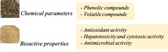
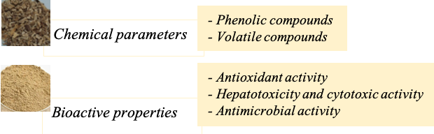
2.3. Chemical Parameters
2.3.1. Phenolic Compounds
2.3.2. Volatile Compounds
2.4. Bioactive Properties
2.4.1. Antioxidant Activity
2.4.2. Hepatotoxicity and Cytotoxic Activity
2.4.3. Antimicrobial Activity
2.5. Statistical Analysis
3. Results and Discussion
3.1. Chemical Characterization of L. officinale Roots
3.1.1. Composition in Phenolic Compounds
3.1.2. Composition in Volatile Compounds
3.2. Bioactive Properties
3.2.1. Antioxidant Activity
3.2.2. Cytotoxic Activity
3.3. Antimicrobial Activity
4. Conclusions
Author Contributions
Funding
Acknowledgments
Conflicts of Interest
References
- Aćimović, M.; Kostadinović, L.; Popović, S.; Dojčinović, N. Apiaceae seeds as functional food. J. Agric. Sci. 2015, 60, 237–246. [Google Scholar]
- Tuncturk, M.; Özgokce, F. Chemical composition of some Apiaceae plants commonly used in herby cheese in Eastern Anatolia. J. Agric. For. 2015, 39, 55–62. [Google Scholar]
- Blank, I.; Schieberle, P. Analysis of the seasoning-like flavour substances of a commercial lovage extract (Levisticum officinale Koch.). Flavour Fragr. J. 1993, 8, 191–195. [Google Scholar] [CrossRef]
- Bylaite, E.; Venskutonis, R.P.; Roozen, J.P. Influence of harvesting time on the composition of volatile components in different anatomical parts of lovage (Levisticum officinale Koch.). J. Agric. Food Chem. 1998, 46, 3735–3740. [Google Scholar] [CrossRef]
- EMA, European Medicines Agency. Community Herbal Monograph on Levisticum officinale Koch, Radix; EMA, European Medicines Agency: Amsterdam, The Netherlands, 2012; Available online: https://www.ema.europa.eu/en/documents/herbal-monograph/draft-community-herbal-monograph-levisticum-officinale-koch-radix_en.pdf (accessed on 15 January 2020).
- Heidarpour, O.; Souri, M.K.; Omidbaigi, R. Changes in content and constituents of essential oil in different plant parts of lovage (Levisticum officinale Koch. Cv. Budakalaszi) Cultivated in Iran. J. Essent. Oil-Bearing Plants 2013, 16, 318–322. [Google Scholar] [CrossRef]
- Miran, M.; Esfahani, H.M.; Farimani, M.M.; Ahmadi, A.A.; Ebrahimi, S.N. Essential Oil Composition and Antibacterial Activity of Levisticum officinale Koch at Different Developmental Stages. J. Essent. Oil-Bearing Plants 2018, 21, 1051–1055. [Google Scholar] [CrossRef]
- Kemzuraite, A.; Venskutonis, P.R.; Baranauskiene, R.; Navikiene, D. Optimization of supercritical CO2 extraction of different anatomical parts of lovage (Levisticum officinale Koch.) using response surface methodology and evaluation of extracts composition. J. Supercrit. Fluids 2014, 87, 93–103. [Google Scholar] [CrossRef]
- Spréa, R.M.; Fernandes, Â.; Calhelha, R.C.; Pereira, C.; Pires, T.C.S.P.; Alves, M.J.; Canan, C.; Barros, L.; Amaral, J.S.; Ferreira, I.C.F.R. Chemical and bioactive characterisation of the aromatic plant: Levisticum officinale W.D.J. Koch: A comprehensive study. Food Funct. 2020, 11, 1292–1303. [Google Scholar]
- Ma, Z.; Bai, L. Anti-inflammatory Effects of Z-Ligustilide Nanoemulsion. Inflammation 2013, 36, 294–299. [Google Scholar] [CrossRef]
- Ma, Z.; Bai, L. The anti-inflammatory effect of Z-Ligustilide in experimental ovariectomised osteopenic rats. Inflammation 2012, 35, 1793–1797. [Google Scholar] [CrossRef]
- Fang, X.; Ma, Q.; Zhang, K.X.; Yao, S.Y.; Feng, Y.; Jin, Y.S.; Liang, S. Synthesis of phthalide derivatives and evaluation on their antiplatelet aggregation and antioxidant activities. J. Asian Nat. Prod. Res. 2019, 1–12. [Google Scholar] [CrossRef] [PubMed]
- Su, Z.Y.; Khor, T.O.; Shu, L.; Lee, J.H.; Saw, C.L.L.; Wu, T.Y.; Huang, Y.; Suh, N.; Yang, C.S.; Conney, A.H.; et al. Epigenetic reactivation of Nrf2 in murine prostate cancer TRAMP C1 cells by natural phytochemicals Z-ligustilide and radix angelica sinensis via promoter CpG demethylation. Chem. Res. Toxicol. 2013, 26, 477–485. [Google Scholar] [CrossRef] [PubMed]
- Long, R.; Yang, F.; Du, J.R.; Qian, Z.M.; Wang, C.Y.; Chen, C. Effects of ligustilide on tumor growth and immune function in institute of cancer research mice. Trop. J. Pharm. Res. 2012, 11, 421–428. [Google Scholar] [CrossRef] [Green Version]
- Long, F.Y.; Shi, M.Q.; Zhou, H.J.; Liu, D.L.; Sang, N.; Du, J.R. Klotho upregulation contributes to the neuroprotection of ligustilide against cerebral ischemic injury in mice. Eur. J. Pharmacol. 2018, 820, 198–205. [Google Scholar] [CrossRef]
- Han, L.; Liu, D.L.; Zeng, Q.K.; Shi, M.Q.; Zhao, L.X.; He, Q.; Kuang, X.; Du, J.-R. The neuroprotective effects and probable mechanisms of Ligustilide and its degradative products on intracerebral hemorrhage in mice. Int. Immunopharmacol. 2018, 63, 43–57. [Google Scholar] [CrossRef] [PubMed]
- Martins, N.; Barros, L.; Ferreira, I.C.F.R. In Vivo antioxidant activity of phenolic compounds: Facts and gaps. Trends Food Sci. Technol. 2016, 48, 1–12. [Google Scholar] [CrossRef] [Green Version]
- Carocho, M.; Barreiro, M.F.; Morales, P.; Ferreira, I.C.F.R. Adding molecules to food, pros and cons: A review on synthetic and natural food additives. Compr. Rev. Food Sci. Food Saf. 2014, 13, 377–399. [Google Scholar] [CrossRef]
- Bessada, S.M.F.; Barreira, J.C.M.; Barros, L.; Ferreira, I.C.F.R.; Oliveira, M.B.P.P. Phenolic profile and antioxidant activity of Coleostephus myconis (L.) Rchb.f.: An underexploited and highly disseminated species. Ind. Crops Prod. 2016, 89, 45–51. [Google Scholar] [CrossRef] [Green Version]
- Falcão, S.; Bacém, I.; Igrejas, G.; Rodrigues, P.J.; Vilas-Boas, M.; Amaral, J.S. Chemical composition and antimicrobial activity of hydrodistilled oil from juniper berries. Ind. Crops Prod. 2018, 124, 878–884. [Google Scholar] [CrossRef] [Green Version]
- Lockowandt, L.; Pinela, J.; Roriz, C.L.; Pereira, C.; Abreu, R.M.V.; Calhelha, R.C.; Alves, M.J.; Barros, L.; Bredol, M.; Ferreira, I.C.F.R. Chemical features and bioactivities of cornflower (Centaurea cyanus L.) capitula: The blue flowers and the unexplored non-edible part. Ind. Crops Prod. 2019, 128, 496–503. [Google Scholar] [CrossRef] [Green Version]
- Abreu, R.M.V.; Ferreira, I.C.F.R.; Calhelha, R.C.; Lima, R.T.; Vasconcelos, M.H.; Adega, F.; Chaves, R.; Queiroz, M.-J. Anti-hepatocellular carcinoma activity using human HepG2 cells and hepatotoxicity of 6-substituted methyl 3-aminothieno[3,2-b]pyridine-2-carboxylate derivatives: In Vitro evaluation, cell cycle analysis and QSAR studies. Eur. J. Med. Chem. 2011, 46, 5800–5806. [Google Scholar] [CrossRef] [Green Version]
- Pires, T.C.S.P.; Dias, M.I.; Barros, L.; Alves, M.J.; Oliveira, M.B.P.P.; Santos-Buelga, C.; Ferreira, I.C.F.R. Antioxidant and antimicrobial properties of dried Portuguese apple variety (Malus domestica Borkh. cv Bravo de Esmolfe). Food Chem. 2018, 240, 701–706. [Google Scholar] [CrossRef] [PubMed] [Green Version]
- Złotek, U.; Szymanowska, U.; Pecio, U.; Kozachok, S.; Jakubczyk, A. Antioxidative and Potentially Anti-inflammatory Activity of Phenolics from Lovage Leaves Levisticum officinale Koch Elicited with Jasmonic Acid and Yeast Extract. Molecules 2019, 24, 1441. [Google Scholar] [CrossRef] [PubMed] [Green Version]
- Spínola, V.; Castilho, P.C. Evaluation of Asteraceae herbal extracts in the management of diabetes and obesity. Contribution of caffeoylquinic acids on the inhibition of digestive enzymes activity and formation of advanced glycation end-products (In Vitro). Phytochemistry 2017, 143, 29–35. [Google Scholar] [PubMed]
- Panthama, N.; Kanokmedhakul, S.; Kanokmedhakul, K. Galloyl and hexahydroxydiphenoyl esters of phenylpropanoid glucosides, phenylpropanoids and phenylpropanoid glucosides from rhizome of Balanophora fungosa. Chem. Pharm. Bull. 2009, 57, 1352–1355. [Google Scholar] [CrossRef] [Green Version]
- Silva, E.D.; Batista, R. Ferulic Acid and Naturally Occurring Compounds Bearing a Feruloyl Moiety: A Review on Their Structures, Occurrence, and Potential Health Benefits. Compr. Rev. Food Sci. Food Saf. 2017, 16, 580–616. [Google Scholar] [CrossRef] [Green Version]
- Cogne, A.L.; Queiroz, E.F.; Wolfender, J.L.; Marston, A.; Mavi, S.; Hostettmann, K. On-line identification of unstable catalpol derivatives from Jamesbrittenia fodina by LC-MS and LC-NMR. Phytochem. Anal. 2003, 14, 67–73. [Google Scholar] [CrossRef]
- Barreira, J.C.M.; Dias, M.I.; Živković, J.; Stojkovic, D.; Soković, M.; Santos-Buelga, C.; Ferreira, I.C.F.R. Phenolic profiling of Veronica spp. grown in mountain, urban and sandy soil environments. Food Chem. 2014, 163, 275–283. [Google Scholar] [CrossRef]
- Olaru, O.T.; Niţulescu, G.M.; Orţan, A.; Dinu-Pîrvu, C.E. Ethnomedicinal, phytochemical and pharmacological profile of anthriscus sylvestris as an alternative source for anticancer lignans. Molecules 2015, 20, 15003–15022. [Google Scholar] [CrossRef] [Green Version]
- Zhao, M.; Tao, J.; Qian, D.; Liu, P.; Shang, E.X.; Jiang, S.; Guo, J.; Su, S.-L.; Duan, J.-A.; Du, L. Simultaneous determination of loganin, morroniside, catalpol and acteoside in normal and chronic kidney disease rat plasma by UPLC-MS for investigating the pharmacokinetics of Rehmannia glutinosa and Cornus officinalis Sieb drug pair extract. J. Chromatogr. B 2016, 1010, 122–129. [Google Scholar] [CrossRef]
- Jiang, B.; Shen, R.F.; Bi, J.; Tian, X.S..; Hinchliffe, T.X.Y. Catalpol: A potential therapeutic for neurodegenerative diseases. Curr. Med. Chem. 2015, 22, 1278–1291. [Google Scholar]
- Shieh, J.P.; Cheng, K.C.; Chung, H.H.; Kerh, Y.F.; Yeh, C.H.; Cheng, J.T. Plasma glucose lowering mechanisms of catalpol, an active principle from roots of rehmannia glutinosa, in streptozotocin-induced diabetic rats. J. Agric. Food Chem. 2011, 59, 3747–3753. [Google Scholar] [CrossRef] [PubMed]
- León, A.; Del-Ángel, M.; Ávila, J.L.; Delgado, G. Phthalides: Distribution in Nature, Chemical Reactivity, Synthesis, and Biological Activity. Prog. Chem. Org. Nat. Prod. 2017, 104, 128–169. [Google Scholar]
- Raal, A.; Arak, E.; Orav, A.; Kailas, T.; Müürisepp, M. Composition of the Essential Oil of Levisticum officinale W.D.J. Koch from Some European Countries. J. Essent. Oil Res. 2008, 20, 37–41. [Google Scholar]
- Li, W.; Wu, Y.; Liu, X.; Yan, C.; Liu, D.; Pan, Y.; Yang, G.; Yin, F.; Weng, Z.; Zhao, D.; et al. Antioxidant properties of cis-Z,Z’-3a.7a’, 7a.3a’-dihydroxyligustilide on human umbilical vein endothelial cells In Vitro. Molecules 2013, 18, 520–534. [Google Scholar] [CrossRef] [PubMed]
- Yu, Y.; Du, J.R.; Wang, C.Y.; Qian, Z.M. Protection against hydrogen peroxide-induced injury by Z-ligustilide in PC12 cells. Exp. Brain Res. 2008, 184, 307–312. [Google Scholar] [CrossRef]
- Abd El-Hamid, S.R.; Abeer, Y.S.H.; Hendawy, S.F. Anti-inflammatory, antioxidant, antitumor and physiological studies on Levisticum officinale-Koch. Planta Med. 2018, 75, 4–5. [Google Scholar]
- Shafaghat, A. Chemical constituents, antimicrobial and antioxidant activity of the hexane extract from root and seed of Levisticum persicum Freyn and Bornm. J. Med. Plant. Res. 2011, 5, 5127–5131. [Google Scholar]
- Guzman, J.D.; Evangelopoulos, D.; Gupta, A.; Prieto, J.M.; Gibbons, S.; Bhakta, S. Antimycobacterials from Lovage root (Ligusticum officinale Koch). Phyther. Res. 2013, 27, 993–998. [Google Scholar] [CrossRef]


| Peak | Rt (min) | λmax (nm) | Molecular Ion [M−H]− (m/z) | MS2 (m/z) | Tentative Identification | Hydroethanolic | Decoction | Hydroethanolic Extract of Hexane Residue | References |
|---|---|---|---|---|---|---|---|---|---|
| 1 | 4.69 | 192, 258 | 341 | 179 (100) | Caffeic acid hexoside 1 | nd | 0.44 ± 0.02 | nd | [24] |
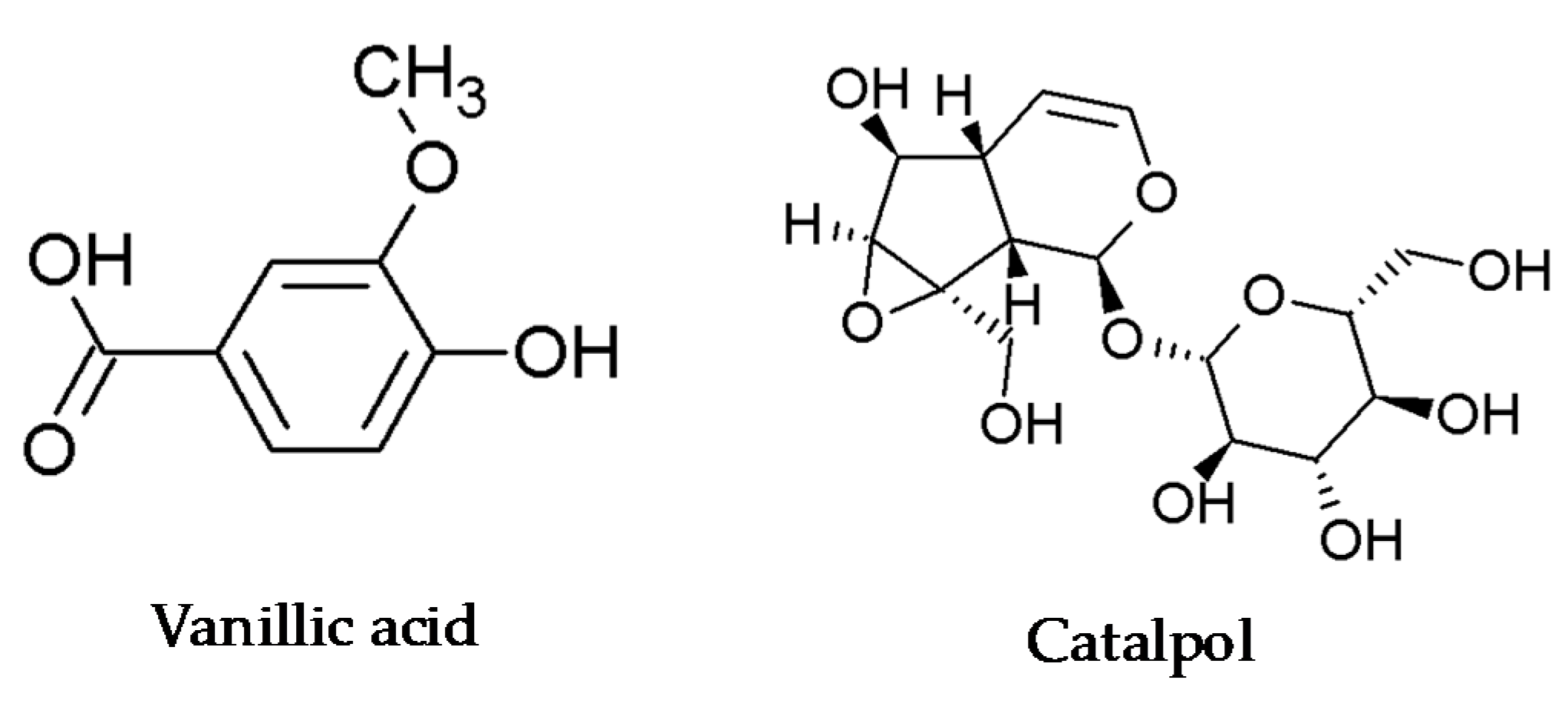
| 2 | 7.04 | 288, 320 | 167 | 123 (100) | Vanillic acid 2 | 1.51 ± 0.03 b | 19.7 ± 0.4 a | 0.49 ± 0.01 c | [1] |
| 3 | 14.07 | 201, 225 | 533 | 515 (5), 371 (100), 353 (13), 209 (7), 191 (5), 179 (3) | di-Caffeoylglucaric acid 1 | 0.056 ± 0.002 b | 0.6163 ± 0.0004 a | 0.025 ± 0.003 c | [25] |
| 4 | 15.44 | 200, 323 | 561 | 367 (100), 193 (26), 191 (13), 173 (100), 129 (60), 111 (2) | Hydroferuloyl-feruloylquinic acid 3 | 0.044 ± 0.002 * | 0.582 ± 0.006 * | nd | [25] |
| 5 | 16.79 | 307 | 935 | 926 (10), 915 (18), 897 (10), 783 (12), 633 (100), 301 (48) | Galloyl-bis-HHDP-glucoside 4 | 0.984 ± 0.001 | nd | nd | [26] |
| 6 | 17.20 | 324 | 547 | 529 (8), 385 (100), 367 (27), 353 (21), 335 (3), 191 (7), 179 (3), 173 (3) | Caffeoyl-feruloylquinic acid 3 | 0.171 ± 0.005 b | 1.62 ± 0.04 a | 0.089 ± 0.002 c | [27] |
| 7 | 20.61 | 196, 233, 280 | 361 | 343 (100), 325 (2), 199 (44), 181 (3) | Catalpol 5 | 0.126 ± 0.005 b | 0.66 ± 0.02 a | 0.059 ± 0.002 c | [28,29] |
| 8 | 22.81 | 234, 282, 322 | 251 | 233 (22), 207 (100), 193 (9), 179 (28), 175 (42), 153 (2) | Unknown | nq | nq | nq | |
| 9 | 25.19 | 233, 281, 322 | 389 | 371 (100), 341 (25), 327 (7), 193 (38) | Methoxylariciresinol 5 | 0.171 ± 0.006 b | 0.639 ± 0.006 a | 0.0484 ± 0.0005 c | [30] |
| TPA | 1.78 ± 0.04 b | 23.1 ± 0.5 a | 0.61 ± 0.01 c | ||||||
| Other compounds | 0.298 ± 0.001 b | 1.295 ± 0.01 a | 0.108 ± 0.002 c |
| Compound | RT (min) | LRI a | LRI b | Relative % c | ||
|---|---|---|---|---|---|---|
| Clevenger | Hexane Extract | |||||
| 1. | Heptanal | 12.66 | 901 | 901 | 0.024 ± 0.005 | - |
| 2. | α-Pinene | 14.25 | 932 | 932 | 0.091 ± 0.006 | - |
| 3. | β-Pinene | 16.47 | 974 | 974 | 0.59 ± 0.04 | 0.097 ± 0.005 |
| 4. | 2-Pentyl furane | 17.31 | 991 | 984 | 0.073 ± 0.004 | - |
| 5. | n-Octanal | 17.90 | 1002 | 998 | 0.046 ± 0.002 | - |
| 6. | p-Cymene | 19.01 | 1023 | 1020 | 0.028 ± 0.003 | - |
| 7. | β-Phellandrene | 19.21 | 1027 | 1025 | 1.26 ± 0.08 | 0.79 ± 0.03 |
| 8. | Linalol | 22.91 | 1099 | 1095 | 0.079 ± 0.001 | - |
| 9. | Nonanal | 23.10 | 1102 | 1100 | 0.095 ± 0.007 | - |
| 10. | β-Fenchol | 23.57 | 1112 | 1118 | 0.02 ± 0.01 | - |
| 11. | α-Canpholenal | 24.19 | 1124 | 1122 | 0.015 ± 0.003 | - |
| 12. | trans-Pinocarveol | 24.84 | 1137 | 1135 | 0.07 ± 0.02 | - |
| 13. | Menthone | 25.60 | 1153 | 1148 | 0.39 ± 0.02 | - |
| 14. | 5-Pentylcyclohexa-1,3-diene | 25.84 | 1154 | - | 0.16 ± 0.01 | |
| 15. | Penthylbenzene | 25.75 | 1156 | 1152 | 1.01 ± 0.06 | - |
| 16. | Pinocarvone | 26.04 | 1162 | 1160 | 0.009 ± 0.002 | - |
| 17. | Menthan-3-one | 26.12 | 1163 | 1158 | 0.28 ± 0.05 | - |
| 18. | Menthol | 26.53 | 1172 | 1167 | 0.494 ± 0.005 | 0.04 ± 0.01 |
| 19. | α-Terpineol | 27.41 | 1189 | 1186 | 0.057 ± 0.002 | - |
| 20. | Myrtenol + estragole | 27.7 | 1195 | 1194 | 0.131 ± 0.005 | - |
| 21. | n-Decanal | 28.11 | 1204 | 1201 | 0.033 ± 0.003 | - |
| 22. | Pulegone | 29.75 | 1239 | 1233 | 0.073 ± 0.001 | - |
| 23. | Carvone | 29.97 | 1243 | 1239 | 0.115 ± 0.002 | - |
| 24. | p-Menth-1-en-7-al | 31.41 | 1274 | 1269 | 0.075 ± 0.001 | - |
| 25. | Anethole | 31.89 | 1284 | 1282 e | 0.34 ± 0.01 | 0.04 ± 0.01 |
| 26. | ρ-Vinyl-guaiacol | 33.21 | 1313 | 1309 | 1.80 ± 0.01 | - |
| 27. | α-Terpinyl acetate | 34.83 | 1350 | 1346 | 0.13 ± 0.01 | |
| 28. | Valerofenone | 35.07 | 1356 | 1359 | 0.96 ± 0.03 | 0.055 ± 0.004 |
| 29. | Cyclosativene | 35.69 | 1369 | 1369 k | 0.080 ± 0.007 | - |
| 30. | α-Copaene | 36.04 | 1378 | 1374 | 0.066 ± 0.002 | - |
| 31. | β-Elemene | 36.72 | 1393 | 1389 | 0.035 ± 0.002 | - |
| 32. | Vanillin | 36.90 | 1397 | 1393 | 0.19 ± 0.02 | |
| 33. | α-Pompene | 37.55 | 1412 | 1407 | 0.097 ± 0.006 | - |
| 34. | α-Guaiene | 38.23 | 1429 | 1431 | 0.109 ± 0.006 | - |
| 35. | Aromadendrene | 38.77 | 1442 | 1444 | 0.061 ± 0.003 | - |
| 36. | β-Acoradiene | 39.93 | 1469 | 1469 | 0.207 ± 0.009 | 0.04 ± 0.01 |
| 37. | 10-epi-β-Acoradiene | 40.24 | 1477 | 1474 | 0.37 ± 0.02 | - |
| 38. | Ar-Curcumene | 40.51 | 1483 | 1479 | 0.49 ± 0.02 | 0.07 ± 0.01 |
| 39. | β-Selinene | 40.75 | 1489 | 1489 | 0.091 ± 0.005 | - |
| 40. | 4-epi-cis-Dihidro agarofurane | 40.87 | 1492 | 1499 | 0.228 ± 0.01 | - |
| 41. | α-Zingiberene | 41.04 | 1496 | 1493 | 0.40 ± 0.03 | - |
| 42. | α-Muurolene | 41.31 | 1503 | 1500 | 0.28 ± 0.02 | - |
| 43. | Cuparene | 41.55 | 1509 | 1504 | 1.07 ± 0.07 | 0.11 ± 0.01 |
| 44. | δ-Cadinene | 42.22 | 1526 | 1522 | 0.84 ± 0.04 | 0.10 ± 0.01 |
| 45. | Kessane | 42.47 | 1532 | 1529 | 2.1 ± 0.1 | 0.30 ± 0.01 |
| 46. | α-Calacorene | 43.01 | 1546 | 1544 | 0.149 ± 0.009 | - |
| 47. | Elemicin | 43.44 | 1557 | 1555 | 0.060 ± 0.002 | - |
| 48. | Spathulenol | 44.41 | 1581 | 1577 | 6.3 ± 0.2 | 1.13 ± 0.03 |
| 49. | Globulol | 44.71 | 1589 | 1590 | 0.96 ± 0.05 | - |
| 50. | 6,6-Dimethyl-cyclooct-4-enone | 46.08 | 1625 | 1618 | 0.40 ± 0.01 | - |
| 51. | 10-epi-γ-Eudesmol | 46.24 | 1629 | 1622 | 0.59 ± 0.02 | - |
| 52. | 1-epi-Cubenol | 46.35 | 1632 | 1627 | 0.41 ± 0.02 | - |
| 53. | Hexahydro-3-butylphthalide | 46.66 | 1640 | 1647 | 1.86 ± 0.08 | 0.46 ± 0.01 |
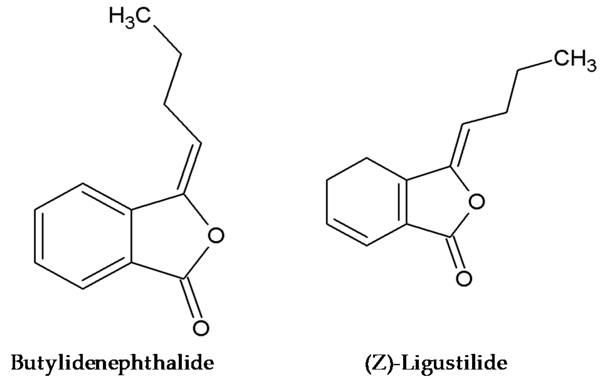
| 54. | 3-Butylphthalide | 47.19 | 1655 | 1647 | 6.8 ± 0.3 | 1.72 ± 0.02 |
| 55. | Z-Butylidenephthalide | 47.98 | 1676 | 1671 | 29 ± 2 | 8.8 ± 0.2 |
| 56. | E-Butylidenefthalide | 49.59 | 1721 | 1717 e | 8.3 ± 0.5 | 3.60 ± 0.05 |
| 57. | Neocnidilide | 50.01 | 1745 | 1722 | 8.9 ± 0.7 | 4.42 ± 0.04 |
| 58. | Z-Ligustilide | 50.1 | 1749 | 1736 | 8.5 ± 0.3 | 20.49 ± 0.02 |
| 59. | E-Ligustilide | 51.49 | 1808 | 1796 | 1.87 ± 0.09 | 25.7 ± 0.1 |
| 60. | Z-Ternine | 52.2 | 1849 | 1844 | 0.26 ± 0.03 | - |
| 61. | n-Hexadecanol | 52.59 | 1881 | 1874 | 0.146 ± 0.007 | - |
| 62. | Metil hexadecanoato | 53.17 | 1927 | 1921 | 0.41 ± 0.05 | - |
| 63. | Metil cis-6-octadecenoato | 54.87 | 2097 | 1921 | 0.70 ± 0.1 | - |
| 64. | Palmitic acid | 53.62 | 1967 | - | - | 4.2 ± 0.5 |
| 65. | Linoleic acid | 55.28 | 2149 | - | - | 18 ± 1 |
| 66. | α-Tocopherol | 63.25 | 3166 | - | - | 0.51 ± 0.06 |
| Total identified | 88.4 ± 0.3 | 91.1 ± 0.4 | ||||
| Monoterpene hydrocarbons | 3.0 ± 0.2 | 0.88 ± 0.0.3 | ||||
| Oxygen-containing monoterpenes | 12.0 ± 0.3 | 0.2 ± 0.02 | ||||
| Sesquiterpene hydrocarbons | 5.3 ± 0.3 | 0.33 ± 0.01 | ||||
| Oxygen-containing sesquiterpenes | 3.4 ± 0.1 | 1.42 ± 0.04 | ||||
| Phthalides | 52.2 ± 2.0 | 65.1 ± 0.2 | ||||
| Others | 12.5 ± 0.8 | 8.9 ± 0.4 | ||||
| Decoction | Hydroethanolic | Hexane | Hydroethanolic Extract of Hexane Residue | Positive Control | |
|---|---|---|---|---|---|
| Antioxidant Activity (EC50, μg/mL) | Trolox | ||||
| DPPH a | 101 ± 2 c | 148 ± 5 b | 469 ± 3 a | 58 ± 1 d | 42 ± 1 |
| Reducing power b | 153 ± 2 b | 153 ± 2 b | 1665 ± 64 a | 114 ± 4 b | 41 ± 1 |
| β-Carotene bleaching inhibition a | 59 ± 34 b | 166 ± 6 a | 188 ± 9 a | 57 ± 4 b | 18 ± 1 |
| TBARS a | 179 ± 11 c | 510 ± 6 b | 3252 ± 49 a | 198 ± 14 c | 23 ± 1 |
| OxHLIA (IC50; µg/mL) | |||||
| Δt = 60 min | 56.0 ± 0.8 b | 41.4 ± 0.5 c | nd | 218 ± 2 a | 19.6 ± 0.1 |
| Δt = 120 min | 100 ± 1 b | 65.1 ± 0.7 c | nd | 343 ± 5 a | 65.1 ± 0.1 |
| Decoction | Hydroethanolic | Hexane | Hydroethanolic Extract of Hexane Residue | Positive Control | |
|---|---|---|---|---|---|
| Cytotoxic Activity (GI50, μg/mL) | Ellipticine | ||||
| HeLa | >400 | >400 | 60 ± 2 | >400 | 0.9 ± 0.1 |
| NCI H460 | >400 | >400 | 69 ± 3 | >400 | 1.03 ± 0.09 |
| MCF7 | >400 | >400 | 48 ± 2 | >400 | 1.21 ± 0.02 |
| HepG2 | >400 | >400 | 67 ± 4 | >400 | 1.10 ± 0.09 |
| Hepatotoxicity (GI50, μg/mL) | |||||
| PLP2 | >400 | >400 | 147 ± 5 | >400 | 2.3 ± 0.2 |
| Decoction | Hydroethanolic | Hexane | Hydroethanolic Extract of Hexane Residue | Ampicilin (20 mg/mL) | Imipenem (1 mg/mL) | Vancomicin (1 mg/mL) | ||||||||
|---|---|---|---|---|---|---|---|---|---|---|---|---|---|---|
| MIC | MBC | MIC | MBC | MIC | MBC | MIC | MBC | MIC | MBC | MIC | MBC | MIC | MBC | |
| Gram-Negative Bacteria | ||||||||||||||
| Escherichia coli | >20 | >20 | 20 | >20 | 20 | >20 | 20 | >20 | <0.15 | <0.15 | <0.0078 | <0.0078 | nt | nt |
| Klebsiella pneumoniae | >20 | >20 | >20 | >20 | >20 | >20 | 20 | >20 | 10 | 20 | <0.0078 | <0.0078 | nt | nt |
| Morganella morganii | >20 | >20 | 20 | >20 | 20 | >20 | 20 | >20 | 20 | >20 | <0.0078 | <0.0078 | nt | nt |
| Proteus mirabilis | >20 | >20 | >20 | >20 | >20 | >20 | 20 | >20 | <0.15 | <0.15 | <0.0078 | <0.0078 | nt | nt |
| Pseudomonas aeruginosa | 20 | >20 | 20 | >20 | >20 | >20 | 20 | >20 | >20 | >20 | 0.5 | 1 | nt | nt |
| Gram-Positive Bacteria | ||||||||||||||
| Enterococcus faecalis | 20 | >20 | 10 | >20 | 10 | >20 | 10 | >20 | <0.15 | <0.15 | nt | nt | <0.0078 | <0.0078 |
| Listeria monocytogenes | 20 | >20 | 10 | >20 | 10 | >20 | 20 | >20 | <0.15 | <0.15 | <0.0078 | <0.0078 | nt | nt |
| MRSA | 20 | >20 | 10 | >20 | 10 | >20 | 10 | >20 | <0.15 | <0.15 | nt | nt | 0.25 | 0.5 |
© 2020 by the authors. Licensee MDPI, Basel, Switzerland. This article is an open access article distributed under the terms and conditions of the Creative Commons Attribution (CC BY) license (http://creativecommons.org/licenses/by/4.0/).
Share and Cite
Spréa, R.M.; Fernandes, Â.; Finimundy, T.C.; Pereira, C.; Alves, M.J.; Calhelha, R.C.; Canan, C.; Barros, L.; Amaral, J.S.; Ferreira, I.C.F.R. Lovage (Levisticum officinale W.D.J. Koch) Roots: A Source of Bioactive Compounds towards a Circular Economy. Resources 2020, 9, 81. https://doi.org/10.3390/resources9070081
Spréa RM, Fernandes Â, Finimundy TC, Pereira C, Alves MJ, Calhelha RC, Canan C, Barros L, Amaral JS, Ferreira ICFR. Lovage (Levisticum officinale W.D.J. Koch) Roots: A Source of Bioactive Compounds towards a Circular Economy. Resources. 2020; 9(7):81. https://doi.org/10.3390/resources9070081
Chicago/Turabian StyleSpréa, Rafael Mascoloti, Ângela Fernandes, Tiane C. Finimundy, Carla Pereira, Maria José Alves, Ricardo C. Calhelha, Cristiane Canan, Lillian Barros, Joana S. Amaral, and Isabel C. F. R. Ferreira. 2020. "Lovage (Levisticum officinale W.D.J. Koch) Roots: A Source of Bioactive Compounds towards a Circular Economy" Resources 9, no. 7: 81. https://doi.org/10.3390/resources9070081
APA StyleSpréa, R. M., Fernandes, Â., Finimundy, T. C., Pereira, C., Alves, M. J., Calhelha, R. C., Canan, C., Barros, L., Amaral, J. S., & Ferreira, I. C. F. R. (2020). Lovage (Levisticum officinale W.D.J. Koch) Roots: A Source of Bioactive Compounds towards a Circular Economy. Resources, 9(7), 81. https://doi.org/10.3390/resources9070081