Article

General Microbiota of the Soft Tick Ornithodoros turicata Parasitizing the Bolson Tortoise (Gopherus flavomarginatus) in the Mapimi Biosphere Reserve, Mexico

by
Sergio I. Barraza-Guerrero
1,
César A. Meza-Herrera
1,
Cristina García-De la Peña
2,*,
Vicente H. González-Álvarez
3,
Felipe Vaca-Paniagua
4,5,6,
Clara E. Díaz-Velásquez
4,
Francisco Sánchez-Tortosa
7,
Verónica Ávila-Rodríguez
2,
Luis M. Valenzuela-Núñez
2 and
Juan C. Herrera-Salazar
2
1
Unidad Regional Universitaria de Zonas Áridas, Universidad Autónoma Chapingo, 35230 Bermejillo, Durango, Mexico
2
Facultad de Ciencias Biológicas, Universidad Juárez del Estado de Durango, 35010 Gómez Palacio, Durango, Mexico
3
Facultad de Medicina Veterinaria y Zootecnia No. 2, Universidad Autónoma de Guerrero, 41940 Cuajinicuilapa, Guerrero, Mexico
4
Laboratorio Nacional en Salud, Diagnóstico Molecular y Efecto Ambiental en Enfermedades Crónico-Degenerativas, Facultad de Estudios Superiores Iztacala, 54090 Tlalnepantla, Estado de México, Mexico
5
Instituto Nacional de Cancerología, 14080 Ciudad de México, Mexico
6
Unidad de Biomedicina, Facultad de Estudios Superiores Iztacala, Universidad Nacional Autónoma de México, 54090 Tlalnepantla, Estado de México, Mexico
7
Departamento de Zoología, Universidad de Córdoba.Edificio C-1, Campus Rabanales, 14071 Cordoba, Spain
*
Author to whom correspondence should be addressed.
Biology 2020, 9(9), 275; https://doi.org/10.3390/biology9090275
Submission received: 30 July 2020 / Revised: 29 August 2020 / Accepted: 3 September 2020 / Published: 5 September 2020
(This article belongs to the Section Conservation Biology and Biodiversity)

Abstract

:
The general bacterial microbiota of the soft tick Ornithodoros turicata found on Bolson tortoises (Gopherus flavomarginatus) were analyzed using next generation sequencing. The main aims of the study were to establish the relative abundance of bacterial taxa in the tick, and to document the presence of potentially pathogenic species for this tortoise, other animals, and humans. The study was carried-out in the Mapimi Biosphere Reserve in the northern-arid part of Mexico. Bolson tortoises (n = 45) were inspected for the presence of soft ticks, from which 11 tortoises (24.4%) had ticks in low loads (1–3 ticks per individual). Tick pools (five adult ticks each) were analyzed through 16S rRNA V3–V4 region amplification in a MiSeq Illumina, using EzBioCloud as a taxonomical reference. The operational taxonomic units (OTUs) revealed 28 phyla, 84 classes, 165 orders, 342 families, 1013 genera, and 1326 species. The high number of taxa registered for O. turicata may be the result of the variety of hosts that this tick parasitizes as they live inside G. flavomarginatus burrows. While the most abundant phyla were Proteobacteria, Actinobacteria, and Firmicutes, the most abundant species were two endosymbionts of ticks (Midichloria-like and Coxiella-like). Two bacteria documented as pathogenic to Gopherus spp. were registered (Mycoplasma spp. and Pasteurella testudinis). The bovine and ovine tick-borne pathogens A. marginale and A. ovis, respectively, were recorded, as well as the zoonotic bacteria A. phagocytophilum, Coxiella burnetii, and Neoehrlichia sp. Tortoises parasitized with O. turicata did not show evident signs of disease, which could indicate a possible ecological role as a reservoir that has yet to be demonstrated. In fact, the defense mechanisms of this tortoise against the microorganisms transmitted by ticks during their feeding process are still unknown. Future studies on soft ticks should expand our knowledge about what components of the microbiota are notable across multiple host–microbe dynamics. Likewise, studies are required to better understand the host competence of this tortoise, considered the largest terrestrial reptile in North America distributed throughout the Chihuahuan Desert since the late Pleistocene.

1. Introduction

Ticks are widely distributed in the world as obligate hematophagous ectoparasites of reptiles, birds, and mammals [1]. In recent years, traditional culture media practices have been replaced by high-throughput techniques such as the next generation sequencing, NGS, used to explore the complete bacterial microbiota of ticks [2]. It is known that tick microbiota is mainly composed of endosymbionts and tick-borne pathogens [3]. Endosymbionts play several beneficial roles to the host (fitness, development, reproduction, immunity, nutritional adaptation, and defense against environmental stress) [4], and tick-borne pathogens are of great concern to animal and human health around the world [3,5]. These two groups of microorganisms form a complex and dynamic ecosystem within ticks, coexisting and interacting with them in a commensal, mutualistic, or pathogenic way [3,6]. From a biological point of view, it is important to know the bacteria that compose of the tick microbiota, but in an epidemiological sense it is fundamental to develop information about the presence and abundance of tick-borne pathogens especially in countries where there is very scarce data.
Tick-borne bacteria have been largely studied in hard ticks (family Ixodidae) Amblyomma spp., Dermacentor spp., Haemaphysalis spp., Hyalomma spp., Ixodes spp., and Rhipicephalus spp., where zoonotic bacteria Anaplasma phagocytophilum, Coxiella burnetii, Ehrlichia chaffensis, Francisella tularensis, and Rickettsia rickettsi have been reported [7,8,9,10]. In soft ticks (family Argasidae), most studies have focused on the genus Ornithodoros, which comprises of more than 60% of the family [1,11]. These ticks usually inhabit sheltered sites (i.e., burrows, caves, cracks, and nests), commonly parasitizing small mammals, birds, reptiles, or bats [12]. Particularly, O. turicata is distributed through the arid regions of the Southern United States and Latin America [13]. It has been reported frequently as a promiscuous feeder parasitizing wild animals such as ground squirrels, prairie dogs, snakes, and desert tortoises [14]. In Mexico, this tick has been reported as an ectoparasite of the Bolson tortoise, Gopherus flavomarginatus [15], an endemic and endangered reptile species restricted to the Chihuahuan desert [16,17]. Gopherus tortoises are considered keystone species because they dig burrows where they spend the most part of its life, providing additional shelter for other animals such as rodents, lagomorphs, birds, and reptiles [18]. However, it is in their burrows where they most likely to acquire soft ticks. Due to the ecological relevance that G. flavomarginatus represents for the Chihuahuan desert ecosystem, it is important to generate information on the possible pathogens with which it may have contact, in this case those transmitted by soft ticks. Since there is no previous information regarding the bacterial microbiota of O. turicata in Mexico, we established the relative abundance of bacterial taxa in this tick. We hypothesized that the O. turicata microbiota will include potentially pathogenic bacteria for the Bolson tortoises, other animals, and even humans.

2. Materials and Methods

2.1. General

All the methods and activities of this study were in strict accordance with accepted guidelines for ethical use, care, and welfare of animals in research at the international level [19]. The federal approval reference number is SEMARNAT-SGPA/DGVS/08406/16. This research has been approved by the Facultad de Ciencias Biológicas UJED ethic committee on 5 February 2017 (ethic code: 0023). The files used in this study (Supplementary files: P1.fasta, P2.fasta and P3.fasta) were deposited into the NCBI Sequence Read Archive (SRA) database (SRA Accession Number: PRJNA649587).

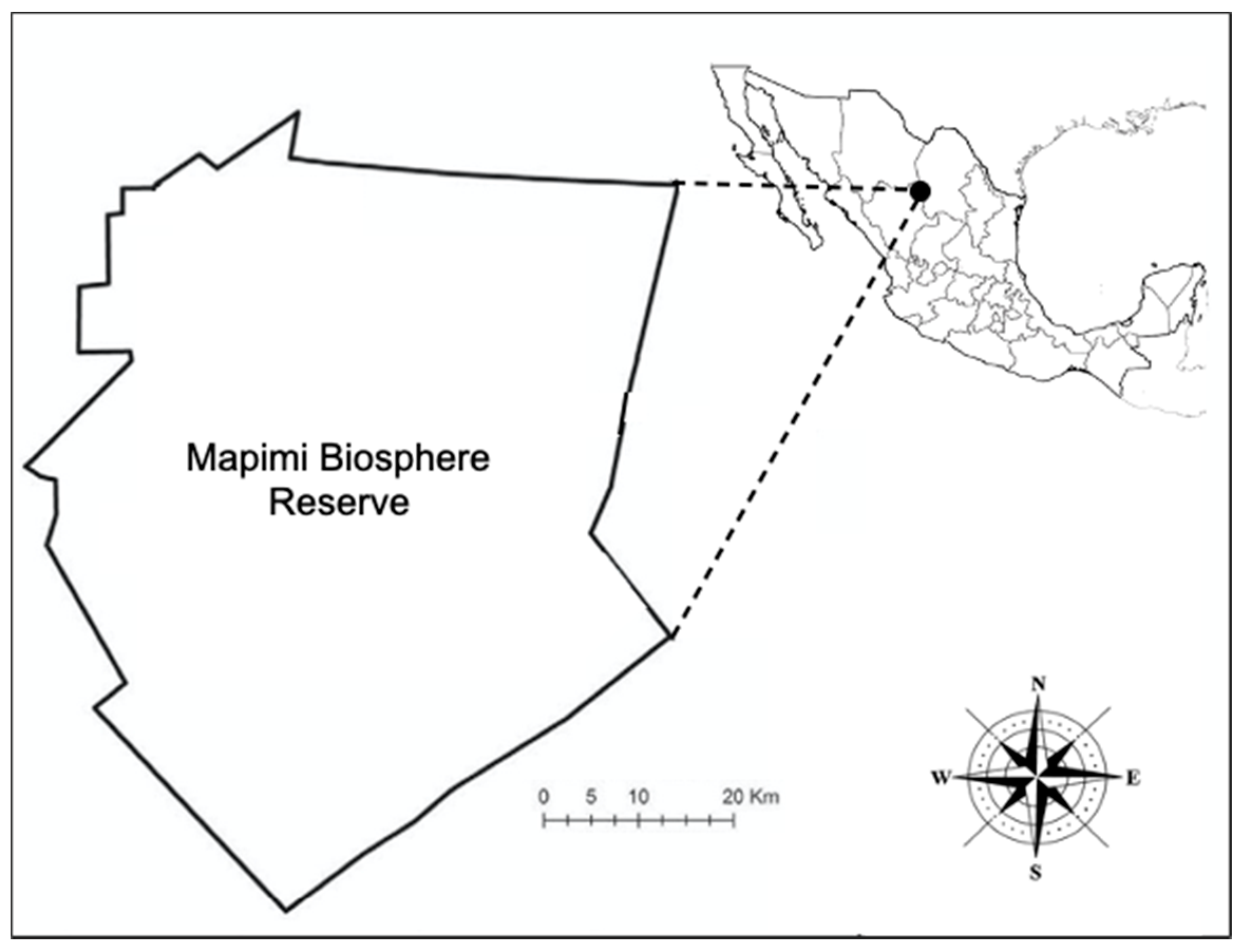
2.2. Location and Environmental Conditions
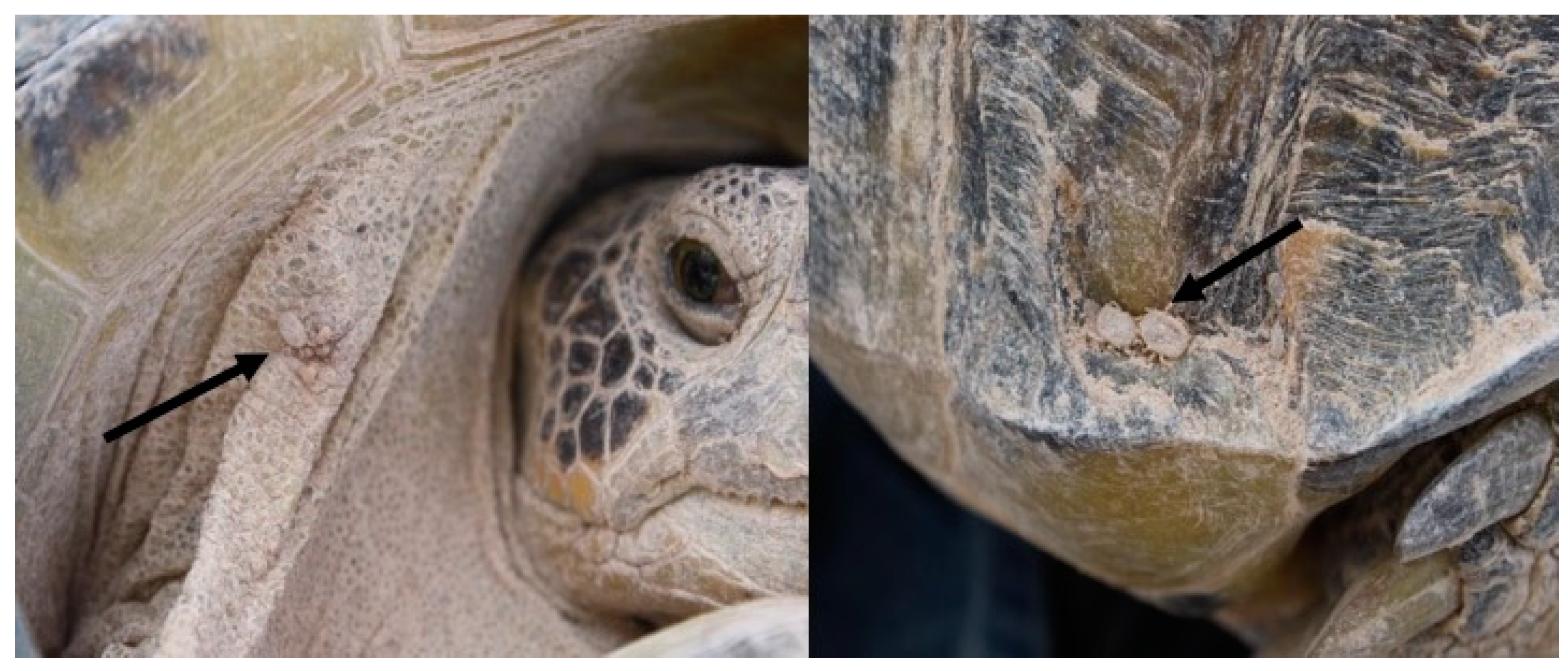
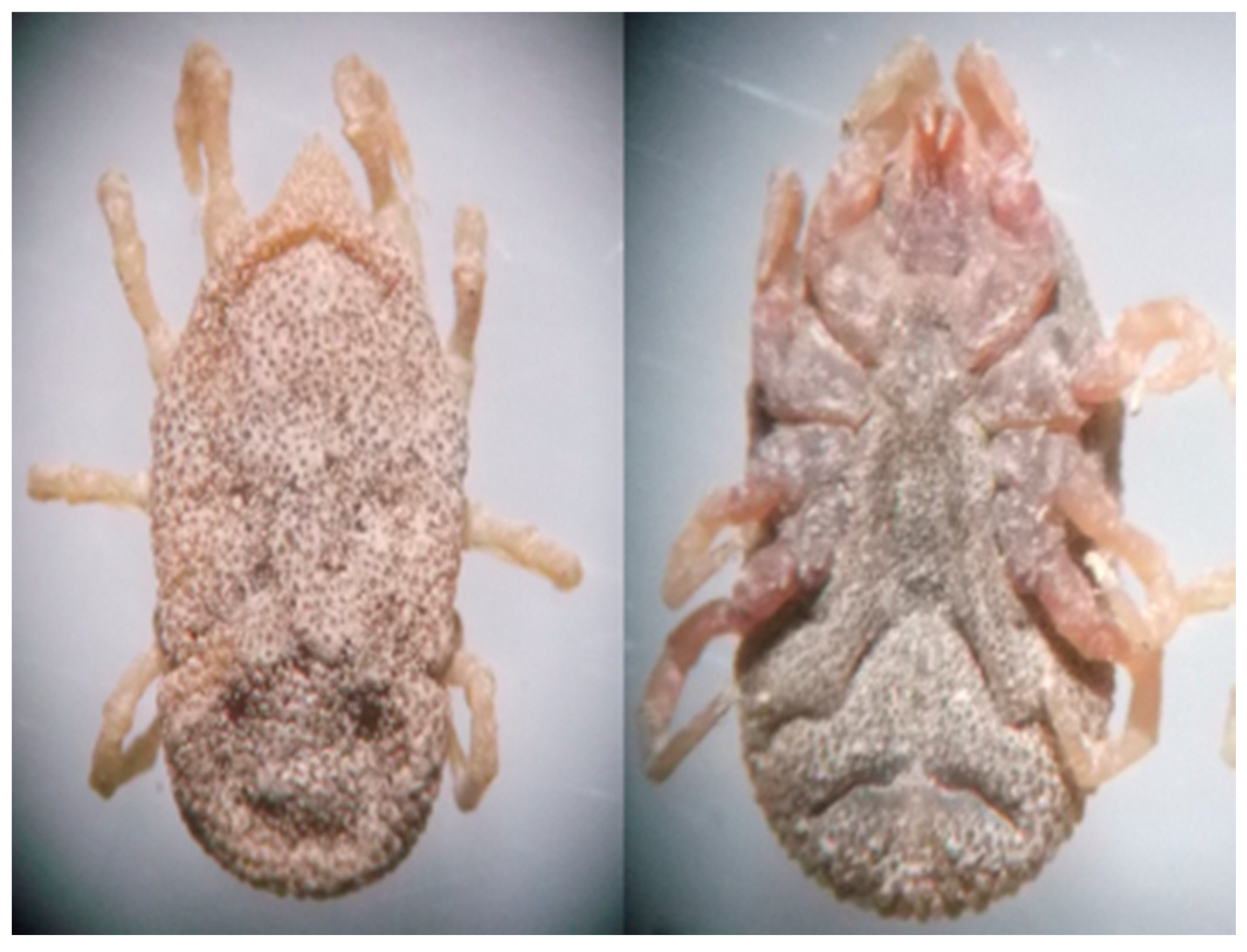
The study was carried-out in the Mapimi Biosphere Reserve in Mexico, which includes part of the states of Chihuahua, Coahuila, and Durango (26°00′ and 26°10′ N, 104°10′ and 103°20′ W; Figure 1). The Bolson de Mapimi has been defined as an endoreic basin, which includes diverse small sub-basins intermixed along valleys with a mean altitude of 1150 m. This area has very arid climate [20], with an average annual temperature of 25.5 °C, and an average annual precipitation of 264 mm [21]. The predominant vegetation in the reserve is rosette and microphile scrub, as well as halophyte, and gypsophila plants [22]. Interestingly, the microregion concentrates the richest herpetofauna across the whole Chihuahuan desert, having diverse endemic species, including the Bolson tortoise [23].

2.3. Field Work

From May to July 2017, Bolson tortoises were captured by hand from 900 to 1300 h and from 1700 to 2100 h. The search for ticks was carefully carried out on the carapace, neck and skin folds of all four limbs. A total of 45 individuals of G. flavomarginatus were captured and revised, but only 11 tortoises (24.4%) carried soft ticks on the carapace or over the skin (Figure 2 and Figure 3). Tortoises carried from one to three ticks per individual. A total of 17 adult ticks were collected and taxonomic keys were used to determine the species [24]. Each tick was placed in an individual 1.5 mL tube containing 500 uL ethanol (70%), 500 uL hydrogen peroxide (H2O2), and 200 uL of ultrapurified H2O; each tube was vortexed for 15 s to remove tick surface contaminants [25]. Later, three pools were formed (five ticks each) and were deposited in BashingBead™ Zymo Research™ cell lysis tubes, containing 750 μL of lysing/stabilizing solution. The tubes were processed in a cellular disruptor (TerraLyzer™) for 20 s. The two extra ticks were deposited in the Entomological collection of the Facultad de Ciencias Biológicas, Universidad Juárez del Estado de Durango, México, for reference purposes.

2.4. DNA Extraction, Visualization, and Quantification

DNA was extracted from the pools using the Xpedition™ Tissue/insect DNA MiniPrep kit (Zymo Research Corp., Irvine, CA, USA) in a laminar UV flow hood in sterile conditions. Then, the DNA was placed on a 1.2% agarose gel at 80 V for 45 min in a BIORAD electrophoresis chamber (Bio-Rad Laboratories, Inc., Hercules, CA, USA). The DNA visualization was carried out in a GelMax™ photo documenter (UVP LLC, Upland, CA, USA). The amount of DNA obtained was measured in a Qubit™ fluorometer (Invitrogen, Carlsbad, CA, USA). Then, the V3-V4 region of the 16S rRNA gene was amplified using the following primers [26]: S-D-Bact-0341-b-S-17, 5′-CCTACGGGNGGCWGCAG-3′ and S-D-Bact-0785-a-A-21, 5′-GACTACHVGGGTATCTAATCC-3′.
The PCR protocol was performed by using 12.5 μL of MyTaq™ Ready Mix 1× (Bioline, London, UK), 1 μL of each primer (10 nM), 5 μL of DNA (50 ng total), and 5.5 μL of molecular grade H2O. The following cycle was used: 95 °C for 3 min; 25 cycles of 95 °C for 30 s, 55 °C for 30 s, 72 °C for 30 s; and 72 °C for 5 min; Illumina (2020a) [27] in a Labnet Multigene™ Gradient PCR thermal cycler (Labnet International, Edison, NJ, USA). Then, 1 μL of each PCR product was placed in an Agilent Bioanalyzer DNA 1000 chip (Agilent Technologies, Santa Clara, CA, USA) to verify the amplicon size (550 bp). The amplicons were purified with Agentcourt® AMPure® XP 0.8% beads (Beckman Coulter Inc., Brea, CA, USA). Thereafter, the Nextera XT Index Kit™ was used to create the library, following the Illumina (2020b) [28] protocol using 25 μL of MyTaq™ Ready Mix 1× (Bioline®), 5 μL of each primer (N7xx and S5xx), 5 μL of DNA, and 10 μL of molecular grade H2O. Each pool considered the following cycle: 95 °C for 3 min; 10 cycles of 95 °C for 30 s, 55 °C for 30 s, 72 °C for 30 s; and 72 °C for 5 min. The libraries were purified with Agencourt® AMPure® XP 1.2% beads. Then, 1 μL of the final PCR product library was randomly selected and placed on a Bioanalyzer DNA 1000 chip to verify the size of the amplicon expecting a size of 630 bp. Thereafter, the amplicon was quantified, normalized, (equimolarity), and sequenced with next generation massive sequencing (MiSeq; Illumina, San Diego, CA, USA) of 2 × 250 paired final readings following the 16S metagenomic protocol [27].

2.5. Bioinformatics Analyses
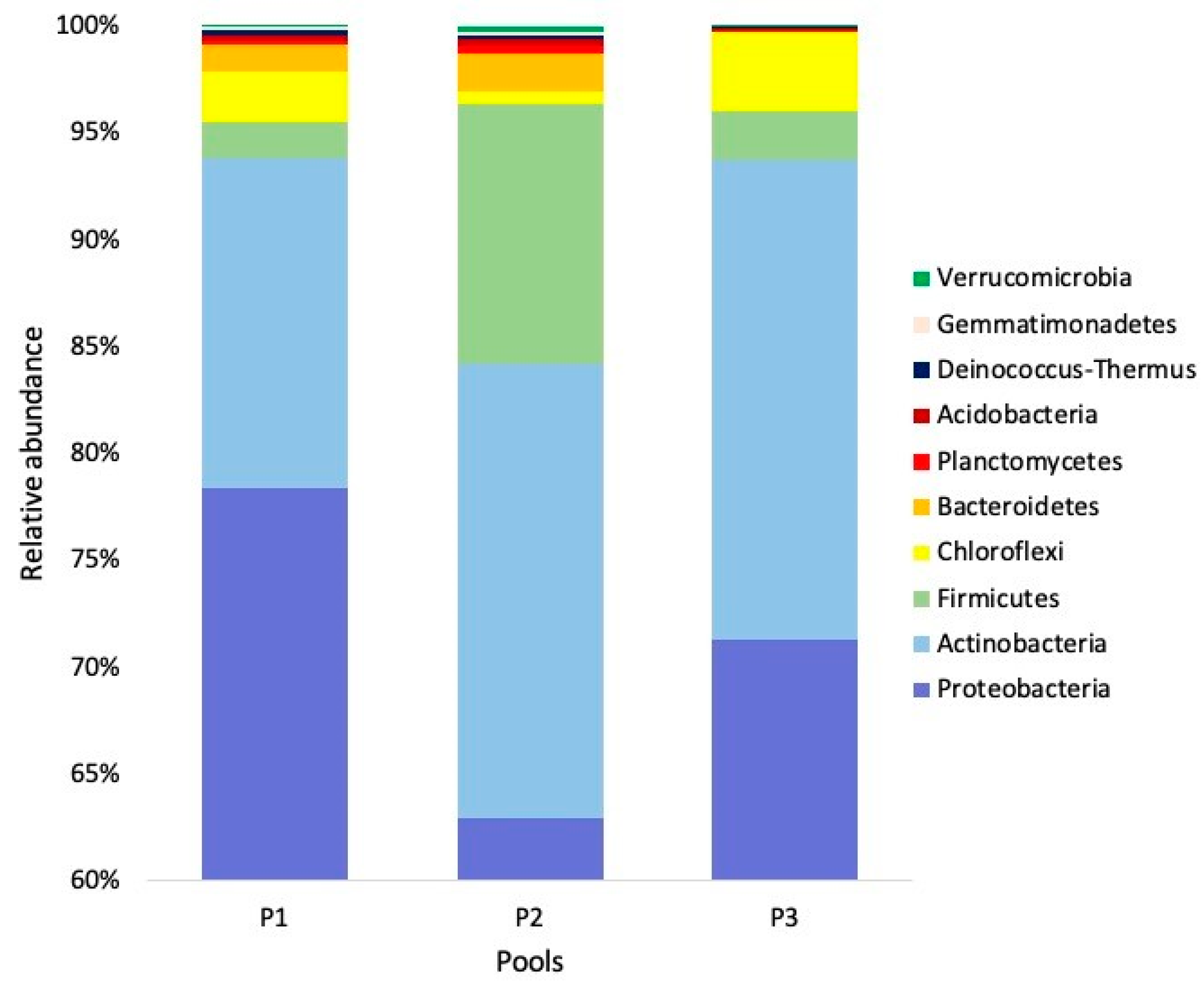
The DNA sequences were analyzed on MGLinux, in a VM Oracle VirtualBox using Quantitative Insights into Microbial Ecology bioinformatics software (QIIME, Boulder, CO, USA) [29]. Both forward and reverse sequences were assembled using the PEAR program [30] with an overlap of 50 bp, a minimum reading length of 430 bp, and a maximum of 470 bp, a quality criterion Q30 (one false base for every 1000 bases) with p < 0.0001. Then the files were converted to the FASTA format, and chimeric sequences were discarded with USEARCH [31]. Thereafter, the operational taxonomic units (OTUs) were selected with the UCLUST method [31] at 97% similarity; a representative sequence for each OTU was obtained, and the taxonomy was assigned and taken as reference to the EzBioCloud database [32]. Next, the OTUs table was built in the Biom format (biological observation matrix) [33] separating domains. A simple random rarefaction process was performed [34] in order to obtain a standardized Biom file for all pools. The Shannon and Simpson alpha diversity indexes were calculated using the standardized Biom file; the mean ± standard deviation for each index was obtained. The relative abundance for the phylum level was represented as a bar chart using Excel, and family and genus levels were visualized as heatmaps using Morpheus software (Broad Institute, Cambridge, MA, USA) [35]; hierarchical clustering (average linkage method with Euclidean distance) was used to visualize pools dendrograms. Each genus and/or species of bacteria registered for O. turicata was consulted in the available literature to indicate its possible pathogenic potential for tortoises or zoonotic potential for humans.

3. Results

The average number of sequences assembled was 196,564. After taxonomic designation an average of 190,769 bacterial sequences was obtained. The average number of OTUs was 44,369 (Table 1). Simple random rarefaction was made at 100,000 sequences, since at this point the number of taxa of the three pools reached asymptotes. From the standardized Biom file, the OTUs resulted in 28 phyla, 84 classes, 165 orders, 342 families, 1013 genera, and 1326 species.
While the Shannon diversity index was 8.12 ± 0.39 (SD), the Simpson diversity index was 0.86 ± 0.05. From the 28 phyla obtained, 27 (96.4%) were taxonomically classified, the rest were classified as “other”. The most abundant phyla were Proteobacteria (mean = 70.75%), Actinobacteria (mean = 19.67%), and Firmicutes (mean = 5.35 %; Figure 4).
At the class level, 84 taxa were obtained of which 82 (97.6%) were taxonomically classified, the rest was classified as “other”. The most abundant classes were Alphaproteobacteria (mean = 56.91%), Actinobacteria (mean = 18.55%), and Gammaproteobacteria (13.65%; Table S1). From the 165 orders obtained, 158 (95.7%) were taxonomically classified, the rest were classified as “other”. The most abundant orders were Rickettsiales (mean = 55.40%), Legionellales (mean = 13.30%), Micrococcales (mean = 5.99%), Propionibacteriales (mean = 5.32%), and Srteptomycetales (mean = 5.11%; Table S1).
At the family level, 342 taxa were obtained of which 325 (95.0%) were taxonomically classified, the rest were classified as “other”. The most abundant families were Midichloriaceae (mean = 51.13%), Coxiellaceae (mean = 13.26%), Nocardioidaceae (mean = 5.29%), Streptomycetaceae (mean = 5.10%), and Anaplasmataceae (mean = 4.29%; Figure 5). From the 1013 genera obtained, 897 (88.54%) were taxonomically classified, the rest were classified as “other”. The most abundant genera were Midichloria (mean = 51.13%), Coxiella (mean = 13.25%), Streptomyces (5.08%), Nocardioides (mean = 5.06%), Anaplasma (mean = 4.29%), and Clostridium (mean = 2.19%; Figure 6; Table S1). The genus Mycoplasma, a potential pathogen of the Gopherus spp., was registered in low abundance (mean = 3.33 × 10⁻⁶ %). Additionally, 1326 species were obtained, 918 (69.2%) were classified as “other”, 268 (20.3%) have a taxonomical key, and 140 (10.5%) have a taxonomical name (Table 2). The most abundant species were Midichloria-like bacteria (mean = 50%), Coxiella-like bacteria (mean = 13%), Streptomyces sp. (mean = 5%), and Nocardioides sp. (mean = 5%). However, considering only bacteria with a complete taxonomical name the most abundant was Midichloria mitochondrii (mean = 0.72%; Table S1). Pasteurella testudinis, a potential pathogen of the Gopherus spp., was registered in low abundance (mean = 1.33 × 10⁻⁵%). The bovine and ovine tick-borne bacteria A. marginale and A. ovis, respectively, were recorded in low abundances; also, the zoonotic bacteria Coxiella burnetii, Anaplasma phagocytophilum, and Neoehrlichia sp. were part of the O. turicata microbiota (see relative abundances in Table S1).

4. Discussion

Our working hypothesis stated that the microbiota of O. turicata would contain potentially pathogenic bacteria for the Bolson tortoise Gopherus flavomarginatus, other animals, and humans. Therefore, and based on the obtained results, this study demonstrated the presence of some potentially pathogenic and zoonotic bacteria in the microbiota of O. turicata.
The most abundant phyla were Proteobacteria, Actinobacteria, and Firmicutes. This is similar to that reported for Ambylomma tuberculatum ticks infesting the Gopher tortoise (G. polyphemus) in Mississippi, USA, with 89%, 9%, and 1% of the relative abundance, respectively [36]. Currently, Proteobacteria is the broadest phylum in the Bacteria domain [37]. Some of the tick species that have shown dominance of this phylum in their microbiota are Argas japonicus in China (42–97%) [38], Hyalomma dromedarii in Saudi Arabia (98%) [39], Dermacentor marginatus and D. reticulatus in Slovakia (60%) [40], D. marginatus (97%), Haemaphysalis punctata (98%), Ixodes ricinus (88%), and Rhipicephalus sanguineus sensu lato (99%) in Spain [41], and I. scapularis in Massachusetts and Texas, USA (73–100%; [8]).
Alphaproteobacteria and Gammaproteobacteria classes were the most abundant in O. turicata microbiota, as has been observed in other tick species [41]. These classes are relevant in the microbiota of all tick species because they group the main intracellular endosymbionts (Arsenophonus-like, Coxiella-like, Fransicella-like, Midichloria-like, Rickettsia-like, and Wolbachia-like) that inhabit the organs of these arthropods [6,42]. These endosymbionts have generally been reported to predominate in the arthropod microbiota and may interfere with the transmission dynamics of pathogenic bacteria [43,44]. In the present study, O. turicata showed only two genera of intracellular endosymbionts, Midichloria and Coxiella. An unidentified species Midichloria-like endosymbiont was the dominant taxon in the O. turicata microbiota, although Candidatus Midichloria mitochondrii (CMM) species was also abundant in this tick. CMM has been reported in a large number of tick genera (Amblyomma spp., Dermacentor spp., Haemaphysalis spp., Hyalomma spp., Ixodes spp., and Rhipicephalus spp.) in different countries of Europe, Asia, and North America [41,45,46]. It is considered a facultative non-pathogenic mutualistic bacterium that lodges in the reproductive tissues of female ticks, specifically in the ovarian mitochondria, where they sometimes invade and destroy this organ [47,48]. By remaining in this organ, vertical transmission to the next generation is carried out. However, its presence has also been recorded in salivary glands from which it is transmitted to vertebrate hosts during tick feeding [49]. Since all the body of O. turicata was analyzed in the present study, it is not possible at this time to determine in which organs occur both unknown Midichloria-like and CMM. However, it is feasible that they occur in the reproductive organs since some studies have analyzed the possible evolutionary process of intracellular endosymbionts of arthropods, and apparently transovarian transmission is the common factor for all species [50]. Bacterial diversity studies in each organ of O. turicata are required to clarify this question.
The second most abundant bacterial species in O. turicata microbiota in our study was a Coxiella-like endosymbiont. This type of endosymbiont does not appear to be pathogenic and are relatively common in the microbiota of various tick species around the world [51,52]. These microorganisms are known to infect tick ovaries and then transmission occurs vertically through the egg cytoplasm, but they have also been found in Malpighi tubules where they may provide essential nutrients to their host [7]. It should be noted the presence of C. burnetii in the present study. This is a zoonotic bacterium that causes Q-fever in both animals and humans. In particular, for the Ornithodoros genus there are reports of C. burnetii in O. tartakovskyi, O. papillipes and O. alactagalis in the former Soviet Union [53], O. moubata in Japan [54], and O. sonrai in Senegal [55]. According to Balashov and Daiter (1973) [53] and Eldin et al. (2017) [56], transmission of C. burnetii is transovarian in most ticks, except for Ixodes holocyclus, O. hermsi, and O. turicata in which it is transestadial. Direct transmission of C. burnetii from infected ticks to humans is not well documented and may occur only rarely in nature [57]. However, the main transmission route is via inhalation of contaminated fecal material from ticks [58]. Since the personnel that monitor G. flavomarginatus populations each year in the Mapimi Biosphere Reserve should manipulate tortoises, it is advisable to take precautions to avoid this airborne infection.
Mycoplasma spp. and Pasteurella spp. are bacterial genera that have been reported as part of the microbiota of hard ticks, as in Ixodes simplex and I. ventalloi, respectively [59,60]. In the present study with O. turicata, the presence of Mycoplasma spp. and Pasteurella testudinis in low abundances was recorded. An interesting fact is that these bacteria along with some viruses have been reported as some of the possible causes of upper respiratory disease (URTD) in tortoises, causing a decrease in the populations of G. agassizii and G. polyphemus in the USA [61,62]. It is still unknown if these bacteria can be transmitted directly to the host, and in the case of G. flavomarginatus if they could cause disease. For now, these bacteria should remain as potential pathogens for this tortoise, until further studies of vector competence discard this possibility.
Other relevant species recorded in O. turicata microbiota were the obligate intracellular bacteria Anaplasma phagocytophilum, A. marginale, A. ovis, and Neoehrlichia sp. These species are important for animal and human health because they typically infect hematopoietic or endothelial cells [63]. Anaplasma phagocytophilum is a tick-borne pathogen that causes human granulocytic anaplasmosis mainly in Asia, Europe, and the USA; A. marginale and A. ovis are worldwide tick-borne bacteria causing bovine and ovine anaplasmosis respectively [64,65]. The genus Neoehrlichia was discovered in 1999 as an Ehrlichia-like bacterium [66], which was later described as Candidatus Neoehrlichia mikurensis [67], an emerging tick-borne pathogen detected in ticks and rodents, causing human systemic inflammatory syndrome in Asia and Europe [68,69]. In America, Candidatus Neoehrlichia lotoris was registered in a free-living raccoon associated with tick-infested populations [70], which suggests that it is transmitted to mammals by ticks. Undoubtedly, specific molecular studies are needed to determine which Neoehrlichia species is the one registered in O. turicata, nevertheless, its zoonotic potential is latent.
According to Gofton et al. (2015b) [71], a limitation of 16S bacterial community profiling in ticks is that a high proportion of sequences will belong to bacterial endosymbionts, as CMM as observed in O. turicata in the present study. This high abundance may mask the presence of less abundant bacteria with zoonotic potential, as for example the genus Borrelia, which was not detected in our study. Species such as B. mazzottii recorded in O. talaje in Mexico [72], as well as B. parkeri in O. parkeri, B. hermsii in O. hermsi, and B. turicatae in O. turicata in the USA [73], are important indicators of the presence of this bacterial genus in the Ornithodoros ticks of America. Perhaps, by carrying out detailed molecular studies in O. turicata in the Mapimi Biosphere Reserve, Borrelia and other zoonotic bacteria such as Rickettsia could be identified. In the same way, since this massive sequencing technique does not reach the species level in many genera documented here for O. turicata, it is necessary to carry out specific PCR studies for the most relevant bacteria involved in the biology of this tick and those bacteria important for the health of animals and humans.
It is important to comment that the present study was carried out at a single time of the year, without distinguishing sexes of O. turicata to provide an initial overview of the microbiota of this tick species. However, various studies indicate that the microbiota of ticks is dynamic and can change among seasons of the year due to the effect of temperature and humidity, between males and females, between different feeding statuses, among life stages, etc. [3,4]. Therefore, it is guaranteed that detailed studies on these issues will further be carried out.
Finally, the high number of bacterial taxa recorded for O. turicata in the present study could be due to the variety of hosts that this tick parasitizes within the burrows of G. flavomarginatus, performing a rapid feeding process that usually lasts an hour on average [14,74]. In this way the general feeding habits of O. turicata may be keeping pathogenic microbes in circulation, thereby ensuring their survival in the ecosystem. In our study, all G. flavomarginatus tortoises found carrying O. turicata ticks were apparently healthy, having the possible ecological role of reservoirs of pathogenic bacteria [75,76]. The fact that tortoises have long lives can favor the maintenance of pathogen cycles under normal conditions [77,78]. However, after thousands of years of coevolution, a microbial balance between O. turicata and G. flavomarginatus must have been reached, although it is not yet known whether this tortoise’s defense mechanism is resistance (i.e., capacity to limit pathogen loads) or tolerance (i.e., capacity to survive damage caused by a given pathogen load) [79,80,81]. To clarify this enquiry, it will be necessary to determine the bacteria that G. flavomarginatus carries in the blood and whether they were transmitted by O. turicata. Studies of resistance mechanisms and vector competence in this tortoise will also be relevant from the point of view of immunity and the eco-epidemiology of zoonotic diseases, respectively.

5. Conclusions

Our study established the general relative abundance of bacterial taxa in the soft tick O. turicata, documenting the presence of potentially pathogenic species for the Bolson tortoise G. flavomarginatus, other animals and humans in the Mapimi Biosphere Reserve, Durango, Mexico. The most abundant phyla were Proteobacteria, Actinobacteria, and Firmicutes. Additionally, Alphaproteobacteria and Gammaproteobacteria classes were the most abundant in O. turicata microbiota. The most abundant species were Midichloria-like and Coxiella-like (endosymbionts of ticks). Mycoplasma spp. and Pasteurella testudinis, both potentially pathogenic to the Bolson tortoise, were registered. Additionally, the bovine and ovine tick-borne pathogens A. marginale and A. ovis, respectively, as well as the zoonotic bacteria A. phagocytophilum, Coxiella burnetii, and Neoehrlichia sp. were founded in O. turicata. Future studies on soft ticks should expand our knowledge about what components of the microbiota are notable across multiple host–microbe dynamics. Likewise, studies are required to better understand the reservoir competence of this tortoise, considered the largest terrestrial reptile in North America distributed throughout the Chihuahuan Desert since the late Pleistocene.

Supplementary Materials

The following are available online at https://www.mdpi.com/2079-7737/9/9/275/s1, Table S1: Relative abundance of bacterial taxa from three pools of the soft tick Ornithodoros turicata as an ectoparasite of the Bolson tortoise (Gopherus flavomarginatus) in the Mapimi Biosphere Reserve, Mexico, Supplementary files (P1.fasta, P2.fasta and P3.fasta). The files used in this study were also deposited into the NCBI Sequence Read Archive (SRA) database (SRA Accession Number: PRJNA649587).

Author Contributions

Conceptualization, C.G.-D.l.P. and C.A.M.-H.; Data curation, S.I.B.-G. and C.A.M.-H.; Formal analysis, S.I.B.-G., C.A.M.-H. and C.G.-D.l.P.; Funding acquisition, C.G.-D.l.P.; Investigation, V.H.G.-Á., C.E.D.-V., V.Á.-R. and J.C.H.-S.; Methodology, V.H.G.-Á., F.V.-P., C.E.D.-V., F.S.-T., V.Á.-R., L.M.V.-N. and J.C.H.-S.; Resources, F.V.-P., F.S.-T. and L.M.V.-N.; Software, F.V.-P. and C.E.D.-V.; Supervision, C.G.-D.l.P. All authors have read and agreed to the published version of the manuscript.

Funding

This research was founded by the National Council of Science and Technology (CONACYT, Mexico) through the Basic Research Fund CIENCIA BÁSICA-CONACYT: 0220658, which greatly contributed to the generation of most of the information presented in this study.

Acknowledgments

We thank Nohemí Villa-Vázquez, Estefanía Garduño-Niño, Romario Martínez-Saldívar, and Irene Pacheco-Torres, for their support in the field work. To Cristino Villarreal-Wislar and the staff of the Administration of the Mapimi Biosphere Reserve for the logistical support during this study. We deeply appreciate the suggestions of anonymous reviewers for the improvement of this manuscript.

Conflicts of Interest

The authors declare that there are no conflicts of interest that could be perceived as prejudicing the impartiality of the research reported in this manuscript.

References

  1. Manzano-Román, R.; Díaz-Martín, V.; de la Fuente, J.; Pérez-Sánchez, R. Soft ticks as pathogen vectors: Distribution, surveillance and control. In Parasitology; Manjur, M., Ed.; Intech: Rijeka, Croatia, 2012; pp. 125–162. [Google Scholar]
  2. Gupta, S.; Mortensen, M.S.; Schjørring, S.; Trivedi, U.; Vestergaard, G.; Stokholm, J.; Bisgaard, H.; Krogfelt, K.A.; Sørensen, S.J. Amplicon sequencing provides more accurate microbiome information in healthy children compared to culturing. Commun. Biol. 2019, 2, 1–7. [Google Scholar] [CrossRef] [PubMed] [Green Version]
  3. Pollet, T.; Sprong, H.; Lejal, E.; Krawczyk, A.I.; Moutailler, S.; Cosson, J.-F.; Vayssier-Taussat, M.; Estrada-Peña, A. The scale affects our view on the identification and distribution of microbial communities in ticks. Parasite. Vector. 2020, 13, 36. [Google Scholar] [CrossRef] [PubMed] [Green Version]
  4. Bonnet, S.I.; Binetruy, F.; Hernández-Jarguín, A.M.; Duron, O. The tick microbiome: Why non-pathogenic microorganisms matter in tick biology and pathogen transmission. Front. Cell. Infect. Microbiol. 2017, 7, 236. [Google Scholar] [CrossRef] [PubMed]
  5. Vayssier-Taussat, M.; Albina, E.; Citti, C.; Cosson, J.F.; Jacques, M.-A.; Lebrun, M.-H.; Le Loir, Y.; Ogliastro, M.; Petit, M.-A.; Roumagnac, P. Shifting the paradigm from pathogens to pathobiome: New concepts in the light of meta-omics. Front. Cell. Infect. Microbiol. 2014, 4, 29. [Google Scholar] [CrossRef] [Green Version]
  6. Narasimhan, S.; Fikrig, E. Tick microbiome: The force within. Trends Parasitol. 2015, 31, 315–323. [Google Scholar] [CrossRef] [Green Version]
  7. Lalzar, I.; Friedmann, Y.; Gottlieb, Y. Tissue tropism and vertical transmission of Coxiella in Rhipicephalus sanguineus and Rhipicephalus turanicus ticks. Environ. Microbiol. 2014, 16, 3657–3668. [Google Scholar] [CrossRef]
  8. Varela-Stokes, A.S.; Park, S.H.; Kim, S.; Ricke, S.C. Microbial communities in North American ixodid ticks of veterinary and medical importance. Front. Vet. Sci. 2017, 4, 179. [Google Scholar] [CrossRef] [Green Version]
  9. Thapa, S.; Zhang, Y.; Allen, M.S. Bacterial microbiomes of Ixodes scapularis ticks collected from Massachusetts and Texas, USA. BMC Microbiol. 2019, 19, 138. [Google Scholar] [CrossRef] [Green Version]
  10. Madison-Antenucci, S.; Kramer, L.D.; Gebhardt, L.L.; Kauffman, E. Emerging Tick-Borne Diseases. Clin. Microbiol. Rev. 2020, 33, 33. [Google Scholar] [CrossRef]
  11. Nava, S.; Venzal, J.M.; Acuña, D.G.; Martins, T.F.; Guglielmone, A.A. Ticks of the Southern Cone of America: Diagnosis, Distribution, and Hosts with Taxonomy, Ecology and Sanitary Importance; Academic Press: Cambridge, MA, USA, 2017; pp. 269–321. [Google Scholar] [CrossRef]
  12. Oliver, J.H., Jr. Biology and systematics of ticks (Acari: Ixodida). Annu. Rev. Ecol. Evol. Syst. 1989, 20, 397–430. [Google Scholar] [CrossRef]
  13. Donaldson, T.G.; de León, A.A.P.; Li, A.I.; Castro-Arellano, I.; Wozniak, E.; Boyle, W.K.; Hargrove, R.; Wilder, H.K.; Kim, H.J.; Teel, P.D. Assessment of the geographic distribution of Ornithodoros turicata (Argasidae): Climate variation and host diversity. PLOS Negl. Trop. Dis. 2016, 10, e0004383. [Google Scholar] [CrossRef] [PubMed] [Green Version]
  14. Milstrey, E.G. Bionomics and Ecology of Ornithodoros (P.) Turicata Americanus (Marx) (Ixodoidea: Argasidae) and Other Commensal Invertebrates Present in the Burrows of the Gopher Tortoise, Gopherus Polyphemus Daudin; University of Florida: Gainesville, FL, USA, 1987. [Google Scholar]
  15. Paredes-León, R.; García-Prieto, L.; Guzmán-Cornejo, C.; León-Regagnon, V.; Pérez, T.M. Metazoan parasites of Mexican amphibians and reptiles. Zootaxa 2008, 1904, 1–166. [Google Scholar] [CrossRef]
  16. SEMARNAT (Secretaria de Medio Ambiente y Recursos Naturales). Norma Oficial Mexicana NOM-059-SEMARNAT-2010, Protección ambiental-Especies nativas de México de flora y fauna silvestres-Categorías de riesgo y especificaciones para su inclusión, exclusión o cambio-Lista de especies en riesgo. Diario Oficial de la Federación 2010, 12, 1–180. [Google Scholar]
  17. Kiester, A.; Palomo-Ramos, R.; Ríos-Arana, J.; Goode, E. Gopherus flavomarginatus. The IUCN Red List of Threatened Species 2018: E.T9402A112660985. Available online: https://dx.doi.org/10.2305/IUCN.UK.2018-2.RLTS.T9402A112660985.en (accessed on 21 August 2020).
  18. Dziadzio, M.C.; Smith, L.L. Vertebrate use of gopher tortoise burrows and aprons. Southeast. Nat. 2016, 15, 586–594. [Google Scholar] [CrossRef]
  19. FASS. Guide for the Care and Use of Agricultural Animals in Agricultural Research and Teaching, 3rd ed.; f Animal Science Journal: Champaing, IL, USA, 2010; p. 177. [Google Scholar]
  20. García, E. Modificaciones al Sistema de Clasificación Climática de Köppen; Universidad Nacional Autónoma de México: Ciudad de México, Mexico, 2004; 246p. [Google Scholar]
  21. Cornet, A. Principales caractéristiques climatiques. In Estudio Integrado de Los Recursos Vegetación, Suelo Y Agua en la Reserva de la Biósfera de Mapimí; Montaña, C., Ed.; Instituto de Ecología, A.C.: Veracruz, México, 1988; pp. 45–76. [Google Scholar]
  22. Rzedowski, J. Vegetación de México, 1st ed.; Comisión Nacional para el Conocimiento y Uso de la Biodiversidad: Ciudad de México, México, 2006; p. 504.
  23. Barnard, S.M.; Durden, L.A. A Veterinary Guide to the Parasites of Reptiles; Krieger: Melbourne, FL, USA, 2000; Volume 2, 288p. [Google Scholar]
  24. Morafka, D.J. A historical biogeography of the Chihuahuan herpetofauna. In A Biogeographical Analysis of the Chihuahuan Desert through Its Herpetofauna; Springer: Dodlerk, The Netherlands, 1977; pp. 159–215. [Google Scholar]
  25. Couper, L.; Swei, A. Tick microbiome characterization by next-generation 16S rRNA amplicon sequencing. JoVE 2018, 138, e58239. [Google Scholar] [CrossRef] [Green Version]
  26. Klindworth, A.; Pruesse, E.; Schweer, T.; Peplies, J.; Quast, C.; Horn, M.; Glockner, F.O. Evaluation of general 16S ribosomal RNA gene PCR primers for classical and next-generation sequencing-based diversity studies. Nucleic. Acids. Res. 2013, 41, e1. [Google Scholar] [CrossRef]
  27. Illumina. 16S Metagenomic Sequencing Library Preparation, Preparing 16S Ribosomal RNA Gene Amplicons for the Illumina MiSeq System. Available online: https://support.illumina.com/documents/documentation/chemistry_documentation/16s/16s-metagenomic-library-prep-guide-15044223-b.pdf (accessed on 21 August 2020).
  28. Illumina. Nextera XT DNA Library Prep Kit Reference Guide. Available online: https://support.illumina.com/content/dam/illumina-support/documents/documentation/chemistry_documentation/samplepreps_nextera/nextera-xt/nextera-xt-library-prep-reference-guide-15031942-05.pdf (accessed on 21 August 2020).
  29. Caporaso, J.G.; Kuczynski, J.; Stombaugh, J.; Bittinger, K.; Bushman, F.D.; Costello, E.K.; Fierer, N.; Pena, A.G.; Goodrich, J.K.; Gordon, J.I. QIIME allows analysis of high-throughput community sequencing data. Nat. Methods 2010, 7, 335. [Google Scholar] [CrossRef] [Green Version]
  30. Zhang, J.; Kobert, K.; Flouri, T.; Stamatakis, A. PEAR: A fast and accurate Illumina Paired-End reAd mergeR. Bioinformatics 2014, 30, 614–620. [Google Scholar] [CrossRef] [Green Version]
  31. Edgar, R.C. Search and clustering orders of magnitude faster than BLAST. Bioinformatics 2010, 26, 2460–2461. [Google Scholar] [CrossRef] [Green Version]
  32. Yoon, S.-H.; Ha, S.-M.; Kwon, S.; Lim, J.; Kim, Y.; Seo, H.; Chun, J. Introducing EzBioCloud: A taxonomically united database of 16S rRNA gene sequences and whole-genome assemblies. Int. J. Syst. Evol. Microbiol. 2017, 67, 1613. [Google Scholar] [CrossRef]
  33. McDonald, D.; Price, M.N.; Goodrich, J.; Nawrocki, E.P.; DeSantis, T.Z.; Probst, A.; Andersen, G.L.; Knight, R.; Hugenholtz, P. An improved Greengenes taxonomy with explicit ranks for ecological and evolutionary analyses of bacteria and archaea. ISME J. 2012, 6, 610–618. [Google Scholar] [CrossRef]
  34. Weiss, S.; Xu, Z.Z.; Peddada, S.; Amir, A.; Bittinger, K.; Gonzalez, A.; Lozupone, C.; Zaneveld, J.R.; Vázquez-Baeza, Y.; Birmingham, A.; et al. Normalization and microbial differential abundance strategies depend upon data characteristics. Microbiome 2017, 5, 27. [Google Scholar] [CrossRef] [PubMed] [Green Version]
  35. Morpheus. Available online: https://software.broadinstitute.org/morpheus/ (accessed on 21 August 2020).
  36. Budachetri, K.; Gaillard, D.; Williams, J.; Mukherjee, N.; Karim, S. A snapshot of the microbiome of Amblyomma tuberculatum ticks infesting the gopher tortoise, an endangered species. Ticks Tick-Borne Dis. 2016, 7, 1225–1229. [Google Scholar] [CrossRef] [PubMed] [Green Version]
  37. Rizzatti, G.; Lopetuso, L.; Gibiino, G.; Binda, C.; Gasbarrini, A. Proteobacteria: A common factor in human diseases. Biomed Res. Int. 2017. [Google Scholar] [CrossRef] [PubMed] [Green Version]
  38. Yan, P.; Qiu, Z.; Zhang, T.; Li, Y.; Wang, W.; Li, M.; Yu, Z.; Liu, J. Microbial diversity in the tick Argas japonicus (Acari: Argasidae) with a focus on Rickettsia pathogens. Med. Vet. Entomol. 2019, 33, 327–335. [Google Scholar] [CrossRef] [PubMed] [Green Version]
  39. Haitham, E.; Faisal, A.; Naser, A.A. A glimpse of the bacteriome of Hyalomma dromedarii ticks infesting camels reveals human Helicobacter pylori pathogen. J. Infect. Dev. Ctries. 2019, 13, 1001–1012. [Google Scholar]
  40. Zhang, Y.-K.; Yu, Z.-J.; Wang, D.; Bronislava, V.; Branislav, P.; Liu, J.-Z. The bacterial microbiome of field-collected Dermacentor marginatus and Dermacentor reticulatus from Slovakia. Parasites Vectors 2019, 12, 325. [Google Scholar] [CrossRef]
  41. Portillo, A.; Palomar, A.M.; de Toro, M.; Santibáñez, S.; Santibáñez, P.; Oteo, J.A. Exploring the bacteriome in anthropophilic ticks: To investigate the vectors for diagnosis. PLoS ONE 2019, 14, e0213384. [Google Scholar] [CrossRef] [Green Version]
  42. Ahantarig, A.; Trinachartvanit, V.; Baimai, V.; Grubhoffer, L. Hard ticks and their bacterial endosymbionts (or would be pathogens). Folia Microbiol. 2013, 58, 419–428. [Google Scholar] [CrossRef]
  43. Haine, E.R. Symbiont-mediated protection. Proc. R. Soc. B-Biol. Sci. 2008, 275, 353–361. [Google Scholar] [CrossRef] [Green Version]
  44. Walker, T.; Johnson, P.; Moreira, L.; Iturbe-Ormaetxe, I.; Frentiu, F.; McMeniman, C.; Leong, Y.S.; Dong, Y.; Axford, J.; Kriesner, P. The wMel Wolbachia strain blocks dengue and invades caged Aedes aegypti populations. Nature 2011, 476, 450–453. [Google Scholar] [CrossRef] [PubMed]
  45. Williams-Newkirk, A.J.; Rowe, L.A.; Mixson-Hayden, T.R.; Dasch, G.A. Presence, genetic variability, and potential significance of “Candidatus Midichloria mitochondrii” in the lone star tick Amblyomma americanum. Exp. Appl. Acarol. 2012, 58, 291–300. [Google Scholar] [CrossRef] [PubMed] [Green Version]
  46. Luo, J.; Liu, M.-X.; Ren, Q.-Y.; Chen, Z.; Tian, Z.-C.; Hao, J.-W.; Wu, F.; Liu, X.-C.; Luo, J.-X.; Yin, H. Micropathogen community analysis in Hyalomma rufipes via high-throughput sequencing of small RNAs. Front. Cell. Infect. Microbiol. 2017, 7, 374. [Google Scholar] [CrossRef] [PubMed]
  47. Beninati, T.; Lo, N.; Sacchi, L.; Genchi, C.; Noda, H.; Bandi, C. A novel alpha-Proteobacterium resides in the mitochondria of ovarian cells of the tick Ixodes ricinus. Appl. Environ. Microbiol. 2004, 70, 2596–2602. [Google Scholar] [CrossRef] [Green Version]
  48. Lo, N.; Beninati, T.; Sassera, D.; Bouman, E.A.; Santagati, S.; Gern, L.; Sambri, V.; Masuzawa, T.; Gray, J.S.; Jaenson, T.G.; et al. Widespread distribution and high prevalence of an alpha-proteobacterial symbiont in the tick Ixodes ricinus. Environ. Microbiol. 2006, 8, 1280–1287. [Google Scholar] [CrossRef] [Green Version]
  49. Mariconti, M.; Epis, S.; Gaibani, P.; Valle, C.D.; Sassera, D.; Tomao, P.; Fabbi, M.; Castelli, F.; Marone, P.; Sambri, V. Humans parasitized by the hard tick Ixodes ricinus are seropositive to Midichloria mitochondrii: Is Midichloria a novel pathogen, or just a marker of tick bite? Pathog. Glob. Health 2012, 106, 391–396. [Google Scholar] [CrossRef] [Green Version]
  50. Moran, N.A.; McCutcheon, J.P.; Nakabachi, A. Genomics and evolution of heritable bacterial symbionts. Annu. Rev. Genet. 2008, 42, 165–190. [Google Scholar] [CrossRef] [Green Version]
  51. Duron, O.; Noël, V.; Mccoy, K.D.; Bonazzi, M.; Sidi-Boumedine, K.; Morel, O.; Vavre, F.; Zenner, L.; Jourdain, E.; Durand, P. The recent evolution of a maternally-inherited endosymbiont of ticks led to the emergence of the Q fever pathogen, Coxiella burnetii. PLoS Pathog. 2015, 11, e1004892. [Google Scholar] [CrossRef] [Green Version]
  52. Trinachartvanit, W.; Maneewong, S.; Kaenkan, W.; Usananan, P.; Baimai, V.; Ahantarig, A. Coxiella-like bacteria in fowl ticks from Thailand. Parasites Vectors 2018, 11, 670. [Google Scholar] [CrossRef]
  53. Balashov, Y.S.; Daiter, A.B. Blood-Feeding Artropods and Rickettsiae; Herald Russian Academy of Sciences; Nauka: Leningrad, Russia, 1973; 251p. [Google Scholar]
  54. Noda, H.; Munderloh, U.G.; Kurtti, T.J. Endosymbionts of ticks and their relationship to Wolbachia spp. and tick-borne pathogens of humans and animals. Appl. Environ. Microbiol. 1997, 63, 3926–3932. [Google Scholar] [CrossRef] [Green Version]
  55. Mediannikov, O.; Fenollar, F.; Socolovschi, C.; Diatta, G.; Bassene, H.; Molez, J.-F.; Sokhna, C.; Trape, J.-F.; Raoult, D. Coxiella burnetii in humans and ticks in rural Senegal. PLOS Negl. Trop. Dis. 2010, 4, e654. [Google Scholar] [CrossRef] [PubMed] [Green Version]
  56. Eldin, C.; Melenotte, C.; Mediannikov, O.; Ghigo, E.; Million, M.; Edouard, S.; Mege, J.-L.; Maurin, M.; Raoult, D. From Q fever to Coxiella burnetii infection: A paradigm change. Clin. Microbiol. Rev. 2017, 30, 115–190. [Google Scholar] [CrossRef] [PubMed] [Green Version]
  57. Duron, O.; Sidi-Boumedine, K.; Rousset, E.; Moutailler, S.; Jourdain, E. The importance of ticks in Q fever transmission: What has (and has not) been demonstrated? Trends Parasitol. 2015, 31, 536–552. [Google Scholar] [CrossRef] [PubMed]
  58. Sprong, H.; Tijsse-Klasen, E.; Langelaar, M.; De Bruin, A.; Fonville, M.; Gassner, F.; Takken, W.; Van Wieren, S.; Nijhof, A.; Jongejan, F.; et al. Prevalence of Coxiella burnetii in ticks after a large outbreak of Q fever. Zoonoses Public Health 2012, 59, 69–75. [Google Scholar] [CrossRef]
  59. Hornok, S.; Szőke, K.; Meli, M.L.; Sándor, A.D.; Görföl, T.; Estók, P.; Wang, Y.; Tu, V.T.; Kováts, D.; Boldogh, S.A.; et al. Molecular detection of vector-borne bacteria in bat ticks (Acari: Ixodidae, Argasidae) from eight countries of the Old and New Worlds. Parasites Vectors 2019, 12, 50. [Google Scholar] [CrossRef] [Green Version]
  60. Díaz-Sánchez, S.; Hernández-Jarguín, A.; Torina, A.; Fernández de Mera, I.G.; Blanda, V.; Caracappa, S.; Gortazar, C.; de la Fuente, J. Characterization of the bacterial microbiota in wild-caught Ixodes ventalloi. Ticks Tick Borne Dis. 2019, 10, 336–343. [Google Scholar] [CrossRef]
  61. Jacobson, E.R.; Berry, K.H.; Wellehan, J.F., Jr.; Origgi, F.; Childress, A.L.; Braun, J.; Schrenzel, M.; Yee, J.; Rideout, B. Serologic and molecular evidence for Testudinid herpesvirus 2 infection in wild Agassiz’s desert tortoises, Gopherus agassizii. J. Wildl. Dis. 2012, 48, 747–757. [Google Scholar] [CrossRef] [Green Version]
  62. Guthrie, A.L.; White, C.L.; Brown, M.B.; deMaar, T.W. Detection of Mycoplasma agassizii in the Texas Tortoise (Gopherus berlandieri). J. Wildl. Dis. 2013, 49, 704–708. [Google Scholar] [CrossRef]
  63. Gofton, A.W.; Doggett, S.; Ratchford, A.; Oskam, C.L.; Paparini, A.; Ryan, U.; Irwin, P. Bacterial profiling reveals novel “Ca. Neoehrlichia”, Ehrlichia, and Anaplasma species in Australian human-biting ticks. PLoS ONE 2015, 10, e0145449. [Google Scholar] [CrossRef] [Green Version]
  64. Atif, F.A. Anaplasma marginale and Anaplasma phagocytophilum: Rickettsiales pathogens of veterinary and public health significance. Parasitol. Res. 2015, 114, 3941–3957. [Google Scholar] [CrossRef]
  65. Cabezas-Cruz, A.; Gallois, M.; Fontugne, M.; Allain, E.; Denoual, M.; Moutailler, S.; Devillers, E.; Zientara, S.; Memmi, M.; Chauvin, A. Epidemiology and genetic diversity of Anaplasma ovis in goats in Corsica, France. Parasites Vectors 2019, 12, 3. [Google Scholar] [CrossRef] [PubMed] [Green Version]
  66. Schouls, L.M.; Van De Pol, I.; Rijpkema, S.G.; Schot, C.S. Detection and Identification of Ehrlichia, Borrelia burgdorferi sensu lato, and Bartonella species in Dutch Ixodes ricinus ticks. J. Clin. Microbiol. 1999, 37, 2215–2222. [Google Scholar] [CrossRef] [PubMed] [Green Version]
  67. Kawahara, M.; Rikihisa, Y.; Isogai, E.; Takahashi, M.; Misumi, H.; Suto, C.; Shibata, S.; Zhang, C.; Tsuji, M. Ultrastructure and phylogenetic analysis of “Candidatus Neoehrlichia mikurensis” in the family Anaplasmataceae, isolated from wild rats and found in Ixodes ovatus ticks. Int. J. Syst. Evol. Microbiol. 2004, 54, 1837–1843. [Google Scholar] [CrossRef] [PubMed]
  68. Silaghi, C.; Beck, R.; Oteo, J.A.; Pfeffer, M.; Sprong, H. Neoehrlichiosis: An emerging tick-borne zoonosis caused by Candidatus Neoehrlichia mikurensis. Exp. Appl. Acarol. 2016, 68, 279–297. [Google Scholar] [CrossRef] [PubMed]
  69. Jha, P.; Kim, C.-M.; Kim, D.-M.; Yoon, N.-R.; Jha, B.; Park, J.W.; Chung, J.K. First detection and identification of Candidatus Neoehrlichia mikurensis in South Korea. PLoS ONE 2018, 13, e0209685. [Google Scholar] [CrossRef] [PubMed]
  70. Yabsley, M.J.; Murphy, S.M.; Luttrell, M.P.; Wilcox, B.R.; Howerth, E.W.; Munderloh, U.G. Characterization of “Candidatus Neoehrlichia lotoris” (family Anaplasmataceae) from raccoons (Procyon lotor). Int. J. Syst. Evol. Microbiol. 2008, 58, 2794. [Google Scholar] [CrossRef]
  71. Gofton, A.W.; Oskam, C.L.; Lo, N.; Beninati, T.; Wei, H.; McCarl, V.; Murray, D.C.; Paparini, A.; Greay, T.L.; Holmes, A.J. Inhibition of the endosymbiont “Candidatus Midichloria mitochondrii” during 16S rRNA gene profiling reveals potential pathogens in Ixodes ticks from Australia. Parasites Vectors 2015, 8, 345. [Google Scholar] [CrossRef]
  72. Davis, G.E. A Relapsing Fever Spirochete, Borrelia mazzottii (sp. nov.), from Ornithodoros talaje from Mexico. Am. J. Hyg. 1956, 63, 13–17. [Google Scholar] [CrossRef]
  73. Krishnavajhala, A.; Armstrong, B.A.; Lopez, J.E. Vector competence of geographical populations of Ornithodoros turicata for the tick-borne relapsing fever spirochete Borrelia turicatae. Appl. Environ. Microbiol. 2018, 84, e01505–e01518. [Google Scholar] [CrossRef] [Green Version]
  74. Zheng, H.; Li, A.Y.; Teel, P.D.; de León, A.A.P.; Seshu, J.; Liu, J. Biological and physiological characterization of in vitro blood feeding in nymph and adult stages of Ornithodoros turicata (Acari: Argasidae). J. Insect. Physiol. 2015, 75, 73–79. [Google Scholar] [CrossRef] [PubMed]
  75. Yadav, M.; Sethi, M. Poikilotherms as reservoirs of Q-fever (Coxiella burnetii) in Uttar Pradesh. J. Wildl. Dis. 1979, 15, 15–17. [Google Scholar] [CrossRef] [PubMed]
  76. Majláthová, V.; Majláth, I.; Hromada, M.; Tryjanowski, P.; Bona, M.; Antczak, M.; Víchová, B.; Dzimko, Š.; Mihalca, A.; Peťko, B. The role of the sand lizard (Lacerta agilis) in the transmission cycle of Borrelia burgdorferi sensu lato. Int. J. Med. Microbiol. 2008, 298, 161–167. [Google Scholar] [CrossRef]
  77. Peter, T.F.; Burridge, M.J.; Mahan, S.M. Competence of the african tortoise tick, Amblyomma marmoreum (acari: Ixodidae), as a vector of the agent of heartwater (Cowdria ruminantium). J. Parasitol. 2000, 86, 438–441. [Google Scholar] [PubMed]
  78. Široký, P.; Kamler, M.; Modrý, D. Long-term occurrence of Hemolivia cf. mauritanica (Apicomplexa: Adeleina: Haemogregarinidae) in captive Testudo marginata (Reptilia: Testudinidae): Evidence for cyclic merogony? J. Parasitol. 2004, 90, 1391–1393. [Google Scholar] [CrossRef]
  79. Råberg, L.; Sim, D.; Read, A.F. Disentangling genetic variation for resistance and tolerance to infectious diseases in animals. Science 2007, 318, 812–814. [Google Scholar] [CrossRef] [PubMed] [Green Version]
  80. Schneider, D.S.; Ayres, J.S. Two ways to survive infection: What resistance and tolerance can teach us about treating infectious diseases. Nat. Rev. Immunol. 2008, 8, 889–895. [Google Scholar] [CrossRef]
  81. Medzhitov, R.; Schneider, D.S.; Soares, M.P. Disease tolerance as a defense strategy. Science 2012, 335, 936–941. [Google Scholar] [CrossRef] [Green Version]
Figure 1. Geographic location of the Mapimi Biosphere Reserve in Northern Mexico.
Biology 09 00275 g001
Figure 2. Ornithodoros turicata soft ticks parasitizing the Bolson tortoise (Gopherus flavomarginatus) in the Mapimi Biosphere Reserve, Mexico.
Biology 09 00275 g002
Figure 3. Ornithodoros turicata soft tick (adult) parasitizing the Bolson tortoise (Gopherus flavomarginatus) in the Mapimi Biosphere Reserve, Mexico. Left is the dorsal view and right is the ventral view.
Biology 09 00275 g003
Figure 4. Relative abundance (%) of bacterial taxa at the phylum level from three pools of the soft tick Ornithodoros turicata as an ectoparasite of the Bolson tortoise (Gopherus flavomarginatus) in the Mapimi Biosphere Reserve, Mexico. Only the 10 most abundant phyla are shown.
Biology 09 00275 g004
Figure 5. Heatmap of bacterial taxa at the family level from three pools of the soft tick Ornithodoros turicata as an ectoparasite of the Bolson tortoise (Gopherus flavomarginatus) in the Mapimi Biosphere Reserve, Mexico. Only the first 50 more abundant families having a taxonomical name are shown.
Biology 09 00275 g005
Figure 6. Heatmap of bacterial taxa at the genus level from three pools of the soft tick Ornithodoros turicata as an ectoparasite of the Bolson tortoise (Gopherus flavomarginatus) in the Mapimi Biosphere Reserve, Mexico. Only the first 50 more abundant genera having a taxonomical name are shown.
Biology 09 00275 g006
Table 1. 16S rRNA V3-V4 region sequences obtained from soft tick Ornithodoros turicata pools as an ectoparasite of the Bolson tortoise (Gopherus flavomarginatus) in the Mapimi Biosphere Reserve, Mexico.
PoolTotalAssembledDiscardedChSQSBSOTUs
P1424,827278,006146,8211580276,426272,33956,998
P2457,353202,476254,8771633200,843195,62241,791
P3159,929109,21150,718278108,616104,34734,318
Mean347,370196,564150,8051164195,295190,76944,369
Abbreviations are ChS = chimeric sequences eliminated, QS = quality sequences after chimeras elimination, BS = bacteria sequences after taxonomy designation, and OTUs = operational taxonomic units.
Table 2. Bacterial species found in the soft tick Ornithodoros turicata as an ectoparasite of the Bolson tortoise (Gopherus flavomarginatus) in the Mapimi Biosphere Reserve, Mexico 1.
Bacterial SpeciesBacterial SpeciesBacterial Species
Actinocorallia libanoticaKribbia dieselivoransPhycicoccus jejuensis
Actinophytocola timorensisKytococcus aerolatusPhytomonospora endophytica
Aeromicrobium panaciterraeKytococcus schroeteriPiscicoccus intestinalis
Agrococcus terreusKytococcus sedentariusPlanosporangium thailandense
Anaerocella delicataLactobacillus sakeiPropionibacterium acnés
Anaplasma marginaleLactococcus lactisRubellimicrobium mesophilum
Anaplasma ovisLentzea kentuckyensisSaccharopolyspora hirsuta
Anaplasma phagocytophilumLeucobacter celerSalmonella entérica
Anseongella ginsenosidimutansLeucobacter chromiireducensSegetibacter koreensis
Arthrobacter haloduransLeucobacter tardusSeinonella peptonophila
Bacillus cecembensisLeucobacter zeaeSerinicoccus profundi
Bacillus halosaccharovoransLysobacter dokdonensisSinomonas mesophila
Bacillus niaciniMagnospira bakiiSkermanella aerolata
Bauldia litoralisMarmoricola bigeumensisSkermanella stibiiresistens
Blautia wexleraeMarmoricola pocheonensisSmaragdicoccus niigatensis
Brachybacterium ginsengisoliMarmoricola terraeSphingoaurantiacus polygranulatus
Brachybacterium phenoliresistensMassilia brevitaleaSphingobacterium changzhouense
Brachybacterium squillarumMethylobacterium oxalidisSphingobacterium hotanense
Brachybacterium zhongshanenseMicromonospora taraxaciSphingobacterium mucilaginosum
Cellulosilyticum lentocellumMidichloria mitochondriiSphingobacterium siyangense
Citricoccus yambaruensisMobilicoccus pelagiusSphingobacterium suaedae
Clavibacter michiganensisMycobacterium rutilumStackebrandtia cavernae
Clostridium butyricumNocardia brevicatenaStaphylococcus cohnii
Clostridium chartatabidumNocardia purisStaphylococcus hominis
Clostridium máximumNocardioides aestuariiStaphylococcus saprophyticus
Coxiella burnetiiNocardioides agariphilusStaphylococcus sciuri
Cronobacter dublinensisNocardioides albertanoniaeStaphylococcus succinus
Dermabacter vaginalisNocardioides daedukensisStenotrophobacter namibiensis
Eubacterium eligensNocardioides glacieisoliStreptomyces bacillaris
Flavobacterium anatolienseNocardioides islandensisStreptomyces baliensis
Geodermatophilus arenariusNocardioides kongjuensisStreptomyces barkulensis
Geodermatophilus nigrescensNocardioides lianchengensisStreptomyces cacaoi
Geodermatophilus obscurusNocardioides luteusStreptomyces drozdowiczii
Geodermatophilus saharensisNocardioides mesophilusStreptomyces fenghuangensis
Geodermatophilus soliNocardioides pyridinolyticusStreptomyces glaucosporus
Glutamicibacter creatinolyticusNocardioides tritoleransStreptomyces mangrovi
Glutamicibacter nicotianaeOrnithinimicrobium humiphilumStreptomyces murinus
Helcobacillus massiliensisOrnithinimicrobium kibberenseStreptomyces thermoviolaceus
Janibacter alkaliphilusOrnithinimicrobium muraleSyntrophomonas curvata
Janibacter corallicolaOrnithinimicrobium pekingenseSyntrophomonas wolfei
Kallotenue papyrolyticumOrnithinimicrobium tianjinenseTaibaiella chishuiensis
Kibdelosporangium aridumPaenarthrobacter nitroguajacolicusTerracoccus luteus
Kineococcus radiotoleransParaburkholderia caledonicaTetrasphaera elongata
Kineosphaera limosaParacoccus saliphilusTetrasphaera remsis
Knoellia aerolataParacoccus sphaerophysaeTetrasphaera vanveenii
Knoellia sinensisPasteurella testudinisThermobaculum terrenum
Kocuria flavaPhycicoccus endophyticus
1 Relative abundance is presented in Table S1

