Article

Kinetics of Enzymatic Synthesis of Cyanidin-3-Glucoside Lauryl Ester and Its Physicochemical Property and Proliferative Effect on Intestinal Probiotics

1
School of Food and Biological Engineering, Hefei University of Technology, Hefei 230009, China
2
Engineering Center of Ministry of Agricultural Products Processing Education, Hefei University of Technology, Hefei 230009, China
*
Authors to whom correspondence should be addressed.
Biology 2020, 9(8), 205; https://doi.org/10.3390/biology9080205
Submission received: 4 July 2020 / Revised: 1 August 2020 / Accepted: 2 August 2020 / Published: 4 August 2020

Abstract

:
The interest in anthocyanins used in food, cosmetic, and pharmaceutical industries has increased the research in order to improve their stability while maintaining bioactivity. In this work, cyanidin-3-glucoside lauryl ester (Cy3glc-C12) was enzymatically synthesized, using Novozym 435 as a catalyst, as well as to obtain a kinetic model for the bioprocess. Its liposolubility, UV–VIS absorbance property, thermostability, and potential proliferative effect on intestinal probiotics were also studied. The maximum conversion yield (68.7 ± 2.1%) was obtained with a molar ratio (substrate:donor) of 1:56, 435 16.5 g/L Novozym, temperature of 56 °C, and a time of 28 h via the acylation occurred at 6′′-OH position of the glucoside. The kinetics of the reaction is consistent with a ping-pong bi-bi mechanism and the parameters of the respective kinetic equations are reported. Compared with native Cy3glc, the liposolubility, pH resistivity and thermostability of Cy3glc-C12 were significantly improved. The growth kinetics of Bifidobacteria and Lactobacillus was established based on the Logistic equation, and Cy3glc-C12 could promote their proliferation especially during the logarithmic growth, in which lower pH and more bacteria population were found compared with those of media without anthocyanins. This research provided a reference for the industrial production of Cy3glc-C12 and extended its application to natural products in lipophilic systems.

Graphical Abstract

1. Introduction

Anthocyanins are a class of water-soluble natural colorants and widely distribute in fruits, vegetables, and flowers [1]. Up to now, there are more than 500 kinds of anthocyanins and 23 aglycons [2]. During the past decades, in vitro and in vivo studies have revealed that anthocyanins have health-promoting effects, including antioxidant, antineoplastic, anticarcinogenic, anti-inflammatory, antibacterial, and hepato-protective activities [3,4,5,6]. In our previous study, anthocyanins from black rice (recovery: 0.86%, purity: 95.93%) were extracted by buffer solutions, and were purified using membrane separation and resin adsorption [7]. The structures of anthocyanins from black rice were characterized as cyanidin-3,5-diglucoside, cyanidin-3-glucoside, cyanidin-3-rutinoside, peonidin-3-glucoside and peonidin-3-rutinoside by LC-MS/MS analysis [8]. Among them, cyanidin-3-glucoside is the most abundant monomer up to 85% [8,9,10]. Meanwhile, we confirmed the anthocyanins from black rice could promote the proliferation of Bifidobacteria and Lactobacillus and be metabolized into phenolic metabolites [10].
Now, anthocyanins have attracted increasing attention in the use as natural food colorants due to their attractive colors, toxicological safety and beneficial health effects [4]. However, anthocyanins are sensitive to pH, temperature, light, and oxidant and metal ions, which limit their application in the food industry [2,11,12]. Various methods, such as acylation [13], microencapsulation [14], and adding auxiliary compounds [15,16], have been attempted to increase the stability of anthocyanins in past decades. Therefore, chemical and enzymatic acylation have made substantial progress in improving the liposolubility and stability of anthocyanins in recent years [17,18,19,20,21,22,23]. Procyanidin B4 and malvidin-3-glucoside stearic acid derivatives were synthesized and characterized [17], and cyanidin-3-glucoside from blueberry was chemically acylated with lauric acid in vitro to produce Cy3glc-C12, in which the reaction (acylation rate: 30.78%) was continuously stirred for 48 h at 4 °C in DMF with Cy3glc (20 mg) and acyl donors, including lauric acid (1 equiv), 1-(3-dimethylaminopropy)-3-ethycarbodiimide (1.5 equiv) and N-hydroxybenzotriazole (1 equiv) [18]. Obviously, low acylated yield was obtained in the chemical acylation of Cy3glc with complex reaction conditions. Meanwhile, the acylated derivative was difficult to separate and purify from the reaction mixture.
In addition, enzymatic acylation of anthocyanins, with mild reaction conditions, high conversion yield, and recyclable materials, has been a better method [19]. In the previous studies, the transesterification reaction (conversion yield: 91%) between black rice anthocyanins and acyl donors (methyl benzoate, methyl salicylate, and methyl cinnamate) was stirred at 30 rpm under vacuum (900 mbar) in 5 mL pyridine for 48 h at 40 °C, using 1 g Novozym 435 lipase [20]. Although the conversion yield was high, pyridine, as a toxic solvent, could affect the safety of acylated products. To compensate for the solvent toxicity of pyridine, tert-butanol, tert-amyl alcohol, and acetonitrile with high hydrophobicity were used in direct esterification, such as malvidin-3-glucoside (yields: 22–40%) [21,22] and cyanidin-3-glucoside with different saturated fatty acids (yields: 25–47%) [23]. In the direct esterification, saturated fatty acids with different carbon chains were chosen as the acyl donors from liquid acid to solid acid. In this case, one hundred molar equivalents of acyl donors compared with anthocyanins were added to make the direct esterification go forward and compensate for the effect of fatty acids with different states on the conversion yield. However, excessive acyl donors (100 equiv.) brought a challenge to the subsequent separation and purification process.
Current research progresses on the enzymatic acylation of cyanidin-3-glucoside by direct esterification reaction mainly focus on its synthesis, identification, evaluation of stability, and in vitro antioxidant capacities [18,23]. There is little information about the effects of reaction conditions, such as the molar ratio, enzyme addition, reaction temperature, and time. In addition, the kinetics of this enzymatic synthesis is less reported while the proliferative effect of the acylated derivative on intestinal probiotics is rarely studied. The existing experimental results are of limited help to the practical application of industrialization, and the research on whether the product has the original bioactivity is still insufficient. In order to obtain an efficient, industrializable, environmentally friendly acylation route, the direct esterification reaction was chosen for enzymatic acylation of Cy3glc, in which tert-amyl alcohol with low toxicity and high hydrophobic coefficient was used as the reaction medium. A Box–Behnken design was applied to evaluate the effects of reaction factors, then Cy3glc-C12 was separated by column chromatograph and characterized by HPLC-MS/MS and NMR. The detailed kinetics of this reaction was studied by Michaelis–Menten model and various kinetic parameters were determined. Its physicochemical properties including liposolubility, UV–VIS absorbance property, and thermostability, were also studied. Furthermore, the potential proliferative effect of Cy3glc-C12 on Bifidobacteria and Lactobacilli were investigated by determining the bacterial populations, media pH, growth kinetics, and metabolic products in vitro.

2. Materials and Methods

2.1. Materials and Reagents

Black rice (Oryza sativa L.) from Yanshou County was purchased from Carrefour Supermarket (Hefei, China). Lauric acid, tert-amyl alcohol, and 4 Å molecular sieves (MS) were purchased from Aladdin (Shanghai, China). Candida antarctica lipase B (CalB, Novozym 435) (≥10,000 U·g−1, recombinant, expressed in Aspergillus niger) was purchased from Novozymes (Copenhagen, Denmark). Bifidobacterium infantis (CICC 6069), Bifidobacterium adolescenties (CICC6070), Bifidobacterium bifidum (CICC 6071), and Lactobacillus acidophilus (CICC 6096) were purchased from China Center of Industrial Culture Collection (Beijing, China). The BS basal nutrient medium (pH 7.2) and MRS basal nutrient medium (pH 5.7) were purchased from Hope Bio-Technology Co., Ltd. (Qingdao, China).

2.2. Enzymatic Synthesis of Cy3glc-C12

Anthocyanins extract from black rice (purity 96%, in which Cy3glc of 85%) were obtained as the method described previously [7]. Anthocyanins and lauric acid of different molar ratios were added to tert-amyl alcohol to give a total volume of 10 mL, followed by MS 4 Å (100 g/L) and different amounts of CalB in a screw-capped glass reaction bottle. The mixture was shaken in a thermostatic oscillator (100 rpm) at different reaction temperatures and times. The reaction mixture was analyzed using a Waters E2695 high-performance liquid chromatograph HPLC system (Water Corp., Milford, MA, USA), equipped with a Waters 2998 diode array detector and an Ultimate XB-C18 reversed-phase column (5 μm, 250 × 4.6 mm). The detailed procedure is described in the Supplementary Materials, and the conversion yield is calculated by Equation (1):
C Y   ( % ) = A 1 A 0 + A 1 × 100
where CY is the conversion yield, A0 is the Cy3glc peak area and A1 is the Cy3glc-C12 peak area.

2.3. Response Surface Methodology (RSM) Optimization Design

A three-level, four-variable Box–Behnken design (BBD) RSM model was employed. Based on the elementary single-factor experiments (Figure S1), the levels of four-variables were demonstrated in Table S1a. The optimization model of conversion yield is expressed in the following quadratic polynomial regression Equation (2):
Y = β 0 + i k β i X i + j k β i i X i 2 + i j k β i j X i X j
where Y is the conversion yield; Xi-j are molar ratio (X1), CalB addition (X2), reaction temperature (X3) and time (X4); k is the number of tested variables (k = 4); β0 is the constant term; βi is the linear coefficient; βii is the quadratic coefficient and βij is the interactive term coefficient.

2.4. Isolation of Cy3glc-C12 by Filtrate Extraction and Semi-Preparative HPLC

The isolation and purification of Cy3glc-C12 were carried out according to the previous reports [19]. After removal of CalB from the mixture, tert-amyl alcohol was then eliminated by a rotary vacuum evaporator at 60 °C. The residue was redissolved in acidified methanol solutions (with 2% HCl) and excess lauric acid was then extracted with heptane. The methanol fraction was, in turn, concentrated, diluted in 20% of aqueous acidified MeOH, and purified by a semi-preparative HPLC system loaded with an EP-C18M reversed-phase silica gel (10 μm, 250 × 10 mm) thermostated at 40 °C. The solvents, wavelengths, and mobile phase gradient were the same as described above in Supplementary Materials. The injection volume was 5 mL while the flow rate was 10 mL/min. The collection solution of Cy3glc-C12 was evaporated at 35 °C to remove organic solvents, freeze-dried, and stored at −80 °C for further use.

2.5. Structural Identification of Cyanidin-3-Glucoside Lauryl Ester

2.5.1. Analysis by ESI-MS/MS

A SCIEX ExionLC™ system (SCIEX Corp., Shanghai, China) with a X500R QTOF mass detector was employed for analysis. The ESI ion source parameters were as follows: curtain gas, 30 Psi; ion spray voltage, 5500 V; temperature, 550 °C; ion source gas 1, 55 Psi; ion source gas 2, 55 Psi. The mass spectrometer was operated in a positive ion mode by scanning ions between 100 and 1000 m/z.

2.5.2. Analysis by Nuclear Magnetic Resonance (NMR)

The freeze-dried Cy3glc and Cy3glc-C12 were analyzed with an Agilent VNMRS600 NMR spectrometer (CA, USA) operating at 600.08 and 150.56 MHz for 1H and 13C. The 1H and 13C spectra were recorded in CD3OD + TMS, and the spectra were processed with MestReNova 10.0 software (Mestrelab Research, Santiago de Compostela, Spain).

2.6. Kinetic Model and Parameters of Enzymatic Synthesis of Cy3glc-C12

Kinetics of enzymatic reactions can be described by the well-known Michaelis–Menten model (Equation (3)). For reactions having two substrates and two products (bi-bi reactions), its application is quite complicated since various mechanisms can be considered according to the order and rate of binding both substrates to the enzyme active sites and releasing the products from them:
v = v max [ A ] [ B ] K ma [ B ] + K mb [ A ] + [ A ] [ B ]
The equation can be expressed as Lineweaver–Burk double reciprocal equation:
1 v = K ma v max 1 [ A ] + K mb v max 1 [ B ] + 1 v max
where v is the initial reaction rate; vmax is the maximum reaction rate; Kma is the kinetic constant of lauric acid; Kmb is the kinetic constant of Cy3gl; [A] is the lauric acid concentration; [B] is the Cy3glc concentration.
In this experiment, substrate concentration effects were analyzed by Ping-Pong Bi-Bi mechanism. In each group, 0.10, 0.15, and 0.20 mmol Cy3glc were added to 10 mL tert-amyl alcohol, followed by 1, 2, 3, 4 and 5 mmol lauric acid, respectively. The reactions were performed with 0.15 g CalB and 1 g MS 4Å of at 56 °C for 1 h. After the reaction finished, the conversion yield was determined by HPLC analysis, and the initial reaction rate is calculated by Equation (5):
v = C 0 C t m × t
where v is the initial reaction rate (mmol/g·min), C0 is the initial Cy3glc concentration (mmol/L), Ct is the Cy3glc concentration at time t (mmol/L), m is the CalB mass (g), and t is the time (min).

2.7. Properties of Cyanidin-3-Glucoside Lauryl Ester

2.7.1. Lipophilic Property

The lipophilicity of Cy3glc from black rice and its acylated derivate Cy3glc-C12 were evaluated by the octanol-water partition coefficient (log P) following the methods described previously [24].

2.7.2. UV–VIS Absorbance Property

In order to simulate a lipophilic system, Cy3glc and Cy3glc-C12 were dissolved in an aqueous 0.1 mol/L SDS solution (pH 3, 5, and 7) to reach a final concentration of 0.1 mmol/L. The detailed procedure is described in the Supplementary Materials.

2.7.3. Thermostability

The effect of temperature on color stability of the anthocyanin solution was determined in a water bath at 65, 80, and 95 °C. After 2, 4, 6, 8, 10, and 12 h, the absorbance at the maximum wavelength (λmax) were measured with a 4802 UV–VIS double beam spectrophotometer (Unico Inc., Shanghai, China). The detailed procedure is described in the Supplementary Materials.

2.8. Effect of Cy3glc-C12 on the proliferation of Bifidobacteria and Lactobacillus

2.8.1. Sample Preparation and Cultivation of the Strains

BS or MRS agar and broth were used as media for isolation and purification of Bifidobacteria and Lactobacillus by repeated plate streaking. Then, B. infantis CICC 6069, B. adolescentis CICC 6070, B. bifidum CICC 6071, and L. acidophilus CICC 6096 were cultivated in BS or MRS broth with an incubation level of 1% (about 1.5 × 10⁶ organisms). Purified Cy3glc and Cy3glc-C12 (density > 1 g/cm3) were sterilized with a filter and added to the media to reach an optimized concentration of 1.0 mg/mL based on our early report [10]. Aliquots of BS or MRS broth with equal amounts of inoculation without samples were prepared as matrix blank samples. For all runs, the culture media were incubated at 37 °C for 36 h under anaerobic conditions (85% N2, 10% CO2, and 5% H2) using an anaerobic chamber (Wanrui Laboratory Equipment Co., Model YQX-III, Shanghai, China).

2.8.2. Calculation of Kinetic Parameters and OD600 Values for Bifidobacteria and Lactobacillus Growth

Bacterial growth during 36 h at 37 °C was monitored by measuring the optical density at 600 nm (OD600) using a spectrophotometer (Unico UV-4802, China). The μ value (specific growth rate) was calculated from the slope of the graph. Bacterial growth period, including the lag, log, and stationary phases, were studied by means of the Logistic equation:
d X d t = μ m X ( 1 X X m )
The equation could be integrated as:
X = X 0 X m e μ m t X m X 0 + X 0 e μ m t
where X is the bacterial OD600 at time t, X0 is the initial bacterial OD600, Xm is the maximum bacterial OD600, t is the fermentation time (h), μm is the maximum specific growth rate (h−1), and d X d t is the bacterial growth rate.

2.8.3. Determination of Bacterial Metabolites and pH Values of Culture Media

The fermentation broth was sampled to measure the media pH at 0, 6, 12, 18, 24, 30, and 36 h, which reflected the acid production of four probiotics. Meanwhile, the corresponding bacterial metabolites were analyzed by GC-MS according to recent publication [10]. The detailed procedure is described in the Supplementary Materials.

2.9. Statistical Analysis

The data were analyzed using SPSS 20.0 (SPSS Inc., New York, NY, USA), and the least significant difference (LSD) post hoc test was conducted. Differences were considered statistically significant as p < 0.05. The results were presented as means ± standard deviation (n = 3).

3. Results and Discussion

3.1. Model Fitting of Lipase-Catalyzed Synthesis of Cy3glc-C12

The response of Y, representing Cy3glc acylation yield (%), was obtained under 29 different combinations in a BBD configuration, of varying levels of molar ratio, enzyme addition, reaction temperature, and time (Table S1b). The modeled Cy3glc acylation yield ranged from 48.19 to 67.32% in Table S1b. The R2 values, F-values, and p-values were calculated by analysis of variance in Table S1c. The R2 value of the model was 0.9885, suggesting that the second-order polynomial model fitted the experimental data well, with significance at a 95% confidence level. The response of the model was statistically acceptable due to significant regression (pm < 0.05) and non-significant lack of fit (plf > 0.05). After fitting the observed data to multiple models, it showed that the relation between acylation yield (response) with independent variables were suitably described with a quadratic polynomial model as in Equation (8):
Y = 348.52 + 4.73 X 1 + 2.86 X 2 + 6.82 X 3 + 5.19 X 4 0.005 X 1 X 2 + 0.013 X 1 X 3 0.001 X 1 X 4 + 0.030 X 2 X 3 0.034 X 2 X 4 0.050 X 3 X 4 0.048 X 1 2 0.102 X 2 2 0.059 X 3 2 0.031 X 4 2

3.2. Optimization of Reaction and Model Validation

Based on the experimental results, the Design Expert software (Stat-Ease Inc., Minneapolis, Minnesota, USA) predicted the optimal levels of the independent variables with desired maximum Cy3glc acylation yield (Figure 1). Therefore, a molar ratio of 1:56, enzyme addition of 16.5 g/L, temperature of 56 °C, and time of 28 h were chosen as the optimized independent variables (Y = 69.2%). To verify the validity and reproducibility of the model, experiments were performed under the predicted optimal conditions and the result was 68.7 ± 2.1%, suggesting the optimal conditions were credible.
In this study, the optimized enzymatic acylation of Cy3glc has a higher conversion yield than the previously reported acylation of anthocyanins. Malvidin-3-glucoside with different saturated fatty acid chain lengths using an enzymatic synthesis were obtained with low yields (22–40%) [21]. Zhao et al. described direct acylation of cyanidin-3-glucoside with lauric acid in blueberry, which was essentially chemical acylation with an acylated yield of 30.8% [18]. On the other hand, the optimal reaction conditions were more economical and efficient than the conditions mentioned in previous studies. The reaction temperature and molar ratio (acyl donor: substrate) was usually chosen as 60 °C and 100:1, such as malvidin-3-glucoside with stearoyl chloride [17], oleic acid [22], and saturated fatty acids of different chain lengths [21], delphindin-3-glucoside with palmitic acid [25], and cyanidin-3-galactoside with lauric acid [26]. Anthocyanins as reaction substrates could be easier to destroy at 60 °C over 24 h than the optimized reaction temperature of 56 °C. Excessive acyl donors might prevent anthocyanins from being fully dissolved in the reaction media, which resulted in the actual concentration of anthocyanins being lower than the apparent concentration, thus reducing the conversion yield [27]. In addition, the optimized enzymatic acylation was environmentally friendly, in which CalB, lauric acid, tert-amyl alcohol, and the 4 Å molecular sieve could be recycled [28]. Our work provided a reference for industrial production of Cy3glc-C12, which was mainly due to the enzymatic acylation method. The enzymatic method could be applied in industry because of the high yield and recoverable feature. The maximum conversion yield (68.74 ± 2.11%) was obtained with a molar ratio (substrate: donor) of 1:56, 16.5 g/L of Novozym 435, temperature of 56 °C, and time of 28 h via the acylation occurring at the 6”-OH position of the glucoside. The detailed amount of Cy3glc-C12 depends on the amount of Cy3glc under the above conditions. In this work, 0.05 mmol Cy3glc was added while 0.034 mmol Cy3glc-C12 was produced. The conversion yield might fluctuate in the industrial production of Cy3glc-C12. Overall, the purpose of this optimization was to obtain an enzymatic acylation route for industrial applications.

3.3. Identification of Cy3glc-C12 by ESI-MS/MS and NMR

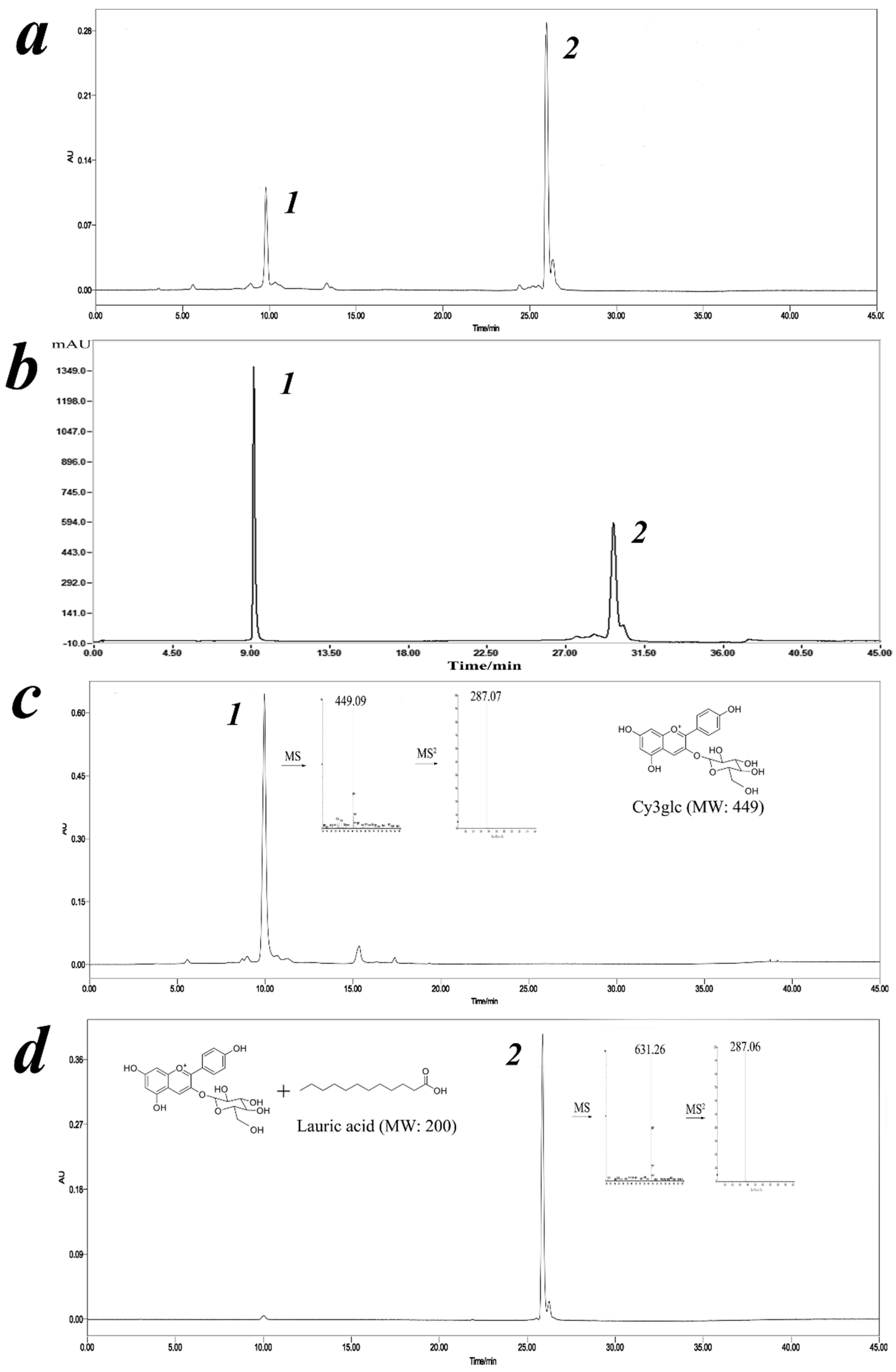
The mixture was analyzed by LC-DAD-ESI/MS in positive ion mode and its characterization was performed bearing their molecular ions and related fragments (Figure 2). A new peak around 26 min was identified as corresponding to a Cy3glc molecule attached to a lauric acid moiety according to its full MS spectrum, [M]+ at m/z 631. In addition, the Cy3glc–lauric acid conjugate revealed an MS2 fragment, [M-344] + at m/z 287, which was in agreement with the Cy3glc aglycone structure (loss of the glucose residue attached to the lauric acid group). This means that acylation of Cy3glc occurred preferentially in the OH groups of the glucose moiety instead of any hydroxyl groups from the flavylium core.
The product was further characterized by 1H and 13C NMR in CD3OD+TMS (Table 1). The enzymatic reaction yielded only a monoester and its regioselectivity was constant, showing that acylation always took place on the primary hydroxyl group present on the glycoside moiety of the molecule (C6ʹʹ-OH). The location of the acylated site has been well documented in previous studies for a range of acyl donors [24,26]. This was confirmed by comparing downfield and upfield shifts between Cy3glc and the Cy3glc-C12 in the same solvent. 13C NMR spectra showed that the acylation on the 6ʹʹ-OH position led to an upfield of the C-6ʹʹ signal by approximately 2.7 ppm (∼62.21 to ∼64.90 ppm) due to the resonance effect towards the carbonyl of the newly generated ester.

3.4. Kinetics of Enzymatic Synthesis of Cy3glc Lauryl Ester

To study the effect of substrate concentration, the reaction kinetics was determined under the optimized conditions: CalB 16.5 g/L, reaction temperature 58 °C and time 28 h. As shown in Figure 3, no substrate inhibition was observed as Cy3glc concentration was less than 25 mmol/L. According to the requirements of dynamic research, the following assumptions were made [12]: Firstly, CalB concentration was very low compared with substrate concentration, so that the formation of substrate complex would not cause significant changes in substrate concentration. Secondly, the production of Cy3glc-C12 and water was irreversible at the beginning of the reaction and when the initial reaction velocity was measured. Thirdly, the concentration of lipase Novozym 435 remained stable during the reaction. Therefore, the reaction process was consistent with the double substrate Ping-Pong mechanism.
A series of basically parallel lines were obtained when the reciprocal of the 1/v was used as the ordinate and the reciprocal of Cy3glc or lauric acid was used as the abscissa for the double-reciprocal fitting curve (Figure 3). Thus, the kinetic equation of acylation reaction between Cy3glc and lauric acid catalyzed by lipase could be calculated with Equation (9):
v = 0.0226 [ A ] [ B ] 2838.56 [ B ] + 54.22 [ A ] + [ A ] [ B ]
The catalyst CalB (E) was firstly combined with lauric acid (A) to produce a binary complex EA. A new complex EAcBP (Ac: lauric acid residue, P: water) was then formed by the frequent collision between EA and Cy3glc (B). The intermediate EAcB appeared after the release of P, and EQ (Q: Cy3glc-C12) was successfully synthesized after the acylation. Finally, the acylated product Q was separated from the binary complex EQ, and the enzyme E was recycled. The enzymatic acylation of Cy3glc with lauric acid to produce Cy3glc-C12 is a double substrate and double product reaction, which is operated by a Ping-Pong mechanism.

3.5. Properties of Cyanidin-3-Glucoside Lauryl Ester

3.5.1. Lipophilic Property

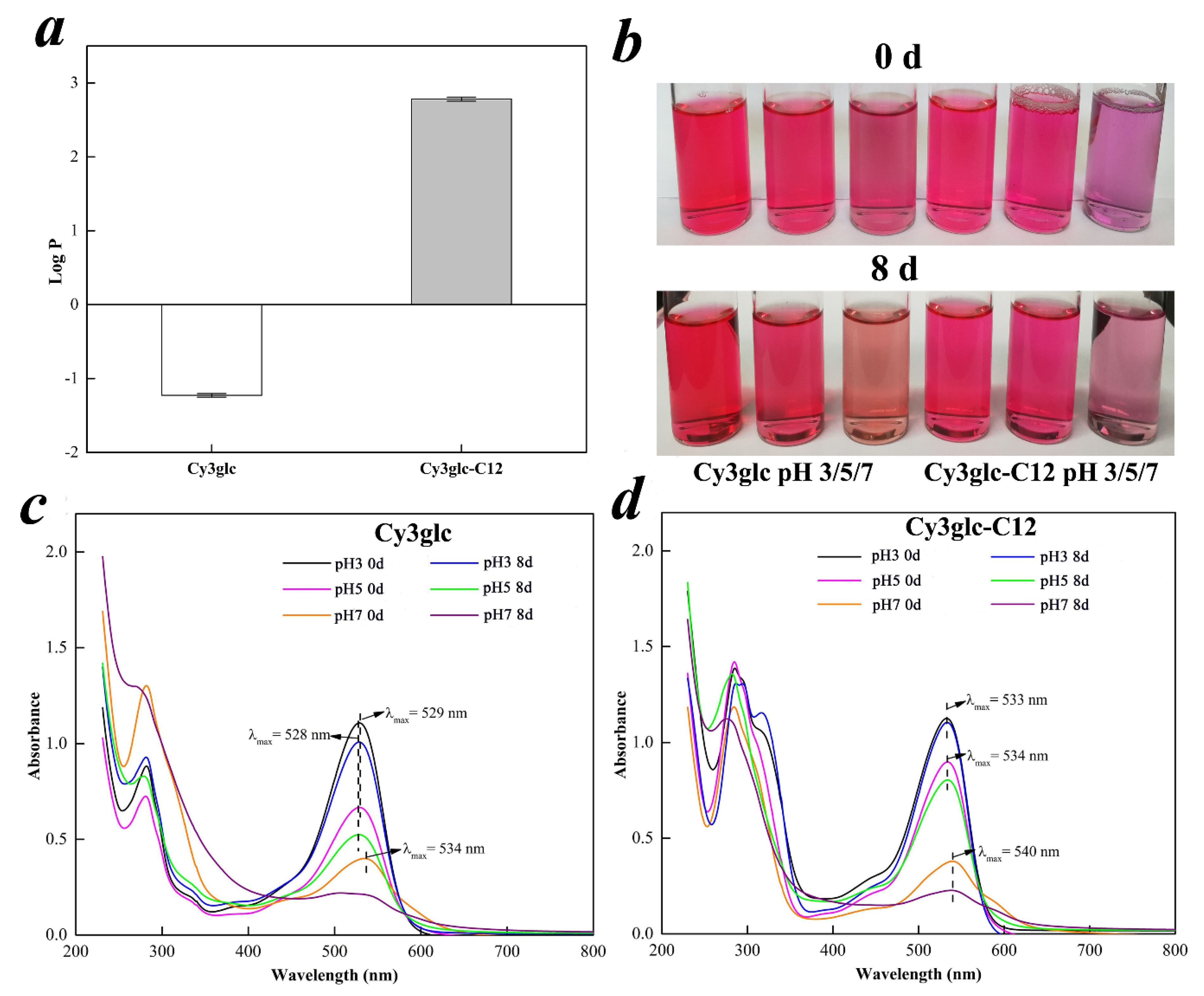
In this experiment, the lipophilicity of Cy3glc and Cy3glc-C12 was evaluated by octanol/water partition coefficient (log P) (Figure 4a). The log P significantly increased from –1.23 to 2.78 after the reaction, indicating that lipophilic Cy3glc-C12 was transformed from hydrophilic Cy3glc. The result was corresponding with the peak delay of Cy3glc-C12 (Figure 2a), in which the retention time mainly depended on substance polarity in HPLC analysis with reversed phase C18 column [29]. Overall, the enzymatic acylation could be a potential method to improve the lipophilicity of anthocyanins.

3.5.2. UV-VIS Absorbance Property

In order to mimic a lipophilic medium, the aqueous SDS model solutions were used to dissolve the anthocyanins. Their chromatic features were photographed (Figure 4b), and all optical wavelengths at room temperature over a period of eight days were preliminarily determined by UV–VIS (Figure 4c,d). In Figure 4b, the color of Cy3glc-C12 was slightly purple on the basis of redness in visual appearance, and significantly different from the color of the native Cy3glc. Their color faded with increasing time, especially in the pH 7 SDS model solutions. Clearly, the absorption showed strong bathochromic shift effect at different pH after the enzymatic acylation (Figure 4c,d). λmax increased from 529 nm (Cy3glc) to 533 nm (Cy3glc-C12) at pH 3 while λmax increased from 528 nm to 534 nm at pH 5. At pH 7, compared with the initial curve, Cy3glc lost its characteristic absorption peak after 8 days. However, Cy3glc-C12 still kept its feature after the same equilibrium period. Overall, cy3glc-C12 had better color intensity and stronger resistance to pH change in the presence of acyl groups linked to the sugar moieties4.

3.5.3. Thermostability

With rising temperature, an increase in rate constant k, and a corresponding decline in half-life t1/2 were recognized in Table 2. The rate constant k of Cy3glc-C12 were 0.074 (pH 3), 0.106 (pH 5) and 0.243 h−1(pH 7) at 95 °C in a water bath, in which the corresponding half-life t1/2 was 9.44, 6.60, and 2.86 h, respectively. Compared with Cy3glc, a lower k and higher t1/2 value of Cy3glc-C12 indicated that its thermostability was significantly increased by enzymatic acylation. Furthermore, the similar trend in kinetic parameters was obtained at the other temperature (65 and 80 °C). Typically, a sandwich structure was formed in the presence of poly-acylation, protecting the molecule from water nucleophilic attack by the package of acyl groups [13]. The intramolecular colorization of anthocyanins could also be achieved, reducing the formation of chalcone or methylamine structure [18]. Altogether, enzymatic acylation of anthocyanins could be applied as an efficient method to significantly increase their thermal stability.

3.6. Proliferative Effect of Cy3glc-C12 on Bifidobacteria and Lactobacillus

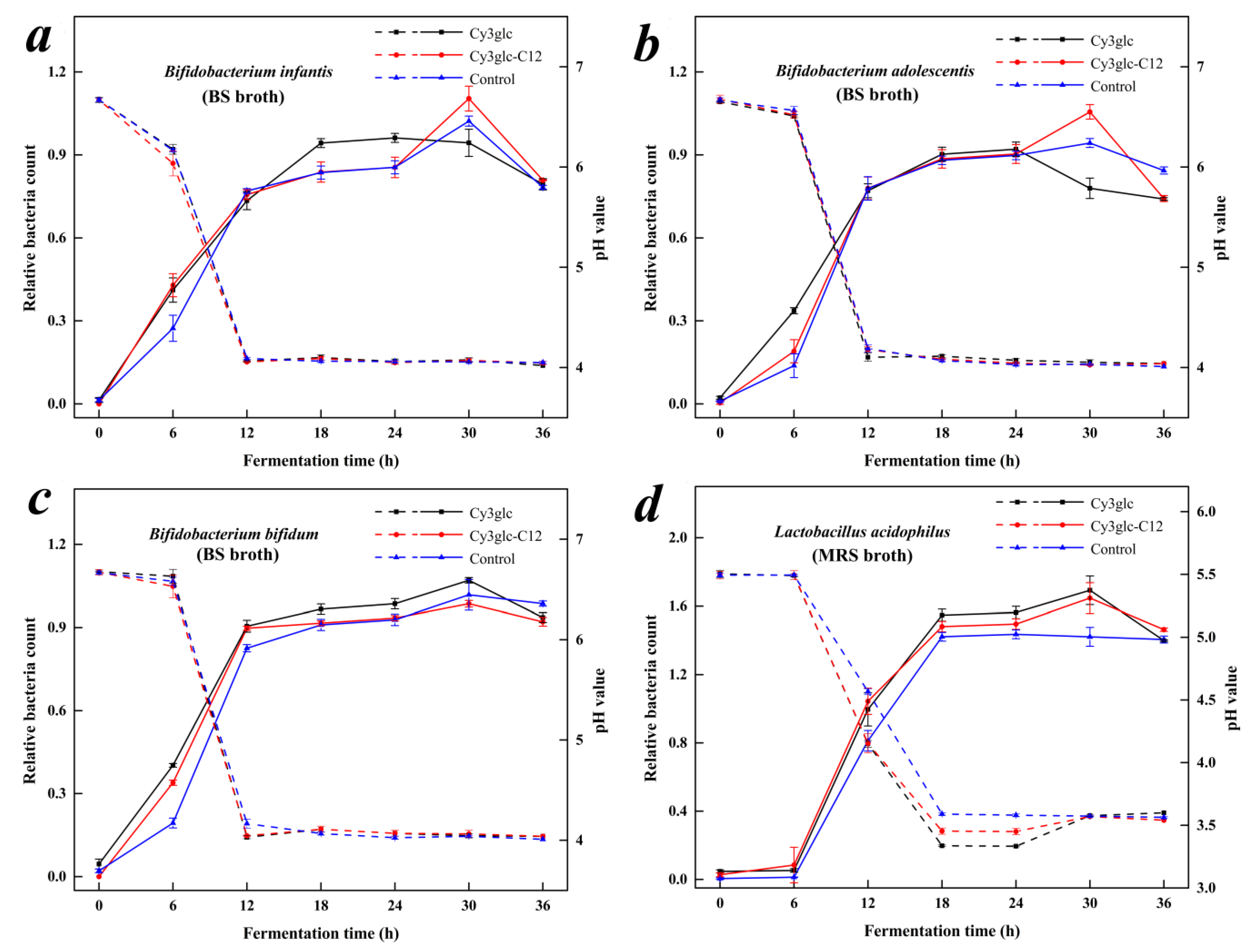
In this experiment, OD600 value with the corresponding pH was adopted to monitor the growth of Bifidobacteria and Lactobacillus (Figure 5), and their growth kinetic parameters were calculated (Table 3). In Figure 5a, B. infantis showed similar growth curves in the media with Cy3glc-C12 and the CG, in which its relative bacteria count (RBC) increased from 0 to 30 h and decreased after incubation for 30 h. Meanwhile, B. infantis had a higher Xm (0.960 ± 0.056) and lower μm (0.319 ± 0.079 h−1) in the media with Cy3glc than those of the CG (p < 0.05). The results indicated that Cy3glc enabled B. infantis to rapidly enter the log phase from the lag phase, so that B. infantis could effectively utilize the media resource in the limited time, which also explained its advanced decline phase and faster decay rate under the circumstance of limited media resources. B. infantis is a common probiotic strain that is associated with a healthy gut and digestion, and it mainly exists in the gut flora of infantis [30]. Lipophilic Cy3glc-C12 was difficult to dissolve in BS broth so that it was not well contacted and metabolized by B. infantis at first, resulting in different proliferative effect between Cy3glc and Cy3glc-C12. With the increase of B. infantis, Cy3glc-C12 could be utilized and further promoted the proliferation of B. infantis according to a maximum RBC after incubation for 30 h.
In the growth of B. adolescentis, there was a significant decrease in μm between the media with Cy3glc (0.390 ± 0.038 h−1) or Cy3glc-C12 (0.536 ± 0.018 h−1) and the CG (Table 3). Even though the media with Cy3glc had the lowest μm value, to some extent, Cy3glc-C12 also enabled B. adolescentis to enter the log phase from the lag phase (Figure 5b). Compared with B. infantis, B. adolescentis had a better capacity in utilizing Cy3glc or Cy3glc-C12, which was reflected in the advanced decline phase after incubation for 24 h (Cy3glc) and a significant μm value (Cy3glc-C12). Even so, the proliferative effect of Cy3glc-C12 on B. adolescentis was mainly found in the later stage of its logarithmic growth based on the maximum RBC after incubation for 30 h, which was similar with that of B. infantis.
B. bifidum showed a higher RBC value in the media with Cy3glc or Cy3glc-C12 than that of the CG during the incubation from 0 to 12 h (Figure 5c). The proliferative effect of Cy3glc on B. bifidum was similar with those of B. infantis and B. adolescentis, in which B. bifidum could rapidly enter the log phase from the lag phase and utilize the limited resources to grow, resulting in a significant increase in Xm (0.979 ± 0.007) and decrease in μm (0.477 ± 0.025 h−1). The growth curve of B. bifidum in the Cy3glc-C12 media was similar to that of Cy3glc media, indicating that both of them played a similar role in promoting the proliferation of B. bifidum. However, the μm value in the Cy3glc-C12 media (0.671 ± 0.044 h−1) was significantly higher than that of the CG (0.566 ± 0.033), revealing that the effect of Cy3glc-C12 focused on the logarithmic growth of B. bifidum, which differed from those of B. infantis and B. adolescentis. B. bifidum is mainly found in the intestinal flora of adults, and its uptake capacity of lipophilic Cy3glc-C12 is better than the other Bifidobacteria, leading to the advanced time of proliferative effects.
As for L. acidophilus, its log phase appeared after incubation for 6 h and lasted from 6 to 18 h, which significantly differed from those of Bifidobacteria (Figure 5d). The growth curves of L. acidophilus were similar in the media with Cy3glc and Cy3glc-C12, and they had higher RBC values in the log phase compared with the CG. In addition, the decline phase of L. acidophilus was observed in the media with samples after incubation for 30 h while L. acidophilus was still in the stationary phase in the CG. Higher Xm values and lower μm values were simultaneously detected in the Cy3glc and Cy3glc-C12 media, indicating that Cy3glc and Cy3glc-C12 had same proliferative effects on L. acidophilus. L. acidophilus is not only found in the stomach, but also a major probiotic in the small intestine. Meanwhile, L. acidophilus could easily contact and metabolize lipophilic Cy3glc-C12 in an aqueous system in vitro, and its utilization capacity of Cy3glc-C12 was better than the three Bifidobacteria species.
Due to the liposolubility of Cy3glc-C12, its proliferative effect on Bifidobacteria and Lactobacillus was different from that of Cy3glc. Lipophilic Cy3glc-C12 was difficult to dissolve in BS or MRS broth and deposited at the bottom of the test tube. To begin with, three Bifidobacteria species could not contact Cy3glc-C12 as easily as Cy3glc. Therefore, Cy3glc could promote the proliferation of Bifidobacteria from making it rapidly enter the log phase from the lag phase and having a longer logarithmic growth after incubation for 24 h. As for Cy3glc-C12, with the growth of Bifidobacteria, Cy3glc-C12 could be gradually contacted and utilized in the log phase so that its proliferative effect was obviously observed in the middle and later log phase. On the other hand, due to the different species of intestinal probiotics, their capacity in utilizing lipophilic Cy3glc-C12 further influenced the growth curve, in which L. acidophilus > B. bifidum > B. infantis > B. adolescentis. Thus, there was no significant difference between the effect of Cy3glc and Cy3glc-C12 on the lag phase of L. acidophilus, and their contact and metabolization by L. acidophilus were nearly synchronous.
Their proliferative effects on Bifidobacteria and Lactobacillus could be explained from two aspects. Firstly, Cy3glc and Cy3glc-C12 could be broken down by Bifidobacteria and Lactobacillus into phenolic metabolites, such as benzoic acid, phenyllactic acid, and phenylalanine (Table 4), which could be used as sources to offer intestinal probiotics better growth conditions [10,31]. Secondly, pH in the media with Cy3glc and Cy3glc-C12 dropped faster than the control. Cy3glc-C12 metabolites, produced by Bifidobacteria and Lactobacilli after incubation for 24 h, were analyzed by GC-MS. About 14 organic acids produced by spontaneous degradation of cyanidin were detected in GC-MS. The results indicated that the type and content of Cy3glc-C12 metabolites were similar to that of Cy3glc metabolites in our previous report [10]. Understandably, Cy3glc and Cy3glc-C12 were metabolized into organic acids by the tested bacteria and the media pH inevitably dropped in the presence of organic acids. As L. acidophilus, the relative contents of main organic acids were benzoic acid (1.47), phenyllactic acid (6.67), and phenylalanine (4.50), respectively. Due to sugar-based connection, Cy3glc or Cy3glc-C12 were not readily absorbed by the digestive tract, but hydrolyzed to aglycones by the intestinal bacterial glycosidase or further degraded into phenolic acids [31,32,33]. An acidic environment (pH 4.5–6.5) is beneficial to the growth of Bifidobacteria and Lactobacillus [34]. Cy3glc and Cy3glc-C12 metabolites not only provided appropriate pH for the growth of intestinal probiotics, but also inhibited the growth of spoilage organisms [5].
Bifidobacteria decreased faster at their decline phase in Cy3glc and Cy3glc-C12 media, which could be attributed to two aspects: Firstly, the nutrients were quickly used up by Bifidobacteria due to the bioactivity of Cy3glc and Cy3glc-C12. After incubation for 24 h, essential nutrients were deficient in the medium, which might be responsible for their faster decay. Secondly, phenolic hydroxyl groups of Cy3glc and Cy3glc-C12 could react with mycoproteins or enzymes through hydrogen bonding, resulting in cytoplasm pyknosis and disintegration [5]. However, no significant difference of relative bacteria count was observed in Cy3glc and Cy3glc-C12 (p > 0.05) after 36 h of incubation. As for the hysteretic decline phase of B. infantis and B. adolescentis in Cy3glc-C12 media, it could be explained from the steric hindrance (the ability of probiotics to ingest Cy3glc-C12) and lipid solubility. The long carbon chain of Cy3glc-C12 could increase the steric hindrance, which limited the proliferative effect of Cy3glc-C12. On the other hand, the lipid solubility of Cy3glc-C12 limited its pathway from water-soluble media to intestinal probiotics. However, Cy3glc-C12 was easier to go through cell membranes of intestinal probiotics because of its lipid solubility (Figure 4a) [26].
Overall, Cy3glc-C12 displayed a similar influence to Cy3glc on proliferation of intestinal probiotics, further confirming well bioactive of Cy3glc-C12. Overall, the results revealed that Cy3glc-C12 could promote the growth of intestinal probiotics, and sustain as well proliferative effect as Cy3glc after the attachment of lauric acid to the 6″-OH position of the glucoside unit.

4. Conclusions

The optimal enzymatic acylation route of 68.7 ± 2.1% conversion yield was obtained by a Box–Behnken RSM design with anthocyanins: lauric acid (molar ratio) 1:56, CalB 16.5 g/L and molecular sieves 4 Å, 100 g/L in tert-amyl alcohol at 56 °C for 28 h. Cyanidin-3-(6′′-dodecanoyl)-glucoside was successfully produced via the acylation occurred at 6′′-OH position of the glucoside. The enzymatic acylation of Cy3glc with lauric acid to produce Cy3glc-C12 is a double substrate and double product reaction, which is operated by a Ping-Pong Bi-Bi mechanism. The liposolubility, pH resistance, and thermostability of Cy3glc-C12 were significantly improved after enzymatic acylation. Furthermore, Cy3glc-C12 could promote the proliferation of Bifidobacteria and Lactobacillus in the middle and later log phase and further be metabolized into phenolic acids. This research established a kinetic model for the enzymatic synthesis of lipophilic, thermostable, and anthocyanin-based pigments and revealed the proliferative effect of Cy3glc-C12 on intestinal probiotics.

Supplementary Materials

The following are available online at https://www.mdpi.com/2079-7737/9/8/205/s1, Figure S1: Effect of reaction parameters on acylation yield of Cy3glc. molar ratio (a), enzyme addition (b), reaction temperature (c), and time (d), Figure S2: 1H, 13C NMR and 2D NMR spectrum (HSQC) for the synthesized compound Cy3glc-C12. (a) 1H NMR, (b) 13C NMR and (c) HSQC 2D NMR, Table S1: Experimental design, results, and variance analysis of response surface optimization. The supplementary materials included the methods about determination of cyanidin-3-glucoside acylation yield, measurement of lipophilicity, UV–VIS absorbance property, thermal stability assay and analysis of secondary metabolites by GC-MS. In addition, the results of single factor test, response surface test and NMR spectrum were also presented. Reference [9] is cited in the supplementary materials.

Author Contributions

Conceptualization: X.Y. and H.S.; methodology: X.Y.; software: X.Y.; validation: H.S. and S.H.; formal analysis: X.Y.; investigation: X.Y.; resources: L.T., Y.J., M.W., S.L., and Y.W.; data curation: X.Y.; writing—original draft preparation: X.Y.; writing—review and editing: H.S. and S.H.; visualization: X.Y.; supervision: Z.Z.; project administration: H.S.; funding acquisition: H.S. All authors have read and agreed to the published version of the manuscript.

Funding

This research was funded by the National Natural Science Foundation of China (grant numbers 31771974, 31701524), the Fundamental Research Funds for the Central Universities, China (no. JZ2018HGTB0245, no. JZ2019YYPY0295), and the Chengdu Technological Innovation Research and Development Project, China (no. 2019-YF05-00965-SN).

Acknowledgments

This work was supported by Hefei University of Technology.

Conflicts of Interest

The authors declare no conflict of interest.

References

  1. Clifford, M.N. Anthocyanins—Nature, occurrence and dietary burden. J. Sci. Food Agric. 2000, 80, 1118–1125. [Google Scholar] [CrossRef]
  2. Castañeda-Ovando, A.; De Lourdes Pacheco-Hernández, M.; Páez-Hernández, M.E.; Rodríguez, J.A.; Galán-Vidal, C.A. Chemical studies of anthocyanins: A review. Food Chem. 2009, 113, 859–871. [Google Scholar] [CrossRef]
  3. Bowen-Forbes, C.S.; Zhang, Y.; Nair, M.G. Anthocyanin content, antioxidant, anti-inflammatory and anticancer properties of blackberry and raspberry fruits. J. Food Compos. Anal. 2010, 23, 554–560. [Google Scholar] [CrossRef]
  4. B¹kowska-Barczak, A. Acylated anthocyanins as stable, natural food colorants—A review. Pol. J. Food Nutr. Sci. 2005, 11, 201–247. [Google Scholar]
  5. Coman, M.M.; Oancea, A.M.; Verdenelli, M.C.; Cecchini, C.; Bahrim, G.E.; Orpianesi, C.; Cresci, A.; Silvi, S. Polyphenol content and in vitro evaluation of antioxidant, antimicrobial and prebiotic properties of red fruit extracts. Eur. Food Res. Technol. 2018, 244, 735–745. [Google Scholar] [CrossRef]
  6. Shao, Y.; Xu, F.; Sun, X.; Bao, J.; Beta, T. Identification and quantification of phenolic acids and anthocyanins as antioxidants in bran, embryo and endosperm of white, red and black rice kernels (Oryza sativa L.). J. Cereal Sci. 2014, 59, 211–218. [Google Scholar] [CrossRef]
  7. He, S.; Lou, Q.; Shi, J.; Sun, H.; Zhang, M.; Li, Q. Water Extraction of Anthocyanins from Black Rice and Purification Using Membrane Separation and Resin Adsorption. J. Food Process. Preserv. 2017, 41, 1–12. [Google Scholar] [CrossRef]
  8. Ito, V.C.; Lacerda, L.G. Black rice (Oryza sativa L.): A review of its historical aspects, chemical composition, nutritional and functional properties, and applications and processing technologies. Food Chem. 2019, 301, 125304. [Google Scholar] [CrossRef]
  9. Hou, Z.; Qin, P.; Zhang, Y.; Cui, S.; Ren, G. Identification of anthocyanins isolated from black rice (Oryza sativa L.) and their degradation kinetics. Food Res. Int. 2013, 50, 691–697. [Google Scholar] [CrossRef]
  10. Zhu, Y.; Sun, H.; He, S.; Lou, Q.; Yu, M.; Tang, M.; Tu, L. Metabolism and prebiotics activity of anthocyanins from black rice (Oryza sativa L.) in vitro. PLoS ONE 2018, 13, e0195754. [Google Scholar] [CrossRef] [Green Version]
  11. Ekici, L.; Simsek, Z.; Ozturk, I.; Sağdıç, O.; Yetim, H.; Sagdic, O. Effects of Temperature, Time, and pH on the Stability of Anthocyanin Extracts: Prediction of Total Anthocyanin Content Using Nonlinear Models. Food Anal. Methods 2014, 7, 1328–1336. [Google Scholar] [CrossRef]
  12. Patras, A.; Brunton, N.; O’Donnell, B.; Tiwari, B. Effect of thermal processing on anthocyanin stability in foods; mechanisms and kinetics of degradation. Trends Food Sci. Technol. 2010, 21, 3–11. [Google Scholar] [CrossRef]
  13. Zhao, C.-L.; Yu, Y.-Q.; Chen, Z.-J.; Wen, G.-S.; Wei, F.-G.; Zheng, Q.; Wang, C.-D.; Xiao, X.-L. Stability-increasing effects of anthocyanin glycosyl acylation. Food Chem. 2017, 214, 119–128. [Google Scholar] [CrossRef] [PubMed]
  14. Ahmad, M.; Ashraf, B.; Gani, A.; Gani, A. Microencapsulation of saffron anthocyanins using β glucan and β cyclodextrin: Microcapsule characterization, release behaviour & antioxidant potential during in-vitro digestion. Int. J. Biol. Macromol. 2018, 109, 435–442. [Google Scholar] [PubMed]
  15. Chung, C.; Rojanasasithara, T.; Mutilangi, W.; McClements, D.J. Stability improvement of natural food colors: Impact of amino acid and peptide addition on anthocyanin stability in model beverages. Food Chem. 2017, 218, 277–284. [Google Scholar] [CrossRef]
  16. Qian, B.-J.; Liu, J.-H.; Zhao, S.-J.; Cai, J.-X.; Jing, P. The effects of gallic/ferulic/caffeic acids on colour intensification and anthocyanin stability. Food Chem. 2017, 228, 526–532. [Google Scholar] [CrossRef]
  17. Cruz, L.; Fernandes, V.C.; Araújo, P.; Mateus, N.; De Freitas, V. Synthesis, characterisation and antioxidant features of procyanidin B4 and malvidin-3-glucoside stearic acid derivatives. Food Chem. 2015, 174, 480–486. [Google Scholar] [CrossRef]
  18. Zhao, L.-Y.; Chen, J.; Wang, Z.-Q.; Shen, R.-M.; Cui, N.; Sun, A. Direct Acylation of Cyanidin-3-Glucoside with Lauric Acid in Blueberry and Its Stability Analysis. Int. J. Food Prop. 2016, 19, 1–12. [Google Scholar] [CrossRef]
  19. Cruz, L.; Benohoud, M.; Rayner, C.M.; Mateus, N.; De Freitas, V.; Blackburn, R.S. Selective enzymatic lipophilization of anthocyanin glucosides from blackcurrant (Ribes nigrum L) skin extract and characterization of esterified anthocyanins. Food Chem. 2018, 266, 415–419. [Google Scholar] [CrossRef] [Green Version]
  20. Yan, Z.; Li, C.; Zhang, L.; Liu, Q.; Ou, S.; Zeng, X. Enzymatic Acylation of Anthocyanin Isolated from Black Rice with Methyl Aromatic Acid Ester as Donor: Stability of the Acylated Derivatives. J. Agric. Food Chem. 2016, 64, 1137–1143. [Google Scholar] [CrossRef]
  21. Cruz, L.; Guimarães, M.; Araújo, P.; Évora, A.; De Freitas, V.; Mateus, N. Malvidin 3-Glucoside–Fatty Acid Conjugates: From Hydrophilic toward Novel Lipophilic Derivatives. J. Agric. Food Chem. 2017, 65, 6513–6518. [Google Scholar] [CrossRef] [PubMed]
  22. Cruz, L.M.; Fernandes, I.; Guimarães, M.; De Freitas, V.; Mateus, N. Enzymatic synthesis, structural characterization and antioxidant capacity assessment of a new lipophilic malvidin-3-glucoside-oleic acid conjugate. Food Funct. 2016, 7, 2754–2762. [Google Scholar] [CrossRef] [PubMed]
  23. Guimarães, M.; Mateus, N.; De Freitas, V.; Cruz, L. Improvement of the Color Stability of Cyanidin-3-glucoside by Fatty Acid Enzymatic Acylation. J. Agric. Food Chem. 2018, 66, 10003–10010. [Google Scholar] [CrossRef]
  24. Yang, W.; Kortesniemi, M.K.; Yang, B.; Zheng, J. Enzymatic Acylation of Anthocyanins Isolated from Alpine Bearberry (Arctostaphylos alpina) and Lipophilic Properties, Thermo-stability and Antioxidant Capacity of the Derivatives. J. Agric. Food Chem. 2018, 66, 2909–2916. [Google Scholar] [CrossRef] [PubMed]
  25. Xu, J.; Qian, J.; Li, S. Enzymatic acylation of isoorientin isolated from antioxidant of bamboo leaves with palmitic acid and antiradical activity of the acylated derivatives. Eur. Food Res. Technol. 2014, 239, 661–667. [Google Scholar] [CrossRef]
  26. Yang, W.; Kortesniemi, M.; Ma, X.; Zheng, J.; Yang, B. Enzymatic acylation of blackcurrant (Ribes nigrum) anthocyanins and evaluation of lipophilic properties and antioxidant capacity of derivatives. Food Chem. 2019, 280, 189–196. [Google Scholar] [CrossRef]
  27. Krishna, S.H.; Divakar, S.; Prapulla, S.G.; Karanth, N.G. Enzymatic synthesis of isoamyl acetate using immobilized lipase from Rhizomucor miehei. J. Biotechnol. 2001, 87, 193–201. [Google Scholar] [CrossRef]
  28. Husson, E.; Garcia-Matilla, V.; Humeau, C.; Chevalot, I.; Fournier, F.; Marc, I. Enzymatic acylation of a bifunctional molecule in 2-methyl-2-butanol: Kinetic modelling. Enzym. Microb. Technol. 2010, 46, 338–346. [Google Scholar] [CrossRef]
  29. Heukelem, L.; Lewitus, A.J.; Kana, T.M.; Craft, N.E. High performance liquid chromatography of phytoplankton pigments using a polymeric reversed phase C18 column. J. Phycol. 2010, 28, 867–872. [Google Scholar] [CrossRef]
  30. Awasthi, S.; Wilken, R.; Patel, F.; German, J.B.; Mills, D.A.; Lebrilla, C.B.; Kim, K.; Freeman, S.L.; Smilowitz, J.T.; Armstrong, A.W.; et al. Dietary supplementation with Bifidobacterium longum subsp. infantis (B. infantis) in healthy breastfed infants: Study protocol for a randomised controlled trial. Trials 2016, 17, 1–11. [Google Scholar]
  31. Cheng, J.-R.; Liu, X.-M.; Chen, Z.-Y.; Zhang, Y.-S.; Zhang, Y.-H. Mulberry anthocyanin biotransformation by intestinal probiotics. Food Chem. 2016, 213, 721–727. [Google Scholar] [CrossRef] [PubMed]
  32. Ryu, D.; Koh, E. Stability of anthocyanins in bokbunja (Rubus occidentalis L.) under in vitro gastrointestinal digestion. Food Chem. 2018, 267, 157–162. [Google Scholar] [CrossRef] [PubMed]
  33. Keppler, K.; Humpf, H.-U. Metabolism of anthocyanins and their phenolic degradation products by the intestinal microflora. Bioorganic Med. Chem. 2005, 13, 5195–5205. [Google Scholar] [CrossRef] [PubMed]
  34. Faria, A.; Fernandes, I.; Norberto, S.; Mateus, N.; Calhau, C. Interplay between Anthocyanins and Gut Microbiota. J. Agric. Food Chem. 2014, 62, 6898–6902. [Google Scholar] [CrossRef]
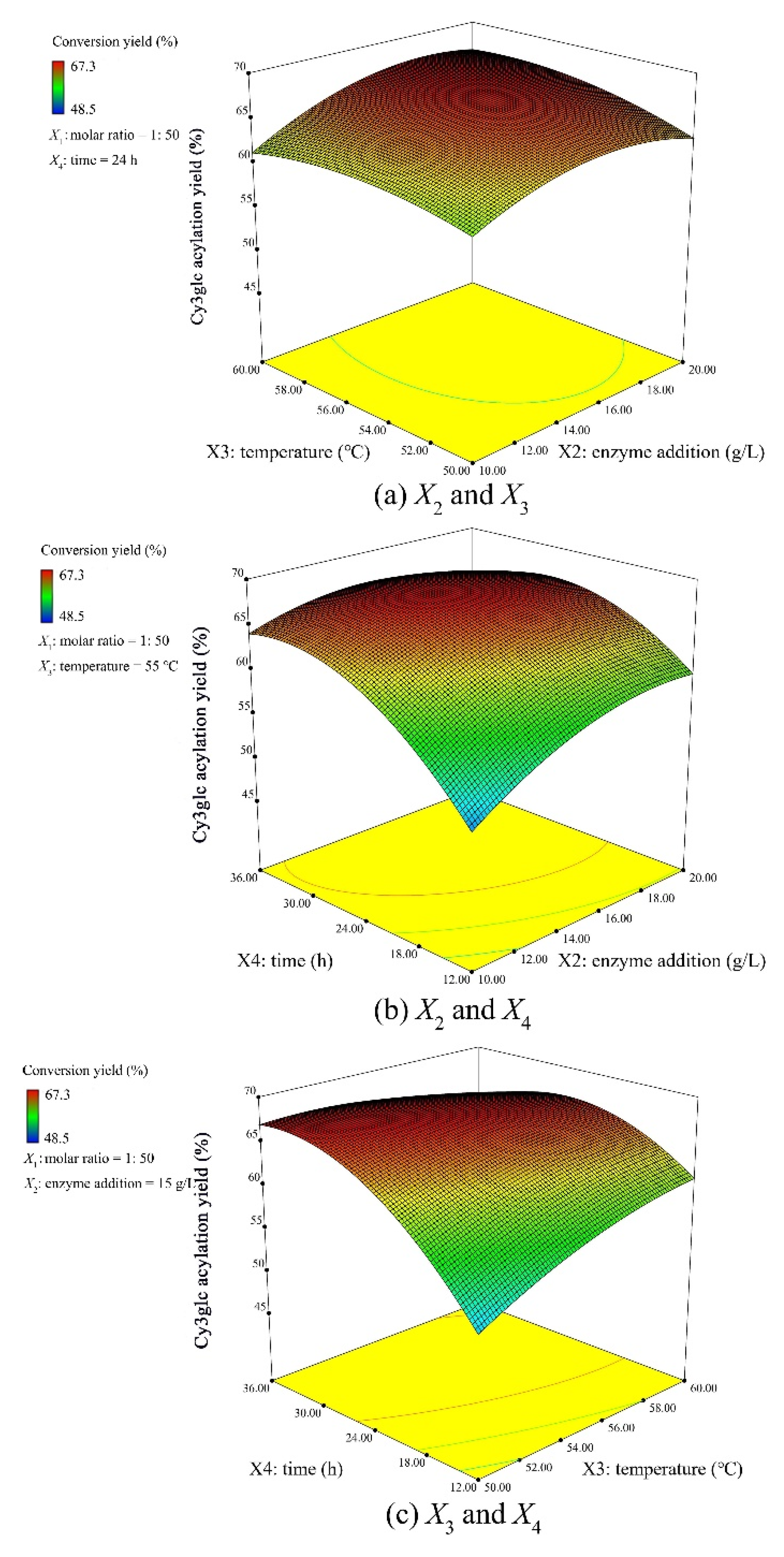
Figure 1. Response surface plots of the interactions between independent variables (a) enzyme addition X2 and temperature X3, (b) enzyme addition X2 and time X4, and (c) temperature X3 and time X4 on Cy3glc acylation yield (%).
Biology 09 00205 g001
Figure 2. HPLC chromatogram of the reaction mixture to determine the Cy3glc acylation yield (a), semi-preparative liquid chromatogram of the reaction mixture to obtain Cy3glc and Cy3glc-C12 (b), and HPLC-MS/MS spectra of Cy3glc (c) and Cy3glc-C12 (d).
Biology 09 00205 g002
Figure 3. Double-reciprocal fit plot of the initial reaction rate with Cy3glc concentration (a) and lauric acid concentration (b) (n = 3).
Biology 09 00205 g003
Figure 4. Lipophilic property (a), color feature (b), and UV-VIS absorbance property of Cy3glc and Cy3glc-C12 (c and d) during storage for eight days in the dark at pH 3, 5, and 7.
Biology 09 00205 g004
Figure 5. The change of strains growth (solid line) and pH value (dotted line) during the fermentation with Cy3glc or Cy3glc-C12. BS, and MRS broth without anthocyanins were kept as the control groups (CG). Error bars represent the standard deviations of the mean of three replicates (n = 3). (a) B. infantis growth in BS broth; (b) B. adolescentis growth in BS broth; (c) B. bifidum growth in BS broth; (d), L. acidophilus growth in MRS broth.
Biology 09 00205 g005
Table 1. 1H and 13C NMR data of Cy3glc and Cy3glc-C12 in CD3OD+TMS.
Biology 09 00205 i001
PositionCy3glcCy3glc-C12
δH (ppm)*δC (ppm)δH (ppm)*δC (ppm)
Cyanidin aglycone
47.50 (s, 1H)132.527.48 (s, 1H)134.31
66.52 (s, 1H)102.966.48 (s, 1H)103.02
86.68 (s, 1H)96.376.68 (s, 1H)96.42
2′6.94 (d, J = 1.9 Hz, 1H)116.576.94 (d, J = 2.1 Hz, 1H)116.40
5′6.83 (d, J = 7.4 Hz, 1H)117.186.85 (d, J = 7.5 Hz, 1H)117.27
6′7.35 (dd, J = 7.4, 2.1 Hz, 1H)126.847.35 (dd, J = 7.4, 2.1 Hz, 1H)125.99
3-O-β-D-glucoside
1′′5.07 (s, 1H)102.505.02 (s, 1H)101.16
2′′3.66 (d, J = 5.1 Hz, 1H)73.393.61 (d, J = 4.9 Hz, 1H)73.47
3′′3.44 (d, J = 4.9 Hz, 1H)76.813.78 (d, J = 5.1 Hz, 1H)76.90
4′′3.40 (d, J = 5.1 Hz, 1H)71.243.55 (d, J = 5.1 Hz, 1H)70.82
5′′3.45 (s, 1H)78.043.72 (s, 1H)75.60
6′′3.82–3.67 (m, 2H)62.214.31 (q, J = 12.6 Hz, 2H)64.90
6′′-O-dodecanoate
CH2 (2′′′)//2.33–2.26 (m, 2H)34.45
CH2 (3′′′)//1.50 (s, 2H)25.48
CH2 (C4-C11)//1.24–1.21 (m, 16H)22.90–31.98
CH3 (C12)//0.88 (s, 3H)14.22
Table 2. Kinetic parameters determined from the thermal-degradation assays of Cy3glc and Cy3glc-C12 in an aqueous SDS solution.
IndicatorsCy3glcCy3glc-C12
65 °C80 °C95 °C65 °C80 °C95 °C
pH3k (h−1)0.078 ± 0.0100.210 ± 0.0040.347 ± 0.0040.054 ± 0.0100.066 ± 0.0050.074 ± 0.005
t1/2 (h)9.04 ± 1.253.30 ± 0.062.00 ± 0.0213.15 ± 2.1910.48 ± 0.759.44 ± 0.61
Ea (kJ·mol−1)52.11 ± 5.00 a13.97 ± 3.23 e
pH5k (h−1)0.150 ± 0.0110.210 ± 0.0110.344 ± 0.0060.077 ± 0.0050.096 ± 0.0100.106 ± 0.011
t1/2 (h)4.65 ± 0.333.31 ± 0.182.01 ± 0.048.97 ± 0.607.29 ± 0.806.60 ± 0.74
Ea (kJ·mol−1)28.70 ± 2.42 c10.70 ± 1.53 e
pH7k (h−1)0.179 ± 0.0100.292 ± 0.0040.342 ± 0.0020.088 ± 0.0040.203 ± 0.0040.243 ± 0.011
t1/2 (h)3.88 ± 0.212.37 ± 0.032.03 ± 0.017.91 ± 0.333.41 ± 0.062.86 ± 0.14
Ea (kJ·mol−1)22.57 ± 1.79 d35.44 ± 1.56 b
Data were obtained from three independent experiments and represented as the mean values ± SD (n = 3). Different lowercase letters indicate significant differences (p < 0.05) in the activation energy.
Table 3. Growth kinetic parameters of three Bifidobacteria and one Lactobacillus species.
ParametersX0 (OD600nm)Xm (OD600nm)μm (h−1)R2 of the Model
Bifidobacterium infantis
CG0.019 ± 0.0030.847 ± 0.0060.504 ± 0.0230.999
Cy3glc0.029 ± 0.012ns0.960 ± 0.056*0.319 ± 0.079*0.974
Cy3glc-C120.015 ± 0.014ns0.832 ± 0.029ns0.501 ± 0.151ns0.986
Bifidobacterium adolescentis
CG0.008 ± 0.0020.889 ± 0.0060.606 ± 0.0190.999
Cy3glc0.006 ± 0.002ns0.915 ± 0.018ns0.390 ± 0.038*0.997
Cy3glc-C120.010 ± 0.001ns0.901 ± 0.006ns0.536 ± 0.018*0.999
Bifidobacterium bifidum
CG0.008 ± 0.0020.926 ± 0.0100.566 ± 0.0330.993
Cy3glc0.018 ± 0.006ns0.979 ± 0.007*0.477 ± 0.025*0.999
Cy3glc-C120.009 ± 0.010ns0.927 ± 0.007ns0.671 ± 0.044*0.999
Lactobacillus acidophilus
CG0.001 ± 0.0011.433 ± 0.0060.805 ± 0.0720.999
Cy3glc0.001 ± 0.001ns1.562 ± 0.025*0.655 ± 0.054*0.999
Cy3glc-C120.002 ± 0.001ns1.495 ± 0.015*0.609 ± 0.043*0.999
Results are expressed as mean ± SD (n = 3). * Significantly different from the CG, p < 0.05 and ns not significantly different from the CG, p > 0.05.
Table 4. Relative content of Cy3glc-C12 metabolites produced by Bifidobacteria and Lactobacilli at 24 h of incubation under anaerobic conditions at 37 °C.
Compound (Relative Content)Retention Time (min)Molecular ion (m/z)Intestinal Probiotics
B. InfantisB. AdolescentisB. BifidumL. Acidophilus
benzoic acid11.261791.051.181.141.47
Phenylacetic acid12.391930.030.010.020.01
Phenylpropanoic acid15.001040.100.310.290.14
Mandelic acid16.181790.020.050.050.05
4-Hydroxybenzaldehyde16.592230.070.060.050.16
Phenethylamine17.941740.020.030.130.02
4-Hydroxyphenylethanol18.071790.060.160.180.16
Phenyllactic acid18.261939.6312.0414.1116.67
Phenylalanine18.962184.312.203.114.50
4-Hydroxybenzoic acid19.012670.060.050.070.16
4-Hydroxyphenylacetic acid19.181790.090.050.050.05
4-Hydroxyphenylpropionic acid20.891790.110.170.180.15
3-Methoxy-4-hydroxybenzoicacid20.932970.080.030.030.12
4-Aminobenzoic acid21.792660.020.030.030.01