Article

Age-Dependence and Aging-Dependence: Neuronal Loss and Lifespan in a C. elegans Model of Parkinson’s Disease

1
Biology Department, Northeastern University, Boston, MA 02115, USA
2
Department of Systems Biology, Harvard Medical School, Boston, MA 02115, USA
*
Authors to whom correspondence should be addressed.
Submission received: 23 November 2017 / Revised: 16 December 2017 / Accepted: 18 December 2017 / Published: 23 December 2017
(This article belongs to the Special Issue Systems Biology of Aging)

Abstract

:
It is often assumed, but not established, that the major neurodegenerative diseases, such as Parkinson’s disease, are not just age-dependent (their incidence changes with time) but actually aging-dependent (their incidence is coupled to the process that determines lifespan). To determine a dependence on the aging process requires the joint probability distribution of disease onset and lifespan. For human Parkinson’s disease, such a joint distribution is not available, because the disease cuts lifespan short. To acquire a joint distribution, we resorted to an established C. elegans model of Parkinson’s disease in which the loss of dopaminergic neurons is not fatal. We find that lifespan is not correlated with the loss of individual neurons. Therefore, neuronal loss is age-dependent and aging-independent. We also find that a lifespan-extending intervention into insulin/IGF1 signaling accelerates the loss of specific dopaminergic neurons, while leaving death and neuronal loss times uncorrelated. This suggests that distinct and compartmentalized instances of the same genetically encoded insulin/IGF1 signaling machinery act independently to control neurodegeneration and lifespan in C. elegans. Although the human context might well be different, our study calls attention to the need to maintain a rigorous distinction between age-dependence and aging-dependence.

1. Introduction

In demographic parlance, the term “age” refers to the time elapsed since the occurrence of a specific developmental event, for example the onset of adulthood. To conform with this meaning of age, the phrase “age-dependent disease” should simply refer to a disease whose risk of onset (or progression to any particular stage) changes with time, usually in a steadily increasing fashion, as is the case for a variety of metabolic and neurodegenerative diseases, as well as many cancers [1]. Yet, the phrase is often used in a way that appears to carry the connotation of age as a “biological time” marking the process of aging, effectively suggesting an aging-dependence, and not just an age-dependence. Such slippage amounts to asserting a causal connection between aging and (say) the onset of a disease. The significance of such a connection derives from the hope that interventions that slow aging might delay disease onset. The main point of our contribution is to show that this frequent slippage from age-dependence to aging-dependence may not be warranted.
Aging refers to a process that (typically) increases the risk of death with the passing of time, i.e., age. The risk of death in turn determines the lifespan distribution and the survival function. This means that the risk of death depends not only on time—which makes it age-dependent—but outright defines aging—which makes it trivially aging-dependent. Having defined aging, it is clear that to determine whether disease X or more precisely any phase of disease X, such as its onset, depends on aging, one has to study the co-variation of disease X and lifespan as it results from aging. This involves first determining the joint probability distribution of the time of onset of X and the time of death from aging, and then assessing, whether or not this joint probability distribution is the product of the respective marginal distributions. A difficulty arises when the progression of an age-dependent and lethal disease caps lifespan. This is known in statistics as a ceiling effect. If an individual dies from a disease, we cannot know how long that individual would have lived had the disease not killed them, but had allowed the aging process to take its course. This counterfactual prevents us from acquiring a joint probability distribution, and seems to have impeded a rigorous determination of whether any age-dependent disease with fatal outcome is actually dependent on the aging process. In the United States, such diseases include the seven leading causes of death among individuals aged 65 years and over, accounting for more than 70% of deaths in this age group [1]. The wide-spread hope of delaying the onset of such diseases by intervening in the aging process betrays a tacit assumption that a dependence on aging must exist.
One way to probe the connection between age- and aging-dependence is to focus on diseases that are not lethal, which might be less pressing. Another approach, and the one we chose, is to exploit the transfer of a disease that is lethal in humans to a model organism in which it is not. Here we aim at a basic analysis of the age- and aging-dependence of a Parkinson’s disease model in the nematode C. elegans. While this does not permit inference to the human case, it nonetheless illustrates the interaction between aging and a model of a time-dependent physiological condition in an organismic context that has a history of illuminating molecular processes in humans.
Parkinson’s disease is perhaps one of the most widely studied age-dependent diseases. In humans, the incidence of Parkinson’s disease increases with age at a nearly exponential rate, with a mean age at onset of 70 years [2]. The pathogenesis of Parkinson’s disease is characterized by the loss of dopaminergic neurons in the substantia nigra of the brain [3]. The progression of Parkinson’s disease involves the loss of essential neurons, leading to the early death of affected individuals [4]. A well-validated C. elegans Parkinson’s disease model has been used previously to identify several genetic determinants of dopaminergic neurodegeneration [5,6], including a gene responsible for familial forms of Parkinson’s disease in humans [7]. However, unlike in humans, dopaminergic neurons are not essential for survival in C. elegans [8]. The disease model provided us therefore with a unique platform to study the poorly understood connections between age-dependent neurodegeneration and the aging process that determines the organism’s lifespan.
The C. elegans model is significant also because its lifespan is controlled by widely-conserved mechanisms. Early studies in C. elegans showed that lifespan can be more than doubled by reduction-of-function mutations in the insulin/IGF1 receptor gene daf-2 [9] or in its downstream signaling effectors, including the age-1 phosphatidylinositol 3-kinase (PI3K) gene [10]. Subsequent studies showed that lowering the action of insulin and IGF1 receptors also extends lifespan in the fruit fly Drosophila melanogaster [11,12] and in mice [13,14]. In worms and flies these lifespan-extending effects are mediated by the activation of FOXO transcription factors, DAF-16 [15,16] and dFOXO [17,18], respectively. This functional conservation appears to extend to humans, since Foxo3A gene variants are associated with human longevity [19,20].
Reducing insulin/IGF1 receptor signaling or increasing FOXO activity specifically in adipose or neuroendocrine tissue can increase lifespan in worms [21,22,23], flies [18,24], and mice [13,25]. This indicates that, across phylogeny, insulin/IGF1 signaling regulates lifespan, at least in part, through the action of systemic signals generated by these tissues. These signals are thought to regulate the cellular processes responsible for the functional decline associated with aging and the associated increase in the risk of death. As such, they may represent ideal entry points for the development of therapies against aging-dependent diseases [26].
In our study, we probed the joint probability distribution of the timing of neuronal loss and the death of the organism in the C. elegans Parkinson’s disease model. We found that the onset of neurodegeneration and aging (as manifest in the timing of death) are independent phenomena. We then investigated the role of insulin/IGF1 signaling in the regulation of the timing of the loss of dopaminergic neurons in the C. elegans model and found, unexpectedly, that long-lived insulin/IGF1 pathway mutants exhibit a more severe degenerative phenotype. These effects are mediated by the action of daf-16. Yet, we also found that this transcription factor regulates neurodegeneration independently from lifespan, suggesting constraints to the organism-wide action of the insulin/IGF1 pathway. In summary, we found that the onset of neurodegeneration and aging (as manifest in the timing of death) are independent phenomena, despite being influenced by the action of distinct and independent insulin/IGF1-dependent processes. This seeming contradiction can be resolved by positing a compartmentalized action of the insulin/IGF1system within the same organism.

2. Materials and Methods

2.1. Parkinson’s Disease Model

To determine whether the timing of neuronal loss and of death are coupled, we used a well-validated C. elegans transgenic model of Parkinson’s disease [5,6]. In this model, human α-synuclein and green fluorescent protein (GFP) are co-expressed in the eight dopaminergic neurons of C. elegans hermaphrodites [5,6]. GFP enables in vivo visualization of these neurons by fluorescence microscopy [27]. α-synuclein is a protein that plays a central role in the etiology and progression of Parkinson’s disease [28]. An increase in the α-synuclein gene dosage causes early-onset forms of familial Parkinson’s disease in humans [29]. Similarly, expression of human α-synuclein in dopaminergic neurons of C. elegans causes age-dependent neurodegeneration [5,6]. This degeneration is mediated by the worm homologue of the familial Parkinson’s disease gene PARK9/ATP13A2, suggesting that the mechanisms leading to the pathological effects of α-synuclein are evolutionarily conserved [7].

2.2. General Methods and Strains

The wild-type strain of C. elegans was Bristol N2. Animals were cultured on NGM agar petri plates containing a lawn of E. coli OP50 bacteria. Unless otherwise noted, animals were reared at 25 °C throughout their lifespan, and were derived from animals reared at 20 °C that were transferred to 25 °C as young adults (defined as the first 4 h of adulthood). The following genes and mutations were used: LG I: daf-16(mu86); LG II: age-1(hx546), fer-15(b26); LG III: daf-2(e1370); LG IV: baIn11[Pdat-1::α-synuclein, Pdat-1::gfp]; other: egIs1[Pdat-1::gfp]; extra-chromosomal: muEx268[Pges-1::gfp-daf-16(+), Podr-1::rfp] (referred to as Pgut::daf-16(+) in the text) [22].

2.3. Construction of Strains

In double mutant construction, baIn11 and egIs1 were assayed by GFP expression in dopaminergic neurons. daf-2(e1370) and age-1(hx546) were assayed by the presence of dauers at 25 °C and 27 °C, respectively. fer-15(b26) was assayed by the presence of all female progeny at 25 °C. muEx268 was assayed by the presence of RFP expressing neurons. daf-16(mu86) and daf-16(+) were distinguished by PCR using allele-specific primers [30].

2.4. Lifespan Assays

Lifespan assays were conducted as described [30], without addition of 5-fluoro-2′-deoxyuridine (FUDR). After microscopy, animals were transferred singly onto plates. We used the L4 molt as age 0 for lifespan analysis. Assessment of viability and movement was performed every day until death, as described [31].

2.5. Microscopy

Age-synchronous cohorts of adults were prepared by hand picking twelve late L4 animals onto single plates. Unless noted, all animals carry the fer-15(b26) mutation, which causes a defect in spermatogenesis at 25 °C that renders the animals self-sterile, facilitating the culture of age-synchronous animals. All live animals in a cohort were imaged at the same age, this ensured that no bias was introduced in the selection of individuals. Animals were mounted for microscopy in a 4 µL drop of M9 buffer on a thin pad of 2% agarose containing 2 mM NaN3 and observed with a 60× and 100× Plan-Apochomat objectives (NA = 1.4) on a Eclipse 80i Microscope (Nikon instruments, Melville, NY, USA) Neurons were identified based on position, and scored as missing when no fluorescence was observed within the cell body or its neurite. Neurons exhibiting dim levels of GFP expression were scored as present. Our strict criterion for neuronal loss is different from criteria used in other studies, where a dimming of GFP expression and abnormalities in cell morphology were considered signs of neuronal degeneration [32,33]. Because daf-2(e1370) mutants form dauer larvae when they develop at 25 °C, we conducted experiments with this mutant at 20 °C, a temperature where they develop into adulthood. Since the fer-15(b26) mutation does not eliminate sperm production at this temperature, we cultured daf-2 mutants and wild-type controls during adulthood in the presence of 16 µM FUDR in order to kill their progeny during embryogenesis and facilitate the isolation of large number of day 7 animals.

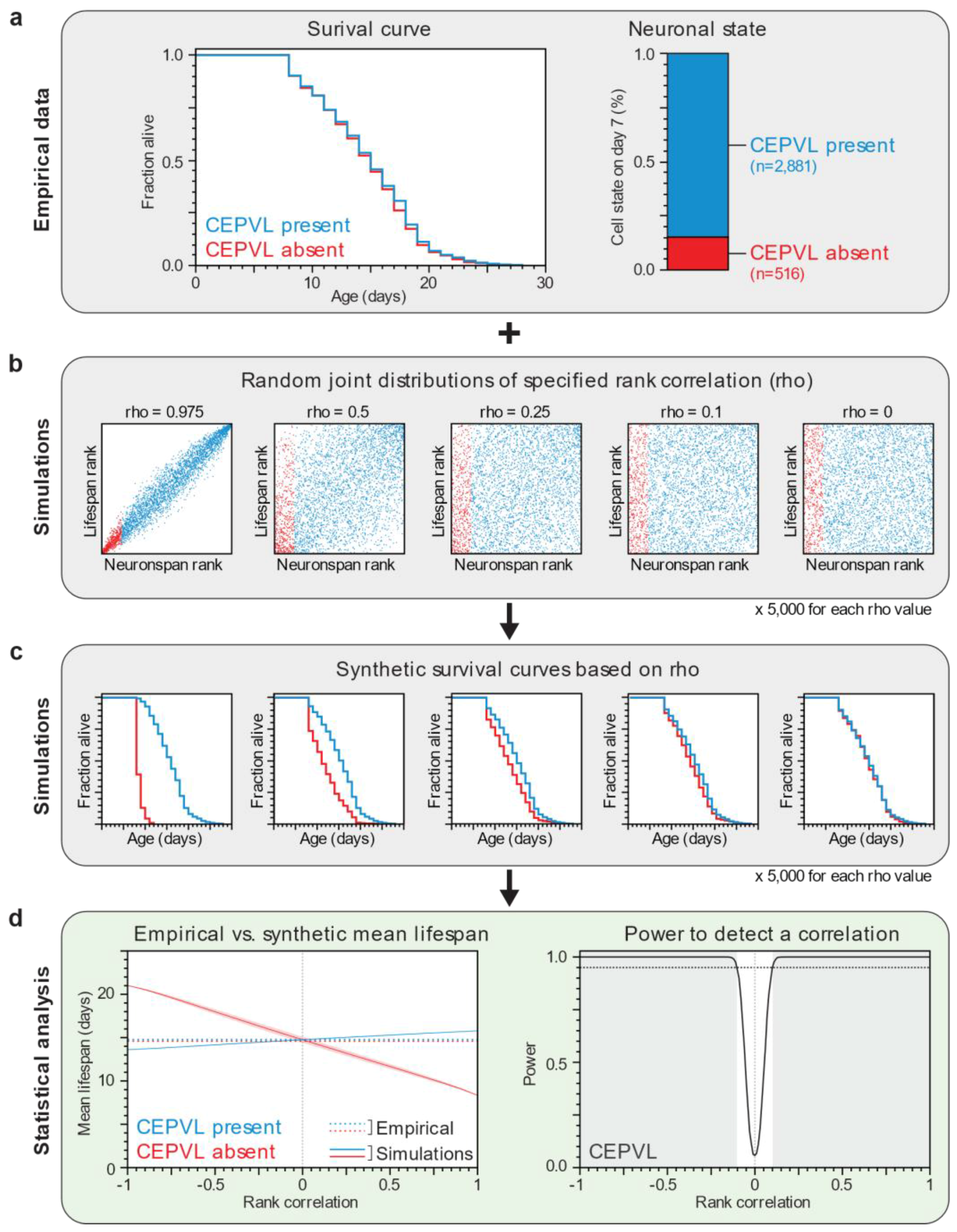
2.6. Computational Power Analysis

Simulations were performed in Matlab (Mathworks). We represented the correlation structure of lifespan and cephalic neuron-span using copulas. We generated random copulas with specific Spearman’s rank correlations using the copularnd function. The marginal distribution of lifespans was the experimentally determined distribution of wild type or age-1 mutants (3397 and 542 animals, respectively). The marginal distribution of neuron-spans was not known, but the number of animals with a present or absent cephalic neuron on day 7 of adulthood was the experimentally determined. Using those values, we partitioned the simulated observations into “cell present” and “cell absent” groups, assigning the lowest neuron-span ranks to the latter. We performed this procedure 5000 times for 75 Spearman’s rank correlations values, which were more densely interspaced as the correlation magnitude approached zero. In each iteration, we calculated the mean lifespan of each group, and compared their survival curves using the Peto-Prentice Wilcoxon test, an extension of the log-rank test is sensitive to early differences in survival. This enabled us to construct confidence intervals for mean lifespans, and to determine the fraction of tests that exhibited a significant difference at p < 0.05 (i.e., the statistical power of our experiment), as a function of the Spearman’s rank correlation. Matlab scripts are available upon request.

2.7. Statistical Analysis

We used JMP 13 software (SAS, Cary, NC, USA) to carry out statistical analysis, to determine means and percentiles, and to perform ordinal logistic regressions. We determined whether the fractions of cells missing in two or more than two groups were equal using Fisher’s Exact test and Pearson’s chi-square test, respectively. We determined whether the survival functions of different groups were equal using the Peto-Prentice Wilcoxon test. p-values were calculated for control and experimental animals examined at the same time. We used the Bonferroni correction to adjust p-values when performing multiple comparisons.

3. Results

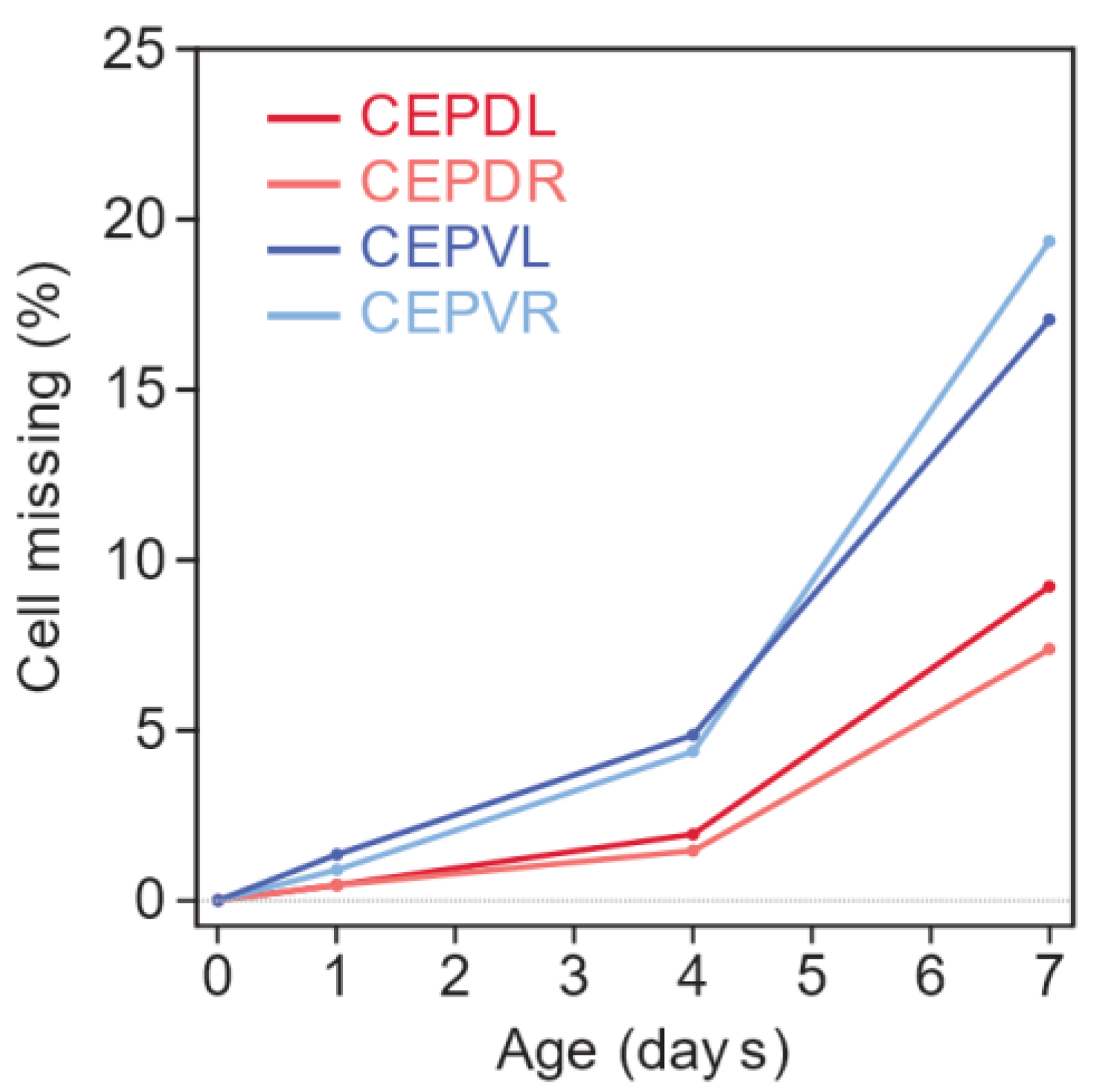
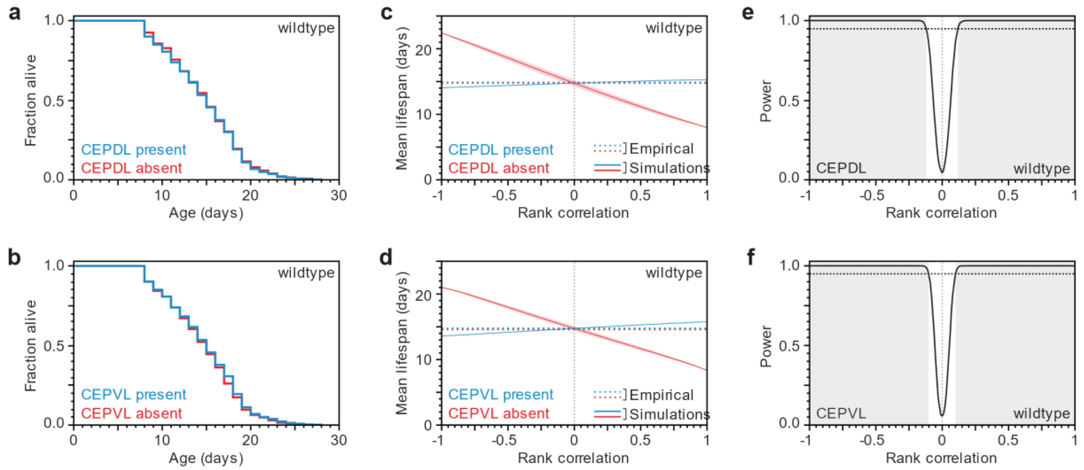
3.1. Lifespan and Neurodegeneration are the Outcomes of Independent Processes

We characterized the age-dependent degeneration of four dopaminergic neurons located in the head of the animal, the so-called cephalic neurons (two dorsal and two ventral) in the C. elegans Parkinson’s disease model [5,6] by examining the neurodegenerative phenotype in larvae and adults of increasing age. The oldest animals we examined, day-7 adults, had lived two thirds of their average expected lifespan. They were post-reproductive and tended to exhibit the reduced and uncoordinated locomotion characteristic of old age [31]. We found that larvae and day-0 adults never exhibit loss in any of the four cephalic neurons (Figure 1 and Table S1, Supplementary Materials). However, as animals grew older, their cephalic neurons were more likely to be absent (Figure 1). Each of the cephalic neurons exhibits this age-dependent neurodegenerative phenotype, although it is more severe in the ventral pair than in the dorsal pair. The neuronal-loss phenotype could be the result of neuronal death or of neuronal dysfunction leading to GFP expression levels below the detection limit. In any case, this phenotype requires α-synuclein expression, as we did not observe a loss of cephalic neurons at any age in animals that express only GFP in these neurons (Table S1, Supplementary Materials).
If the timing of α-synuclein-induced neuronal loss and of organismic death were indeed coordinated, then neuronal loss would depend on aging, not just age. Specifically, animals in which a particular cephalic neuron is present at a given age would have a different lifespan from those in which that neuron is absent. In addition, the magnitude of the difference in lifespan between these groups would provide information about the sign and strength of the correlation between the (model) disease process and the aging process. We therefore compared the lifespans of animals that experienced neuronal loss with the lifespans of those that did not.
We imaged 3397 seven-day old adults, and determined whether each of the four cephalic neurons was present or absent in each animal. We then recovered each animal and determined its lifespan. We found that animals in which a specific cephalic neuron is present have lifespans that are indistinguishable from those where that neuron is absent (Figure 2a,b, Figure S1a–d and Table S2, Supplementary Materials). We also did not observe a difference in lifespan among these animals when we group them (i) by whether any of the four cephalic neurons is missing or not; (ii) by the number of missing dorsal cephalic neurons; (iii) by the number of missing ventral cephalic neurons; or (iv) by the total number of missing cephalic neurons (Figure S2 and Table S2, Supplementary Materials). These findings strongly suggest that the timing of neuronal loss and organismic death are independent.
To ascertain that lifespan distributions (survival functions) in the presence or absence of cephalic neurons are indistinguishable, we tested (using the Wilcoxon test) whether the two survival functions arose from the same underlying distribution, which gave us a p-value indicating that the null hypothesis (no statistical difference) holds. Since this result is critical for our thesis that death and neuronal-loss times are uncorrelated, we needed to evaluate whether our experiment had enough power, i.e., we wanted to know how often our experiment would yield indistinguishable (p > 0.05) survival functions by chance, if there actually was a correlation between death and neuronal-loss times. To assess the statistical power of our experiment, we ideally would like to computationally repeatedly sample 3397 (the population size in our experiment) death times and neuronal-loss times that are consistent with our empirical data but with varying correlation, then test for statistical equality, and determine, for each correlation value, the frequency with which testing would not miss that there was, in fact, a correlation. However, in our dataset we did not have a full joint distribution of death times and neuronal-loss times; we only had a death-time distribution and we knew the number of individuals that had experienced neuronal loss by age (day) 7 (Figure 3a). We therefore used rank correlations and (for each sample) generated a joint rank distribution of 3397 points with specified rank correlations (Figure 3b). We then read off the lifespan ranks for points corresponding up to day 7 of neuronal loss and the lifespan ranks for points from day 7 onward, and converted those ranks back into death times by matching them to the ranks of our empirical lifespan data. This process yielded a survival curve for both the subpopulation that has and that has not experienced neuronal loss up to day 7 (Figure 3c and Video S1, Supplementary Materials). Finally, we tested the two survival curves against a null hypothesis of no statistical difference. In this way, sampling 5000 joint rank distributions per rank correlation value, we estimated that our experiment was powered to detect a correlation between death and neuronal-loss times greater than 0.13 with probability 0.95 for each of the cephalic neurons (Figure 3d, Figure 2c–f and Figure S1e–m, Supplementary Materials). Thus, knowing when a specific neuron is lost appears to provide very little, if any, information about organismic lifespan. We conclude that lifespan and neuronal loss are not coordinated by systemic signals; instead, they are the outcomes of independent processes.

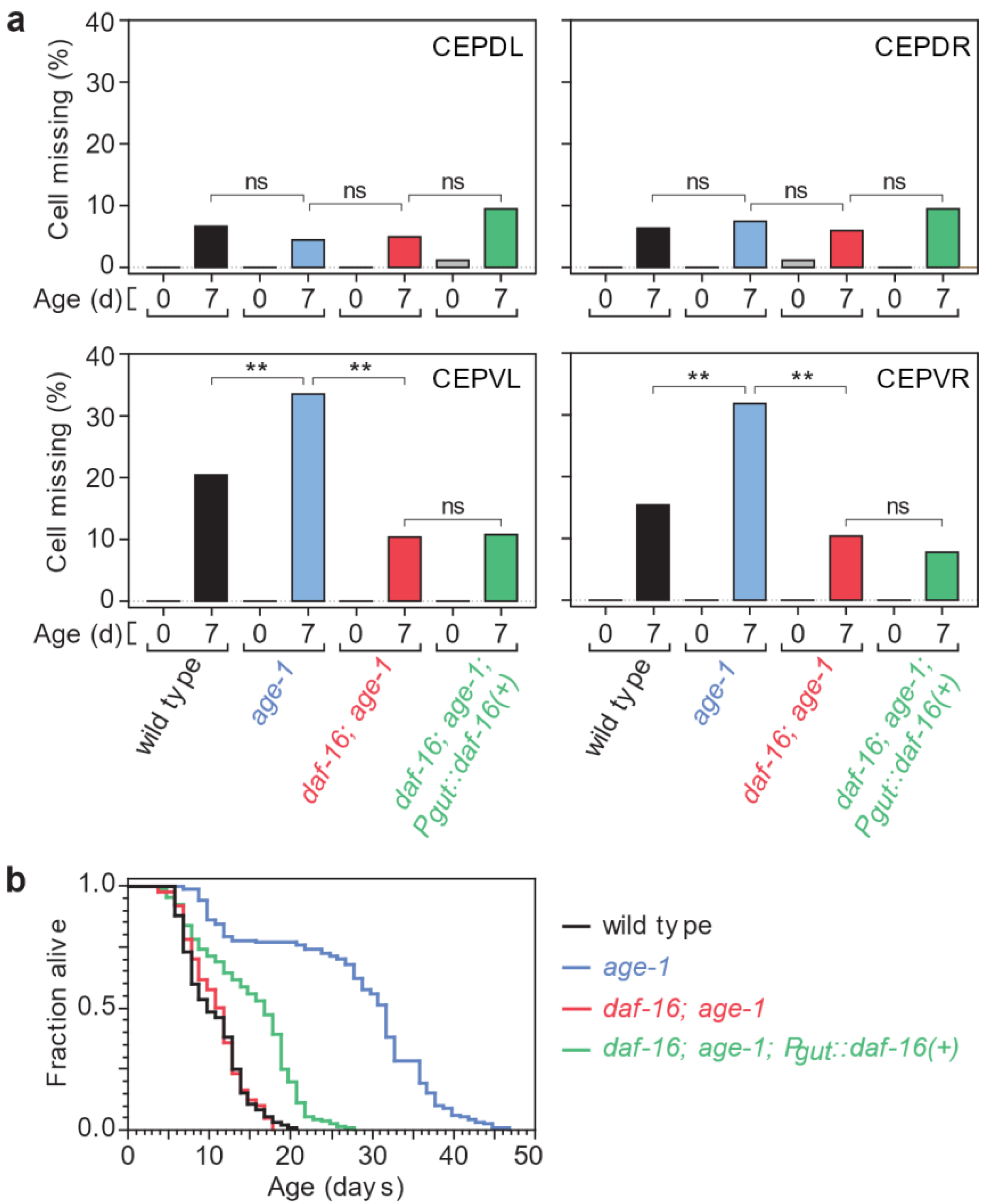
3.2. Tissue-Specific Insulin/IGF1 Signaling Has Neuron-Specific Effects on Age-Dependent Dopaminergic Loss

The existence of a lifespan-altering mutation that also impacts the timing of disease onset is often taken as a strong indication that the disease is coupled to the aging process [34,35,36,37]. We therefore first investigated whether and in what way genetic interventions that are well-known to alter lifespan also affect the timing of neurodegeneration. Subsequently, we studied whether any joint effects support a causal coupling.
Specifically, we probed the role of insulin/IGF1 signaling in the control of the timing of neuronal loss by testing the effect of the age-1(hx546) reduction-of-function mutation. As expected, in the Parkinson’s disease model this mutation causes a large, 2.6 fold, lifespan extension (Figure 4b and Table S3, Supplementary Materials). Surprisingly, these long-lived mutants exhibit a more severe neurodegenerative phenotype than wild-type animals: the fraction of missing ventral cephalic neurons in seven-day old age-1(hx546) mutants is much higher than in wild-type animals (Figure 4a and Table S4, Supplementary Materials). In contrast, the fraction of missing dorsal cephalic neurons is not affected by age-1(hx546) (Figure 4a and Table S4, Supplementary Materials). In daf-2(e1370) mutants, where insulin/IGF1 signaling is more strongly reduced than in age-1(hx546) mutants [38,39], we also observed increased loss of ventral cephalic neurons in seven-day old animals (Table S5, Supplementary Materials). In addition, the fraction of missing dorsal cephalic neurons in seven-day old daf-2(e1370) mutants was lower than in wild-type animals (Table S5, Supplementary Materials). As a result, the proportion of animals in which all four cephalic neurons are present was unaffected (62% in wild-type animals and daf-2 mutants, p > 0.1, Fisher Exact Test). In a recent study using the same Parkinson’s model but examining the two ADE dopaminergic neurons in addition to the four CEP neurons we examined, a larger proportion of daf-2(e1370) mutants than wild-type animals were found to have none of these dopaminergic neurons affected on day 7 (40% vs. 15%, respectively) [32]. In another study, daf-2 mutants were found to delay degeneration of dopaminergic neurons expressing α-synuclein with the A53T mutation [33]. Differences with these two studies may stem from differences of the effect of daf-2 on the timing of ADE loss, in the strictness of the criteria used for determining the loss of each neuron, or in experimental conditions (see Materials and Methods). Taken together, we conclude that, in our hands, insulin/IGF1 signaling, a modulator of aging, has indeed an effect—albeit opposite to what was expected, and cell-type specific—on the age-dependent loss of dopaminergic neurons in the C. elegans Parkinson’s disease model.
Activation of FOXO transcription factors mediates the lifespan-extending effects of reduced insulin/IGF-1 signaling in C. elegans and Drosophila [9,40,41,42]. We therefore asked whether the sole C. elegans FOXO transcription factor [15,16], DAF-16, plays a similar role in mediating the effects of reduced insulin/IGF1 signaling on neuronal loss by examining the effect of the daf-16(mu86) null allele [15]. The observed fraction of missing ventral cephalic neurons was dramatically lower in seven-day old daf-16(mu86); age-1(hx546) double mutants than in age-1(hx546) single mutants (Figure 4a and Table S4, Supplementary Materials). Therefore, daf-16 is required for the increased loss of ventral cephalic neurons observed in age-1(hx546) mutants. Our findings thus indicate that activation of daf-16 mediates both lifespan extending and neurodegenerative phenotypes associated with reduced insulin/IGF1 signaling. We conclude that the divergent effects of this pathway on the timing of neurodegeneration and lifespan are generated downstream of the action of daf-16.
Tissue-restricted activation of FOXO transcription factors is sufficient to extend lifespan in C. elegans [22,43] and Drosophila [17,18,24,44]. In C. elegans, FOXO activation in one tissue can influence gene expression in other tissues via cell-to-cell signaling mechanisms that involve daf-16 in the receiving tissue (FOXO-to-FOXO signaling) and by signaling mechanisms that do not (FOXO-to-other signaling) [22,23,43,45]. FOXO-to-other signaling modulates lifespan, since daf-16 expression in the intestine is sufficient to extend lifespan in animals that lack daf-16 in all other tissues [22,23]. FOXO-to-other signaling from the intestine also delays the age-dependent paralysis caused by muscle-specific expression of the human Alzheimer’s protein Aβ1-42 [43], but does not affect axon regeneration in GABA motor neurons [46]. We therefore asked whether this type of systemic signaling mechanism affects the timing of neuronal loss in the Parkinson’s disease model.
We found that expressing daf-16(+) only in the intestine extends the lifespans of daf-16(mu86); age-1(hx546) double mutants by 40% in the Parkinson’s disease model (Figure 4b and Table S3, Supplementary Materials). However, this treatment does not affect the loss of any of the four cephalic neurons on seven-day old adults (Figure 4a and Table S4, Supplementary Materials). Hence, tissues other than the intestine must mediate the effect of insulin/IGF1 signaling on α-synuclein induced neuronal loss.

3.3. After a Lifespan-Extending Intervention, Lifespan and Neurodegeneration Are Still Independent

Having established that reduction of insulin/IGF1 signaling impacts both lifespan and neuronal loss, we asked whether this affects the independence of these processes, which we observed in the wild-type background. To test this possibility, we stratified a population of 542 seven-day old age-1(hx546) adults according to the presence or absence of each cephalic neuron, and then determined their lifespans, following the same scheme as in the wild-type case.
We found that age-1(hx546) mutants in which a cephalic neuron is present have lifespans that are indistinguishable from those where that neuron is absent (Figure 5a,b, Figure S3a–d and Table S2, Supplementary Materials). In addition, we found that the lifespan of age-1(hx546) mutant individuals was not predicted by the extent of neurodegeneration they exhibited (Figure S4, Supplementary Materials). As in the wild-type case, we carried out numerical simulations to estimate how sampling noise would impact the conclusions we drew from our experiment as a function of rank correlation (Figure 5c–f and Figure S3e–m, Supplementary Materials). This analysis indicates that our experiment was well powered to detect a significant difference in lifespan for at least one of the four neurons if rank correlations were greater than 0.15 in magnitude (Figure S3m, Supplementary Materials). We conclude that lifespan and neurodegeneration are at most weakly coordinated, if at all, in age-1(hx546) mutants.

4. Discussion

In this study, we probed the causal links between neurodegeneration and lifespan in a C. elegans model of Parkinson’s disease. We found that, while the loss of dopaminergic neurons increases with age, it is a phenomenon independent of aging. Yet, despite this independence, the timescales of neuronal loss and organismic lifespan are both regulated by insulin/IGF-1 signaling, pointing to a compartmentalization of this pathway’s action. Our findings have potential implications for our understanding of a broad class of age-dependent and lethal human diseases whose aging-dependence remains poorly understood.

4.1. Age-Dependence of Neuronal Loss in a C. elegans Parkinson’s Disease Model

We first review our observations on the C. elegans Parkinson’s disease model. We found that specific sets of dopaminergic neurons are affected differently by age. Dorsal cephalic neurons are less likely to exhibit α-synuclein-induced loss than ventral cephalic neurons as time progresses. In addition, these neurons are affected differently by insulin/IGF1 signaling. In particular, compared to wild type, age-1/PI3K mutants exhibit higher neurodegeneration in ventral cephalic neurons but not in dorsal cephalic neurons. In humans, too, though for reasons unknown, specific dopaminergic neurons exhibit distinct susceptibility to Parkinson’s disease [47]. Our findings in C. elegans suggest that this specificity could stem from differences in the sensitivity of specific dopaminergic neurons to insulin or IGF1.
In addition to extending lifespan in C. elegans, reducing insulin/IGF1 signaling also delays the onset of a wide variety of phenotypes associated with normal aging, including many biochemical [48,49], morphological [31,50], and behavioral impairments [9,51,52,53,54]. Neurons are affected too, as in the case of the reduction of the age-dependent incidence of neurite sprouting in ALM and PLM mechanosensory neurons [55,56,57], and axon regeneration in GABA motor neurons [46]. Diminishing the action of the insulin/IGF1-signaling pathway also postpones age-dependent changes in some disease models, including models of age-dependent paralysis due to the expression in body wall muscles of an aggregation-prone polyglutamine-containing protein or the human protein Aβ1-42 linked to Alzheimer’s disease [58,59]. We were therefore surprised to find that reducing insulin/IGF1 signaling augments the loss of ventral cephalic neurons in the Parkinson’s disease model. Previous studies have shown that DAF-16/FOXO-to-other signaling from the intestine affects both lifespan [22] and the timing of body paralysis, due to expression in body wall muscles of the human Aβ1-42 protein [43]. In contrast, this type of signaling from the intestine does not affect axon regeneration in GABA motor neurons [46]. Similarly, we find that FOXO-to-other signaling from the intestine does not affect the loss of neurons in the C. elegans Parkinson’s disease model.

4.2. Aging-Independence of Neuronal Loss in a C. elegans Parkinson’s Disease Model

Perhaps most strikingly, we did not find a correlation between lifespan and neurodegeneration in the disease model with wild-type background. It is important to emphasize, once again, that age simply refers to physical time whose origin (time zero) is defined by the occurrence of a specific developmental event, which in C. elegans is the onset of adulthood. However, in informal usage, age often appears to carry a connotation of “biological time”. At the present state of knowledge, any notion of biological time in the context of aging must refer to the risk of death. Thus, the risk of any event X, other than death, can be said to depend on “biological time” only insofar as it carries information about the risk of death (and vice versa). The risk of death tracks the aging process, and the risk of loss of dopaminergic neurons tracks the disease process (or a pathological condition for the worm). Both risks change with time, that is, with the age of an individual. An interesting question is whether the disease is not only age-dependent, but also dependent on aging—the very process that determines lifespan. In this study, we showed that the Parkinson’s disease model in C. elegans is independent of aging.
We also found something that might seem puzzling at first: that both processes underlying the respective event risks are influenced by manipulation of insulin/IGF1 signaling (e.g., in age-1(hx546) mutants), and yet remain independent. The key here is that causal relations between one phenotype (aging) and another (disease onset) are captured by their joint probability distribution. Whether the marginal distributions of both, one, or neither of these phenotypes are influenced by insulin/IGF1 signaling (or something else) does not provide information about the joint-distribution of aging and disease onset (in the presence or absence of reduced insulin/IGF1 signaling). In principle, one could find an intervention that affects the timing of only one of these phenotypes; say, death occurs twice as late. The existence of such an intervention would (i) indicate that the timescale of death and disease onset can be influenced differentially; and (ii) identify a causal link between the intervention and one of the phenotypes; but (iii) say nothing about the causal relations between the two phenotypes, which requires knowledge of their joint distribution. At the level of abstraction of genetics, the age-1 gene might be viewed as a common cause (all else equal) of its effects on neurodegeneration and aging in the C. elegans Parkinson’s disease model. However, in the context of a fully developed organism, tissue-specific realizations of the insulin/IGF1 signaling pathway may well have distinct effects on neuronal loss and lifespan, in particular if these realizations are “compartmentalized” in the sense of not exchanging state information.

5. Conclusions

In humans, the progression of Parkinson’s disease and other neurodegenerative diseases often includes the loss of neurons involved in essential functions, thereby resulting in early death [4]. It is therefore not possible to measure the joint probability distribution of Parkinson’s disease onset and lifespan as determined by aging. This has made it difficult to study whether systemic signals coordinate the onset of such diseases with the timing of death from aging. One approach to addressing this impasse would be to use an animal model in which the disease is not fatal, thereby allowing the acquisition of joint probability distributions. This is the case in the C. elegans Parkinson’s disease model because, unlike in humans, dopaminergic neurons are not essential for survival in the worm [8]. While the disease model in C. elegans might serve well for the study of molecular mechanisms that are associated with the disease and potentially conserved in humans, it may fail to evidence a connection with aging that might be present in humans, perhaps because of a more complex aging process. That said, our results regarding the distinction of age- and aging-dependence suggest at least a cautionary stance with regard to the widely held, yet untested, view that the onset of neurodegenerative diseases, such as Parkinson’s and Alzheimer’s, is mechanistically linked to the aging process that determines lifespan. This assumption is the basis for the expectation that disease onset will be postponed by pharmaceutical, dietary or genetic interventions that extend lifespan [34,35,36,37]. Our results call for prudence when using the term “disease of aging”, which is often tacitly based on the notion of an overall aging process that couples all age-related phenotypes—a notion that our work demonstrates does not apply to C. elegans.

Supplementary Materials

The following are available online at www.mdpi.com/2079-7737/7/1/1/s1, Figure S1: Independence of neuronal loss and organismic death in wild type animals, Figure S2: Independence of neuronal loss and organismic death in wild type animals, Figure S3: Independence of neuronal loss and organismic death in age-1 mutants, Figure S4: Independence of neuronal loss and organismic death in age-1 mutants, Table S1: Age-dependent neurodegeneration in wild-type animals expressing α-synuclein in dopaminergic neurons, Table S2: Independence of lifespan and cephalic neurodegeneration in animals expressing α-synuclein in dopaminergic neurons, Table S3: Statistical analysis of adult lifespans of animals expressing α-synuclein in dopaminergic neurons, Table S4: Percent of cephalic neurons missing in wild type and mutants expressing α-synuclein in dopaminergic neurons, Table S5: Percent of cephalic neurons missing in wild type and daf-2(e1370) mutants expressing α-synuclein in dopaminergic neurons, Video S1: Simulations of the joint variation of the timing of neuronal loss and lifespan.

Acknowledgments

We thank Guy Caldwell for providing the baIn11 strain; Jennifer Whangbo, Rebecca Ward and members of the Fontana lab for comments on the manuscript and stimulating discussions. Some of the C. elegans strains used in this work were provided by the Caenorhabditis Genetics Center, which is funded by US National Institutes of Health (NIH) Office of Research Infrastructure Programs (P40 OD010440). This work was funded by the NIH through grant R01 AG034994.

Author Contributions

J.A. Conducted experiments, simulations and statistical analysis. J.A. and W.F. conceived the project, interpreted data, and wrote the manuscript.

Conflicts of Interest

The authors declare no conflict of interest.

References

  1. Heron, M. Deaths: Leading causes for 2010. Natl. Vital Stat. Rep. 2013, 62, 1–96. [Google Scholar] [PubMed]
  2. Driver, J.A.; Logroscino, G.; Gaziano, J.M.; Kurth, T. Incidence and remaining lifetime risk of Parkinson disease in advanced age. Neurology 2009, 72, 432–438. [Google Scholar] [CrossRef] [PubMed]
  3. Olanow, C.W.; Stern, M.B.; Sethi, K. The scientific and clinical basis for the treatment of Parkinson disease (2009). Neurology 2009, 72, S1–S136. [Google Scholar] [CrossRef] [PubMed]
  4. Poewe, W. The natural history of Parkinson’s disease. J. Neurol. 2006, 253, VII2–VII6. [Google Scholar] [CrossRef] [PubMed]
  5. Cao, S.; Gelwix, C.C.; Caldwell, K.A.; Caldwell, G.A. Torsin-mediated protection from cellular stress in the dopaminergic neurons of Caenorhabditis elegans. J. Neurosci. Off. J. Soc. Neurosci. 2005, 25, 3801–3812. [Google Scholar] [CrossRef] [PubMed]
  6. Hamamichi, S.; Rivas, R.N.; Knight, A.L.; Cao, S.; Caldwell, K.A.; Caldwell, G.A. Hypothesis-based RNAI screening identifies neuroprotective genes in a Parkinson’s disease model. Proc. Natl. Acad. Sci. USA 2008, 105, 728–733. [Google Scholar] [CrossRef] [PubMed]
  7. Gitler, A.D.; Chesi, A.; Geddie, M.L.; Strathearn, K.E.; Hamamichi, S.; Hill, K.J.; Caldwell, K.A.; Caldwell, G.A.; Cooper, A.A.; Rochet, J.C.; et al. Alpha-synuclein is part of a diverse and highly conserved interaction network that includes park9 and manganese toxicity. Nat. Genet. 2009, 41, 308–315. [Google Scholar] [CrossRef] [PubMed]
  8. Sawin, E.R.; Ranganathan, R.; Horvitz, H.R. C. elegans locomotory rate is modulated by the environment through a dopaminergic pathway and by experience through a serotonergic pathway. Neuron 2000, 26, 619–631. [Google Scholar] [CrossRef]
  9. Kenyon, C.; Chang, J.; Gensch, E.; Rudner, A.; Tabtiang, R.A. C. elegans mutant that lives twice as long as wild type. Nature 1993, 366, 461–464. [Google Scholar] [CrossRef] [PubMed]
  10. Friedman, D.B.; Johnson, T.E. A mutation in the age-1 gene in Caenorhabditis elegans lengthens life and reduces hermaphrodite fertility. Genetics 1988, 118, 75–86. [Google Scholar] [PubMed]
  11. Clancy, D.J.; Gems, D.; Harshman, L.G.; Oldham, S.; Stocker, H.; Hafen, E.; Leevers, S.J.; Partridge, L. Extension of life-span by loss of chico, a drosophila insulin receptor substrate protein. Science 2001, 292, 104–106. [Google Scholar] [CrossRef] [PubMed]
  12. Tatar, M.; Kopelman, A.; Epstein, D.; Tu, M.P.; Yin, C.M.; Garofalo, R.S. A mutant drosophila insulin receptor homolog that extends life-span and impairs neuroendocrine function. Science 2001, 292, 107–110. [Google Scholar] [CrossRef] [PubMed]
  13. Bluher, M.; Kahn, B.B.; Kahn, C.R. Extended longevity in mice lacking the insulin receptor in adipose tissue. Science 2003, 299, 572–574. [Google Scholar] [CrossRef] [PubMed]
  14. Holzenberger, M.; Dupont, J.; Ducos, B.; Leneuve, P.; Geloen, A.; Even, P.C.; Cervera, P.; Le Bouc, Y. IGF-1 receptor regulates lifespan and resistance to oxidative stress in mice. Nature 2003, 421, 182–187. [Google Scholar] [CrossRef] [PubMed]
  15. Lin, K.; Dorman, J.B.; Rodan, A.; Kenyon, C. Daf-16: An HNF-3/forkhead family member that can function to double the life-span of Caenorhabditis elegans. Science 1997, 278, 1319–1322. [Google Scholar] [CrossRef] [PubMed]
  16. Ogg, S.; Paradis, S.; Gottlieb, S.; Patterson, G.I.; Lee, L.; Tissenbaum, H.A.; Ruvkun, G. The fork head transcription factor daf-16 transduces insulin-like metabolic and longevity signals in C. elegans. Nature 1997, 389, 994–999. [Google Scholar] [CrossRef] [PubMed]
  17. Giannakou, M.E.; Goss, M.; Junger, M.A.; Hafen, E.; Leevers, S.J.; Partridge, L. Long-lived drosophila with overexpressed dFOXO in adult fat body. Science 2004, 305. [Google Scholar] [CrossRef] [PubMed]
  18. Hwangbo, D.S.; Gersham, B.; Tu, M.P.; Palmer, M.; Tatar, M. Drosophila dFOXO controls lifespan and regulates insulin signalling in brain and fat body. Nature 2004, 429, 562–566. [Google Scholar] [CrossRef] [PubMed]
  19. Willcox, B.J.; Donlon, T.A.; He, Q.; Chen, R.; Grove, J.S.; Yano, K.; Masaki, K.H.; Willcox, D.C.; Rodriguez, B.; Curb, J.D. FOXO3A genotype is strongly associated with human longevity. Proc. Natl. Acad. Sci. USA 2008, 105, 13987–13992. [Google Scholar] [CrossRef] [PubMed]
  20. Flachsbart, F.; Caliebe, A.; Kleindorp, R.; Blanche, H.; von Eller-Eberstein, H.; Nikolaus, S.; Schreiber, S.; Nebel, A. Association of FOXO3A variation with human longevity confirmed in German centenarians. Proc. Natl. Acad. Sci. USA 2009, 106, 2700–2705. [Google Scholar] [CrossRef] [PubMed]
  21. Apfeld, J.; Kenyon, C. Cell nonautonomy of C. elegans daf-2 function in the regulation of diapause and life span. Cell 1998, 95, 199–210. [Google Scholar] [CrossRef]
  22. Libina, N.; Berman, J.R.; Kenyon, C. Tissue-specific activities of C. elegans daf-16 in the regulation of lifespan. Cell 2003, 115, 489–502. [Google Scholar] [CrossRef]
  23. Iser, W.B.; Gami, M.S.; Wolkow, C.A. Insulin signaling in Caenorhabditis elegans regulates both endocrine-like and cell-autonomous outputs. Dev. Biol. 2007, 303, 434–447. [Google Scholar] [CrossRef] [PubMed]
  24. Alic, N.; Tullet, J.M.; Niccoli, T.; Broughton, S.; Hoddinott, M.P.; Slack, C.; Gems, D.; Partridge, L. Cell-nonautonomous effects of dFOXO/DAF-16 in aging. Cell Rep. 2014, 6, 608–616. [Google Scholar] [CrossRef] [PubMed] [Green Version]
  25. Kappeler, L.; De Magalhaes Filho, C.; Dupont, J.; Leneuve, P.; Cervera, P.; Perin, L.; Loudes, C.; Blaise, A.; Klein, R.; Epelbaum, J.; et al. Brain IGF-1 receptors control mammalian growth and lifespan through a neuroendocrine mechanism. PLoS Biol. 2008, 6, e254. [Google Scholar] [CrossRef] [PubMed] [Green Version]
  26. Kenyon, C.J. The genetics of ageing. Nature 2010, 464, 504–512. [Google Scholar] [CrossRef] [PubMed]
  27. Nass, R.; Hall, D.H.; Miller, D.M., 3rd; Blakely, R.D. Neurotoxin-induced degeneration of dopamine neurons in Caenorhabditis elegans. Proc. Natl. Acad. Sci. USA 2002, 99, 3264–3269. [Google Scholar] [CrossRef] [PubMed]
  28. Deng, H.; Yuan, L. Genetic variants and animal models in SNCA and Parkinson disease. Ageing Res. Rev. 2014, 15C, 161–176. [Google Scholar] [CrossRef] [PubMed]
  29. Singleton, A.B.; Farrer, M.; Johnson, J.; Singleton, A.; Hague, S.; Kachergus, J.; Hulihan, M.; Peuralinna, T.; Dutra, A.; Nussbaum, R.; et al. Alpha-synuclein locus triplication causes Parkinson’s disease. Science 2003, 302. [Google Scholar] [CrossRef] [PubMed]
  30. Apfeld, J.; O’Connor, G.; McDonagh, T.; DiStefano, P.S.; Curtis, R. The AMP-activated protein kinase AAK-2 links energy levels and insulin-like signals to lifespan in C. elegans. Genes Dev. 2004, 18, 3004–3009. [Google Scholar] [CrossRef] [PubMed]
  31. Herndon, L.A.; Schmeissner, P.J.; Dudaronek, J.M.; Brown, P.A.; Listner, K.M.; Sakano, Y.; Paupard, M.C.; Hall, D.H.; Driscoll, M. Stochastic and genetic factors influence tissue-specific decline in ageing C. elegans. Nature 2002, 419, 808–814. [Google Scholar] [CrossRef] [PubMed]
  32. Knight, A.L.; Yan, X.; Hamamichi, S.; Ajjuri, R.R.; Mazzulli, J.R.; Zhang, M.W.; Daigle, J.G.; Zhang, S.; Borom, A.R.; Roberts, L.R.; et al. The glycolytic enzyme, GPI, is a functionally conserved modifier of dopaminergic neurodegeneration in Parkinson’s models. Cell Metab. 2014, 20, 145–157. [Google Scholar] [CrossRef] [PubMed]
  33. Cooper, J.F.; Dues, D.J.; Spielbauer, K.K.; Machiela, E.; Senchuk, M.M.; Van Raamsdonk, J.M. Delaying aging is neuroprotective in Parkinson’s disease: A genetic analysis in C. elegans models. NPJ Parkinson’s Dis. 2015, 1. [Google Scholar] [CrossRef] [PubMed]
  34. Alavez, S.; Lithgow, G.J. Pharmacological maintenance of protein homeostasis could postpone age-related disease. Aging Cell 2012, 11, 187–191. [Google Scholar] [CrossRef] [PubMed]
  35. Butler, R.N.; Miller, R.A.; Perry, D.; Carnes, B.A.; Williams, T.F.; Cassel, C.; Brody, J.; Bernard, M.A.; Partridge, L.; Kirkwood, T.; et al. New model of health promotion and disease prevention for the 21st century. BMJ 2008, 337. [Google Scholar] [CrossRef] [PubMed]
  36. Blagosklonny, M.V. Validation of anti-aging drugs by treating age-related diseases. Aging 2009, 1, 281–288. [Google Scholar] [CrossRef] [PubMed]
  37. Finkel, T. Radical medicine: Treating ageing to cure disease. Nat. Rev. Mol. Cell Biol. 2005, 6, 971–976. [Google Scholar] [CrossRef] [PubMed]
  38. Gems, D.; Sutton, A.J.; Sundermeyer, M.L.; Albert, P.S.; King, K.V.; Edgley, M.L.; Larsen, P.L.; Riddle, D.L. Two pleiotropic classes of daf-2 mutation affect larval arrest, adult behavior, reproduction and longevity in Caenorhabditis elegans. Genetics 1998, 150, 129–155. [Google Scholar] [PubMed]
  39. Patel, D.S.; Garza-Garcia, A.; Nanji, M.; McElwee, J.J.; Ackerman, D.; Driscoll, P.C.; Gems, D. Clustering of genetically defined allele classes in the Caenorhabditis elegans daf-2 Insulin/IGF-1 receptor. Genetics 2008, 178, 931–946. [Google Scholar] [CrossRef] [PubMed]
  40. Morris, J.Z.; Tissenbaum, H.A.; Ruvkun, G. A phosphatidylinositol-3-OH kinase family member regulating longevity and diapause in Caenorhabditis elegans. Nature 1996, 382, 536–539. [Google Scholar] [CrossRef] [PubMed]
  41. Slack, C.; Giannakou, M.E.; Foley, A.; Goss, M.; Partridge, L. dFOXO-independent effects of reduced insulin-like signaling in Drosophila. Aging Cell 2011, 10, 735–748. [Google Scholar] [CrossRef] [PubMed]
  42. Yamamoto, R.; Tatar, M. Insulin receptor substrate chico acts with the transcription factor FOXO to extend Drosophila lifespan. Aging Cell 2011, 10, 729–732. [Google Scholar] [CrossRef] [PubMed]
  43. Zhang, P.; Judy, M.; Lee, S.J.; Kenyon, C. Direct and indirect gene regulation by a life-extending FOXO protein in C. elegans: Roles for gata factors and lipid gene regulators. Cell Metab. 2013, 17, 85–100. [Google Scholar] [CrossRef] [PubMed]
  44. Demontis, F.; Perrimon, N. FOXO/4E-BP signaling in drosophila muscles regulates organism-wide proteostasis during aging. Cell 2010, 143, 813–825. [Google Scholar] [CrossRef] [PubMed]
  45. Murphy, C.T.; Lee, S.J.; Kenyon, C. Tissue entrainment by feedback regulation of insulin gene expression in the endoderm of Caenorhabditis elegans. Proc. Natl. Acad. Sci. USA 2007, 104, 19046–19050. [Google Scholar] [CrossRef] [PubMed]
  46. Byrne, A.B.; Walradt, T.; Gardner, K.E.; Hubbert, A.; Reinke, V.; Hammarlund, M. Insulin/IGF1 signaling inhibits age-dependent axon regeneration. Neuron 2014, 81, 561–573. [Google Scholar] [CrossRef] [PubMed]
  47. Dawson, T.M.; Ko, H.S.; Dawson, V.L. Genetic animal models of parkinson’s disease. Neuron 2010, 66, 646–661. [Google Scholar] [CrossRef] [PubMed]
  48. Yasuda, K.; Adachi, H.; Fujiwara, Y.; Ishii, N. Protein carbonyl accumulation in Aging Dauer Formation—Defective (daf) mutants of Caenorhabditis elegans. J. Gerontol. Ser. A Biol. Sci. Med. Sci. 1999, 54, B47–B51. [Google Scholar] [CrossRef]
  49. Gerstbrein, B.; Stamatas, G.; Kollias, N.; Driscoll, M. In Vivo spectrofluorimetry reveals endogenous biomarkers that report healthspan and dietary restriction in Caenorhabditis elegans. Aging Cell 2005, 4, 127–137. [Google Scholar] [CrossRef] [PubMed]
  50. Garigan, D.; Hsu, A.L.; Fraser, A.G.; Kamath, R.S.; Ahringer, J.; Kenyon, C. Genetic analysis of tissue aging in Caenorhabditis elegans: A role for heat-shock factor and bacterial proliferation. Genetics 2002, 161, 1101–1112. [Google Scholar] [PubMed]
  51. Duhon, S.A.; Johnson, T.E. Movement as an index of vitality: Comparing wild type and the age-1 mutant of Caenorhabditis elegans. J. Gerontol. Ser. A Biol. Sci. Med. Sci. 1995, 50, B254–B261. [Google Scholar] [CrossRef]
  52. Huang, C.; Xiong, C.; Kornfeld, K. Measurements of age-related changes of physiological processes that predict lifespan of Caenorhabditis elegans. Proc. Natl. Acad. Sci. USA 2004, 101, 8084–8089. [Google Scholar] [CrossRef] [PubMed]
  53. Kauffman, A.L.; Ashraf, J.M.; Corces-Zimmerman, M.R.; Landis, J.N.; Murphy, C.T. Insulin signaling and dietary restriction differentially influence the decline of learning and memory with age. PLoS Biol. 2010, 8, e1000372. [Google Scholar] [CrossRef] [PubMed]
  54. Hahm, J.H.; Kim, S.; DiLoreto, R.; Shi, C.; Lee, S.J.V.; Murphy, C.T.; Nam, H.G. C. elegans maximum velocity correlates with healthspan and is maintained in worms with an insulin receptor mutation. Nat. Commun. 2015, 6. [Google Scholar] [CrossRef] [PubMed]
  55. Pan, C.L.; Peng, C.Y.; Chen, C.H.; McIntire, S. Genetic analysis of age-dependent defects of the Caenorhabditis elegans touch receptor neurons. Proc. Natl. Acad. Sci. USA 2011, 108, 9274–9279. [Google Scholar] [CrossRef] [PubMed]
  56. Tank, E.M.; Rodgers, K.E.; Kenyon, C. Spontaneous age-related neurite branching in Caenorhabditis elegans. J. Neurosci. Off. J. Soc. Neurosci. 2011, 31, 9279–9288. [Google Scholar] [CrossRef] [PubMed]
  57. Toth, M.L.; Melentijevic, I.; Shah, L.; Bhatia, A.; Lu, K.; Talwar, A.; Naji, H.; Ibanez-Ventoso, C.; Ghose, P.; Jevince, A.; et al. Neurite sprouting and synapse deterioration in the aging Caenorhabditis elegans nervous system. J. Neurosci. Off. J. Soc. Neurosci. 2012, 32, 8778–8790. [Google Scholar] [CrossRef] [PubMed]
  58. Morley, J.F.; Brignull, H.R.; Weyers, J.J.; Morimoto, R.I. The threshold for polyglutamine-expansion protein aggregation and cellular toxicity is dynamic and influenced by aging in Caenorhabditis elegans. Proc. Natl. Acad. Sci. USA 2002, 99, 10417–10422. [Google Scholar] [CrossRef] [PubMed]
  59. Cohen, E.; Bieschke, J.; Perciavalle, R.M.; Kelly, J.W.; Dillin, A. Opposing activities protect against age-onset proteotoxicity. Science 2006, 313, 1604–1610. [Google Scholar] [CrossRef] [PubMed]
Figure 1. Age-dependent loss of cephalic neurons in animals expressing α-synuclein in the dopaminergic neurons (Parkinson’s disease model). C. elegans has four cephalic (CEP) neurons, located dorsally (D) or ventrally (V), on the left (L) or the right (R) side of the worm. Ordinal logistic regression indicates that the proportion of animals with an absent neuron increases in an age-dependent manner (p = 0.0009 between days 1 and 4, and p < 0.0001 between days 4 and 7) and that dorsal neurons have a more severe phenotype than ventral neurons (p < 0.0001), while left-right position has no impact on phenotype (p > 0.05). Animals that do not express α-synuclein do not exhibit neuronal loss (Table S1, Supplementary Materials). For additional statistics see Table S1, Supplementary Materials.
Biology 07 00001 g001
Figure 2. Independence of neuronal loss and organismic death in wild-type animals. (a,b) No difference in lifespan was observed when 3397 seven-day old wild-type animals expressing α-synuclein in the dopaminergic neurons were grouped according to whether the specified cephalic neuron was present (blue line) or absent (red line). For statistics see Table S2, Supplementary Materials; (c,d) Using computer simulations (Figure 3), we predicted how the mean lifespans of seven-day old animals with present (blue line) and absent (red line) cephalic neurons are influenced by the Spearman’s rank correlation between the timing of neuronal loss and lifespan. The shaded areas represent the 95% point-wise confidence interval of the predicted mean lifespans. The observed mean lifespans (dotted lines) are only consistent with correlations of low magnitude; (e,f) The statistical power of our experiment is the fraction of simulations with a significant (p < 0.05) difference in survival between the neuron present and absent groups. The shaded region denotes the Spearman’s rank correlation values where statistical power is greater than 95% (dotted line). For the analysis based on the state of CEPDR and CEPVR on day 7, see Figure S3, Supplementary Materials.
Biology 07 00001 g002
Figure 3. Statistical analysis strategy based on empirical data and computational simulations. (a) This panel shows the two types of data that we collected for each individual expressing α-synuclein in the dopaminergic neurons: its lifespan and the state of each of its four cephalic neurons on day 7 of adulthood. These data were used to construct the empirical distribution of lifespan (plotted as a survival curve) and neuronal state; (b) We generated collections of random joint distributions of varying Spearman rank correlation values. Each point represents a simulated animal that has a relative lifespan and neuronspan. Simulated animals where a neuron is absent on day 7 have the lowest neuronspans (red points), while those were a neuron was present have the highest neuronspans (blue points). The joint distribution size equals the number of individuals in the empirical data set and the proportion of points labelled as “present” or “absent” equals the observed proportion of neuronal states; (c) We generated synthetic survival curves by dividing the empirical lifespans based on their rank, using the simulated lifespan ranks of the present (blue) and absent (red) groups in each random joint distribution; (d) The collection of synthetic survival curves was analyzed statistically to model how the mean lifespan of each group varies with Spearman rank correlation value. The shaded areas represent the 95% point-wise confidence interval of the mean lifespans of the synthetic curves. The statistical power of our experiment is the fraction of simulations with a statistically significant (p < 0.05) difference in survival between the neuron present and absent groups. The shaded region denotes the Spearman’s rank correlation values where statistical power is greater than 95% (dotted line). The empirical data and statistical analysis shown in this figure is also shown in Figure 2b,d,f.
Biology 07 00001 g003
Figure 4. Effect of insulin/IGF1 signaling on neurodegeneration and lifespan. (a) Each panel shows the percentage of animals with the indicated cephalic neuron missing in young adults (day 0) and old adults (day 7). These percentages are paired for each specified genotype. Except for age-1 (CEPDL) and daf-16; age-1 (CEPDL and CEPDR), all pairs show a statistically significant difference of at least p = 0.004 between young and old. The double asterisk (**) indicates p ≤ 0.001; “ns” indicates that no significant difference was observed (p > 0.05). For additional statistics see Table S3; (b) Survival curves for wild-type and mutant animals expressing α-synuclein in the dopaminergic neurons (Parkinson’s disease model). For statistics see Table S3.
Biology 07 00001 g004
Figure 5. Independence of neuronal loss and organismic death in age-1 mutant animals. (a,b) No difference in lifespan was observed when 542 seven-day old age-1(hx546) mutants expressing α-synuclein in the dopaminergic neurons were grouped according to whether the specified cephalic neuron was present (blue line) or absent (red line). For statistics see Table S2; (c,d) Using computer simulations (Figure 3), we predicted how the mean lifespans of seven-day old animals with present (blue line) and absent (red line) cephalic neurons are influenced by the Spearman’s rank correlation between the timing of neuronal loss and lifespan. The shaded areas represent the 95% point-wise confidence interval of the predicted mean lifespans. The observed mean lifespans (dotted lines) are only consistent with correlations of low magnitude; (e,f) The statistical power of our experiment is the fraction of simulations with a significant (p < 0.05) difference in survival between the neuron present and absent groups. The shaded region denotes the Spearman’s rank correlation values where statistical power is greater than 95% (dotted line). For the analysis of based on the state of CEPDR and CEPVR on day 7 see Figure S4, Supplementary Materials.
Biology 07 00001 g005

