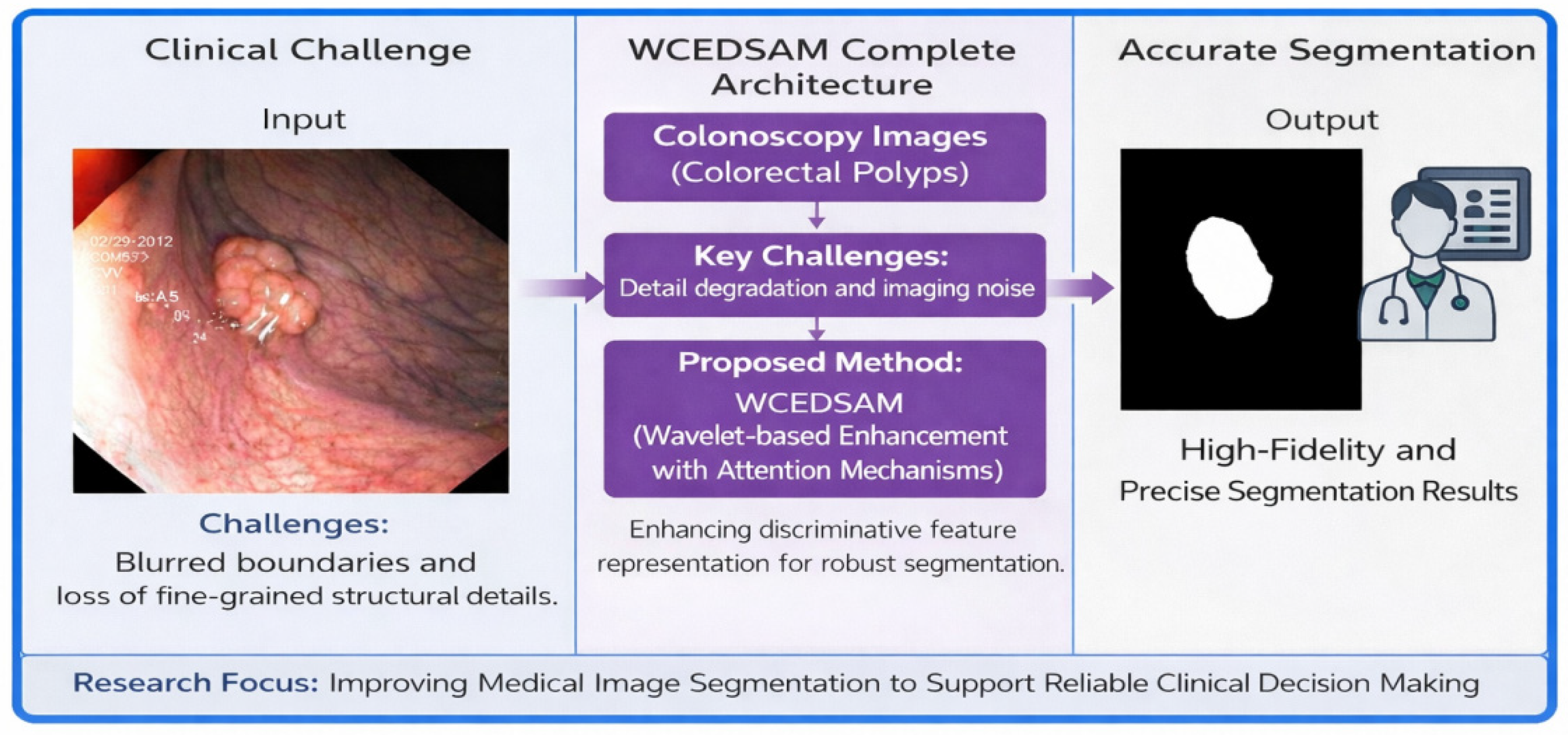
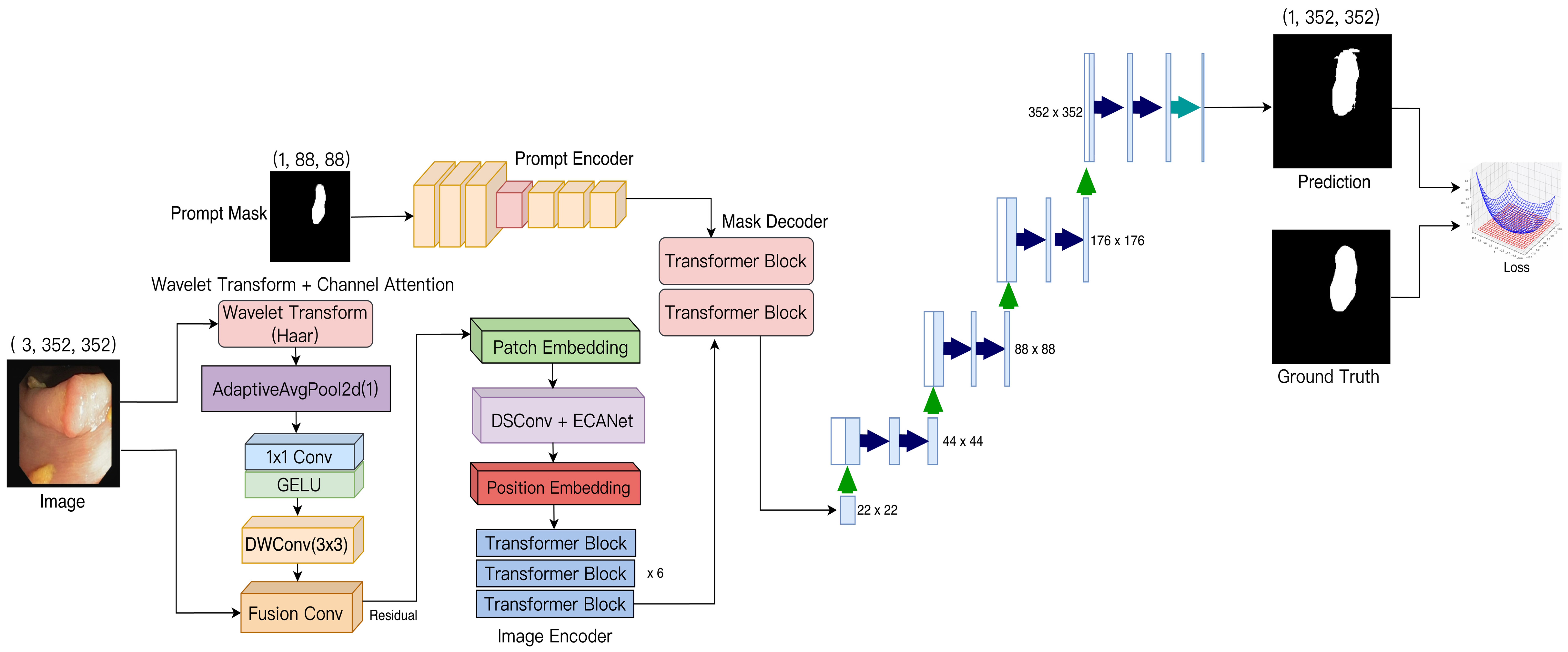
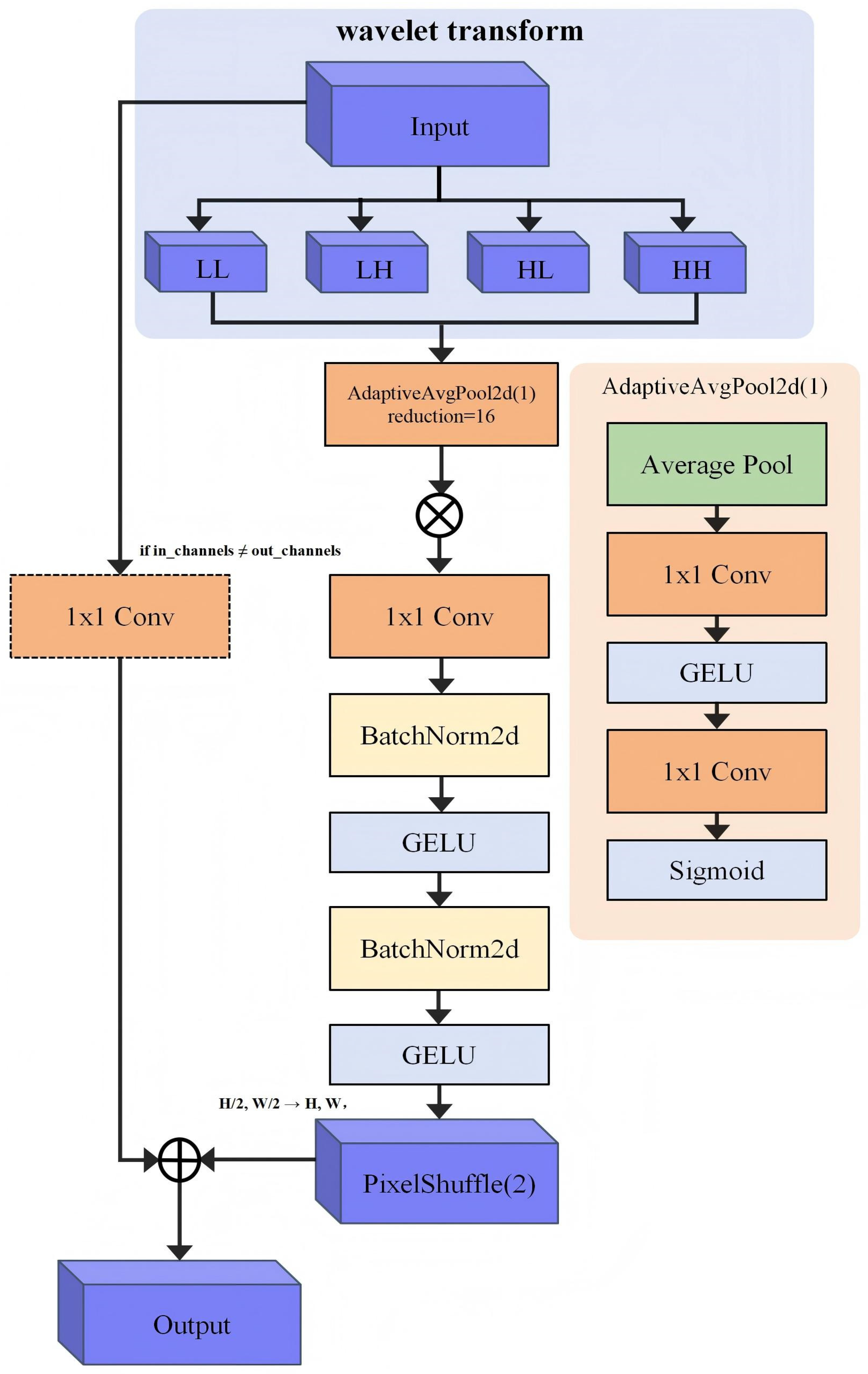
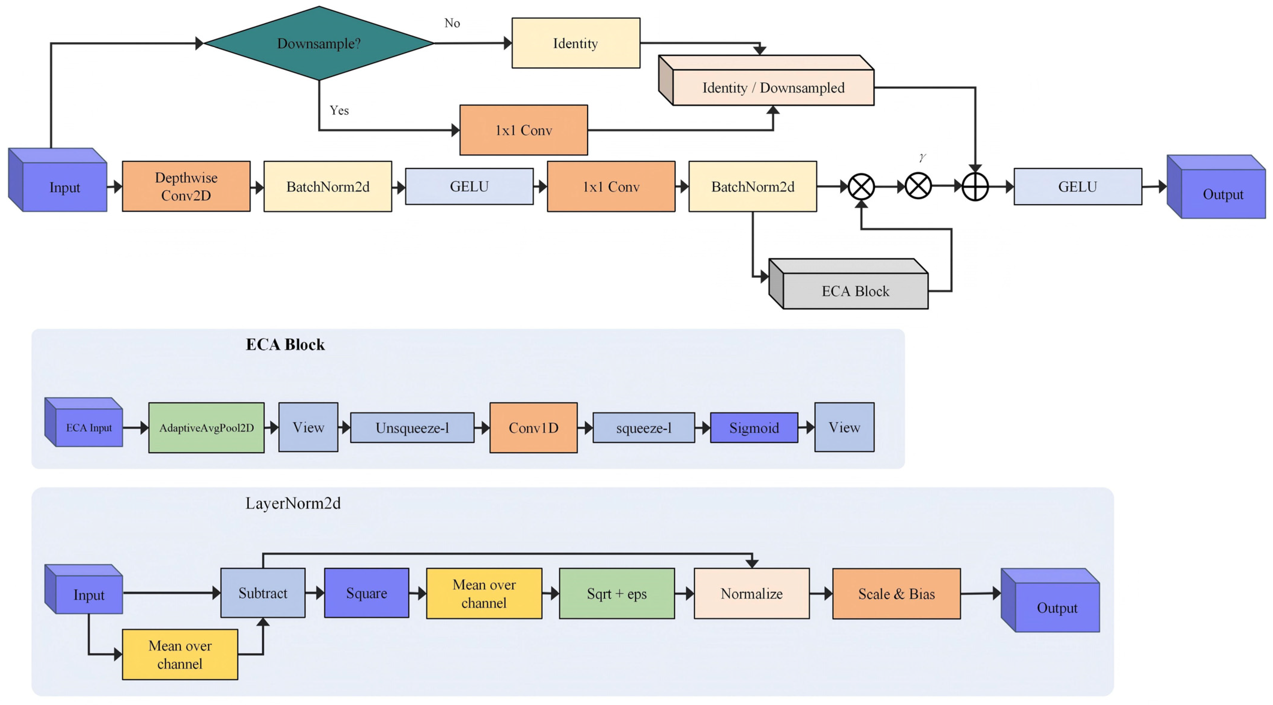
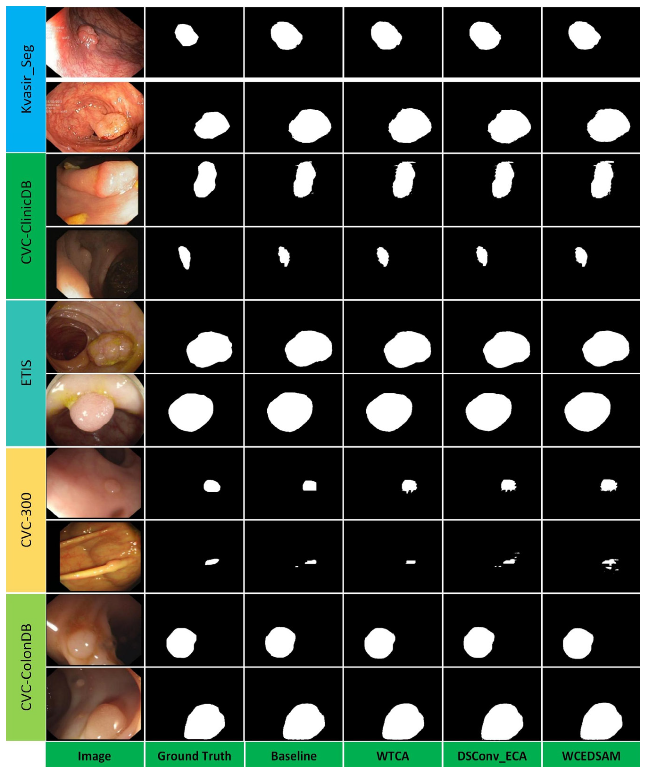
To demonstrate the effectiveness of the proposed module, a lightweight baseline model was first established. The baseline was based on MedSAM, which was substantially reduced using deep compression techniques. Specifically, the embedding dimension of the image encoder was decreased from 768 to 384, the number of transformer layers was reduced from 12 to 6, and the number of attention heads was adjusted from 12 to 6. To preserve effective feature representation despite the model reduction, the modified image encoder was initialized with pre-trained weights from ViT-S/16. This strategy facilitates the transfer and retention of visual priors learned through large-scale pre-training, while significantly reducing the model’s parameter count. These modifications produced a parameter-efficient version of MedSAM, serving as the foundation for subsequent ablation experiments.
Accordingly, a two-part ablation study was systematically conducted. The experimental design followed an incremental approach, first evaluating the individual effects of the two proposed core modules—the WTCA module and the DSConv_ECA module—before assessing their combined impact on the segmentation performance of the model. Experiments were conducted across five benchmark datasets: Kvasir-Seg, CVC-ClinicDB, CVC-ColonDB, CVC-300, and ETIS. Segmentation performance was quantitatively assessed using metrics including the mean Dice coefficient (mDice), mean Intersection over Union (mIoU), precision, and recall, alongside the evaluation of model parameter count.
4.5.1. Quantitative Analysis
Ablation experiments were conducted on the Kvasir-Seg dataset to evaluate the effectiveness of the proposed modules. The baseline model achieved an mDice of 0.9309, an mIoU of 0.9020, a precision of 0.9441, and a recall of 0.9249, with 15.22 million parameters and a computational cost of 9.370 G. The model operated at 2.65 FPS with an inference latency of 377.23 ms. Incorporation of the WTCA module improved the model’s recall to 0.9302 and precision to 0.9492, while mDice and mIoU increased to 0.9338 and 0.9041, respectively. The number of parameters remained at 15.22 million, and computational cost increased slightly to 9.384 G. The frame rate improved to 2.99 FPS, and inference time decreased to 334.13 ms. These results indicate that the WTCA module enhances the model’s ability to capture target regions, reduces false negatives, and improves inference efficiency. When the DSConv_ECA module was introduced independently, precision increased further to 0.9495, recall slightly rose to 0.9278, and mDice reached 0.9342. The number of parameters increased to 15.38 million, and computational cost rose to 9.447 G, with a frame rate of 2.92 FPS and an inference time of 342.39 ms. This demonstrates that DSConv_ECA improves the model’s accuracy in identifying lesion features by optimizing feature extraction and channel attention. When WTCA and DSConv_ECA were combined, the model achieved optimal performance, with mDice, mIoU, and recall reaching 0.9383, 0.9074, and 0.9345, respectively, while maintaining a high precision of 0.9484. The model comprised 15.38 million parameters and required 9.461 G of computational cost, operating at 3.33 FPS with an inference time of 300.50 ms. These results fully validate the synergistic effect of the two modules in enhancing segmentation overlap, reducing false negatives, and achieving an effective balance between accuracy and inference efficiency with minimal additional computational overhead (
Table 5).
Ablation experiments conducted on the CVC-ClinicDB dataset further clarified the individual contributions of each module. The baseline model achieved mDice and mIoU scores of 0.9238 and 0.8695, respectively, operating at 4.73 FPS with an inference latency of 211.60 ms. Upon incorporation of the WTCA module, recall increased from 0.8945 to 0.9000, and mIoU improved to 0.8740, while precision slightly decreased to 0.9591, and FPS marginally decreased to 4.70. Inference latency remained largely unchanged at 212.62 ms. These results indicate that the WTCA module expands the model’s detection range, effectively reducing false negatives, while introducing a small number of false positives and slightly increasing computational overhead. When the DSConv_ECA module was applied independently, recall increased substantially to 0.9092, and mIoU and mDice improved to 0.8819 and 0.9312, respectively. Despite a reduction in FPS to 3.43 and an increase in inference time to 291.92 ms, these findings demonstrate that DSConv_ECA significantly enhances the representation of boundary features and suppresses background interference, even with added computational cost. Ultimately, the WCEDSAM model, integrating both modules, achieved comprehensive improvements: mDice, mIoU, and recall reached 0.9376, 0.8943, and 0.9206, respectively, while precision recovered to 0.9614. The model operated at 3.29 FPS with an inference time of 303.98 ms. These results demonstrate that the complementary effects of the two modules not only maximize lesion detection but also restore segmentation accuracy via feature calibration, achieving optimal segmentation performance within an acceptable inference time (
Table 6).
Ablation experiments conducted on the CVC-ColonDB dataset further validated the effectiveness of each module. The baseline model attained mDice and mIoU scores of 0.9044 and 0.8607, respectively, with precision and recall of 0.9230 and 0.8964. It operated at 3.56 FPS with an inference latency of 280.90 ms. Following the incorporation of the WTCA module, recall improved significantly to 0.9110, with mDice and mIoU increasing to 0.9141 and 0.8766, respectively, and precision rising slightly to 0.9237. The model’s FPS increased to 4.04, and inference time decreased to 247.67 ms, indicating that WTCA effectively expands the detection range, reduces false negatives, and enhances inference efficiency. Independent application of the DSConv_ECA module increased precision to 0.9264, recall to 0.9065, and mDice and mIoU to 0.9119 and 0.8754, respectively. FPS increased to 4.29, and inference time decreased to 232.99 ms, indicating that DSConv_ECA optimizes feature extraction via depthwise separable convolutions and channel attention mechanisms, enhances lesion feature identification, and improves inference speed. When WTCA and DSConv_ECA were combined, the model achieved optimal performance, with mDice, mIoU, and recall improving to 0.9189, 0.8855, and 0.9132, respectively, and precision further increasing to 0.9304. FPS reached 4.52, and inference time decreased to 221.01 ms (
Table 6). These results fully validate the synergistic effects of the two modules in enhancing segmentation overlap, reducing false negatives, and simultaneously optimizing segmentation accuracy and inference speed.
Ablation experiments conducted on the CVC-300 dataset further validated the effectiveness of the proposed modules. The baseline model achieved a recall of 0.8017, with mDice and mIoU of 0.8721 and 0.7891, respectively, operating at 2.39 FPS and an inference latency of 418.62 ms. Upon incorporation of the WTCA module alone, recall increased substantially to 0.8502, mIoU improved to 0.8198, and mDice reached 0.8906, while precision decreased slightly from 0.9650 to 0.9454. FPS increased to 3.44 and inference time decreased to 299.47 ms, demonstrating that WTCA effectively expands the detection range and identifies potential lesion areas while enhancing inference speed. These results reaffirm the primary contribution of the WTCA module in expanding detection coverage and enhancing lesion identification, although a slight reduction in precision is observed. Introduction of the DSConv_ECA module independently increased recall to 0.8365, mIoU to 0.8127, and mDice to 0.8870, while precision slightly declined to 0.9507. The model operated at 2.41 FPS with an inference time of 415.60 ms, indicating that DSConv_ECA enhances the representation of complex features through depthwise separable convolutions and channel attention, albeit with a higher computational cost. When WTCA and DSConv_ECA were combined in WCEDSAM, the model achieved optimal performance: mDice, mIoU, and recall reached 0.8961, 0.8284, and 0.8704, respectively, while precision decreased to 0.9265. The model operated at 2.04 FPS with an inference time of 490.73 ms. In medical imaging segmentation, high recall and segmentation overlap are of greater clinical importance, demonstrating that the combined modules improve sensitivity and lesion coverage, even at the expense of increased computational overhead (
Table 7).
Ablation experiments conducted on the ETIS dataset further demonstrated that each module contributes substantially to performance enhancements. The baseline model achieved mDice and mIoU scores of 0.7584 and 0.7098, respectively, operating at 3.22 FPS with an inference latency of 310.60 ms. Incorporation of the WTCA module alone increased mDice to 0.7727, mIoU to 0.7279, recall from 0.7450 to 0.7583, and precision slightly to 0.8035. FPS decreased to 2.43 and inference time increased to 411.07 ms, indicating that WTCA expands detection coverage and maintains localization accuracy, albeit with additional computational cost. These results indicate that on more challenging datasets, WTCA effectively enhances the detection range while preserving precise localization, despite increased computational demands. Application of the DSConv_ECA module independently increased mDice to 0.7724, mIoU to 0.7288, recall to 0.7610, and precision to 0.8033. The model operated at 2.92 FPS with an inference time of 342.22 ms, confirming the module’s effectiveness in optimizing feature channel weights and enhancing key feature representation. These findings further corroborate the effectiveness of DSConv_ECA in enhancing feature representation and channel weighting. When the two modules were combined in the WCEDSAM model, mDice, mIoU, and recall improved to 0.7765, 0.7326, and 0.7667, respectively, with precision maintained at 0.7981. The model achieved 4.12 FPS with an inference time of 242.77 ms, demonstrating enhanced efficiency alongside accuracy. These results demonstrate that in complex, small-object scenarios, the synergy between WTCA and DSConv_ECA maximizes segmentation completeness and boundary alignment, achieving simultaneous improvements in accuracy and inference efficiency.
In summary, the ablation experiments clearly demonstrate the critical roles and complementary contributions of the WTCA and DSConv_ECA modules in polyp segmentation. The primary contribution of the WTCA module is its ability to expand the model’s receptive field over target regions via an attention mechanism, thereby significantly reducing false negatives while enhancing both recall and precision. The DSConv_ECA module enhances the model’s capacity to capture lesion boundaries and critical features through depthwise separable convolutions combined with an efficient channel attention mechanism, optimizing feature representation and further improving recall. The integrated WCEDSAM model achieved superior performance, with the highest mDice, mIoU, and recall values across four datasets of varying sizes and complexity. These results indicate that the global contextual awareness provided by WTCA and the fine-grained feature extraction of DSConv_ECA act synergistically, maximizing lesion detection completeness while maintaining precise boundary delineation. Collectively, these findings provide an efficient and robust solution for medical image segmentation tasks.