1. Introduction
As an essential trace element, zinc (Zn) acts as a crucial cofactor for over 300 enzymes, playing an indispensable role in sustaining growth performance and physiological functions in both terrestrial animals and teleosts [
1]. Zn deficiency adversely affects various species by retarding growth, impairing metabolic functions, and compromising both antioxidant capacity and immunity [
2,
3]. At the molecular level, Zn uptake is facilitated by specific transporters, which are vital for maintaining intracellular homeostasis and regulating metabolic processes [
4,
5]. The systemic and cellular Zn levels are meticulously regulated by Zn transporter (ZnT) families, which mediate Zn flux across biological membranes to preserve systemic balance [
6,
7,
8]. Consequently, determining precise dietary Zn requirements is fundamental to ensuring metabolic homeostasis and optimal growth across diverse species.
In teleost fish, maintaining Zn homeostasis is critical for physiological equilibrium and aquaculture success. Although fish can absorb trace amounts of waterborne Zn via their gills, this is rarely sufficient to meet the high metabolic demands of rapidly growing cultured species, making a precise dietary supply imperative [
9]. Disruption of this balance—either through deficiency or excessive accumulation—severely impairs fish health, leading to growth retardation, metabolic dysfunction, or heavy metal toxicity [
10]. Metabolically, adequate dietary Zn significantly enhances nutrient assimilation by upregulating the activities of key digestive enzymes, such as amylase, trypsin, and lipase, in the hepatopancreas and intestine [
11,
12]. Furthermore, intensive aquaculture often exposes fish to environmental stressors, triggering the overproduction of reactive oxygen species (ROS) and subsequent oxidative stress. In response, Zn serves a pivotal function in the antioxidant defense system [
13]. It protects against oxidative damage and tissue injury by activating the Nrf2 signaling pathway, which upregulates downstream target molecules and antioxidant enzymes, including catalase, heme oxygenase-1 (HO-1), and glutathione [
11,
12], a protective mechanism widely conserved across diverse species [
14,
15,
16,
17].
Beyond its metabolic and antioxidant functions, Zn is also fundamental to maintaining intestinal health, immunomodulation, and the suppression of inflammation. Intestinal health is intrinsically dependent on structural integrity. Extensive research demonstrates that optimal dietary Zn fortifies the intestinal barrier by upregulating tight junction proteins, such as occludin and claudins [
18,
19]. Concurrently, Zn enhances host intestinal immune defenses by stimulating the activity of immunomodulatory factors, including lysozyme (LZM) and complement components 3 and 4 (C3, C4) [
20]. Furthermore, Zn mitigates intestinal inflammation by optimizing the cytokine profile. Specifically, it suppresses the expression of pro-inflammatory mediators, such as tumor necrosis factor-α (TNF-α) and interleukin-1β (IL-1β), while promoting the secretion of anti-inflammatory cytokines, such as interleukin-10 (IL-10) and transforming growth factor-β1 (TGF-β1) [
20,
21,
22]. Mechanistically, this immunomodulation is largely achieved through the regulation of inflammatory signaling cascades. Pro-inflammatory stimuli frequently activate the p38 MAPK signaling pathway, which governs vital cellular processes including differentiation and apoptosis [
11,
12]. Numerous studies have evidenced that sufficient dietary Zn inhibits the overactivation of the p38 MAPK pathway, thereby curbing the excessive production of inflammatory factors and alleviating inflammation-induced tissue damage in the intestine [
22,
23].
Black carp (
Mylopharyngodon piceus) is a commercially vital freshwater carnivorous teleost widely cultured in China, playing a crucial role in the aquaculture industry. Although recent studies have explored its nutritional requirements for amino acids [
24], vitamins [
20,
25], and certain minerals [
26], the precise dietary Zn requirement for this species remains completely undefined. Given its unique molluscivorous feeding habits and the intensive nature of its culture, extrapolating Zn requirements from other cyprinid species may lead to inaccurate feed formulations, potentially resulting in suboptimal growth, impaired Zn homeostasis, or compromised immunity. Therefore, establishing the optimal dietary Zn inclusion level specifically for
M. piceus is highly necessary. The specific objectives of this study were to determine the precise dietary Zn requirement for juvenile black carp based on growth performance and systematically evaluate the impacts of graded dietary Zn levels on serum biochemistry, Zn transporter gene expression, hepato-intestinal antioxidant status, and intestinal immune and inflammatory responses. Ultimately, these findings will provide a crucial scientific basis for optimizing Zn supplementation in practical aquafeeds, thereby enhancing the health, growth, and economic sustainability of black carp aquaculture.
2. Materials and Methods
2.1. Animals, Diets and Treatments
Six isonitrogenous and isoenergetic experimental diets were formulated to evaluate the dietary Zn requirements of juvenile black carp (
M.
piceus) (
Table 1). Casein and gelatin were used as the primary protein sources, fish oil as the lipid source, and dextrin as the carbohydrate source. To strictly control the basal Zn level, a vitamin premix and a Zn-free mineral premix were added to all diets. Zinc sulfate heptahydrate (ZnSO
4·7H
2O) was supplemented into the basal diet at graded levels of 0, 10, 20, 40, 80, and 160 mg/kg (designated as Zn0, Zn10, Zn20, Zn40, Zn80, and Zn160). Although no exogenous zinc was supplemented in the mineral premix, a basal level of zinc was still present in the control diet, which was attributed to the endogenous Zn contained in the dietary protein sources.
According to the Inductively Coupled Plasma Mass Spectrometry (ICP-MS) analysis, the actual dietary Zn concentrations were 27.95, 34.38, 44.90, 66.52, 116.14, and 199.56 mg/kg, respectively. The basal Zn content (27.95 mg/kg) in the unsupplemented control group (Zn0) originated purely from the inherent background Zn present in the raw dietary ingredients. All dry ingredients were finely ground, sieved, thoroughly mixed with oil and water, and extruded into 2.0 mm pellets. The pellets were air-dried and stored at −20 °C until further use. The production and storage protocols strictly followed the guidelines described by Jia et al. [
25].
2.2. Fish and Feeding Trial
All experimental procedures received prior approval from the Ethics Committee of Huzhou Normal University (Protocol No. 068/2023) and adhered to standard protocols for animal welfare. The experimental procedures in this study were adapted from those previously described by Wu et al. [
20] and Jia et al. [
25]. Healthy black carp (
M. piceus) were bought from Xingwang Fish Breeding Farm (Huzhou, China).
Before the experiment, all fish were acclimated in 500 L tanks for two weeks and fed the control diet (Zn0). Subsequently, 540 fish of uniform size (initial body weight: 2.88 ± 0.12 g) were randomly distributed into 18 tanks (30 fish per tank), representing six dietary treatments with three biological replicates (tanks) per treatment. The feeding trial lasted for 60 days. Fish were hand-fed to apparent satiation (approximately 4% of body weight) three times daily (08:00, 12:00, and 17:00). To accurately calculate the feed conversion ratio (FCR), uneaten feed residues were siphoned out one hour after each feeding, oven-dried, and weighed. Feces were removed daily. Strict water quality management was maintained throughout the trial with a daily water exchange rate of approximately 25%. The water quality parameters were monitored daily and maintained within the following ranges: water temperature at 26.0 ± 1.0 °C, dissolved oxygen (DO) > 6.0 mg/L, pH at 7.6 ± 0.1, and total ammonia nitrogen < 0.1 mg/L. The experiment was conducted in an indoor flow-through system with a natural photoperiod.
2.3. Sample Collection and Compositional Analysis
At the end of the feeding trial, all fish were fasted for 24 h to empty their digestive tracts. Following anesthesia with tricaine methanesulfonate [
27], all fish from each tank were counted and batch-weighed to evaluate growth performance. Subsequently, 10 fish were randomly sampled from each tank and pooled together to form one composite sample per tank, representing one biological replicate (
n = 3 biological replicates per dietary treatment). Serum, liver, and intestine samples were immediately harvested on ice, flash-frozen in liquid nitrogen, and stored at −80 °C. The proximate compositions of the experimental diets and whole fish bodies (moisture, crude protein, crude lipid, and ash) were analyzed following standard AOAC methods as described in our previous studies [
20,
25].
2.4. Biochemical and Enzymatic Assays
Serum samples (n = 3 biological replicates per group, each pooled from 10 fish) were analyzed for the levels of albumin (ALB), total bile acid (TBA), glucose (GLU), and the activities of aspartate aminotransferase (AST), alkaline phosphatase (ALP), lactate dehydrogenase (LDH), and alanine aminotransferase (ALT). These indices were measured using an automated clinical chemistry analyzer (Model C400n, Dirui, Changchun, China).
For digestive enzyme analysis, frozen liver and intestinal tissues were pulverized in liquid nitrogen and homogenized in a 9-fold volume of ice-cold phosphate-buffered saline (PBS, pH 7.4). The homogenates were centrifuged at 3000× g for 20 min at 4 °C, and the supernatants were collected. The activities of lipase (LPS), amylase (AMS), trypsin (TRY), and chymotrypsin (CYT) were determined using commercial assay kits (Nanjing Jiancheng Bioengineering Institute, Nanjing, China) with three technical replicates per sample.
2.5. Analysis of Antioxidative, Oxidative, Immune, and Inflammatory Markers
The hepatic and intestinal antioxidant statuses were evaluated by determining the activities of superoxide dismutase (SOD), catalase (CAT), glutathione reductase (GR), glutathione S-transferase (GST), glutathione peroxidase (GPX), as well as the levels of glutathione (GSH), total antioxidant capacity (T-AOC), and malondialdehyde (MDA). Intestinal immune parameters, including lysozyme (LZM), immunoglobulin M (IgM), acid phosphatase (ACP), and alkaline phosphatase (ALP), were also assessed. All biochemical analyses were conducted using commercial kits (Nanjing Jiancheng, Bioengineering Institute, Nanjing, China) strictly according to the manufacturer’s instructions.
Furthermore, the intestinal concentrations of complement components (C3, C4) and inflammatory cytokines (TGF-β1, TNF-α, IL-1β, IL-6, IL-8, IL-12, IL-17, and IFN-γ) were quantified using specific Fish ELISA kits (Hengyuan Biotech, Shanghai, China). For all biochemical and ELISA analyses, samples from n = 3 tanks per treatment were utilized to ensure true biological replication.
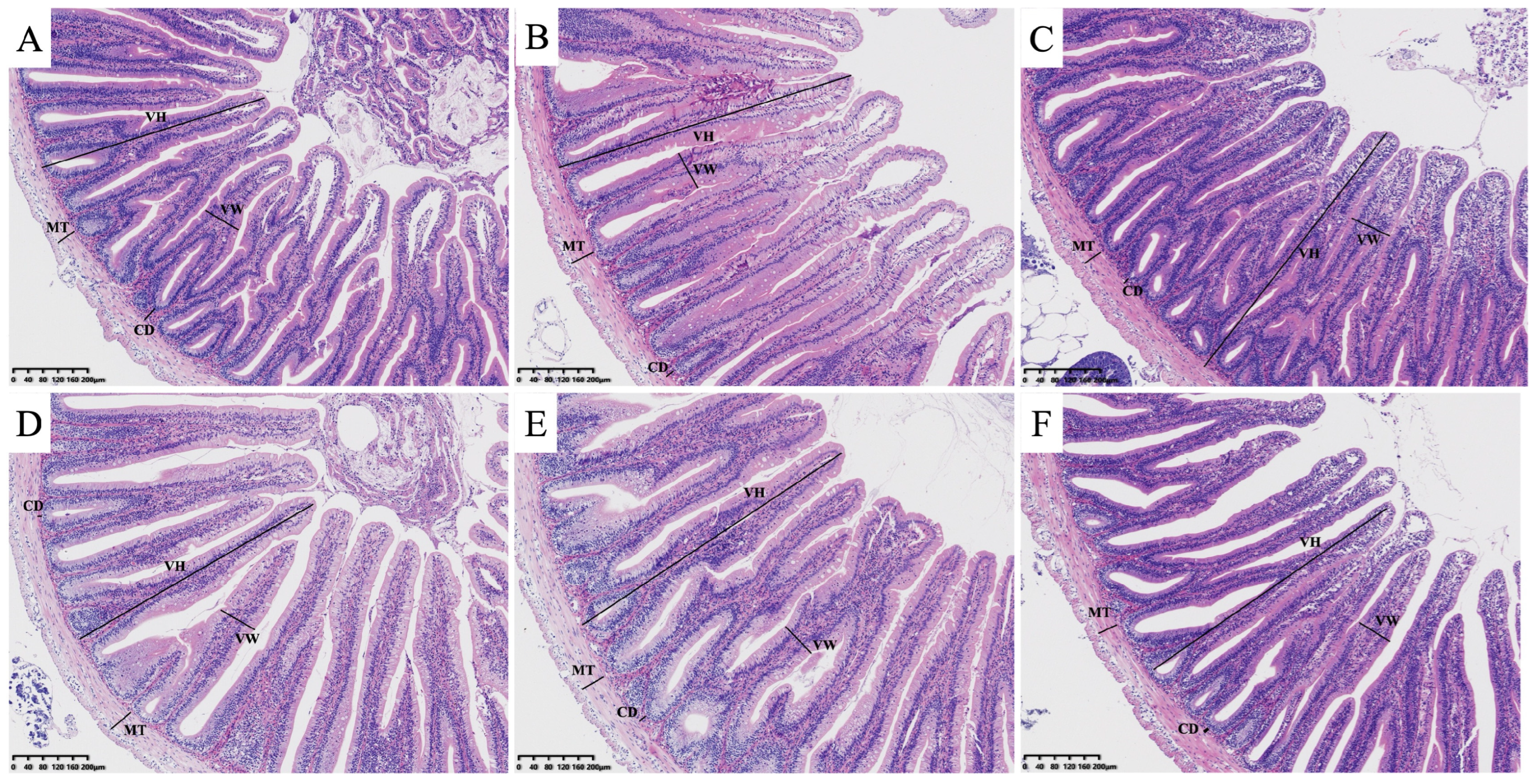
For histological analysis, fixed intestine samples (4% paraformaldehyde) were dehydrated, embedded in paraffin, and sectioned at 5 μm thickness. Sections were stained with hematoxylin and eosin (H&E) and examined under a light microscope (Olympus BX53, Tokyo, Japan) to evaluate morphological integrity. Specifically, morphometric parameters including villus height, villus width, muscular thickness, and crypt depth were quantified using cellSens Standard software version 1.18 (Olympus, Tokyo, Japan). For each section, at least 5 intact and well-oriented villi were randomly selected to measure and calculate the average values.
2.6. Analysis of Gene Expression
Total RNA from hepatic and intestinal tissues (
n = 3 biological replicates per group) was extracted using the TRIzol reagent (Invitrogen, Carlsbad, CA, USA). First-strand cDNA was synthesized using the MonScript RT-PCR Kit (Monad Biotech, Wuhan, China). Specific primer sequences (
Table 2) were synthesized by Biosune Biotech (Shanghai, China). Quantitative real-time PCR (qPCR) was performed in triplicate (technical replicates) on a CFX96 Real-Time PCR System (Bio-Rad, Hercules, CA, USA) using SYBR Green Master Mix (Takara, Dalian, China). Thermal cycling conditions followed Wu et al. [
20], and relative mRNA expression was calculated via the 2
−ΔΔCt method normalized to β-actin.
2.7. Data Analysis
All data are presented as means ± standard deviations (SDs). Statistical analyses were performed using IBM SPSS software version 25.0 (Chicago, IL, USA). The normality and homogeneity of variance were verified prior to analysis. Differences among dietary treatments were evaluated using one-way ANOVA, followed by Duncan’s multiple range test. Orthogonal polynomial contrasts were utilized to determine linear and quadratic dose–response trends. A difference was considered statistically significant at p < 0.05. The optimal dietary Zn requirement was estimated using a broken-line regression model based on weight gain and specific growth rate.
4. Discussion
Zinc functions as an indispensable micronutrient that underpins the somatic growth of diverse aquatic species [
9,
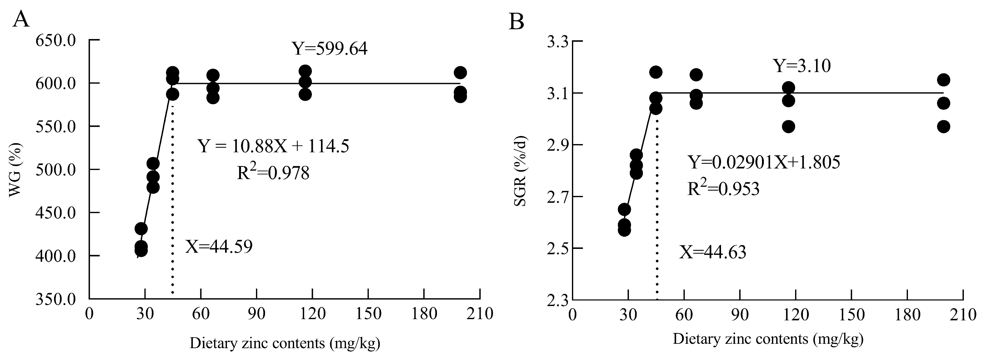
28]. In the current study, broken-line regression analysis of both WG and SGR revealed the optimal dietary Zn requirement for juvenile black carp (
M. piceus) ranges from 44.59 to 44.63 mg/kg. Systematic comparison across fish species reveals interesting patterns in Zn requirements reflecting differences in phylogeny, feeding ecology, and metabolism [
29]. The Zn requirement of black carp (
M. piceus) was higher than that of yellow catfish (
Pelteobagrus fulvidraco) [
30] and rainbow trout (
Oncorhynchus mykiss) [
31], similar to grass carp (
Ctenopharyngodon idella) [
32] and Jian carp (
Cyprinus carpio var. Jian) [
33], but lower than that of largemouth bass (
Micropterus salmoides) [
34]. These variations might be attributed to multiple factors, including basal dietary Zn content, Zn source bioavailability, growth stage, metabolic rate, and species-specific digestive physiology [
29,
35,
36]. The relatively high Zn requirement in black carp might be related to its specialized molluscivorous feeding habit, which demands enhanced synthesis of digestive enzymes and intestinal absorptive capacity to process hard-shelled prey.
Mechanistically, the growth-promoting properties of Zn are multifaceted, intricately linked to the optimized assimilation of nutrients and energy metabolism. Although no marked effects were presented on the contents of crude protein and ash in the whole fish body, fish body lipid content was significantly decreased at optimal and higher contents of dietary Zn, consistent with findings in yellow catfish (
P. fulvidraco Richardson) [
30]. The improved FCR suggests that adequate Zn levels in feed can enhance feed utilization efficiency and nutrient assimilation [
30]. The liver and intestine are crucial organs for digestive enzyme secretion and nutrient absorption [
37,
38]. Zn plays key roles in the synthesis and activity regulation of digestive enzymes, which are closely related to digestive, metabolic, absorptive and growth processes in all fish species [
39]. Previous research has found Zn-deficiency feeding could significantly reduce activities of TRY, CYT and AMS in pig [
40], yellow catfish (
P. fulvidraco Richardson) [
30] and Siberian sturgeon (
Acipenser baerii) [
39], which is consistent with the present experimental results. Together with the improved FCR and overall growth, these findings suggest that adequate Zn inclusion significantly enhances digestive enzyme activities, thereby driving the superior growth performance of juvenile black carp.
Cellular Zn homeostasis is precisely maintained through coordinated regulation of Zn transporter (ZnT/
Slc30a) families, which control Zn movement across cellular membranes in response to different levels of dietary Zn [
41,
42]. In teleosts, ZnT-regulating responses have been documented in zebrafish (
Danio rerio) embryos and grass carp [
43,
44,
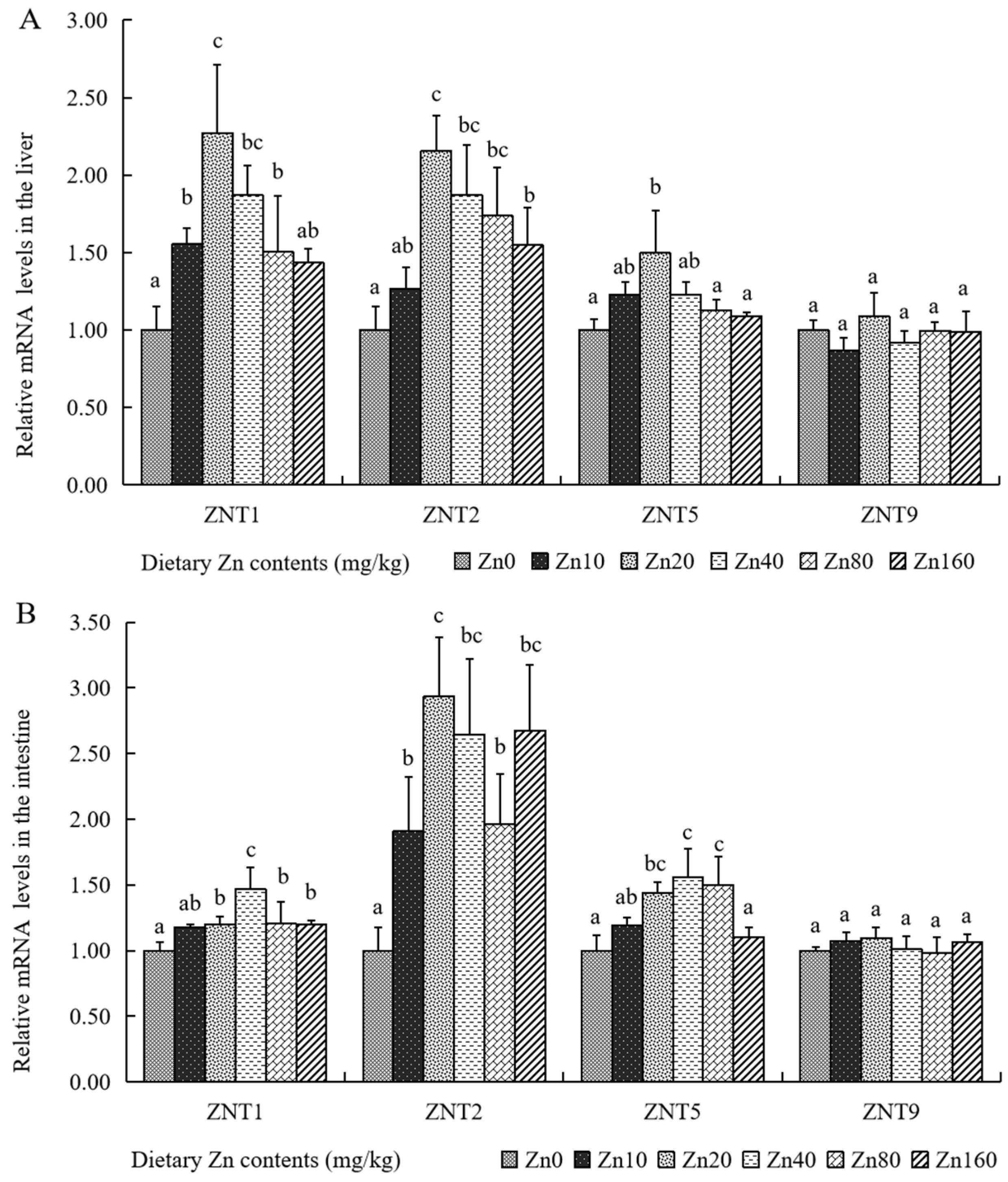
45]. In the present study, optimal dietary Zn supplementation significantly upregulated the transcription of key zinc transporter genes (
ZNT1,
ZNT2, and
ZNT5) in both the hepatic and intestinal tissues. This indicates that optimal dietary Zn supplementation is associated with the modulation of
ZnT expression profiles to maintain integrated Zn homeostasis in black carp [
37]. Conversely, relative lower expression levels of
ZNT1,
ZNT2, and
ZNT5 were observed in the Zn160 group, aligning with observations in yellow catfish [
37]. This transcriptional downregulation under high-Zn conditions may serve as a protective mechanism to limit further Zn influx and avoid heavy metal overload in fish [
46,
47].
Serum biochemical indicators have been widely used to evaluate the metabolic homeostasis and nutritional status of fish [
48]. Numerous studies have shown that relatively higher serum ALT, ALP, and AST activities can reflect the functional status of metabolic organs, particularly the liver [
26,
49,
50]. Hepatic injury compromises hepatocyte membrane integrity, leading to the leakage of intracellular enzymes and a subsequent elevation in serum ALT and AST activities [
51]. In this study, activities of ALT and AST declined in the serum of Zn-supplemented groups, which agrees with earlier findings in soft-shelled turtle (
Pelodiscus sinensis) [
52], indicating that optimal dietary Zn could improve the health status of hepatic cells in black carp (
M. piceus) [
53]. Functionally, HDL-C mediates reverse cholesterol transport from peripheral tissues to hepatic cells for subsequent catabolism, while LDL-C delivers hepatic cholesterol to other tissues for cellular utilization [
54]. As we know, cholesterol can be coupled with bile acids to generate related mixtures and play important roles in lipid digestion and absorption [
55]. Similar to results in soft-shelled turtle (
P. sinensis) [
52], higher HDL-C levels coupled with lower LDL-C levels were observed in these Zn-supplemented groups, indicating that adequate dietary Zn might be beneficial for serum lipid transport into hepatic cells in black carp (
M. piceus). As the primary lipid molecules synthesized in the liver, TG and TC play indispensable roles in driving lipid metabolism and maintaining systemic metabolic homeostasis [
56]. However, there was no significant difference in TG and TC levels in the Zn-added groups, which differs from results in soft-shelled turtle (
P. sinensis) [
52] and largemouth bass (
M. salmoides) [
57]. These divergences may be mediated by differences in fish species. Additionally, ALP is a Zn-dependent enzyme, and its significantly increased activity in the Zn-added groups indirectly reflects improved Zn metabolism and utilization [
58], consistent with findings in golden pompano (
Trachinotus ovatus) [
59]. Furthermore, serum GLU levels were notably decreased in the Zn-added groups, consistent with previous findings in
Pangasius hypophthalmus [
60] and largemouth bass (
M. salmoides) [
57]. This hypoglycemic effect likely stems from Zn-mediated regulation of glucose metabolism, though further metabolic studies are required to fully elucidate this mechanism in fish.
It is widely recognized that reactive oxygen species (ROS) are continuously generated during the aerobic metabolism of nutrients, including Zn, which significantly influences redox homeostasis in animals [
61,
62]. Acting as crucial components of the antioxidant defense system, key enzymes such as SOD, CAT, and GPX catalyze the conversion of toxic oxygen radicals into harmless H
2O and O
2, thereby mitigating oxidative stress in fish [
55,
63]. In juvenile black carp (
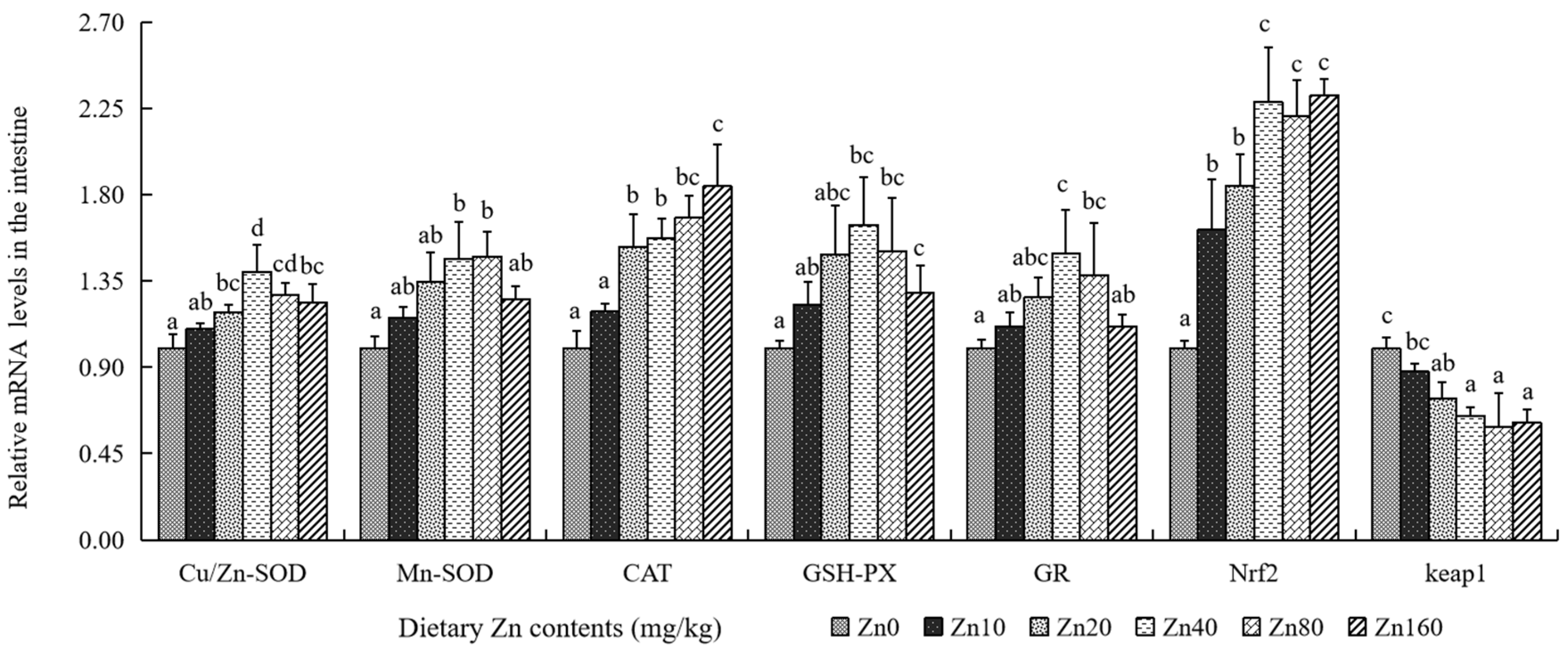
M. piceus), the activities of T-SOD, GPX, and CAT were notably upregulated in both hepatic and intestinal tissues following dietary Zn supplementation. Similar Zn-induced enhancements in these antioxidant enzymes have been widely reported in other teleosts, including rainbow trout (
Oncorhynchus mykiss) [
64], Jian carp (
Cyprinus carpio var. Jian) [
65], half-smooth tongue sole (
Cynoglossus semilaevis) [
66], and golden pompano (
T. ovatus) [
59]. These consistent results strongly suggest that adequate dietary Zn plays a vital role in activating and bolstering the primary enzymatic antioxidant defense system in fish [
64]. Beyond primary enzymes, the glutathione system is essential for sustaining cellular redox balance. GR facilitates the reduction of glutathione disulfide (GSSG) into GSH [
67], while GST drives the generation of glutathione S-conjugates for detoxification [
68]. Elevated GST typically serves as an adaptive physiological response to oxidative damage or inflammatory stimuli [
69]. In our study, Zn supplementation significantly increased GSH content and T-AOC in both the liver and intestine. The observed decrease in GST alongside the increase in GR activity suggests that Zn promotes the regeneration of reduced GSH, thereby expanding the cellular antioxidant pool to maintain redox homeostasis [
65]. Under oxidative stress, excessive ROS damages cell membranes, leading to lipid peroxidation, which is reliably quantified by MDA content [
70]. As a vital antioxidant, GSH scavenges oxygen radicals and organic peroxides, protecting tissues and preventing harmful MDA accumulation [
71]. Previous studies confirm that dietary Zn levels significantly affect GSH synthesis and effectively reduce MDA concentrations [
72,
73]. Consistently, our results showed that Zn supplementation significantly reduced hepatic and intestinal MDA levels, aligning with findings in Jian carp (
C. carpio var. Jian) [
65] and golden pompano (
T. ovatus) [
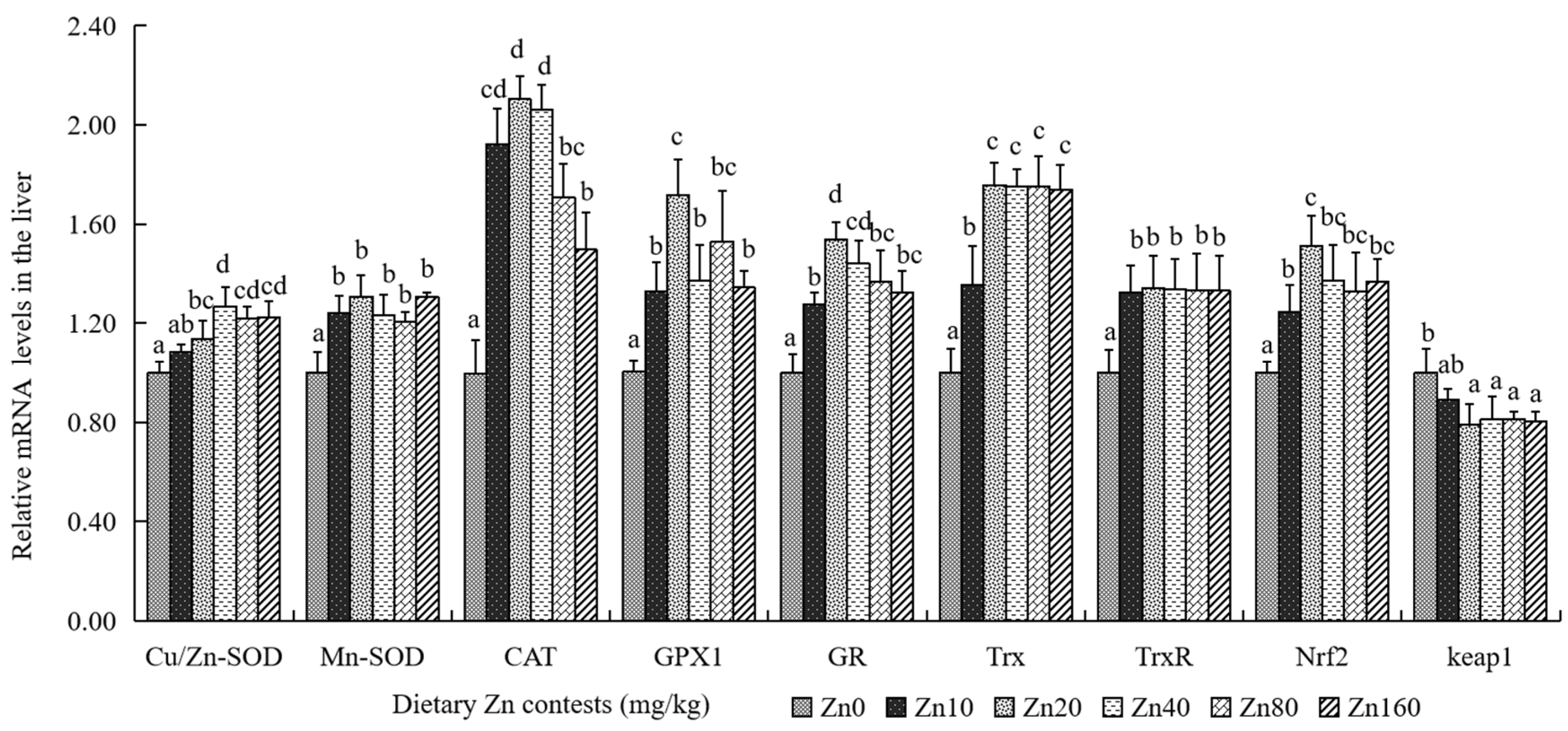
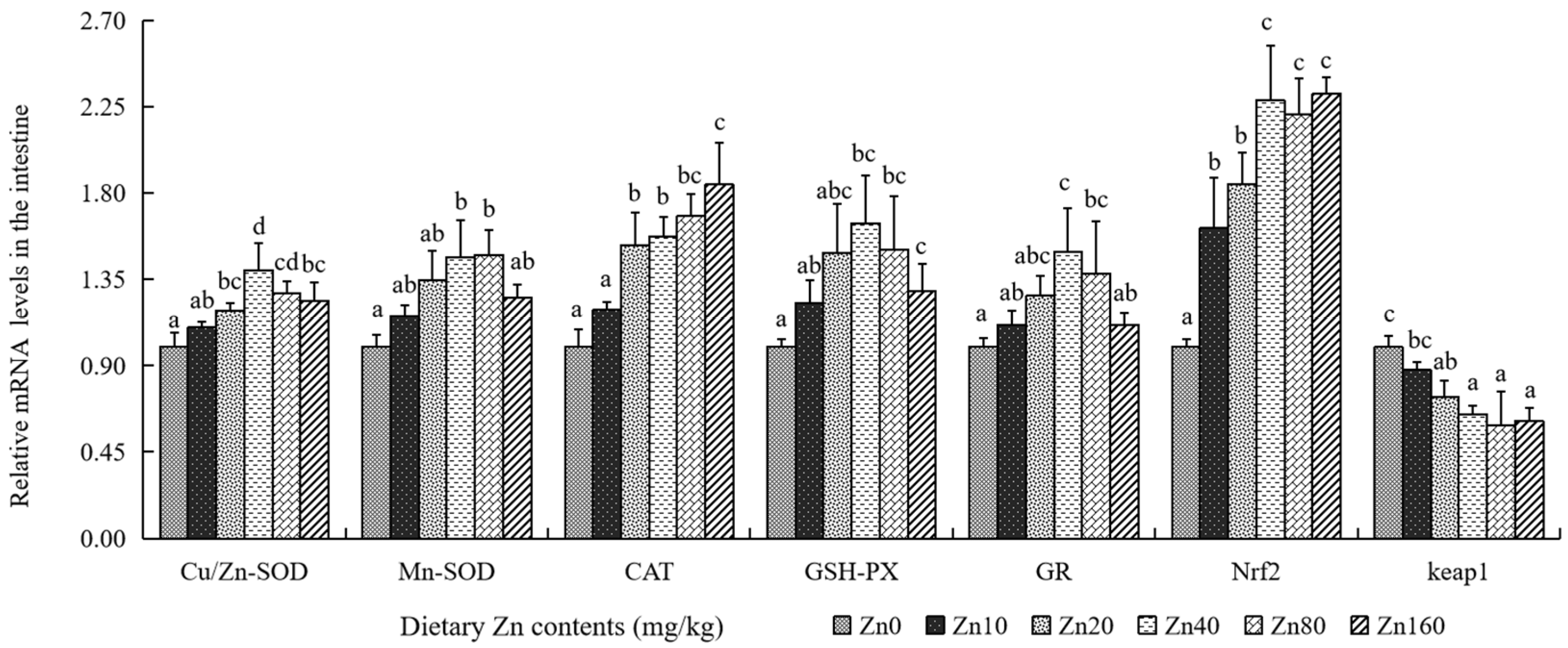
59]. At the molecular level, these biochemical adaptations are governed by the
Nrf2/Keap1 axis, a master regulator of cellular antioxidant responses [
74,
75]. Dietary Zn supplementation not only significantly enhanced
Nrf2 transcription but also downregulated
Keap1 mRNA expression, corroborating previous results in yellow catfish (
Pelteobagrus fulvidraco) [
73]. Taken together, our findings demonstrate that adequate dietary Zn enhances antioxidant capacity and mitigates lipid peroxidation in black carp by stimulating antioxidant enzyme activities and increasing cellular GSH content, a process likely mediated through the activation of the Nrf2/Keap1 signaling pathway.
The integrity of the intestinal physical barrier is primarily governed by the tight junction (TJ) complex, including zonula occludens (ZOs) and claudins (CLDNs), alongside the chemical barrier formed by mucins [
25,
76]. These proteins act as essential biomarkers for evaluating the functional integrity of the intestinal barrier [
77,
78], and are indispensable for regulating epithelial permeability and supporting external nutrient absorption [
79]. Specifically, as a peripheral membrane protein, ZO-1 is fundamental in constructing and stabilizing tight junctions between epithelial cells [
80]. Concurrently, CLDNs function as key transmembrane components that sustain cell-to-cell barriers, manage intercellular communication, and preserve cellular polarity [
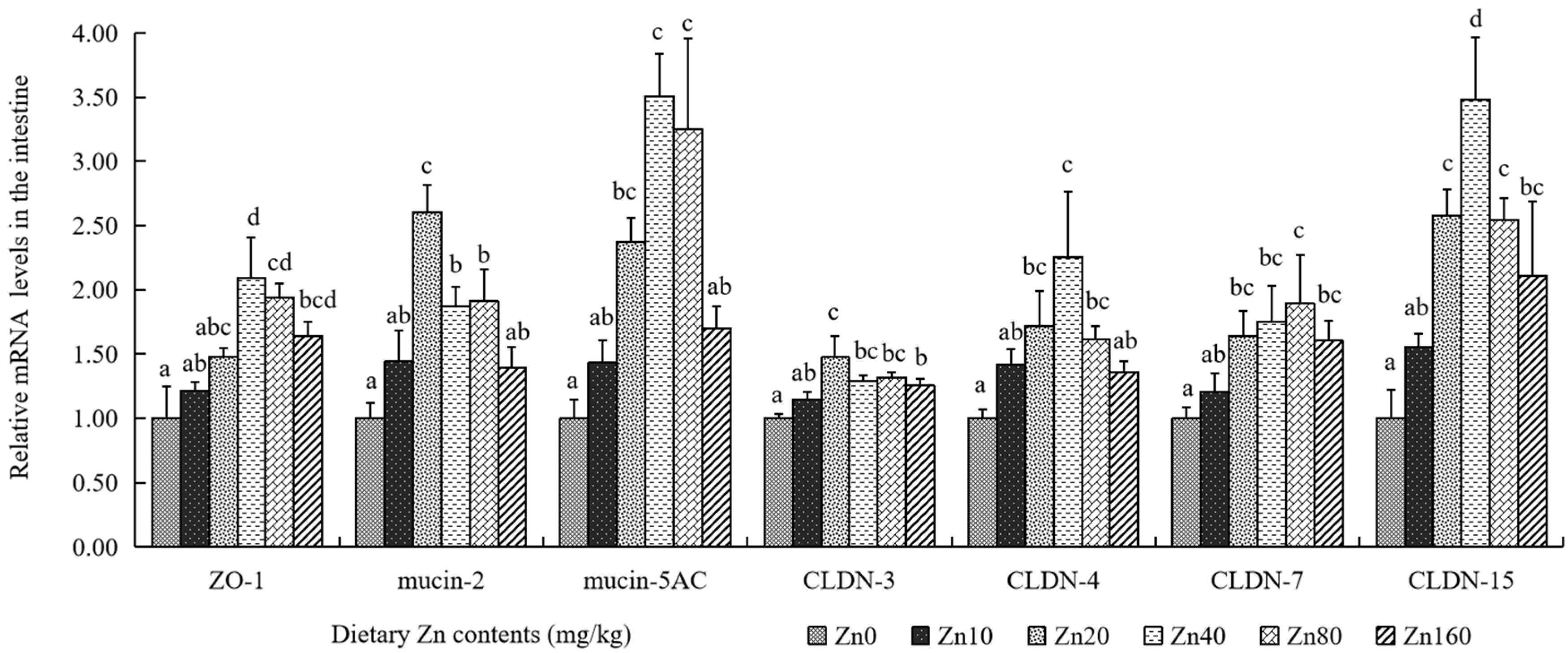
81]. In our present study, optimal dietary Zn supplementation, particularly in the Zn40 group, markedly upregulated the mRNA expressions of
ZO-1, as well as multiple claudin members (
CLDN-3,
CLDN-4,
CLDN-7, and
CLDN-15). Furthermore, the transcriptions of
mucin-2 and
mucin-5AC, which act as crucial components of the intestinal chemical barrier, exhibited a synchronized upregulation under adequate Zn intake. This observation is highly consistent with prior investigations in Nile tilapia (
Oreochromis niloticus) [
82], rainbow trout (
O. mykiss) [
83], and grass carp (
C. idella) [
45]. These transcriptional profiles indicate that an adequate dietary intake of Zn is associated with improved intestinal barrier function by simultaneously reinforcing the physical barrier (
ZO-1 and
CLDNs) and supporting chemical mucus secretion.
A growing body of evidence indicates that augmenting the levels of key immune effectors fundamentally bolsters host immunity, including complement components (C3, C4, and CFB) and humoral immune-related genes (
LZM,
NRAMP,
IgH, and
Igλ1) [
57,
77,
84] In the present study, Zn-supplemented diets effectively counteracted potential damage by enhancing intestinal immunity, evidenced by increased C3, C4, IgM, and LZM activities. This immunomodulatory effect of adequate Zn aligns strongly with similar findings reported in largemouth bass (
M. salmoides) [
57]. Furthermore, appropriate dietary Zn content significantly increased intestinal ACP and ALP activities, aligning with findings in rats [
85] and various fish species, such as largemouth bass (
M. salmoides) juveniles supplemented with dietary soybean lecithin [
77] and black carp (
M. piceus) fed selenium yeast [
26]. It is extensively indicated in the literature that Zn deprivation triggers metabolic stress and physiological disturbances [
86], ultimately compromising the survival, proliferation, and maturation of immune cells [
87]. Central to regulating these immune responses and inflammatory processes is the strict homeostatic balance of inflammatory cytokines [
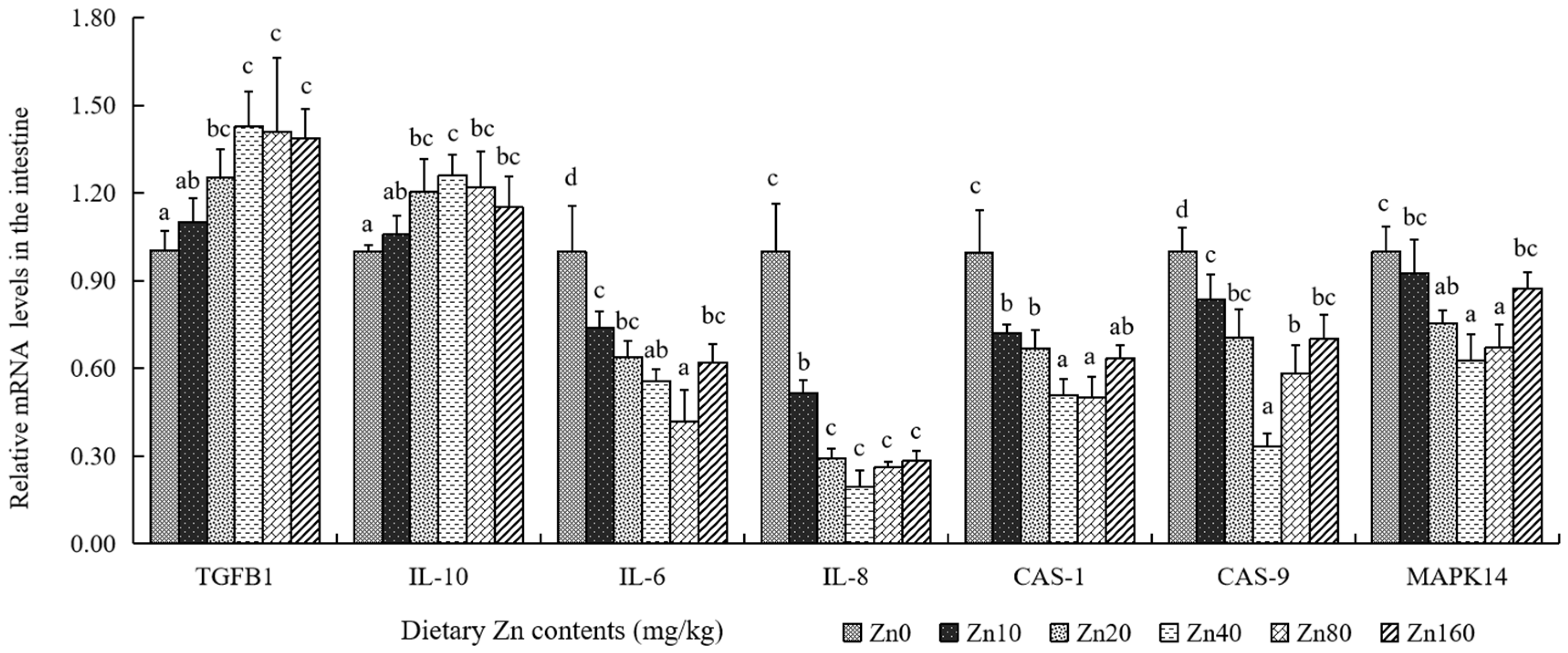
86]. In our results,
IL-10 and
TGF-β1, two typical anti-inflammatory cytokines, showed increasing then stabilizing trends with dietary Zn increases, similar to results in largemouth bass (
M. salmoides) treated with dietary Zn [
74]. Conversely, the expression levels of pro-inflammatory cytokines (
IL-1β,
IL-6,
IFN-γ, and
TNF-α) exhibited a pattern of initial suppression followed by a plateau, corroborating previous observations in largemouth bass (
M. salmoides) [
74], indicating adequate dietary Zn could suppress the contents of these pro-inflammatory cytokines at both transcriptional and protein levels in black carp (
M. piceus).
Crucially, inflammatory and apoptotic responses are meticulously regulated by distinct signaling cascades. In our study, adequate dietary Zn downregulated the mRNA expression of
CAS-1 and
CAS-9. It is well established that Caspase-1 plays a vital role in the inflammasome complex, facilitating the maturation and secretion of IL-1β, whereas Caspase-9 functions primarily as an initiator caspase in the mitochondrial apoptosis pathway [
74]. The concomitant reduction in
CAS-1 and
IL-1β transcription suggests that adequate Zn may help alleviate intestinal inflammation, while the downregulation of
CAS-9 points to a potential attenuation of cellular apoptosis. Mechanistically, the production of pro-inflammatory cytokines is largely governed by the p38 MAPK signaling cascade. Our data showed that adequate Zn supplementation downregulated
MAPK14 mRNA expression, similar to findings in largemouth bass [
74]. Although further investigation at the protein phosphorylation level is required to confirm functional pathway activity, the current transcriptional evidence suggests that adequate dietary Zn may be involved in mitigating intestinal inflammation, potentially by modulating the p38 MAPK cascade and Caspase-1 signaling in black carp.



 ,
,