Article

Occurrence and Antimicrobial Resistance Profiles of Escherichia coli Isolated from Commercial Poultry Farm in West Kazakhstan


1
Scientific Research and Production Laboratory of Veterinary and Biological Safety (BSL-2) Laboratory, Faculty Veterianry and Agrotechnology, Non-Profit Joint-Stock Company “Zhangir Khan West Kazakhstan Agrarian-Technical University”, Zhangir Khan Street 51, Uralsk 090009, Kazakhstan
2
The Microbiome and Mucosal Defence Research Unit, Institut de Recherche Clinique de Montréal, Montreal, QC H2W 1R7, Canada
3
Research Institute for Biological Safety Problems LLP, «QazBioPharm» National Holding, 15 B. Momyshuly Street, Gvardeiskiy Village, Kordai District, Zhambyl Region 080409, Kazakhstan
*
Authors to whom correspondence should be addressed.
Biology 2026, 15(11), 877; https://doi.org/10.3390/biology15110877
Submission received: 21 April 2026 / Revised: 22 May 2026 / Accepted: 27 May 2026 / Published: 2 June 2026
(This article belongs to the Section Infection Biology)

Simple Summary

Antimicrobial resistance (AMR) is one of the most serious global health threats today. The overuse of antibiotics in poultry farming can lead to the emergence of bacteria that no longer respond to treatment. This study investigated the presence of antibiotic-resistant Escherichia coli on a commercial chicken farm (egg production) in the West Kazakhstan region. A total of 100 samples were collected from chickens (cloacal swabs, fresh feces, and internal organs), the farm environment, and wild pigeons. The samples were analyzed using cultural and genetic methods to detect resistance genes. Of the 40 E. coli isolates confirmed, ten (25%) carried at least one resistance gene, and all ten carried resistance genes associated with resistance to multiple antimicrobial classes. The most common resistance genes were blaTEM (80%), aadA17 (90%), and int1-a-marko (80%). Genes providing resistance to disinfectants (qacEΔ1) were also found in 80% of the resistant isolates. Importantly, similar resistance genes were detected in E. coli from pigeons, suggesting that wild birds may act as carriers and contribute to the spread of resistant bacteria between farms and the environment. These findings highlight the need for responsible antibiotic use and improved biosecurity measures on poultry farms to protect both animal and human health.

Abstract

The widespread use of antimicrobials in poultry production exerts selective pressure for the emergence of resistant bacteria, yet molecular data on resistance determinants in Escherichia coli from commercial farms in Central Asia remain scarce. This study aimed to characterize antimicrobial resistance genes and mobile genetic elements in E. coli isolates from a layer poultry farm in the West Kazakhstan region. A total of 100 samples (cloacal swabs, fresh feces, internal organs, and wild pigeons) were collected. E. coli was isolated using selective media and confirmed by qPCR targeting the uidA gene. Forty isolates were confirmed, of which ten (25%) carried at least one resistance gene as determined by a panel of 57 primer sets. All ten isolates carrying antimicrobial resistance-associated genes (100%) harbored determinants associated with multidrug resistance. The most frequently detected genes were blaTEM (80%), aadA17 (90%), and intI1-a-marko (80%). A high prevalence of qacEΔ1 (80%) was also observed. At least one class 1 integron-associated gene was detected in all resistant isolates and those carrying both integron variants harbored the highest numbers of resistance genes (14–19). Resistance gene profiles in pigeon isolates mirrored those from poultry, suggesting wild birds as potential vectors. The correlation between detected resistance genes and farm antimicrobial use (enrofloxacin, trimethoprim/sulfonamide combinations) indicates strong selective pressure. These findings suggest that poultry farms may serve as environments supporting the persistence and dissemination of antimicrobial resistance-associated genes in E. coli and underscore the need for enhanced biosecurity and antimicrobial stewardship within a One Health framework.

1. Introduction

Antimicrobial resistance (AMR) is recognized as one of the most critical global health threats of the 21st century. The World Health Organization (WHO) has declared AMR a top priority, warning that without effective intervention, common infectious diseases may once again become untreatable, leading to increased morbidity, mortality, and healthcare costs worldwide [1]. The emergence and rapid spread of resistant bacteria, particularly among the family Enterobacteriaceae, have been driven largely by the misuse and overuse of antimicrobial agents in human medicine, veterinary practice, and food production livestock [2].
Poultry production is one of the fastest-growing livestock sectors globally and represents a major consumer of veterinary antimicrobials [3]. In many countries, antibiotics are administered not only for therapeutic purposes but also for routine prophylaxis and growth promotion, creating strong selective pressure for the emergence and persistence of resistant bacterial strains. Escherichia coli, a commensal bacterium of the intestine of humans and animals, is widely used as an indicator organism for monitoring AMR due to its ability to acquire and disseminate resistance genes through horizontal gene transfer [4].
The problem of antimicrobial resistance in poultry production is particularly acute in low- and middle-income countries, where regulatory frameworks for antimicrobial use may be less stringent and surveillance systems remain underdeveloped [2]. In Kazakhstan, as in many Central Asian countries, the poultry industry has expanded rapidly in recent years to meet domestic demand for meat and eggs. However, comprehensive data on antimicrobial use in livestock production and the molecular epidemiology of AMR in poultry-associated bacteria remain extremely limited. This knowledge gap is significant, as the transmission pathways of resistant food-borne pathogens have been extensively reviewed [5], and similar resistance patterns have been documented in poultry and cattle in Bangladesh [6]. Yet, there is a notable lack of molecular characterization of resistance genes and mobile genetic elements in E. coli isolates from poultry farms in the West Kazakhstan region.
To our knowledge, this is the first study in the West Kazakhstan region to simultaneously characterize antimicrobial resistance genes, mobile genetic elements, and disinfectant-associated resistance determinants in poultry-associated E. coli isolates from poultry, farm-associated environments, and wild birds within a One Health framework.
Regional surveillance data indicate increasing circulation of extended-spectrum β-lactamase (ESBL)-producing E. coli carrying blaCTX-M, blaSHV, and blaTEM genes in clinical settings in Kazakhstan [7]. However, the contribution of poultry production systems to the dissemination of these resistance determinants in Kazakhstan remains insufficiently studied. Furthermore, wild birds, particularly pigeons, have been increasingly recognized as potential reservoirs and vectors of antimicrobial-resistant bacteria, facilitating the spread of resistance genes across different environments and between livestock operations [8]. A recent systematic review revealed that urban-adapted birds, especially pigeons and gulls, frequently carry clinically relevant antimicrobial-resistant bacteria including ESBL-producing E. coli [9]. However, studies investigating the genetic relatedness of E. coli isolates from poultry and wild birds in Central Asia are scarce.
The characterization of resistance genes and mobile genetic elements is essential for understanding the mechanisms underlying AMR and its potential for horizontal gene transfer. Class 1 integrons (intI1) and insertion sequences such as IS26 and IS6100 are known to facilitate the capture, expression, and dissemination of resistance gene cassettes [10,11]. The presence of plasmid-mediated quinolone resistance (PMQR) genes, including qnrB and qepA, is particularly concerning due to their potential for rapid spread and association with critically important antimicrobials for human medicine [12,13]. High-throughput real-time PCR arrays, such as the panel of 57 primer sets developed by Stedtfeld et al. [14], offer a powerful tool for the simultaneous detection of multiple resistance genes and mobile genetic elements, enabling comprehensive molecular profiling of resistant bacterial isolates.
The aim of this study was to conduct molecular surveillance of antimicrobial resistance-associated genes and mobile genetic elements in E. coli isolates obtained from poultry, environmental, feed, water, and wild bird samples collected at a commercial layer poultry farm in the West Kazakhstan region. Wild bird samples consisted of cloacal swabs collected from pigeons located near the poultry feed storage area.
As part of a broader antimicrobial resistance monitoring project conducted in collaboration with the poultry farm, this study was designed as an initial molecular screening investigation to assess the potential circulation of antimicrobial resistance determinants within the farm environment and associated biological sources. The findings were intended to provide baseline data for preliminary risk assessment and future surveillance activities within a One Health framework.

2. Materials and Methods

2.1. Ethical Statement

The present study was conducted at the Veterinary and Biological Safety Laboratory of Zhangir Khan West Kazakhstan Agrarian Technical University, Kazakhstan.
The study protocols and procedures involving animals were reviewed and approved by the Local Commission on Biological Ethics of Kazakh Scientific Research Veterinary Institute (West Kazakhstan Research Veterinary Station branch). The approval was granted at the meeting held on 22 October 2024 (Protocol No. 1).
All procedures involving experimental animals complied with international ethical standards, including the European Convention for the Protection of Vertebrate Animals Used for Experimental and Other Scientific Purposes and the WHO Guidelines for Ethics Committees Reviewing Biomedical Research, as well as European Commission Recommendation 2007/526/EC on the housing and care of animals used for scientific purposes.

2.2. Study Design and Sampling

This cross-sectional study was conducted in the West Kazakhstan region, Republic of Kazakhstan, at a commercial layer poultry farm located in the vicinity of Uralsk city (51.23° N, 51.37° E). Due to confidentiality agreements with the commercial poultry producer, the exact identity and precise location of the farm are not disclosed.
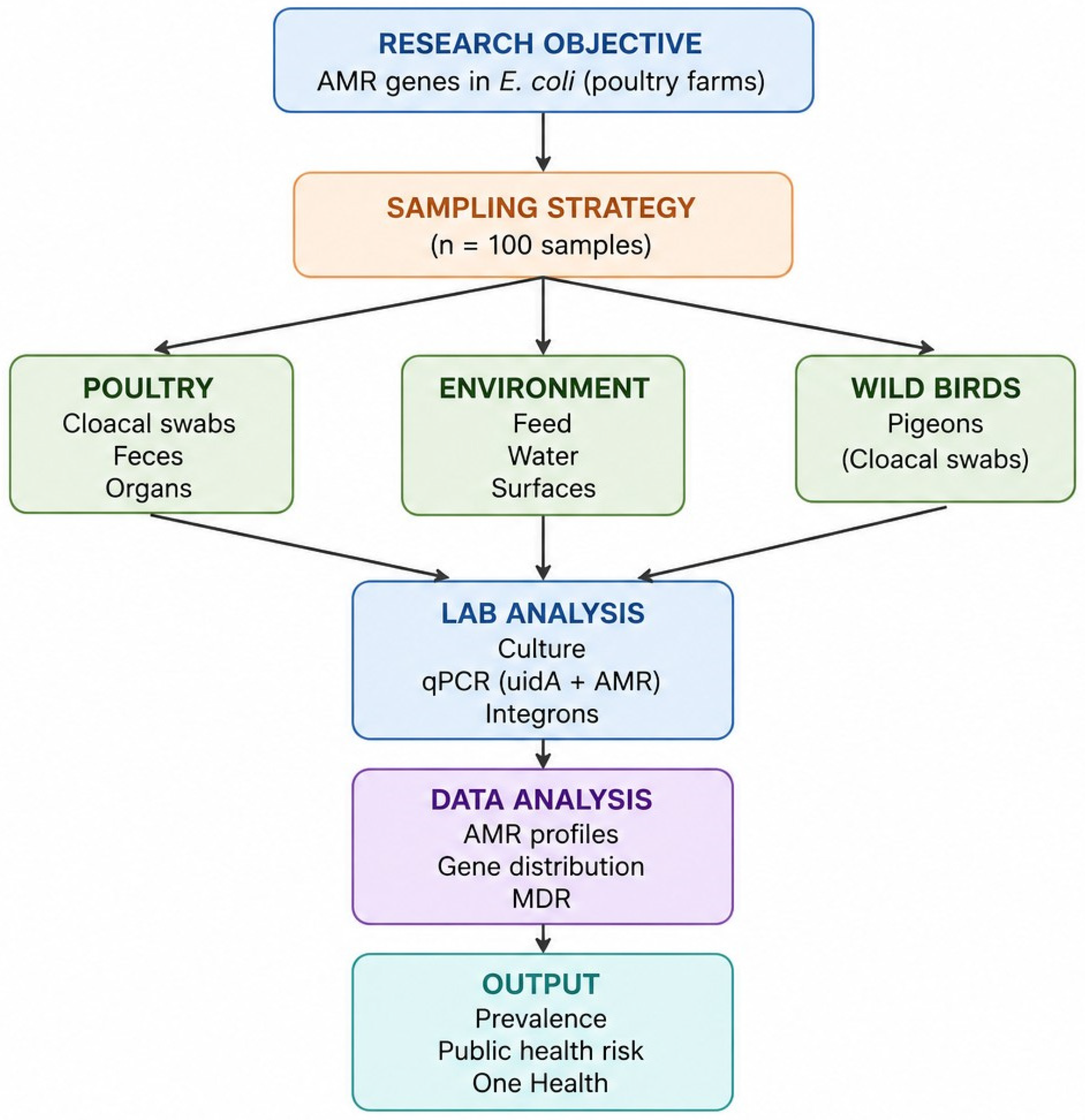
A schematic overview of the study design and sampling framework is presented in Figure 1.
For confidentiality reasons and in accordance with farm management agreements, the name of the participating farm is anonymized and referred to as a commercial layer poultry farm throughout the manuscript. The farm operated a commercial layer production system and housed Dekalb White laying hens. Adult birds were maintained in conventional four-tier cage batteries equipped with Big Dutchman housing systems under standard commercial management conditions. Each poultry house accommodated approximately 6000–7000 laying hens, while replacement pullets were housed separately. Feed was provided through an automated feeding system using commercially formulated layer rations supplemented with vitamin–mineral premixes, and water was supplied via nipple drinking systems with ad libitum access. Poultry houses were equipped with regulated ventilation, lighting, and sanitation systems in accordance with routine commercial biosecurity practices.
Field sampling was conducted during two visits to capture potential seasonal variation: September 2025 and January 2026. Approximately 50 samples were collected during each sampling visit.
A total of 100 microbiological specimens were collected from the poultry farm, including specimens from internal organs, the environment, feed, water, and wild birds. Each collected specimen (cloacal swab, organ sample, fecal sample, feed, water, or environmental swab) was considered an independent microbiological sample for bacteriological analysis. To obtain a comprehensive representation of the farm environment, specimens were collected from different biological and environmental matrices.
From live birds: Cloacal swab samples were collected from 40 clinically healthy laying hens randomly selected from different cage sections of the poultry house. Sterile cotton swabs moistened with sterile phosphate-buffered saline were gently inserted into the cloaca and rotated before placement into sterile transport tubes. In addition, eight freshly voided fecal samples were collected aseptically from poultry house floor surfaces immediately after defecation to minimize environmental contamination.
From internal organs: Ten birds showing mild clinical signs, including lethargy and ruffled feathers, were provided by farm staff for diagnostic examination. These birds were humanely euthanized according to standard veterinary practice for diagnostic purposes [15]. Post-mortem examinations were performed under aseptic conditions, and samples from the cecum, small intestine, large intestine, and liver were collected for bacteriological analysis.
From the environment: Two environmental samples were collected using sterile swabs moistened with sterile buffer solution from poultry cage floor surfaces and other areas in direct contact with birds, including feeders, drinkers, cages, walls, and floors of poultry houses.
Production inputs: Two drinking water samples were collected from the farm water supply system, and two feed samples containing commercial premix supplements were collected from feeders under aseptic conditions.
Wildlife sampling was conducted to assess the potential role of wild birds in bacterial dissemination within the farm environment. Five clinically normal pigeons (Columba livia) observed near the feed storage and processing areas were captured using a net-based trapping method [16]. A total of 10 samples, including cloacal swabs and intestinal samples, were aseptically collected from these birds using the same procedures as for poultry sampling. Most birds were released immediately after sampling to minimize stress, while one pigeon was humanely euthanized for collection of internal organ samples. The limited number of wild birds included in the study reflects the exploratory nature of this preliminary surveillance investigation and the restricted availability of birds for safe capture during the sampling period. Wild bird sampling was conducted during the same farm visits as poultry sampling to ensure temporal comparability between sample sources.
All bird sampling procedures were performed in accordance with standard veterinary ethical practices and with the permission of farm management. Immediately after collection, all samples were placed in sterile labeled containers, transported to the laboratory under refrigerated conditions (2–8 °C), and processed within 24 h to ensure bacterial viability.

2.3. Study Farm and Housing Conditions

The study was conducted at a commercial layer poultry farm located in the West Kazakhstan region, Republic of Kazakhstan. The farm housed Dekalb White laying hens maintained in a conventional cage production system equipped with Big Dutchman housing systems (Big Dutchman International GmbH, Vechta-Calveslage, Germany). Adult laying hens were housed in four-tier cage batteries under standard commercial management conditions. The farm included separate poultry houses for replacement pullets and adult layers, with approximately 6000–7000 hens per house.
Feed was produced on-site and provided through automated feeding systems using commercially formulated layer rations supplemented with vitamin–mineral premixes. Water was supplied through automated nipple drinking systems with ad libitum access. Poultry houses were maintained under controlled environmental conditions, including regulated ventilation, temperature, humidity, and lighting. Routine biosecurity measures included sanitation barriers, controlled farm access, and vaccination programs against major poultry infectious diseases.

2.4. Microbiological Isolation and Identification

Samples were pre-enriched in 10 mL of Buffered Peptone Water (BPW; Sigma-Aldrich, St. Louis, MO, USA) and incubated at 37 °C for 18–24 h [17]. For internal organ samples, aseptic tissue fragments from the liver, cecum, intestine, and other parenchymal organs were collected using sterile instruments and suspended in sterile physiological saline prior to pre-enrichment. All samples, including internal organs, cloacal swabs, feces, environmental, feed, and water samples, subsequently underwent the same pre-enrichment and culture procedure. For isolation of E. coli, a loopful of the pre-enrichment culture was streaked onto MacConkey Agar (MCA; HiMedia, Mumbai, India) and Eosin Methylene Blue Agar (EMB; HiMedia, Mumbai, India) [18]. Plates were incubated at 37 °C for 24–48 h.
Presumptive E. coli colonies (lactose-positive on MCA and exhibiting a metallic sheen on EMB) were subcultured onto Tryptic Soy Agar (TSA; HiMedia) to obtain pure cultures [19].
Pure isolates were identified using conventional biochemical tests, including Triple Sugar Iron Agar (TSI), Simmons Citrate, Methyl Red, Voges–Proskauer, indole production, urease, and lysine decarboxylase tests [20].
Isolates with biochemical profiles consistent with E. coli (Indole+, MR+, VP, Citrate, TSI A/A, H2S) were stored at −80 °C for further analysis [21].

2.5. Molecular Confirmation by qPCR

Molecular confirmation of presumptive isolates was performed by real-time polymerase chain reaction (qPCR) targeting the uidA gene. DNA was extracted from overnight pure cultures by thermal lysis: bacterial colonies were suspended in 100 µL of nuclease-free water, heated at 95 °C for 10 min, and centrifuged, after which the supernatant was used as a template [22,23].
Real-time PCR was performed using a QuantStudio™ 5 Real-Time PCR System (Applied Biosystems, Thermo Fisher Scientific, Waltham, MA, USA). Amplification reactions were carried out in a final volume of 20 µL using BioMaster UDG HS-qPCR Lo-ROX SYBR Green (2×) (Biolabmix, Novosibirsk, Russia), according to the manufacturer’s instruction. Each reaction mixture contained 10 µL of 2× SYBR MasterMix, 0.5 µM of each forward and reverse primer, 2 µL of template DNA, and nuclease-free water to the final volume.
Species-specific primers targeting the uidA gene of E. coli were used: uidA-F: 5′-AAA ACG GCA AGA AAA AGC AG-3′ and uidA-R: 5′-ACG CGT GGT TGA GTA GCT GA-3′ (amplicon size: 147 bp) [24]. Thermal cycling conditions consisted of initial denaturation at 95 °C for 5 min, followed by 40 cycles of 95 °C for 15 s and 60 °C for 30 s with fluorescence acquisition. A melting curve analysis (65–95 °C) was performed at the end of amplification to confirm product specificity. Positive controls (E. coli ATCC 25922), negative controls, and no-template controls were included in each run. All qPCR reactions were performed in duplicate.

2.6. Molecular Screening of Antimicrobial Resistance Genes

The presence of antimicrobial resistance genes was assessed using real-time PCR targeting genes associated with resistance to β-lactams, aminoglycosides, tetracyclines, sulfonamides, fluoroquinolones, macrolides, phenicols, and disinfectants. Mobile genetic elements, including class 1 integrons and insertion sequences, were also investigated.
A panel of 57 primer pairs was used for the detection of antimicrobial resistance genes. Custom standard DNA oligonucleotides (25 nmol scale; catalog no. A15612) were synthesized by Thermo Fisher Scientific (Applied Biosystems, Thermo Fisher Scientific, Waltham, MA, USA) and supplied through ZALMA Ltd. (Almaty, Kazakhstan).
The following genes were included in the analysis: β-lactam resistance genes (blaTEM, blaCTX-M, blaCMY, blaSHV, blaOXA, blaACC-1, blaDHA, blaFOX, blaPER, blaIMP, blaNDM, KPC); aminoglycoside resistance genes (aadA, aadB, aadD, aphA1, aph(3″)-Ia, ArmA, aacA43, strA, strB); tetracycline resistance genes (tetA, tetB, tetM, tetO, tetX); sulfonamide and trimethoprim resistance genes (sul1, sul2, dfrA variants); quinolone resistance genes (qnrB, qepA, oqxA); phenicol resistance genes (catA1, cmlA1, floR); macrolide-lincosamide resistance genes (ermB, ermF, mphA); disinfectant resistance genes (qac); and mobile genetic elements, including class 1 integrons (intI1), insertion sequences (IS26, IS6100, IS6/257), and plasmid-associated genes (tra, mob).
Primer sequences (Table S1) were obtained from Stedtfeld et al. (2018) [14]. Real-time PCR was performed in a total reaction volume of 25 µL containing 12.5 µL of BioMaster UDG HS-qPCR (2×) Lo-ROX SYBR Master Mix, primer mix, 5.0 µL of DNA template (3 ng), and nuclease-free water.
Amplification conditions consisted of an initial denaturation at 95 °C for 3 min, followed by 30 cycles of denaturation at 95 °C for 15 s, annealing at 62 °C for 20 s, and elongation at 72 °C for 10 s. Melt curve analysis was performed from 65 °C to 95 °C with 0.5 °C increments to confirm amplification specificity. A threshold cycle (Ct) value of 28 was used as the cutoff for positive amplification, as recommended by the original authors.
All reactions were performed in triplicate. Each run included previously characterized positive control isolates carrying representative antimicrobial resistance genes, as well as negative and no-template controls.

2.7. Farm Hygiene and Biosecurity Assessment

A standardized on-site evaluation of husbandry practices and biosecurity measures was conducted at the poultry farm (layer operation) using an observational checklist adapted from FAO biosecurity guidelines [25]. The assessment covered infrastructure, access control, sanitation protocols, health management, and feed/water systems.

2.7.1. Zoohygienic Conditions

Microclimate parameters (temperature, humidity, air velocity, gas composition, dust concentration, illumination) were measured using standard equipment: an August psychrometer (SATO Keiryoki Mfg. Co., Ltd., Tokyo, Japan), a fur aspirator “AM-5M” (MS GO Ekran LLC, Moscow, Russia) with indicator tubes, a thermo-anemometer (AZ-8906, AZ Instrument Corp., Taichung, Taiwan), a handheld luxmeter (model 8581, AZ Instrument Corp., Taichung City, Taiwan), and the gravimetric method with an aspirator (PU-3E, NIKI MLT LLC, Saint Petersburg, Russia), according to previously described methods.
Microclimate parameters. Measurements of microclimate parameters in poultry houses showed values within zootechnical standards: air temperature ranged from 18–20 °C for adult laying hens; relative humidity was maintained at 60–70%; air velocity ranged from 0.2–0.3 m/s in winter and up to 0.5 m/s in summer. Carbon dioxide concentration did not exceed 0.25%, ammonia levels ranged from 5–20 mg/m3, hydrogen sulfide concentration was up to 5 mg/m3, and dust concentration did not exceed 3.0 mg/m3. Illumination was maintained at 10–15 lux during rearing and 15–20 lux for laying hens, with a regulated light regime of 16–17 h of light per day.
Biosecurity measures. The farm implemented comprehensive biosecurity protocols including: a single entry point with disinfection barriers (footbath and wheel dip); an “all-in/all-out” system with 14-day sanitary breaks between production cycles; sequential sanitation of poultry houses after each cycle (litter removal, mechanical cleaning, washing, disinfection with 3% hot solution of “Macrodez” or “Miracid”); insect control using “Perteid 25%”; and rodent control using “Shtorm”. These measures ensured maintenance of high sanitary status and prevention of infectious disease transmission.
Vaccination program. Preventive vaccination was administered from day 0 to 21 against Newcastle disease (La Sota strain), infectious bronchitis (H-120 strain), and infectious bursal disease (“Hipragumboro-GM97”).

2.7.2. Veterinary, Sanitary Measures and Antimicrobial Use (AMU)

Data on antimicrobial use over a 5-year period (2020–2025) were collected from farm records. The assessment also included documentation of vaccination protocols, disinfection procedures, pest control measures, and biosecurity practices, following the methodology recommended by the World Organisation for Animal Health (WOAH) [2] and the European Food Safety Authority (EFSA) [26]. Analysis of farm records over a 5-year period (2020–2025) documented the routine prophylactic use of antimicrobial agents in drinking water. The following antibiotics were used: enrofloxacin (“Enroxil”), trimethoprim/sulfonamide combinations (“Tromexin”), and trimethoprim. For layer operations, antimicrobial prophylaxis was administered during the first 100 days of rearing at a dosage of 10 mg active substance per kg of body weight (equivalent to 0.5–1 mL of 10% solution per 1 L of water).

2.8. Statistical Analysis

Descriptive statistics were used to summarize the distribution of E. coli isolates and antimicrobial resistance-associated genes among different sample sources. Isolation rates were calculated as percentages based on the number of positive isolates relative to the total number of samples collected from each source. Data visualization and descriptive analyses were performed using Microsoft Excel 2019 (Microsoft Corp., Redmond, WA, USA). Due to the limited number of resistant isolates included in the qPCR screening analysis, no inferential statistical tests were performed.

3. Results

3.1. Isolation and Identification of E. coli

A total of 100 microbiological specimens were collected from various sources, including internal organs (cecum, n = 26; liver, n = 10), cloacal swabs (n = 40), fresh feces (n = 8), environmental samples (n = 2), water samples (n = 2), feed samples (n = 2), and wild bird microbiological specimens obtained from five pigeons (Columba livia, n = 10 specimens). From these, 40 E. coli isolates were confirmed by qPCR targeting the uidA gene. The distribution of E. coli isolates by sample source is presented in Table 1.
Environmental samples included poultry cage floor swabs, water samples from drinking systems, and feed samples mixed with premixes. The highest isolation rate was observed in cloacal swabs (65.0%, 26/40), followed by environmental samples, water, and feed samples (50.0% each), wild birds (40.0%, 4/10), cecum samples (19.2%, 5/26), fresh feces (12.5%, 1/8), and liver samples (10.0%, 1/10).
All 40 isolates were confirmed as E. coli by qPCR targeting the uidA gene. The specificity of amplification was confirmed by melt curve analysis, which showed a single peak at 80.5 ± 0.3 °C for all positive samples, indicating specific product formation without primer–dimer artifacts. The positive control (E. coli ATCC 25922) exhibited consistent amplification, while no amplification was observed in negative controls (no template).

3.2. Detection of Antimicrobial Resistance Genes

A panel of 57 primer sets targeting antimicrobial resistance genes and mobile genetic elements was used to screen 40 E. coli isolates confirmed by qPCR. Among these, ten isolates (25%) were positive for at least one resistance gene. The resistant isolates were designated as isolates 535, 543v, 544E, and 492, 575, 589, 610, 616, 625, 640.
Representative resistance determinants are summarized in Table 2, while complete qPCR profiles are presented in Supplementary Table S2.
The most frequently detected resistance genes were aadA17 (90%, 9/10), followed by blaTEM, int1-a-marko, qacEΔ1, IS26, blaCMY, and sul2 (80% each). Genes qnrB46,47,48 were detected in 70% of isolates, whereas sul1 and intI1F165_clinical were identified in 60% of isolates (Table 3).
A descriptive correspondence was observed between the presence of class 1 integrons (int1-a-marko, intI1F165_clinical) and multiple resistance genes. All ten isolates carried at least one integron gene (complete integron profiles are presented in Supplementary Table S2), and isolates with both integron variants (535, 543v, 544E, 575, 616, 640) harbored the highest numbers of resistance genes (14–19 genes). All ten isolates carried resistance genes associated with resistance to three or more antimicrobial classes, indicating the presence of resistance determinants associated with multiple antimicrobial classes.

3.3. Association Between Detected AMR Genes and Antimicrobial Use Practices

The resistance genes identified in E. coli isolates directly corresponded to the antimicrobials used on the farm (Table 4):
The high prevalence of fluoroquinolone resistance genes (qnrB46,47,48 in 70% of resistant isolates) is consistent with the routine use of enrofloxacin on the farm. Similarly, the frequent detection of sulfonamide (sul1, sul2) and trimethoprim (dfrA1, dfra14) resistance genes corresponds to the prophylactic administration of trimethoprim/sulfonamide combinations. The presence of β-lactamase genes (blaTEM, blaCMY, blaCTX-M) despite the absence of prophylactic β-lactam use may reflect co-selection with other resistance determinants on mobile genetic elements or environmental contamination.

4. Discussion

The present study provides a comprehensive analysis of antimicrobial resistance genes in isolates from a commercial layer poultry farm in Western Kazakhstan. A total of 40 E. coli isolates were confirmed by qPCR, of which 10 (25%) harbored at least one resistance gene, and all positive isolates carried resistance gene combinations associated with multidrug resistance.
The highest isolation rate of E. coli was observed in cloacal swabs (65.0%), followed by wild birds (40.0%) and fresh feces (12.5%). The detection of E. coli in pigeons captured near feed storage areas (40.0%) is particularly noteworthy, as it suggests that wild birds may contribute to bacterial circulation within the farm environment. Similar findings have been reported in other studies, where wild birds were identified as reservoirs and disseminators of antimicrobial-resistant E. coli in agricultural settings [27,28,29,30].
This prevalence is comparable to findings from other poultry studies in Central Asia and Eastern Europe, where resistance rates in E. coli range from 20% to 35% [31]. The most frequently detected resistance genes were blaTEM (80%), aadA17 (90%), and int1-a-marko (80%). The high prevalence of blaTEM, a common broad-spectrum β-lactamase gene, is consistent with global trends indicating its widespread distribution among Enterobacteriaceae in livestock production [32]. The detection of blaCTX-M (30%) and blaCMY (70%) in this study is of particular concern, as these genes confer resistance to extended-spectrum cephalosporins, which are critically important for human medicine [33]. The presence of ESBL-producing E. coli in poultry has been increasingly reported worldwide and represents a potential food safety concern due to the risk of foodborne transmission [34].
Regional surveillance data from the West Kazakhstan region support these findings. According to regional antimicrobial resistance monitoring data obtained within the framework of the present research project and based on anonymized reports from a local sanitary-epidemiological laboratory in Uralsk, clinical isolates collected in 2025 demonstrated increasing circulation of ESBL-producing E. coli carrying blaCTX-M, blaSHV, and blaTEM genes in clinical settings. The detection of blaCTX-M (30%), blaTEM (80%), and blaSHV-11 (20%) in our poultry isolates reflects this regional trend. In addition, acute intestinal infections among children accounted for 76.2% of more than 5000 registered cases during the first five months of 2025, highlighting the public health relevance of antimicrobial resistance in enteric bacteria.
The high prevalence of aadA17 (90%) among resistant isolates indicates widespread aminoglycoside resistance, likely driven by co-selection with other resistance genes located on the same mobile genetic elements, as aminoglycosides are not commonly used in poultry [35,36]. Tetracycline resistance genes (tetA, tetB, tetO) were detected at moderate frequencies (30% each). Tetracyclines have been widely used in livestock for decades, and the continued detection of tetracycline resistance genes reflects their long-term persistence in the farm environment, even after reduced usage [37].
Fluoroquinolone resistance genes (qnrB46,47,48, qepA_1_2) were detected in 70% and 30% of resistant isolates, respectively. The presence of plasmid-mediated quinolone resistance (PMQR) genes is particularly concerning because they can facilitate the spread of reduced susceptibility to fluoroquinolones even in the absence of direct selective pressure [13]. The routine prophylactic use of enrofloxacin on the farm during the first 100 days of rearing may contribute to the maintenance of these PMQR genes within the farm environment [38].
Sulfonamide resistance genes (sul1, sul2) were detected in 70% of resistant isolates, and trimethoprim resistance genes (dfrA1, dfra14) were detected in 60% and 40%, respectively. These findings are consistent with the documented use of trimethoprim/sulfonamide combinations (“Tromexin”) on the farm. The high prevalence of these genes is consistent with other studies reporting widespread sulfonamide and trimethoprim resistance in poultry-associated E. coli [39].
The detection of qacEΔ1 in 80% of resistant isolates is of particular interest. The farm routinely uses disinfectants containing quaternary ammonium compounds (QACs), such as “Macrodez” and “Miracid”. The presence of qacEΔ1 has been associated with reduced susceptibility to QACs and may contribute to the survival and persistence of resistant bacteria in disinfected farm environments. This finding highlights the potential role of biocide use in co-selecting for antibiotic resistance [40].
All ten resistant isolates carried at least one class 1 integron gene (int1-a-marko or intI1F165_clinical), and isolates with both integron variants (535, 543v, 544E, 575, 616, 640) harbored the highest numbers of resistance genes (14–19 genes). Class 1 integrons are well-established hotspots for the capture and expression of resistance gene cassettes and play a central role in the dissemination of multidrug resistance among Gram-negative bacteria [37]. The presence of insertion sequences (IS26, IS6100) further supports the potential for horizontal gene transfer [41].
A descriptive correspondence was observed between the antimicrobials used on the farm and the corresponding resistance genes detected in E. coli isolates. Enrofloxacin use was associated with the presence of qnrB46,47,48 and qepA_1_2; trimethoprim/sulfonamide use correlated with dfrA1, dfra14, sul1, and sul2; and β-lactamase genes (blaTEM, blaCMY, blaCTX-M) were detected despite the absence of prophylactic β-lactam use, suggesting co-selection with other resistance determinants or environmental contamination [36]. These findings align with the growing body of evidence demonstrating that antimicrobial use in food-producing animals selects for resistance in commensal and pathogenic bacteria, with potential implications for human health [42].
The detection of similar resistance gene profiles in E. coli isolates from poultry and pigeons (e.g., blaTEM, int1-a-marko, aadA17, qacEΔ1) suggests that wild birds may act as environmental carriers and contribute to the circulation of antimicrobial resistance genes within and between poultry farms [42]. This finding underscores the need for enhanced biosecurity measures to limit wild bird access to feed storage areas and poultry houses [25].
The detection of E. coli in feed samples may reflect environmental or fecal contamination occurring during feed storage, handling, or exposure to wild birds and farm-associated dust particles.
The concordance between resistance gene profiles in poultry isolates and clinical isolates from the same region supports the One Health concept, recognizing the interconnectedness of human, animal, and environmental health. The high proportion of acute intestinal infections in children, coupled with the detection of ESBL-associated resistance genes in poultry-associated E. coli, suggests that poultry products may contribute to the dissemination of resistant bacteria through the food chain [43]. This study contributes to the national antimicrobial resistance surveillance efforts under the “Roadmap for Containment of Antimicrobial Resistance (2023–2027)” and aligns with European Food Safety Authority (EFSA) and National Antimicrobial Resistance Monitoring System (NARMS) data, which report high levels of fluoroquinolone and tetracycline resistance in poultry-associated E. coli across multiple countries [44].
Several limitations should be acknowledged. First, this study was conducted at a single poultry farm, which limits the generalizability of the findings to other farms or regions. Second, phenotypic antimicrobial susceptibility testing was not performed; therefore, the detected resistance genes indicate the genetic potential for antimicrobial resistance rather than confirmed phenotypic resistance. Finally, the absence of direct comparison with human clinical isolates limits assessment of potential zoonotic transmission pathways. Environmental, feed, and water sampling was exploratory in nature and intended to provide preliminary screening data rather than statistically representative prevalence estimates.

5. Conclusions

This study demonstrated the presence of Escherichia coli carrying multiple antimicrobial resistance determinants and mobile genetic elements in a commercial poultry farm in Western Kazakhstan. All resistant isolates carried resistance determinants associated with multidrug resistance and frequently carried integron-associated resistance determinants, suggesting a high potential for horizontal dissemination within poultry production environments.
The co-occurrence of resistance genes, class 1 integrons, and insertion sequences suggests substantial potential for horizontal gene transfer and persistence under antimicrobial and disinfectant selective pressure. These findings provide important baseline molecular surveillance data for Kazakhstan and support the implementation of integrated One Health-based antimicrobial stewardship and biosecurity strategies under commercial poultry production conditions.
These findings suggest that long-term prophylactic use of critically important antimicrobials in poultry production exerts selective pressure, favoring the persistence and dissemination of corresponding resistance genes in E. coli populations.

Supplementary Materials

The following supporting information can be downloaded at: https://www.mdpi.com/article/10.3390/biology15110877/s1, Table S1: Primer sequences for detection of antibiotic resistance genes in Escherichia coli; Table S2: Complete antimicrobial resistance gene and mobile genetic element profiles of Escherichia coli isolates detected by qPCR array.

Author Contributions

Conceptualization, L.D., K.M., A.K., A.I. and K.D.M.; methodology, L.D., K.M., A.K., R.K., A.N., B.S., A.I. and K.D.M.; validation, L.D., K.M., A.K., R.K., A.N., B.S., A.I., R.S., G.D., M.G. and K.D.M.; formal analysis, L.D., K.M., A.I., R.S. and K.D.M.; investigation, L.D., K.M., A.K., R.K., A.N., B.S., A.I., R.S., G.D., M.G. and K.D.M.; resources, L.D., K.M., A.K., A.I. and K.D.M.; data curation, L.D., K.M., A.I., R.S. and K.D.M.; writing—original draft preparation, L.D., K.M., A.I., R.S. and K.D.M.; writing—review and editing, L.D., K.M., A.K., R.K., A.N., B.S., A.I., R.S., G.D., M.G. and K.D.M.; visualization, L.D., K.M., A.I. and K.D.M.; supervision, L.D., K.M., A.K., A.I. and K.D.M.; project administration, L.D., K.M., A.K. and A.I.; funding acquisition, L.D., K.M., A.K. and A.I. All authors have read and agreed to the published version of the manuscript.

Funding

This research has been funded by the Science Committee of the Ministry of Science and Higher Education of the Republic of Kazakhstan (Grant No. AP26101263 «Determination of antimicrobial resistance and whole-genome characterization of Salmonella spp. and E. coli isolates circulating in poultry farms in the West Kazakhstan region»).

Institutional Review Board Statement

The animal study protocol was approved by the Local Commission on Biological Ethics of Kazakh Scientific Research Veterinary Institute (West Kazakhstan Research Veterinary Station branch) on 22 October 2024 (Protocol No. 1).

Informed Consent Statement

Not applicable.

Data Availability Statement

The original data presented in this study are included in the article and Supplementary Materials. Further inquiries can be directed to the corresponding author.

Conflicts of Interest

Author Rasul Sidikhov was employed by Research Institute for Biological Safety Problems LLP, “QazBioPharm” National Holding. The remaining authors declare that the research was conducted in the absence of any commercial or financial relationships that could be construed as a potential conflict of interest.

Abbreviations

The following abbreviations are used in this manuscript:
AMRAntimicrobial resistance
AMUAntimicrobial use
BPWBuffered Peptone Water
CIConfidence interval
CLSIClinical and Laboratory Standards Institute
CtCycle threshold
EFSAEuropean Food Safety Authority
EMBEosin Methylene Blue Agar
ESBLExtended-spectrum β-lactamase
FAOFood and Agriculture Organization
MCAMacConkey Agar
MDRMultidrug resistance
MGEMobile genetic elements
MLSTMultilocus sequence typing
MRMethyl red
NARMSNational Antimicrobial Resistance Monitoring System
PCRPolymerase chain reaction
PMQRPlasmid-mediated quinolone resistance
QACQuaternary ammonium compound
qPCRReal-time polymerase chain reaction
TSATryptic Soy Agar
TSITriple Sugar Iron Agar
VPVoges–Proskauer
WGSWhole-genome sequencing
WHOWorld Health Organization
WOAHWorld Organisation for Animal Health

References

  1. World Health Organization (WHO). WHO List of Medically Important Antimicrobials for Human Medicine; WHO: Geneva, Switzerland, 2024; Available online: https://www.who.int/publications/i/item/9789240093461 (accessed on 18 May 2026).
  2. Van Boeckel, T.P.; Pires, J.; Silvester, R.; Zhao, C.; Song, J.; Criscuolo, N.G.; Laxminarayan, R. Global trends in antimicrobial resistance in animals in low- and middle-income countries. Science 2019, 365, eaaw1944. [Google Scholar] [CrossRef]
  3. Landers, T.F.; Cohen, B.; Wittum, T.E.; Larson, E.L. A review of antibiotic use in food animals: Perspective, policy, and potential. Public Health Rep. 2012, 127, 4–22. [Google Scholar] [CrossRef]
  4. Poirel, L.; Madec, J.Y.; Lupo, A.; Schink, A.K.; Kieffer, N.; Nordmann, P.; Schwarz, S. Antimicrobial resistance in Escherichia coli. Microbiol. Spectr. 2018, 6, 10–1128. [Google Scholar] [CrossRef]
  5. Dushayeva, L.Z. Antimicrobial resistance in foodborne Escherichia coli and Salmonella spp. from animal-origin foods: Transmission pathways, global surveillance gaps, and alternative therapeutic strategies. Vet. World 2025, 18, 3288–3305. [Google Scholar] [CrossRef]
  6. Sultana, S.; Hossain, M.A.; Hossain, M.T. Antibiotic resistance and virulence profiles of Escherichia coli isolated from poultry and cattle in Bangladesh. Vet. World 2022, 12, 165–173. [Google Scholar]
  7. Aleshina, Y.; Yeleussizova, A.; Mendybayeva, A.; Shevchenko, P.; Rychshanova, R. Prevalence and antimicrobial resistance of Enterobacteriaceae in the north of Kazakhstan. Open Vet. J. 2024, 14, 604–616. [Google Scholar] [CrossRef] [PubMed]
  8. Wang, J.; Ma, Z.B.; Zeng, Z.L.; Yang, X.W.; Huang, Y.; Liu, J.H. The role of wildlife (wild birds) in the global transmission of antimicrobial resistance genes. Zool. Res. 2017, 38, 55–80. [Google Scholar] [CrossRef] [PubMed]
  9. Swift, B.M.; Bennett, M.; Waller, K.; Dodd, C.; Murray, A.; Gomes, R.L.; Humphreys, B.; Hobman, J.L.; Jones, M.A.; Whitlock, S.E.; et al. Anthropogenic environmental drivers of antimicrobial resistance in wildlife. Sci. Total Environ. 2019, 649, 12–20. [Google Scholar] [CrossRef] [PubMed]
  10. Cooper-Beknazarova, M.; van den Akker, B.; Drigo, B.; Ross, K.; Whiley, H.; Williams, C.R. Systematic review of wild birds as reservoirs of antimicrobial resistance: Implications for roof-harvested rainwater safety. Int. J. Hyg. Environ. Health 2026, 273, 114743. [Google Scholar] [CrossRef]
  11. Gillings, M.R. Integrons: Past, present, and future. Microbiol. Mol. Biol. Rev. 2014, 78, 257–277. [Google Scholar] [CrossRef]
  12. He, S.; Hickman, A.B.; Varani, A.M.; Siguier, P.; Chandler, M.; Dekker, J.P.; Dyda, F. Insertion sequence IS26 reorganizes plasmids in clinically isolated multidrug-resistant bacteria. mBio 2015, 6, e00762-15. [Google Scholar] [CrossRef]
  13. Jacoby, G.A.; Strahilevitz, J.; Hooper, D.C. Plasmid-mediated quinolone resistance. Microbiol. Spectr. 2014, 2, 475–503. [Google Scholar] [CrossRef]
  14. Stedtfeld, R.D.; Guo, X.; Stedtfeld, T.M.; Sheng, H.; Williams, M.R.; Hauschild, K.; Gunturu, S.; Tift, L.; Wang, F.; Howe, A.; et al. Primer set 2.0 for highly parallel qPCR array targeting antibiotic resistance genes and mobile genetic elements. FEMS Microbiol. Ecol. 2018, 94, fiy130. [Google Scholar] [CrossRef] [PubMed]
  15. AVMA (American Veterinary Medical Association). AVMA Guidelines for the Euthanasia of Animals, 2020 ed.; American Veterinary Medical Association: Schaumburg, IL, USA, 2020. [Google Scholar]
  16. WOAH (World Organisation for Animal Health). Terrestrial Animal Health Code. Chapter 7.6: Welfare of Animals During Killing for Disease Control Purposes; World Organisation for Animal Health: Paris, France, 2023. [Google Scholar]
  17. Coxen, C.L.; Collins, D.P.; Carleton, S.A. Capture and injury rates of Band-tailed Pigeons using whoosh nets. Wilson J. Ornithol. 2018, 130, 321–326. [Google Scholar] [CrossRef]
  18. Foster, G.; Hopkins, G.F.; Gunn, G.J.; Ternent, H.E.; Thomson-Carter, F.; Knight, H.I.; Graham, D.J.L.; Edge, V.; Synge, B.A. A comparison of two pre-enrichment media prior to immunomagnetic separation for the isolation of Escherichia coli O157 from bovine faeces. J. Appl. Microbiol. 2003, 95, 155–159. [Google Scholar] [CrossRef]
  19. Bale, M.J.; McLaws, S.M.; Fenn, J.P.; Matsen, J.M. Use of and cost savings with morphologic criteria and the spot indole test as a routine means of identification of Escherichia coli. Diagn. Microbiol. Infect. Dis. 1984, 2, 187–191. [Google Scholar] [CrossRef]
  20. Fricker, C.R.; Warden, P.S.; Eldred, B.J. Use of the ISO 9308-1 procedure for the detection of E. coli in water utilizing two incubation temperatures and two confirmation procedures and comparison with defined substrate technology. J. Water Health 2008, 6, 389–397. [Google Scholar] [CrossRef]
  21. ISO 16649-2:2001; Microbiology of Food and Animal Feeding Stuffs—Horizontal Method for the Enumeration of β-Glucuronidase-Positive Escherichia coli—Part 2: Colony-Count Technique at 44 °C Using 5-Bromo-4-chloro-3-indolyl β-D-Glucuronide. International Organization for Standardization: Geneva, Switzerland, 2001. [CrossRef]
  22. Queipo-Ortuño, M.I.; De Dios Colmenero, J.; Macias, M.; Bravo, M.J.; Morata, P. Preparation of bacterial DNA template by boiling and effect of immunoglobulin G as an inhibitor in real-time PCR for serum samples from patients with brucellosis. Clin. Vaccine Immunol. 2008, 15, 293–296. [Google Scholar] [CrossRef]
  23. Wang, L.; Li, Y.; Mustapha, A. Detection of viable Escherichia coli O157:H7 by ethidium monoazide real-time PCR. J. Appl. Microbiol. 2009, 107, 1719–1728. [Google Scholar] [CrossRef] [PubMed]
  24. Pavlovic, M.; Luze, A.; Konrad, R.; Berger, A.; Sing, A.; Busch, U.; Huber, I. Development of a duplex real-time PCR for differentiation between E. coli and Shigella spp. J. Appl. Microbiol. 2011, 110, 1245–1251. [Google Scholar] [CrossRef]
  25. Food and Agriculture Organization of the United Nations. Biosecurity Guidelines for Sustainable Animal Production; FAO: Rome, Italy, 2020; Available online: https://www.fao.org/documents/card/en/c/cb1341en (accessed on 18 May 2026).
  26. EFSA Panel on Biological Hazards (BIOHAZ). Maximum levels of cross-contamination for 24 antimicrobial active substances in non-target feed. Part 1: Methodology, general data gaps and uncertainties. EFSA J. 2021, 19, e06852. [Google Scholar] [CrossRef]
  27. Atterby, C.; Börjesson, S.; Ny, S.; Järhult, J.D.; Drobni, M. ESBL-producing Escherichia coli in Swedish gulls—A case of environmental pollution from humans? PLoS ONE 2017, 12, e0187800. [Google Scholar] [CrossRef]
  28. Guardia, T.; Varriale, L.; Minichino, A.; Balestrieri, R.; Mastronardi, D.; Russo, T.P.; Dipineto, L.; Fioretti, A.; Borrelli, L. Wild birds and the ecology of antimicrobial resistance: An approach to monitoring. J. Wildl. Manag. 2024, 88, e22588. [Google Scholar] [CrossRef]
  29. Chantziaras, I.; Boyen, F.; Callens, B.; Dewulf, J. Correlation between veterinary antimicrobial use and antimicrobial resistance in food-producing animals: A report on seven countries. J. Antimicrob. Chemother. 2014, 69, 827–834. [Google Scholar] [CrossRef]
  30. Cole, D.; Drum, D.J.V.; Stalknecht, D.E.; White, D.G.; Lee, M.D.; Ayers, S.; Sobsey, M.; Maurer, J.J. Free-living Canada geese and antimicrobial resistance. Emerg. Infect. Dis. 2005, 11, 935–938. [Google Scholar] [CrossRef]
  31. Dierikx, C.M.; van der Goot, J.A.; Smith, H.E.; Kant, A.; Mevius, D.J. Presence of ESBL/AmpC-producing Escherichia coli in the broiler production pyramid: A descriptive study. PLoS ONE 2018, 13, e0203415. [Google Scholar] [CrossRef]
  32. Dorado-García, A.; Mevius, D.J.; Jacobs, J.J.H.; Van Geijlswijk, I.M.; Mouton, J.W.; Wagenaar, J.A.; Heederik, D.J. Quantitative assessment of antimicrobial resistance in livestock during the course of a nationwide antimicrobial use reduction in the Netherlands. J. Antimicrob. Chemother. 2016, 71, 3607–3619. [Google Scholar] [CrossRef] [PubMed]
  33. Shang, K.; Kim, J.-H.; Park, J.-Y.; Choi, Y.-R.; Kim, S.-W.; Cha, S.-Y.; Jang, H.-K.; Wei, B.; Kang, M. Comparative Studies of Antimicrobial Resistance in Escherichia coli, Salmonella, and Campylobacter Isolates from Broiler Chickens with and without Use of Enrofloxacin. Foods 2023, 12, 2239. [Google Scholar] [CrossRef] [PubMed]
  34. Racewicz, P.; Majewski, M.; Biesiada, H.; Nowaczewski, S.; Wilczyński, J.; Wystalska, D.; Kubiak, M.; Pszczoła, M.; Madeja, Z.E. Prevalence and characterisation of antimicrobial resistance genes and class 1 and 2 integrons in multiresistant Escherichia coli isolated from poultry production. Sci. Rep. 2022, 12, 6062. [Google Scholar] [CrossRef]
  35. Buffet-Bataillon, S.; Tattevin, P.; Bonnaure-Mallet, M.; Jolivet-Gougeon, A. Emergence of resistance to antibacterial agents: The role of quaternary ammonium compounds—A critical review. Int. J. Antimicrob. Agents 2012, 39, 381–389. [Google Scholar] [CrossRef]
  36. Ploy, M.C.; Lambert, T.; Couty, J.P.; Denis, F. Integrons: An antibiotic resistance gene capture and expression system. Clin. Chem. Lab. Med. 2000, 38, 483–487. [Google Scholar] [CrossRef] [PubMed]
  37. Baker, M.; Zhang, X.; Maciel-Guerra, A.; Babaarslan, K.; Dong, Y.; Wang, W.; Hu, Y.; Renney, D.; Liu, L.; Li, H.; et al. Convergence of resistance and evolutionary responses in Escherichia coli and Salmonella enterica co-inhabiting chicken farms in China. Nat. Commun. 2024, 15, 206. [Google Scholar] [CrossRef]
  38. Spagnolo, A.M.; Palma, F.; Amagliani, G.; Panunzio, M.F.; Montagna, M.T.; Alonzo, E.; Bonaccorsi, G.; Cairella, G.; Guberti, E.; Schiavano, G.F.; et al. Antimicrobial Resistance Along the Food Chain: Spread and Integrated Strategies for Mitigation and Control. Antibiotics 2026, 15, 311. [Google Scholar] [CrossRef]
  39. Cocoș, D.I.; Dumitrescu, E.; Muselin, F.; Brezovan, D.; Degi, J.; Boldura, O.M.; Cristina, R.T. Prevalence and Antimicrobial Resistance of Enterobacteriaceae in Wild Birds Across Europe: A Systematic Review. Antibiotics 2025, 14, 905. [Google Scholar] [CrossRef]
  40. Liu, Y.Y.; Wang, Y.; Walsh, T.R.; Yi, L.X.; Zhang, R.; Spencer, J.; Doi, Y.; Tian, G.; Dong, B.; Huang, X.; et al. Emergence of plasmid-mediated colistin resistance mechanism MCR-1 in animals and human beings in China: A microbiological and molecular biological study. Lancet Infect. Dis. 2016, 16, 161–168. [Google Scholar] [CrossRef] [PubMed]
  41. Robinson, T.P.; Bu, D.P.; Carrique-Mas, J.; Fèvre, E.M.; Gilbert, M.; Grace, D.; Hay, S.I.; Jiwakanon, J.; Kakkar, M.; Kariuki, S.; et al. Antibiotic resistance is the quintessential One Health issue. Trans. R. Soc. Trop. Med. Hyg. 2016, 110, 377–380. [Google Scholar] [CrossRef] [PubMed] [PubMed Central]
  42. Lazarus, B.; Paterson, D.L.; Mollinger, J.L.; Rogers, B.A. Do human extraintestinal Escherichia coli infections resistant to expanded-spectrum cephalosporins originate from food-producing animals? A systematic review. Clin. Infect. Dis. 2015, 60, 439–452. [Google Scholar] [CrossRef]
  43. EFSA (European Food Safety Authority); ECDC (European Centre for Disease Prevention and Control). The European Union Summary Report on Antimicrobial Resistance in zoonotic and indicator bacteria from humans, animals and food in 2020. EFSA J. 2022, 20, e07209. [Google Scholar] [CrossRef]
  44. NARMS (National Antimicrobial Resistance Monitoring System). NARMS Integrated Report: 2018–2019; U.S. Food and Drug Administration: Rockville, MD, USA, 2020.
Figure 1. Schematic overview of the study design, sampling strategy, laboratory workflow, and molecular characterization of antimicrobial resistance genes in Escherichia coli isolates from poultry, environmental, and wild bird samples.
Biology 15 00877 g001
Table 1. Distribution of E. coli isolates by sample source.
SourceDetection SiteNumber of Microbiological SpecimensNumber of Confirmed E. coli IsolatesIsolation Rate (%)
Cloacal swabsLive birds402665.0
Wild birds (pigeons)Feed storage area10440.0
Fresh fecesPoultry house floor8112.5
Cecum, small intestine, and large intestineInternal organs26519.2
LiverInternal organs10110.0
Environmental samplesPoultry cage floor surfaces2150.0
WaterDrinking water system2150.0
FeedFeed mixed with premixes2150.0
Total10040 isolates40.0
Table 2. Summary of resistance gene profiles in E. coli isolates.
IsolateTotal Detected Resistance Determinants (Including MGEs)Representative Resistance Genes and MGEs Detected
153519blaTEM, blaCMY, aadA17, sul1, qnrB, qepA, floR, mphA, qacEΔ1, IS26
2543v15blaTEM, blaCMY, tetA, aadA17, sul1, qnrB, floR, qacEΔ1, IS26
3544E16blaTEM, blaCTX-M, tetB, qnrB, qepA, dfrA1, qacEΔ1, IS6100
44929blaTEM, ArmA, aadA17, tetO, sul1, IS26
557515blaTEM, blaCMY, tetA, aadA17, sul1, qnrB, floR, qacEΔ1
658915blaCMY, blaSHV-11, aadA17, sul1, qnrB, qepA, floR, mphA, qacEΔ1, IS6100
76106blaCMY, blaCTX-M, tetB, qnrB, sul2, qacEΔ1
861615blaTEM, blaCMY, tetA, aadA17, sul2, dfrA1, qacEΔ1, IS6100
96259blaTEM, ArmA, aadA17, tetO, sul1, IS26
1064016blaTEM, blaCTX-M, aadA17, dfrA14, qnrB, qepA, qacEΔ1, IS6100
Note: Only representative resistance genes and mobile genetic elements (MGEs) are shown. Total counts and antimicrobial resistance classes were determined based on the complete qPCR profiles provided in Supplementary Table S2. Letter suffixes in isolate identifiers represent internal laboratory coding and do not indicate different phenotypes.
Table 3. Most frequently detected resistance genes in E. coli isolates (n = 10).
GeneFrequency (n = 10)Resistance Group
1aadA179 (90%)Aminoglycoside
2blaTEM8 (80%)β-lactamase
3int1-a-marko8 (80%)Class 1 integron
4qacEΔ18 (80%)Disinfectant resistance
5IS268 (80%)Insertion sequence
6blaCMY8 (80%)AmpC β-lactamase
7sul28 (80%)Sulfonamide
8qnrB46,47,487 (70%)Fluoroquinolone
9sul16 (60%)Sulfonamide
10intI1F165_clinical6 (60%)Class 1 integron
Note: Only genes detected in ≥60% of isolates are shown.
Table 4. Correspondence between AMU and detected resistance genes.
Antimicrobial ClassAntibiotics UsedDetected Resistance GenesFrequency (%)
FluoroquinolonesEnrofloxacinqnrB46,47,48, qepA_1_270%, 30%
SulfonamidesTrimethoprim/sulfonamidesul1,sul270%, 70%
TrimethoprimTrimethoprim, TromexindfrA1, dfra1460%, 40%
β-lactams(not used prophylactically)blaTEM, blaCMY, blaCTX-M90%, 70%, 30%
Disclaimer/Publisher’s Note: The statements, opinions and data contained in all publications are solely those of the individual author(s) and contributor(s) and not of MDPI and/or the editor(s). MDPI and/or the editor(s) disclaim responsibility for any injury to people or property resulting from any ideas, methods, instructions or products referred to in the content.