Article

Molecular Phylogeny, Divergence Time Estimation, and Biogeography of Moelleriella (Clavicipitaceae, Hypocreales) with Taxonomic Insights


1
State Key Laboratory of Agricultural and Forestry Biosecurity, College of Life Sciences, Fujian Agriculture and Forestry University, Fuzhou 350002, China
2
Department of Biological Sciences, University of Illinois, Chicago, IL 60607, USA
3
College of Humanities and Law, Fuzhou Technology and Business University, Fuzhou 350715, China
4
Fujian Academy of Forestry, Fuzhou 350012, China
5
Key Laboratory of Ministry of Education for Genetics, Breeding and Multiple Utilization of Crops, College of Horticulture, Fujian Agriculture and Forestry University, Fuzhou 350002, China
*
Authors to whom correspondence should be addressed.
Biology 2026, 15(10), 739; https://doi.org/10.3390/biology15100739
Submission received: 17 April 2026 / Revised: 3 May 2026 / Accepted: 3 May 2026 / Published: 7 May 2026

Simple Summary

This study describes one new species and two new Chinese records within the insect pathogenic genus, Moelleriella. Ancestral-area reconstruction suggests an Asian origin for Moelleriella during the Late Cretaceous (91.60 Mya), with subsequent dispersal to North America and South America via the Bering land bridge and to Africa via the Arabian Peninsula land bridge. These findings provide a phylogenetic framework for understanding the origin and evolutionary history of this genus.

Abstract

The Clavicipitaceae family, including saprobes and insect and myco-pathogens, are widely distributed in nature across various trophic regions, and play important roles in insect population control, plant interactions, and symbiotic evolution. Members of the genus Moelleriella within this family primarily specialize in infecting scale insects and whiteflies. Using five genomic loci (SSU, LSU, tef1-α, rpb1, and rpb2), we report on the inferred divergence times among members of Clavicipitaceae using molecular dating analyses. Molecular clock estimates revealed that the ancestor of Moelleriella likely emerged in the Late Cretaceous (91.60 Mya; 95% highest posterior density of 79.29–100.13 Mya). Historical biogeographic reconstruction of Moelleriella, performed using the Bayesian Binary Markov chain Monte Carlo (BBM) method, indicates that it most likely originated in Asia. Moreover, based on taxonomic and phylogenetic analyses, we describe three species within the genus Moelleriella, including one new species (Moelleriella microstroma) and two new records for China (Moelleriella chiangmaiensis and Moelleriella phukhiaoensis).

1. Introduction

The genus Moelleriella Bres. (Ascomycota, Hypocreales, Clavicipitaceae) comprises entomopathogenic fungi that parasitize scale insects (Coccidae, Lecaniidae) and whiteflies (Aleyrodidae) [1]. These fungi are predominantly distributed in tropical ecosystems and are occasionally found in subtropical regions [2,3]. Originally classified under Hypocrella Sacc., the genus was subsequently segregated based on molecular phylogenetic analyses and morphological revisions from allied genera (e.g., Samuelsia P. Chaverri & K.T. Hodge, Hypocrella P.A. Saccardo, and Orbiocrella D. Johnson, G.H. Sung, Hywel-Jones & Spatafora) [2,4]. Moelleriella sulphurea Bres. was designated as the type species of the genus [5]. Moelleriella differs from Samuelsia and Hypocrella in that its ascospores are fibrillar, multiseptate, and disarticulate at the septa within the ascus, whereas those of Samuelsia and Hypocrella remain intact, appearing filiform or long fusiform [1,6]. Furthermore, the anamorph of Moelleriella produces fusiform conidia, contrasting sharply with the allantoid conidia of Samuelsia [7,8]. Another notable characteristic of Moelleriella species is their brightly colored and morphologically diverse stromata, which often exhibit vivid hues of yellow, orange, or white [6]. Stromatal structures range from globose, thick pulvinate, tuberculate, and convex-to-thin pulvinate, serving as key diagnostic traits for species identification [9]. These morphological distinctions, combined with molecular evidence from loci such as the large subunit of ribosomal RNA (LSU) gene, the translation elongation factor 1-α (tef1-α) gene, and the largest subunit of RNA polymerase II (rpb1), support the taxonomic distinction of this genus [2].
These fungi exhibit dual trophic association strategies: necrotrophic parasitism on insects and biotrophic interactions with host plants, which combine to significantly enhance their survival and dispersal in complex environments [3,10]. According to the Index Fungorum database (https://indexfungorum.org/, accessed on 26 January 2026), Moelleriella currently comprises 83 species, with transcontinental distributions in some species (e.g., Moelleriella libera, found in Bolivia, Panama and regions of China; Moelleriella ochracea in Honduras and Brazil; and Moelleriella raciborskii in Ghana and Thailand) suggesting historical dispersal or vicariance events [2]. Such dispersals can be addressed via biogeographic studies by reconstructing the origins, speciation, and distribution patterns of organisms (fungi). Hyde et al. proposed a series of evolutionary timeframes [11], which indicated that fungal phyla originated >550 Mya, subphyla of Ascomycota emerged ~400–550 Mya, classes ~300–400 Mya, and subclasses ~250–300 Mya. Through molecular dating and ancestral reconstruction, the ancestor of the Schizoparmaceae family within the Diaporthales (sac fungi, includes several important plant pathogens and saprobes) was inferred to have likely originated in Africa during the Late Cretaceous, approximately 75.7 Mya (60.3–91.3 Mya) [12]. Similarly, investigations of the fungal spider pathogens, Akanthomyces sensu lato within the family Cordycipitaceae, suggests that the ancestor of this group appeared in the Paleogene, around 34.57 Mya (31.41–37.67 Mya), most likely originating in Asia [13]. Thailand and China have emerged as biodiversity hotspots for Moelleriella, with recent studies reporting thirteen new species from Yunnan, Hainan and Sichuan provinces in China [3,9,14], suggesting a continued biodiversity of the genus, and providing a valuable dataset for biogeographic studies.
Here, we isolated and purified specimens collected from Guizhou, Hunan, and Jiangxi provinces in China, and analyzed nucleotide sequences from three genetic loci (LSU, tef1-α and rpb1). Based on morphological characteristics and molecular data, we describe one new species and two new records in China, providing detailed illustrations and taxonomic descriptions. Furthermore, using Paleoophiocordyceps coccophagus (G.H. Sung, Poinar & Spatafora) as a fossil calibration point, we estimated the divergence time and origin of the genus Moelleriella, thereby offering new insights into its evolutionary history, biogeography, and likely routes of dispersal.

2. Materials and Methods

2.1. Collections and Isolation

Moelleriella specimens were collected from the undersides of dicotyledonous plant leaves at the following three locations in China: (1) Maolan Reserve, Libo County, Guizhou Province (107°52′10″–108°45′40″ E, 25°09′20″–25°20′50″ N); (2) Taoyuandong Reserve, Yanling County, Hunan Province (113°56′30″–114°06′20″ E, 26°18′00″–26°35′30″ N); and (3) Jiulianshan Reserve, Longnan County, Jiangxi Province (114°30′–114°50′ E, 24°30′–24°50′ N). Specimens were placed into 50 mL tubes and brought to the laboratory for isolation and purification by tissue isolation. Specimens collected in the field were first washed with sterile water on a sterile workbench, and using sterilized forceps and a scalpel, a portion of the stroma was excised and immersed in 75% ethanol (Sinopharm Chemical Reagent Co., Ltd., Shanghai, China) for 30 s, followed by rinsing in ddH2O for 60 s. After drying on sterile filter paper, the stroma was placed in a sterile Petri dish, and 200 μL of ddH2O was added. The stroma was then gently crushed with the handle of a sterilized scalpel, and the resulting suspension was spread onto potato dextrose agar (PDA) plates supplemented with 10 mg/mL ampicillin (Solarbio Science and Technology Co., Ltd., Beijing, China). After incubation at 25 °C for 3–5 d, the growing edge of a single fungal colony was transferred to a fresh PDA plate to obtain a pure culture. Dried specimens and purified isolates were deposited in the Mycological Herbarium, Institute of Mycology, Chinese Academy of Sciences (HMAS), and the China General Microbiological Culture Collection Center (CGMCC).

2.2. Morphological Observations

Fresh specimens were photographed in the field using a Canon EOS 6D Mark II camera (Canon Inc., Tokyo, Japan) for preliminary morphological documentation. Macroscopic features were examined and photographed in the laboratory under a stereomicroscope (Nikon SMZ74, Tokyo, Japan). After fungal pure cultures were inoculated onto PDA plates and incubated at 25 °C for 21 d, colony photographs were taken using the same Canon camera, and the resultant images used to measure colony diameters [5]. Tissue sections of specimens were mounted on glass slides with lactic acid–cotton blue (Solarbio Science and Technology Co., Ltd., Beijing, China), and microscopic structures were observed and photographed using a Nikon Ni-U compound microscope (Ni-U, Tokyo, Japan) [15]. All images were analyzed using Digimizer software (v5.4.4). The lengths and widths of thirty conidia and pycnidia were measured/sampled.

2.3. DNA Extraction and PCR Amplification

Small pieces of tissue were taken from the PDA plates of each sample, crushed in a mortar with liquid nitrogen, and total genomic DNA was extracted using the Fungal DNA Mini Kit (OMEGA–D3390, Feiyang Biological Engineering Corporation, Guangzhou, China) following the manufacturer’s instructions. The concentration and purity of total DNA were measured using a Nano-400A Ultramicro nucleic acid analyzer (AllSheng Company, Hangzhou, China). For molecular/phylogenetic determination, the sequences of the nuclear genomic loci were examined: (i) the nuclear ribosomal large subunit (LSU), (ii) translation elongation factor1-α gene (tef1-α), and (iii) RNA polymerase II largest subunits (rpb1) [2,16]. The primers and thermocycler conditions for PCR amplification are given in Table 1. Using the 2 × Rapid Taq Master Mix kit (Vazyme, Nanjing, China), target genes were amplified via polymerase chain reaction (PCR) using a thermal cycler (Bio-Rad, Hercules, CA, USA). For each sample, a 25 μL reaction volume included 12.5 μL of 2 × Rapid Taq Master Mix, 1 μL each of forward and reverse primers (10 μM) (QingKe Biotech Co., Ltd., Beijing, China), 1 μL of genomic DNA (100–200 ng/μL), and 9.5 μL of sterile water. The PCR products underwent purification and Sanger sequencing by BioSune (BioSune Co., Ltd., Shanghai, China). New sequences generated in this study have been deposited in GenBank (https://www.ncbi.nlm.nih.gov/, accessed on 28 January 2026) with accession numbers given in Table 2.

2.4. Sequence Alignment and Phylogenetic Analyses

Based on the Sanger sequencing results, the sequences of the LSU, tef1-α, and rpb1 genes were manually adjusted using MEGA v.7.0 and BioEdit v.7.2.6.1 [22,23]. The adjusted sequences were subjected to a BLAST (https://blast.ncbi.nlm.nih.gov/Blast.cgi/, accessed on 28 January 2026) search via the NCBI (National Center for Biotechnology Information). In combination with the reported literature, GenBank accession numbers for phylogenetic analysis were downloaded from the NCBI (Table 2). MAFFT v.7.11 (https://mafft.cbrc.jp/alignment/software/, accessed on 28 January 2026) was used to align each locus individually, and the primer-derived sequences were then trimmed using MEGA 7.0 [24]. The trimmed sequences were concatenated using Phylosuite v1.2 [25]. Phylogenetic analyses were performed on the concatenated three-gene dataset using Maximum Likelihood (ML) and Bayesian Inference (BI). ML was conducted in IQtree 1.6.8 with automatic model selection and 1000 bootstrap iterations. BI was performed using MrBayes 3.2.6. The best-fit evolutionary model for each partition was selected under the Akaike Information Criterion (AIC) using PartitionFinder 2 [26]. Four parallel Markov chain Monte Carlo (MCMC) simulations were run for 2,000,000 generations with a sampling frequency of every 100th generation. The burn-in phase was set at 10% of the total runs [27]. Finally, the phylogenetic trees were visualized using FigTree v.1.4.3 and edited with Adobe Illustrator CS 6.0 (Adobe Systems Inc., San Jose, CA, USA).

2.5. Divergence Time Estimation and Inferring Historical Biogeography

The divergence times of Moelleriella species were inferred using the Bayes MCMC algorithm in the BEAST 2.7.5 software package. The analysis was based on the SSU + LSU + tef1-α + rpb1+ rpb2 concatenated sequence dataset from 159 specimens [28]. MrModeltestv.2.3 was used to select the best-fitting evolutionary model (GTR + G + I) for the dataset. The Extensible Markup Language (XML) file was imported into BEAUti v2.0 for parameter configuration. A relaxed clock log normal model was applied [29,30]. Fossil calibration was performed using P. cocophagus, a fungal parasite of a scale insect from the Cretaceous period (99–105 Mya) [31]. A gamma-distributed prior was applied for fossil node calibration. This calibration was used to estimate the molecular clock ages of the nodes for species within the family Clavicipitaceae. The BEAST analysis ran for 200 million generations, logging parameters every 10,000 generations. Convergence and stationarity of the resulting log files were checked using Tracer v.1.7 soft, with ESS values ≥ 200 indicating convergence [12]. The first 10% of trees, representing the initial and unreliable results, were discarded as burn-in. Finally, a maximum clade credibility tree was constructed using TreeAnnotator v. 2.6.7 [13,32].
To reconstruct the ancestral geographical distribution and infer the historical biogeography of Moelleriella, the Bayesian Binary Markov chain Monte Carlo (BBM) method in the Reconstruct Ancestral State in Phylogenies (RASP v.4.3 software) package was used for analysis [33,34]. The parameters were set to 10 million generations, and the first 10% of samples were discarded as burn-in. Statistically, the geographic distributions of Moelleriella were identified in four areas: (A) Asia, (B) Africa, (C) North America, and (D) South America.

3. Results

3.1. Phylogenetic Analyses

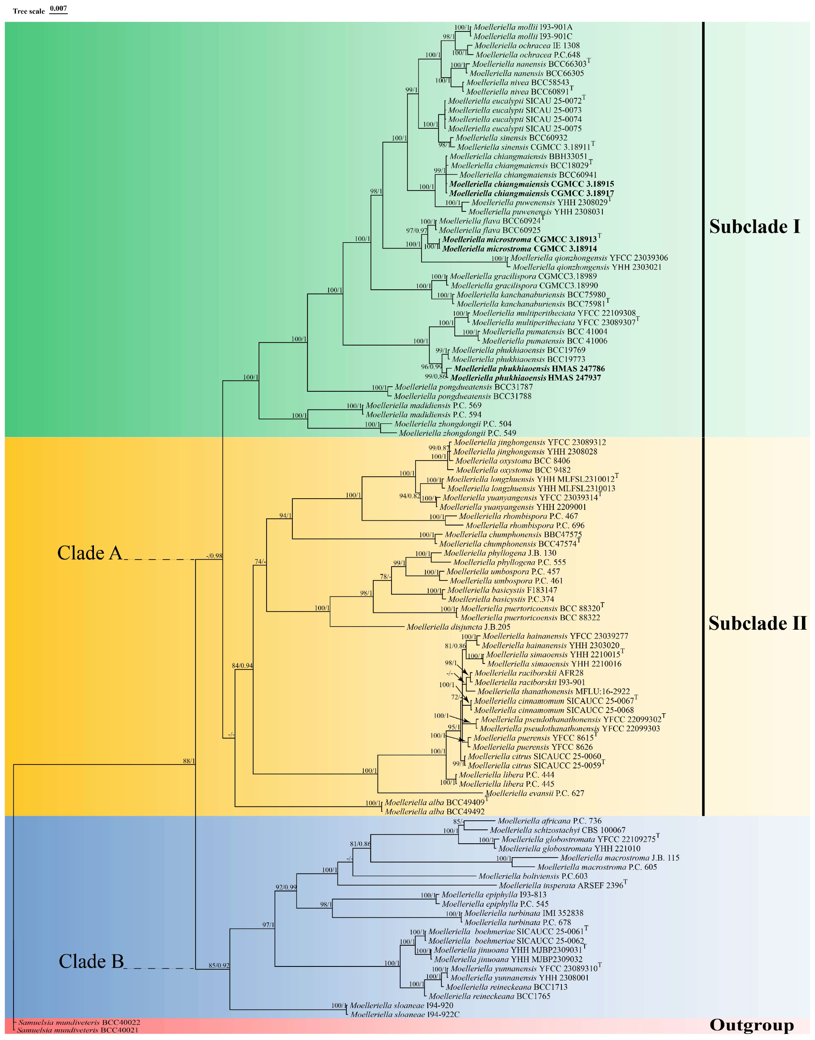
Phylogenetic analyses based on ML (Maximum Likelihood) and BI (Bayesian Inference) were conducted on a dataset of 108 concatenated nuclear gene sequences (LSU + tef1-α + rpb1) from 54 species of Moelleriella, with Samuelsia mundiveteris BCC 40021 and Samuelsia mundiveteris BCC 40022 designated as outgroups. The concatenated three-gene sequence dataset had a total length of 2468 bp, including 869 bp for LSU, 886 bp for tef1-α and 713 bp for rpb1. The topologies inferred from the ML and BI phylogenetic analyses were largely consistent across most branches, with strong support for most branches (Figure 1). The results show that Moelleriella species can be divided into two distinct clades (Clade A and Clade B), with Clade A further split into two sister subclades (Subclade I and Subclade II). Sequences generated from a new species described in the present study (Moelleriella microstroma) and two new records of species (Moelleriella chaiangmaiensis and Moelleriella phukhiaoensis) were included in the analyses (two isolates of each, six sets of sequences total) and all were found to be located within Subclade I of Clade A. Molecular analyses show that the newly described species M. microstroma sp. nov. is closely related to M. flava, with high levels of support (BP = 97%, PP = 0.97). Two samples formed a monophyletic clade with M. chiangmaiensis, which was recently reported in Thailand (BP = 99%, PP = 1), while two other samples were closely related to M. phukhiaoensis (BP = 96%, PP = 0.99).

3.2. The Divergence Time Estimation of Moelleriella

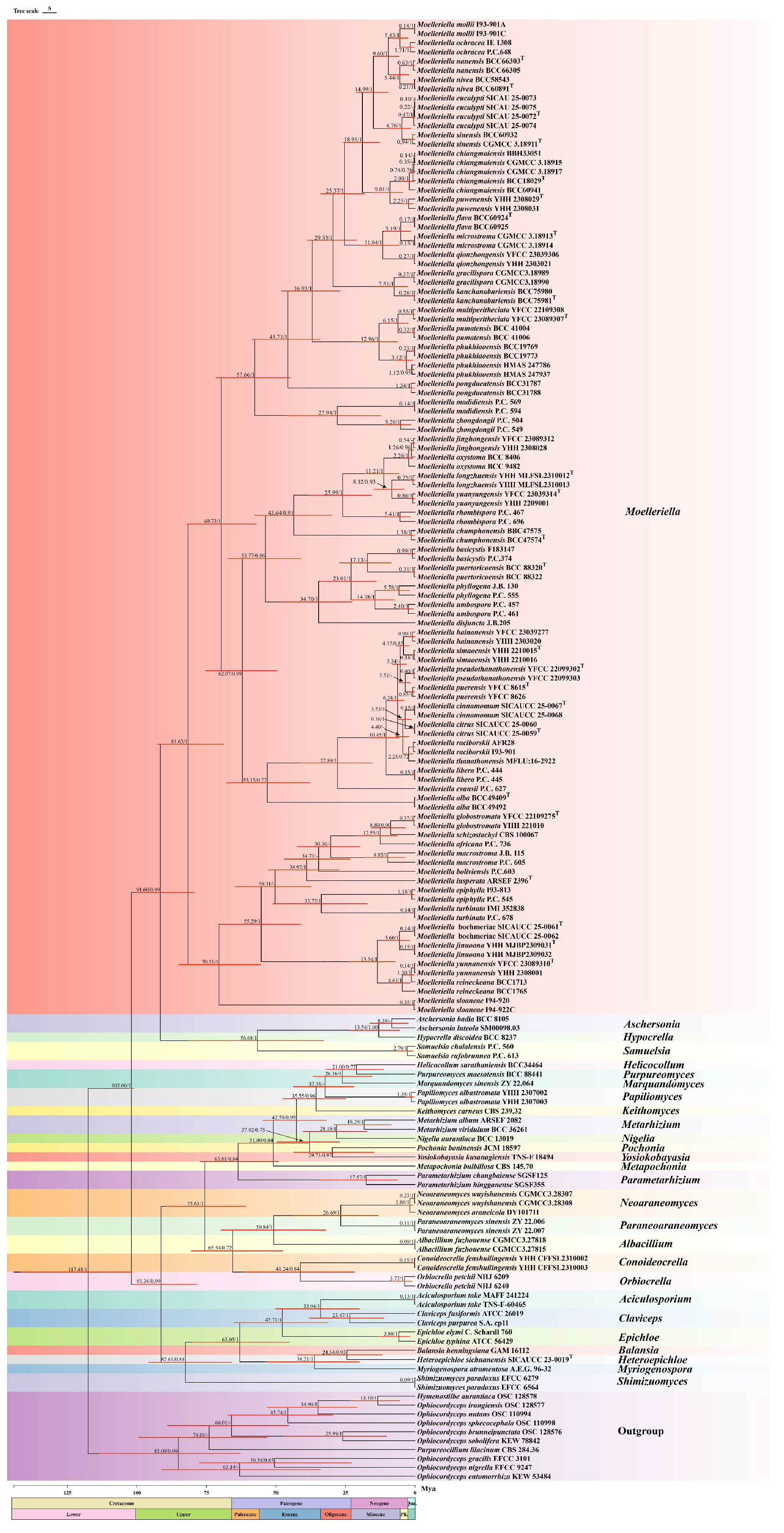
Based on prior studies, additional members of the family Clavicipitaceae were selected to investigate the evolutionary history, origin, and internal systematics of Moelleriella [19,35]. Divergence time estimation analyses confirmed that Clavicipitaceae diverged during the Early Cretaceous period (Figure 2). Further analyses indicated that the divergence time of Moelleriella from other genera of Clavicipitaceae is closely associated, with a mean stem age of 91.60 Mya (95% HPD = 79.29–100.13 Mya; PP = 0.99) and a mean crown age of 81.63 Mya (95% HPD = 68.88–92.68 Mya; PP = 1.00). The initial diversification of Moelleriella occurred in the Upper Cretaceous period (66–100.5 Mya), whereas the divergence of most Moelleriella species took place primarily during the Neogene period (2.58–23.03 Mya).

3.3. The Historical Biogeography of Moelleriella

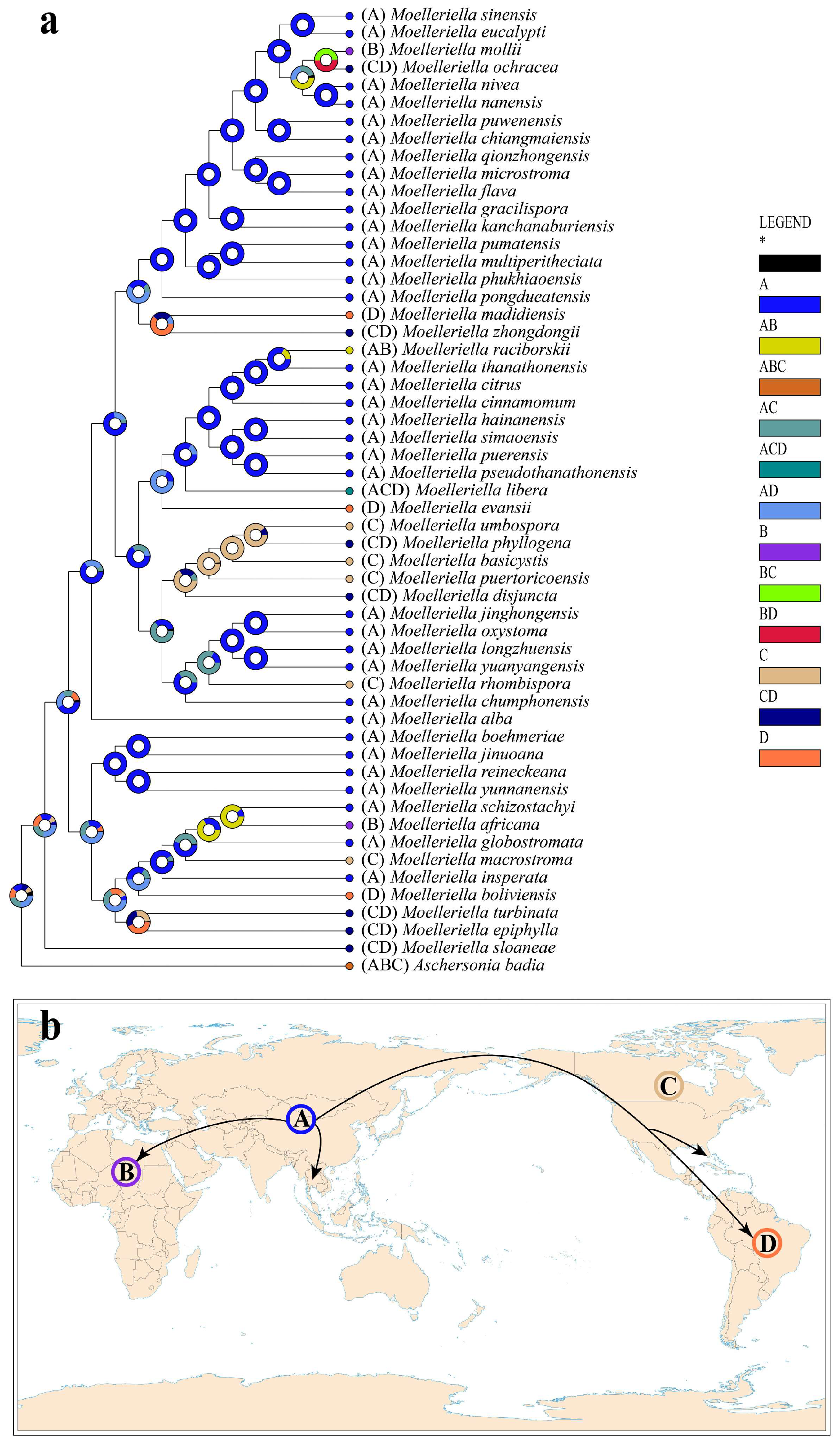
Historical biogeographic scenarios of Moelleriella were reconstructed using RASP (Figure 3). Results from Bayesian Binary Markov chain Monte Carlo (BBM) analysis suggest an Asian origin for Moelleriella, with at least 25 dispersal events and 10 vicariance events shaping its current distribution. To date, 37 species have been recorded in Asia, 13 in North America, 11 in South America, and three in Africa, confirming Asia as the center of diversity for this genus.

3.4. Taxonomy

3.4.1. Moelleriella microstroma X. Y. Wei, Y. S. Lin and J. Z. Qiu, sp. nov. (Figure 4)

MycoBank. MB861130.
Etymology. Microstroma refers to the small macroscopic morphological characteristics of the stroma.
Diagnosis. Moelleriella microstroma stroma color and conidial morphology are like those of Moelleriella flava, but M. microstroma produces multiple conidiomata on the stroma, whereas M. flava possesses only a single conidioma.
Type. China. Hunan Province: Zhuzhou City, Yanling County, Taoyuandong National Nature Reserve, 113°56′30″–114°06′20″ E, 26°18′00″–26°35′30″ N, collected from the abaxial surface of dicot leaves. 15 October 2017; coll. X. Y. Wei and J. Z. Qiu (holotype HMAS 247794, ex-type living culture CGMCC 3.18913).
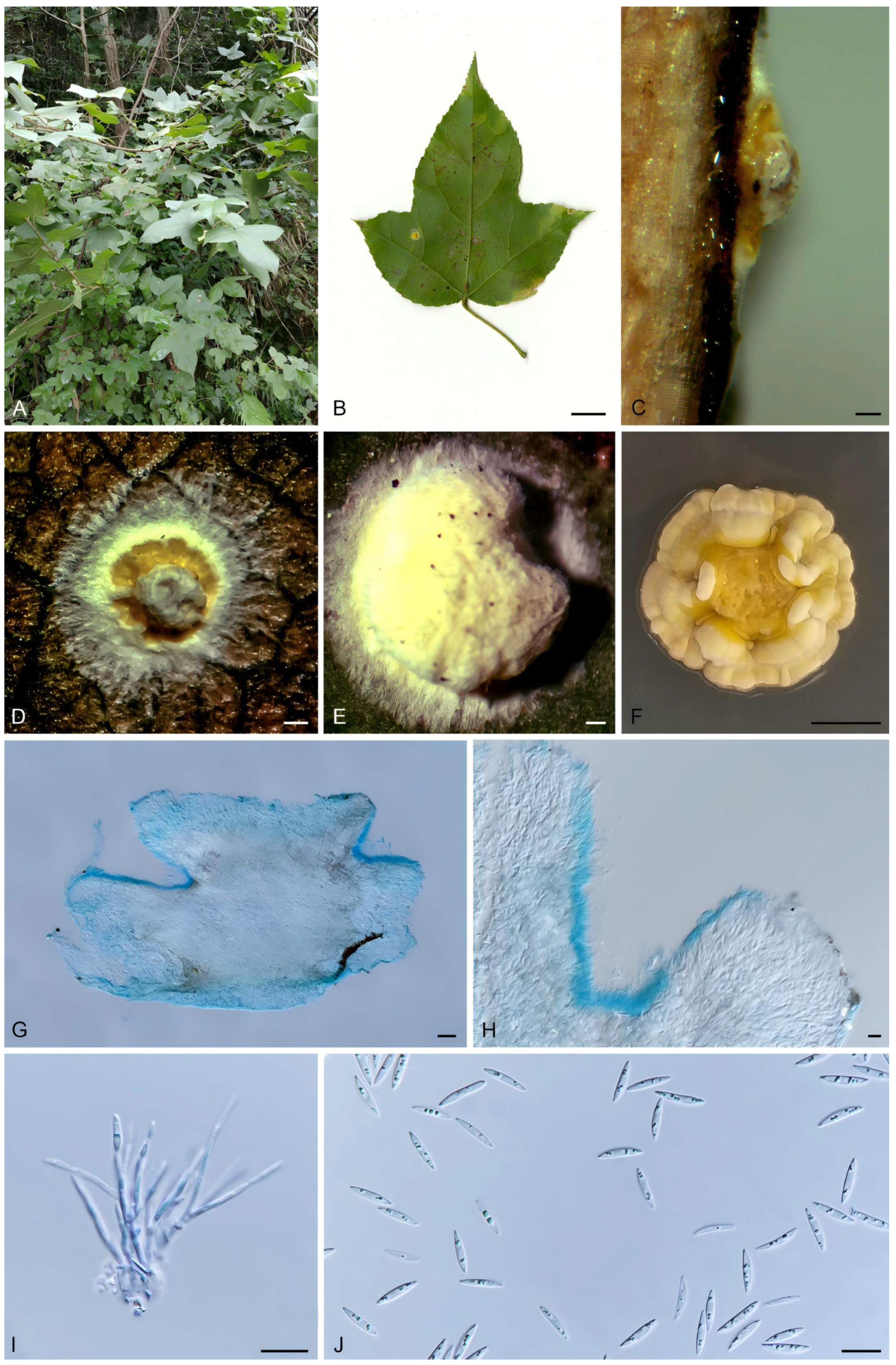
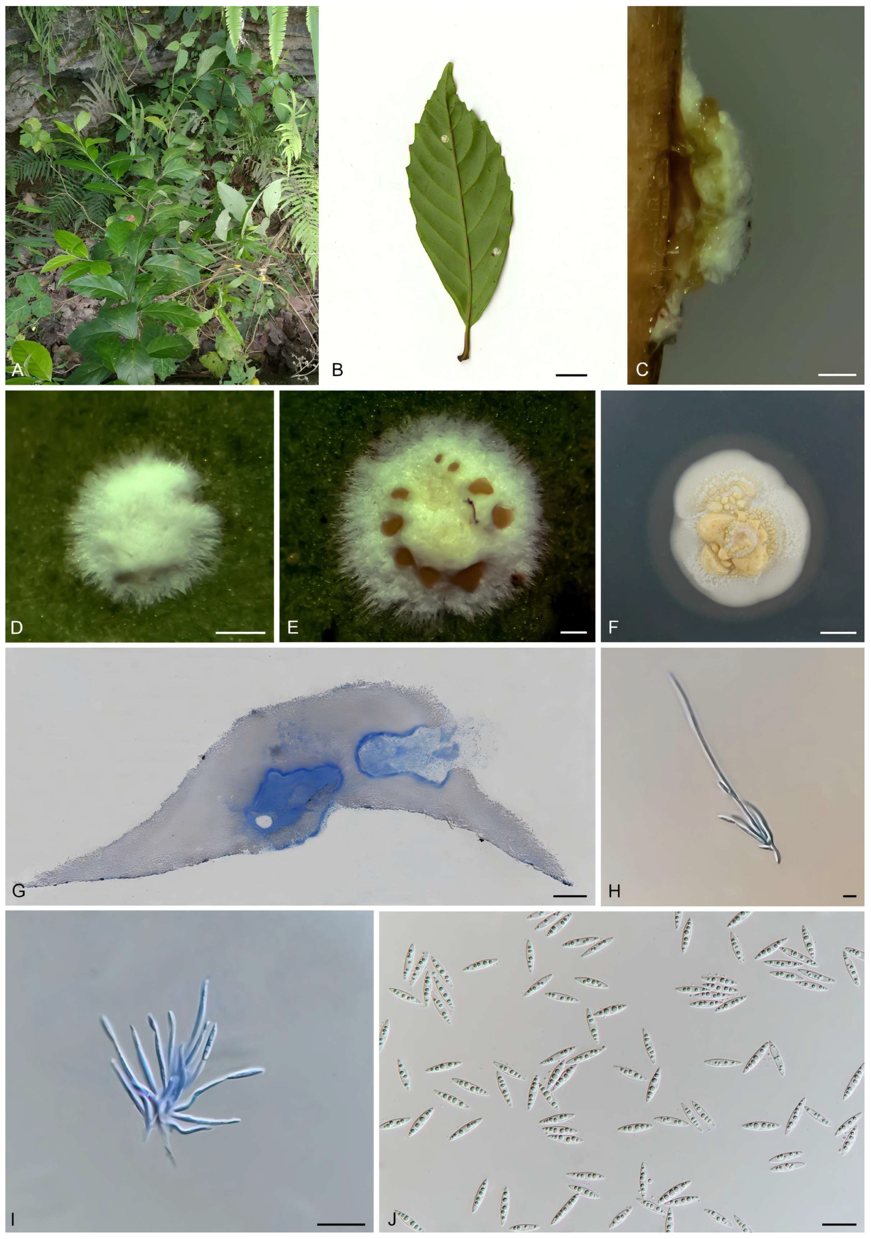
Description. Anamorph stromata when immature pale yellow, subglobose, becoming nearly cylindrical with a depressed apex at maturity, 1.18 (0.78–1.64) × 1.03 (0.56–1.59) mm in diameter, and 0.51 (0.39–0.73) mm high; with a pale yellow hypothallus and multiple orange acervular conidiomata; hypothallus 0.31 (0.13–0.60) mm wide. Multiple conidiomata per stroma; conidiomatal chambers opening irregularly, arranged on both sides of the stroma. Conidia fusoid, slightly curved on one side, 11.42 (10.09–13.07) × 1.99 (1.51–2.40) μm, L/W ratio = 5.8.
Teleomorph: Unknown.
Culture characteristics. Colonies on PDA slow-growing, attaining a diameter of 1.2–1.5 cm in 21 d at 25 °C. Stromatic colonies pulvinate, margin white to pale yellow, conidial masses pale yellow.
Habitat. On scale insects (Coccidae, Hemiptera) or whiteflies (Aleyrodidae, Homoptera), found on lower leaf surface of dicotyledons.
Distribution. China, Hunan Province, Zhuzhou City.
Additional specimen examined. China: Hunan Province, Zhuzhou City, Yanling County, Taoyuandong Reserve, 113°56′30″–114°06′20″ E, 26°18′00″–26°35′30″ N, found on abaxial surface of dicot leaves, 5 October 2017, J. Z. Qiu, paratype HMAS 247795; exparatype living culture CGMCC 3.18914.
Notes. In this study, the new species M. microstroma was strongly supported (100% ML/1 PP) based on phylogenetic analyses using sequences of three gene regions, and it formed a sister clade to M. flava (Figure 1). Morphologically, although the stromatal color and the shape and size of the conidia in the anamorph of M. microstroma are similar to those of M. flava, stromata of M. microstroma (1.18 (0.78–1.64) × 1.03 (0.56–1.59) mm) are slightly smaller than those of M. flava (1–4.5 mm). Furthermore, M. microstroma is distinguished by the presence of multiple orange acervuli (Figure 4D) and multiple conidiomata (Figure 4G) on each stroma, which contrast markedly with the condition in M. flava. Accordingly, this fungus is described here as a new species.
Figure 4. Morphology of M. microstroma. (A) Habitat; (B) stroma on the underside of leaves; (C) flank of stroma; (D,E) frontage of stroma; (F) colony on PDA; (G,H) section of stromata showing conidiomata; (I) conidiogenous cells; (J) conidia. Scale bars: (B) = 1 cm; (CE) = 0.2 mm; (F) = 0.5 cm; (G) = 100 μm; (H) = 20 μm; (I,J) = 10 μm.
Biology 15 00739 g004

3.4.2. Moelleriella phukhiaoensis, Mongkol.,Thanakitp & Luangsa-ard, Fungal Diversity 2016, 78, 1–237 [36] (Figure 5)

Description. Stromata pulvinate, yellow, 2.14 (1.23–3.46) × 1.95 (1.19–2.89) mm in diameter and 0.55 (0.30–0.81) mm height; without hypothallus; hyphae compact. Conidiomata numerous, tear-shaped, regularly arranged in the central region of the stroma, 183.79 (103.45–234.48) × 77.95 (55.17–106.9) μm. Conidia fusiform, 11.75 (9.17–13.1) × 1.60 (1.31–2.02) μm, L/W ratio = 7.52.
Teleomorph: Unknown.
Culture characteristics. Colonies on PDA slow-growing, attaining a diameter of 1.5 cm in 21 d at 25 °C. Hyphae are pale yellow, and abundant yellow conidial masses are visible on the colony surface.
Specimen examined. China: Hunan Province, Zhuzhou City, Yanling County, Taoyuandong Reserve, 113°56′30″–114°06′20″ E, 26°18′00″–26°35′30″ N; on underside of dicotyledonous leaves, 15 October 2017, J. Z. Qiu (paratype: HMAS 247937; exparatype living culture: CGMCC 3.19091). China: Jiangxi Province, Longnan County, Jiulian Mountain Nature Reserve, 114°30′–114°50′ E, 24°30′–24°50′ N, 23 September 2017, J. Z. Qiu (paratype: HMAS 247786).
Notes. Moelleriella phukhiaoensis was originally found on the underside of dicotyledonous leaves in Bueng Pan Protect Forest Unit, Phu Khiao Wildlife Sanctuary, Chaiyaphum Province, Thailand. Hosts are scale insect nymphs (Hemiptera). In this study, two specimens, HMAS 247937 and HMAS 247786, formed a sister clade with M. phukhiaoensis (BCC 19769, BCC 19773) in the phylogenetic analysis (Figure 1). Morphologically, both HMAS 247937 and HMAS 247786 share the yellow, stromata flattened pulvinate with the holotype of M. phukhiaoensis (BBH 17305, ex-type living culture BCC 19769). The shapes of the conidiomata and conidia of the anamorph are also like those of M. phukhiaoensis BBH 17305, although the conidiomata and conidia of the two Chinese collections are shorter. However, the two isolates characterized herein were highly similar to M. phukhiaoensis BCC 19769 in LSU (99.88%) and rpb1 (99.53%). Therefore, we identify our isolates as M. phukhiaoensis, marking the first record of M. phukhiaoensis in China.
Figure 5. Morphology of Moelleriella phukhiaoensis. (A) Habitat; (B) stroma on the underside of leaves; (C) frontage of stroma; (D) flank of stroma; (EH) section of stromata showing conidiomata; (I) colony on PDA; (J) conidiogenous cells; (K) conidia. Scale bars: (B) = 1 cm; (C,D) = 0.2 mm; (E,F) = 100 μm; (G,H) = 20 μm; (I) = 0.5 cm; (J,K) = 10 μm.
Biology 15 00739 g005

3.4.3. Moelleriella chiangmaiensis, Khonsanit, A., Noisripoom, W., Mongkolsamrit, S., Phosrithong, N., and Luangsa-ard, J.J., Mycol Progress 2021, 20, 847–867 [3] (Figure 6)

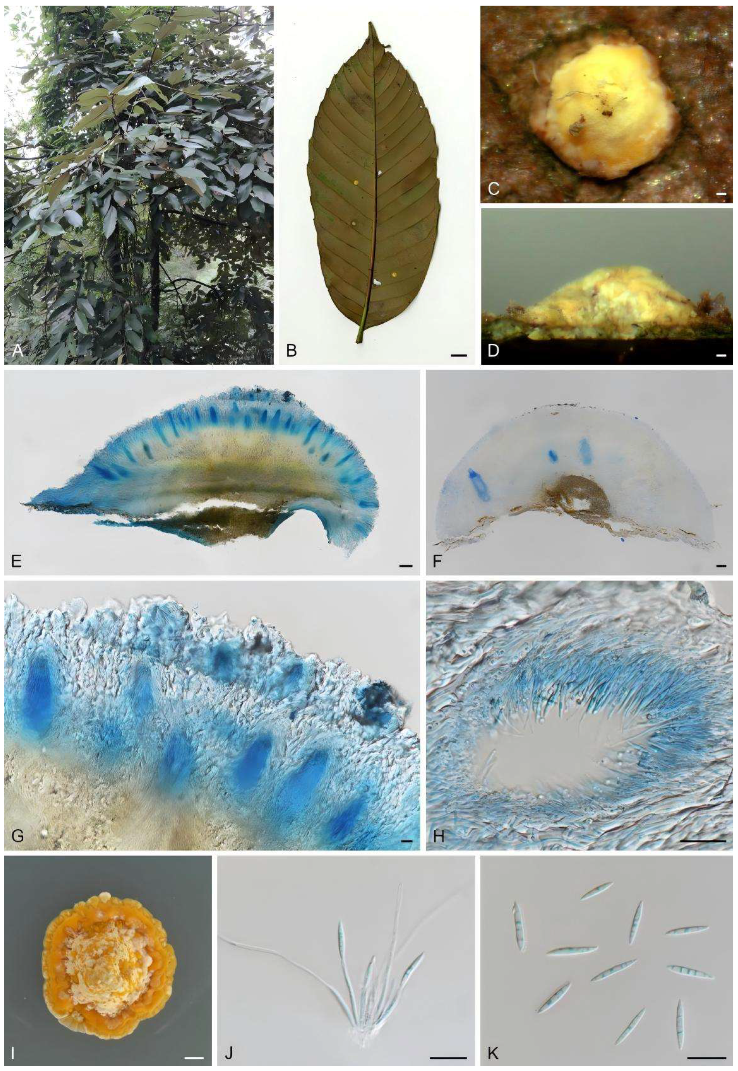
Description. Stromata pulvinate, white, 1.66 (0.68–2.96) × 1.40 (0.66–2.03) mm in diameter, 0.36 (0.13–0.61) mm high with a white hypothallus, 0.44 (0.21–0.64) mm wide. The margin of the stromata bears white floccose hyphae, and the surface of mature stromata displays multiple regularly distributed yellow acervuli. Conidiomata are irregular in shape and located at the base of the stromata, 294.87 (208.76–312.82) × 150 (114.67–168.17) µm, containing abundant fusiform conidia 8.53 (6.90–10.10) × 1.83 (1.40–2.20) µm, L/W ratio = 4.75.
Culture characteristics. Colonies on PDA fast-growing at 25 °C, attaining 2 cm diameter in 21 d. The mycelium is white, flocose, and loosely textured. Abundant pale yellow conidial masses are observed on the colony surface. Transparent hydrolytic zones, approximately 0.2–0.4 cm in diameter, are present around the colony.
Specimen examined. China: Guizhou Province, Libo County, Maolan Reserve, 107°52′10″–108°45′40″ E, 25°09′20″–25°20′50″ N, found on the underside leaves of a dicotyledonous plant, 20 October 2017, J. Z. Qiu, paratype HMAS 247792, HMAS 247793; exparatype living culture CGMCC 3.18915, CGMCC 3.18917.
Notes: Moelleriella chiangmaiensis was originally discovered on the underside of dicotyledonous leaves in Doi Inthanon National Park, Chiang Mai Province, Thailand. In this study, the specimens identified as M. chiangmaiensis (HMAS 247792, HMAS 247793) were also collected from the underside of dicotyledonous leaves. Morphologically, strains CGMCC 3.18915 and CGMCC 3.18917 share with M. chiangmaiensis white-to-moderately yellow pulvinate stromata and fusiform conidia of a comparable size (8.53 (6.90–10.10) × 1.83 (1.40–2.20) μm vs. (7–)8–10(–11) × 1.5–2 μm, respectively). Phylogenetic analysis revealed that strains CGMCC 3.18915 and CGMCC 3.18917 formed a monophyletic clade with M. chiangmaiensis (BCC 60941, BCC 18029, and BBH 33051) with relatively strong statistical support (99% ML/1 PP, Figure 1). Strains CGMCC 3.18915 and CGMCC 3.18917 exhibit high sequence identity with the ex-type living culture of M. chiangmaiensis BCC 18029 in LSU (99.18%) and tef1-α (100%). Furthermore, M. chiangmaiensis forms a sister clade with M. puwenensis. For better resolution, we conducted an additional comparison of strains CGMCC 3.18915 and CGMCC 3.18917 with M. puwenensis YHH 2308029. Although the three strains are similar in stromatal morphology and color, strains CGMCC 3.18915 and CGMCC 3.18917 have smaller conidiomata (294.87 (208.76–312.82) × 150 (114.67–168.17) μm than those of M. puwenensis (350–545 × 185–250 μm)) and smaller conidia (8.53 (6.90–10.10) × 1.83 (1.40–2.20) μm vs. 10–15 × 1.2–1.9 μm in M. puwenensis). On a molecular level, sequence alignment revealed that strains CGMCC 3.18915 and CGMCC 3.18917 show 99.09% sequence identity with M. puwenensis YHH 2308029 in the LSU. However, they exhibit lower identity in rpb1 (98.64%) and tef1-α (97.00%), which represents high identity but lower than that seen towards M. chiangmaiensis. We therefore identify our isolates as M. chiangmaiensis, which represents the first record of this species in China.
Figure 6. Morphology of Moelleriella chiangmaiensis. (A) Habitat; (B) stroma on the underside of leaves; (C) flank of stroma; (D,E) frontage of stroma; (F) colony on PDA; (G) section of stromata showing conidiomata; (H,I) conidiogenous cells; (J) conidia. Scale bars: (B) = 1 cm; (CE) = 0.2 mm; (F) = 0.5 cm; (G) = 100 μm; (H,I) = 20 μm; (J) = 10 μm.
Biology 15 00739 g006

4. Discussion

The genus Moelleriella is divided into the effuse and globose clades (A and B, respectively) with the former separated into two sister subclades (I and II) [2,3]. Phylogenetic analyses identified 41 species within Clade A: 19 in Subclade I (including one new species) and 22 in subclade II. Morphologically, members of this clade are characterized by effuse-to-thin, pulvinate stromata that are predominantly cream-colored, occasionally pale yellow to orange [37,38]. Most species have obpyriform or flask-shaped perithecia and fusoid conidia. Clade B encompasses 13 species, which typically exhibit globose, deep yellow to brown stromata and small conidia, occasionally producing large part-spores [3,9]. However, none of these traits are exclusive to either clade. In recent years, 23 new Moelleriella species have been reported: 18 from China and five from Thailand [3,9,14]. In the present study, specimens collected from Guizhou, Hunan, and Jiangxi provinces in China were examined morphologically and all were placed within Subclade I of the effuse (A) clade. These included a new species, Moelleriella microstroma, which differed significantly from its closest relative, M. flava, in both morphology and DNA sequence data. Additionally, two species previously reported in Thailand, M. chiangmaiensis and M. phukhiaoensis, are reported and illustrated here in China.
In recent years, molecular dating methods that combine analyses of fungal fossils with characterization of selected molecular loci have been widely applied to estimate the divergence times of fungi, significantly enhancing our understanding of fungal evolution at the taxonomic level [39,40,41]. Ancestral state reconstruction and divergence time estimation have inferred that Hypocrealean fungi likely existed in the Early Jurassic [31], with the crown node age estimated at ~193 Mya (158–232 Mya). These analyses suggested that the familial lineages within this group primarily originated in the Late Jurassic and underwent diversification during the Cretaceous. The crown age of Clavicipitaceae was estimated to be at least ~117 Mya (95–144 Mya), dating back to the Early Cretaceous. Additional estimations of the crown age of Hypocreales are believed to be around 200 Mya (174–232 Mya), with the crown node age of Clavicipitaceae refined to 107 Mya (90–126 Mya) [35]. The latter study also provided evidence that the direction of host shifts in Hypocrealean fungi occurred from plants (endophytes) to fungi (pathogens), and then possibly to animals (pathogens). The Cretaceous diversification and cladogenesis within this family gave rise to several subclades, reflecting multiple inter-kingdom host shifts among the three major eukaryotic kingdoms—animals, plants, and fungi [42]. Based on divergence time analysis, our analyses estimate the mean crown age of Clavicipitaceae at ~102.00 Mya (101.00–103.91 Mya), consistent with previous studies. The mean crown age of Moelleriella was estimated at ~81.63 Mya (68.88–92.68 Mya), suggesting a Late Cretaceous origin for its ancestor with most species emerging during the Neogene. Furthermore, the mean crown ages of the effuse clade and the globose clade within Moelleriella were similar, estimated at ~69.73 Mya and ~70.51 Mya, respectively, suggesting a critical adaptive branching occurred within that timeframe.
Based on our biogeographic analyses, Asia is inferred to be the most probable ancestral region of Moelleriella, with Southeast Asia likely representing the center of origin. The now-submerged Bering land bridge (BLB) may have facilitated the dispersal of Moelleriella from East Asia to North America [43]. Further analysis indicates that by the Early Oligocene (approximately 33.9 Mya), M. disjuncta, M. epiphylla, and M. turbinata already occurred in both North and South America. Although a direct land bridge between North and South America did not exist during this period, Moelleriella may have dispersed via transoceanic or long-distance dispersal (LDD) [44]. Although such latter mechanisms are generally considered rare, this hypothesis may better explain the distribution patterns of fungi across various contexts and geographical regions. Additionally, three species of Moelleriella (M. africana, M. mollii, and M. raciborskii) were present in Africa during the Neogene (23.03–2.58 Mya). The collision of the Arabian Plate with the Eurasian Continent around 15 Mya formed a land bridge, which may have facilitated the dispersal of Moelleriella species from Asia into Africa via this Arabian Peninsula land bridge [45,46]. However, it is also possible that our dataset may have some (locational) sampling bias, and that additional collection worldwide would alter some of the biogeographic analyses. Overall, our data provide new insights into the diversity, ecology, and evolution of Moelleriella, suggesting pathways for species dispersal, vicariance, and expansion that may be associated with the emergence of land bridges as well as long-distance dispersal. Additional sampling and coverage across biogeographic areas combined with additional fossil evidence would help support and/or revise these findings. As these fungi are insect pathogens characterized by brightly colored stromata and tend to occur in tropical/subtropical old-growth forests, where they have specialized to infect scales and whiteflies (causing epizootic infections), they likely help control populations of these insects in forests and may therefore serve as important novel biological control agents.

5. Conclusions

Based on the combination of morphological observations and phylogenetic analyses, we identified Moelleriella species collected from three provinces in China, recognizing one new species and two new records of species. Simultaneously, divergence time molecular-clock analyses coupled with ancestral state reconstruction (RASP) infers that the genus Moelleriella originated in Asia during the Late Cretaceous, approximately 91.60 Mya, and subsequently dispersed to North America, South America, and Africa. This study enriches our understanding of the diversity of Moelleriella species and provides a theoretical framework for elucidating the origin and evolution of the genus.

Author Contributions

Conceptualization, Y.L. and J.Q.; methodology, Y.L., L.Z. and Z.Q.; software, J.Y., S.C. and F.S.; validation, Y.L., X.W. and J.Q.; formal analysis, L.W., X.G. and Y.Y.; investigation, Y.L. and X.W.; resources, X.W. and J.Q.; data curation, X.W. and Y.L.; writing—original draft preparation, Y.L. and Z.Q.; writing—review and editing, N.O.K., Z.Q. and J.Q.; visualization, Y.L.; supervision, J.Q.; project administration, J.Q.; funding acquisition, X.G. and J.Q. All authors have read and agreed to the published version of the manuscript.

Funding

This research was funded by the National Natural Science Foundation of China, grant numbers 32570023 and 32270029; the National Forestry and Grassland Administration, grant number Min [2025] TG24; Science and Technology Plan Project of Alashan League, grant number AMKJ2025-03; a Key Project from the Fujian Provincial Department of Science and Technology, grant number 2025I0010; Fujian Provincial Environmental Protection Science and Technology Plan Project, grant number 2026R017; the Science and Technology Innovation Special Fund of Fujian Agriculture and Forestry University, grant numbers KFB23084, CXZX2019059S, and CXZX2019060G.

Institutional Review Board Statement

Not applicable.

Informed Consent Statement

Not applicable.

Data Availability Statement

Data are available at NCBI (https://www.ncbi.nlm.nih.gov/, accessed on 28 January 2026).

Conflicts of Interest

The authors declare no conflicts of interest.

References

  1. Chen, Y.X.; Xue, Q.L.; Xie, Y.X.; Keyhani, N.O.; Guan, X.Y.; Wei, X.Y.; Zhao, J.; Yan, Z.; Chen, H.; Qiu, J.Z.; et al. Moelleriella sinensis sp. nov. (Clavicipitaceae, Ascomycota) from southeast China. Phytotaxa 2020, 429, 289–296. [Google Scholar] [CrossRef] [Scilit]
  2. Chaverri, P.; Liu, M.; Hodge, K.T. A monograph of the entomopathogenic genera Hypocrella, Moelleriella, and Samuelsia gen. nov. (Ascomycota, Hypocreales, Clavicipitaceae), and their aschersonia-like anamorphs in the Neotropics. Stud. Mycol. 2008, 60, 1–66. [Google Scholar] [CrossRef] [Scilit]
  3. Khonsanit, A.; Noisripoom, W.; Mongkolsamrit, S.; Phosrithong, N.; Luangsa-ard, J.J. Five new species of Moelleriella infecting scale insects (Coccidae) in Thailand. Mycol. Prog. 2021, 20, 847–867. [Google Scholar] [CrossRef] [Scilit]
  4. Johnson, D.; Sung, G.H.; Hywel-Jones, N.L.; Luangsa-Ard, J.J.; Bischoff, J.F.; Kepler, R.M.; Spatafora, J.W. Systematics and evolution of the genus Torrubiella (Hypocreales, Ascomycota). Mycol. Res. 2009, 113, 279–289. [Google Scholar] [CrossRef] [Scilit] [PubMed]
  5. Mongkolsamrit, S.; Khonsanit, A.; Noisripoom, W.; Luangsa-ard, J.J. Two new entomogenous species of Moelleriella with perithecia in tubercles from Thailand. Mycoscience 2015, 56, 66–74. [Google Scholar] [CrossRef] [Scilit]
  6. Wang, Z.Q.; Ma, J.M.; Yang, Z.L.; Zhao, J.; Yu, Z.Y.; Li, J.H.; Yu, H. Morphological and phylogenetic analyses reveal three new species of entomopathogenic fungi belonging to Clavicipitaceae (Hypocreales, Ascomycota). J. Fungi 2024, 10, 423. [Google Scholar] [CrossRef] [Scilit] [PubMed]
  7. Tadych, M.; Chaverri, P.; Bergen, M.; White, J.F. Moelleriella zhongdongii: Stroma development and identification of hirsutella-like and Aschersonia synanamorphs. Mycol. Res. 2009, 113, 611–615. [Google Scholar] [CrossRef] [Scilit]
  8. Mongkolsamrit, S.; Luangsa-ard, J.J.; Hywel-Jones, N.L. Samuelsia mundiveteris sp. nov. from Thailand. Mycologia 2011, 103, 921–927. [Google Scholar] [CrossRef] [Scilit]
  9. Wang, Z.Q.; Yang, Z.L.; Zhao, J.; Ma, J.M.; Tang, D.X.; Liang, Z.L.; Li, J.H.; Zhou, X.M.; Yu, H. Taxonomy and phylogeny of entomopathogenic fungi from China—Revealing two new genera and thirteen new species within Clavicipitaceae (Hypocreales, Ascomycota). MycoKeys 2025, 117, 121–169. [Google Scholar] [CrossRef] [Scilit]
  10. Hywel-Jones, N.L.; Samuels, G.J. Three species of Hypocrella with large stromata pathogenic on scale insects. Mycologia 1998, 90, 36–46. [Google Scholar] [CrossRef] [Scilit]
  11. Hyde, K.D.; Maharachchikumbura, S.S.N.; Hongsanan, S.; Samarakoon, M.C.; Lücking, R.; Pem, D.; Harishchandra, D.; Jeewon, R.; Zhao, R.L.; Xu, J.C.; et al. The ranking of fungi: A tribute to David L. Hawksworth on his 70th birthday. Fungal Divers. 2017, 84, 1–23. [Google Scholar] [CrossRef] [Scilit]
  12. Mu, T.; Lin, Y.; Pu, H.; Keyhani, N.O.; Dang, Y.; Lv, H.; Zhao, Z.; Heng, Z.; Wu, Z.; Xiong, C.; et al. Molecular phylogenetic and estimation of evolutionary divergence and biogeography of the family Schizoparmaceae and allied families (Diaporthales, Ascomycota). Mol. Phylogenet. Evol. 2024, 201, 108211. [Google Scholar] [CrossRef] [Scilit]
  13. Pu, H.; Yang, J.; Keyhani, N.O.; Yang, L.; Zheng, M.; Qiu, C.; Mao, Y.; Shang, J.; Lin, Y.; Xiong, C.; et al. Molecular phylogenetics and estimation of evolutionary divergence and biogeography of the family Cordycipitaceae (Ascomycota, Hypocreales). J. Fungi 2025, 11, 28. [Google Scholar] [CrossRef] [Scilit] [PubMed]
  14. Yang, C.; Xu, X.; Li, X.; Liu, F.; Zeng, Z.; Xiao, Q.; Liu, Y. Morphological and phylogenetic analyses reveal novel entomopathogenic fungi infecting scale insects and aphids in China. IMA Fungus 2025, 16, e170123. [Google Scholar] [CrossRef] [Scilit] [PubMed]
  15. Qiu, J.Z.; Sun, C.Y.; Guan, X. A new pathogen of scale insects, Aschersonia fusispora sp. nov. (Clavicipitaceae) from Guangxi Province, China. Mycotaxon 2010, 113, 81–85. [Google Scholar] [CrossRef] [Scilit]
  16. Castlebury, L.A.; Rossman, A.Y.; Sung, G.H.; Hyten, A.S.; Spatafora, J.W. Multigene phylogeny reveals new lineage for Stachybotrys chartarum, the indoor air fungus. Mycol. Res. 2004, 108, 864–872. [Google Scholar] [CrossRef] [Scilit]
  17. Vilgalys, R.; Hester, M. Rapid genetic identification and mapping of enzymatically amplified ribosomal DNA from several Cryptococcus species. J. Bacteriol. 1990, 172, 4238–4246. [Google Scholar] [CrossRef] [Scilit]
  18. Rehner, S.A.; Samuels, G.J. Taxonomy and phylogeny of Gliocladium analysed from nuclear large subunit ribosomal DNA sequences. Mycol. Res. 1994, 98, 625–634. [Google Scholar] [CrossRef] [Scilit]
  19. Sung, G.H.; Hywel-Jones, N.L.; Sung, J.M.; Luangsa-Ard, J.J.; Shrestha, B.; Spatafora, J.W. Phylogenetic classification of Cordyceps and the clavicipitaceous fungi. Stud. Mycol. 2007, 57, 5–59. [Google Scholar] [CrossRef] [Scilit]
  20. Bischoff, J.F.; Rehner, S.A.; Humber, R.A. Metarhizium frigidum sp. nov.: A cryptic species of M. anisopliae and a member of the M. flavoviride complex. Mycologia 2006, 98, 737–745. [Google Scholar] [CrossRef]
  21. Wang, Z.; Wang, Y.; Dong, Q.; Fan, Q.; Dao, V.M.; Yu, H. Morphological and phylogenetic characterization reveals five new species of Samsoniella (Cordycipitaceae, Hypocreales). J. Fungi 2022, 8, 747. [Google Scholar] [CrossRef] [Scilit]
  22. Thompson, J.D.; Gibson, T.J.; Plewniak, F.; Jeanmougin, F.; Higgins, D.G. The CLUSTAL_X windows interface: Flexible strategies for multiple sequence alignment aided by quality analysis tools. Nucleic Acids Res. 1997, 25, 4876–4882. [Google Scholar] [CrossRef] [Scilit] [PubMed]
  23. Tamura, K.; Stecher, G.; Peterson, D.; Filipski, A.; Kumar, S. MEGA6: Molecular evolutionary genetics analysis version 6.0. Mol. Biol. Evol. 2013, 30, 2725–2729. [Google Scholar] [CrossRef] [Scilit]
  24. Katoh, K.; Rozewicki, J.; Yamada, K.D. MAFFT online service: Multiple sequence alignment, interactive sequence choice and visualization. Brief. Bioinform. 2019, 20, 1160–1166. [Google Scholar] [CrossRef] [Scilit]
  25. Zhang, D.; Gao, F.; Jakovlić, I.; Zou, H.; Zhang, J.; Li, W.X.; Wang, G.T. PhyloSuite: An integrated and scalable desktop platform for streamlined molecular sequence data management and evolutionary phylogenetics studies. Mol. Ecol. Resour. 2020, 20, 348–355. [Google Scholar] [CrossRef] [Scilit] [PubMed]
  26. Ronquist, F.; Teslenko, M.; van der Mark, P.; Ayres, D.L.; Darling, A.; Höhna, S.; Larget, B.; Liu, L.; Suchard, M.A.; Huelsenbeck, J.P. MrBayes 3.2: Efficient bayesian phylogenetic inference and model choice across a large model space. Syst. Biol. 2012, 61, 539–542. [Google Scholar] [CrossRef] [Scilit]
  27. Ronquist, F.; Huelsenbeck, J.P. MrBayes 3: Bayesian phylogenetic inference under mixed models. Bioinformatics 2003, 19, 1572–1574. [Google Scholar] [CrossRef] [Scilit]
  28. Bouckaert, R.; Heled, J.; Kühnert, D.; Vaughan, T.; Wu, C.H.; Xie, D.; Suchard, M.A.; Rambaut, A.; Drummond, A.J. BEAST 2: A software platform for bayesian evolutionary analysis. PLoS Comput. Biol. 2014, 10, e1003537. [Google Scholar] [CrossRef] [Scilit] [PubMed]
  29. Drummond, A.J.; Ho, S.Y.W.; Phillips, M.J.; Rambaut, A. Relaxed phylogenetics and dating with confidence. PLoS Biol. 2006, 4, e88. [Google Scholar] [CrossRef] [Scilit]
  30. Lepage, T.; Bryant, D.; Philippe, H.; Lartillot, N. A general comparison of relaxed molecular clock models. Mol. Biol. Evol. 2007, 24, 2669–2680. [Google Scholar] [CrossRef] [Scilit]
  31. Sung, G.H.; Poinar, G.O.; Spatafora, J.W. The oldest fossil evidence of animal parasitism by fungi supports a Cretaceous diversification of fungal–arthropod symbioses. Mol. Phylogenet. Evol. 2008, 49, 495–502. [Google Scholar] [CrossRef] [Scilit]
  32. Xie, J.; Chen, Y.; Cai, G.; Cai, R.; Hu, Z.; Wang, H. Tree visualization by one table (tvBOT): A web application for visualizing, modifying and annotating phylogenetic trees. Nucleic Acids Res. 2023, 51, W587–W592. [Google Scholar] [CrossRef] [Scilit]
  33. Yu, Y.; Harris, A.J.; Blair, C.; He, X. RASP (Reconstruct Ancestral State in Phylogenies): A tool for historical biogeography. Mol. Phylogenet. Evol. 2015, 87, 46–49. [Google Scholar] [CrossRef] [Scilit]
  34. Yu, Y.; Blair, C.; He, X. RASP 4: Ancestral state reconstruction tool for multiple genes and characters. Mol. Biol. Evol. 2020, 37, 604–606. [Google Scholar] [CrossRef] [Scilit] [PubMed]
  35. Wei, D.; Gentekaki, E.; Wanasinghe, D.; Tang, S.; Hyde, K. Diversity, molecular dating and ancestral characters state reconstruction of entomopathogenic fungi in Hypocreales. Mycosphere 2023, 13, 281–351. [Google Scholar] [CrossRef] [Scilit]
  36. Li, G.J.; Hyde, K.D.; Zhao, R.L.; Hongsanan, S.; Abdel-Aziz, F.A.; Abdel-Wahab, M.A.; Alvarado, P.; Alves-Silva, G.; Ammirati, J.F.; Ariyawansa, H.A.; et al. Fungal Diversity Notes 253–366: Taxonomic and phylogenetic contributions to fungal taxa. Fungal Divers. 2016, 78, 1–237. [Google Scholar] [CrossRef] [Scilit]
  37. Yang, Z.L.; Wang, Z.Q.; Ma, J.M.; Yu, H. Moelleriella Simaoensis, a new entomogenous species of Moelleriella (Clavicipitaceae, Hypocreales) from southwestern China. Phytotaxa 2023, 603, 60–68. [Google Scholar] [CrossRef] [Scilit]
  38. Yuan, H.S.; Lu, X.; Dai, Y.C.; Hyde, K.D.; Kan, Y.H.; Kušan, I.; He, S.H.; Liu, N.G.; Sarma, V.V.; Zhao, C.L.; et al. Fungal diversity notes 1277–1386: Taxonomic and phylogenetic contributions to fungal taxa. Fungal Divers. 2020, 104, 1–266. [Google Scholar] [CrossRef] [Scilit]
  39. Li, J.; Han, L.H.; Liu, X.B.; Zhao, Z.W.; Yang, Z.L. The saprotrophic Pleurotus ostreatus species complex: Late Eocene origin in East Asia, multiple dispersal, and complex speciation. IMA Fungus 2020, 11, 10. [Google Scholar] [CrossRef] [Scilit]
  40. Zhao, H.; Zhou, M.; Liu, X.Y.; Wu, F.; Dai, Y.C. Phylogeny, divergence time estimation and biogeography of the genus Onnia (Basidiomycota, Hymenochaetaceae). Front. Microbiol. 2022, 13, 907961. [Google Scholar] [CrossRef] [Scilit]
  41. Zhang, Z.; Liu, X.; Tao, M.; Liu, X.; Xia, J.; Zhang, X.; Meng, Z. Taxonomy, phylogeny, divergence time estimation, and biogeography of the family Pseudoplagiostomataceae (Ascomycota, Diaporthales). J. Fungi 2023, 9, 82. [Google Scholar] [CrossRef] [Scilit]
  42. Spatafora, J.W.; Sung, G.H.; Sung, J.M.; Hywel-Jones, N.L.; White, J.F. Phylogenetic evidence for an animal pathogen origin of ergot and the grass endophytes. Mol. Ecol. 2007, 16, 1701–1711. [Google Scholar] [CrossRef] [Scilit] [PubMed]
  43. Cai, Q.; Tulloss, R.E.; Tang, L.P.; Tolgor, B.; Zhang, P.; Chen, Z.H.; Yang, Z.L. Multi-locus phylogeny of lethal amanitas: Implications for species diversity and historical biogeography. BMC Evol. Biol. 2014, 14, 143. [Google Scholar] [CrossRef] [Scilit]
  44. Golan, J.J.; Pringle, A. Long-distance dispersal of fungi. Microbiol. Spectr. 2017, 5, 10. [Google Scholar] [CrossRef] [Scilit]
  45. Portik, D.M.; Papenfuss, T.J. Historical biogeography resolves the origins of endemic Arabian toad lineages (Anura: Bufonidae): Evidence for ancient vicariance and dispersal events with the Horn of Africa and South Asia. BMC Evol. Biol. 2015, 15, 152. [Google Scholar] [CrossRef] [Scilit] [PubMed]
  46. Hugonnot, V.; Pépin, F.; Freedman, J. Bryophyte flora of AlUla county (Saudi Arabia)-distribution, ecology, and conservation. Plants 2025, 14, 170. [Google Scholar] [CrossRef] [Scilit]
Figure 1. Phylogenetic analysis of Moelleriella based on Maximum Likelihood (ML) and Bayesian Inference (BI) using three-gene combination (LSU, tef1-α, and rpb1). The values of the BI posterior probability (≥0.70) and ML bootstrap proportions (≥70%) are indicated at the nodes (BP/PP). Fungal isolates from this study are shown in bold. The ex-type, ex-epitype, ex-neotype strains are marked with “T”.
Biology 15 00739 g001
Figure 2. Divergence time estimates of Clavicipitaceae inferred from molecular clock analyses based on a five-locus dataset (SSU, LSU, tef1-α, rpb1, rpb 2). Horizontal red bars at nodes represent 95% highest posterior density (HPD) intervals for estimated divergence times. Mean divergence ages and Bayesian posterior probabilities (BPP) ≥ 0.70 are indicated at each node. Time scale in millions of years (Mya). The ex-type, ex-epitype, ex-neotype strains are marked with “T”.
Biology 15 00739 g002
Figure 3. Ancestral state reconstruction and divergence time estimates of Moelleriella. (a) Pie charts at nodes represent inferred ancestral distributions from Bayesian Binary Markov chain Monte Carlo (BBM) analysis implemented in RASP. “*” represents other ancestral ranges. (b) Putative dispersal routes of Moelleriella.
Biology 15 00739 g003
Table 1. PCR primers and their annealing temperatures used in this study.
GenePrimers NameSequence (5′-3′)Annealing Temperature
LSULR0RfGTACCCGCTGAACTTAAGC50 °C [17,18]
LR5rATCCTGAGGGAAACTTC
tef1EF1α-983fGCYCCYGGHCAYCGTGAYTTYAT55 °C [19,20]
EF1α-2218rATGACACCRACRGCRACRGTYTG
rpb1RPB1-AfCAYCCWGGYTTYATCAAGAA50 °C [19,20,21]
RPB1-CrCCNGCDATNTCRTTRTCCATRTA
Table 2. List of species and GenBank accession numbers of sequences used in this study.
SpeciesStrainOriginGenBank Accession no.
LSUSSUtef1-αrpb1rpb2
Aciculosporium takeMAFF 241224JapanLC571753-LC572034-LC572048
A. takeTNS-F-60465JapanLC571756-LC572035-LC572049
Albacillium fuzhouenseCGMCC3.27818ChinaPQ425618PQ425616PQ469143PQ469145PQ469147
A. fuzhouenseCGMCC3.27815ChinaPQ425619PQ425617PQ469144PQ469146PQ469148
Aschersonia badiaBCC 8105ThailandDQ518752DQ522537DQ522317DQ522363DQ522411
A. placentaBCC 7869ThailandJN940907JN940988-JN987885
Balansia henningsianaGAM 16112USAAY545727AY545723AY489610AY489643DQ522413
Claviceps fusiformisATCC 26019ZimbabweU17402DQ522539DQ522320DQ522366
C. purpureaS.A. cp11GermanyEF469075EF469122EF469058EF469087EF469105
Conoideocrella fenshuilingensisYHH CFFSL2310002ChinaPP178583-PP776168PP776158-
C. fenshuilingensisYHH CFFSL2310003ChinaPP178584-PP776169PP776159-
Epichloe elymiC. Schardl 760USAAY986924-AY986951DQ000352-
E. typhinaATCC 56429USAU17396U32405AF543777AY489653DQ522440
Helicocollum surathaniensisBCC34464ThailandKT222329-KT222337--
Heteroepichloe sichuanensisSICAUCC 23-0019TChinaOR405917OR405932OR531518-OR531523
Hymenostilbe aurantiacaOSC 128578USADQ518770DQ522556DQ522345DQ522391DQ522445
Hypocrella discoideaBCC 8237ThailandDQ384937-DQ384977DQ385000DQ452461
Keithomyces carneusCBS 239.32FranceNG_057769EF468988EF468789EF468894EF468938
Marquandomyces sinensisZY 22.064USAOR680607-OR858937-OR842958
Metapochonia bulbillosaCBS 145.70DenmarkAF339542AF339591EF468796-EF468943
Metarhizium albumARSEF 2082IndonesiaDQ518775DQ522560DQ522352DQ522398DQ522452
M. viridulumBCC 36261ThailandMN781827MN781930MN781680MN781737MN781781
Moelleriella africanaP.C. 736GhanaAY986917-AY986943DQ000344-
M. albaBCC49409TThailandJQ269646-KX254423JQ256906-
M. albaBCC49492ThailandJQ269645-KX254424JQ256905-
M. basicystisF183147PanamaEU392577-EU392653--
M. basicystisP.C.374Costa RicaAY986903-AY986928DQ000329-
M. boliviensisP.C.603BoliviaAY986923-AY986950DQ000351-
M. boehmeriaeSICAUCC 25-0061TChinaPV124784-PV153478PV153492-
M. boehmeriaeSICAUCC 25-0062ChinaPV124785-PV153479PV153493-
M. chiangmaiensisCGMCC 3.18915ChinaPQ877335-PV505213PV505209-
M. chiangmaiensisCGMCC 3.18917ChinaPQ877336-PV505214PV505210-
M. chiangmaiensisBCC60941ThailandMT659361-MT672278MT672270-
M. chiangmaiensisBCC18029TThailandMT659360-MW091560--
M. chiangmaiensisBBH33051ThailandMT659362-MT672277MT672269-
M. chumphonensisBCC47574TThailandJQ269647-KX254421JQ256907-
M. chumphonensisBBC47575ThailandJQ269648-KX254422JQ256908-
M. cinnamomumSICAUCC 25-0067TChinaPV124788-PV153484PV153494-
M. cinnamomumSICAUCC 25-0068ChinaPV124789-PV153485PV153495-
M. citrusSICAUCC 25-0059TChinaPV124782-PV153476PV153490-
M. citrusSICAUCC 25-0060ChinaPV124783-PV153477PV153491-
M. disjunctaJ.B.205PanamaEU392578-EU392654--
M. epiphyllaP.C. 545BoliviaEU392585-EU392660EU392711-
M. epiphyllaI93-813GuyanaEU392583-EU392656EU392707-
M. eucalyptiSICAU 25-0072TChinaPV124778 PV153470PV153486
M. eucalyptiSICAU 25-0073ChinaPV124779 PV153471PV153487
M. eucalyptiSICAU 25-0074ChinaPV124780 PV153472PV153488
M. eucalyptiSICAU 25-0075ChinaPV124781 PV153473PV153489
M. evansiiP.C. 627EcuadorAY986916-AY986942DQ000343-
M. flavaBCC60924TThailandKF951146-KX254430MT672271-
M. flavaBCC60925ThailandKF951147-KX254431MT672272-
M. globostromataYFCC 22109275TChinaOR828408-OR831942OR831952-
M. globostromataYHH 221010ChinaOR828403-OR831940OR831950-
M. gracilisporaCGMCC3.18989ChinaKC964202-KC964191KC964179-
M. gracilisporaCGMCC3.18990ChinaKC964203-KC964192KC964180-
M. hainanensisYHH 2303020ChinaOR828400-OR831938OR831948-
M. hainanensisYFCC 23039277TChina--OR831939OR831949-
M. insperataARSEF 2396TPhilippinesAY518374-DQ070029EU392713-
M. jinghongensisYFCC 23089312TChina--OR854253OR837093-
M. jinghongensisYHH 2308028ChinaOR828410-OR854256OR837096-
M. jinuoanaYHH MJBP2309031TChinaPP178643-PP177170PP177160-
M. jinuoanaYHH MJBP2309032ChinaPP178644-PP177171PP177161-
M. kanchanaburiensisBCC75980ThailandMT659364-MT672280MT843901-
M. kanchanaburiensisBCC75981TThailandMT659365-MT672281--
M. liberaP.C. 444MexicoEU392591-EU392662EU392714-
M. liberaP.C. 445MexicoAY986900-AY986925DQ000326-
M. longzhuensisYHH MLFSL2310012TChinaPP178646-PP177173PP177163-
M. longzhuensisYHH MLFSL2310013ChinaPP178647-PP177174PP177164-
M. macrostromaJ.B. 115Costa RicaAY986920-AY986947DQ000348-
M. macrostromaP.C. 605BoliviaAY986919-AY986946DQ000347-
M. madidiensisP.C. 569BoliviaAY986915-AY986941DQ000342-
M. madidiensisP.C. 594BoliviaEU392595-EU392666EU392718-
M. microstromaCGMCC 3.18913TChinaPQ877337-PV505215PV505211-
M. microstromaCGMCC 3.18914ChinaPQ877338-PV505216PV505212-
M. molliiI93-901ACôte D’IvoireEU392599-EU392667EU392719-
M. molliiI93-901CCôte D’IvoireEU392600-EU392668EU392720-
M. multiperitheciataYFCC 23089307TChinaOR828407-OR832085OR837089-
M. multiperitheciataYFCC 22109308ChinaOR828406-OR832086OR837090-
M. nanensisBCC66303TThailandKX298236-KX254427MW085940-
M. nanensisBCC66305ThailandMW080317-KX254428MW085941-
M. niveaBCC60891TThailandMW080318-MT672282MW085942-
M. niveaBCC58543ThailandMT659366-MT672283MT672274-
M. ochraceaIE 1308MexicoEU392601-EU392669EU392721-
M. ochraceaP.C.648HondurasEU392605-EU392671EU392723-
M. oxystomaBCC 8406IndiaDQ384943-DQ384978DQ384993-
M. oxystomaBCC 9482IndiaDQ377986-DQ384957DQ385013-
M. phukhiaoensisHMAS 247937ChinaPV817736-PV832432PV832430-
M. phukhiaoensisHMAS 247786ChinaPV817737-PV868264PV832431-
M. phukhiaoensisBCC19769ThailandKT880502--KT880506-
M. phukhiaoensisBCC19773ThailandKT880503--KT880507-
M. phyllogenaP.C. 555BoliviaEU392610-EU392674EU392726-
M. phyllogenaJ.B. 130PanamaEU392608-EU392672EU392724-
M. pongdueatensisBCC31787TThailandKT880500-KX254433KT880504-
M. pongdueatensisBCC31788ThailandKT880501-KX254434KT880505-
M. pseudothanathonensisYFCC 22099302TChina--OR842379OR837103-
M. pseudothanathonensisYFCC 22099303China--OR842380OR837104-
M. puerensisYFCC 8615TChinaMW786748-MW815596MW815595-
M. puerensisYFCC 8626ChinaMW786750-MW815598MW815594-
M. puertoricoensisBCC 88320Puerto RicoMN954683-MN944389--
M. puertoricoensisBCC 88322Puerto RicoMN954682-MN944391--
M. pumatensisBCC 41004Vietnam--HQ722026--
M. pumatensisBCC 41006Vietnam--HQ722027--
M. puwenensisYHH 2308029TChinaOR828412-OR854257OR831953-
M. puwenensisYHH 2308031ChinaOR828414-OR854259OR831955-
M. qionzhongensisYHH 2303021ChinaOR828399-OR831936OR831946-
M. qionzhongensisYFCC 23039306TChina--OR831937OR831947-
M. raciborskiiAFR28GhanaDQ070113-EU392675EU392727-
M. raciborskiiI93-901Côte D’IvoireEU392611-EU392676EU392728-
M. reineckeanaBCC1713Thailand--DQ384968DQ385008-
M. reineckeanaBCC1765Thailand--DQ384958DQ385010-
M. rhombisporaP.C. 467Costa RicaAY986908-AY986933DQ000334-
M. rhombisporaP.C. 696HondurasEU392618-EU392680EU392732-
M. schizostachyiCBS 100067ThailandAY986921-AY986948DQ000349-
M. simaoensisYHH 2210015TChinaOQ621807-OQ623179OQ616915-
M. simaoensisYHH 2210016ChinaOQ621808-OQ623180OQ616916-
M. sinensisCGMCC 3.18911ChinaMK412091--MK412101-
M. sinensisBCC60932ThailandMT659368--MT672275-
M. sloaneaeI94-920GuatemalaEU392621-EU392682EU392734-
M. sloaneaeI94-922CBelizeEU392622-EU392683EU392735-
M. thanathonensisMFLU:16-2922Thailand--KY646200--
M. turbinataIMI 352838MexicoEU392625-EU392685EU392737-
M. turbinataP.C. 678HondurasEU392627-EU392687EU392739-
M. umbosporaP.C. 461MexicoEU392628-EU392688EU392740-
M. umbosporaP.C. 457MexicoAY986904-AY986929DQ000330-
M. yuanyangensisYFCC 23039314TChinaOR828405-OR831945OR837097-
M. yuanyangensisYHH 2209001China--OR831944OR837098-
M. yunnanensisYFCC 23089310TChina--OR832093OR837102-
M. yunnanensisYHH 2308001ChinaOR828416-OR832091OR837100-
M. zhongdongiiP.C. 504Costa RicaEU392631-EU392689EU392741-
M. zhongdongiiP.C. 549BoliviaEU392632-EU392690EU392742-
Myriogenospora atramentosaA.E.G. 96–32CubaAY489733AY489701AY489628AY489665DQ522455
Neoaraneomyces araneicolaDY101711ChinaMW730609-MW753033MW753024MW753026
N. wuyishanensisCGMCC3.28307ChinaPQ278803PQ286042PQ301444PQ316536PQ334680
N. wuyishanensisCGMCC3.28308ChinaPQ278804PQ286043PQ301445PQ316537PQ334681
Nigelia aurantiacaBCC 13019ThailandGU979948GU979939GU979957GU979966GU979971
Ophiocordyceps brunneipunctataOSC 128576USADQ518756DQ522542DQ522324DQ522369DQ522420
O. entomorrhizaKEW 53484USAEF468809EF468954EF468749EF468857EF468911
O. gracilisEFCC 3101USAEF468810EF468955EF468750EF468858EF468913
O. irangiensisOSC 128577USADQ518760DQ522546DQ522329DQ522374DQ522427
O. nutansOSC 110994USADQ518763DQ522549DQ522333DQ522378-
O. soboliferaKEW 78842USAEF468828EF468972-EF468875EF468925
O. sphecocephalaOSC 110998USADQ518765DQ522551DQ522336DQ522381DQ522432
O. nigrellaEFCC 9247USAEF468818EF468963EF468758EF468866EF468920
Orbiocrella petchiiNHJ 6240ThailandEU369038EU369103EU369022EU369060EU369082
O. petchiiNHJ 6209ThailandEU369039EU369104EU369023EU369061EU369081
Papiliomyces albastromataYHH 2307002ChinaOR770504OR770494PP479838PP203269PP479841
P. albastromataYHH 2307003ChinaOR770503OR770493PP479837PP203268PP479840
Parametarhizium changbaienseSGSF125ChinaMN589994MN59023MN908589MN917168MT921829
Parametarhizium hingganenseSGSF355ChinaMN061635-MN065770MN917170-
Paraneoaraneomyces sinensisZY 22.006ChinaOQ709260OQ709248OQ719626-OQ719621
P.s sinensisZY 22.007ChinaOQ709261OQ709249OQ719627-OQ719622
Pochonia boninensisJCM 18597JapanAB709831AB758255AB758463AB758666AB758693
Purpureocillium lilacinumCBS 284.36USA-AY526475EF468792EF468898EF468941
P. maesotensisBCC 88441ThailandMN781877-MN781734MN781779MN781824
Samuelsia chalalensisP.C. 560BoliviaEU392637-EU392691EU392743-
S. mundiveterisBCC40021ThailandGU552152-GU552145--
S. mundiveterisBCC40022ThailandGU552153-GU552146--
S. rufobrunneaP.C. 613BoliviaAY986918-AY986944DQ000345-
Shimizuomyces paradoxusEFCC 6279KoreaEF469084EF469131EF469071EF469100EF469117
S. paradoxusEFCC 6564KoreaEF469083EF469130EF469072EF469101EF469118
Yosiokobayasia kusanagiensisTNS-F 18494JapanJF415972JF415954JF416014JN049890-
Note: Newly generated sequences are in bold. The ex-type, ex-epitype, ex-neotype strains are marked with “T”.
Disclaimer/Publisher’s Note: The statements, opinions and data contained in all publications are solely those of the individual author(s) and contributor(s) and not of MDPI and/or the editor(s). MDPI and/or the editor(s) disclaim responsibility for any injury to people or property resulting from any ideas, methods, instructions or products referred to in the content.