Article

First Record of Clonostachys rosea as an Entomopathogenic Fungus of the Cephus fumipennis (Hymenoptera: Cephidae) in China

1
Qinghai Provincial Key Laboratory of Comprehensive Management of Agricultural Pests, Qinghai University, Xining 810016, China
2
Plant Protection Institute, Qinghai Academy of Agricultural and Forestry Sciences, Xining 810016, China
*
Author to whom correspondence should be addressed.
Biology 2025, 14(9), 1240; https://doi.org/10.3390/biology14091240
Submission received: 31 July 2025 / Revised: 4 September 2025 / Accepted: 9 September 2025 / Published: 10 September 2025

Simple Summary

Cephus fumipennis is a significant pest of wheat crops. An entomopathogenic fungus, identified as Clonostachys sp., based on morphological and molecular analyses, was isolated from dead C. fumipennis larvae. A spore suspension derived from this isolate was used to inoculate third-instar larvae. Mycelial growth rate and sporulation served as key parameters to determine optimal growth conditions. Results demonstrated that strain CF01 is pathogenic to C. fumipennis larvae. As no prior studies have documented entomopathogenic fungi infecting C. fumipennis, these findings provide a novel approach for its biological control.

Abstract

Cephus fumipennis, a significant pest of highland spring wheat, damages crops through larval boring and feeding within wheat stalks. This activity disrupts nutrient and water transport, causing severe yield reductions. To find microbial biocontrol agents targeting this pest, primary entomopathogenic microorganisms were isolated and identified from naturally infected, deceased C. fumipennis larvae. Morphological examination and ITS-based phylogenetic analysis tentatively identified the isolate as the entomopathogenic fungus Clonostachys sp. (strain CF01). Third-instar larvae of C. fumipennis were inoculated with conidial suspensions of the CF01 strain at concentrations of 1 × 10⁵, 1 × 10⁶, 1 × 10⁷, and 1 × 10⁸ spores/mL. Spore suspensions of different concentrations demonstrated pathogenicity against third-instar larvae of C. fumipennis. The optimal growth conditions for strain CF01 were identified as follows: PPDA medium, 25 °C, fructose as the carbon source, and yeast extract as the nitrogen source. Photoperiod exhibited no significant effect on either mycelial growth or sporulation. These findings indicate that the CF01 strain possesses considerable potential for the biocontrol of C. fumipennis.

1. Introduction

Cephus fumipennis Eversmann, commonly known as the gray-winged wheat stem wasp (Hymenoptera: Cephidae), is a major pest of wheat in northwestern China. Its primary distribution encompasses Qinghai and Gansu provinces, with additional occurrences reported in Shaanxi, Henan, and Shanxi, among others [1,2,3]. In 2016, C. fumipennis was first documented in the Tacheng district, Xinjiang Uygur Autonomous Region. Severe infestations by this wasp cause wheat failure, white ears, reduced thousand-grain weight, and spike weight, ultimately diminishing yield and quality, with recorded losses reaching 24%. Infestation rates average 7.9% in Gansu and 12.5% in Qinghai. Notably, in Tacheng district, Xinjiang, C. fumipennis infests approximately 50% of spring wheat fields, with rates exceeding 70% in severely affected areas; its impact has intensified annually [4]. Consequently, C. fumipennis significantly threatens wheat production in northwestern China, leading to substantial economic losses.
In Qinghai province’s spring wheat-producing areas, C. fumipennis exhibits a univoltine life cycle. Mature larvae overwinter in diapause within thin cocoons located in wheat stubble or stem bases. Pupation occurs in April of the following year, with adult emergence and mating peaking in mid-to-late May. Using their ovipositors, female wasps saw small openings in wheat stems to deposit eggs scattered along the stem’s inner wall. Upon hatching, larvae feed extensively within the stems. At maturity, larvae migrate downward to the stem base, where they perform girdling behavior—severing vascular tissues while retaining epidermal connection—before constructing overwintering cocoons in the stubble [5,6]. Field distribution of C. fumipennis follows an aggregated pattern, demonstrating significant edge effects. Adults predominantly cluster near field margins and exhibit rapid migration to adjacent wheat fields, resulting in more severe infestations along field edges [7].
Integrated Pest Management (IPM) for C. fumipennis prioritizes resistant cultivar development combined with post-harvest deep plowing of stubble, supplemented by chemical control [8]. Nevertheless, chemical methods remain predominant in field applications. Biological control investigations identify Collyria catoptron Wahl as the dominant parasitoid, achieving average parasitism rates of 9.6% in Gansu and 1.6% in Qinghai [9]. For the Nearctic Cephus cinctus, the parasitoids Bracon cephi Gahan and B. lissogaster Muesebeck serve as key natural enemies [10,11]. The beetle Phyllobaenus dubius Wolcott preys upon wheat stem sawflies; however, its unconfirmed life history and limited rearing potential necessitate further evaluation of its biocontrol utility [12]. Entomopathogenic nematodes (EPNs) demonstrate efficacy against sawfly larvae and pupae, particularly when enhanced with chemical additives [13]. Pathogenic fungi studies reveal Fusarium spp. induce high larval mortality in C. cinctus-infested spring wheat [14]; five Fusarium species cause 90% larval mortality at high concentrations [15,16]. However, given Fusarium’s detrimental impact on wheat growth and yield, its practical application requires a thorough risk-benefit assessment.
Entomopathogenic fungi infect insects through cuticular penetration, proliferating within or on the host body to cause mortality. These fungi represent promising biological control agents due to their pest-specific lethality and minimal environmental impact [17,18]. They contribute significantly to agricultural pest management as key components of IPM systems [19]. Approximately 100 genera encompassing 1000 fungal species have been identified as entomopathogens, with at least 750 species demonstrating insecticidal activity [20,21,22]. Commercially deployed species include Beauveria bassiana (Bals.-Criv.) Vuill., Metarhizium anisopliae (Metschn.) Sorokin, Paecilomyces fumosoroseus (Wize) Kepler, B. Shrestha & Spatafora., and Akanthomyces lecanii (Zimm.) Spatafora, Kepler & B. Shrestha [23]. Clonostachys spp. are multifunctional microorganisms that play key ecological and biological control roles. They occur widely across diverse habitats, with a pronounced prevalence in soil [24]. Regarding insect pathogenicity, certain Clonostachys spp. can infect Hemiptera, Hymenoptera, and Coleoptera. Known insect hosts remain limited: initial documentation in 2006 identified C. rosea infestations causing mortality in the hemipteran Oncometopia tucumana [25], Lu et al. [26] isolated a highly virulent strain of C. rosea from larvae of Cephalcia chuxiongica (Hymenoptera).
In this study, diseased larvae of the gray-winged wheat stem sawfly, C. fumipennis, were collected from spring wheat fields. A fungal isolate obtained and purified from these larvae was tentatively identified—based on morphological and ITS sequence analyses—as C. rosea, and its pathogenicity to C. fumipennis was confirmed. The optimal culture medium and conditions for this isolate were subsequently determined. Our findings indicate that this strain is a promising candidate for the biological control of C. fumipennis.

2. Materials and Methods

2.1. Fungal Isolation and Purification

Cephus fumipennis larvae exhibiting disease-induced mortality were collected from root stubble in spring wheat fields of Tal Town, Datong County, Xining City, Qinghai Province (37°1′41″ N, 101°36′46″ E; elevation 2511.8 m). Specimens were transported to the laboratory for fungal isolation and purification.

2.2. Strain Isolation and Purification

Dead C. fumipennis larvae were maintained in sterile Petri dishes under controlled conditions (20 °C, 80% RH) until conidiogenesis occurred. Hyphae and conidia emerging on cadaver surfaces were aseptically transferred using an inoculation loop within a super-clean worktable. The fungal material was inoculated onto a potato dextrose agar plate (PDA; formulation detailed in Table 1) and incubated at 25 °C. After 7 days, a spore suspension was prepared from the plate and subjected to a series of gradient dilutions. A 100 μL aliquot of the suspension was pipetted onto the center of a culture medium plate and spread evenly. Following 3 days of additional incubation, well-isolated colonies became visible on plates with appropriate dilution densities. Distinct colonies exhibiting well-defined margins were selected with a sterile inoculation needle and transferred to fresh plates, resulting in pure single-spore isolates. Final purified isolates were preserved on PDA slants at 4 °C for long-term storage.

2.3. Morphological Characterization

Strain morphology was assessed by light microscopy, examining hyphal structures, conidiogenous apparatus, and conidial dimensions. Colony characteristics (pigmentation, texture, growth pattern) on PDA were documented photographically after 10 days at 25 °C. The pathogen was identified morphologically following the Manual of Fungal Identification [27].

2.4. Molecular Identification

Genomic DNA Extraction of Strain CF01. The ITS region was amplified with universal ITS1/ITS4 primers under the following protocol: reaction mixture (25 μL): 1 μL each primer (10 μM), 1 μL DNA template, 12.5 μL PCR MasterMix, 9.5 μL ddH2O; the PCR reaction program: initial denaturation at 98 °C for 3 min, 35 cycles of denaturation at 98 °C for 10 s, annealing at 55 °C for 15 s, and extension at 72 °C for 30 s/kb; final extension at 72 °C for 5 min. Amplification products were electrophoresed on 1% agarose gels, and the target amplicon was excised from the gel and submitted to Beijing Qingke Biotechnology Co., Ltd. (Beijing, China) for Sanger sequencing. Resulting sequences were aligned against the NCBI database via BLASTn. Phylogenetic reconstruction used the neighbor-joining algorithm in MEGA 7.0 with the Kimura 2-parameter model, bootstrap-validated (1000 replicates).

2.5. Larval Bioassay with Fungal Strains

The isolate obtained in Section 2.2 was cultured on PDA plates and incubated at 25 °C for 10 days. Conidia were harvested using 0.05% Tween-80 sterile aqueous solution and adjusted to 1 × 10⁵, 1 × 10⁶, 1 × 10⁷, and 1 × 10⁸ spores/mL. Healthy third-instar C. fumipennis larvae were individually reared and inoculated via immersion in conidial suspensions (15 s) across graded concentrations. After removing excess moisture with sterile filter paper, larvae were maintained at 22 ± 1 °C, 75% RH, under a 16:8 h (L:D) photoperiod. Fresh wheat straw segments were provided as the sole diet and replaced every three days to ensure food freshness. Each treatment included three replicates of 10 larvae, with 0.05% Tween-80 sterile solution serving as the negative control. Mortality was recorded daily until the 15th day. Cadavers were surface-sterilized with 75% ethanol for 10 s, incubated at high humidity (>90% RH), and monitored for signs of mycosis. Cumulative mortality and corrected mortality rates were calculated based on daily mortality counts, respectively.
M o r t a l i t y   r a t e = T o t a l   n u m b e r   o f   d e a d   i n s e c t s T o t a l   n u m b e r   o f   i n s e c t s   i n   t r e a t m e n t s × 100 %
C o r r e c t e d   m o r t a l i t y   r a t e = T r e a t m e n t   m o r t a l i t y   r a t e C o n t r o l   m o r t a l i t y   r a t e 1 C o n t r o l   m o r t a l i t y   r a t e × 100 %

2.6. Optimization of the Culture Conditions of the Isolated Fungus

2.6.1. Influence of Medium on Fungal Growth and Sporulation

The optimal medium for both fungal growth and sporulation of C. rosae strain CF01 was evaluated using six culture media (Table 1). A mycelial plug (7 mm in diameter) obtained from the actively growing margin of a fungal colony was placed at the center of each plate and incubated at 25 °C for 7 days. Colony morphology was observed daily, and the colony diameter was measured along two perpendicular diameters to calculate the average daily growth rate. All tests were repeated three times. After the incubation period, spores were harvested under sterile conditions using a 10 mL 0.05% Tween-80 solution, and a spore suspension was prepared. Sporulation yield was determined using a hemocytometer under an optical microscope.

2.6.2. Optimizing the Carbon and Nitrogen Sources for Fungal Growth and Sporulation

Using PDA as the base medium, equimolar concentrations of fructose, maltose, sucrose, or soluble starch replaced glucose as carbon sources; nitrogen-equivalent amounts of (NH4)2SO4, NH4NO3, urea, or yeast extract replaced peptone. Mycelial plugs (7 mm in diameter) were cultured on media with different carbon and nitrogen sources at 25 °C in the dark for 7 days. All tests had three repeats. The fungal growth rate and sporulation yield were calculated as described in Section 2.6.1.

2.6.3. Effect of Temperature on Fungal Growth and Sporulation

Mycelial plugs containing mycelia (d = 7 mm) were inoculated on PDA plates at 15, 20, 25, and 30 °C, as described in Section 2.6.1, and the fungal growth rate and sporulation yield were calculated. Each test was repeated three times.

2.6.4. Optimization of Photoperiod for Fungal Growth and Sporulation

PDA plates inoculated with fungal isolates were incubated under three different photoperiod regimes: continuous light (24 L:0 D), alternating light/dark (12 L:12 D), and continuous darkness (0 L:24 D), all maintained at 25 °C for 7 days. The calculation of mycelial growth rate and sporulation yield is the same as in Section 2.6.1.

2.7. Statistical Analysis

Data were organized in Excel, SPSS 27.0 and GraphPad Prism 9.0. and used for statistical analysis. Analysis of variance (ANOVA) and least significant difference (LSD) tests were performed for fungal growth, spore production, and insect mortality rates. LT50, LC50, and their 95% confidence limits (CI) were calculated by probit probability analysis. Graphs were prepared GraphPad Prism 9.0.

3. Results

3.1. Morphological Identification of Strains

Morphological characteristics of the isolate were observed on PDA medium. Colonies displayed white floccose surfaces with irregular hyphal density (Figure 1A), exuding droplets; reverse sides were light yellow (Figure 1B). After 15 days of incubation, light pink conidial layers developed. Microscopic examination revealed conidiophores with Penicillate-type conidiophores and Verticillate-type conidiophores (Figure 1C) and ovate (diameter: 1.5–2.5 μm) or subglobose (3–5 μm × 2–3.5 μm) conidia; conidia adhere to one another and form aggregates (Figure 1D). Among these, the penicillate-type conidiophores are scattered and vary in length. They exhibit swollen bases and slender necks. The apical regions of the main stems branch multiple times, forming a broom-like structure measuring 9–25.5 μm × 1.5–2 μm. The verticillate-type conidiophores are symmetrically branched, slightly inflated at the base, and gradually taper toward the apex, with dimensions of 8.5–20.5 μm × 1–2 μm.

3.2. Molecular Biological Identification of the Strain

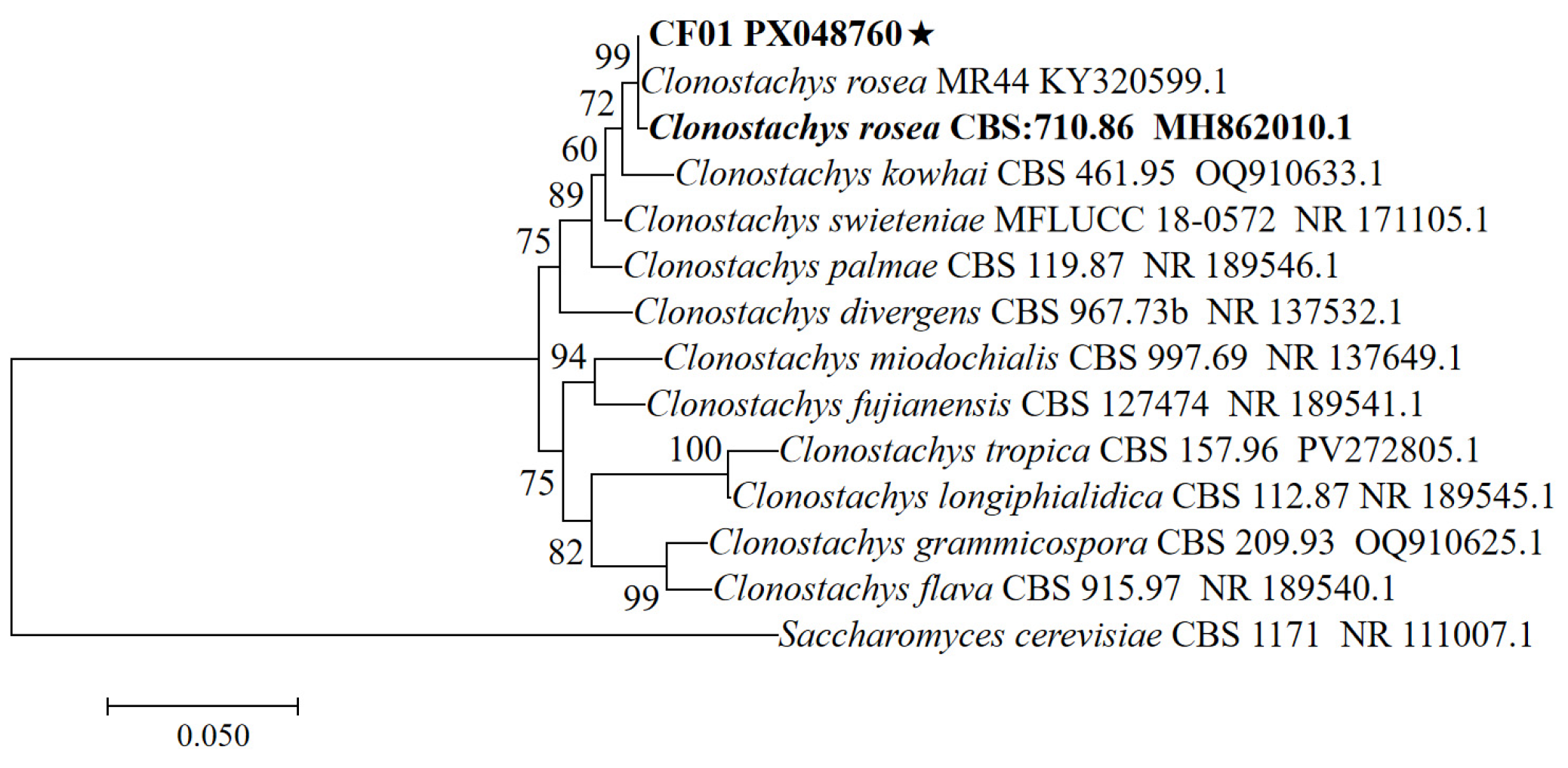
Genomic DNA was extracted from the isolate. Amplification of the ITS region yielded a 576 bp amplicon. Relevant sequences were aligned, as shown in Figure 2. ITS sequences showed 99% similarity between the isolate and C. rosea (KY320599) as well as its type strain (MH862010). The sequences obtained in this study were submitted to GenBank with the accession number PX048760.

3.3. Pathogenicity of C. rosea CF01 Against C. fumipennis Larvae

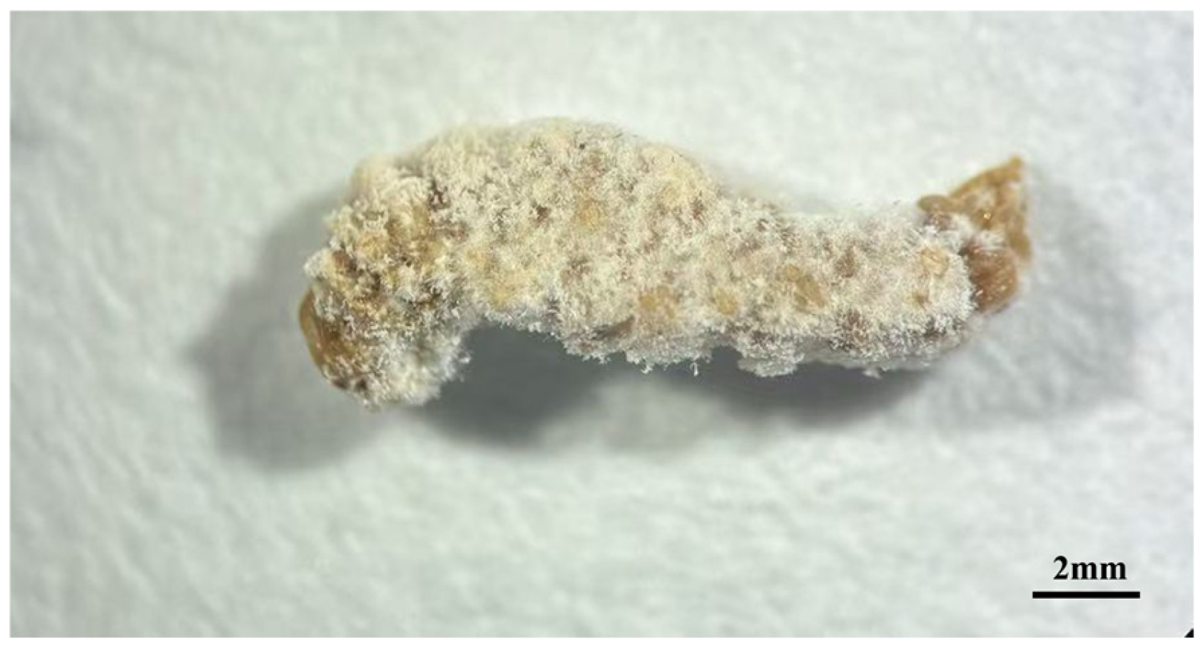
After larvae of C. fumipennis were inoculated with the CF01 strain, cadavers were collected daily, surface-disinfected, and incubated under high humidity. Mycelia first emerged from the head and intersegmental membranes, gradually enveloping the entire larva to form a mummified cadaver (Figure 3).
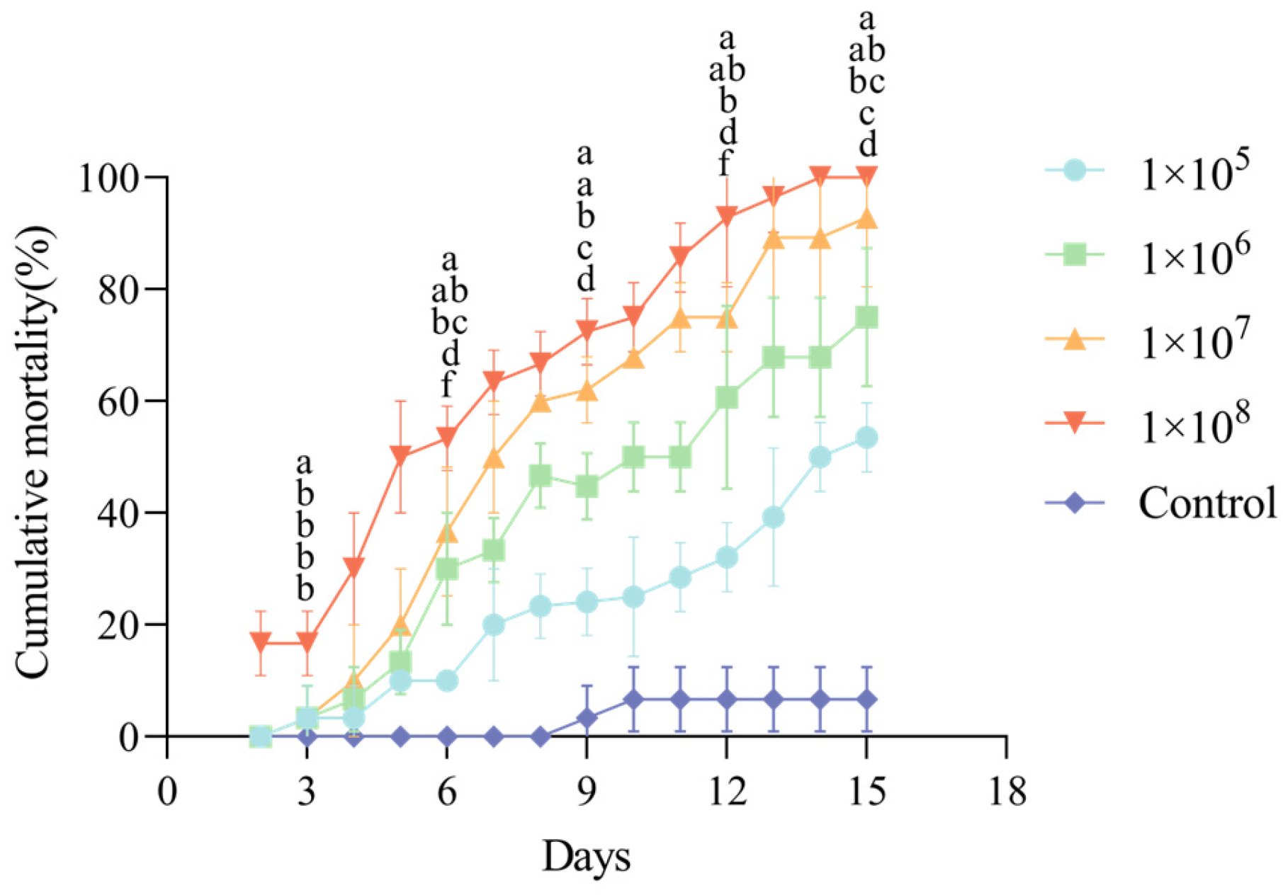
The corrected cumulative mortality of third-instar C. fumipennis larvae exposed to CF01 strain spore suspensions at different concentrations over time is shown in Figure 4. A clear dose- and time-dependent effect was observed; at every tested concentration, mortality rose gradually over time. Statistical analysis revealed that, beginning on day 4, the mortality rate in all treatment groups was significantly higher than that in the control group (p < 0.05).
Spore suspensions of different concentrations demonstrated pathogenicity against third-instar larvae of C. fumipennis. At a concentration of 1 × 10⁸ spores/mL, strain CF01 showed the fastest lethal effect, with an LT50 value of 5.368 days (Table 2). The LT50 increased as the spore concentration decreased. Table 3 presents the LC50 values of strain CF01 against third-instar C. fumipennis larvae at 7, 10, and 14 days post-infection, which decreased over time.

3.4. Optimization of the Culture Conditions of the Isolated Fungus

3.4.1. Optimization of Culture Medium for Strain CF01

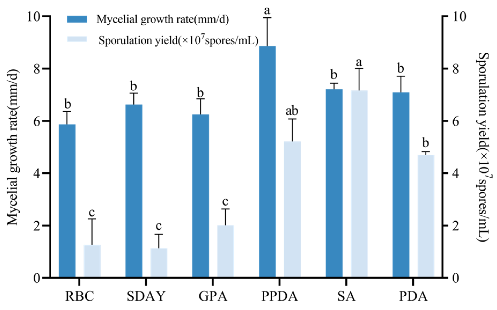
Following 7-day incubation on varied media, strain CF01 exhibited significantly different growth and conidiation. PPDA medium yielded maximum mycelial growth, with the fastest growth rate being 8.86 mm/d (F5,12 = 8.203, p < 0.05), exceeding all other media. Conidial production peaked on SA medium (7.12 × 10⁷ spores/mL), with PPDA and PDA generating 5.22 × 10⁷ and 4.70 × 10⁷ spores/mL, respectively. Minimal sporulation occurred on RBC, SDAY, and GPA media (F5,12 = 35.23, p < 0.001) (Figure 5). Based on the analysis of growth rate and spore production, the optimal medium for the CF01 strain growth is PPDA medium.

3.4.2. Effects of Temperature, Photoperiod, Carbon, and Nitrogen Sources on Growth and Sporulation of Strain CF01

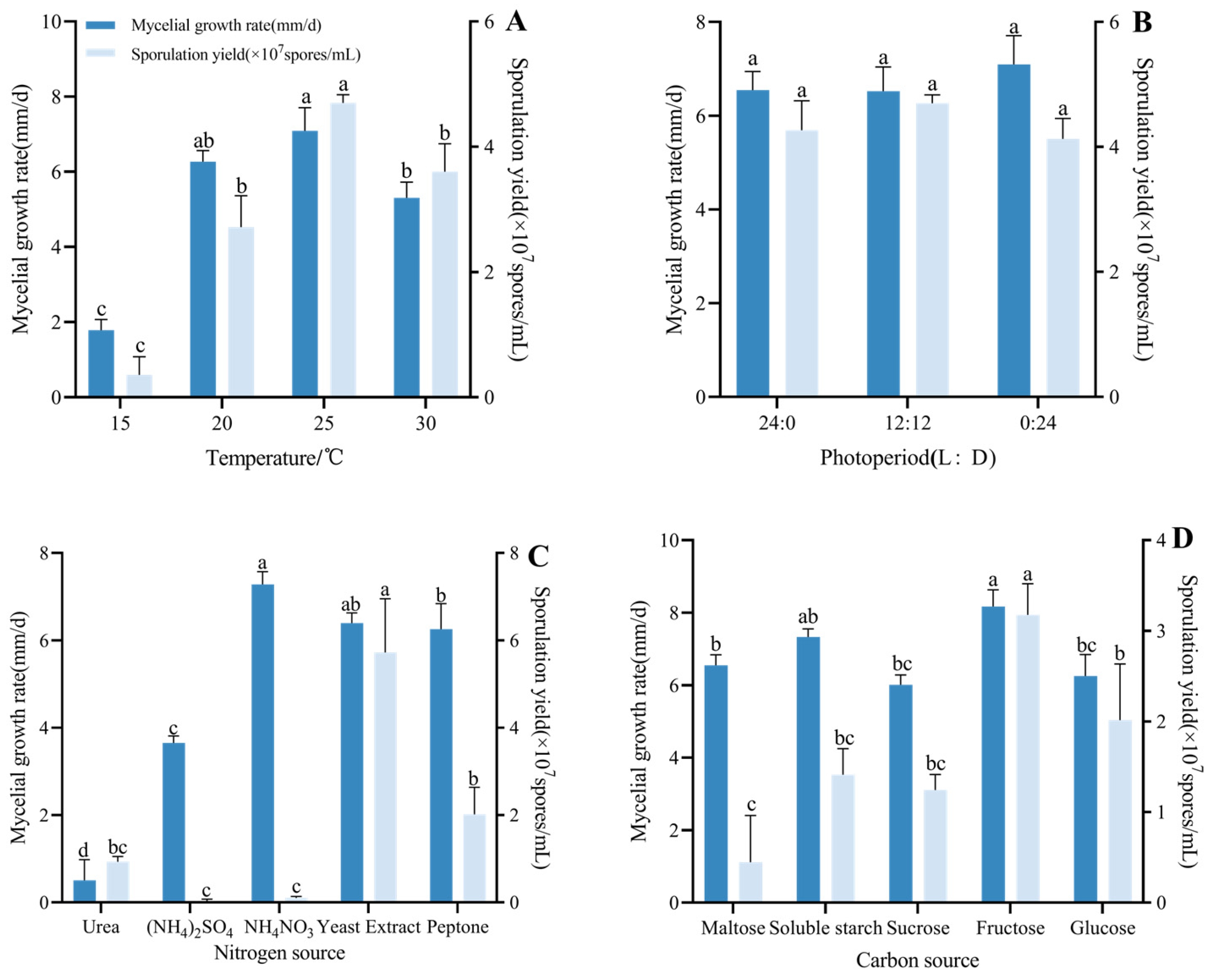
The CF01 strain was capable of growth across a temperature range of 15 to 30 °C. Statistical analysis revealed significant differences in both growth rates and conidial yields among the different temperature treatments (F3,8 = 90.57, p < 0.001). Optimal mycelial growth rate occurred at 25 °C (7.09 mm/d), which was not statistically different from 20 °C, while 15 °C showed a minimal growth rate (Figure 6A). Conidiation peaked at 25 °C (4.70 × 10⁷ spores/mL), significantly exceeding other temperatures (F3,8 = 73.80, p < 0.001). The strains exhibited no significant differences in growth or spore production across the different photoperiod regimes. Maximal mycelial growth rate occurred under continuous darkness (0 L:24 D), while moderate conidiation was observed at 12 L:12 D (Figure 6B).
Regarding nitrogen sources, colonies exhibited the fastest mean daily growth rate when supplied with yeast extract or NH4NO3, demonstrating no significant difference between these two sources. Ammonium nitrate supported a growth rate of 7.32 mm/d. Growth was slowest on urea-containing medium (F4,10 = 148.6, p < 0.001). Furthermore, spore production under yeast leaching powder reached 5.72 × 10⁷ spores/mL, significantly exceeding that of all other tested nitrogen sources (F4,10 = 43.26, p < 0.001). In contrast, ammonium sulfate as a nitrogen source resulted in no spore production, while ammonium nitrate yielded substantially fewer spores at 0.10 × 10⁷ spores/mL; these findings are presented in Figure 6C. Among carbon sources, colonies grew significantly faster on media containing fructose or soluble starch compared to those containing maltose or sucrose. Fructose supported the highest growth rate, attaining 8.17 mm/d (F4,10 = 15.22, p < 0.001). Spore production was also maximal with fructose, reaching 3.18 × 10⁷ spores/mL and significantly exceeding all other tested carbon sources (F4,10 = 17.59, p < 0.001) (Figure 6D). In summary, the optimal carbon source for the CF01 strain is fructose, and the optimal nitrogen source is yeast leaching powder.

4. Discussion

In this study, an entomopathogenic fungal strain was isolated from diseased C. fumipennis larvae. Based on morphological and molecular analyses, the isolate was preliminarily identified as Clonostachys sp. This study relied solely on ITS sequences for species identification, which represents a limitation of our work. Further multi-locus phylogenetic analysis (including genes such as TUB2 and TEF1-α) will be required in subsequent studies to clarify the taxonomic status of strain CF01. This fungus demonstrated notable pathogenicity against C. fumipennis larvae, suggesting its promising potential as a biological control agent. At a conidial concentration of 1 × 10⁸/mL, strain CF01 achieved the most rapid lethality, with a median lethal time (LT50) of 5.368 days. The experiment conducted by Lu et al. also confirmed the pathogenicity of C. rosea against C. chuxiongica, a hymenopteran insect.
Entomopathogenic fungi exhibit broad parasitic host ranges and low resistance development risk, rendering them advantageous for agricultural and forestry pest biocontrol. Despite this potential, C. fumipennis remains understudied regarding its pathogenic fungi. This work presents the first characterization of C. rosea as a larval pathogen of C. fumipennis. Notably, our results contrast with Lu et al. [26] regarding optimal sporulation conditions: whereas they reported ammonium nitrate and urea as maximal sporulation-promoting nitrogen sources at 25 °C with null sporulation on yeast extract powder, we observed peak spore production specifically with yeast extract powder. The data reported by Lu et al. were collected in Xundian county, Kunming city, Yunnan province, China—a region with markedly distinct climatic conditions from those at our study site. Xundian County is characterized by elevated temperatures, greater humidity, and a lower altitude in contrast to the conditions at our study site. These climatic and topographic disparities represent plausible factors contributing to the divergent outcomes between the studies. Nevertheless, the observed mycelial growth kinetics and colonial morphology align with other reports [25].
Clonostachys rosea (Ascomycota, Hypocreales, Bionectriaceae) is a biocontrol fungus widely distributed in Chinese soils. As an endophytic fungus, C. rosea parasitizes diverse hosts, including plants, insects, and nematodes [24], while suppressing phytopathogens such as Sclerotinia sclerotiorum, Fusarium spp., and Rhizoctonia spp. [28]. This fungus colonizes plant roots to provide sustained protection [29], enhances plant growth and seed germination [30], and induces systemic resistance [31]. Its applications target fruit/vegetable gray mold [32], root rot of soybean [33], black scurf on potato [34], barley spot blotch [35], nematodes [36,37], and arthropod pests. C. rosea has been documented as an entomopathogenic fungus isolated from cadavers of various insect hosts, including Oncometopia tucumana and Bemisia tabaci (Hemiptera), C. chuxiongica (Hymenoptera), and Ophrida xanthospilota (Coleoptera) [38]. Additionally, it exhibits pathogenicity against Lepidoptera pests such as the diamondback moth (Plutella xylostella) and Coleoptera, including the grain beetle (Trogoderma granarium) [39,40]. The CF01 strain investigated in this study was isolated from carcasses of deceased C. fumipennis larvae. This finding further expands the known host range and geographical distribution of C. rosea while also contributing new evidence regarding its entomopathogenic potential.
The CF01 strain isolated in this study demonstrated dual functionality: it infests Cephus fumipennis (wheat stem sawfly) while concurrently suppressing Fusarium graminearum (Gibberella zeae), the causal agent of wheat scab [41,42]. This fungus exhibits mycoparasitic activity against F. graminearum [43], providing effective wheat scab control. Its broad-spectrum inhibition of phytopathogenic fungi enables simultaneous management of wheat stem sawfly and associated fungal diseases.
Studies of plant systems confirm that endophytic fungi reduce insect survival [44,45], prompting recent efforts to exploit endophytic colonization for enhanced pest control. For instance, endophytic establishment of Beauveria bassiana in banana plants significantly increased larval mortality of the banana weevil Cosmopolites sordidus [46]. These fungi enhance pest resistance through secondary metabolite production and induce systemic resistance [47,48], while indirectly impacting insect viability via plant-mediated defense responses [49]. Given that C. rosea is a widely colonizing endophyte, its application could similarly improve control efficiency. Colonization strategies for wheat include foliar sprays, root immersion in spore suspensions, or seed treatments, with pre-inoculation leaf surface wounding enhancing endophytic establishment [50]. Such colonization would increase C. fumipennis exposure to fungal spores within wheat tissues, potentially reducing infestation rates [51]. Since C. fumipennis larvae reside concealed within wheat stalks—presenting control challenges—the endophytic pathway represents a promising biological control strategy.
This study confirms the pathogenicity of the CF01 strain toward C. fumipennis larvae. Limitations include the single-generation design, which precluded assessment of genetic and phenotypic stability across successive cycles, and without conducting field experiments, it is difficult to know whether the strain will interact with other soil microorganisms in wheat fields. Future research should therefore evaluate non-target effects on beneficial insects in wheat fields, elucidate its pathogenic mechanisms, include spore germination rate as a critical parameter, and verify whether the CF01 strain can colonize wheat plants to enhance pest-control efficacy under field conditions.

5. Conclusions

In this study, a fungal strain isolated from C. fumipennis was identified as Clonostachys sp. strain CF01 through integrated morphological characterization and ITS rDNA sequence analysis. These findings provide a foundation for deploying entomopathogenic fungi in biological control programs targeting C. fumipennis and support subsequent investigations into its pathogenic mechanisms and rhizosphere colonization dynamics in wheat.

Author Contributions

Conceptualization, M.L. and Y.L.; methodology, M.L.; software, J.L.; validation, M.L. and J.L.; formal analysis, Z.A. and S.W.; investigation, M.L. and Y.L.; resources, Y.L.; data curation, M.L. and J.L.; writing—original draft preparation, M.L.; writing—review and editing, Y.L.; visualization, J.L.; supervision, Y.L.; project administration, Y.L.; funding acquisition, Y.L. All authors have read and agreed to the published version of the manuscript.

Funding

This research was funded by the Applied Fundamental Research Fund of Qinghai Provincial Department of Science and Technology (Grant NO. 2023ZJ739).

Institutional Review Board Statement

Not applicable.

Informed Consent Statement

Not applicable.

Data Availability Statement

Data are contained within the article.

Acknowledgments

The authors would like to thank all the students and teachers in the Key Laboratory of Comprehensive Management of Agricultural Pests in Qinghai Province for their help, with special thanks to the students who collected and reared the insects.

Conflicts of Interest

The authors declare no conflicts of interest.

References

  1. Naito, T.; Smith, D.R.; Huang, F.S. Records of Cephidae (Hymenoptera) from China and Southeastern Asia with two new species of Janus Stephens. Jpn. J. Syst. Entomol. Matsuyama 1998, 4, 237–242. [Google Scholar]
  2. Hoelmer, K.A.; Shanower, T.G. Foreign exploration for natural enemies of cephid sawflies. J. Agric. Urban Entomol. 2004, 21, 223–238. [Google Scholar]
  3. Zhao, L.M. Morphology of Cephus fumipennis (Hymenoptera: Cephidae) andComparison with Other Three Cephids. Northwest J. Agric. 2011, 20, 167–173. [Google Scholar]
  4. Zhao, L.M.; Zhang, H.L. Economic-Injury Level and Economic Threshold of Cephus fumipennis (Hymenoptera: Cephidae). Northwest J. Agric. 2008, 17, 65–69. [Google Scholar]
  5. Chen, S.; Hoelmer, K.A.; Chen, H.; Liu, A. A Review of Wheat Stem Sawfly (Hymenoptera: Cephidae). J. Agric. Urban Entomol 2004, 21, 249–256. [Google Scholar]
  6. Rand, T.A.; Morrill, W.L.; Runyon, J.B.; Hoelmer, K.A.; Shanower, T.G.; Littlefield, J.L.; Weaver, D.K. Assessing phenological synchrony between the Chinese sawfly, Cephus fumipennis (Hymenoptera: Cephidae), its egg-larval parasitoid, Collyria catoptron (Hymenoptera: Ichneumonidae), and the North American sawfly, Cephus cinctus: Implications for biological control. Can. Entomol. 2016, 148, 482–492. [Google Scholar]
  7. Wu, X.H.; Héctor, C.A.; Pang, B.P. Progress in wheat stem wasp research. Plant Prot. 2016, 42, 18–26. [Google Scholar]
  8. Limin, Z. Identification of the natural resistance of several spring wheat varieties to Cephus fumipennis Eversmann. Acta Agric. Boreali-Occident. Sin. 2000, 9, 39–40. [Google Scholar]
  9. Wahl, D.B.; Shanower, T.G.; Hoelmer, K.A. A new species of Collyria Schiødte (Hymenoptera: Ichneumonidae: Collyriinae), a parasitoid of Cephus fumipennis (Hymenoptera: Cephidae) in China, and potential biological control agent for Cephus cinctus in North America. J. Kans. Entomol. Soc. 2007, 80, 43–50. [Google Scholar] [CrossRef]
  10. Runyon, J.; Hurley, R.; Morrill, W.; Weaver, D. Distinguishing adults of Bracon cephi and Bracon lissogaster (Hymenoptera: Braconidae), parasitoids of the wheat stem sawfly (Hymenoptera: Cephidae). Can. Entomol. 2001, 133, 215–217. [Google Scholar] [CrossRef]
  11. Nelson, W.; Farstad, C. Biology of Bracon cephi (Gahan) (Hymenoptera: Braconidae), An Important Native Parasite of the Wheat Stem Sawfly, Cephus cinctus Nort. (Hymenoptera: Cephidae), in Western Canada. Can. Entomol. 1953, 85, 103–107. [Google Scholar] [CrossRef]
  12. Morrill, W.L.; Weaver, D.K.; Irish, N.J.; Barr, W.F. Phyllobaenus dubius (Wolcott) (Coleoptera: Cleridae), a new record of a predator of the wheat stem sawfly (Hymenoptera: Cephidae). J. Kans. Entomol. Soc. 2001, 73, 181–183. [Google Scholar]
  13. Portman, S.L.; Krishnankutty, S.M.; Reddy, G.V. Entomopathogenic nematodes combined with adjuvants presents a new potential biological control method for managing the wheat stem sawfly, Cephus cinctus (Hymenoptera: Cephidae). PLoS ONE 2016, 11, e0169022. [Google Scholar] [CrossRef] [PubMed]
  14. Wenda-Piesik, A.; Sun, Z.; Grey, W.E.; Weaver, D.K.; Morrill, W.L. Mycoses of wheat stem sawfly (Hymenoptera: Cephidae) larvae by Fusarium spp. isolates. Environ. Entomol. 2009, 38, 387–394. [Google Scholar] [CrossRef] [PubMed]
  15. Piesik, D.; Wenda-Piesik, A.; Weaver, D.K.; Macedo, T.B.; Morrill, W.L. Influence of Fusarium and wheat stem sawfly infestation on volatile compounds production by wheat plants. J. Plant Prot. Res. 2009, 49, 167–174. [Google Scholar] [CrossRef]
  16. Sun, Z. The Pathogenicity of Fusarium spp. to Wheat Stem Sawfly, Cephus Cinctus Norton (Hymenoptera: Cephidae); Montana State University: Bozeman, MT, USA, 2008. [Google Scholar]
  17. Jaber, L.R.; Ownley, B.H. Can we use entomopathogenic fungi as endophytes for dual biological control of insect pests and plant pathogens? Biol. Control 2018, 116, 36–45. [Google Scholar] [CrossRef]
  18. Singh, D.; Raina, T.K.; Singh, J. Entomopathogenic fungi: An effective biocontrol agent for management of insect populations naturally. J. Pharm. Sci. Res. 2017, 9, 833. [Google Scholar]
  19. Maina, U.; Galadima, I.; Gambo, F.; Zakaria, D. A review on the use of entomopathogenic fungi in the management of insect pests of field crops. J. Entomol. Zool. Stud. 2018, 6, 27–32. [Google Scholar]
  20. Koul, O. Biorationals and Biopesticides: Pest Management; Springer International Publishing: Berlin, Germany, 2023. [Google Scholar]
  21. Shahid, A.A.; Rao, Q.A.; Bakhsh, A.; Husnain, T. Entomopathogenic fungi as biological controllers: New insights into their virulence and pathogenicity. Arch. Biol. Sci. 2012, 64, 21–42. [Google Scholar] [CrossRef]
  22. Wang, C.; Feng, M.-G. Advances in fundamental and applied studies in China of fungal biocontrol agents for use against arthropod pests. Biol. Control 2014, 68, 129–135. [Google Scholar] [CrossRef]
  23. Mora, M.A.E.; Castilho, A.M.C.; Fraga, M.E. Classification and infection mechanism of entomopathogenic fungi. Arq. Do Inst. Biológico 2017, 84, e0552015. [Google Scholar] [CrossRef]
  24. Sun, Z.-B.; Li, S.-D.; Ren, Q.; Xu, J.-L.; Lu, X.; Sun, M.-H. Biology and applications of Clonostachys rosea. J. Appl. Microbiol. 2020, 129, 486–495. [Google Scholar] [CrossRef] [PubMed]
  25. Toledo, A.V.; Virla, E.; Humber, R.A.; Paradell, S.L.; Lastra, C.L. First record of Clonostachys rosea (Ascomycota: Hypocreales) as an entomopathogenic fungus of Oncometopia tucumana and Sonesimia grossa (Hemiptera: Cicadellidae) in Argentina. J. Invertebr. Pathol. 2006, 92, 7–10. [Google Scholar] [CrossRef] [PubMed]
  26. Lu, J.; Xia, J.; Zhuang, F.; Xu, R.; Zeng, T.; Li, Y. Identification and biological characterization of mycoparasitic fungus Clonostachys rosea isolated from sawfly Cephalcia chuxiongica. J. Plant Prot. 2018, 45, 622–631. [Google Scholar] [CrossRef]
  27. Wei, J.C. Fungi Identification Manual; Shanghai Scientific & Technical Publishers: Shanghai, China, 1979; pp. 58–90. [Google Scholar]
  28. Rodriguez, M.A.; Rothen, C.; Lo, T.E.; Cabrera, G.M.; Godeas, A.M. Suppressive soil against Sclerotinia sclerotiorum as a source of potential biocontrol agents: Selection and evaluation of Clonostachys rosea BAFC1646. Biocontrol Sci. Technol. 2015, 25, 1388–1409. [Google Scholar] [CrossRef]
  29. Dubey, M.K.; Jensen, D.F.; Karlsson, M. Hydrophobins are required for conidial hydrophobicity and plant root colonization in the fungal biocontrol agent Clonostachys rosea. BMC Microbiol. 2014, 14, 18. [Google Scholar] [CrossRef]
  30. Roberti, R.; Veronesi, A.; Cesari, A.; Cascone, A.; Di Berardino, I.; Bertini, L.; Caruso, C. Induction of PR proteins and resistance by the biocontrol agent Clonostachys rosea in wheat plants infected with Fusarium culmorum. Plant Sci. 2008, 175, 339–347. [Google Scholar] [CrossRef]
  31. Walters, D.R.; Ratsep, J.; Havis, N.D. Controlling crop diseases using induced resistance: Challenges for the future. J. Exp. Bot. 2013, 64, 1263–1280. [Google Scholar] [CrossRef]
  32. Borges, Á.V.; Saraiva, R.M.; Maffia, L.A. Biocontrol of gray mold in tomato plants by Clonostachys rosea. Trop. Plant Pathol. 2015, 40, 71–76. [Google Scholar] [CrossRef]
  33. Bienapfl, J.; Floyd, C.; Percich, J.; Malvick, D. First report of Clonostachys rosea causing root rot of soybean in the United States. Plant Dis. 2012, 96, 1700. [Google Scholar] [CrossRef]
  34. Salamone, A.L.; Gundersen, B.; Inglis, D.A. Clonostachys rosea, a potential biological control agent for Rhizoctonia solani AG-3 causing black scurf on potato. Biocontrol Sci. Technol. 2018, 28, 895–900. [Google Scholar] [CrossRef]
  35. Jensen, B.; Lübeck, P.S.; Jørgensen, H.J. Clonostachys rosea reduces spot blotch in barley by inhibiting prepenetration growth and sporulation of Bipolaris sorokiniana without inducing resistance. Pest Manag. Sci. 2016, 72, 2231–2239. [Google Scholar] [CrossRef]
  36. Zhang, L.; Yang, J.; Niu, Q.; Zhao, X.; Ye, F.; Liang, L.; Zhang, K.-Q. Investigation on the infection mechanism of the fungus Clonostachys rosea against nematodes using the green fluorescent protein. Appl. Microbiol. Biotechnol. 2008, 78, 983–990. [Google Scholar] [CrossRef]
  37. Iqbal, M.; Dubey, M.; McEwan, K.; Menzel, U.; Franko, M.A.; Viketoft, M.; Jensen, D.F.; Karlsson, M. Evaluation of Clonostachys rosea for control of plant-parasitic nematodes in soil and in roots of carrot and wheat. Phytopathology 2018, 108, 52–59. [Google Scholar] [CrossRef]
  38. Anwar, W.; Ali, S.; Nawaz, K.; Iftikhar, S.; Javed, M.A.; Hashem, A.; Alqarawi, A.A.; Abd_Allah, E.F.; Akhter, A. Entomopathogenic fungus Clonostachys rosea as a biocontrol agent against whitefly (Bemisia tabaci). Biocontrol Sci. Technol. 2018, 28, 750–760. [Google Scholar] [CrossRef]
  39. Mohammed, A.A.; Younus, A.S.; Ali, A.N. Efficacy of Clonostachys rosea, as a promising entomopathogenic fungus, against coleopteran stored product insect pests under laboratory conditions. Egypt. J. Biol. Pest Control 2021, 31, 55. [Google Scholar] [CrossRef]
  40. Zhang, Z.Y.; Lu, J.J.; Yang, M.G.; Ma, Y.Q.; Tian, Y.J.; Gou, C.J.; Liu, M. Virulence Determination and Optimization of Toxin Production Conditions of Biocontrol Strains of Plutella xylostella Larvae. Shandong Agric. Sci. 2024, 56, 132–137. [Google Scholar] [CrossRef]
  41. Hue, A.; Voldeng, H.; Savard, M.; Fedak, G.; Tian, X.; Hsiang, T. Biological control of fusarium head blight of wheat with Clonostachys rosea strain ACM941. Can. J. Plant Pathol. 2009, 31, 169–179. [Google Scholar] [CrossRef]
  42. Kulik, T.; Treder, K.; Rochoń, M.; Załuski, D.; Sulima, P.; Olszewski, J.; Bilska, K.; Elena, G.; Kowalski, T. Measurement of the effectiveness of Clonostachys rosea in reducing Fusarium biomass on wheat straw. J. Appl. Genet. 2024, 65, 937–947. [Google Scholar] [CrossRef]
  43. Demissie, Z.A.; Witte, T.; Robinson, K.A.; Sproule, A.; Foote, S.J.; Johnston, A.; Harris, L.J.; Overy, D.P.; Loewen, M.C. Transcriptomic and exometabolomic profiling reveals antagonistic and defensive modes of Clonostachys rosea action against Fusarium graminearum. Mol. Plant-Microbe Interact. 2020, 33, 842–858. [Google Scholar] [CrossRef]
  44. Braman, S.; Duncan, R.; Engelke, M.; Hanna, W.; Hignight, K.; Rush, D. Grass species and endophyte effects on survival and development of fall armyworm (Lepidoptera: Noctuidae). J. Econ. Entomol. 2002, 95, 487–492. [Google Scholar] [CrossRef] [PubMed]
  45. Afkhami, M.E.; Rudgers, J.A. Endophyte-mediated resistance to herbivores depends on herbivore identity in the wild grass Festuca subverticillata. Environ. Entomol. 2009, 38, 1086–1095. [Google Scholar] [CrossRef] [PubMed]
  46. Akello, J.; Dubois, T.; Coyne, D.; Kyamanywa, S. Endophytic Beauveria bassiana in banana (Musa spp.) reduces banana weevil (Cosmopolites sordidus) fitness and damage. Crop Prot. 2008, 27, 1437–1441. [Google Scholar] [CrossRef]
  47. Arnold, A.E.; Mejía, L.C.; Kyllo, D.; Rojas, E.I.; Maynard, Z.; Robbins, N.; Herre, E.A. Fungal endophytes limit pathogen damage in a tropical tree. Proc. Natl. Acad. Sci. USA 2003, 100, 15649–15654. [Google Scholar] [CrossRef]
  48. Rodriguez, R.; White, J., Jr.; Arnold, A.E.; Redman, R.S. Fungal endophytes: Diversity and functional roles. New Phytol. 2009, 182, 314–330. [Google Scholar] [CrossRef]
  49. Hartley, S.E.; Eschen, R.; Horwood, J.M.; Gange, A.C.; Hill, E.M. Infection by a foliar endophyte elicits novel arabidopside-based plant defence reactions in its host, Cirsium arvense. New Phytol. 2015, 205, 816–827. [Google Scholar] [CrossRef]
  50. Anderson, C.M.; McGee, P.A.; Nehl, D.B.; Mensah, R.K. The fungus Lecanicillium lecanii colonises the plant Gossypium hirsutum and the aphid Aphis gossypii. Aust. Mycol. 2007, 26, 65–70. [Google Scholar]
  51. Portman, S.L.; Jaronski, S.T.; Weaver, D.K.; Reddy, G.V. Advancing biological control of the wheat stem sawfly: New strategies in a 100-yr struggle to manage a costly pest in the Northern Great Plains. Ann. Entomol. Soc. Am. 2018, 111, 85–91. [Google Scholar] [CrossRef]
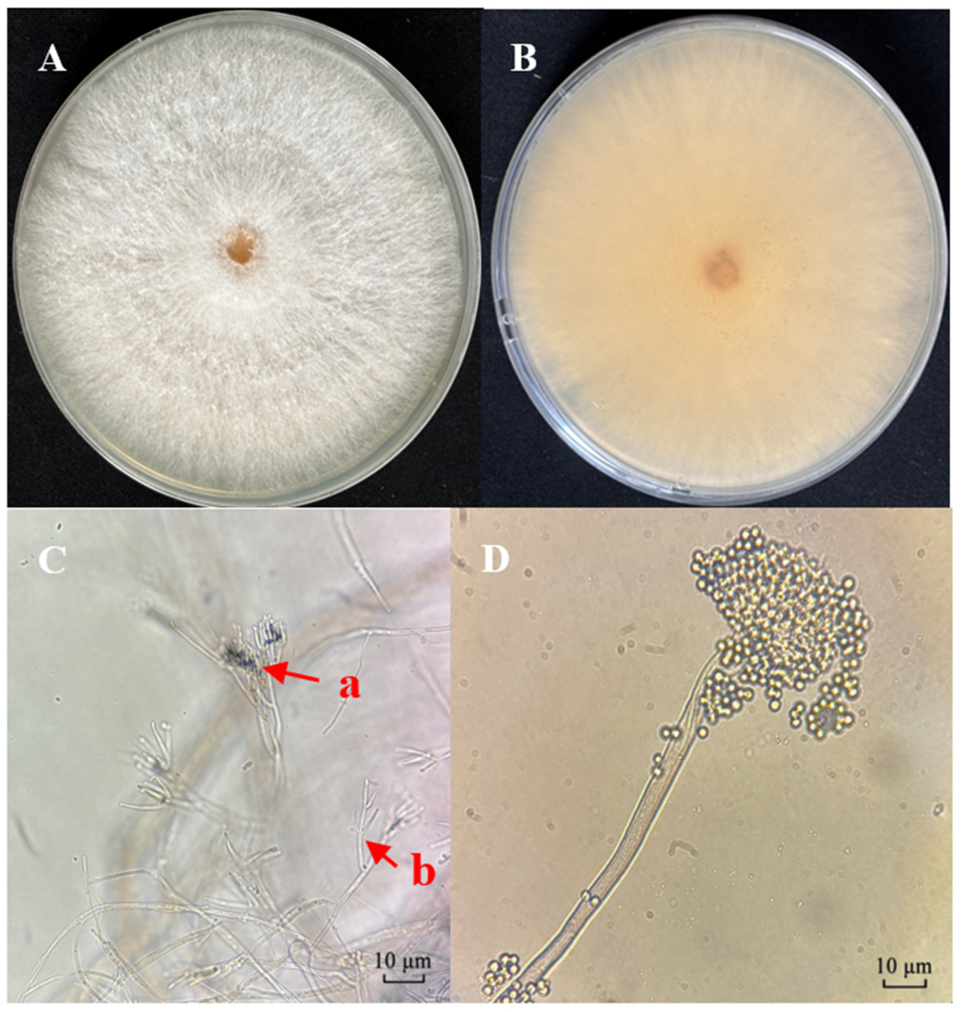
Figure 1. Morphological characteristics of C. rosea strain CF01. (A) Colony surface on PDA medium after 10 days. (B) Colony reverse on PDA medium. (C) (a) Penicillate-type conidiophores. (b) Verticillate-type conidiophore. (D) Conidia.
Biology 14 01240 g001
Figure 2. Phylogenetic analysis of the CF01 strain and related taxa based on ITS sequence alignment. Tree reconstructed using the neighbor-joining method (bootstrap values from 1000 replicates shown at nodes). The scale bar indicates 0.05 substitutions per site. MH862010 is C. rosea’s type strain. The asterisk denotes the CF01 strain.
Biology 14 01240 g002
Figure 3. Mycosis progression in third-instar Cephus fumipennis larvae inoculated with CF01 strain (1 × 10⁷ spores/mL).
Biology 14 01240 g003
Figure 4. Pathogenicity of the CF01 strain on third instar larvae of the C. fumipennis. The mortality rate trend over time (in days) after insect treatment with four different concentrations of CF01. Data are mean ± SE. Letters on the error bars indicate significant differences analyzed using ANOVA with the LSD test (p < 0.05).
Biology 14 01240 g004
Figure 5. Mycelial growth rate and sporulation yield of CF01 on different media. Data are mean ± SE. Letters on the error bars indicate significant differences analyzed by ANOVA with the LSD test (p < 0.05).
Biology 14 01240 g005
Figure 6. Effects of different temperatures (A), photoperiods (B), carbon (C), and nitrogen (D) sources on mycelial growth rate and sporulation yield in the CF01 strain. Data are mean ± SE. Letters on the error bars indicate significant differences analyzed by ANOVA with the LSD test (p < 0.05).
Biology 14 01240 g006
Table 1. Test media and their formula.
MediumMedium Component
Potato Dextrose Agar (PDA)Peeled potato 200 g/L, Dextrose 20 g/L, Agar 18 g/L, pH unadjusted
Starch Agar (SA)soluble starch 40 g/L, yeast paste 5 g/L, Agar 20 g/L, pH unadjusted
Dextrose Peptone Agar (GPA)Peptone 10 g/L, Dextrose 40 g/L, Agar 20 g/L, pH unadjusted
Rose Bengal Chloramphenicol Agar (RBC)Peptone 5.0 g/L, Glucose 10.0 g/L, KH2PO4
1.0 g/L, MgSO4 0.5 g/L, 1/3000 Rose Bengal red 100 mL/L, Agar 18.0 g/L, pH unadjusted
Peptone Potato Dextrose Agar (PPDA)Peeled potato 200 g/L, Glucose 20 g/L, Peptone 20 g/L, Agar 18 g/L, pH unadjusted
Sabouraud Dextrose Agar with Yeast Extract (SDAY)Glucose 40 g/L, Yeast paste 10 g/L, Peptone 10 g/L, Agar 20 g/L, pH unadjusted
Table 2. Virulence regression equations for LT50 values of C. rosea CF01 against C. fumipennis.
Concentration (Spores/mL)Virulence Regression EquationLT50 (Days)R295%CI (Days)
1 × 10⁸y = 3.566x − 2.6035.3680.8694.356–6.307
1 × 10⁷y = 4.500x − 3.9367.4930.9866.467–8.513
1 × 10⁶y = 3.462x − 3.4279.7680.9768.343–11.792
1 × 10⁵y = 2.860x − 3.40715.5350.95412.252–27.060
Table 3. Virulence regression equations for LC50 values of C. rosea CF01 against C. fumipennis.
DaysVirulence Regression EquationLC50 (Days)R295%CI (Days)
7y = 0.397x − 2.8121.217 × 1070.9987.178 × 105–5.285 × 10¹⁵
10y = 0.452x − 2.8141.682 × 1060.9499.011 × 103–2.725 × 10⁷
14y = 0.720x − 3.7061.405 × 1050.9794.220 × 102–6.688 × 10⁵
Disclaimer/Publisher’s Note: The statements, opinions and data contained in all publications are solely those of the individual author(s) and contributor(s) and not of MDPI and/or the editor(s). MDPI and/or the editor(s) disclaim responsibility for any injury to people or property resulting from any ideas, methods, instructions or products referred to in the content.