Article

Dysregulation of Protein Kinase CaMKI Leads to Autism-Related Phenotypes in Synaptic Connectivity, Sleep, Sociality, and Aging-Dependent Degeneration in Drosophila

1
Department of Biological Sciences, University of Maryland, Baltimore County, Baltimore, MD 21250, USA
2
Department of Chemistry and Biochemistry, University of Maryland, College Park, MD 20742, USA
3
Department of Neurosurgery, Yale School of Medicine, New Haven, CT 06510, USA
*
Author to whom correspondence should be addressed.
These authors contributed equally to this work.
Current address: Medical College of Wisconsin, Pharmacy School, Milwaukee, WI 53226, USA.
§
Current address: School of Medicine, University of Maryland, Baltimore, MD 21201, USA.
Biology 2025, 14(9), 1228; https://doi.org/10.3390/biology14091228
Submission received: 17 July 2025 / Revised: 13 August 2025 / Accepted: 30 August 2025 / Published: 9 September 2025

Simple Summary

This study explores the role of the gene CaMKI (calcium/calmodulin-dependent protein kinase I) in fruit flies, which is similar to the human gene CAMK4. CAMK4 has recently been linked to autism spectrum disorder (ASD), a condition that affects social behavior and is often accompanied by sleep problems and motor difficulties. While it is known that CAMK4 is connected to ASD, the exact ways it influences the brain and behavior are still unclear. We found that reducing CaMKI levels in flies affected their sleep patterns, day/night activity cycles, and social interactions. Because ASD patients are also more likely to develop dementia, we tested if altering CaMKI would influence age-related changes. Indeed, flies with reduced CaMKI showed faster declines in movement ability and signs of cell damage as they aged. Another theory about ASD suggests that it may involve problems with how connections between nerve cells, called synapses, are refined during development. In our experiments, reducing CaMKI caused wiring errors in the fly nervous system, which supports this idea. We also analyzed the proteins present in flies with altered CaMKI and identified several molecules that could explain these effects. Overall, our results suggest that CaMKI influences both early development and age-related brain changes. This work may provide new clues about how genetic changes in CAMK4 might contribute to ASD in humans and could help guide the development of future treatments.

Abstract

Autism spectrum disorder (ASD) encompasses a range of conditions, primarily marked by deficits in social behaviors, along with several comorbidities such as sleep abnormalities and motor dysfunction. Recent studies have identified genetic risk factors associated with ASD, including the CAMK4 (calcium/calmodulin-dependent protein kinase 4). However, the molecular mechanisms linking CAMK4 dysregulation and ASD-associated phenotypes remain poorly understood. Here, we used Drosophila melanogaster as a model system to investigate ASD-associated phenotypes in flies with dysregulated CaMKI, the fly homolog of mammalian CAMK4. We show that CaMKI manipulations affect sleep, circadian rhythmicity, and social behavior. Consistent with the higher prevalence of dementia observed in autistic patients, we also observed a significantly enhanced behavioral decline in motor performance and dendritic degeneration in flies expressing RNAi-based CaMKI knockdown in flight motoneurons, suggesting a link between developmental and degenerative processes. As aberrant synaptic pruning is hypothesized to underlie the synaptic phenotypes observed in brains of autistic patients, we examined synaptic phenotypes following CaMKI manipulations using the larval neuromuscular junction (NMJ) and observed miswiring phenotypes suggesting aberrant synaptic refinement. We performed shotgun mass-spectrometry proteomics and identified various molecular candidates, particularly molecules involved in cytoskeleton regulation and chemorepulsion, likely to regulate the phenotypes described here. Thus, our results suggest that CaMKI plays a role in developmental processes and influences aging-dependent degenerative processes, possibly providing mechanistic insight into the genetic basis of ASD etiology and the development of effective treatments.

1. Introduction

Postmortem studies reveal increased density of synapses in brains of individuals with autism spectrum disorder (ASD) [1,2,3]. These observations have led to the hypothesis that defects in synaptic pruning may contribute to ASD pathology [4,5,6,7,8]. Synaptic pruning, also called synaptic elimination or refinement, is a neuroplastic process involving the removal of ectopic synapses formed in the initial stages of neuronal development [9,10]. Studies in vertebrates and invertebrates indicate that a crucial factor regulating synaptic pruning is neuronal activity [11,12,13], which was first hypothesized by Nobel laureates Hubel and Wiesel in the 1960s [14,15]. Studies at the vertebrate retina [16,17] as well as the mammalian and Drosophila neuromuscular junction (NMJ) [18,19] indicate that waves of prenatal electrical activity and calcium (Ca2+) oscillations regulate the withdrawal of off-target synaptic contacts [13]. Ca2+ oscillations regulate Ca2+-dependent molecular factors such as kinases (e.g., CaMKII [18,19]), phosphatases (e.g., Calcineurin [19]), and Ca2+-dependent adenylyl cyclases (e.g., Rutabaga [20]), which in turn regulate intracellular cyclic AMP (cAMP) levels for synaptic pruning in mammalian visual neurons [21,22] and at the Drosophila NMJ [20]. Whereas a model to explain the refinement of synaptic connections often involves mechanisms based on Hebbian-like, spike-timing correlation between synaptic partners, where asynchronous inputs are removed [23], alternative non-Hebbian mechanisms have also been proposed that rely on activity-dependent modulation of chemorepulsion [13]. Although some molecular aspects underlying activity-dependent synaptic refinement have recently been unraveled [13,24,25], what molecules link dynamic levels of neuronal activity and the refinement of synaptic contacts during development remain to be elucidated. Whereas some anatomical differences are observed between brains of humans and animal models, including rodents and flies, a large body of the literature indicates a high level of conservation in basic organizational principles for neuronal network structure, assembly and function, ranging from neuronal and synaptic anatomy to biophysical and electrical properties [26,27,28,29,30,31]. Consistent with its contributions to advance our understanding of synaptic development, Drosophila melanogaster has emerged as a suitable genetic model system for investigating fundamental neuronal mechanisms associated with ASD [32,33,34,35].
CAMK4 (calcium/calmodulin-dependent protein kinase 4) has recently been identified as a risk gene for autism spectrum disorder [36]. Evidence from various studies using whole-exome sequencing, candidate gene association, and copy number variation indicates numerous genetic variations in CAMK4 in populations diagnosed with ASD [37,38,39,40]. Consistently, the Simons Foundation Autism Research Initiative (SFARI) gives CAMK4 a SFARI Gene Score of two, signifying it as a strong candidate autism risk gene. Although CAMK4 is known to encode a serine/threonine protein kinase involved in a Ca2+-calmodulin dependent pathway that regulates synaptic plasticity and other neuronal functions, experimental evidence linking CAMK4 function with autism-related phenotypes remains elusive. CAMK4 is evolutionary conserved from yeast, worms, and flies to zebrafish, rodents, and humans, including the Drosophila ortholog CaMKI. In response to social experience, Drosophila CaMKI is known to activate the histone-acetyl transferase CBP, which enhances the efficacy of Juvenile Hormone and the transcription of fruM in male olfactory neurons to modulate pheromone detection and courtship behavior [41,42]. However, whether dysregulation of CaMKI signaling leads to autism-associated phenotypes remains an unexplored question.
Here, we used Drosophila as a model to test CaMKI manipulations and their effect on autism-related phenotypes. Despite limitations of experimental animal models in resembling human neurotypical behaviors and clinical symptoms, their contribution to the understanding of the etiology and pathogenesis of human diseases, including ASD, is widely accepted considering the high degrees of evolutionary conservation of the nervous system and behaviors [43,44]. Consistently, social behavior is observed in various animal taxa ranging from insects to large mammals [44,45,46], and sleep has been well documented throughout the entire animal kingdom, ranging from the nematode worm Caenorhabditis elegans to elephants [47]. ASD involves a spectrum of disorders/conditions, mainly characterized by deficits in social behaviors, which can also include various comorbidities such as sleep abnormalities [48,49,50,51,52]. Therefore, we tested whether CaMKI manipulations had an effect on ASD-related behaviors such as sleep and sociality in adult CaMKI loss-of-function mutants (CaMKILOF) as well as in animals expressing pan-neuronal CaMKI knockdown. Following both manipulations, we observed that the disruption of CaMKI function led to increased sleep levels, but CaMKILOF flies showed decreases in sociality and circadian rhythmicity.
Moreover, recent studies indicate a higher prevalence of dementia diagnosis in ASD patients [53,54,55,56,57]. However, whether genes that regulate neuronal development can affect neurodegeneration processes remains poorly understood. Consistent with previous studies establishing Drosophila as a key model for aging research [58,59], we tested the effects of CaMKI manipulations on aging flies and observed that flight performance declined more severely in flies expressing RNAi-knockdown of CaMKI. Anatomical analysis revealed that this behavioral phenotype correlated with enhanced degeneration of motoneuron dendritic architecture. These phenotypes in the motor network and locomotion performance are consistent with the high prevalence of motor dysfunction observed in ASD patients [60,61], considered a comorbidity in ASD. From our proteomic analysis, we identified the β-amyloid precursor protein binding protein 1 (APP-BP1) [62] as well as the kinase Par1 [63] as molecular candidates linking neurodevelopment and degeneration. Thus, our results suggest that CaMKI plays a role in developmental processes and influences aging-dependent degenerative processes.
To assess synaptic perturbations at the anatomical level, we used the larval neuromuscular junction (NMJ) as previous studies have established it as a powerful model to study various aspects of synaptic connectivity including activity-dependent synaptic refinement and plasticity [13,64,65,66,67,68,69,70,71,72,73,74]. Thus, we performed anatomical analysis of NMJ connectivity and observed an increase in the frequency of ectopic connections in CaMKILOF mutants as well as in animals expressing pan-neuronal CaMKI knockdown. Epistatic genetic interaction tests suggest that CaMKI may be part of a different molecular pathway than the activity-dependent regulation of PlexB/Sema2a chemorepulsion [13]. Therefore, to identify molecular mechanisms underlying the phenotypic alterations described here, we performed shotgun proteomics on CaMKILOF mutants to reveal genes that show significant changes in protein levels and phospho-peptides. We identified CaMKI-dependent interactions with the cAMP-phosphodiesterase Dunce and the phosphatase CanB that we have previously described to regulate synaptic refinement [19,20]. Thus, dynamic phosphorylation levels of target proteins can be hypothesized to be regulated by an interplay between CaMKI-dependent phosphorylation and CanB-dependent dephosphorylation. Interestingly, additional neuronal proteins identified in our dataset known to regulate neuronal anatomy and cytoskeleton are the small GTPase Cdc42 that regulates the actin cytoskeleton and cell polarity [75], and Futsch, a MAP1B-like protein required for dendritic and axonal development implicated in the regulation of microtubule dynamics [76]. Furthermore, we identified several proteins known to regulate repulsive axon guidance mediated by the presynaptic Plexin A (PlexA) receptor [77,78]. These include the actin regulatory oxidation–reduction enzyme MICAL-like that directly bind and disassemble actin filaments (F-actin) [79], the A kinase anchoring protein (AKAP) Nervy [80], and the phospho-binding protein 14-3-3ε [81]. Therefore, our results point to an alternative signaling pathway that involves PlexA in the regulation of synaptic connectivity.

2. Materials and Methods

2.1. Fly Stocks

Flies were kept at 25 °C in a 12/12 h light/dark incubator on standard plastic vials, using a regular cornmeal/molasses food as described before [82,83]. As the wildtype line, we used DGRP-774 (RRID:BDSC_25205) from the Drosophila Genome Reference Panel lines, which are fully sequenced inbred stocks derived from a natural population from Raleigh, NC [84]. The following fly lines were obtained from the Bloomington Drosophila Stock Center: CaMKILOF (RRID:BDSC_16799) [41]; elavC155-GAL4; UAS-Dicer (RRID:BDSC_25750); UAS-CaMKI-RNAi-1 (RRID:BDSC_41900) [85,86]; UAS-CaMKI-RNAi-2 (RRID:BDSC_35362) [41,85,86,87]. Mutant lines used in genetic interaction tests as previously described [18,19] are as follows: CaMKIILOF (RRID:BDSC_60770), CanA-14DLOF (RRID:BDSC_22025), cacLOF (cacNT27), napts; TipE (Na(v)1 voltage-gated Na channel mutants mlenap-ts; tipE; [88]), Sema2aLOF (Sema2aB65), w1118. The C380-GAL4, UAS-mCD8-GFP; Cha-GAL80 expresses GAL4 predominately in adult motoneurons as previously described [89]. The CaMKILOF mutant line was created by a single P-element insertion in the CaMKI gene as part of the Gene Disruption Project [90], and the loss-of-function (LOF) nature of this mutation was confirmed in our proteomic analysis as described below. We preferred the use of CaMKILOF mutants for most of our experiments as we expected stronger phenotypes than RNAi-based knockdown manipulations. The only exception involved the quantification of flight performance decline and motoneuron dendritic degeneration that was assessed only in RNAi flies due to the possibility to knock down CaMKI exclusively in motoneurons and estimate CaMKI-dependent effects on the motor network in a more precise manner. Generally, RNAi manipulations were primarily used to provide evidence of the specificity of the CaMKI-dependent effects in behavioral (sleep) and anatomical (NMJ miswiring) assays.

2.2. CaMK Homologue Visualization

Visualization of CaMK domains and orthology was performed using IBS2.0 web server [91]. The protein domain map of Drosophila CaMKI (Q7JMV3) and Human CAMK4 (Q16566) are shown. Identity of domains calculated from alignment of CaMKI (NP_524622.1) and CAMK4 (NP_001310303.1) on DIPOT (v9) [92].

2.3. Sleep and Circadian Rhythm Experiments

Sleep timing and sleep bouts were assessed in <10-day old male flies using the TriKinetics Drosophila Activity Monitoring System (DAMS; TriKinetics, Waltham, MA, USA). Data were collected with DAMS every 1 min for 3 or 4 consecutive days in 12-h light/12-h dark (LD) followed by 5 or 6 days in constant darkness (DD). Flies, initially kept at a standard 12:12 light/dark cycle, exhibit bimodal behavior. Zeitgeber is the German word used to indicate a rhythmically occurring natural phenomenon, onset/offset of light in this study, which acts as a cue in the regulation of the body’s circadian rhythms. In LD conditions, Zeitgeber time (ZT) is used to indicate the time in 24 h cycle, with ZT0 being the “lights on” time, ZT12 the “lights off” time, and ZT24 the “lights on” time again. In the absence of external cues though, such as in constant dark conditions, subjective day is the portion of that 24 h cycle that the organism perceives as daytime [93]. Given that the DD condition is followed by the standard 12:12 LD condition, the flies are “primed” to perceive ZT0-ZT12 as their subjective day and ZT12-ZT24 as their subjective night in the absence of light cues. Therefore, subjective day or night is defined as the interval in DD that corresponds to the day or night phase under prior LD cycling conditions. Indeed, in the absence of external light cues, time is not defined in reference to a Zeitgeber but in reference to the internal circadian rhythmicity; therefore, “Zeitgeber Time” (ZT) is substituted by “Circadian Time” (CT). Data were analyzed using Visualization and ANalysis of timE SerieS dAta—Drosophila Activity Monitors (VANESSA-DAM, v1.0.3) [94] to generate actograms and sleep/circadian metrics. Specifically, VANESSA-DAM-CRA was used for circadian rhythm analysis, and VANESSA-DAM-SA was used for sleep analysis. Sleep was defined as five or more consecutive minutes of inactivity [95,96,97]. Daytime sleep was classified as sleep occurring during the lights-on phase (Zeitgeber Time (ZT) 0–ZT12), while nighttime sleep was defined as sleep occurring during the lights-off phase (ZT12–ZT0). Sleep parameters, including total nighttime sleep and the number and duration of sleep bouts, were averaged for each genotype. The power–significance threshold value, often referred to as the relative power, indicates how much stronger the observed rhythmic signal is compared to the background noise level, and it is used as an indicator of rhythm robustness. Using the VANESSA-DAM software, under chi-square analysis of periodograms, for a given p-value (p < 0.05), we calculated a power threshold based on number of data points, variance, and distribution of power across the tested periods. Then, the significance threshold line was plotted on the periodogram, and any fly with a power above this line is rhythmic. The robustness of the circadian rhythm was then determined by computing the difference between the peak chi-squared value and the value expected by chance. The rhythmicity index (RI), another measure of robustness of circadian rhythmicity, was also calculated using autocorrelation-based periodograms [94], and a frequency distribution was obtained (RIs were binned according to the following criteria: RI < 0.1-arrhythmic, 0.1 < RI < 0.3-weakly rhythmic, and RI > 0.3-rhythmic). The Fisher exact test applied to the RI frequency distributions also confirmed a significant difference in rhythmicity between the controls and CaMKILOF mutants (statistical analysis not shown). In order to examine possible locomotion defects, we calculated the activity index (or waking activity index), which is a measure of activity counts per waking minute. Activity index calculations and graphs were directly obtained from the VANESSA software for DAM data analysis.

2.4. Anatomical Approaches and Flight Performance

Adult flight motoneurons were labeled via the C380-GAL4, UAS-mCD8-eGFP; Cha-GAL80 driver [89] that drives the expression of GAL4-dependent, membrane-bound eGFP mostly in motoneurons for anatomical analysis. The Cha-GAL80 transgene was used to block GAL4 in cholinergic neurons, preventing GAL4-dependent manipulations to affect presynaptic cholinergic neurons. Dissection and analysis followed as we previously described in [98,99]. Briefly, flies were dissected in PBS, fixed in 4% paraformaldehyde, and mounted in glycerol [100]. Optical sections of motoneuron dendrites were acquired using identical settings with a 63× oil-immersion lens on a Leica SP5 confocal laser scanning microscope. For each Z-stack, maximum intensity projections were created, and motoneuron arborizations were selected within the dendritic region of interest using the free-hand selection tool of Fiji/ImageJ software (v1.54f, NIH, Bethesda, MD, USA). GFP fluorescence was measured as the mean gray value within that region, including subtraction of background fluorescence as described in [98,99]. The flight assay was performed as we previously described in [98,101] based on the method described in [102] and as modified by Nelson et al. [103]. For NMJ analyses, third instar larvae were dissected, and neuromuscular synapses were analyzed as previously described in [104,105,106]. Whereas phalloidin has been used in previous publications [20] to counterstain muscle tissue, we visualized neuronal tissue and synaptic boutons using Goat anti-HRP (1:250; Jackson ImmunoResearch #123-005-021) and Cy5-conjugated Donkey anti-goat (1:250; Jackson ImmunoResearch #705-175-147). As described in previous publications [18,88], although ectopic contacts were observed throughout the musculature, the scoring and quantification of the miswiring phenotype were restricted to muscles 7 and 6 in the abdominal segments A2–A7 as previously described [19,20]. Although some anatomical differences in the number of native boutons between A2 and A7 abdominal segments have been observed, no particular differences in the anatomical features or frequency of ectopic contacts have been observed across abdominal segments. Frequency of ectopic contacts is presented as mean ± SEM.

2.5. Social Assay

We performed a social attraction assay modified from [107]. Briefly, sociality chambers were created using 60 mm petri dishes filled with Sylgard. Using UV glue, ten flies (five males and five females) were glued to one side of the dish acting as attractor flies in the “attached” condition. An empty (no flies attached) Sylgard dish was used in the “empty” condition. Ten 4- to 5-day-old free walking flies of the same sex were transferred to the dishes. After 30 min of habituation, the movement of the flies in the dish was recorded every 15 s for 3 h. Social attraction was defined as the proportion of times the free-walking flies on the side of the immobilized flies. For each frame, the preference index (PI) was calculated as follows: PI = (total # of free walking flies − # of free walking flies on the side without attached flies)/total # of free walking flies. Thus, PI > 0 indicates attraction, whereas PI < 0 repulsion. The averaged PI value for each run was calculated.

2.6. Proteomics Sample Preparation

Flies aged 3–5 days old were flash frozen on liquid nitrogen and spontaneously vortexed to remove their heads. Heads were removed from bodies and wings using No. 25 sieves (Cole-Parmer (Vernon Hills, IL, USA)). For mass spectrometry, three separate samples for each genotype were prepared. Each sample contains 25 heads from males and 25 heads from females mixed for 50 heads total in each sample. Each individual sample was run independently, and LFQ was performed using the three samples from each genotype combined. Proteomic analysis was performed on these three samples from each noted genotype that were collected, extracted, and digested in parallel on the same day. Proteomic analysis was not repeated on another set of samples for cross validation. The frozen heads were weighed, and RIPA (Thermo Fisher Scientific (Waltham, MA, USA)), supplemented with protease inhibitor cocktail (Roche Diagnostics GmbH (Mannheim, Germany)), was added at 7.5 µL per milligram of tissue and hand homogenized with mini pestles. Homogenate was incubated at 4 °C for 30 min followed by 20-min centrifugation (at 4 °C) to clarify the lysate. Supernatant was collected and quantified using a Pierce BCA protein assay kit (Thermo). Supernatant was stored at −20 °C until usage.
Samples were prepped for MS analysis using S-Trap micro MS sample prep kit (Protifi (Fairport, NY, USA)). For each genotype, three samples were prepared using 100 µg of lysate following the standard S-Trap kit protocol and digested overnight using Trypsin Lys-C Mix (Promega (Madison, WI, USA)) with 10 µg per sample added at a concentration of 0.5 µg per µL of S-Trap micro MS sample prep kit digestion buffer (Protifi (Fairport, NY, USA)). Peptides were eluted in standard kit buffers and frozen at −20 °C until dried down and reconstituted for injection.

2.7. Proteomics Mass Spectrometry Acquisition and Data Analysis

Samples were analyzed on a Thermo Eclipse mass spectrometer (San José, CA, USA) with FAIMS interface, coupled to an Ultimate3000 HPLC. Then, 5 μL (1.5 μg total) injections were loaded onto a PepMap 100, 75 μm id × 2 cm C18 trap column (Thermo Fisher Scientific) at 3 μL/min for 10 min with 2% acetonitrile (v/v) and 0.05% formic acid (v/v). Chromatographic separation was performed using an Easy-Spray 75 μm id × 75 cm length, 2 μm, 100 Å C18 column (Thermo Fisher Scientific) at 60 °C. Samples were resolved using a 200 min gradient that began with an increase from 5% to 10% mobile phase B (80% acetonitrile + 0.1% formic acid in water) over the course of 10 min, followed by a linear increase to 90% mobile phase B at 161 min at a flow rate of 300 nL/min. The Thermo Eclipse was operated in positive polarity and data-dependent mode (topN, 3 s cycle time) with a dynamic exclusion of 60 s (with 5 ppm error). FAIMS CVs were set to −50 V, −65 V, and −85 V, with the 3 s cycle time split between each CV. MS1 scan resolution using the Orbitrap was set at 240,000, and the mass range was set to 350 to 2000 m/z. Normalized AGC target was set to 250%, with maximum injection time set to auto. Monoisotopic peak determination was used, specifying peptides, and an intensity threshold of 5 × 10³ was used for precursor selection. Data-dependent MS2 fragmentation was performed using higher-energy collisional dissociation (HCD) at a collision energy of 28% with quadrupole isolation at 1 m/z width. Ion trap scan rate was set to turbo, with a maximum injection time of 35 ms.
Untargeted proteomics data analysis was performed with Thermo Scientific Proteome Discoverer (v2.5) [108]. Database search was performed with Sequest [109] against the Drosophila melanogaster canonical and isoform fasta (UP000000803) downloaded from Uniprot [110] (15 December 2024) with an MS1 tolerance of 10 ppm and MS2 tolerance of 0.6 Da. A variable protein N-terminal acetylation was also included in the search along with oxidation on methionines and a static carbamidomethylation on cystines. FDR control was achieved utilizing the Percolator [111,112] node with 0.01 and 0.05 cutoffs for high and medium confidence. PTM localization was performed with IMP-ptmRS [113]. Label free quantitation was performed with the Minora feature detection node.
Visualization of PPI pathway enrichment was performed in Metascape [114] (v3.5) and edited using Cytoscape [115] (v3.10.3). Selected gene interaction visualization was performed in Cytoscape (v3.10.3) using selected genes input with a 0.4 confidence cutoff and a maximum of 8 additional interactors. Edge length was calculated using the STRING confidence score and was set equal to 1 − confidence score, where the longest edges will be the most distant interactions and the shortest will be the more confident direct interactions.
Mass spectrometry data were deposited in PRIDE under dataset identifier PXD061426.

2.8. Statistical Analysis

We used GraphPad Prism v10.4.1 (GraphPad Software Inc., San Diego, CA, USA) for all our statistical analyses unless noted otherwise. Descriptive statistics and normality distribution tests were carried out to assess the appropriate statistical analysis on all data collected. For sleep behavior, under light/dark (LD) conditions, the number of sleep bouts and total sleep time were compared using ANOVA, while Mann–Whitney U tests were used for comparing non-parametric data such as average sleep bout length. Circadian rhythm analyses under LD and constant darkness (DD) conditions included comparisons of period length and rhythmicity strength (power–significance threshold) using Mann–Whitney tests, and Fisher’s exact test was applied to rhythmicity index (RI) frequency distributions. In the social behavior assay, the preference index (PI) was analyzed using a t-test to compare control and CaMKILOF flies under “attached” versus “empty” conditions. For aging-dependent flight performance, comparisons across age groups (2, 10, and 30 days) were made using the Kruskal–Wallis test, a non-parametric alternative to ANOVA, due to non-normal data distribution. Dendritic fluorescence intensities were analyzed using one-way ANOVA to detect differences between control and CaMKILOF flies. At the larval neuromuscular junction (NMJ), frequencies of ectopic synaptic contacts were analyzed using one-way ANOVA or the Kruskal–Wallis test depending on data distribution. For proteomic and phosphoproteomic analyses, volcano plots were generated using adjusted p-values (FDR ≤ 0.05) and log2 fold changes, with statistical significance assessed via Proteome Discoverer software tools. To summarize, the parametric t-test, ANOVA, and Mann–Whitney multiple t-test, or the non-parametric Kruskal–Wallis test, were used to compare experimental genotypes to their respective controls as described in the corresponding figure legends.
No artificial intelligence software was used at any time for the writing of the manuscript or data analysis.

3. Results

3.1. Loss of CaMKI Increases Sleep Duration and Alters Sleep Architecture in Drosophila

The CAMK4 (calcium/calmodulin-dependent protein kinase 4) is considered a risk gene for ASD [36,37,38,39,40], but the molecular mechanisms linking CAMK4 dysregulation and ASD-associated phenotypes remain poorly understood. Therefore, we used Drosophila melanogaster as a model system to investigate ASD-related phenotypes in flies with dysregulated CaMKI, the fly homolog of mammalian CAMK4. Drosophila CaMKI displays 47% identity and 62% similarity to the human CAMK4 protein (based on the DIOPT ortholog mapping online resource [116]). The CaMK family of proteins was initially defined by the presence of an intrinsic regulatory Ca2+/calmodulin-dependent (CaM) domain; however, it is now well documented that many family members either do not contain this domain or are not Ca2+/calmodulin-dependent [117]. Evolutionarily, human CAMK1 and CAMK4 are closely related as defined primarily by sequence of catalytic domain supplemented with information regarding biological function [118,119]. Consistently, compared to human CAMK4, Drosophila CaMKI shows a highly conserved sequence of the catalytic domain, lacking the CaM binding domain (Figure 1A). The biological role of CaMKI in Drosophila is not well studied, likely due to the challenges associated with its location on the fourth chromosome [120,121,122], but its transcriptional role in Or47b neurons to modulate pheromone detection and courtship behavior in males is well established [41,42].
Sleep is a highly conserved biological process regulated by both circadian rhythms and homeostatic mechanisms [47,123]. Sleep disturbances are common to neurodevelopmental and neurodegenerative conditions and are thought to contribute to cognitive impairment, behavioral issues, and disease progression [124,125]. It is extensively documented that ASD patients experience altered sleep behavior, with the majority reporting insomnia, fragmented sleep patterns, and longer sleep onset, all generally leading to less sleep duration, while others experience hyper-insomnia, difficulties waking up, and longer sleep duration [126]. As many ASD patients suffer from sleep disturbances, we aimed to assess the role, if any, of CaMKI in sleep behavior by comparing the sleep patterns of CaMKILOF flies to controls (Figure 1B,C). In Drosophila, sleep is defined as five or more minutes of inactivity in which flies show an increased arousal threshold [95]. Sleep parameters were assessed under 12-h light–12-h dark (LD) cycles and constant darkness (DD) to examine sleep homeostasis. CaMKILOF mutants exhibit an increased fraction of time spent sleeping during the dark phase in LD conditions (Figure 1B). CaMKILOF mutants appeared to struggle with wake time anticipation as the fraction of time sleeping in the control groups starts decreasing earlier than the mutant groups, suggesting that the mutant groups might rely more on the light cue to wake up rather than on their internal clock (Figure 1B).
Additionally, these mutants failed to maintain the typical bimodal circadian rhythm seen in controls under DD conditions (Figure 1C). Sleep fraction analysis revealed that CaMKILOF flies exhibited significantly decreased number of sleep bouts during the LD dark phase, but increased sleep bout length and time spent sleeping at night compared to controls (Figure 1F–H). Under constant darkness (DD) conditions, CaMKILOF flies exhibited increased sleep during the subjective day relative to light–dark (LD), indicating a disruption in circadian regulation and a diminished capacity to sustain endogenous rhythmicity compared to controls (Figure 1H,I). Sleep behavior and circadian rhythm were also assessed in flies expressing pan-neuronal RNAi-based CaMKI knockdown and compared to their genetic control (see Figure 1D,E; Supplementary Figure S1). Consistently, CaMKI-RNAi flies showed similar trends to the sleep defects observed in the corresponding mutant genotype (Figure 1D,E; Supplementary Figure S1).
Since sleep is estimated based on inactivity periods, we measured the activity index, which indicates the level of activity of the fly, in order to test whether the observed sleep phenotype is a consequence of broader motor dysfunction or actual sleep-specific effects. We observed no difference in the level of activity between CaMKILOF mutants, CaMKI-RNAi flies, and their control groups, suggesting that the phenotype we observed was likely due to sleep/circadian disruptions and not motor dysfunction, overall fitness, or health conditions (Supplementary Figure S2). Overall, the data show that flies with dysregulated CaMKI exhibited altered sleep behavior, with fewer sleep bouts but longer sleep durations and increased time spent sleeping at night compared to control flies under normal light/dark conditions. The decreased number of sleep bouts and longer sleep duration during nighttime compared to control in our study suggests higher-arousal threshold and, therefore, altered homeostatic regulation, which is consistent with some of the sleep disturbances observed in ASD individuals [126].
Furthermore, CaMKILOF mutants displayed increased subjective daytime sleep under constant darkness. The nearly complete loss of the difference in sleep time between subjective day and subjective night that we observed in CaMKILOF under constant dark conditions (Figure 1D), and, therefore, the difficulties of the mutant flies to maintain a bimodal pattern of activity independent of the presence of light cues, offered partial insights on the rhythmicity strength of these flies. Therefore, we proceeded to investigate circadian rhythmicity further.

3.2. CaMKI Dysregulation Disrupts Circadian Locomotor Rhythms

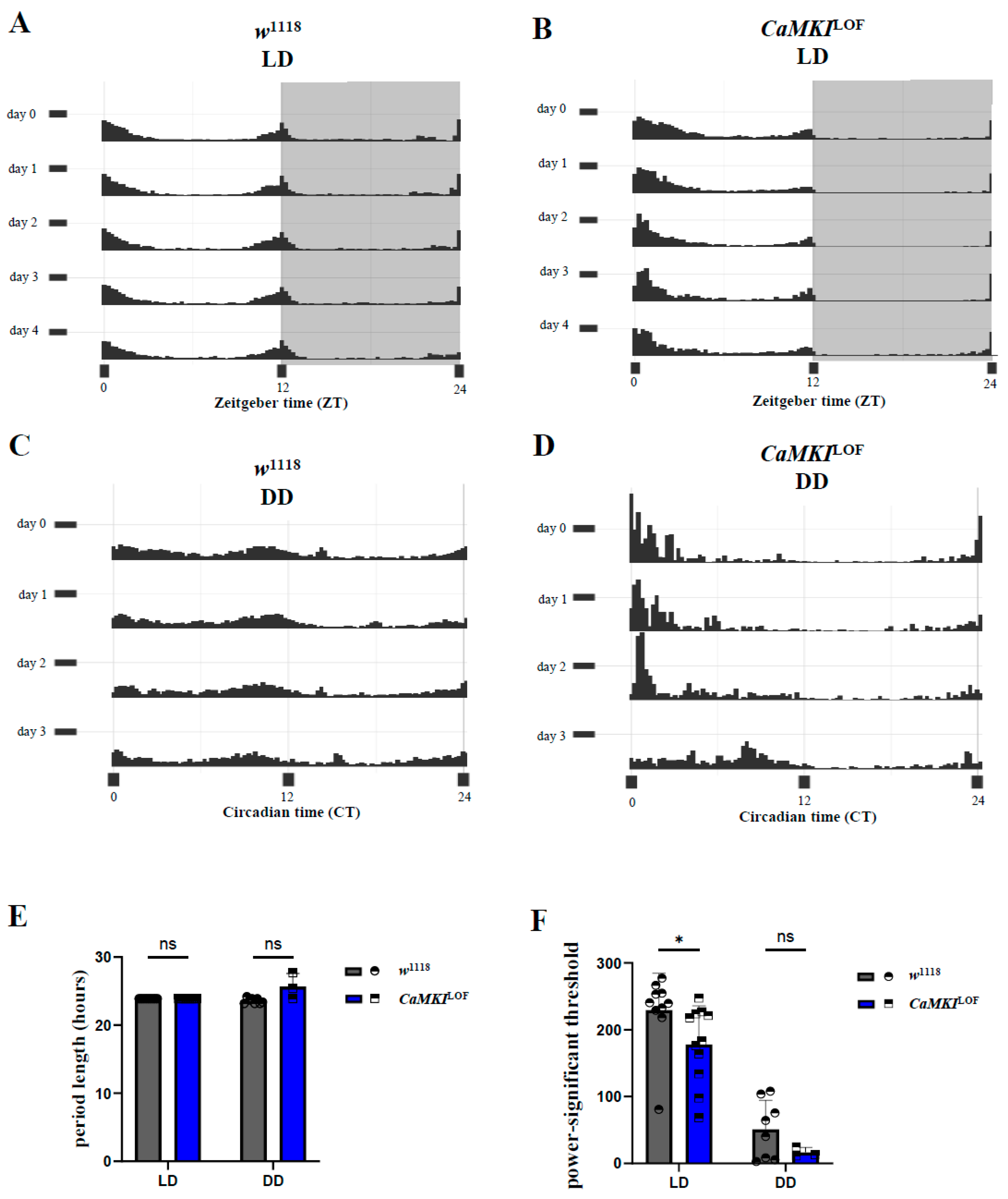
To assess whether CaMKI dysregulation affected circadian rhythm, we analyzed the circadian strength under LD (Figure 2A,B) and DD (Figure 2C,D) conditions as described in previous studies [95]. Additional analysis on circadian rhythmicity revealed a disrupted internal clock regulation in CaMKILOF mutants. First, the number of rhythmic flies, determined through chi-square analysis of periodograms (see methods), revealed how CaMKILOF mutants were less rhythmic compared to the control flies in the absence of light cues (% rhythmic flies—LD: control flies → 100%, CaMKILOF → 100%; DD: control flies → 80%, CaMKILOF → 33%). Then, control flies exhibited robust morning and evening anticipatory activity peaks under LD, while CaMKI mutants displayed attenuated morning anticipation (Figure 2A,B). Although CaMKILOF mutants exhibited the same circadian period length as control flies, their circadian strength—measured by the power–significance threshold (see methods) (Figure 2E,F) and the rhythmicity index—was significantly lower under standard 12h:12h light/dark conditions, indicating a weaker internal regulatory mechanism in the mutants. Consistently, CaMKI-RNAi flies showed a similar effect on circadian regulation as the mutant genotype (Supplementary Figure S1). Together, these findings demonstrate that CaMKI dysregulation disrupts normal sleep homeostasis, intended as the regulation and balancing of sleep-to-wake states, and circadian rhythm regulation in Drosophila. The observed increase in sleep duration and impaired circadian rhythmicity suggests that CaMKI plays a critical role in neuronal circuits governing sleep–wake transitions and endogenous rhythmicity.

3.3. Social Behavior Is Affected in Adult CaMKILOF Mutant Flies

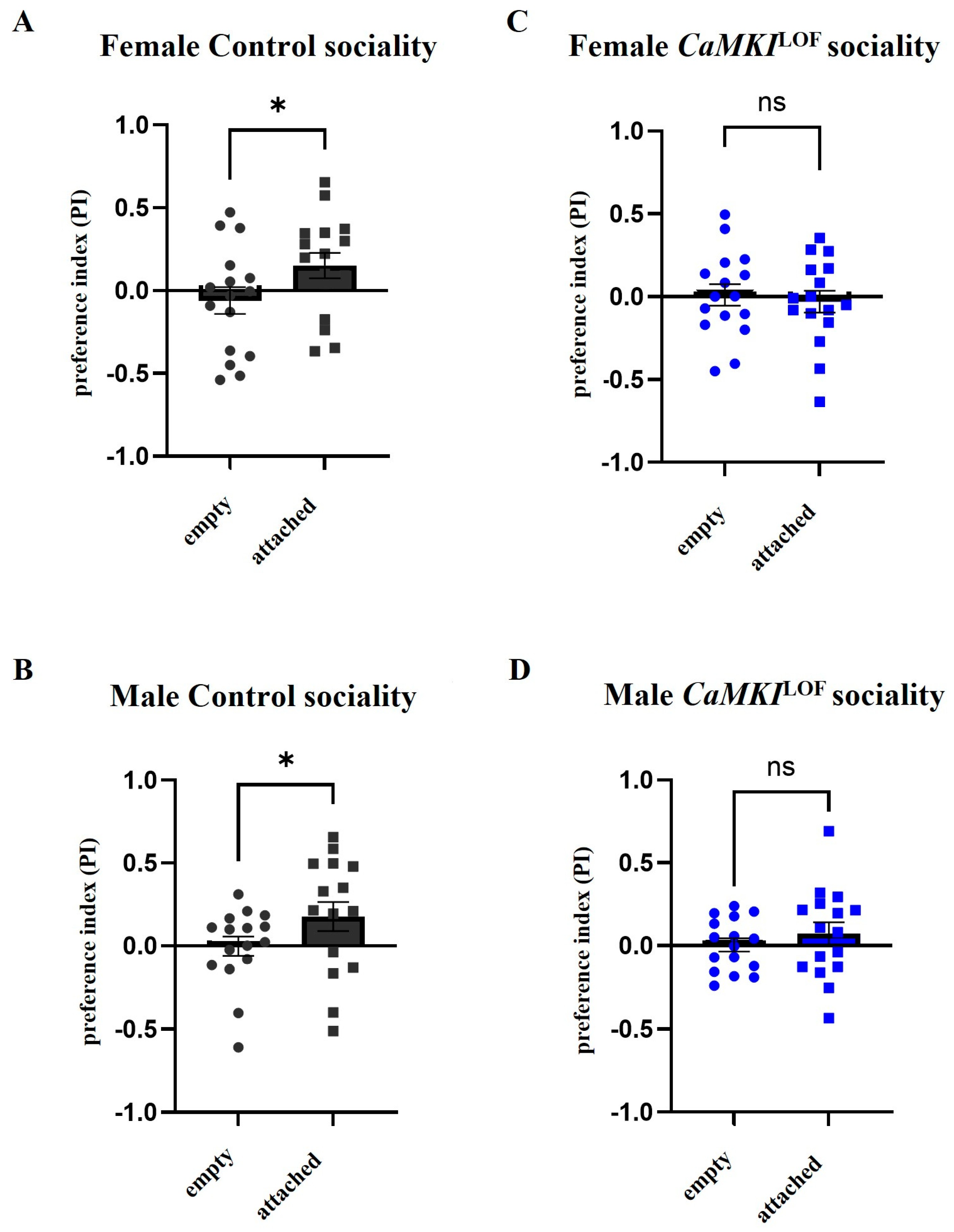
As deficits in social interaction is one of the main criteria for diagnosing ASD, we used a recently developed paradigm to study social approach behavior [107] to examine sociality interactions in CaMKILOF mutants. For this, we measured social attraction when 10 flies were tethered to one half of a shallow circular chamber to serve as attractor flies and measure whether free-walking flies spend significantly more time on the same side as attractors. By contrast, free-walking flies exhibit no bias to either side of the chamber in an empty chamber with no attractor flies. As shown in Figure 3, control females (Figure 3A) and males (Figure 3B) spent significantly more time on the same side as attractors, whereas no bias to either side was observed in an empty chamber, indicating that control flies had a strong tendency to associate with other flies, as previously described [107]. Interestingly, CaMKILOF males or females (Figure 3C,D) showed no bias to either side of the chamber in the presence of the attached flies or in an empty chamber, suggesting no tendency for sociality. No additional anxiety-like, repetitive behaviors (e.g., grooming) were observed, although this was not rigorously quantified.

3.4. CaMKI Dysregulation Enhances Behavioral Decline and Neurodegeneration

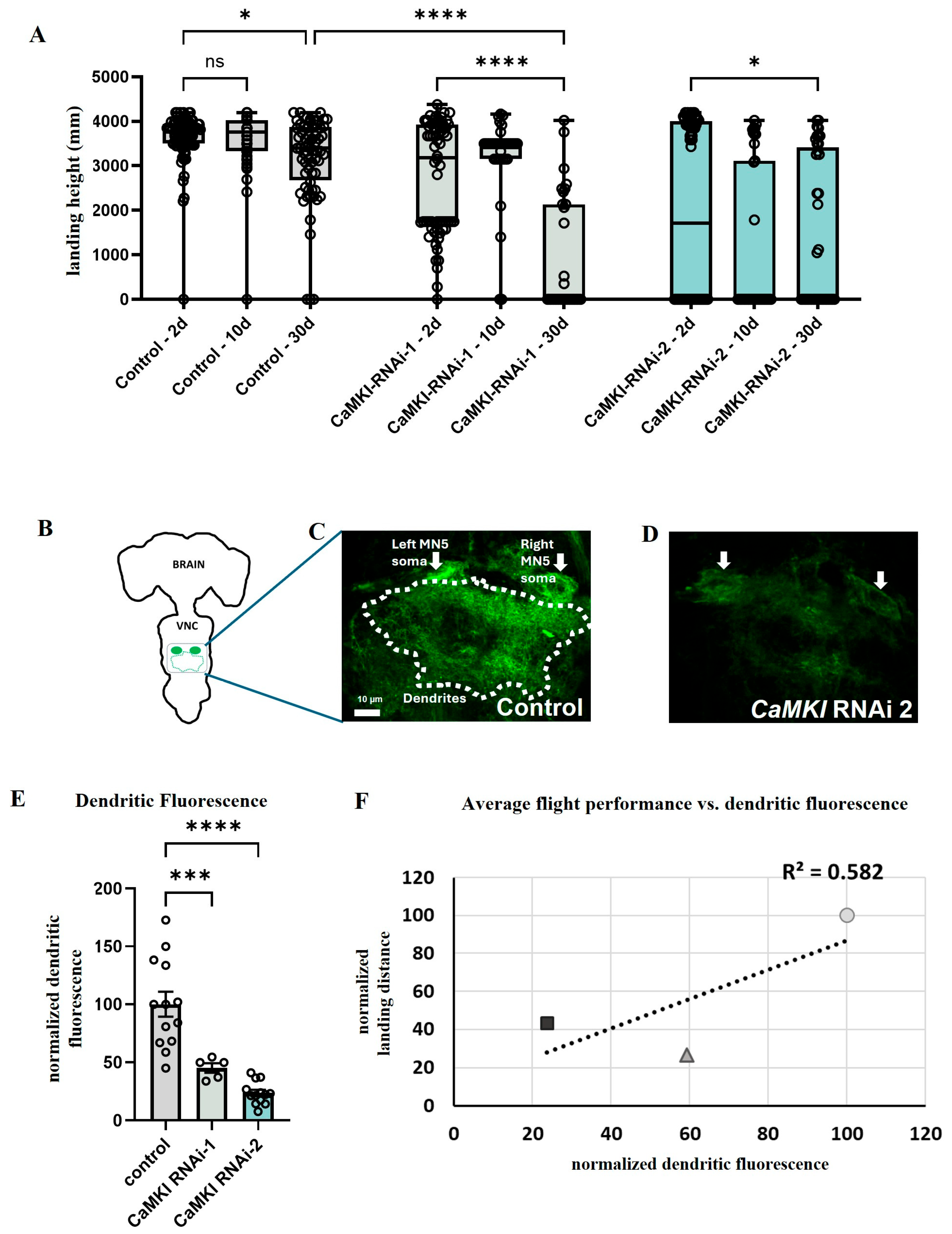
A link between autism and aging-dependent conditions such as dementia and Alzheimer’s disease has been proposed in numerous recent studies [53,54,55,56,57]. We took advantage of the strengths of the Drosophila model for aging research [58] to investigate the effects of CaMKI dysregulation on aging-dependent processes. We focused on the motor network and examined locomotion performance, which is also consistent with the prevalence of motor dysfunction in ASD patients [60,61] and in other ASD animal models [8]. For this, we quantified the decline in flight performance as behavioral readout as well as the intensity of dendritic fluorescence in adult flight motoneurons as a means of anatomical integrity as previously described in our publications [83,98,99,101,127,128] and as used in anatomical studies of mammalian brains [129]. Interestingly, the decline in flight performance was significantly more pronounced in flies expressing CaMKI RNAi-knockdown in adult motoneurons than their corresponding controls (Figure 4A). This locomotion defect was phenocopied using a second RNAi construct. We next quantified the relative fluorescence intensity of the dendritic field in controls and in CaMKI-knockdown flies to test for changes in dendritic anatomy (Figure 4B–E). Consistent with our behavioral data, flies expressing either of the two RNAi constructs to knock down CaMKI showed a significant decrease in dendritic fluorescence as compared to controls (Figure 4E), suggesting that CaMKI dysregulation may enhance aging-dependent dendritic degeneration. Thus, a strong correlation between poor flight performance and increased dendritic degeneration was observed (Figure 4F). In contrast to CaMKI knockdown, such a decrease in dendritic fluorescence and flight performance was not observed in flies expressing different RNAi constructs that target other genes not involved in degeneration that we analyzed for a different study.

3.5. Loss of CaMKI Affects Synaptic Development Increasing Ectopic Synapses

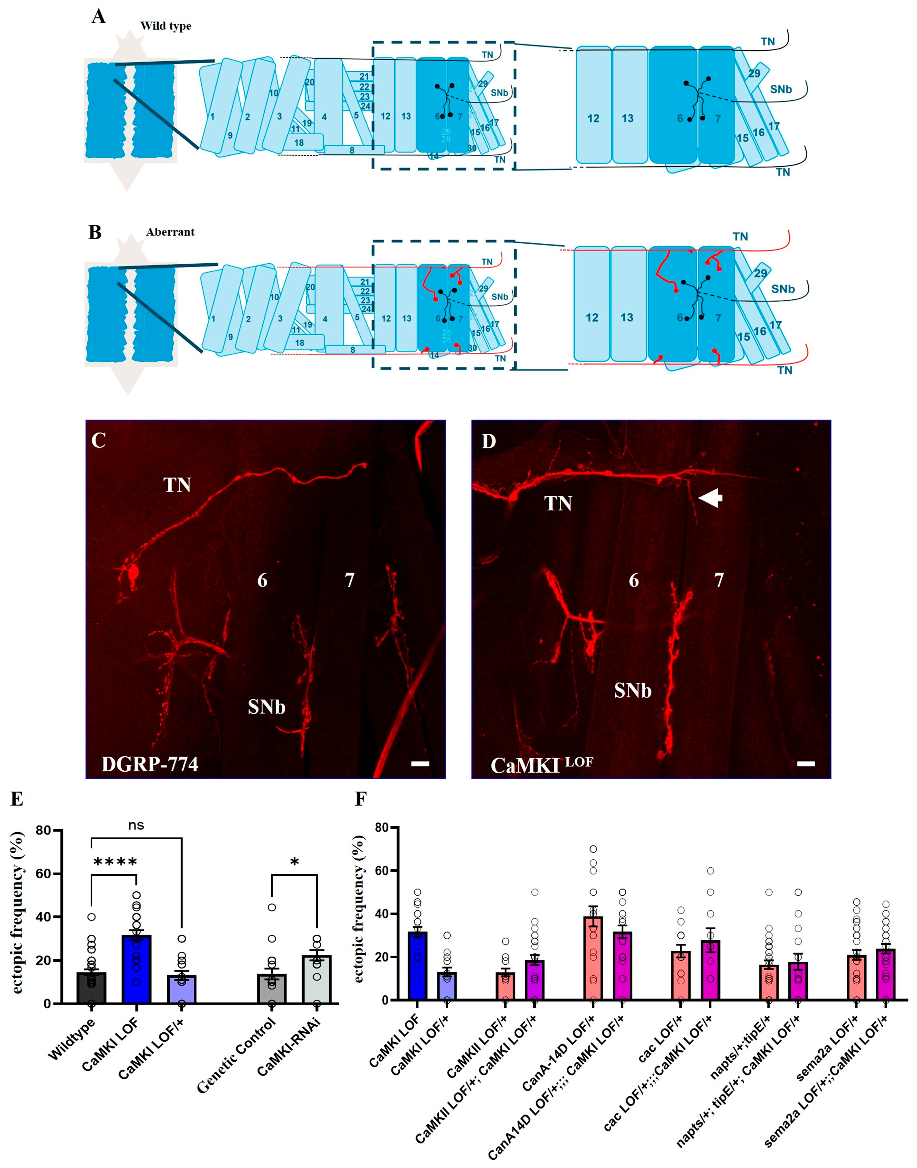
Aberrant synaptic refinement during early development has been proposed to underlie the synaptic phenotypes observed in brains of autistic patients [1,2,3]. To investigate whether CaMKI disruption affects synaptic anatomy, we used the well-characterized stereotypic anatomy of larval neuromuscular junction (NMJ) to examine changes in synaptic connectivity. During early embryonic development, motoneuron growth cones extend their filopodia and innervate not only their target muscles but also off-target muscle fibers [130,131,132]. These temporary off-target contacts have to be removed in an activity-dependent manner, otherwise they are stabilized to become functional ectopic synapses [13,18,19,88] (Figure 5A,B). Compared to control larvae (Figure 5C), CaMKI mutants showed an elevated frequency of ectopic contacts (Figure 5D,E). Pan-neuronal expression of an RNAi construct to knock down CaMKI phenocopied miswiring, leading to increased ectopic frequency in knockdown animals as compared to controls (Figure 5E). As part of the activity-dependent mechanism regulating synaptic refinement, recent studies have identified numerous molecules acting downstream of neuronal activity and calcium (Ca2+). These include the Ca2+-dependent adenylyl cyclase Rutabaga [20], the Ca2+/calmodulin-dependent serine/threonine kinase CaMKII [18,19], and its counterpart, the protein phosphatase Calcineurin (CaN) [19]. These activity-dependent molecules are thought to act downstream of presynaptic Ca2+ and cAMP oscillations to regulate the neuron’s responsiveness to chemorepulsion exerted by the postsynaptic muscle via the secreted chemorepellent Sema2a and its PlexB receptor [13,18,19,20]. Therefore, we performed genetic interaction tests to examine whether the molecular components identified in this pathway may functionally interact with CaMKI. For this, we followed a well-established method of using various heterozygous mutants used in previous publications that provide a sensitized genetic background to test for interactions with CaMKILOF heterozygotes, including CaMKIILOF/+, CanA14D/+, cacLOF/+, napts/+ TipE/+, and Sema2a/+ heterozygotes [18,19,20,88]. These genetic interaction assays test functional relationships between the molecules, pointing to an epistatic interaction when non-additive effects of their individual effects are observed. As shown in Figure 5F, control-like levels and no significant enhancement of the ectopic contact phenotype were observed in any of the double heterozygotes (Figure 5F), suggesting that CaMKI may be acting in a different molecular pathway to regulate synaptic connectivity (see below).

3.6. Proteomic Analyses Reveal Proteins Dysregulated by CaMKI Loss

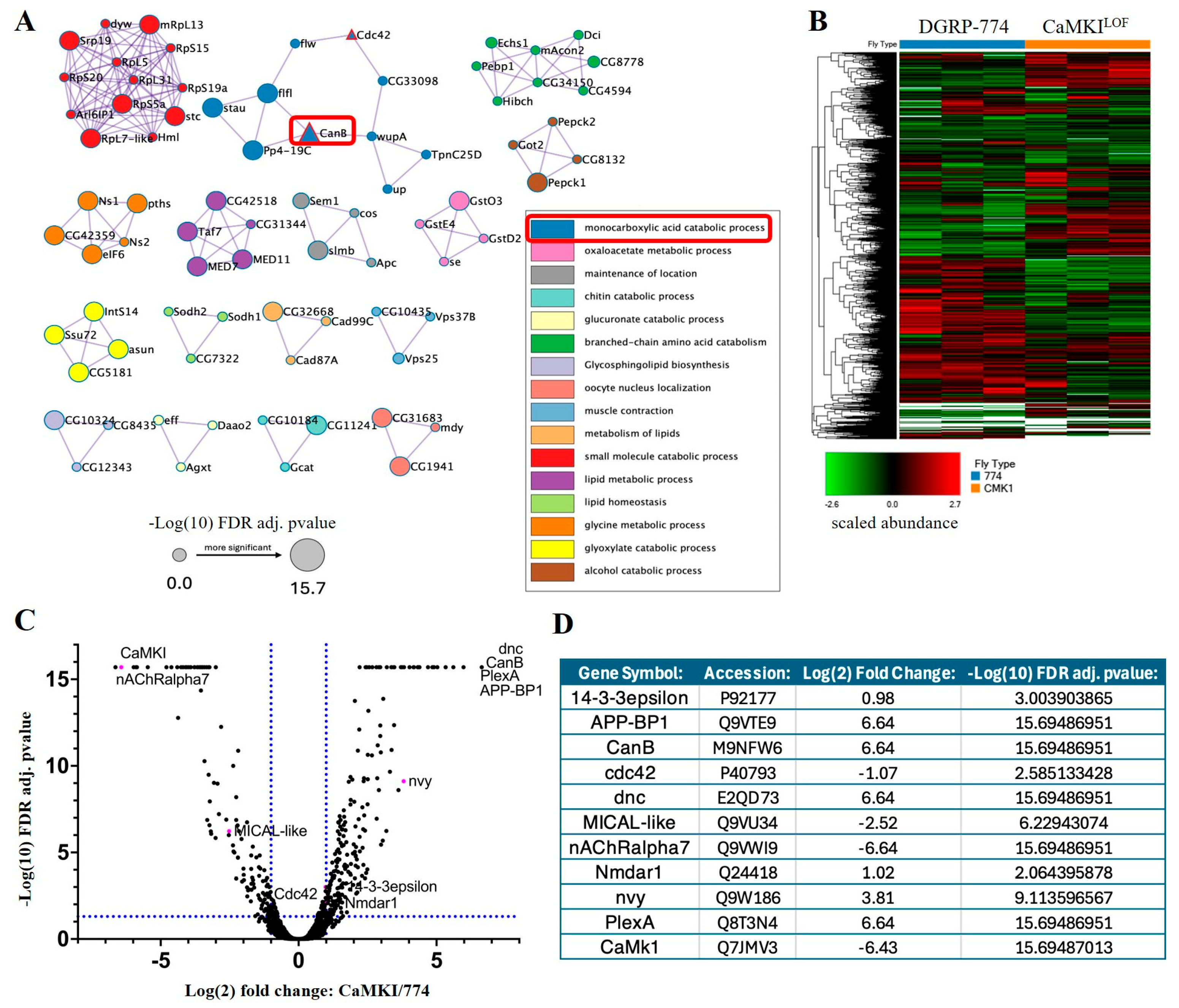
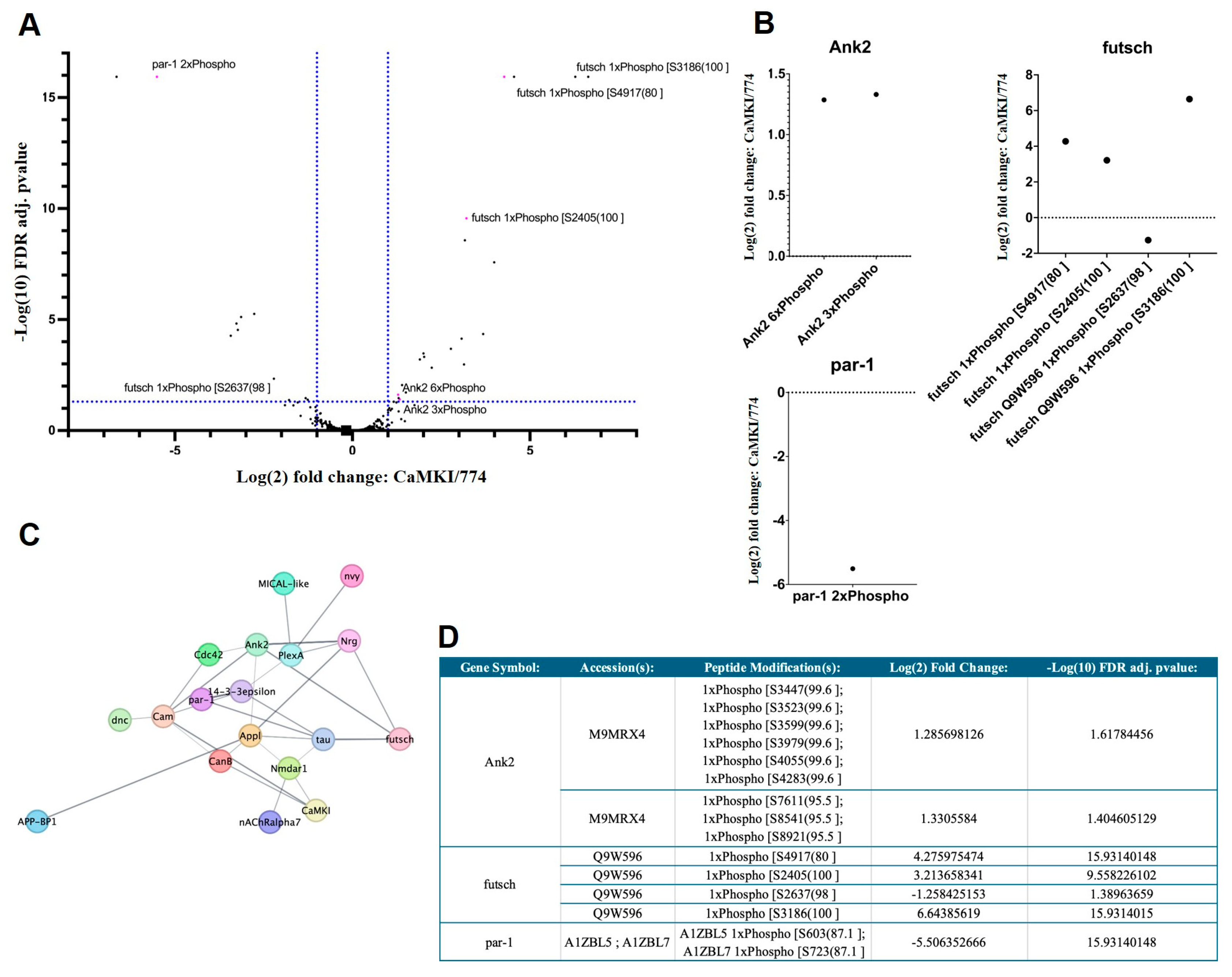
Finally, to further elucidate molecular pathways interacting with CaMKI in the regulation of the processes described above, we performed shotgun proteomics of CaMKILOF mutants. Our analysis showed that 639 protein accession numbers were identified to display significant changes in protein levels (FDR adj. p-value ≤ 0.05; Figure 6; including the expected decrease in CaMKI levels confirming the CaMKILOF mutant background) as well as in phosphorylation levels of peptides (Figure 7). All eight peptides detected for CaMKI are unique. In the CaMKI mutants, peaks are detected for some of these peptides suggesting that portions of the protein may be expressed. However, LFQ finds that CaMKI is downregulated significantly (Log(2) FC of −6.43 compared to control). Although the presence of CaMKI peptides does suggest portions of the protein are potentially present, the results in this study are able to show that the significant change in CaMKI is enough to generate both anatomical and behavioral phenotypes.
As the top candidate molecules dysregulated in CaMKI mutants, we identified the cAMP-phosphodiesterase Dunce (Dnc) and the phosphatase CanB (Figure 6C,D) that we have previously described to regulate synaptic refinement [19,20]. This result suggests the possibility of dynamic phosphorylation levels of target proteins to be regulated by an interplay between CaMKI-dependent phosphorylation and CanB-dependent dephosphorylation. Interestingly, we observed a significant change in phosphorylation levels in various molecules known to regulate cytoskeleton dynamics (Figure 7A–D), such as Futsch, Ankyrin2, and Par1. The microtubule associated protein Futsch (peptide modification: S4917(80), log2FC = 4.28; adj. p value = 15.93), a MAP1B-like protein required for dendritic and axonal development, is implicated in the regulation of microtubule dynamics [76] and is known to be phosphorylated by the GSK3beta-homolog Shaggy [133]. The cytoskeletal adapter Ankyrin2 (Ank2) is involved in neuronal morphogenesis [134], whereas Par1 is a kinase involved in cell polarity and microtubule stability [135] as well as in tau phosphorylation and toxicity [63]. Additional neuronal genes identified in our dataset known to regulate neuronal anatomy and cytoskeleton are the small GTPase Cdc42 that regulates actin cytoskeleton and cell polarity [75] and the neuronal Plexin A (PlexA) receptor known to promote axonal repulsion [77,78]. Interestingly, various molecules previously described to interact with PlexA also showed significantly different expression levels in our proteomic analyses. These include the actin regulatory oxidation–reduction enzyme MICAL-like that directly bind and disassemble actin filaments (F-actin) [79], the A kinase anchoring protein (AKAP) nervy that regulates repulsive axon guidance [80], and the phospho-binding protein 14-3-3ε necessary for PlexA-dependent repulsion and axon guidance [81]. Regarding degenerative processes, we also identified the β-amyloid precursor protein binding protein 1 (APP-BP1) to interact with CaMKI, providing a molecular link between ASD-associated phenotypes and aging-dependent degeneration due to its known interactions with Cullin-3 (Cul3), a ubiquitin ligase strongly associated with ASD [136,137]. APP-BP1 is also known to interact with the Drosophila β-amyloid precursor protein (Appl), a transmembrane protein associated with degeneration and phenotypes associated with Alzheimer’s disease (AD) [62,138,139].

4. Discussion

4.1. Drosophila as a Model to Study Autism-Associated Phenotypes

Autism spectrum disorder (ASD) refers to a broad range of conditions including challenges with social skills, repetitive behaviors, speech, and nonverbal communication [140,141]. Studies using animal models have investigated basic aspects of such complex behaviors, providing insight into fundamental aspects underlying ASD etiology [142,143,144]. The Drosophila genome is 60% homologous to that of humans and includes homologs of about 75% of genes associated with human diseases [145,146]. In particular, considering the strong genetic link to ASD, Drosophila has emerged as a valuable invertebrate model to expand our understanding of ASD-associated phenotypes and conserved molecular mechanisms [32,33,34,35]. Our study provides experimental evidence of loss-of-function mutations in the fly homolog CaMKI in ASD-associated phenotypes, providing initial steps to expand our understanding of potential roles of CAMK4 in ASD-relevant pathways, paving the way for future research to confirm a clear etiology for CAMK4-related ASD. It is worth noting that, whereas monoallelic mutations in CAMK4 have been identified in ASD patients, the CaMKILOF mutants used in our study showed strong phenotypic defects in homozygotes, while CaMKILOF heterozygotes showed control-like phenotypes. This apparent discrepancy can be explained by the functional consequences of the different allelic mutations as well as genetic regulatory mechanisms such as penetrance, expressivity, and pleiotropy that may differ between mutations, species, and individuals. This includes differences in epigenetic regulation of the homologous locus as well as compensatory effects by other genes in the required signaling pathways in a cell- or non-cell autonomous manner that may vary among species. Multiple examples of haploinsufficient mutations in genes leading to disease in humans without affecting heterozygous animals are well documented in validated mammalian models such as, for example, mutations in the zinc-finger transcription factors GATA4 and GATA6 associated with congenital heart defects in humans [147], mutations in the macrophage colony-stimulating factor receptor (CSF1R) associated with adult onset leukoencephalopathy [148], or mutations in the PITX1 bicoid homeodomain transcription factor associated with clubfoot in humans [149]. Therefore, the insights provided in this study regarding CaMKI function in the nervous system may contribute to a better understanding of the fundamental functions of CAMK4 and its association with ASD.
At the behavioral level, flies engage in various social behaviors including courtship, aggression, mating, sociality, and egg-laying, while the effects of group-housing vs. isolation are well documented [41,42,150]. Consistently, we observed disrupted sociality responses in CaMKILOF mutant females and males when compared to controls. Future follow-up studies will address the modulation of the observed CaMKILOF mutant phenotypes by different genetic backgrounds as well as by rescue experiments via functional CaMKI expression in CaMKILOF mutant flies. Furthermore, studies in Drosophila have examined the regulation of sleep [97,123,151], motor control [152,153,154], and neuronal activity [19,100,155], providing mechanistic insight into ASD-associated comorbidities such as sleep dysfunction, motor impairments, and epilepsy [60]. Consistently, Drosophila has been useful for characterizing the in vivo roles of ASD-associated genes and their regulatory networks involved in these behaviors [32,33,34,101,128,136,156]. We observed aberrant sleep and circadian patterns in CaMKILOF mutant flies, consistent with observations in ASD patients [48,52], which involve an increased number of awakenings during the night, sleep onset latency, and reduced sleep efficiency [157]. The sleep and circadian alterations that we observed suggest that CaMKI may play a role in the function of morning cells, a key circadian subgroup comprising the PDF-positive s-LNvs. The altered sleep patterns that we observed, including disrupted morning anticipation and increased sleep during the subjective day in constant darkness, mirror phenotypes previously reported in studies manipulating morning cells [158,159]. These parallels raise the possibility that CaMKI activity contributes to morning cell output or identity. Future studies should directly assess CaMKI function within identified circadian neurons to determine whether its loss specifically impairs morning cell signaling or broader circadian circuit dynamics.
At the anatomical level, postmortem studies of autistic patients that have mutations in genes associated with ASD showed increased synaptic density and longer dendritic spines [1,2,4,7], suggesting a link between ASD and aberrant synaptic refinement during early development. Drosophila has served as a suitable model to elucidate the molecular mechanisms underlying many aspects of anatomical remodeling including synaptic pruning [13] and dendrite regulation [160]. Given the highly conserved nature of basic signaling pathways, insight gained in Drosophila studies has the potential to be translated into novel ASD treatments by advancing our understanding of the mammalian and human brain.

4.2. Link Between Developmental Deficits and Aging-Dependent Degeneration

Although an ASD diagnosis in children is considered reliable at 24 months of age, several anatomical and behavioral differences can be observed between infants at high-risk for ASD and low-risk controls from age 6 to 12 months (reviewed in [161]). For example, using high-resolution brain magnetic resonance imaging (MRI), increased rates of surface area expansion from age 6 to 12 months, followed by an increased rate of total brain volume from 12 to 24 months, were detected [162]. Also, functional MRI (fMRI) studies showed reduced functional connectivity between the amygdala and canonical targets [163] and atypical connectivity in language areas [164] in children with ASD. Thus, consistent with the notion that ASD is considered a neurodevelopmental disorder, we observed behavioral changes in sleep, circadian rhythms, and sociality in young CaMKILOF mutant flies as well as miswiring indicating defects in synaptic refinement during the development of precise connectivity. On the other hand, recent studies indicated a higher prevalence of identified dementia diagnoses in ASD individuals [53,54,55,56,57]. Consistently, we observed experimental evidence of increased anatomical degeneration as well as enhanced behavioral decline in flies with disrupted CaMKI, suggesting that CaMKI may play an instructive role during development in addition to a neuroprotective role in aging flies or, alternatively, that CaMKI-dependent defects during development may promote aging-dependent degeneration. This is consistent with a strong cooccurrence of neurodevelopmental and neurodegenerative conditions that has been observed in other disease-related conditions such as, for example, in the well-characterized neurodevelopmental roles of the β-amyloid precursor protein (APP) associated with the formation of β-amyloid plaques and Alzheimer’s disease [139,165]. Interestingly, our proteomic results indicate an interaction between CaMKI and the β-amyloid precursor protein binding protein 1 (APP-BP1). Various molecular functions have been characterized for APP-BP1, including ubiquitin activation functions via interactions with Uba3 that in turn activates the ubiquitin-like protein NEDD8 in humans [166] and flies [62]. These interactions are required for neddylation of the ubiquitin ligase Cullin-3 (Cul3) [167], a gene strongly associated with ASD [136,137]. Moreover, during development, APP-BP1 is known to interact with the β-amyloid precursor protein-like (Appl) [62], the Drosophila ortholog for APP, a molecule associated with Alzheimer’s disease in humans [138]. Human APP and fly Appl share numerous conserved properties such as proteolytic processing, Aβ peptide formation, plaque buildup, memory regulation, and neurodegeneration (reviewed in [139,168,169,170]). However, the role of aberrant developmental processes in Alzheimer’s disease has not been considerably appreciated [139].
An additional link between neurodevelopment and degeneration is supported by our observation of significant changes in Par1 phosphorylation levels in CaMKI mutants. On the one hand, the Par1 kinase regulates synaptic development at the larval NMJ by phosphorylating scaffold proteins such as presynaptic Brp [171] and postsynaptic PSD-95/Dlg [172]. Regulation of anatomical changes in these processes can involve Par1 roles in regulating cell polarity and microtubule stability [135]. Additional structural roles include the Par1-dependent regulation of the cytoskeleton via interactions with the small GTPase Cdc42 [75] or with the microtubule binding protein Futsch, which has been proposed to be a likely substrate of Par1 [135] and shows decreased phosphorylation levels in our proteomic results. Future studies will examine the role of Par1 and Futsch in modulating the developmental defects observed in CaMKILOF mutants. Furthermore, Par1 is also involved in Tau phosphorylation and toxicity in Drosophila neurons [63], implicating its aberrant activation in the pathogenesis of AD and related tauopathies. Such roles of Par1 are highly conserved in its human homolog, the microtubule affinity regulating kinase (MARK), whose dysregulation has previously been associated with autism [173] as well as Alzheimer’s disease [174,175,176]. Therefore, our results provide initial mechanistic insight into the molecular means linking developmental processes and aging-dependent degeneration.

4.3. Molecular Pathways Involving CaMKI During Neurodevelopment and Degeneration

Our results indicate that dysregulation of CaMKI has various effects at the behavioral level, including aberrant sleep, circadian rhythms, sociality, and flight performance, in addition to cellular defects during synaptic development and neuronal network degeneration. Whereas most of the molecular interactions of CaMKI in the pathways regulating such processes remain to be tested, the results from our shotgun proteomic analysis revealed more than 600 proteins that have a statistically significant difference in the amount and phosphorylation levels in CaMKI mutants. This provides a list of various candidate genes and signaling pathways that may interact with CaMKI to regulate the biological processes described above.
In contrast to previous studies at the Drosophila NMJ that identified chemorepulsion exerted by the presynaptic receptor Plexin B as a key factor in synaptic refinement [18,19,20], the results from our genetic interaction tests with molecules involved in activity-dependent synaptic refinement indicated that CaMKI may regulate synaptic connectivity by acting in different molecular networks. From our proteomic data, we identified a group of genes that might represent an alternative chemorepulsion pathway regulating precise synaptic connectivity. These involve the neuronal Plexin A (PlexA) receptor, involved in axonal repulsion [77,78], the cytosolic flavoprotein MICAL, required for PlexA repulsive axon guidance [79], the phospho-binding protein 14-3-3ε, necessary for PlexA-mediated axon guidance and repulsion [81], and the A kinase anchoring protein (AKAP) Nervy (nvy), which antagonizes PlexA-mediated axonal repulsion [177]. Whether PlexA-mediated chemorepulsion may represent a separate pathway parallel to the PlexB-dependent pathway previously identified or, alternatively, whether there is a link between both pathways remains to be tested. The latter scenario is supported by our proteomic results showing dysregulation of the phosphatase CanB and the cAMP-phosphodiesterase Dunce (Dnc), both described in our previous studies to regulate PlexB-mediated synaptic refinement at the NMJ [19,20]. Whether a balance of phosphorylation levels between CaMKI-dependent kinase activity and CanB-dependent dephosphorylation regulates the phenotypes described above will be the focus of future research.
For the regulation of neuronal anatomy, an interesting set of genes worth highlighting include molecules known to regulate cytoskeleton dynamics, such as the microtubule associated protein Futsch [76], the cytoskeletal adapter Ankyrin2 [134] (Ank2; previously associated with ASD in a study using whole exome sequencing [40]), and the small GTPase Cdc42 [75] known to regulate actin cytoskeleton. This suggests molecular means for the CaMKI-dependent regulation of synaptic connectivity, which may involve the histone-acetyl transferase CBP and Juvenile Hormone [41,42], a hormone recently describe to regulate the maturation of naïve ethanol olfactory preference in young flies [178]. While most of the molecules described here are known to play a role during early stages of development, how CaMKI dysregulation affects aging-dependent degeneration remains an open question. As mentioned above, our proteomic analysis revealed changes in protein levels of APP-BP1 and phosphorylation levels of Par1 in CaMKILOF mutants. Thus, follow-up experiments will address the question whether the enhanced anatomical degeneration and behavioral decline are influenced by aberrant neurodevelopment or by molecular interactions between CaMKI, APP-BP1, Par1, and other molecules involved in aging and degenerative processes such as Appl.

5. Conclusions

Our data suggest that CaMKI plays a role in developmental processes and influences aging-dependent degenerative processes. These results will shed light on fundamental molecular pathways involving genes during developmental and degenerative processes, possibly providing new candidate genes for expanding our understanding of ASD etiology and the development of effective treatments.

Supplementary Materials

The following supporting information can be downloaded at https://www.mdpi.com/article/10.3390/biology14091228/s1: Supplementary Figure S1. CaMKI knockdown phenocopies effects on sleep and circadian rhythmicity. (A,B) Quantification of sleep parameters, including the number of sleep bouts and average sleep bout length (minutes), under 12 h:12 h light/dark (LD) conditions. (C,D) Time spent sleeping and time spent awake comparing genotypes, control, and knockdown under LD and DD conditions. Sleep was measured in genetic control (elavc155-Gal4 > w1118) and CaMKI RNAi (elavc155-Gal4 > CaMKI_RNAi; BDSC_35362) flies. Statistical analysis was performed using ANOVA to compare number of sleep bouts and time spent sleeping, while non-parametric multiple comparison Mann–Whitney t-test was performed to compare average sleep bout length between genotypes. The analysis of sleep parameters for DD condition was carried out averaging the data in a 24 h cycle and not based on the observed genotype period cycle. (E–H) Actograms showing sleep–wake activity rhythms over 24 h in LD and DD conditions. Genetic control and CaMKI RNAi flies are displayed separately. (I,J) Quantification of circadian rhythm parameters, including period length (hours) and rhythmicity strength defined as power–significance threshold, for genetic control and CaMKI RNAi flies in LD and DD conditions. White and shaded gray areas in LD graphs represent light and dark phases, respectively. Bars represent mean + SEM. Statistical significance is indicated (* p < 0.05). Statistical analysis was performed using the non-parametric Mann–Whitney test for multiple t-tests to compare circadian parameters between genotypes. Analysis performed for genetic control and CaMKI RNAi flies in LD and DD conditions. Supplementary Figure S2. Activity index suggests normal levels of activity in mutants compared to controls in LD conditions. Activity index (total awake time activity counts/total awake minutes) of different days (left y axis), plotted as violin plots, suggests no difference in levels of activity between CaMKILOF and w1118 (A), as well as between CaMKI RNAi and its control (B) under LD conditions. S3—SUPP_Metascape GOs all PPI Data excel. S4—SUPP_allProteomics_Data. S5—SUPP_phospho all.

Author Contributions

Conceptualization, C.G., Z.M.S., K.M.-G., and F.J.V.; methodology, C.G., Z.M.S., Z.K., C.J., K.M.-G., and F.J.V.; formal analysis, C.G., Z.M.S., A.C., Z.K., C.J., K.M.-G., and F.J.V.; investigation, C.G., Z.M.S., A.C., Z.K., C.J., K.M.-G., and F.J.V.; data curation, C.G., Z.M.S., A.C., Z.K., C.J., K.M.-G., and F.J.V.; writing—original draft preparation, C.G., Z.M.S., and F.J.V.; writing—review and editing, C.G., Z.M.S., and F.J.V.; visualization, C.G., Z.M.S., and F.J.V.; supervision, K.M.-G. and F.J.V.; project administration, K.M.-G. and F.J.V.; funding acquisition, K.M.-G. and F.J.V. All authors have read and agreed to the published version of the manuscript.

Funding

Z.K. was supported by the Undergraduate Research Award program at UMBC, which was funded by the Division of Undergraduate Academic Affairs. A.C. was supported by the LSAMP Program at UMBC, which was funded through an award from the National Science Foundation (Award #1002566), and by the MARC*U/U-RISE Program at UMBC. URISE at UMBC was supported by the National Institute of General Medical Sciences of the National Institutes of Health (NIGMS/NIH) under Award T34GM136497.

Institutional Review Board Statement

Not applicable.

Informed Consent Statement

Not applicable.

Data Availability Statement

Mass spectrometry data deposited to PRIDE under dataset identifier PXD061426.

Acknowledgments

We thank members of the Vonhoff lab for helpful comments on the manuscript. No artificial intelligence software was used at any time for the writing of the manuscript or data analysis.

Conflicts of Interest

The authors declare no conflicts of interest.

References

  1. Hutsler, J.J.; Zhang, H. Increased dendritic spine densities on cortical projection neurons in autism spectrum disorders. Brain Res. 2010, 1309, 83–94. [Google Scholar] [CrossRef] [PubMed]
  2. Tang, G.; Gudsnuk, K.; Kuo, S.H.; Cotrina, M.L.; Rosoklija, G.; Sosunov, A.; Sonders, M.S.; Kanter, E.; Castagna, C.; Yamamoto, A.; et al. Loss of mTOR-dependent macroautophagy causes autistic-like synaptic pruning deficits. Neuron 2014, 83, 1131–1143. [Google Scholar] [CrossRef] [PubMed] [PubMed Central]
  3. Weir, R.K.; Bauman, M.D.; Jacobs, B.; Schumann, C.M. Protracted dendritic growth in the typically developing human amygdala and increased spine density in young ASD brains. J. Comp. Neurol. 2018, 526, 262–274. [Google Scholar] [CrossRef] [PubMed] [PubMed Central]
  4. Zoghbi, H.Y.; Bear, M.F. Synaptic dysfunction in neurodevelopmental disorders associated with autism and intellectual disabilities. Cold Spring Harb. Perspect. Biol. 2012, 4, a009886. [Google Scholar] [CrossRef] [PubMed] [PubMed Central]
  5. Bourgeron, T. A synaptic trek to autism. Curr. Opin. Neurobiol. 2009, 19, 231–234. [Google Scholar] [CrossRef] [PubMed]
  6. Faust, T.E.; Gunner, G.; Schafer, D.P. Mechanisms governing activity-dependent synaptic pruning in the developing mammalian CNS. Nat. Rev. Neurosci. 2021, 22, 657–673. [Google Scholar] [CrossRef] [PubMed] [PubMed Central]
  7. Penzes, P.; Cahill, M.E.; Jones, K.A.; VanLeeuwen, J.E.; Woolfrey, K.M. Dendritic spine pathology in neuropsychiatric disorders. Nat. Neurosci. 2011, 14, 285–293. [Google Scholar] [CrossRef] [PubMed] [PubMed Central]
  8. James, E.J.; Gu, J.; Ramirez-Vizcarrondo, C.M.; Hasan, M.; Truszkowski, T.L.; Tan, Y.; Oupravanh, P.M.; Khakhalin, A.S.; Aizenman, C.D. Valproate-induced neurodevelopmental deficits in Xenopus laevis tadpoles. J. Neurosci. 2015, 35, 3218–3229. [Google Scholar] [CrossRef] [PubMed] [PubMed Central]
  9. Purves, D.; Lichtman, J.W. Elimination of synapses in the developing nervous system. Science 1980, 210, 153–157. [Google Scholar] [CrossRef] [PubMed]
  10. Rakic, P.; Bourgeois, J.P.; Eckenhoff, M.F.; Zecevic, N.; Goldman-Rakic, P.S. Concurrent overproduction of synapses in diverse regions of the primate cerebral cortex. Science 1986, 232, 232–235. [Google Scholar] [CrossRef] [PubMed]
  11. Cang, J.; Feldheim, D.A. Developmental mechanisms of topographic map formation and alignment. Annu. Rev. Neurosci. 2013, 36, 51–77. [Google Scholar] [CrossRef] [PubMed]
  12. Kano, M.; Hashimoto, K. Synapse elimination in the central nervous system. Curr. Opin. Neurobiol. 2009, 19, 154–161. [Google Scholar] [CrossRef] [PubMed]
  13. Vonhoff, F.; Keshishian, H. Activity-Dependent Synaptic Refinement: New Insights from Drosophila. Front. Syst. Neurosci. 2017, 11, 23. [Google Scholar] [CrossRef] [PubMed] [PubMed Central]
  14. Wiesel, T.N.; Hubel, D.H. Effects of Visual Deprivation on Morphology and Physiology of Cells in the Cats Lateral Geniculate Body. J. Neurophysiol. 1963, 26, 978–993. [Google Scholar] [CrossRef] [PubMed]
  15. Espinosa, J.S.; Stryker, M.P. Development and plasticity of the primary visual cortex. Neuron 2012, 75, 230–249. [Google Scholar] [CrossRef] [PubMed] [PubMed Central]
  16. Arroyo, D.A.; Feller, M.B. Spatiotemporal Features of Retinal Waves Instruct the Wiring of the Visual Circuitry. Front. Neural Circuits 2016, 10, 54. [Google Scholar] [CrossRef] [PubMed] [PubMed Central]
  17. Ackman, J.B.; Burbridge, T.J.; Crair, M.C. Retinal waves coordinate patterned activity throughout the developing visual system. Nature 2012, 490, 219–225. [Google Scholar] [CrossRef] [PubMed] [PubMed Central]
  18. Carrillo, R.A.; Olsen, D.P.; Yoon, K.S.; Keshishian, H. Presynaptic activity and CaMKII modulate retrograde semaphorin signaling and synaptic refinement. Neuron 2010, 68, 32–44. [Google Scholar] [CrossRef] [PubMed] [PubMed Central]
  19. Vonhoff, F.; Keshishian, H. In Vivo Calcium Signaling during Synaptic Refinement at the Drosophila Neuromuscular Junction. J. Neurosci. 2017, 37, 5511–5526. [Google Scholar] [CrossRef]
  20. Vonhoff, F.; Keshishian, H. Cyclic nucleotide signaling is required during synaptic refinement at the Drosophila neuromuscular junction. Dev. Neurobiol. 2017, 77, 39–60. [Google Scholar] [CrossRef] [PubMed] [PubMed Central]
  21. Nicol, X.; Voyatzis, S.; Muzerelle, A.; Narboux-Nême, N.; Südhof, T.C.; Miles, R.; Gaspar, P. cAMP oscillations and retinal activity are permissive for ephrin signaling during the establishment of the retinotopic map. Nat. Neurosci. 2007, 10, 340–347. [Google Scholar] [CrossRef] [PubMed]
  22. Nicol, X.; Muzerelle, A.; Rio, J.P.; Métin, C.; Gaspar, P. Requirement of adenylate cyclase 1 for the ephrin-A5-dependent retraction of exuberant retinal axons. J. Neurosci. 2006, 26, 862–872. [Google Scholar] [CrossRef] [PubMed] [PubMed Central]
  23. Stent, G.S. A physiological mechanism for Hebb’s postulate of learning. Proc. Natl. Acad. Sci. USA 1973, 70, 997–1001. [Google Scholar] [CrossRef] [PubMed] [PubMed Central]
  24. Cheadle, L.; Tzeng, C.P.; Kalish, B.T.; Harmin, D.A.; Rivera, S.; Ling, E.; Nagy, M.A.; Hrvatin, S.; Hu, L.; Stroud, H.; et al. Visual Experience-Dependent Expression of Fn14 Is Required for Retinogeniculate Refinement. Neuron 2018, 99, 525–539.e10. [Google Scholar] [CrossRef] [PubMed] [PubMed Central]
  25. Yap, E.L.; Greenberg, M.E. Activity-Regulated Transcription: Bridging the Gap between Neural Activity and Behavior. Neuron 2018, 100, 330–348. [Google Scholar] [CrossRef] [PubMed] [PubMed Central]
  26. Paulin, M.G.; Cahill-Lane, J. Events in Early Nervous System Evolution. Top. Cogn. Sci. 2021, 13, 25–44. [Google Scholar] [CrossRef] [PubMed]
  27. Sousa, A.M.M.; Meyer, K.A.; Santpere, G.; Gulden, F.O.; Sestan, N. Evolution of the Human Nervous System Function, Structure, and Development. Cell 2017, 170, 226–247. [Google Scholar] [CrossRef] [PubMed] [PubMed Central]
  28. Niven, J.E.; Chittka, L. Evolving understanding of nervous system evolution. Curr. Biol. 2016, 26, R937–R941. [Google Scholar] [CrossRef] [PubMed]
  29. Assaf, Y.; Bouznach, A.; Zomet, O.; Marom, A.; Yovel, Y. Conservation of brain connectivity and wiring across the mammalian class. Nat. Neurosci. 2020, 23, 805–808. [Google Scholar] [CrossRef] [PubMed]
  30. Kozol, R.A.; Conith, A.J.; Yuiska, A.; Cree-Newman, A.; Tolentino, B.; Benesh, K.; Paz, A.; Lloyd, E.; Kowalko, J.E.; Keene, A.C.; et al. A brain-wide analysis maps structural evolution to distinct anatomical module. eLife 2023, 12, e80777. [Google Scholar] [CrossRef] [PubMed] [PubMed Central]
  31. Burkhardt, P.; Sprecher, S.G. Evolutionary origin of synapses and neurons—Bridging the gap. Bioessays 2017, 39, 1700024. [Google Scholar] [CrossRef] [PubMed]
  32. Bellosta, P.; Soldano, A. Dissecting the genetics of autism spectrum disorders: A Drosophila perspective. Front. Physiol. 2019, 10, 987. [Google Scholar] [CrossRef] [PubMed] [PubMed Central]
  33. Mariano, V.; Achsel, T.; Bagni, C.; Kanellopoulos, A.K. Modelling Learning and Memory in Drosophila to Understand Intellectual Disabilities. Neuroscience 2020, 445, 12–30. [Google Scholar] [CrossRef] [PubMed]
  34. Ueoka, I.; Pham, H.T.N.; Matsumoto, K.; Yamaguchi, M. Autism Spectrum Disorder-Related Syndromes: Modeling with Drosophila and Rodents. Int. J. Mol. Sci. 2019, 20, 4071. [Google Scholar] [CrossRef] [PubMed] [PubMed Central]
  35. Coll-Tané, M.; Krebbers, A.; Castells-Nobau, A.; Zweier, C.; Schenck, A. Intellectual disability and autism spectrum disorders ‘on the fly’: Insights from. Dis. Model. Mech. 2019, 12, dmm039180. [Google Scholar] [CrossRef] [PubMed] [PubMed Central]
  36. Kaiser, J.; Risteska, A.; Muller, A.G.; Sun, H.; Lei, B.; Nay, K.; Means, A.R.; Cousin, M.A.; Drewry, D.H.; Oakhill, J.S.; et al. Convergence on CaMK4: A Key Modulator of Autism-Associated Signaling Pathways in Neurons. Biol. Psychiatry 2024, 97, 439–449. [Google Scholar] [CrossRef] [PubMed]
  37. Alawneh, I.; Amburgey, K.; Gonorazky, H.; Gorodetsky, C. CAMK4-related Case of Hyperkinetic Movement Disorder. Mov. Disord. Clin. Pract. 2023, 10, 707–709. [Google Scholar] [CrossRef] [PubMed] [PubMed Central]
  38. Zech, M.; Lam, D.D.; Weber, S.; Berutti, R.; Poláková, K.; Havránková, P.; Fečíková, A.; Strom, T.M.; Růžička, E.; Jech, R.; et al. A unique de novo gain-of-function variant in CAMK4 associated with intellectual disability and hyperkinetic movement disorder. Cold Spring Harb. Mol. Case Stud. 2018, 4, a003293. [Google Scholar] [CrossRef] [PubMed] [PubMed Central]
  39. Zech, M.; Bardakjian, T.M.; Stoklosa, M.; Ploski, R.; Jech, R.; Gonzalez-Alegre, P.; Winkelmann, J. A Neurodevelopmental Disorder with Dystonia and Chorea Resulting from Clustering CAMK4 Variants. Mov. Disord. 2021, 36, 520–521. [Google Scholar] [CrossRef] [PubMed]
  40. Iossifov, I.; O’Roak, B.J.; Sanders, S.J.; Ronemus, M.; Krumm, N.; Levy, D.; Stessman, H.A.; Witherspoon, K.T.; Vives, L.; Patterson, K.E.; et al. The contribution of de novo coding mutations to autism spectrum disorder. Nature 2014, 515, 216–221. [Google Scholar] [CrossRef] [PubMed] [PubMed Central]
  41. Zhao, S.; Deanhardt, B.; Barlow, G.T.; Schleske, P.G.; Rossi, A.M.; Volkan, P.C. Chromatin-based reprogramming of a courtship regulator by concurrent pheromone perception and hormone signaling. Sci. Adv. 2020, 6, eaba6913. [Google Scholar] [CrossRef] [PubMed] [PubMed Central]
  42. Sethi, S.; Lin, H.H.; Shepherd, A.K.; Volkan, P.C.; Su, C.Y.; Wang, J.W. Social Context Enhances Hormonal Modulation of Pheromone Detection in Drosophila. Curr. Biol. 2019, 29, 3887–3898.e4. [Google Scholar] [CrossRef] [PubMed] [PubMed Central]
  43. Ergaz, Z.; Weinstein-Fudim, L.; Ornoy, A. Genetic and non-genetic animal models for autism spectrum disorders (ASD). Reprod. Toxicol. 2016, 64, 116–140. [Google Scholar] [CrossRef] [PubMed]
  44. Alexander, R.D. The Evolution of Social Behavior. Annu. Rev. Ecol. Evol. Syst. 1974, 5, 325–383. [Google Scholar] [CrossRef]
  45. Hamilton, W.D. The Evolution of Altruistic Behavior. Am. Nat. 1963, 97, 354–356. [Google Scholar] [CrossRef]
  46. Wheeler, W.M. The Social Insects: Their Origin and Evolution; K. Paul, Trench, Trubner: London, UK, 1928. [Google Scholar]
  47. Keene, A.C.; Duboue, E.R. The origins and evolution of sleep. J. Exp. Biol. 2018, 221, jeb159533. [Google Scholar] [CrossRef] [PubMed] [PubMed Central]
  48. Ballester, P.; Richdale, A.L.; Baker, E.K.; Peiró, A.M. Sleep in autism: A biomolecular approach to aetiology and treatment. Sleep Med. Rev. 2020, 54, 101357. [Google Scholar] [CrossRef] [PubMed]
  49. Carmassi, C.; Palagini, L.; Caruso, D.; Masci, I.; Nobili, L.; Vita, A.; Dell’Osso, L. Systematic Review of Sleep Disturbances and Circadian Sleep Desynchronization in Autism Spectrum Disorder: Toward an Integrative Model of a Self-Reinforcing Loop. Front. Psychiatry 2019, 10, 366. [Google Scholar] [CrossRef] [PubMed] [PubMed Central]
  50. Deliens, G.; Leproult, R.; Schmitz, R.; Destrebecqz, A.; Peigneux, P. Sleep Disturbances in Autism Spectrum Disorders. Rev. J. Autism Dev. Disord. 2015, 2, 343–356. [Google Scholar] [CrossRef]
  51. Krakowiak, P.; Goodlin-Jones, B.; Hertz-Picciotto, I.; Croen, L.A.; Hansen, R.L. Sleep problems in children with autism spectrum disorders, developmental delays, and typical development: A population-based study. J. Sleep Res. 2008, 17, 197–206. [Google Scholar] [CrossRef] [PubMed] [PubMed Central]
  52. Martinez-Cayuelas, E.; Gavela-Pérez, T.; Rodrigo-Moreno, M.; Losada-Del Pozo, R.; Moreno-Vinues, B.; Garces, C.; Soriano-Guillén, L. Sleep Problems, Circadian Rhythms, and Their Relation to Behavioral Difficulties in Children and Adolescents with Autism Spectrum Disorder. J. Autism Dev. Disord. 2024, 54, 1712–1726. [Google Scholar] [CrossRef] [PubMed] [PubMed Central]
  53. Pagni, B.A.; Walsh, M.J.M.; Ofori, E.; Chen, K.; Sullivan, G.; Alvar, J.; Monahan, L.; Guerithault, N.; Delaney, S.; Braden, B.B. Effects of age on the hippocampus and verbal memory in adults with autism spectrum disorder: Longitudinal versus cross-sectional findings. Autism Res. 2022, 15, 1810–1823. [Google Scholar] [CrossRef] [PubMed]
  54. Vivanti, G.; Tao, S.; Lyall, K.; Robins, D.L.; Shea, L.L. The prevalence and incidence of early-onset dementia among adults with autism spectrum disorder. Autism Res. 2021, 14, 2189–2199. [Google Scholar] [CrossRef] [PubMed] [PubMed Central]
  55. Vivanti, G.; Lee, W.L.; Ventimiglia, J.; Tao, S.; Lyall, K.; Shea, L.L. Prevalence of Dementia Among US Adults with Autism Spectrum Disorder. JAMA Netw. Open 2025, 8, e2453691. [Google Scholar] [CrossRef] [PubMed] [PubMed Central]
  56. Rhodus, E.K.; Barber, J.; Abner, E.L.; Duff, D.M.C.; Bardach, S.H.; Caban-Holt, A.; Lightner, D.; Rowles, G.D.; Schmitt, F.A.; Jicha, G.A. Behaviors Characteristic of Autism Spectrum Disorder in a Geriatric Cohort with Mild Cognitive Impairment or Early Dementia. Alzheimer Dis. Assoc. Disord. 2020, 34, 66–71. [Google Scholar] [CrossRef] [PubMed] [PubMed Central]
  57. Powell, P.S.; Klinger, L.G.; Klinger, M.R. Patterns of Age-Related Cognitive Differences in Adults with Autism Spectrum Disorder. J. Autism Dev. Disord. 2017, 47, 3204–3219. [Google Scholar] [CrossRef] [PubMed]
  58. Shukla, A.K.; Johnson, K.; Giniger, E. Common features of aging fail to occur in Drosophila raised without a bacterial microbiome. iScience 2021, 24, 102703. [Google Scholar] [CrossRef] [PubMed] [PubMed Central]
  59. Piper, M.D.W.; Partridge, L. Drosophila as a model for ageing. Biochim. Biophys. Acta Mol. Basis Dis. 2018, 1864, 2707–2717. [Google Scholar] [CrossRef] [PubMed]
  60. Maski, K.P.; Jeste, S.S.; Spence, S.J. Common neurological co-morbidities in autism spectrum disorders. Curr. Opin. Pediatr. 2011, 23, 609–615. [Google Scholar] [CrossRef] [PubMed] [PubMed Central]
  61. Van Waelvelde, H.; Oostra, A.; Dewitte, G.; Van Den Broeck, C.; Jongmans, M.J. Stability of motor problems in young children with or at risk of autism spectrum disorders, ADHD, and or developmental coordination disorder. Dev. Med. Child. Neurol. 2010, 52, e174–e178. [Google Scholar] [CrossRef] [PubMed]
  62. Kim, H.J.; Kim, S.H.; Shim, S.O.; Park, E.; Kim, C.; Kim, K.; Tanouye, M.A.; Yim, J. Drosophila homolog of APP-BP1 (dAPP-BP1) interacts antagonistically with APPL during Drosophila development. Cell Death Differ. 2007, 14, 103–115. [Google Scholar] [CrossRef] [PubMed]
  63. Nishimura, I.; Yang, Y.; Lu, B. PAR-1 Kinase Plays an Initiator Role in a Temporally Ordered Phosphorylation Process that Confers Tau Toxicity in Drosophila. Cell 2004, 116, 671–682. [Google Scholar] [CrossRef] [PubMed]
  64. DePew, A.T.; Mosca, T.J. Conservation and Innovation: Versatile Roles for LRP4 in Nervous System Development. J. Dev. Biol. 2021, 9, 9. [Google Scholar] [CrossRef] [PubMed] [PubMed Central]
  65. Menon, K.P.; Carrillo, R.A.; Zinn, K. Development and plasticity of the Drosophila larval neuromuscular junction. Wiley Interdiscip. Rev. Dev. Biol. 2013, 2, 647–670. [Google Scholar] [CrossRef] [PubMed] [PubMed Central]
  66. Chou, V.T.; Johnson, S.A.; Van Vactor, D. Synapse development and maturation at the drosophila neuromuscular junction. Neural Dev. 2020, 15, 11. [Google Scholar] [CrossRef] [PubMed] [PubMed Central]
  67. Vicidomini, R.; Serpe, M. Local BMP signaling: A sensor for synaptic activity that balances synapse growth and function. Curr. Top. Dev. Biol. 2022, 150, 211–254. [Google Scholar] [CrossRef] [PubMed] [PubMed Central]
  68. Harris, K.P.; Littleton, J.T. Transmission, Development, and Plasticity of Synapses. Genetics 2015, 201, 345–375. [Google Scholar] [CrossRef] [PubMed] [PubMed Central]
  69. Orr, B.O.; Hauswirth, A.G.; Celona, B.; Fetter, R.D.; Zunino, G.; Kvon, E.Z.; Zhu, Y.; Pennacchio, L.A.; Black, B.L.; Davis, G.W. Presynaptic Homeostasis Opposes Disease Progression in Mouse Models of ALS-Like Degeneration: Evidence for Homeostatic Neuroprotection. Neuron 2020, 107, 95–111.e6. [Google Scholar] [CrossRef] [PubMed] [PubMed Central]
  70. DePew, A.T.; Bruckner, J.J.; O’Connor-Giles, K.M.; Mosca, T.J. Neuronal LRP4 directs the development, maturation and cytoskeletal organization of Drosophila peripheral synapses. Development 2024, 151, dev202517. [Google Scholar] [CrossRef] [PubMed] [PubMed Central]
  71. Yu, X.M.; Gutman, I.; Mosca, T.J.; Iram, T.; Ozkan, E.; Garcia, K.C.; Luo, L.; Schuldiner, O. Plum, an immunoglobulin superfamily protein, regulates axon pruning by facilitating TGF-β signaling. Neuron 2013, 78, 456–468. [Google Scholar] [CrossRef] [PubMed] [PubMed Central]
  72. Restrepo, L.J.; DePew, A.T.; Moese, E.R.; Tymanskyj, S.R.; Parisi, M.J.; Aimino, M.A.; Duhart, J.C.; Fei, H.; Mosca, T.J. γ-secretase promotes Drosophila postsynaptic development through the cleavage of a Wnt receptor. Dev. Cell 2022, 57, 1643–1660.e7. [Google Scholar] [CrossRef] [PubMed] [PubMed Central]
  73. Wang, Y.; Lobb-Rabe, M.; Ashley, J.; Anand, V.; Carrillo, R.A. Structural and Functional Synaptic Plasticity Induced by Convergent Synapse Loss in the. J. Neurosci. 2021, 41, 1401–1417. [Google Scholar] [CrossRef] [PubMed] [PubMed Central]
  74. Meng, J.L.; Wang, Y.; Carrillo, R.A.; Heckscher, E.S. Temporal transcription factors determine circuit membership by permanently altering motor neuron-to-muscle synaptic partnerships. eLife 2020, 9, e56898. [Google Scholar] [CrossRef] [PubMed] [PubMed Central]
  75. Harris, K.P.; Tepass, U. Cdc42 and Par proteins stabilize dynamic adherens junctions in the Drosophila neuroectoderm through regulation of apical endocytosis. J. Cell Biol. 2008, 183, 1129–1143. [Google Scholar] [CrossRef] [PubMed] [PubMed Central]
  76. Roos, J.; Hummel, T.; Ng, N.; Klämbt, C.; Davis, G.W. Drosophila Futsch Regulates Synaptic Microtubule Organization and Is Necessary for Synaptic Growth. Neuron 2000, 26, 371–382. [Google Scholar] [CrossRef]
  77. Chak, K.; Kolodkin, A.L. Function of the Drosophila receptor guanylyl cyclase Gyc76C in PlexA-mediated motor axon guidance. Development 2014, 141, 136–147. [Google Scholar] [CrossRef] [PubMed] [PubMed Central]
  78. Winberg, M.L.; Noordermeer, J.N.; Tamagnone, L.; Comoglio, P.M.; Spriggs, M.K.; Tessier-Lavigne, M.; Goodman, C.S. Plexin A Is a Neuronal Semaphorin Receptor that Controls Axon Guidance. Cell 1998, 95, 903–916. [Google Scholar] [CrossRef]
  79. Wu, H.; Yesilyurt, H.G.; Yoon, J.; Terman, J.R. The MICALs are a Family of F-actin Dismantling Oxidoreductases Conserved from Drosophila to Humans. Sci. Rep. 2018, 8, 937. [Google Scholar] [CrossRef] [PubMed] [PubMed Central]
  80. Wildonger, J.; Mann, R.S. Evidence that nervy, the Drosophila homolog of ETO/MTG8, promotes mechanosensory organ development by enhancing Notch signaling. Dev. Biol. 2005, 286, 507–520. [Google Scholar] [CrossRef] [PubMed]
  81. Yang, T.; Terman, J.R. 14-3-3ε couples protein kinase A to semaphorin signaling and silences plexin RasGAP-mediated axonal repulsion. Neuron 2012, 74, 108–121. [Google Scholar] [CrossRef] [PubMed] [PubMed Central]
  82. Oyeyinka, A.; Kansal, M.; O’Sullivan, S.M.; Gualtieri, C.; Smith, Z.M.; Vonhoff, F.J. Corazonin Neurons Contribute to Dimorphic Ethanol Sedation Sensitivity in Drosophila melanogaster. Front. Neural Circuits 2022, 16, 702901. [Google Scholar] [CrossRef] [PubMed] [PubMed Central]
  83. Remy, N.Q.; Guevarra, J.A.; Vonhoff, F.J. Food supplementation with wheat gluten leads to climbing performance decline in Drosophila melanogaster. MicroPubl. Biol. 2022, 2022, 10-17912. [Google Scholar] [CrossRef] [PubMed] [PubMed Central]
  84. Mackay, T.F.; Richards, S.; Stone, E.A.; Barbadilla, A.; Ayroles, J.F.; Zhu, D.; Casillas, S.; Han, Y.; Magwire, M.M.; Cridland, J.M.; et al. The Drosophila melanogaster Genetic Reference Panel. Nature 2012, 482, 173–178. [Google Scholar] [CrossRef] [PubMed] [PubMed Central]
  85. Malpe, M.S.; McSwain, L.F.; Kudyba, K.; Ng, C.L.; Nicholson, J.; Brady, M.; Qian, Y.; Choksi, V.; Hudson, A.G.; Parrott, B.B.; et al. G-protein signaling is required for increasing germline stem cell division frequency in response to mating in Drosophila males. Sci. Rep. 2020, 10, 3888. [Google Scholar] [CrossRef] [PubMed] [PubMed Central]
  86. Wang, Z.H.; Zhao, W.; Combs, C.A.; Zhang, F.; Knutson, J.R.; Lilly, M.A.; Xu, H. Mechanical stimulation from the surrounding tissue activates mitochondrial energy metabolism in Drosophila differentiating germ cells. Dev. Cell 2023, 58, 2249–2260.e9. [Google Scholar] [CrossRef] [PubMed] [PubMed Central]
  87. Cho, J.H.; Jo, M.G.; Kim, E.S.; Lee, N.Y.; Kim, S.H.; Chung, C.G.; Park, J.H.; Lee, S.B. CBP-Mediated Acetylation of Importin α Mediates Calcium-Dependent Nucleocytoplasmic Transport of Selective Proteins in Drosophila Neurons. Mol. Cells 2022, 45, 855–867. [Google Scholar] [CrossRef] [PubMed] [PubMed Central]
  88. Jarecki, J.; Keshishian, H. Role of neural activity during synaptogenesis in Drosophila. J. Neurosci. 1995, 15, 8177–8190. [Google Scholar] [CrossRef] [PubMed] [PubMed Central]
  89. Vonhoff, F.; Kuehn, C.; Blumenstock, S.; Sanyal, S.; Duch, C. Temporal coherency between receptor expression, neural activity and AP-1-dependent transcription regulates Drosophila motoneuron dendrite development. Development 2013, 140, 606–616. [Google Scholar] [CrossRef] [PubMed] [PubMed Central]
  90. Bellen, H.J.; Levis, R.W.; Liao, G.; He, Y.; Carlson, J.W.; Tsang, G.; Evans-Holm, M.; Hiesinger, P.R.; Schulze, K.L.; Rubin, G.M.; et al. The BDGP gene disruption project: Single transposon insertions associated with 40% of Drosophila genes. Genetics 2004, 167, 761–781. [Google Scholar] [CrossRef] [PubMed] [PubMed Central]
  91. Xie, Y.; Li, H.; Luo, X.; Gao, Q.; Zhang, L.; Teng, Y.; Zhao, Q.; Zuo, Z.; Ren, J. IBS 2.0: An upgraded illustrator for the visualization of biological sequences. Nucleic Acids Res. 2022, 50, W420–W426. [Google Scholar] [CrossRef] [PubMed] [PubMed Central]
  92. Hu, Y.; Comjean, A.; Rodiger, J.; Chen, W.; Kim, A.R.; Qadiri, M.; Gao, C.; Zirin, J.; Mohr, S.E.; Perrimon, N. FlyRNAi.org 2025 update-expanded resources for new technologies and species. Nucleic Acids Res. 2025, 53, D958–D965. [Google Scholar] [CrossRef] [PubMed] [PubMed Central]
  93. Vitaterna, M.H.; Takahashi, J.S.; Turek, F.W. Overview of circadian rhythms. Alcohol. Res. Health 2001, 25, 85–93. [Google Scholar] [PubMed] [PubMed Central]
  94. Ghosh, A.; Sheeba, V. VANESSA-Shiny Apps for Accelerated Time-series Analysis and Visualization of. J. Biol. Rhythm. 2022, 37, 222–231. [Google Scholar] [CrossRef] [PubMed]
  95. Hendricks, J.C.; Finn, S.M.; Panckeri, K.A.; Chavkin, J.; Williams, J.A.; Sehgal, A.; Pack, A.I. Rest in Drosophila Is a Sleep-like State. Neuron 2000, 25, 129–138. [Google Scholar] [CrossRef]
  96. Shaw, P.J.; Cirelli, C.; Greenspan, R.J.; Tononi, G. Correlates of sleep and waking in Drosophila melanogaster. Science 2000, 287, 1834–1837. [Google Scholar] [CrossRef] [PubMed]
  97. Harbison, S.T.; McCoy, L.J.; Mackay, T.F. Genome-wide association study of sleep in Drosophila melanogaster. BMC Genom. 2013, 14, 281. [Google Scholar] [CrossRef] [PubMed] [PubMed Central]
  98. Mishra-Gorur, K.; Çağlayan, A.O.; Schaffer, A.E.; Chabu, C.; Henegariu, O.; Vonhoff, F.; Akgümüş, G.T.; Nishimura, S.; Han, W.; Tu, S.; et al. Mutations in KATNB1 cause complex cerebral malformations by disrupting asymmetrically dividing neural progenitors. Neuron 2014, 84, 1226–1239. [Google Scholar] [CrossRef] [PubMed] [PubMed Central]
  99. Gabrawy, M.M.; Westbrook, R.; King, A.; Khosravian, N.; Ochaney, N.; DeCarvalho, T.; Wang, Q.; Yu, Y.; Huang, Q.; Said, A.; et al. Dual treatment with kynurenine pathway inhibitors and NAD precursors synergistically extends life span in Drosophila. Aging Cell 2024, 23, e14102. [Google Scholar] [CrossRef] [PubMed] [PubMed Central]
  100. Ryglewski, S.; Vonhoff, F.; Scheckel, K.; Duch, C. Intra-neuronal Competition for Synaptic Partners Conserves the Amount of Dendritic Building Material. Neuron 2017, 93, 632–645.e6. [Google Scholar] [CrossRef] [PubMed] [PubMed Central]
  101. Williams, A.A.; Mehler, V.J.; Mueller, C.; Vonhoff, F.; White, R.; Duch, C. Apoptotic Activity of MeCP2 Is Enhanced by C-Terminal Truncating Mutations. PLoS ONE 2016, 11, e0159632. [Google Scholar] [CrossRef] [PubMed] [PubMed Central]
  102. Drummond, D.R.; Hennessey, E.S.; Sparrow, J.C. Characterisation of missense mutations in the Act88F gene of Drosophila melanogaster. Mol. Gen. Genet. 1991, 226, 70–80. [Google Scholar] [CrossRef] [PubMed]
  103. Nelson, H.B.; Heiman, R.G.; Bolduc, C.; Kovalick, G.E.; Whitley, P.; Stern, M.; Beckingham, K. Calmodulin point mutations affect Drosophila development and behavior. Genetics 1997, 147, 1783–1798. [Google Scholar] [CrossRef] [PubMed] [PubMed Central]
  104. Berke, B.; Wittnam, J.; McNeill, E.; Van Vactor, D.L.; Keshishian, H. Retrograde BMP signaling at the synapse: A permissive signal for synapse maturation and activity-dependent plasticity. J. Neurosci. 2013, 33, 17937–17950. [Google Scholar] [CrossRef] [PubMed] [PubMed Central]
  105. Berke, B.; Le, L.; Keshishian, H. Target-dependent retrograde signaling mediates synaptic plasticity at the Drosophila neuromuscular junction. Dev. Neurobiol. 2019, 79, 895–912. [Google Scholar] [CrossRef] [PubMed] [PubMed Central]
  106. Sukhanova, T.; Renkemeyer, M.K.; Pritchett, N.; Berke, B.; Keshishian, H. The Roles of the Numb Protein in Synaptic Development and Plasticity. Dev. Neurobiol. 2025, 85, e22988. [Google Scholar] [CrossRef] [PubMed] [PubMed Central]
  107. Sun, Y.; Qiu, R.; Li, X.; Cheng, Y.; Gao, S.; Kong, F.; Liu, L.; Zhu, Y. Author Correction: Social attraction in Drosophila is regulated by the mushroom body and serotonergic system. Nat. Commun. 2020, 11, 5738. [Google Scholar] [CrossRef] [PubMed] [PubMed Central]
  108. Orsburn, B.C. Proteome Discoverer-A Community Enhanced Data Processing Suite for Protein Informatics. Proteomes 2021, 9, 15. [Google Scholar] [CrossRef] [PubMed] [PubMed Central]
  109. Eng, J.K.; McCormack, A.L.; Yates, J.R. An approach to correlate tandem mass spectral data of peptides with amino acid sequences in a protein database. J. Am. Soc. Mass Spectrom. 1994, 5, 976–989. [Google Scholar] [CrossRef]
  110. Consortium, U. UniProt: The Universal Protein Knowledgebase in 2025. Nucleic Acids Res. 2025, 53, D609–D617. [Google Scholar] [CrossRef] [PubMed] [PubMed Central]
  111. The, M.; MacCoss, M.J.; Noble, W.S.; Käll, L. Fast and Accurate Protein False Discovery Rates on Large-Scale Proteomics Data Sets with Percolator 3.0. J. Am. Soc. Mass Spectrom. 2016, 27, 1719–1727. [Google Scholar] [CrossRef] [PubMed] [PubMed Central]
  112. Käll, L.; Storey, J.D.; MacCoss, M.J.; Noble, W.S. Assigning Significance to Peptides Identified by Tandem Mass Spectrometry Using Decoy Databases. J. Proteome Res. 2008, 7, 29–34. [Google Scholar] [CrossRef]
  113. Taus, T.; Köcher, T.; Pichler, P.; Paschke, C.; Schmidt, A.; Henrich, C.; Mechtler, K. Universal and confident phosphorylation site localization using phosphoRS. J. Proteome Res. 2011, 10, 5354–5362. [Google Scholar] [CrossRef] [PubMed]
  114. Zhou, Y.; Zhou, B.; Pache, L.; Chang, M.; Khodabakhshi, A.H.; Tanaseichuk, O.; Benner, C.; Chanda, S.K. Metascape provides a biologist-oriented resource for the analysis of systems-level datasets. Nat. Commun. 2019, 10, 1523. [Google Scholar] [CrossRef] [PubMed] [PubMed Central]
  115. Shannon, P.; Markiel, A.; Ozier, O.; Baliga, N.S.; Wang, J.T.; Ramage, D.; Amin, N.; Schwikowski, B.; Ideker, T. Cytoscape: A software environment for integrated models of biomolecular interaction networks. Genome Res. 2003, 13, 2498–2504. [Google Scholar] [CrossRef] [PubMed] [PubMed Central]
  116. Hu, Y.; Flockhart, I.; Vinayagam, A.; Bergwitz, C.; Berger, B.; Perrimon, N.; Mohr, S.E. An integrative approach to ortholog prediction for disease-focused and other functional studies. BMC Bioinform. 2011, 12, 357. [Google Scholar] [CrossRef] [PubMed] [PubMed Central]
  117. Bayer, K.U.; Schulman, H. CaM Kinase: Still Inspiring at 40. Neuron 2019, 103, 380–394. [Google Scholar] [CrossRef] [PubMed] [PubMed Central]
  118. Manning, G.; Whyte, D.B.; Martinez, R.; Hunter, T.; Sudarsanam, S. The protein kinase complement of the human genome. Science 2002, 298, 1912–1934. [Google Scholar] [CrossRef] [PubMed]
  119. Manning, G.; Plowman, G.D.; Hunter, T.; Sudarsanam, S. Evolution of protein kinase signaling from yeast to man. Trends Biochem. Sci. 2002, 27, 514–520. [Google Scholar] [CrossRef]
  120. Stinchfield, M.J.; Weasner, B.P.; Weasner, B.M.; Zhitomersky, D.; Kumar, J.P.; O’Connor, M.B.; Newfeld, S.J. Fourth Chromosome Resource Project: A comprehensive resource for genetic analysis in Drosophila that includes humanized stocks. Genetics 2024, 226, iyad201. [Google Scholar] [CrossRef] [PubMed] [PubMed Central]
  121. Sousa-Neves, R.; Lukacsovich, T.; Mizutani, C.M.; Locke, J.; Podemski, L.; Marsh, J.L. High-resolution mapping of the Drosophila fourth chromosome using site-directed terminal deficiencies. Genetics 2005, 170, 127–138. [Google Scholar] [CrossRef] [PubMed] [PubMed Central]
  122. Riddle, N.C.; Leung, W.; Haynes, K.A.; Granok, H.; Wuller, J.; Elgin, S.C. An investigation of heterochromatin domains on the fourth chromosome of Drosophila melanogaster. Genetics 2008, 178, 1177–1191. [Google Scholar] [CrossRef] [PubMed] [PubMed Central]
  123. Shafer, O.T.; Keene, A.C. The Regulation of Drosophila Sleep. Curr. Biol. 2021, 31, R38–R49. [Google Scholar] [CrossRef] [PubMed]
  124. Al Lihabi, A. A literature review of sleep problems and neurodevelopment disorders. Front. Psychiatry 2023, 14, 1122344. [Google Scholar] [CrossRef] [PubMed] [PubMed Central]
  125. Winsky-Sommerer, R.; de Oliveira, P.; Loomis, S.; Wafford, K.; Dijk, D.J.; Gilmour, G. Disturbances of sleep quality, timing and structure and their relationship with other neuropsychiatric symptoms in Alzheimer’s disease and schizophrenia: Insights from studies in patient populations and animal models. Neurosci. Biobehav. Rev. 2019, 97, 112–137. [Google Scholar] [CrossRef] [PubMed]
  126. Singer, E.V.; Niarchou, M.; Maxwell-Horn, A.; Hucks, D.; Johnston, R.; Sutcliffe, J.S.; Davis, L.K.; Malow, B.A. Characterizing sleep disorders in an autism-specific collection of electronic health records. Sleep Med. 2022, 92, 88–95. [Google Scholar] [CrossRef] [PubMed] [PubMed Central]
  127. Ryglewski, S.; Kadas, D.; Hutchinson, K.; Schuetzler, N.; Vonhoff, F.; Duch, C. Dendrites are dispensable for basic motoneuron function but essential for fine tuning of behavior. Proc. Natl. Acad. Sci. USA 2014, 111, 18049–18054. [Google Scholar] [CrossRef] [PubMed] [PubMed Central]
  128. Vonhoff, F.; Williams, A.; Ryglewski, S.; Duch, C. Drosophila as a model for MECP2 gain of function in neurons. PLoS ONE. 2012, 7, e31835. [Google Scholar] [CrossRef] [PubMed] [PubMed Central]
  129. Scott, C.A.; Rossiter, J.P.; Andrew, R.D.; Jackson, A.C. Structural abnormalities in neurons are sufficient to explain the clinical disease and fatal outcome of experimental rabies in yellow fluorescent protein-expressing transgenic mice. J. Virol. 2008, 82, 513–521. [Google Scholar] [CrossRef] [PubMed] [PubMed Central]
  130. Halpern, M.E.; Chiba, A.; Johansen, J.; Keshishian, H. Growth cone behavior underlying the development of stereotypic synaptic connections in Drosophila embryos. J. Neurosci. 1991, 11, 3227–3238. [Google Scholar] [CrossRef] [PubMed] [PubMed Central]
  131. Sink, H.; Whitington, P.M. Pathfinding in the central nervous system and periphery by identified embryonic Drosophila motor axons. Development 1991, 112, 307–316. [Google Scholar] [CrossRef] [PubMed]
  132. Chiba, A.; Hing, H.; Cash, S.; Keshishian, H. Growth cone choices of Drosophila motoneurons in response to muscle fiber mismatch. J. Neurosci. 1993, 13, 714–732. [Google Scholar] [CrossRef] [PubMed] [PubMed Central]
  133. Gögel, S.; Wakefield, S.; Tear, G.; Klämbt, C.; Gordon-Weeks, P.R. The Drosophila microtubule associated protein Futsch is phosphorylated by Shaggy/Zeste-white 3 at an homologous GSK3beta phosphorylation site in MAP1B. Mol. Cell. Neurosci. 2006, 33, 188–199. [Google Scholar] [CrossRef] [PubMed]
  134. Schwartz, S.; Wilson, S.J.; Hale, T.K.; Fitzsimons, H.L. Ankyrin2 is essential for neuronal morphogenesis and long-term courtship memory in Drosophila. Mol. Brain 2023, 16, 42. [Google Scholar] [CrossRef] [PubMed] [PubMed Central]
  135. Doerflinger, H.; Benton, R.; Shulman, J.M.; St Johnston, D. The role of PAR-1 in regulating the polarised microtubule cytoskeleton in the Drosophila follicular epithelium. Development 2003, 130, 3965–3975. [Google Scholar] [CrossRef] [PubMed]
  136. Tener, S.J.; Lin, Z.; Park, S.J.; Oraedu, K.; Ulgherait, M.; Van Beek, E.; Martínez-Muñiz, A.; Pantalia, M.; Gatto, J.A.; Volpi, J.; et al. Neuronal knockdown of Cullin3 as a Drosophila model of autism spectrum disorder. Sci. Rep. 2024, 14, 1541. [Google Scholar] [CrossRef] [PubMed] [PubMed Central]
  137. O’Roak, B.J.; Vives, L.; Girirajan, S.; Karakoc, E.; Krumm, N.; Coe, B.P.; Levy, R.; Ko, A.; Lee, C.; Smith, J.D.; et al. Sporadic autism exomes reveal a highly interconnected protein network of de novo mutations. Nature 2012, 485, 246–250. [Google Scholar] [CrossRef] [PubMed] [PubMed Central]
  138. Chow, N.; Korenberg, J.R.; Chen, X.N.; Neve, R.L. APP-BP1, a novel protein that binds to the carboxyl-terminal region of the amyloid precursor protein. J. Biol. Chem. 1996, 271, 11339–11346. [Google Scholar] [CrossRef] [PubMed]
  139. Jalali, D.; Guevarra, J.A.; Martinez, L.; Hung, L.; Vonhoff, F.J. Nutraceutical and Probiotic Approaches to Examine Molecular Interactions of the Amyloid Precursor Protein APP in. Int. J. Mol. Sci. 2021, 22, 7022. [Google Scholar] [CrossRef] [PubMed] [PubMed Central]
  140. Association, A.P. Diagnostic and Statistical Manual of Mental Disorders; American Psychiatric Association: Washington, DC, USA, 2013. [Google Scholar]
  141. McDonald, T.A.M. The broader autism phenotype constellations-disability matrix paradigm: Theoretical model for autism and the broader autism phenotype. Med. Hypotheses 2021, 146, 110456. [Google Scholar] [CrossRef] [PubMed] [PubMed Central]
  142. Kazdoba, T.M.; Leach, P.T.; Yang, M.; Silverman, J.L.; Solomon, M.; Crawley, J.N. Translational Mouse Models of Autism: Advancing Toward Pharmacological Therapeutics. Curr. Top. Behav. Neurosci. 2016, 28, 1–52. [Google Scholar] [CrossRef] [PubMed] [PubMed Central]
  143. Berg, E.L.; Silverman, J.L. Chapter 8—Animal models of autism. In The Neuroscience of Autism; Kana, R.K., Ed.; Academic Press: Cambridge, MA, USA, 2022; pp. 157–196. [Google Scholar]
  144. Silverman, J.L.; Thurm, A.; Ethridge, S.B.; Soller, M.M.; Petkova, S.P.; Abel, T.; Bauman, M.D.; Brodkin, E.S.; Harony-Nicolas, H.; Wöhr, M.; et al. Reconsidering animal models used to study autism spectrum disorder: Current state and optimizing future. Genes Brain Behav. 2022, 21, e12803. [Google Scholar] [CrossRef] [PubMed] [PubMed Central]
  145. Ugur, B.; Chen, K.; Bellen, H.J. Drosophila tools and assays for the study of human diseases. Dis. Models Mech. 2016, 9, 235–244. [Google Scholar] [CrossRef] [PubMed] [PubMed Central]
  146. Mirzoyan, Z.; Sollazzo, M.; Allocca, M.; Valenza, A.M.; Grifoni, D.; Bellosta, P. Drosophila melanogaster: A Model Organism to Study Cancer. Front. Genet. 2019, 10, 51. [Google Scholar] [CrossRef] [PubMed] [PubMed Central]
  147. Xin, M.; Davis, C.A.; Molkentin, J.D.; Lien, C.L.; Duncan, S.A.; Richardson, J.A.; Olson, E.N. A threshold of GATA4 and GATA6 expression is required for cardiovascular development. Proc. Natl. Acad. Sci. USA 2006, 103, 11189–11194. [Google Scholar] [CrossRef] [PubMed] [PubMed Central]
  148. Patkar, O.L.; Caruso, M.; Teakle, N.; Keshvari, S.; Bush, S.J.; Pridans, C.; Belmer, A.; Summers, K.M.; Irvine, K.M.; Hume, D.A. Analysis of homozygous and heterozygous Csf1r knockout in the rat as a model for understanding microglial function in brain development and the impacts of human CSF1R mutations. Neurobiol. Dis. 2021, 151, 105268. [Google Scholar] [CrossRef] [PubMed] [PubMed Central]
  149. Lanctôt, C.; Moreau, A.; Chamberland, M.; Tremblay, M.L.; Drouin, J. Hindlimb patterning and mandible development require the Ptx1 gene. Development 1999, 126, 1805–1810. [Google Scholar] [CrossRef] [PubMed]
  150. Billeter, J.C.; Bailly, T.P.M.; Kohlmeier, P. The social life of Drosophila melanogaster. Insectes Sociaux 2024, 72, 127–140. [Google Scholar] [CrossRef]
  151. Cirelli, C.; Bushey, D. Sleep and Wakefulness inDrosophila melanogaster. Ann. N. Y. Acad. Sci. 2008, 1129, 323–329. [Google Scholar] [CrossRef]
  152. Couton, L.; Mauss, A.S.; Yunusov, T.; Diegelmann, S.; Evers, J.F.; Landgraf, M. Development of connectivity in a motoneuronal network in Drosophila larvae. Curr. Biol. 2015, 25, 568–576. [Google Scholar] [CrossRef] [PubMed] [PubMed Central]
  153. Cheong, H.S.J.; Eichler, K.; Stürner, T.; Asinof, S.K.; Champion, A.S.; Marin, E.C.; Oram, T.B.; Sumathipala, M.; Venkatasubramanian, L.; Namiki, S.; et al. Transforming Descending Input into Behavior: The Organization of Premotor Circuits in the Drosophila Male Adult Nerve Cord Connectome; eLife Sciences Publications, Ltd.: Cambridge, UK, 2024. [Google Scholar]
  154. Hürkey, S.; Niemeyer, N.; Schleimer, J.H.; Ryglewski, S.; Schreiber, S.; Duch, C. Gap junctions desynchronize a neural circuit to stabilize insect flight. Nature 2023, 618, 118–125. [Google Scholar] [CrossRef] [PubMed] [PubMed Central]
  155. Bell, C.; Kilo, L.; Gottschalk, D.; Arian, J.; Deneke, L.; Kern, H.; Rickert, C.; Kobler, O.; Strauß, J.; Heine, M.; et al. Specific presynaptic functions require distinct Drosophila Cav2 splice isoforms. eLife 2025, 13, RP100394. [Google Scholar] [CrossRef] [PubMed]
  156. Cukier, H.N.; Perez, A.M.; Collins, A.L.; Zhou, Z.; Zoghbi, H.Y.; Botas, J. Genetic modifiers of MeCP2 function in Drosophila. PLoS Genet. 2008, 4, e1000179. [Google Scholar] [CrossRef] [PubMed] [PubMed Central]
  157. Ballester, P.; Martínez, M.J.; Javaloyes, A.; Inda, M.D.; Fernández, N.; Gázquez, P.; Aguilar, V.; Pérez, A.; Hernández, L.; Richdale, A.L.; et al. Sleep problems in adults with autism spectrum disorder and intellectual disability. Autism Res. 2019, 12, 66–79. [Google Scholar] [CrossRef] [PubMed]
  158. Parisky, K.M.; Agosto, J.; Pulver, S.R.; Shang, Y.; Kuklin, E.; Hodge, J.J.; Kang, K.; Liu, X.; Garrity, P.A.; Rosbash, M.; et al. PDF cells are a GABA-responsive wake-promoting component of the Drosophila sleep circuit. Neuron 2008, 60, 672–682. [Google Scholar] [CrossRef] [PubMed] [PubMed Central]
  159. Brown, M.P.; Verma, S.; Palmer, I.; Guerrero Zuniga, A.; Mehta, A.; Rosensweig, C.; Keles, M.F.; Wu, M.N. A subclass of evening cells promotes the switch from arousal to sleep at dusk. Curr. Biol. 2024, 34, 2186–2199.e3. [Google Scholar] [CrossRef] [PubMed] [PubMed Central]
  160. Furusawa, K.; Emoto, K. Scrap and Build for Functional Neural Circuits: Spatiotemporal Regulation of Dendrite Degeneration and Regeneration in Neural Development and Disease. Front. Cell Neurosci. 2020, 14, 613320. [Google Scholar] [CrossRef] [PubMed] [PubMed Central]
  161. Wolff, J.J.; Jacob, S.; Elison, J.T. The journey to autism: Insights from neuroimaging studies of infants and toddlers. Dev. Psychopathol. 2018, 30, 479–495. [Google Scholar] [CrossRef] [PubMed] [PubMed Central]
  162. Hazlett, H.C.; Gu, H.; Munsell, B.C.; Kim, S.H.; Styner, M.; Wolff, J.J.; Elison, J.T.; Swanson, M.R.; Zhu, H.; Botteron, K.N.; et al. Early brain development in infants at high risk for autism spectrum disorder. Nature 2017, 542, 348–351. [Google Scholar] [CrossRef] [PubMed] [PubMed Central]
  163. Shen, M.D.; Li, D.D.; Keown, C.L.; Lee, A.; Johnson, R.T.; Angkustsiri, K.; Rogers, S.J.; Müller, R.A.; Amaral, D.G.; Nordahl, C.W. Functional Connectivity of the Amygdala Is Disrupted in Preschool-Aged Children with Autism Spectrum Disorder. J. Am. Acad. Child. Adolesc. Psychiatry 2016, 55, 817–824. [Google Scholar] [CrossRef] [PubMed] [PubMed Central]
  164. Lombardo, M.V.; Pierce, K.; Eyler, L.T.; Carter Barnes, C.; Ahrens-Barbeau, C.; Solso, S.; Campbell, K.; Courchesne, E. Different functional neural substrates for good and poor language outcome in autism. Neuron 2015, 86, 567–577. [Google Scholar] [CrossRef] [PubMed] [PubMed Central]
  165. Schor, N.F.; Bianchi, D.W. Neurodevelopmental Clues to Neurodegeneration. Pediatr. Neurol. 2021, 123, 67–76. [Google Scholar] [CrossRef] [PubMed] [PubMed Central]
  166. Chen, Y.; McPhie, D.L.; Hirschberg, J.; Neve, R.L. The amyloid precursor protein-binding protein APP-BP1 drives the cell cycle through the S-M checkpoint and causes apoptosis in neurons. J. Biol. Chem. 2000, 275, 8929–8935. [Google Scholar] [CrossRef] [PubMed]
  167. Du, J.; Zhang, J.; Su, Y.; Liu, M.; Ospina, J.K.; Yang, S.; Zhu, A.J. In vivo RNAi screen reveals neddylation genes as novel regulators of Hedgehog signaling. PLoS ONE 2011, 6, e24168. [Google Scholar] [CrossRef] [PubMed] [PubMed Central]
  168. Cassar, M.; Kretzschmar, D. Analysis of Amyloid Precursor Protein Function in Drosophila melanogaster. Front. Mol. Neurosci. 2016, 9, 61. [Google Scholar] [CrossRef] [PubMed] [PubMed Central]
  169. Copenhaver, P.F.; Kögel, D. Role of APP Interactions with Heterotrimeric G Proteins: Physiological Functions and Pathological Consequences. Front. Mol. Neurosci. 2017, 10, 3. [Google Scholar] [CrossRef] [PubMed] [PubMed Central]
  170. Preat, T.; Goguel, V. Role of Drosophila Amyloid Precursor Protein in Memory Formation. Front. Mol. Neurosci. 2016, 9, 142. [Google Scholar] [CrossRef] [PubMed] [PubMed Central]
  171. Barber, K.R.; Hruska, M.; Bush, K.M.; Martinez, J.A.; Fei, H.; Levitan, I.B.; Dalva, M.B.; Wairkar, Y.P. Levels of Par-1 kinase determine the localization of Bruchpilot at the Drosophila neuromuscular junction synapses. Sci. Rep. 2018, 8, 16099. [Google Scholar] [CrossRef] [PubMed] [PubMed Central]
  172. Zhang, Y.; Guo, H.; Kwan, H.; Wang, J.W.; Kosek, J.; Lu, B. PAR-1 kinase phosphorylates Dlg and regulates its postsynaptic targeting at the Drosophila neuromuscular junction. Neuron 2007, 53, 201–215. [Google Scholar] [CrossRef] [PubMed] [PubMed Central]
  173. Maussion, G.; Carayol, J.; Lepagnol-Bestel, A.M.; Tores, F.; Loe-Mie, Y.; Milbreta, U.; Rousseau, F.; Fontaine, K.; Renaud, J.; Moalic, J.M.; et al. Convergent evidence identifying MAP/microtubule affinity-regulating kinase 1 (MARK1) as a susceptibility gene for autism. Hum. Mol. Genet. 2008, 17, 2541–2551. [Google Scholar] [CrossRef] [PubMed]
  174. Lund, H.; Gustafsson, E.; Svensson, A.; Nilsson, M.; Berg, M.; Sunnemark, D.; von Euler, G. MARK4 and MARK3 associate with early tau phosphorylation in Alzheimer’s disease granulovacuolar degeneration bodies. Acta Neuropathol. Commun. 2014, 2, 22. [Google Scholar] [CrossRef] [PubMed] [PubMed Central]
  175. Seshadri, S.; Fitzpatrick, A.L.; Ikram, M.A.; DeStefano, A.L.; Gudnason, V.; Boada, M.; Bis, J.C.; Smith, A.V.; Carassquillo, M.M.; Lambert, J.C.; et al. Genome-wide analysis of genetic loci associated with Alzheimer disease. JAMA 2010, 303, 1832–1840. [Google Scholar] [CrossRef] [PubMed] [PubMed Central]
  176. Chin, J.Y.; Knowles, R.B.; Schneider, A.; Drewes, G.; Mandelkow, E.M.; Hyman, B.T. Microtubule-affinity regulating kinase (MARK) is tightly associated with neurofibrillary tangles in Alzheimer brain: A fluorescence resonance energy transfer study. J. Neuropathol. Exp. Neurol. 2000, 59, 966–971. [Google Scholar] [CrossRef] [PubMed]
  177. Terman, J.R.; Kolodkin, A.L. Nervy links protein kinase a to plexin-mediated semaphorin repulsion. Science 2004, 303, 1204–1207. [Google Scholar] [CrossRef] [PubMed]
  178. Marini-Davis, A.; Oliver, R.; Godwin, B.; Chase, S.; Khan, Z.; Vonhoff, F.J. Juvenile hormone regulates the maturation of sexually dimorphic naive ethanol olfactory preference in Drosophila melanogaster. R. Soc. Open Sci. 2025, 12, 242217. [Google Scholar] [CrossRef] [PubMed] [PubMed Central]
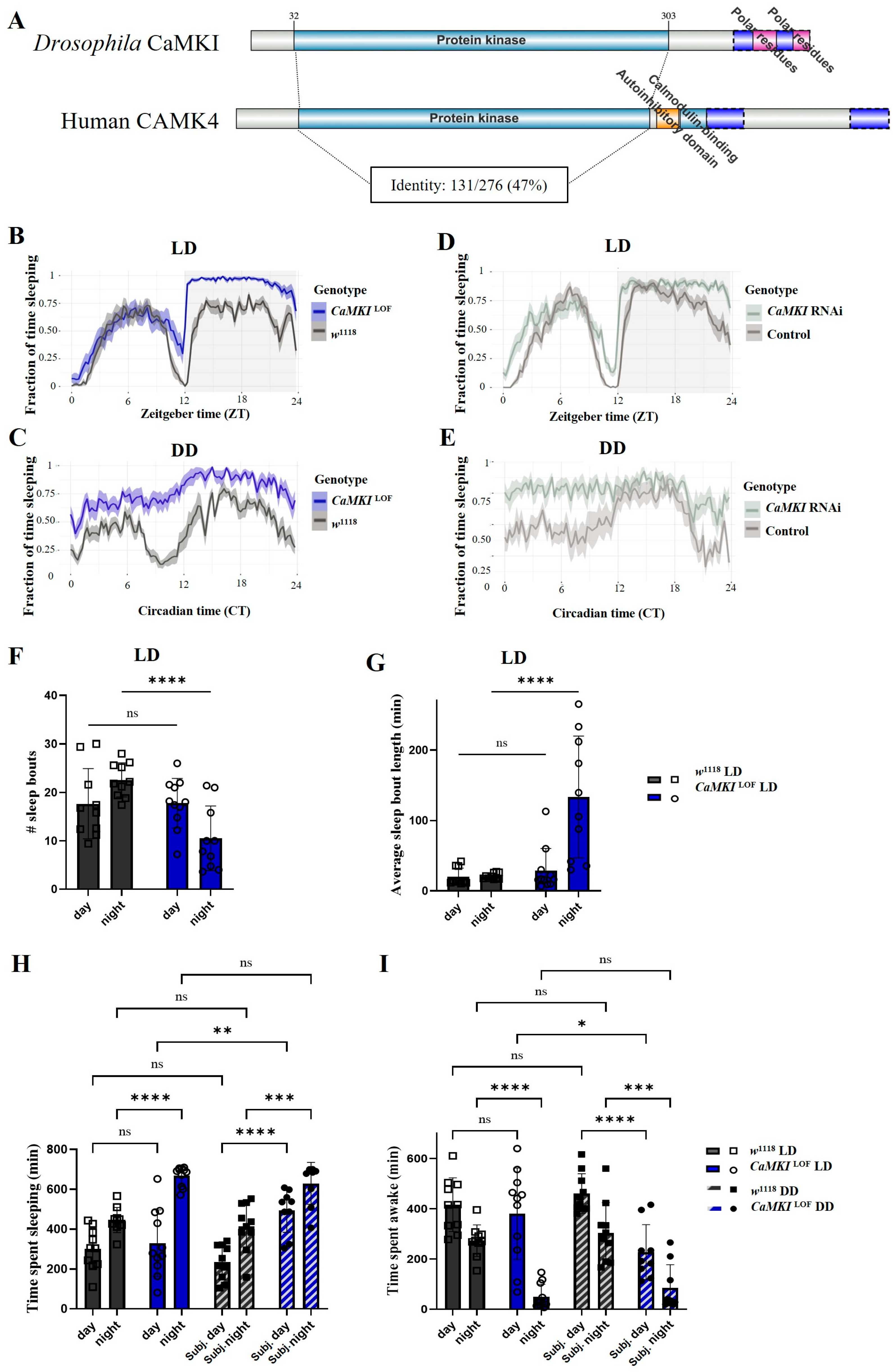
Figure 1. CaMKI dysregulation affects sleep behavior. (A) IBS2.0 protein domain map of Drosophila CaMKI (Q7JMV3) and human CAMK4 (Q16566). Domains annotated based on UniProt entries, on human CAMK4, there is a region of multiple domain overlap between an Autoinhibitory domain (residues 305–321), a PP2A-binding domain (not depicted on the diagram) (residues 306–323), and a Calmodulin-binding domain (residues 322–341). Identity of domains calculated from alignment of CaMKI (NP_524622.1) and CAMK4 (NP_001310303.1) on DIPOT (v9). (B) Sleep fraction over a 24-h period in 12 h:12 h light–dark (LD) conditions for CaMKI mutants (CaMKILOF, blue; n = 11) and control (w1118, black; n = 10) flies. Shaded regions represent the standard error of the mean (SEM). (C) Sleep fraction over a 24-h period in 12 h:12 h dark–dark (DD) conditions for CaMKILOF and w1118 males. (D) Sleep fraction over a 24-h period in LD conditions for CaMKI_RNAi (light-gray; elavc155-Gal4 > CaMKI_RNAi; BDSC_35362; n = 11) and genetic controls (gray; elavc155-Gal4 > w1118; n = 10) males. (E) Sleep fraction over a 24-h period in DD conditions for CaMKI RNAi and control flies. (F,G) Quantification of number of sleep bouts and average sleep bout length in each genotype under LD conditions. (H,I) Total time spent sleeping and time spent awake comparing genotypes, control and CaMKILOF, under LD and DD conditions. Error bars represent ±SEM. Statistical significance is indicated (* p < 0.05, ** p < 0.01, *** p < 0.001, and **** p < 0.0001). Statistical analysis was performed using ANOVA to compare number of sleep bouts and time spent sleeping, while non-parametric multiple comparison Mann–Whitney t-test was performed to compare average sleep bout length between genotypes. The analysis of sleep parameters for DD condition was carried out averaging the data in a 24 h cycle and not based on the observed genotype period cycle.
Biology 14 01228 g001
Figure 2. Circadian rhythm is disrupted following CaMKI dysregulation. (A) Actograms of w1118 control males over a 48-h period in LD conditions. White and shaded gray areas represent light and dark phases, respectively. (B) Actograms of CaMKILOF males in LD conditions over 48 h, showing altered activity patterns compared to controls. (C) Actograms of w1118 males in constant darkness (DD) over 24 h, displaying sustained rhythmic activity. (D) Actograms of CaMKILOF males in DD conditions, showing disrupted rhythmic activity. (E,F) Quantification of circadian period length and power–significance threshold under LD and DD conditions. Bars represent mean + SEM. Statistical significance is indicated (* p < 0.05). Statistical analysis was performed using the non-parametric Mann–Whitney test for multiple t-tests to compare circadian parameters between genotypes.
Biology 14 01228 g002
Figure 3. Social attraction is disrupted in CaMKILOF mutants. Quantification of social approach behavior by calculating the preference index of free-walking flies as they are attracted to immobilized flies on a dish (attached condition; squares) as compared to their response on a dish with no immobilized flies (empty condition; circles) in w1118 controls ((A,B); dark gray) and CaMKILOF mutants ((C,D); blue). Whereas social attraction was observed in control females (A) and males (B), no social attraction was observed in female (C) or male (D) CaMKILOF mutants. Each dot indicates the mean preference index (PI) from a run containing 10 free-walking flies (n = 16 runs for each group). Bars indicate mean ± SEM. Statistical analysis was performed using t-test (* p < 0.05).
Biology 14 01228 g003
Figure 4. CaMKI dysregulation enhances behavioral decline and dendritic degeneration in aging flies. (A) Expression of CaMKI-RNAi in adult motoneurons controlling wing muscles (using the C380-GAL4 driver) resulted in severely impaired flight response, as assessed by the landing distance in 2, 10, and 30-day-old males from controls (C380-GAL4, UAS-CD8-GFP/Y; Cha-GAL80/+; n = 99, 43, 63 flies for 2, 10, and 30 day-old) and in CaMKI knockdown flies (CaMKI-RNAi-1: BDSC_41900, n = 64, 34, 35 flies for 2, 10, and 30 day-old; CaMKI-RNAi-2: BDSC_35362, n = 62, 39, 41 flies for 2, 10, and 30 day-old). Data are presented as median and quartiles. Statistical analysis was performed using Kruskal–Wallis test. (* p < 0.05, **** p < 0.0001) (B) Schematic showing an adult brain and ventral nerve cord (VNC) in addition to the location of motoneurons that innervate the flight muscle. Green circles indicate cell bodies of the identified motoneuron MN5 with the area of the dendritic fields represented by the green dotted line. Dotted square indicates area of pictures shown in (C,D). (C,D) Maximum projection images from confocal stacks showing the two MN5 somata (arrows) and the dendritic area from all 10 DLM motoneurons (dotted line) in control (C380-GAL4, UAS-CD8-GFP/Y; Cha-GAL80/+) and in CaMKI knockdown males (C380-GAL4, UAS-CD8-GFP/Y; Cha-GAL80/UAS-CaMKI-RNAi2-BDSC_35362) both from 30-day-old flies. (E) Quantification of dendritic fluorescence normalized to the baseline fluorescent value in control flies vs. the two different groups of CaMKI knockdown flies as shown in (C,D) (n = 13, 3, 13). Error bars represent ± SEM. Statistical analysis was performed using one-way ANOVA. (*** p < 0.001, **** p < 0.0001) (F) Correlation of landing height (normalized to control) with dendritic fluorescence (normalized to control mean gray values) from 30-day-old flies of controls (circle), CaMKI-RNAi-1 (triangle) and CaMKI-RNAi-2 (square), indicating that poor flight performance correlated with decreased dendritic fluorescence as we previously described [99].
Biology 14 01228 g004
Figure 5. CaMKI disruption leads to NMJ miswiring. (A,B) Schematic diagrams of the transverse nerve (TN) and segmental nerve b (SNb) motor neuron innervations, illustrating their structural organization, key branch points, and synaptic contacts on target muscles. The upper schematic represents the control condition in which the native SNb motor neurons innervate muscles 6 and 7, while the TN passes muscles 6 and 7 without innervating them. The lower schematic highlights alterations (red markings) observed under specific genetic perturbations, where the TN presents innervations and ectopic synaptic contacts onto muscles 6 and/or 7. (C,D) Confocal images of TN and SNb axon projections and synaptic boutons on muscles in controls (DGRP-774) and CaMKILOF mutants. In CaMKILOF mutants (D), the TN shows ectopic synaptic contacts onto muscle 7 (white arrow). Scale bars: 20 µm. (E) Frequency of ectopic contacts in controls (n = 51), CaMKILOF homozygotes (n = 25), and heterozygotes (n = 22), as well as in larvae expressing pan-neuronal CaMKI-RNAi BDSC_35362 (n = 14) and its corresponding genetic control (elavC155-Gal4/w1118; n = 19). Bars represent mean values ± SEM with statistical significance indicated. (F) Quantification of ectopic innervations under different genetic conditions for genetic interaction tests involving genes previously identified to regulate activity-dependent synaptic refinement [18,19,20] (n = 25, 22, 20, 26, 20, 25, 54, 10, 34, 18, 34, and 29). Bars represent mean values ± SEM with statistical significance indicated. Statistical analysis was carried out performing one-way ANOVA and non-parametric Kruskal–Wallis test depending on data distribution (* p < 0.05, **** p < 0.0001).
Biology 14 01228 g005
Figure 6. Proteome dysregulation is observed in CaMKI mutants. Data represent proteome-wide comparisons between CaMKILOF mutants and DGRP-774. (A) Metascape v3.5 [114] visualization of significant differentially expressed proteins (p < 0.05) using protein–protein interaction (PPI)-grouped pathways. Genes of interest noted by red bordered triangles. Various pathways associated with clustering listed in legend and defined in *S3_SUPP—Metascape GO all PPl* (B) Clustering of proteome in samples of each genotype visualizing scaled protein abundance. Created using Proteome Discoverer. (C) Volcano plot of differentially expressed proteins between CaMKILOF mutants and DGRP-774 flies. Proteins of interest highlighted in magenta with name adjacent. Generated in GraphPad Prism 10 using abundance ratios and adjusted significance values generated in Proteome Discoverer. Full protein target list and associated values available in *S4_SUPP—allproteomics data*. (D) Table listing proteins of interest along with fold change and significance values highlighted in panel (C).
Biology 14 01228 g006
Figure 7. Phosphorylation differences are observed in CaMKILOF mutants. (A) Volcano plot of differentially expressed phospho-peptides in CaMKILOF mutants compared to DGRP-774. Peptides of interest highlighted in magenta with gene name and modification listed. Plot created in GraphPad Prism 10 using data generated from Proteome Discoverer, all phospho-peptide data available in *S5—phospho all data*. (B) Relative abundances of selected peptides of interest in CaMKILOF and DGRP-774. Quantified using Proteome Discoverer and visualized using GraphPad Prism 10. Graphs show fold change. (C) Protein–protein interaction network of candidate genes identified from proteomic and phosphopeptidomic analyses (Cytoscape v3.10.3). Nodes represent proteins, and edge length is proportional to 1—STRING confidence score, where weaker interactions appear more distant, using a 0.4 confidence cutoff and maximum of 8 additional interactors. (D) Table of selected peptides and associated fold change with significance (all data available in S5_SUPP—phospho all).
Biology 14 01228 g007
Disclaimer/Publisher’s Note: The statements, opinions and data contained in all publications are solely those of the individual author(s) and contributor(s) and not of MDPI and/or the editor(s). MDPI and/or the editor(s) disclaim responsibility for any injury to people or property resulting from any ideas, methods, instructions or products referred to in the content.