Article

Comparative Study of Lefaxin Family in Two Asian Leeches: Hirudinaria manillensis and Whitmania pigra

1
School of Life Sciences, Key Laboratory of Jiangxi Province for Biological Invasion and Biosecurity, Jinggangshan University, Ji’an 343009, China
2
College of Life Sciences, Jiangxi Normal University, Nanchang 330022, China
3
Engineering Research Center for Exploitation and Utilization of Leech Resources in Universities of Yunnan Province, School of Agronomy and Life Sciences, Kunming University, Kunming 650000, China
*
Authors to whom correspondence should be addressed.
Biology 2025, 14(8), 918; https://doi.org/10.3390/biology14080918
Submission received: 18 June 2025 / Revised: 11 July 2025 / Accepted: 21 July 2025 / Published: 23 July 2025
(This article belongs to the Section Biochemistry and Molecular Biology)

Simple Summary

The hematophagous buffalo leech (Hirudinaria manillensis) and the non-hematophagous medicinal leech (Whitmania pigra) have long been utilized in traditional Chinese medicine for treating blood-stasis-related diseases. Despite their widespread use, the underlying pharmacological mechanisms remain insufficiently understood. This study combined next-generation sequencing and recombinant expression to systematically compare the lefaxin family (a Factor Xa inhibitor) between both species. Three lefaxin genes (designated lefaxin_Hman1–3 and lefaxin_Wpig1–3) were identified in each sequenced genome of each species, with a focus on intraspecific variation, molecular evolution, gene expression, and anticoagulant activity. The comparative analysis reveals that lefaxin genes in both leech species are subject to purifying selection, emphasizing their conserved functions and roles in anticoagulation. Lefaxins derived from W. pigra demonstrate more vigorous anticoagulant activity compared to those from H. manillensis. These findings challenge traditional assumptions regarding the medicinal efficacy of leeches, propose lefaxins as a promising therapeutic target for thrombotic diseases, and underscore the need to reevaluate the medical potential of non-hematophagous leech species.

Abstract

Thrombotic diseases represent a significant global health burden, particularly for middle-aged and elderly populations. Medicinal leeches, such as Hirudinaria manillensis and Whitmania pigra, have been traditionally used for their anticoagulant properties. The genomes of these leeches each harbor three lefaxin genes, which are designated lefaxin_Hman1–3 and lefaxin_Wpig1–3, respectively. We conducted genomic and transcriptomic sequencing on wild populations of both species. Bioinformatics tools were employed to analyze intraspecific variation, molecular evolution, and protein structures. We expressed recombinant lefaxin proteins in Pichia pastoris and assessed their anticoagulant activities using in vitro coagulation assays. H. manillensis exhibited greater genetic diversity and stability, whereas W. pigra showed higher expression levels and hydrophilicity. Both species exhibited purifying selection, indicating conserved function, and their lefaxin structures are similar to the archetypal lefaxin (UniProt No. P86681.1). W. pigra lefaxins bound Factor Xa more effectively. W. pigra lefaxins exhibited more robust anticoagulant activity in vitro compared to those from H. manillensis. W. pigra, a non-hematophagous leech, shows potent anticoagulant activity through lefaxins, challenging traditional views on leech efficacy. This study underscores the potential of lefaxins as therapeutic targets for thrombotic diseases and highlights the need to reconsider the use of various leech species in medicine.

1. Introduction

The incidence, disability rate, and mortality associated with thrombotic diseases have consistently increased, positioning them as a significant global health concern, especially among middle-aged and elderly populations [1]. Thrombi, abnormal intravascular clots that impede circulatory flow, can induce ischemic tissue necrosis and organ dysfunction by reducing blood supply in various pathologies [2]. While current anticoagulants (e.g., heparin, warfarin) remain mainstay therapies, their limitations, including bleeding risks and narrow therapeutic windows, indicate that there must be novel treatments targeting alternative pathways in the coagulation cascade [3]. Leeches (Annelida: Clitellata: Hirudinida) are an ancient therapeutic tool with long-standing medicinal use across many cultures, particularly in traditional bloodletting practices [4]. Recognized in modern complementary and alternative medicine [5,6,7], leeches hold significant value in traditional Chinese medicine (TCM) for their hematological effects. The Shen-Nong-Ben-Cao-Jing first documented the use of leeches for treating “blood stasis, amenorrhea, abdominal masses, and fluid retention” [8]. Leeches exhibit diverse pharmacological effects, including anticoagulation, vasodilation, and menstrual regulation [9]. Contemporary applications of leeches focus on cardiovascular and cerebrovascular diseases, as well as thrombotic disorders, through their blood-activating and stasis-resolving properties [10].
Leeches exhibit remarkable species diversity. Those commonly employed in TCM include Hirudo nipponia, Whitmania pigra, Whitmania acranulata, and the Asian buffalo leech (Hirudinaria manillensis) [11]. Of these, H. nipponia, W. pigra, and W. acranulata are officially listed in the Chinese Pharmacopoeia [12]. Although H. manillensis is not included in the Chinese Pharmacopoeia, its medicinal value has been recognized and utilized regionally, with documented inclusion in the local standards for Chinese medicinal materials in Guangxi [13] and Yunnan provinces [14].
Leeches can be broadly classified, based on their feeding habits, into hematophagous (blood-feeding) and non-hematophagous types [15]. H. manillensis predominantly parasitizes humans, domestic animals, and other vertebrates [16,17]. This species has a wide distribution across South and Southeast Asia, as well as southern China, primarily inhabiting lowland ecosystems, including rice paddies, swamps, and ponds [18]. In contrast, W. pigra represents a non-hematophagous species that feeds on mollusk hemolymph, particularly from snails and clams [19]. The species W. pigra is primarily distributed across most regions of China and certain areas of Japan [20].
H. manillensis and W. pigra are the dominant species in the Chinese medicinal leech market, being the most frequently used for medicinal purposes [21,22]. By comparison, the smaller size and slower reproduction rate of H. nipponia result in reduced aquaculture profitability, with current populations inadequate for market requirements [23]. Although studies demonstrate the superior antithrombotic efficacy of H. manillensis over both H. nipponia and some non-hematophagous leeches [24], such findings should not diminish the research potential of non-hematophagous species. Processed leeches such as W. pigra have maintained historical significance in medicinal applications [25]. While thermal processing often denatures polypeptides and protein-based components, such as hirudin, W. pigra exhibits enhanced anticoagulant activity post-decoction, contrasting with H. manillensis, which shows reduced anticoagulant capacity after heat treatment [26]. These differential responses highlight the distinct therapeutic merits of non-hematophagous leeches that merit comprehensive investigation.
Recent advances in modern medicine have identified a diverse array of antithrombotic proteins and peptides in leeches, with hirudin being the most prominent [27]. Hirudin consists of approximately 65 amino acids and has a molecular weight of around 7000 Daltons [28]. It functions as a potent natural thrombin inhibitor. The high specificity of hirudin for thrombin enables it to effectively inhibit the conversion of fibrinogen to fibrin catalyzed by thrombin [29]. In addition to hirudin, other bioactive substances have been identified in leeches, including antistasin [30], a Factor Xa inhibitor; decorsin [31], a platelet aggregation inhibitor; and bdellastasin [32], an anticoagulant protein. These substances are capable of exerting anticoagulant effects either directly or indirectly.
Factor Xa, a vitamin-K-dependent coagulation protease, catalyzes thrombin generation from prothrombin in the presence of Factor Va, calcium, and phospholipids [33]. This central role in coagulation cascade activation has positioned Factor Xa as a key therapeutic target for anticoagulant development, particularly in managing thrombotic disorders such as stroke, myocardial infarction, and deep vein thrombosis [34]. Lefaxin, a 30 kDa Factor Xa inhibitor (isoelectric point~5.7) isolated from Haementeria depressa salivary glands, demonstrates significant anticoagulant potential by specifically inhibiting Factor Xa activity within the prothrombinase complex [35].
Studies have demonstrated that the anticoagulant activities of leech genes may differ significantly even within the same species. For example, while multiple hirudin genes exist in the H. manillensis genome, only select variants demonstrate anticoagulant properties [36]. This functional divergence suggests that structural or sequence variations in amino acid composition may influence hirudin efficacy [37]. Interspecies genetic variations further contribute to the differential composition, functionality, and evolutionary patterns of antithrombotic genes across leech species, where homologous genes might be expressed in one species but silenced in another through genomic absence or regulatory differences [21].
Our previous research has identified at least 72 antithrombotic genes within the genome of H. manillensis [37], spanning 21 gene families. The lefaxin genes in H. manillensis comprise three members (lefaxin_Hman1–3). The genome of W. pigra contains at least 79 antithrombotic genes distributed across 20 gene families [38], and the lefaxin genes comprise three members (lefaxin_Wpig1–3). This study aims to investigate whether the lefaxin genes exhibit antithrombotic activity in these two leech species through bioinformatics analysis, gene expression profiling, and in vitro anticoagulant activity assays and to compare the similarities and differences between the lefaxins of H. manillensis and W. pigra.

2. Materials and Methods

2.1. DNA and RNA Sequencing

We sampled wild populations of H. manillensis and W. pigra from various locations. Specimens of H. manillensis were collected from Yulin, Guangxi (GXYL, 110.57° E, 22.81° N); Zhanjiang, Guangdong (GDZJ, 110.46° E, 21.25° N); and Honghe, Yunnan (YNHH, 102.58° E, 23.31° N). We also collected W. pigra specimens from Baodi, Tianjin (TJBD, 117.48° E, 39.47° N); Yibin, Sichuan (SCYB, 105.31° E, 28.18° N); and Wuhan, Hubei (HBWH, 114.57° E, 30.59° N). After a two-week acclimatization period under laboratory conditions (temperature maintained at 25 ± 1 °C, 70 ± 5% relative humidity, 12 h light/dark cycle, and daily water renewal), 10 adult individuals were randomly selected from each of the collection sites. The anterior one-third of the head tissue was excised from each specimen, and any residual blood was washed away using sterile water. We extracted total DNA using the DNeasy Blood and Tissue Kit (Qiagen, Germany). We checked the quality and condition of the DNA using agarose gel electrophoresis, NanoDrop spectrophotometry (NanoDrop Technologies, Wilmington, DE, USA), and Qubit fluorometry (Thermo Fisher Scientific, Waltham, MA, USA). High-quality genomic DNA was then used to construct sequencing libraries for the PacBio and Illumina platforms.
For RNA-Seq, total RNA was extracted from individual leeches using TRIzol reagent (Thermo Fisher Scientific Inc.) and purified with the RNeasy Mini Kit (Qiagen, Chatsworth, CA, USA). We constructed and sequenced a 350 bp insert DNA library on the Illumina HiSeq 2000 platform using paired-end 150 bp reads. We processed the raw sequences using fastp v0.20.0 [39] to remove adapters and low-quality regions, yielding clean reads for further analysis.

2.2. Sequence Extraction

We performed de novo assembly of genome resequencing clean reads using MEGAHIT v1.2.9 [40], which generated contigs for each sample. For transcriptome data, we assembled clean reads using Trinity v2.9.0 [41] to obtain unigene sequences. We used published lefaxin genes as bait sequences for subsequent analysis. We formatted the unigene sequences into a BLAST database and identified homologous sequences using BLAST v2.13.0+ [42].
Using MEGA v11.0.13 [43], we aligned the homologous sequences and examined variant sites. Based on the alignment results and intron boundary information (GT-AG rule), we extracted coding regions. For genes with low expression levels that lacked complete coding sequences, we used exons ±50 bp as bait to retrieve homologous contigs through BLAST searches, followed by alignment in MEGA. To resolve highly variable regions that likely resulted from assembly gaps, we employed Mirabait [44] to extract and align related reads, thereby improving sequence accuracy.

2.3. Intraspecific Variation Analysis

We compiled the coding region sequences of each gene into FASTA format and aligned them using the “Align by MUSCLE (Codons)” function in MEGA. We then determined the number of variable sites (VS) and number of haplotypes (HN) for each gene using DnaSP [45]. VS reflects mutation frequency within gene sequences, while HN indicates genetic diversity. We calculated Watterson’s Theta diversity (WD) index, which measures genetic variation within populations, using DAMBE v7.3.5 [46]. For further analysis, we changed gene sequences into amino acid sequences in MEGA and then calculated VS, HN, and WD for these sequences using DAMBE.

2.4. Molecular Evolution Analysis

Lefaxin genes from four leech species (H. manillensis, W. pigra, H. nipponia, and Hirudo medicinalis) were aligned, and a phylogenetic tree was constructed using MEGA. Based on the relationships among the four species [47], a species tree of these lefaxins was built using TreeGraph v2.15 [48]. The dN/dS (ω) ratios were estimated using PAML-X [49] to evaluate the selective pressures acting on each lefaxin gene. Synonymous substitutions are assumed to be neutral, whereas nonsynonymous substitutions may be subject to purifying (ω < 1), neutral (ω = 1), or positive (ω > 1) selection.
Three models from the CODEML module of PAML were applied: the branch model, branch-site model, and site model. In the branch and branch-site models, branches of H. manillensis and W. pigra were set as foreground, with the others as background. The site model analysis employed three models: M0, M7, and M8. By examining the ω ratios derived from these models, we elucidated how different types of selective pressures may have influenced various members of the lefaxin family during their evolutionary history.

2.5. Gene Expression Analysis

We constructed sequence indices using the Salmon v1.0.0 [50] based on coding region sequences from whole-genome annotation data. We aligned the transcriptome reads from each sample to these indices using a k-mer value of 31, which yielded transcripts per million (TPM) values for each coding region sequence. This information was used to determine and compare the relative expression levels of genes across samples.
We analyzed differences in the levels of lefaxin gene expression among populations within species using SPSS v25 [51]. A one-sample Kolmogorov–Smirnov test indicated that lefaxin expression levels significantly deviated from normality (p < 0.05). Given this result, we employed non-parametric methods: first, we used the Several-Related-Samples test (Friedman test) to assess significant differences among genes. If significant differences were detected, we performed pairwise comparisons using the Two-Related-Samples test (Wilcoxon test).
Differences in lefaxin gene expression between species were analyzed using SPSS software. The one-sample Kolmogorov–Smirnov test was used to check for a normal distribution (p > 0.05). If the data had a normal distribution and the variances were homogeneous (as determined by Levene’s test, p > 0.05), we used an Independent Samples T-test. If not, we applied the non-parametric Mann–Whitney U test.

2.6. Analysis of Protein Biochemical Properties

We translated the six lefaxin genes into their corresponding protein sequences. We used SignalP [52], an online tool, to predict signal peptide regions and evaluate the presence of signal peptide domains. Additionally, we used the ProtParam online tool to calculate the isoelectric point (pI), instability index (II), aliphatic index, and grand average of hydropathy (GRAVY) for each protein [53].

2.7. Protein Structure Alignment Analysis

We predicted the protein structures of six lefaxins using the AlphaFold Protein Structure Database, along with the archetypal lefaxin (UniProt No. P86681.1). To assess their structural similarity, we employed the FATCAT method [54], which allows flexible protein alignment by detecting hinges and internal rearrangements.
We utilized parameters such as root mean square deviation (RMSD), FATCAT score, and p-value during the alignment process to assess structural similarity. These parameters quantitatively measured structural conservation and highlighted specific similarities and differences among lefaxins from different species. This analysis offers insights into their evolutionary and functional relationships.

2.8. Protein Docking Analysis

The ZDOCK server [55] docked the archetypal lefaxin and six lefaxins with human Factor Xa, treating Factor Xa as the receptor and lefaxins as ligands. ZDOCK is a protein docking algorithm that predicts protein–protein binding modes through energy minimization. After docking, we used PyMOL 3.1 [56] to visualize protein structures and analyze interaction interfaces. Subsequent analysis of the docking results included calculating the binding free energy and evaluating the characteristics of amino acid residues at the binding sites to assess the stability and biological significance of the docking modes.

2.9. Pichia Pastoris Expression

Plasmids containing the six genes were synthesized and ordered from Sangon Biotech (Shanghai, China), retaining the necessary sequences for full gene expression. We cultured Escherichia coli in a liquid LB medium overnight with shaking to amplify the plasmid DNA. The plasmid DNA was extracted using a commercial kit, linearized with the SpeedyCut SacI enzyme, and purified using a corresponding purification kit.
The linearized plasmid DNA was chemically transformed into GS115-competent cells, which were then plated on Yeast Extract Peptone Dextrose (YEPD) medium with 0.25% geneticin. Yeast colonies appeared after 3–5 days. The successfully transformed yeast cells underwent stepwise resistance selection on a YEPD medium with increasing concentrations of geneticin (0.5%, 1%, and 2%). We inoculated the selected yeast cells into a buffered glycerol-complex medium for expansion. We extracted yeast genomic DNA using a rapid extraction kit and confirmed the integration of the target gene into the yeast genome by PCR amplification and agarose gel electrophoresis. After confirming successful integration, we transferred the yeast cells to the buffered methanol-complex medium for fermentation, which induces the expression of the target gene. We purified the expressed proteins by salting out and desalting them to obtain the final product. We separated the six recombinant proteins by SDS-PAGE and confirmed the presence of the target protein by comparing the molecular weight of the obtained bands with those of the target bands.

2.10. Anticoagulation Test

We evaluated the anticoagulant activity of the target proteins using a Sienco hemorheology analyzer (Viscell, UK), which measures clotting kinetics and platelet function using ultrasonic viscoelastic detection. We prepared 500 μL of citrated pig blood (3.8% sodium citrate) and mixed it with 100 μL of the target protein. To reverse anticoagulation, we added 20 μL of 0.25 M calcium chloride to reinitiate clotting capability. We transferred the mixture into a test cup containing 360 μL of whole blood and analyzed it with the Sonoclot system. The instrument’s ultrasonic sensor (200 Hz oscillation) monitored real-time changes in blood viscosity and elasticity during clot formation, generating a coagulation signal curve.
We tested each protein sample in triplicate and recorded the following parameters: (1) Activated clotting time (ACT): Normal range, 100–240 s; values exceeding 240 s indicate prolonged coagulation. (2) Clot rate (CR): Normal range 10–35; values < 10 suggest impaired clotting. (3) Platelet function (PF): PF is quantitatively derived from the timing and quality of clot retraction, reflecting platelet contractile activity during the late phase of coagulation. Values > 1 indicate normality, while values < 1 indicate bleeding risk. All three parameters were automatically calculated by the Sonoclot system based on viscoelastic changes observed during the clotting process. We inferred anticoagulant activity based on delayed or inhibited clot formation after calcium addition, as evidenced by divergence from control curves. This method allowed us to assess the target protein’s effects on hemostasis quantitatively.

3. Results

3.1. Intraspecific Variation

In the reference genomes of H. manillensis and W. pigra, six lefaxin genes were identified and named as lefaxin_Hman13 and lefaxin_Wpig13, respectively. The coding sequences and amino acid sequences of these genes, alongside those of H. nipponia and H. medicinalis, were aligned to enable comparison across species (Supplementary File S1). Complete coding sequences for lefaxin_Hman1–3 (all 32 H. manillensis samples, Supplementary File S2) and lefaxin_Wpig1–3 (all 35 W. pigra samples, Supplementary File S3) were successfully obtained. After translating and aligning the valid sequences to analyze sequence variation, a total of 20 variable sites were identified at the DNA level, and 5 were identified at the protein sequence level. The VS in the lefaxin genes of H. manillensis was significantly higher than that in W. pigra at both the DNA and protein levels. Additionally, 30 DNA haplotypes and 11 amino acid haplotypes were identified, with H. manillensis exhibiting a significantly higher HN at both levels compared to W. pigra. The WD index for both the DNA and protein levels also indicated significantly higher diversity in H. manillensis (Table 1).
In summary, the lefaxin genes in H. manillensis demonstrated greater genetic diversity and more haplotypes at both the nucleotide and amino acid levels. In contrast, although W. pigra also exhibited some genetic variation, it had fewer variation sites and haplotypes in the lefaxin genes compared to H. manillensis. These findings offered helpful information about the molecular evolutionary processes and ecological adaptive differences between these two leech species.

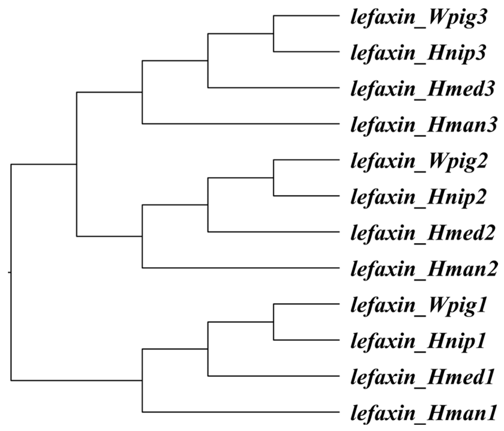
3.2. Molecular Evolution

As shown in Figure 1, a species tree was reconstructed based on lefaxin gene sequences, revealing the phylogenetic relationships among H. manillensis, W. pigra, H. nipponia, and H. medicinalis. The branch model analysis revealed that all six lefaxin genes exhibited low ω values (≤0.08985), suggesting strong purifying selection and a high degree of conservation. The branch-site analysis further showed that, among the six lefaxin genes, lefaxin_Hman2 had only 3.0776% of its sites under purifying selection, with the majority of sites exhibiting neutral selection. In contrast, the other five lefaxin genes exhibited 86.746–95.156% of their sites under purifying selection (ω ≤ 0.04615), with minimal neutral selection (ω = 1). The Bayes Empirical Bayes analysis identified several codon sites with potential positive selection (Table 2).
The site model analysis (M0, M7, M8) corroborated these results. The M0 model yielded an average ω of 0.06006, reflecting overall purifying selection. Models M7 and M8 confirmed that most sites were under purifying selection (ω = 0.00011–0.25513), with negligible positive selection (ω = 81.13, probability = 0.00001).
In summary, lefaxins were highly conserved, with most sites under strong purifying selection, highlighting their functional significance. The molecular evolution of lefaxins in W. pigra and H. manillensis was similar, dominated by purifying selection, likely due to their essential physiological roles in leeches.

3.3. Gene Expression

The mean TPM values ± standard deviations for the lefaxin genes are presented in Table 3. The total expression levels of the lefaxin genes in H. manillensis were 10,013.0 ± 5930.2, whereas in W. pigra, they were 29,041.4 ± 13,030.8. The total expression of the lefaxin genes was significantly higher in W. pigra than in H. manillensis (Z = −3.814, p < 0.001).
In H. manillensis, the Friedman test revealed a highly significant overall difference in the expression levels of the three lefaxin genes (Chi-Square = 51.063, df = 2, p < 0.0001). The Wilcoxon test showed significant differences in expression levels between all pairwise gene comparisons (Z < −2.281, p < 0.023). In W. pigra, the Friedman test also showed a highly significant overall difference in the expression levels of the three lefaxin genes (Chi-Square = 70, df = 2, p < 0.001). The Wilcoxon test showed significant differences in expression levels across all pairwise gene comparisons (Z < −5.159, p < 0.001).
A Mann–Whitney U test showed no significant difference in the expression levels of lefaxin_Hman1 and lefaxin_Wpig1. However, highly significant differences in expression levels were observed between lefaxin_Hman2 and lefaxin_Wpig2, as well as between lefaxin_Hman3 and lefaxin_Wpig3 (Table 3).

3.4. Protein Biochemical Properties

The sequence lengths of the homologous lefaxin genes were identical in H. manillensis and W. pigra. However, the molecular weights of lefaxins in W. pigra were generally slightly higher than those in H. manillensis. The pI values of the two species were not significantly different (p = 0.567). They were both below the physiological pH of mammals (7.35–7.45), suggesting that lefaxins carried a net positive charge under physiological conditions. The II values, which range from 0 to 40 for stable proteins, fell between 27.27 and 38.34 for lefaxins, indicating overall stability. Notably, lefaxins in H. manillensis exhibited slightly lower II than those in W. pigra, suggesting greater structural stability. The aliphatic indices ranged from 68.25 to 80.32. The aliphatic index reflects the volume occupied by aliphatic side chains in a protein, and higher values are commonly linked to enhanced thermal stability. H. manillensis showed marginally higher values, indicating enhanced thermal stability [57]. The GRAVY scores of all lefaxins were negative (−0.524 to −0.658), reflecting a general tendency to interact with water molecules. A more negative GRAVY score indicates greater hydrophilicity of the protein. Lefaxins in W. pigra exhibited slightly higher hydrophilicity values than those in H. manillensis (Table 4).
Differences in molecular weight, stability, and hydrophilicity between the two species may have reflected adaptive divergence in response to their ecological niches. H. manillensis lefaxins appeared to be more thermally and structurally stable, whereas W. pigra lefaxins displayed greater hydrophilicity and solubility, which may have supported their species-specific physiological functions.

3.5. Protein Structure Alignment

Structural alignment using the FATCAT algorithm revealed significant similarity between the archetypal lefaxin and six variants from H. manillensis and W. pigra, with all p-values ≤ 0.005. FATCAT scores ranged from 106.50 to 107.63, and RMSD values from 1.75 Å to 1.86 Å, reflecting minimal structural variation across 56 aligned residues. Notably, lefaxin_Hman2 and lefaxin_Wpig2 exhibited the highest FATCAT scores (107.63 and 107.57), while lefaxin_Wpig1 displayed the lowest RMSD (1.75 Å). These results suggested that the archetypal lefaxin exhibited substantial structural similarity with all six lefaxins, potentially supporting conserved biological functions at the molecular level.

3.6. Protein Docking

We performed protein–protein docking between the archetypal lefaxin and Factor Xa using ZDOCK and visualized the interacting residues with PyMOL. We then selected the identical residues from the archetypal complex as reference points and used them to compare the docking results of the six lefaxin variants (Table 5). The docking analysis revealed that many of these key residues were consistently involved across all six proteins, indicating that they played a crucial role in binding to Factor Xa and contributed to the stability of the docking interaction.
We evaluated docking quality using ZDOCK scores, where higher values indicated more stable interactions and lower energy conformations. Lefaxin_Hman2 and lefaxin_Wpig2 obtained relatively higher scores. Overall, the lefaxins from W. pigra showed slightly higher docking scores than those from H. manillensis, implying that they may have possessed greater structural stability or stronger affinity for protein–protein interactions.

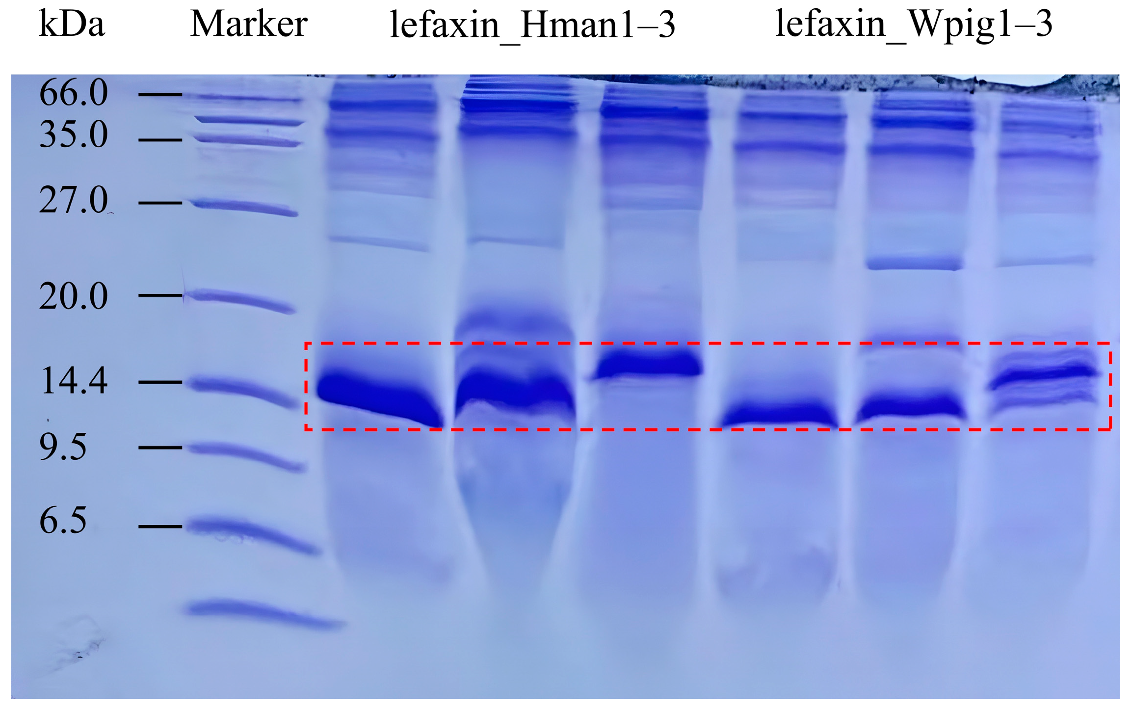
3.7. Recombinant Protein Synthesis

We successfully expressed six recombinant lefaxins (lefaxin_Hman1–3 and lefaxin_Wpig1–3) in Pichia pastoris. After purification, the protein concentrations were 13.73, 30.40, 24.20, 25.44, 28.06, and 22.62 mg/mL, respectively. We predicted their molecular weights to be 13.86, 13.77, and 14.80 kDa for lefaxin_Hman1–3, and 13.90, 13.86, and 14.95 kDa for lefaxin_Wpig1–3. SDS-PAGE analysis confirmed that all recombinant proteins appeared at their expected molecular weights (Figure 2).

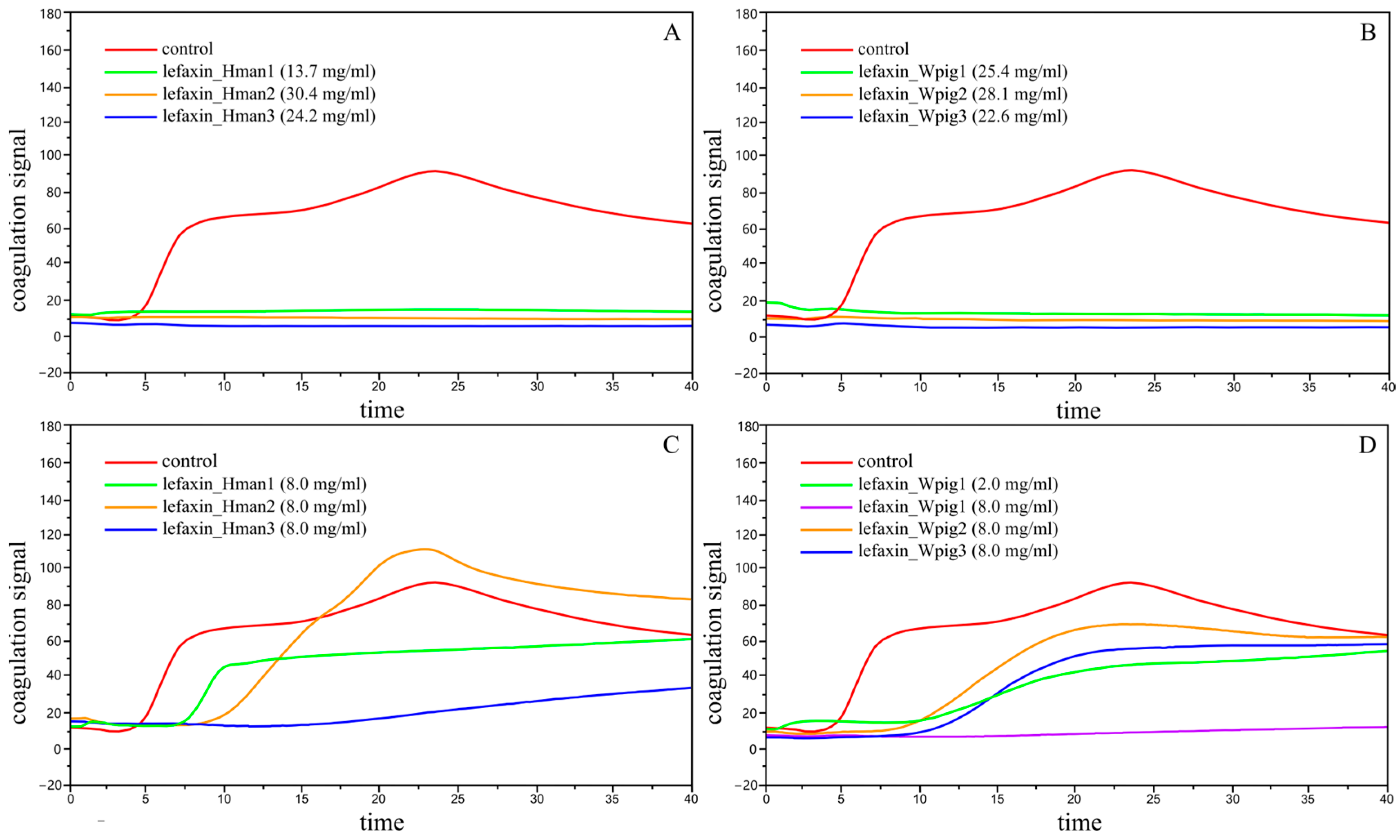
3.8. Anticoagulation of Lefaxins

To evaluate the anticoagulant properties of recombinant lefaxins, we conducted three sets of qualitative and three sets of quantitative replicate experiments, including all six target proteins from H. manillensis (lefaxin_Hman1–3) and W. pigra (lefaxin_Wpig1–3). Each assay was performed in three biological and three technical replicates. Anticoagulation performance was assessed using three clinical indicators: ACT, CR, and PF.
In qualitative tests, the parameters for the control group remained within the physiological range, confirming normal coagulation function. In contrast, all target proteins at their original concentrations resulted in complete suppression of coagulation, rendering ACT, CR, and PF values unmeasurable (Table 6). As shown in Figure 3A,B, the coagulation signal of the control group increased sharply after approximately 3 min, peaked, and then declined, indicating successful clot formation. However, the lefaxin-treated groups maintained flat signal curves, indicating vigorous anticoagulant activity.
For quantitative analysis, all lefaxins were tested at a standardized concentration of 8 mg/mL. As shown in Table 6 and Figure 3C,D, lefaxins from W. pigra exhibited markedly stronger anticoagulant activity than those from H. manillensis. In particular, lefaxin_Wpig1 completely suppressed coagulation at 8 mg/mL, with no measurable coagulation parameters due to full inhibition. Remarkably, even when diluted to 2 mg/mL, lefaxin_Wpig1 still maintained substantial anticoagulant activity, surpassing the performance of other proteins tested at 8 mg/mL.
In summary, lefaxin_Wpig1 exhibited the most potent anticoagulant effect among all tested proteins, and lefaxins from W. pigra overall displayed more vigorous anticoagulant activity than those from H. manillensis.

4. Discussion

This study compared the lefaxin family in two Asian leech species, H. manillensis and W. pigra, using next-generation sequencing techniques and recombinant expression assays. Molecular evolutionary analysis indicated that the dN/dS ratios for all lefaxins were significantly below 1 in both species, suggesting that these proteins were under strong purifying selection and tended to be conserved during evolution. Specific sites under neutral or positive selection indicated potential functional diversification. Further analysis revealed that H. manillensis lefaxins exhibited higher numbers of gene variant sites at both the DNA and protein levels, as well as greater haplotype numbers and a higher Watterson’s Theta diversity index compared to W. pigra. This divergence likely reflects the ecological habits and physiological traits of H. manillensis. As a tropical species, H. manillensis inhabits environments with higher ambient temperatures. As a poikilotherm, it has a relatively high metabolic rate [58]. The elevated metabolic rate likely increases the genomic mutation rate [59], thereby contributing to the higher genetic diversity observed in the lefaxin genes of H. manillensis.
Differences in lefaxin gene expression and anticoagulant activity between the two species suggest distinct evolutionary trajectories and molecular adaptation strategies. The elevated lefaxin expression in W. pigra may be phylogenetically attributed to its closer affinity to hematophagous leech genera, implying a blood-feeding ancestral lineage reliant on lefaxin-mediated anticoagulant activity. Although W. pigra has evolved into a non-bloodsucking species, the expression pattern of the lefaxin gene retains its ancestral characteristics due to its high conservation and short functional degradation.
Structural biology studies help explain why W. pigra lefaxins are better at preventing blood clots due to their similar structure, strong binding ability, and important interactions between specific parts of the molecules. Structurally, W. pigra lefaxins exhibit a high degree of similarity to the archetypal lefaxin, serving as a conserved scaffold that facilitates recognizing and binding to specific coagulation Factor Xa regions. Binding affinity determines the strength of their interaction [60], with W. pigra lefaxins demonstrating significantly higher affinity than other homologs, indicating their enhanced capacity to form stable complexes with Factor Xa. Key residue interactions mediate the anticoagulant function, stabilizing lefaxin–Factor Xa binding through multiple non-covalent bonds [61]. Critical binding sites in W. pigra lefaxin (e.g., K96 and Y99) are highly conserved relative to the archetypal lefaxin, correlating with its markedly superior anticoagulant activity compared to other homologs. These three factors act synergistically: structural similarity provides an optimal framework for residue functionality, while residue interactions reinforce binding affinity. Collectively, this molecular interplay confers high anticoagulant efficacy on the W. pigra lefaxins (especially lefaxin_Wpig1), effectively inhibiting blood coagulation in vitro.
In contrast, the low expression of lefaxins in H. manillensis may be due to a functional reallocation of anticoagulation strategies. As a hematophagous species, H. manillensis depends on blood ingestion for nutrient acquisition, necessitating rapid and potent anticoagulant mechanisms to sustain its feeding behavior. This species likely relies primarily on specialized anticoagulant proteins (e.g., hirudin) for immediate hemostatic suppression, thereby reducing selective pressure on lefaxin expression. The interplay of ancestral phylogenetic constraints and lineage-specific functional adaptations shapes the divergence observed between the two species. Specifically, W. pigra retains molecular traits constrained by its ancestral genetic architecture, whereas H. manillensis has undergone evolutionary specialization to prioritize functional efficiency in blood-feeding.
The non-hematophagous nature of W. pigra has long been a subject of skepticism within TCM, since its inclusion in the Chinese Pharmacopoeia [62]. According to TCM theory, leeches that suck blood are believed to possess special healing properties due to their bloodsucking behavior. Bloodsucking leeches, such as H. manillensis and H. nipponia, are thought to contain more active substances that help prevent blood clots, making them highly valuable in medicine. This study found that W. pigra does not rely on bloodsucking for sustenance, but its lefaxin proteins exhibit more vigorous anticoagulant activity. In contrast, H. manillensis, a hematophagous leech, exhibits weaker anticoagulant activity. This finding provides new scientific evidence supporting the medicinal value of W. pigra. It may alter the TCM perception of its efficacy, prompting the TCM community to reevaluate the criteria and theoretical basis for using leeches in medicine.
In molecular evolution, it is widely accepted that genes with more critical functions evolve more slowly than those with less important functions [63]. This principle is based on the neutral theory, which suggests a negative correlation between the functional importance of a gene and its rate of evolution. Specifically, molecules or segments with less functional importance tend to evolve more rapidly than those with more significant functional importance [64]. Since the anticoagulant function of lefaxins is vital for the survival of leeches, its sequence and structure undergo purifying selection during evolution, resulting in fewer variations. This evolutionary pattern is consistent with that of many other conserved genes. Lefaxin is more stable than other anticoagulant proteins, such as hirudin, and its well-preserved sequence suggests it has a reliable function in the organism [65]. Therefore, lefaxin may be an ideal target for the development of novel anticoagulant drugs.
From a pharmacological perspective, lefaxin offers potential advantages over classical anticoagulants such as hirudin. While hirudin acts as a direct thrombin inhibitor, lefaxin targets the upstream activator Factor Xa, thereby indirectly modulating thrombin generation. This mechanism may provide a more controllable anticoagulant effect and potentially reduce the risk of bleeding complications [66]. The evolutionary stability, structural conservation, and demonstrated anticoagulant activity of lefaxin underscore its promise as a novel target for the development of anticoagulant drugs.
This study closely compared the lefaxin family in two types of leeches, showing that lefaxins in W. pigra have more vigorous anticoagulant activity. This finding not only provides a scientific basis for the medicinal value of W. pigra but also has the potential to alter TCM’s perception of the efficacy of leeches. Lefaxin shows promise as a novel target for anticoagulant drugs. Future research could further explore the structure–function relationship of lefaxin, combined with drug design and optimization, to potentially develop more efficient anticoagulant drugs. Studying how lefaxin has evolved and been selected by different leech species will enhance our overall understanding of its development and function, providing new insights and opportunities for utilizing leech resources.

5. Conclusions

This study reveals that the non-hematophagous leech W. pigra exhibits more vigorous anticoagulant activity via its lefaxin proteins than the hematophagous H. manillensis. The findings challenge traditional assumptions about the medicinal efficacy of leech species and highlight lefaxins as a promising therapeutic target for thrombotic diseases. The research highlights the importance of reevaluating leech species for medicinal applications, providing a scientific basis for the inclusion of W. pigra in the development of anticoagulant drugs and expanding the potential of leech-derived biomolecules in modern medicine.

Supplementary Materials

The following supporting information can be downloaded at https://www.mdpi.com/article/10.3390/biology14080918/s1. File S1: Coding and amino acid sequences of lefaxins from H. manillensis, W. pigra, H. nipponia, and H. medicinalis; File S2: lefaxin coding sequences from 32 H. manillensis samples; File S3: lefaxin coding sequences from 35 W. pigra samples.

Author Contributions

Conceptualization, G.L. and Z.H.; formal analysis, T.Y. and F.Z.; funding acquisition, Z.H. and G.L.; investigation, T.Y., M.X. and J.Y.; methodology, G.L.; resources, R.A., L.T. and Z.L.; data curation, F.Z., L.T. and Z.L.; writing—original draft preparation, T.Y.; writing—review and editing, G.L.; visualization, M.X., J.Y. and R.A.; supervision, L.T. and Z.L.; project administration, F.Z. and Z.H. All authors have read and agreed to the published version of the manuscript.

Funding

This research was funded by the National Natural Science Foundation of China (No. 82260742), the Double Thousand Plan of Jiangxi Province (No. jxsq2023201063), the Foundation of Yunnan International Joint Laboratory with South and Southeast Asia for the Integrated Development of Animal-derived Anti-thrombosis Chinese Medicine (No. 202503AP140025), and the Key Laboratory of Jiangxi Province for Biological Invasion and Biosecurity (No. 2023SSY02111).

Institutional Review Board Statement

Not applicable.

Informed Consent Statement

Not applicable.

Data Availability Statement

Data are contained within the article and Supplementary Files.

Conflicts of Interest

The authors declare no conflicts of interest.

Abbreviations

TCMtraditional Chinese medicine
VSnumber of variable sites
HNnumber of haplotypes
WDWatterson’s Theta diversity
TPMtranscripts per million
pIisoelectric point
IIinstability index
GRAVYgrand average of hydropathy
RMSDroot mean square deviation
YEPDYeast Extract Peptone Dextrose
ACTactivated clotting time
CRclot rate
PFplatelet function

References

  1. Xu, Y.H.; Zhang, Y.Q. Advances on the application of artificial intelligence in thromboembolic diseases. Chin. J. Thromb. Hemost. 2023, 29, 231–236. (In Chinese) [Google Scholar] [CrossRef]
  2. Furie, B.; Furie, B.C. Mechanisms of thrombus formation. N. Engl. J. Med. 2008, 359, 938–949. [Google Scholar] [CrossRef] [PubMed]
  3. Pu, Y.; Liu, H.; Zhou, Y.; Peng, J.; Li, Y.; Li, P.; Li, Y.; Liu, X.; Zhang, L. In silico discovery of novel FXa inhibitors by pharmacophore modeling and molecular docking. Nat. Prod. Bioprospect. 2017, 7, 249–256. [Google Scholar] [CrossRef] [PubMed]
  4. Whitaker, I.S.; Rao, J.; Izadi, D.; Butler, P.E. Historical Article: Hirudo medicinalis: Ancient origins of, and trends in the use of medicinal leeches throughout history. Br. J. Oral. Maxillofac. Surg. 2004, 42, 133–137. [Google Scholar] [CrossRef] [PubMed]
  5. Okka, B. Hirudotherapy from past to present. Eur. J. Basic Med. Sci. 2013, 3, 61–65. [Google Scholar] [CrossRef] [PubMed]
  6. Zaidi, S.; Jameel, S.; Zaman, F.; Jilani, S.; Sultana, A.; Khan, S.A. A systematic overview of the medicinal importance of sanguivorous leeches. Altern. Med. Rev. 2011, 16, 59–65. [Google Scholar] [PubMed]
  7. Sig, A.K.; Guney, M.; Uskudar Guclu, A.; Ozmen, E. Medicinal leech therapy-an overall perspective. Integr. Med. Res. 2017, 6, 337–343. [Google Scholar] [CrossRef] [PubMed]
  8. Wu, S.H.; Zhou, Y.Y.; Wang, Y.; Zhang, Z.P. Therapeutic Potentials of Medicinal Leech in Chinese Medicine. Am. J. Chin. Med. 2024, 52, 1027–1051. [Google Scholar] [CrossRef] [PubMed]
  9. Wang, C.; Gao, R.L.; Qiu, G.X. Guidelines for the Prevention and Treatment of Thrombotic Diseases in China; Peking Union Medical College Press: Beijing, China, 2022. (In Chinese) [Google Scholar]
  10. Lu, Z.; Shi, P.; You, H.; Liu, Y.; Chen, S. Transcriptomic analysis of the salivary gland of medicinal leech Hirudo nipponia. PLoS ONE 2018, 13, e0205875. [Google Scholar] [CrossRef] [PubMed]
  11. Zhang, W.; Liu, J.; Li, X.; Zheng, J.; Zhou, W.; Huang, C.; Liu, Q.; Pan, H. Comparative Study on Differentially Expressed Genes of Hirudinaria manillensis, Hirudo nipponia, and Whitmania pigra. Genom. Appl. Biol. 2024, 43, 1413–1429. (In Chinese) [Google Scholar] [CrossRef]
  12. Commission, C.P. Pharmacopoeia of the People’s Republic of China; Medicine Science and Technology Press: Beijing, China, 2020. (In Chinese) [Google Scholar]
  13. Guangxi Zhuang Autonomous Region Drug Administration. Guangxi Zhuang Autonomous Region Medicine Quality Standards; Guangxi Science and Technology Press: Nanning, China, 2011; Volume 2. (In Chinese) [Google Scholar]
  14. Yunnan Provincial Drug Administration. Yunnan Chinese Medicinal Materials Standard; Yunnan Provincial Drug Administration: Kunming, China, 2013. (In Chinese) [Google Scholar]
  15. Ma, C.J.; Li, X.; Chen, H. Research progress in the use of leeches for medical purposes. Tradit. Med. Res. 2021, 6, 15. [Google Scholar] [CrossRef]
  16. Shan, H.; Ren, K.; Liu, J.; Rehman, S.; Yan, X.; Ma, X.; Zheng, Y.; Feng, T.; Wang, X.; Li, Z. Comprehensive Transcriptome Sequencing Analysis of Hirudinaria manillensis in Different Growth Periods. Front. Physiol. 2022, 13, 897458. [Google Scholar] [CrossRef] [PubMed]
  17. Chen, C.; Qi, M.; Yang, X.J. Research Progress on Composition and Breeding Technology of Poecilobdella manillensis. Feed. Ind. 2021, 42, 59–64. (In Chinese) [Google Scholar] [CrossRef]
  18. Feng, X.; Kong, W.J.; Yang, M.H.; Ouyang, Z. Advances in the Study of Pharmacological Effects and Mechanisms of Poecilobdella manillensis. Cent. South Pharm. 2013, 11, 750–753. (In Chinese) [Google Scholar]
  19. Sun, Y.; Wang, Y.W.; Liu, J.Y.; Yu, J.Y.; Zhao, Y.T.; Li, J.H. Research Progress in the Analysis of Anticoagulant Active Substances in Leeches and the Extraction and Purification of Hirudin. Acta Neuropharmacol. 2021, 11, 30–33. (In Chinese) [Google Scholar]
  20. Tan, E.G. Zoogeoraphy of Hirudinidae in China. Acta Sci. Nat. Univ. Sunyatseni 2007, 46, 100–104. (In Chinese) [Google Scholar]
  21. Tong, L.; Dai, S.-X.; Kong, D.-J.; Yang, P.-P.; Tong, X.; Tong, X.-R.; Bi, X.-X.; Su, Y.; Zhao, Y.-Q.; Liu, Z.-C. The genome of medicinal leech (Whitmania pigra) and comparative genomic study for exploration of bioactive ingredients. BMC Genom. 2022, 23, 76. [Google Scholar] [CrossRef] [PubMed]
  22. He, M.X.; Li, Z.Y. Extraction and SDS-PAGE Electrophoresis of Anticoagulant Components from Poecilobdella manillensis Lesson in Hainan. J. South China Univ. Trop. Agric. 2019, 10, 204–207. (In Chinese) [Google Scholar] [CrossRef]
  23. Yan, X.Y.; Ren, K.; Ma, X.C.; Shan, H.Q.; Zheng, Y.L.; Liu, Q.Y.; Zhou, W.G.; Zheng, J.H. Comparison on differentially expressed genes in larval, juvenile and adult Hirudo nipponia at different growth stages. J. South. Agric. 2022, 53, 2714–2724. (In Chinese) [Google Scholar] [CrossRef]
  24. Zhang, B.; Wang, B.; Gong, Y.; Yu, X.; Lv, J.Y. Anticoagulant Active Substances Extraction and Anti-Thrombin Activity Analysis of Several Species of Leeches. Acta Sci. Nat. Univ. Sunyatseni 2012, 51, 92–96. [Google Scholar]
  25. Jiang, Q.; Wang, L.N.; Liu, Q.; Yang, C.M.; Zhang, Y.Q. Research progress on processing history evolution, chemical constituents, and pharmacological effects of Hirudo. Chin. J. Chin. Mater. Med. 2022, 47, 5806–5816. (In Chinese) [Google Scholar] [CrossRef]
  26. Ma, J.F.; Wang, D.; Chen, Z.; Fan, M.D.; Li, Y.; Yan, Y.M. Research Progress on Pharmacological Pechanism of Shuizhi (Hirudo) in the Treatment of Ischemic Stroke. J. Liaoning Univ. Tradit. Chin. Med. 2022, 24, 79–82. [Google Scholar] [CrossRef]
  27. Markwardt, F. Untersuchungen über hirudin. Naturwissenschaften 1955, 42, 537–538. [Google Scholar] [CrossRef]
  28. Montinari, M.R.; Minelli, S. From ancient leech to direct thrombin inhibitors and beyond: New from old. Biomed. Pharmacother. 2022, 149, 112878. [Google Scholar] [CrossRef] [PubMed]
  29. Mory, R.N.; Mindell, D.; Bloom, D.A. The leech and the physician: Biology, etymology, and medical practice with Hirudinea medicinalis. World J. Surg. 2000, 24, 878–883. [Google Scholar] [CrossRef] [PubMed]
  30. Han, J.H.; Law, S.W.; Keller, P.M.; Kniskern, P.J.; Silberklang, M.; Tung, J.-S.; Gasic, T.B.; Gasic, G.J.; Friedman, P.A.; Ellis, R.W. Cloning and expression of cDNA encoding antistasin, a leech-derived protein having anti-coagulant and anti-metastatic properties. Gene 1989, 75, 47–57. [Google Scholar] [CrossRef] [PubMed]
  31. Seymour, J.; Henzel, W.; Nevins, B.; Stults, J.; Lazarus, R. Decorsin. A potent glycoprotein IIb-IIIa antagonist and platelet aggregation inhibitor from the leech Macrobdella decora. J. Biol. Chem. 1990, 265, 10143–10147. [Google Scholar] [CrossRef] [PubMed]
  32. Moser, M.; Auerswald, E.; Mentele, R.; Eckerskorn, C.; Fritz, H.; Fink, E. Bdellastasin, a serine protease inhibitor of the antistasin family from the medical leech (Hirudo medicinalis) primary structure, expression in yeast, and characterisation of native and recombinant inhibitor. Eur. J. Biochem. 1998, 253, 212–220. [Google Scholar] [CrossRef] [PubMed]
  33. Manithody, C.; Yang, L.; Rezaie, A.R. Identification of exosite residues of factor Xa involved in recognition of PAR-2 on endothelial cells. Biochemistry 2012, 51, 2551–2557. [Google Scholar] [CrossRef] [PubMed]
  34. Gustafsson, D.; Bylund, R.; Antonsson, T.; Nilsson, I.; Nyström, J.-E.; Eriksson, U.; Bredberg, U.; Teger-Nilsson, A.-C. A new oral anticoagulant: The 50-year challenge. Nat. Rev. Drug Discov. 2004, 3, 649–659. [Google Scholar] [CrossRef] [PubMed]
  35. Faria, F.; Kelen, E.M.A.; Sampaio, C.A.; Bon, C.; Duval, N.; Chudzinski-Tavassi, A.M. A new factor Xa inhibitor (lefaxin) from the Haementeria depressa leech. Thromb. Haemost. 1999, 82, 1469–1473. [Google Scholar] [CrossRef] [PubMed]
  36. Zhao, F.; Tang, L.Z.; He, B.; Liu, Z.C.; Wu, Q.; Huang, Z.H.; Lin, G.H. Intraspecific variation in the hirudin gene family of the asian buffalo leech (Hirudinaria manillensis). J. Jinggangshan Univ. Nat. Sci. 2024, 45, 38–45. (In Chinese) [Google Scholar]
  37. Liu, Z.; Zhao, F.; Huang, Z.; Hu, Q.; Meng, R.; Lin, Y.; Qi, J.; Lin, G. Revisiting the Asian buffalo leech (Hirudinaria manillensis) genome: Focus on antithrombotic genes and their corresponding proteins. Genes 2023, 14, 2068. [Google Scholar] [CrossRef] [PubMed]
  38. Liu, Z.; Zhao, F.; Huang, Z.; He, B.; Liu, K.; Shi, F.; Zhao, Z.; Lin, G. A chromosome-level genome assembly of the non-hematophagous leech Whitmania pigra (Whitman 1884): Identification and expression analysis of antithrombotic genes. Genes 2024, 15, 164. [Google Scholar] [CrossRef] [PubMed]
  39. Chen, S.; Zhou, Y.; Chen, Y.; Gu, J. fastp: An ultra-fast all-in-one FASTQ preprocessor. Bioinformatics 2018, 34, i884–i890. [Google Scholar] [CrossRef] [PubMed]
  40. Li, D.; Liu, C.-M.; Luo, R.; Sadakane, K.; Lam, T.-W. MEGAHIT: An ultra-fast single-node solution for large and complex metagenomics assembly via succinct de Bruijn graph. Bioinformatics 2015, 31, 1674–1676. [Google Scholar] [CrossRef] [PubMed]
  41. Grabherr, M.G.; Haas, B.J.; Yassour, M.; Levin, J.Z.; Thompson, D.A.; Amit, I.; Adiconis, X.; Fan, L.; Raychowdhury, R.; Zeng, Q. Full-length transcriptome assembly from RNA-Seq data without a reference genome. Nat. Biotechnol. 2011, 29, 644–652. [Google Scholar] [CrossRef] [PubMed]
  42. Camacho, C.; Coulouris, G.; Avagyan, V.; Ma, N.; Papadopoulos, J.; Bealer, K.; Madden, T.L. BLAST+: Architecture and applications. BMC Bioinform. 2009, 10, 421. [Google Scholar] [CrossRef] [PubMed]
  43. Kumar, S.; Stecher, G.; Li, M.; Knyaz, C.; Tamura, K. MEGA X: Molecular evolutionary genetics analysis across computing platforms. Mol. Biol. Evol. 2018, 35, 1547–1549. [Google Scholar] [CrossRef] [PubMed]
  44. Chevreux, B.; Wetter, T.; Suhai, S. Genome Sequence Assembly Using Trace Signals and Additional Sequence Information. In Proceedings of the German Conference on Bioinformatics, Hannover, Germany, 4–6 October 1999. [Google Scholar]
  45. Rozas, J.; Ferrer-Mata, A.; Sánchez-DelBarrio, J.C.; Guirao-Rico, S.; Librado, P.; Ramos-Onsins, S.E.; Sánchez-Gracia, A. DnaSP 6: DNA sequence polymorphism analysis of large data sets. Mol. Biol. Evol. 2017, 34, 3299–3302. [Google Scholar] [CrossRef] [PubMed]
  46. Xia, X. DAMBE5: A Comprehensive Software Package for Data Analysis in Molecular Biology and Evolution. Mol. Biol. Evol. 2013, 30, 1720–1728. [Google Scholar] [CrossRef] [PubMed]
  47. Zhao, F.; Huang, Z.; Tang, L.; Zhang, W.; Liu, Z.; Lin, G. Comparative genomics of three non-hematophagous leeches (Whitmania spp.) with emphasis on antithrombotic biomolecules. Front. Genet. 2025, 16, 1548006. [Google Scholar] [CrossRef] [PubMed]
  48. Stöver, B.C.; Müller, K.F. TreeGraph 2: Combining and visualizing evidence from different phylogenetic analyses. BMC Bioinform. 2010, 11, 7. [Google Scholar] [CrossRef] [PubMed]
  49. Xu, B.; Yang, Z. pamlX: A Graphical User Interface for PAML. Mol. Biol. Evol. 2013, 30, 2723–2724. [Google Scholar] [CrossRef] [PubMed]
  50. Patro, R.; Duggal, G.; Love, M.I.; Irizarry, R.A.; Kingsford, C. Salmon provides fast and bias-aware quantification of transcript expression. Nat. Methods 2017, 14, 417–419. [Google Scholar] [CrossRef] [PubMed]
  51. Green, S.B.; Salkind, N.J. Using SPSS for Windows and Macintosh; Pearson: Boston, MA, USA, 2013. [Google Scholar]
  52. Teufel, F.; Almagro Armenteros, J.J.; Johansen, A.R.; Gíslason, M.H.; Pihl, S.I.; Tsirigos, K.D.; Winther, O.; Brunak, S.; von Heijne, G.; Nielsen, H. SignalP 6.0 predicts all five types of signal peptides using protein language models. Nat. Biotechnol. 2022, 40, 1023–1025. [Google Scholar] [CrossRef] [PubMed]
  53. Yu, Y.; Tang, L.; Xiao, M.; Yin, J.; Ye, T.; Sun, R.; Ai, R.; Zhao, F.; Huang, Z.; Lin, G. Genetic Variation and Gene Expression of the Antimicrobial Peptide Macins in Asian Buffalo Leech (Hirudinaria manillensis). Biology 2025, 14, 517. [Google Scholar] [CrossRef] [PubMed]
  54. Ye, Y.; Godzik, A. Flexible structure alignment by chaining aligned fragment pairs allowing twists. Bioinformatics 2003, 19, ii246–ii255. [Google Scholar] [CrossRef] [PubMed]
  55. Chen, R.; Li, L.; Weng, Z. ZDOCK: An initial-stage protein-docking algorithm. Proteins 2003, 52, 80–87. [Google Scholar] [CrossRef] [PubMed]
  56. DeLano, W.L. Pymol: An open-source molecular graphics tool. CCP4 Newsl. Protein Crystallogr. 2002, 40, 82–92. [Google Scholar]
  57. Wu, H.; Li, S.Y.; Jiang, J.M.; Wang, H.; Wu, G.S.; Li, L.B.; Chen, Q.L.; Du, L.; Wang, F.Y.; Chen, S. Genetic diversity of Hainan cattle population based on whole genome resequencing and screening of candidate genes related to heat adaptation. J. South. Agric. 2024, 55, 3707–3717. (In Chinese) [Google Scholar]
  58. Kühnhold, H.; Steinmann, N.; Huang, Y.-H.; Indriana, L.; Meyer, A.; Kunzmann, A. Temperature-induced aerobic scope and Hsp70 expression in the sea cucumber Holothuria scabra. PLoS ONE 2019, 14, e0214373. [Google Scholar] [CrossRef] [PubMed]
  59. Chak, S.T.; Baeza, J.A.; Barden, P. Eusociality shapes convergent patterns of molecular evolution across mitochondrial genomes of snapping shrimps. Mol. Biol. Evol. 2021, 38, 1372–1383. [Google Scholar] [CrossRef] [PubMed]
  60. Zhao, L.; Zhu, Y.; Wang, J.; Wen, N.; Wang, C.; Cheng, L. A brief review of protein–ligand interaction prediction. Comput. Struct. Biotechnol. J. 2022, 20, 2831–2838. [Google Scholar] [CrossRef] [PubMed]
  61. De Paula, V.S.; Sgourakis, N.G.; Francischetti, I.M.; Almeida, F.C.; Monteiro, R.Q.; Valente, A.P. NMR structure determination of Ixolaris and factor X (a) interaction reveals a noncanonical mechanism of Kunitz inhibition. Blood 2019, 134, 699–708. [Google Scholar] [CrossRef] [PubMed]
  62. He, C.H.; Chen, X.Y.; Zhang, X.M.; Wang, L.; Hu, S.M. A textual research on Whitmania pigra Whitman as the origin of Hirudo. Chin. Med. Her. 2021, 18, 112–115. (In Chinese) [Google Scholar] [CrossRef]
  63. Chivot, L.; Mathieux, N.; Cosson, A.; Bridier-Nahmias, A.; Favennec, L.; Gelly, J.-C.; Clain, J.; Coppée, R. CONSTRUCT: An algorithmic tool for identifying functional or structurally important regions in protein tertiary structure. Bioinformatics 2025, 41, btaf166. [Google Scholar] [CrossRef] [PubMed]
  64. Zhang, J.; Yang, J.-R. Determinants of the rate of protein sequence evolution. Nat. Rev. Genet. 2015, 16, 409–420. [Google Scholar] [CrossRef] [PubMed]
  65. Beltran, A.; Jiang, X.e.; Shen, Y.; Lehner, B. Site-saturation mutagenesis of 500 human protein domains. Nature 2025, 637, 885–894. [Google Scholar] [CrossRef] [PubMed]
  66. Shoeb, M.; Fang, M.C. Assessing bleeding risk in patients taking anticoagulants. J. Thromb. Thrombolysis 2013, 35, 312–319. [Google Scholar] [CrossRef] [PubMed]
Figure 1. Species tree constructed from lefaxin gene nucleotide sequences of Hirudinaria manillensis (lefaxin_Hman1–3), Whitmania pigra (lefaxin_Wpig1–3), Hirudo nipponia (lefaxin_Hnip1–3), and Hirudo medicinalis (lefaxin_Hmed1–3). Three lefaxin homologs from each species were included to infer their evolutionary relationships at the gene level.
Biology 14 00918 g001
Figure 2. SDS-PAGE analysis of recombinant lefaxins. Lanes: M, protein marker; 1–6: lefaxin_Hman1, lefaxin_Hman2, lefaxin_Hman3, lefaxin_Wpig1, lefaxin_Wpig2, lefaxin_Wpig3. All recombinant proteins appeared as bands between 14.4 and 9.5 kDa, consistent with predicted molecular weights based on sequence length. Red dashed box indicates expected size range of target proteins.
Biology 14 00918 g002
Figure 3. Coagulation signals of the control and recombinant lefaxins. (A) Coagulation curves for H. manillensis lefaxins (lefaxin_Hman1–lefaxin_Hman3) at their initial concentrations (13.7–30.4 mg/mL). (B) Coagulation curves for W. pigra lefaxins (lefaxin_Wpig1–3) at their initial concentrations (22.6–28.1 mg/mL). (C) Coagulation curves for H. manillensis lefaxins (lefaxin_Hman1–3) at a uniform concentration of 8.0 mg/mL. (D) Coagulation curves for W. pigra lefaxins (lefaxin_Wpig1–3) at 8.0 mg/mL, and for lefaxin1_Wpig1 at a reduced concentration of 2.0 mg/mL. The y-axis indicates coagulation signal intensity, and the x-axis represents time (min).
Biology 14 00918 g003
Table 1. Intraspecific genetic variants of lefaxin genes and their encoded proteins.
GeneSequence LengthCoding SequenceProtein Sequence
VSHNWDVSHNWD
lefaxin_Hman1363450.00529010.00000
lefaxin_Hman2363350.00397120.00826
lefaxin_Hman33787100.00655230.01058
lefaxin_Wpig1363350.00397120.00826
lefaxin_Wpig2363230.00367120.00826
lefaxin_Wpig3378120.00265010.00000
Total22082030511
Note: VS, number of variable sites; HN, number of haplotypes; WD, Watterson’s Theta diversity.
Table 2. Molecular evolutionary analysis of lefaxin genes under different selection models.
GeneBranch ModelBranch-Site ModelBayesian Empirical Bayesian Analysis
Foreground ωSite Class, Proportion, Foreground ωPositive Sites (Probability. ω > 1)
lefaxin_Hman10.028630, 0.95156, 0.04615
1, 0.04844, 1.00000
2a, 0.00000, 1.00000
2b, 0.00000, 1.00000
41 T, 0.569
lefaxin_Hman20.017930, 0.03077, 0.04615
1, 0.00157, 1.00000
2a, 0.92080, 1.00000
2b, 0.04687, 1.00000
lefaxin_Hman30.089850, 0.86746, 0.03985
1, 0.04522, 1.00000
2a, 0.08299, 1.00000
2b, 0.00433, 1.00000
80 H, 0.557
81 A, 0.590
lefaxin_Wpig10.125750, 0.88938, 0.04210
1, 0.04605, 1.00000
2a, 0.06139, 1.00000
2b, 0.00318, 1.00000
27 N, 0.518
30 K, 0.551
61 E, 0.507
lefaxin_Wpig20.081710, 0.94221, 0.04591
1, 0.04738, 1.00000
2a, 0.00991, 1.00000
2b, 0.00050, 1.00000
87 K, 0.666
lefaxin_Wpig30.05920, 0.95156, 0.04615
1, 0.04844, 1.00000
2a, 0.00000, 1.00000
2b, 0.00000, 1.00000
48 T, 0.504
Table 3. Statistical analysis of lefaxin gene expression in H. manillensis and the W. pigra.
GeneTPM (Mean ± SD)Mann–Whitney U Test
H. manillensisW. pigraZp
lefaxin15394.3 ± 2064.9 a5729.7 ± 2729.1 b−0.4020.688
lefaxin24540.1 ± 3822.4 a23,205.8 ± 10,273.6 a−6.803<0.001
lefaxin378.6 ± 42.9 b105.9 ± 28.1 c−3.289<0.001
total10,013.0 ± 5930.229,041.4 ± 13,030.8−3.814<0.001
Note: Different superscript letters show statistically significant difference between TPM values of different genes; lefaxin13 indicate lefaxin_Hman1–3 or lefaxin_Wpig13.
Table 4. Basic biochemical properties of the lefaxin proteins.
ProteinSequence LengthMolecular Weight (Da)pIIIAliphatic IndexGRAVY
lefaxin_Hman112013,856.545.6429.5576.33−0.524
lefaxin_Hman212013,772.505.6927.2769.08−0.588
lefaxin_Hman312514,800.675.7534.5580.32−0.626
lefaxin_Wpig112013,904.555.7734.0573.08−0.600
lefaxin_Wpig212013,858.555.6927.7968.25−0.631
lefaxin_Wpig312514,947.905.7238.3478.00−0.658
Note: pI, isoelectric point; II, instability index; GRAVY, grand average of hydropathicity.
Table 5. Protein–protein docking residue interactions and ZDOCK scores between Factor Xa and six lefaxin variants from H. manillensis and W. pigra.
ProteinInteracting Residue Pairs Between Factor Xa and Each LefaxinZDOCK Scores
lefaxin_Hman1G2-G216, N107-G218, M1-D189881.685
lefaxin_Hman2S70-Q192, S70-R143, S68-G218, S68-G216, L120-Q61, K66-K96, K66-Y991028.977
lefaxin_Hman3R41-G218, D45-K96669.794
lefaxin_Wpig1E103-K96, E103-Y99, G2-G216, K110-R222932.989
lefaxin_Wpig2M1-G218, M1-D189, M1-A190, T112-N107, T112-R2221123.996
lefaxin_Wpig3G114-S173, M1-S195, H3-Y99, I5-K96, N2-D102, R4-I175955.779
Table 6. Anticoagulant activities (mean ± SD) of recombinant lefaxins at different concentrations.
TreatACTCRPF
Control228.3 ± 14.723.6 ± 1.92.5 ± 0.4
lefaxin_Hman1 (13.7 mg/mL)
lefaxin_Hman2 (30.4 mg/mL)
lefaxin_Hman3 (24.2 mg/mL)
lefaxin_Wpig1 (25.4 mg/mL)
lefaxin_Wpig2 (28.1 mg/mL)
lefaxin_Wpig3 (22.6 mg/mL)
lefaxin_Hman1 (8.0 mg/mL)371.7 ± 35.516.6 ± 2.40.3 ± 0.0
lefaxin_Hman2 (8.0 mg/mL)453.7 ± 93.113.0 ± 1.71.3 ± 0.5
lefaxin_Hman3 (8.0 mg/mL)863.7 ± 344.24.2 ± 2.20.3 ± 0.0
lefaxin_Wpig1 (8.0 mg/mL)
lefaxin_Wpig1 (2.0 mg/mL)547.3 ± 80.76.4 ± 2.40.3 ± 0.0
lefaxin_Wpig2 (8.0 mg/mL)539.0 ± 46.88.0 ± 0.30.3 ± 0.0
lefaxin_Wpig3 (8.0 mg/mL)690.3 ± 124.66.6 ± 2.10.3 ± 0.0
Note: ACT, activated clotting time; CR, clot rate; PF, platelet function; “∞” indicates complete inhibition of coagulation, with no detectable clotting signal.
Disclaimer/Publisher’s Note: The statements, opinions and data contained in all publications are solely those of the individual author(s) and contributor(s) and not of MDPI and/or the editor(s). MDPI and/or the editor(s) disclaim responsibility for any injury to people or property resulting from any ideas, methods, instructions or products referred to in the content.

