Article

Effects of Hypoxia and Reoxygenation on Hypoxia-Responsive Genes, Physiological and Biochemical Indices in Hybrid Catfish (Pelteobagrus vachelli ♀ × Leiocassis longirostris ♂)

1
College of Marine Science and Engineering, Nanjing Normal University, Nanjing 210098, China
2
School of Agriculture and Bioengineering, Heze University, Heze 274015, China
3
Co-Innovation Center for Marine Bio-Industry Technology, Lian Yungang 222005, China
*
Authors to whom correspondence should be addressed.
These authors contributed equally to this work and should be considered co-first authors.
Biology 2025, 14(8), 915; https://doi.org/10.3390/biology14080915
Submission received: 3 June 2025 / Revised: 25 June 2025 / Accepted: 25 June 2025 / Published: 23 July 2025
(This article belongs to the Special Issue Nutrition, Environment, and Fish Physiology)

Simple Summary

This study explored how hybrid catfish respond to low oxygen levels (hypoxia) and subsequent recovery (reoxygenation), a common challenge in aquatic environments due to factors like pollution and climate change. We examined changes in genes and physiological processes in the brain, liver, and muscle tissues of the fish under controlled hypoxia and reoxygenation conditions. We found that low oxygen triggered adaptive responses, such as switching energy production from aerobic to anaerobic pathways in the liver, while the brain reduced anaerobic activity to avoid harmful lactate buildup. Muscle tissue showed the most severe effects, including oxidative stress, mitochondrial damage, and increased cell death. After oxygen levels were restored, the fish recovered, but signs of stress lingered in some tissues. These findings help explain how hybrid catfish tolerate fluctuating oxygen levels, which is crucial for improving aquaculture practices and ensuring fish health in changing environments. The study provides insights that could aid in breeding more resilient fish species, supporting sustainable fisheries and food security.

Abstract

Hypoxia represents a critical environmental stressor in aquaculture, significantly disrupting aquatic organisms’ physiological homeostasis and thereby constraining the sustainable development of aquaculture industries. To elucidate the mechanisms underlying hypoxia-induced metabolic regulation in aquatic species, this study employed hybrid yellow catfish (Pelteobagrus vachelli ♀ × Leiocassis longirostris ♂) as a model organism to systematically investigate the multidimensional physiological responses in brain, liver, and muscle tissues under hypoxia (0.7 mg/L) and reoxygenation (7.0 mg/L) conditions. Through qRT-PCR and enzymatic activity analyses, we comprehensively assessed molecular alterations associated with oxygen sensing (HIF-1α gene), respiratory metabolism (PFKL, HK1, PK, CS, and LDHA genes and corresponding enzyme activities), oxidative stress (SOD1, SOD2, GSH-PX, and CAT genes, along with LPO, MDA, PCO, T-SOD, GSH-PX, and CAT levels), apoptosis (Caspase-3, Bax/Bcl-2), inflammatory response (IL-1β, IKKβ), and mitochondrial function (COXIV, PGC-1α, ATP5A1). Key findings demonstrated pronounced HIF-1α activation across all examined tissues. Hepatic tissues exhibited adaptive metabolic reprogramming from aerobic to anaerobic metabolism, whereas cerebral tissues displayed suppressed anaerobic glycolysis during prolonged hypoxia, and muscular tissues manifested concurrent inhibition of both glycolytic and aerobic metabolic pathways. Notably, skeletal muscle exhibited marked oxidative stress accompanied by mitochondrial dysfunction, exacerbated inflammation, and apoptosis activation during hypoxia/reoxygenation cycles. This study delineates tissue-specific adaptive mechanisms to hypoxia in yellow catfish, providing theoretical foundations for both piscine hypoxia physiology research and aquaculture practices.

1. Introduction

Dissolved oxygen (DO) content in aquatic ecosystems is a critical factor for the survival, growth, and development of aquatic organisms [1,2]. However, DO concentration in aquatic ecosystems exhibits greater spatial and temporal variability compared to terrestrial environments due to factors such as global warming, eutrophication, pollution, photosynthesis, temperature, salinity, and seasonal variations [3]. Fish, as aquatic organisms, are highly susceptible to low-oxygen stress, and severe hypoxia can lead to large-scale mortality, resulting in a decline in fishery production. Therefore, hypoxia in aquatic ecosystems has become a significant concern in aquaculture and fishery science research.
Hypoxic conditions can significantly impact various physiological processes in fish, including behavior, growth, development, metabolism, and immune responses, potentially leading to mortality. To cope with dynamic fluctuations in dissolved oxygen (DO) concentrations, fish have evolved sophisticated adaptive mechanisms through long-term evolutionary processes [4,5]. For instance, fish can enhance hypoxia tolerance by regulating the transcription of relevant genes through the hypoxia-inducible factor (HIF) signaling pathway [6]. As a core component of oxygen-sensing proteins, HIF-1α is activated under hypoxic conditions and subsequently modulates the expression of downstream target genes, such as glycolysis-related genes (phosphofructokinase, liver type (PFKL), hexokinase 1 (HK1), pyruvate kinase (PK), lactate dehydrogenase A (LDHA)) and the tricarboxylic acid (TCA) cycle key enzyme gene (citrate synthase (CS)), thereby adjusting energy metabolism patterns [7,8]. Studies have demonstrated significant upregulation of HIF-1αA gene expression in the white muscle tissue of Gymnocypris eckloni under hypoxic conditions [9]. Similarly, Hexagrammos otakii exhibited markedly elevated serum HIF-1α levels following hypoxic stress [10]. Furthermore, fish can shift their respiratory metabolic mode from aerobic to anaerobic respiration to sustain energy supply under oxygen deprivation. For example, Oncorhynchus mykiss [11] and Megalobrama amblycephala [12] displayed reduced aerobic respiration rates, increased lactate concentrations, and decreased adenosine triphosphate (ATP) production rates under hypoxic conditions. Concurrently, fish enhance their antioxidant capacity to mitigate oxidative damage. Key enzymes in the antioxidant defense system, including superoxide dismutase (SOD), catalase (CAT), and glutathione peroxidase (GSH-PX), scavenge reactive oxygen species (ROS) and minimize oxidative injury [13,14]. Notably, Larimichthys crocea [15] showed significantly elevated activities of SOD, CAT, and GSH-PX under hypoxia, with similar antioxidant enzyme activity enhancements observed in G. przewalskii [16] and Micropterus salmoides [17]. These findings collectively demonstrate that fish achieve adaptive regulation to variable oxygen environments by activating a series of complex biological processes, thereby maintaining homeostasis.
Nevertheless, research on the adaptive mechanisms of hybrid fish to hypoxic environments, particularly the molecular regulatory mechanisms, remains relatively scarce. Significant variations exist in the hypoxic adaptability among different fish species, which may be attributed to their genetic backgrounds and physiological characteristics. For instance, darkbarbel catfish (Pelteobagrus vachelli) and longsnout catfish (Leiocassis longirostris), two species with distinct ecological adaptations, may produce hybrid offspring exhibiting enhanced hypoxia tolerance. P. vachelli belongs to the order Siluriformes, family Bagridae, and genus Pelteobagrus, while L. longirostris is classified under the same order and family but within the genus Leiocassis. The latter exhibits significantly faster growth rates and larger adult body sizes compared to the former. Interspecific hybridization breeding was conducted using 3-year-old L. longirostris as the paternal parent and P. vachelli as the maternal parent. The resulting hybrid offspring exhibited heterosis, demonstrating significantly faster growth rates compared to both parental species [18]. This study employs hybrid catfish as the experimental model to systematically investigate the effects of hypoxia stress and reoxygenation on oxygen-sensing proteins, respiratory metabolism, oxidative stress, apoptosis, inflammatory responses, and mitochondrial function in the brain, liver, and muscle tissues. The research aims to elucidate the dynamic changes in hypoxia-responsive genes and physiological–biochemical indicators, as well as to preliminarily clarify the molecular regulatory mechanisms underlying the responses of brain, liver, and muscle tissues to hypoxic stress.

2. Materials and Methods

2.1. Experimental Materials

The experimental 1-year-old hybrid yellow catfish (Pelteobagrus vachelli ♀ × Leiocassis longirostris ♂) were obtained from the Lukou Base of Nanjing Institute of Fisheries Sciences in Jiangsu Province, China (Nanjing, China). The fish had an average body weight of 20 ± 1.1 g and an average body length of 12 ± 0.7 cm. A total of 150 healthy and vigorous individuals were randomly allocated into three recirculating aquaculture glass tanks (equipped with biofiltration systems; dimensions: 1.2 m × 0.85 m × 0.55 m), with 50 fish per tank. The fish were acclimatized for 3 days under controlled conditions: water temperature was maintained at 25 ± 1 °C, dissolved oxygen (DO) at 7.0 mg/L, and pH at 7.5. During acclimation, the fish were fed twice daily (08:00 and 17:00) with formulated feed (Tongwei Yellow Catfish Floating Feed, Chengdu, China). This study was approved by the Nanjing Normal University Animal Ethics Committee (approval no. SYXK [Jiangsu] 2015-0028).

2.2. Hypoxic Stress Treatment

Prior to the formal experiments, preliminary trials were conducted to establish the hypoxia tolerance thresholds of P. vachelli, L. longirostris, and their hybrid under controlled temperature conditions (25 ± 1 °C). During the procedure, the aeration and water circulation systems in the aquaculture tanks were temporarily suspended, while high-purity nitrogen (99.99%) was introduced via a precision gas flow controller (Aosong Electronics, Guangzhou, China, AS200; regulation accuracy: ±0.05 L/min). The initial nitrogen flow rate was set at 3.0 L/min to rapidly reduce dissolved oxygen (DO) levels below 1.0 mg/L. As DO approached the target concentration (±0.2 mg/L), the flow rate was reduced to 0.8 L/min to prevent abrupt hypoxia-induced stress in the fish. Real-time DO monitoring was performed using an LDO101 (HACH, Singapore, Singapore, HQ1130) probe, with automated data logging at 10 s intervals. A PID feedback control system dynamically adjusted the nitrogen–air mixture ratio to maintain DO fluctuations within ±0.03 mg/L. Threshold breaches triggered automatic compensatory measures: fine-tuning nitrogen flow (±0.1 L/min) or brief air infusion (0.5 L/min). Results demonstrated species-specific surfacing responses: L. longirostris surfaced at 0.64 mg/L, P. vachelli at 0.61 mg/L, and the hybrid at 0.60 mg/L. Each experiment was replicated three times to ensure data robustness.
Preliminary experiments determined the surfacing point of hybrid yellow catfish to be 0.60 mg/L DO. Accordingly, the hypoxic stress condition was set at 0.7 mg/L DO, ensuring sustained and stable hypoxic exposure. Experimental groups included a normoxic control (C, 7.0 mg/L DO); hypoxic groups (H2, H4, H6 at 0.7 mg/L DO for 2, 4, and 6 h, respectively); and reoxygenation groups (R2, R4, R6 at 7.0 mg/L DO for 2, 4, and 6 h post-hypoxia). Each group contained three biological replicates (n = 5 fish per replicate). Prior to hypoxia induction, 15 fish (5 from each of 3 tanks at 7.0 mg/L DO) were sampled as controls. Brain, liver, and muscle tissues were collected, rinsed with ice-cold physiological saline (0.8%), and pooled as composite samples (5 fish/sample) in 1.5 mL cryotubes before flash-freezing in liquid nitrogen and storage at −80 °C. Following the termination of aeration and water circulation systems, pure nitrogen gas was immediately infused into the recirculating aquaculture glass tanks to rapidly reduce dissolved oxygen (DO) to 0.7 mg/L within 30 min. The hypoxic exposure period commenced upon reaching the target DO level, which was continuously monitored using an oxygen meter. Precise regulation of nitrogen and air infusion rates maintained the desired DO concentration throughout the experiment. Biological sampling was performed at three hypoxic timepoints (2 h, 4 h, and 6 h), with 15 specimens collected at each interval using identical procedures to the control group. After 6 h of hypoxic exposure, nitrogen infusion was discontinued and the system was restored through activation of water circulation with atmospheric aeration, maintaining DO at 7.0 mg/L during reoxygenation. Subsequent sampling was conducted at 2 h, 4 h, and 6 h reoxygenation intervals (15 specimens per timepoint), adhering to the standardized sampling protocol.

2.3. Temporal Gene Expression Analysis

All brain, liver, and muscle tissue samples were retrieved from −80°C storage and homogenized into powder using liquid nitrogen in pre-chilled mortars. Total RNA was extracted from brain, liver, and muscle tissues using the FastPure RNA Kit (Nanjing Vazyme, China RC101-01, Nanjing, China). Briefly, 10–20 mg of tissue was homogenized in 500 μL Buffer RL, followed by gDNA removal and RNA purification through column filtration. RNA quality was verified by spectrophotometry (OD260/280 ratio 1.8–2.0) and agarose gel electrophoresis. cDNA was synthesized with 1 μg total RNA using the Hifair® III cDNA Synthesis Kit (Hubei Yeasen, Wuhan, China), including gDNA digestion (42 °C, 2 min) and reverse transcription (25 °C for 5 min, 55 °C for 15 min, 85 °C for 5 min). Gene-specific primers (Table 1) were designed with Premier 5.0, and qRT-PCR was performed using SYBR Green Master Mix (Hubei Yeasen, China Yeasen) under standard cycling conditions (95 °C for 5 min; 40 cycles of 95 °C/10 s and 60 °C/30 s). Relative expression was calculated via the 2−ΔΔCt method with β-actin as the reference.

2.4. Enzyme Activity Assay

A precise weight of 0.1 g of tissue was measured and homogenized mechanically in a 1:9 weight-to-volume ratio with 0.9% physiological saline under ice-bath conditions to prepare a 10% homogenate. The homogenate was centrifuged at 4 °C and 2500 r/min for 10 min, and the supernatant was collected for enzyme activity determination. The enzyme activity assay kits used in this experiment, including those for hexokinase (HK), citrate synthase (CS), lactate dehydrogenase (LDH), pyruvate kinase (PK), lipid peroxidase (LPO), malondialdehyde (MDA), protein carbonyl (PCO), total superoxide dismutase (T-SOD), glutathione peroxidase (GSH-PX), and catalase (CAT), were all sourced from Nanjing Jiancheng Bioengineering Institute (Nanjing, China). The specific detection methods followed the instructions provided with the kits (Table S1).

2.5. Data Processing

Statistical analysis of the data was performed using SPSS 27.0 software. One-way ANOVA and Dunnett t-tests were conducted to analyze significant differences between the control group and the hypoxia and reoxygenation groups. A p-value of less than 0.05 was considered statistically significant (denoted by *). Experimental data are presented as mean ± SD. Graphs were generated using GraphPad Prism 9 based on the analysis results.

3. Results

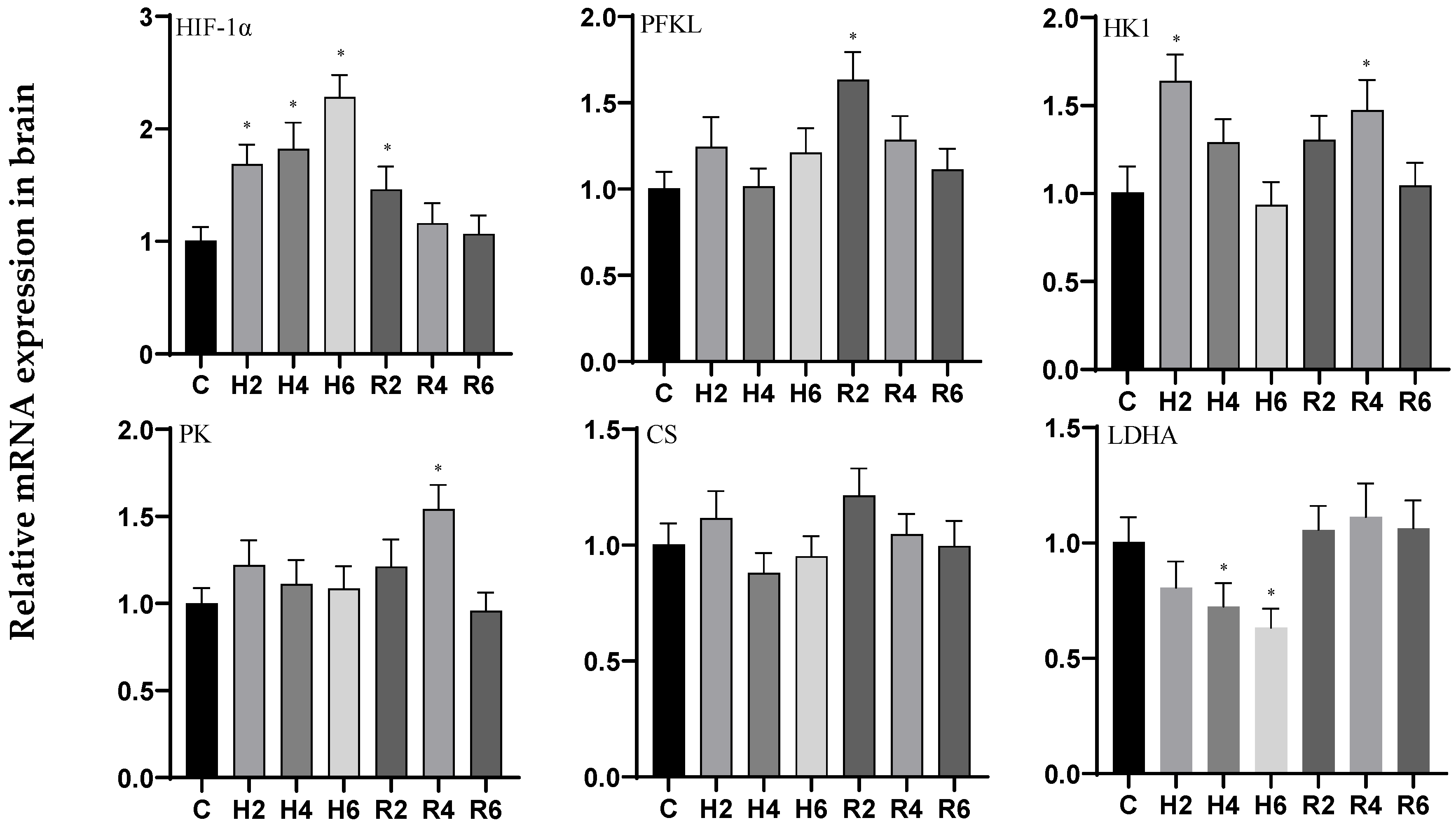
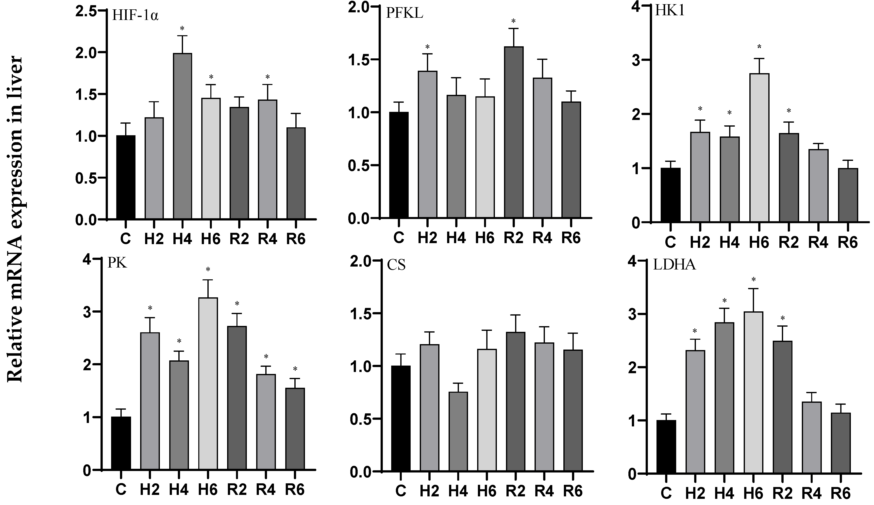
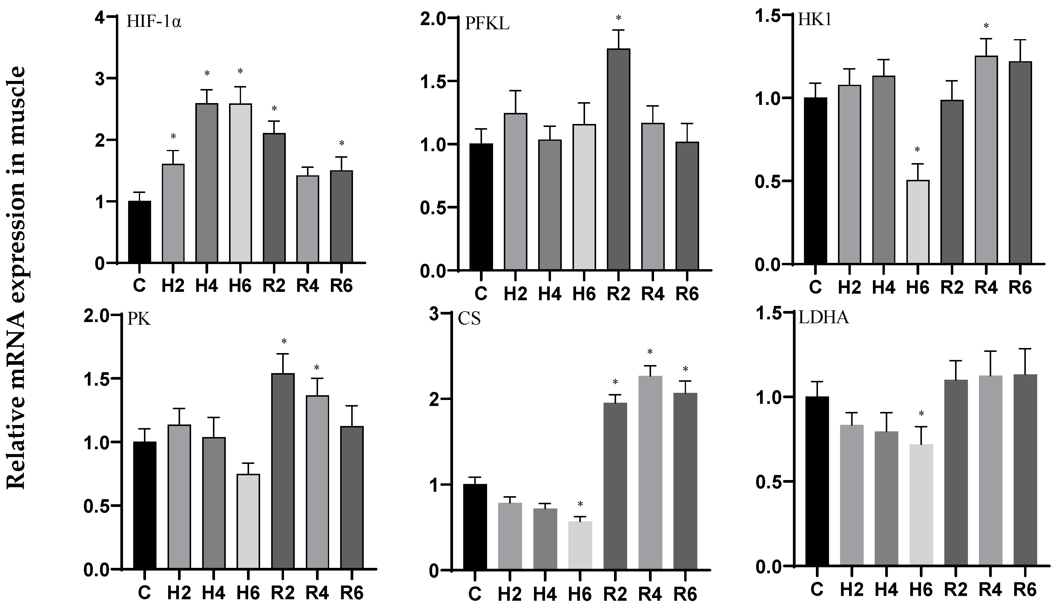
3.1. Effects of Hypoxia and Reoxygenation on Oxygen-Sensing Proteins and Respiratory Metabolism-Related Genes in Hybrid Catfish

Under acute hypoxia, the expression of the HIF-1α gene in brain tissue exhibited a significant upward trend, peaking at H6 (p < 0.05), with a 2.27-fold increase compared to the control group. This elevated expression remained significantly different from the control group at R2 (p < 0.05) before gradually returning to baseline levels. The HK1 gene exhibited a fluctuating expression pattern, peaking at H2 (p < 0.05) with a 1.63-fold increase relative to the control group, followed by a decline. Upon reoxygenation, its expression rebounded and remained significantly elevated at R4 (p < 0.05) compared to the control. In contrast, the PFKL, PK, and CS genes showed no significant changes under hypoxic conditions (p > 0.05). However, the PFKL and PK genes were significantly upregulated at R2 and R4, respectively (p < 0.05), before returning to control levels. Conversely, the LDHA gene displayed a downward trend, with significant downregulation observed at H6 (p < 0.05), reaching 0.63 times the control level, and gradually returning to baseline levels after reoxygenation (Figure 1).
In liver tissue under acute hypoxia, the expression of the HIF-1α and PFKL genes initially increased, peaking at H4 and H2, respectively (p < 0.05), with 1.97- and 1.39-fold increases compared to the control group. These levels remained significantly elevated at R4 and R2, respectively (p < 0.05), following reoxygenation. The HK1 and PK genes exhibited a fluctuating upward trend, while the LDHA gene showed a consistent upward trend, all reaching their peaks at H6 (p < 0.05), with 2.74-, 3.31-, and 3.03-fold increases relative to the control group, respectively. These levels remained significantly higher than the control group at R2 (p < 0.05) after reoxygenation. In contrast, the CS gene showed no significant changes under either hypoxic or reoxygenation conditions (Figure 2).
In muscle tissue under acute hypoxia, the expression of the HIF-1α and HK1 genes initially increased, peaking at H4, and then decreased to a minimum at H6 (p < 0.05), with 2.58- and 0.50-fold changes relative to the control group, respectively. These levels remained significantly elevated at R2 and R4 (p < 0.05) following reoxygenation. The PFKL gene exhibited an initial increase followed by a decrease, showing no significant changes compared to the control group. However, it was significantly upregulated at R2 after reoxygenation before returning to baseline levels. Similarly, the PK gene initially increased and then decreased, showing no significant changes compared to the control group, but was significantly higher than the control group at R2 (p < 0.05) after reoxygenation before returning to baseline levels. In contrast, the CS and LDHA genes both displayed a downward trend, with significant downregulation observed at H6 (p < 0.05), reaching 0.57 and 0.72 times the control levels, respectively. After reoxygenation, the CS gene was significantly upregulated compared to the control group (p < 0.05), while the LDHA gene showed no significant changes (Figure 3).

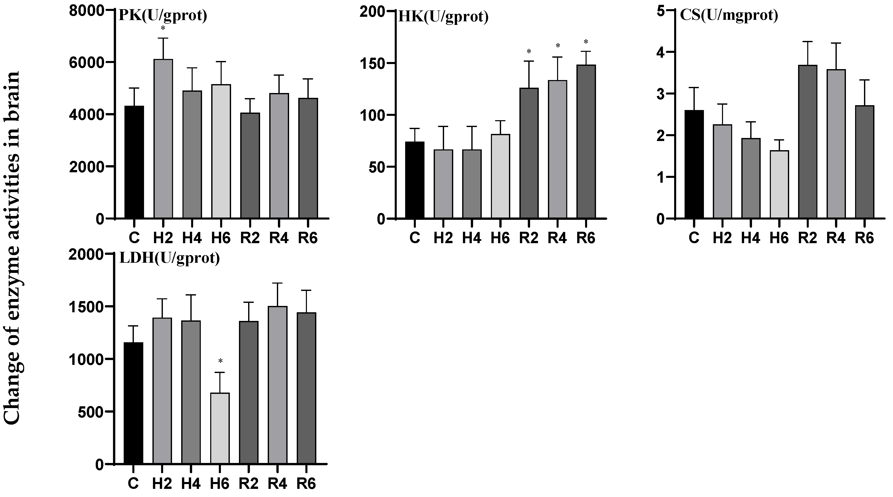
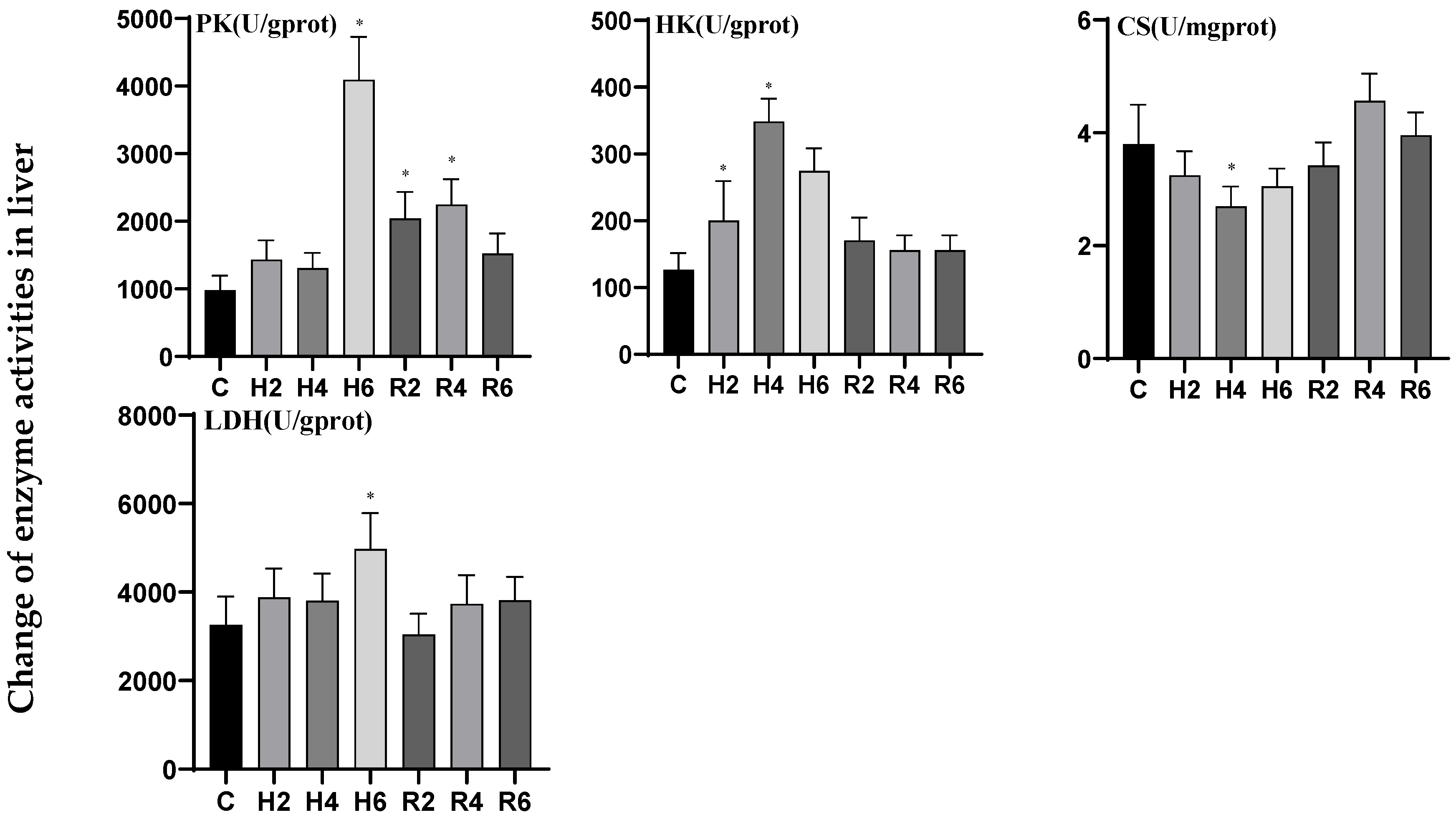
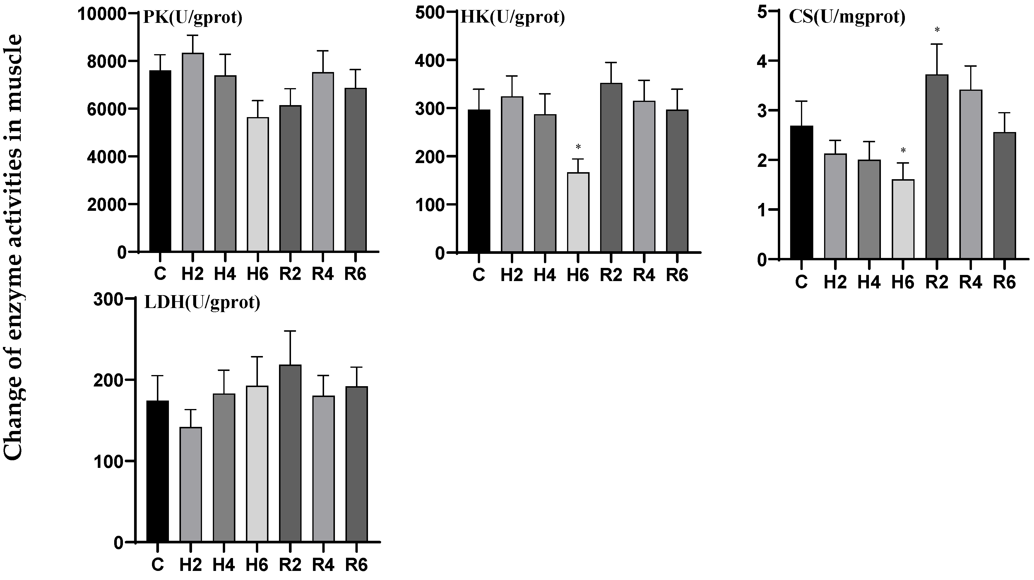
3.2. Effects of Hypoxia and Reoxygenation on Respiratory Metabolism-Related Enzyme Activities in Hybrid Catfish

Under acute hypoxia, the activity of pyruvate kinase (PK) in brain tissue initially increased, peaking at H2 (p < 0.05), reaching 1.45 times the level of the control group, followed by a gradual decline and eventual return to baseline levels after reoxygenation. In contrast, the activities of hexokinase (HK) and lactate dehydrogenase (LDH) exhibited an initial decrease followed by an increase. Although HK activity did not significantly differ from the control group under hypoxic conditions, it peaked at R6 following reoxygenation, demonstrating a significant difference compared to the control group (p < 0.05). LDH activity reached its minimum at H6, decreasing to 0.56 times the control level, but no significant difference was observed compared to the control group after reoxygenation. Citrate synthase (CS) activity displayed a downward trend during hypoxia and an upward trend following reoxygenation, with no significant differences observed compared to the control group under either condition (Figure 4).
In liver tissue under acute hypoxia, the activities of pyruvate kinase (PK) and lactate dehydrogenase (LDH) generally exhibited an upward trend, peaking at H6 (p < 0.05), reaching 4.16 and 1.53 times the levels of the control group (C), respectively. Following reoxygenation, PK activity peaked at R4, demonstrating a significant difference compared to the control group (p < 0.05), whereas LDH activity showed no significant difference. Hexokinase (HK) activity initially increased, significantly upregulated at H2 (p < 0.05), and peaked at H4, reaching 2.76 times the control level (C), before gradually returning to baseline levels after reoxygenation. The citrate synthase (CS) activity exhibited an initial decline followed by a subsequent increase, reaching its minimum value at H4 (p < 0.05), which was 0.71-fold of the control group (C). The activity gradually recovered to control levels following reoxygenation (Figure 5).
In muscle tissue under acute hypoxia, hexokinase (HK) activity initially increased, reaching its minimum at H6 (p < 0.05), decreasing to 0.56 times the control level (C), and gradually returned to baseline levels after reoxygenation. Citrate synthase (CS) activity exhibited a downward trend, reaching its minimum at H6, decreasing to 0.60 times the control level (C), and subsequently peaked at R2 after reoxygenation, demonstrating a significant difference compared to the control group (p < 0.05). The activities of pyruvate kinase (PK) and lactate dehydrogenase (LDH) exhibited no significant differences compared to the control group under either hypoxic or reoxygenation conditions (Figure 6).

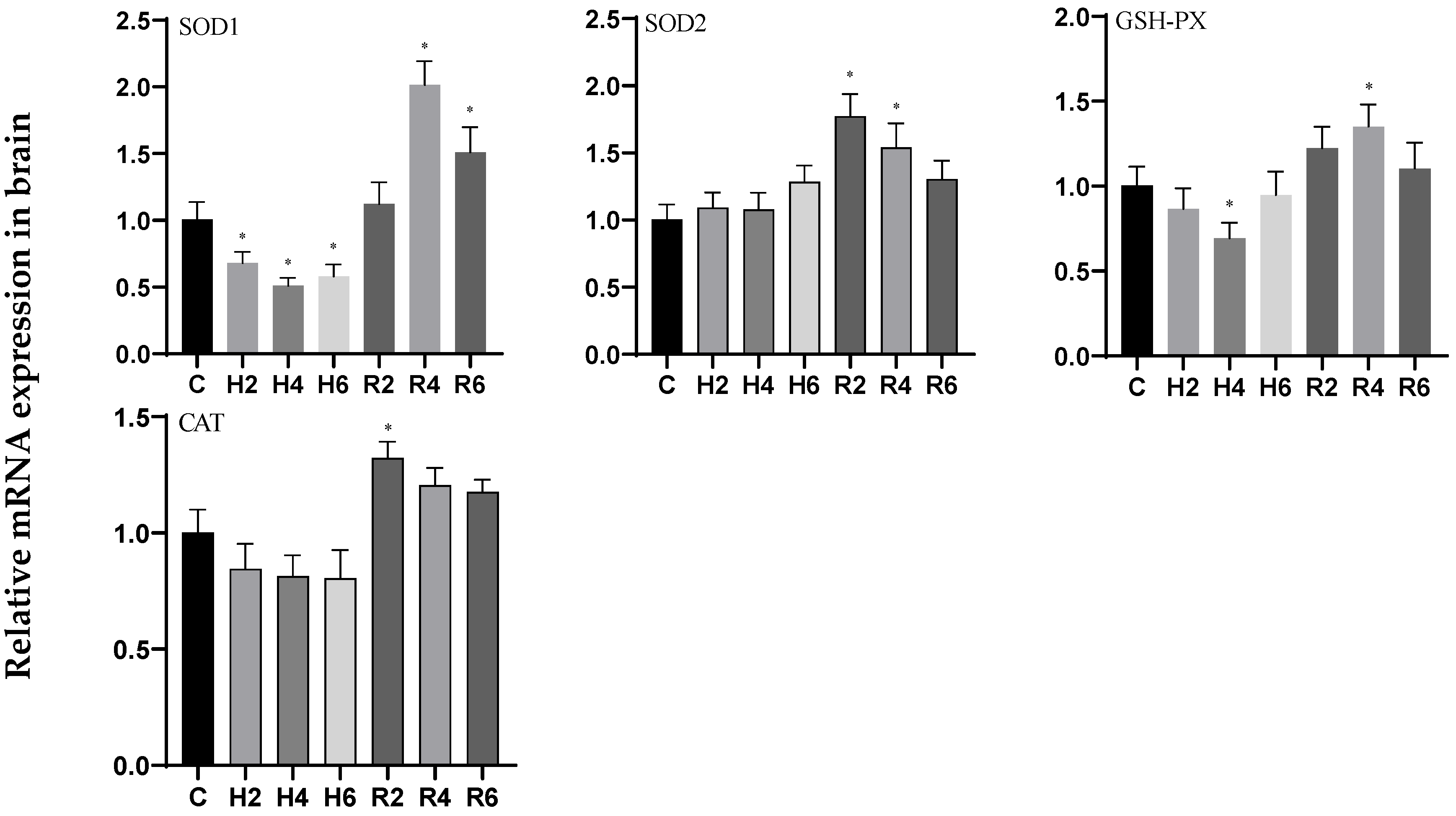
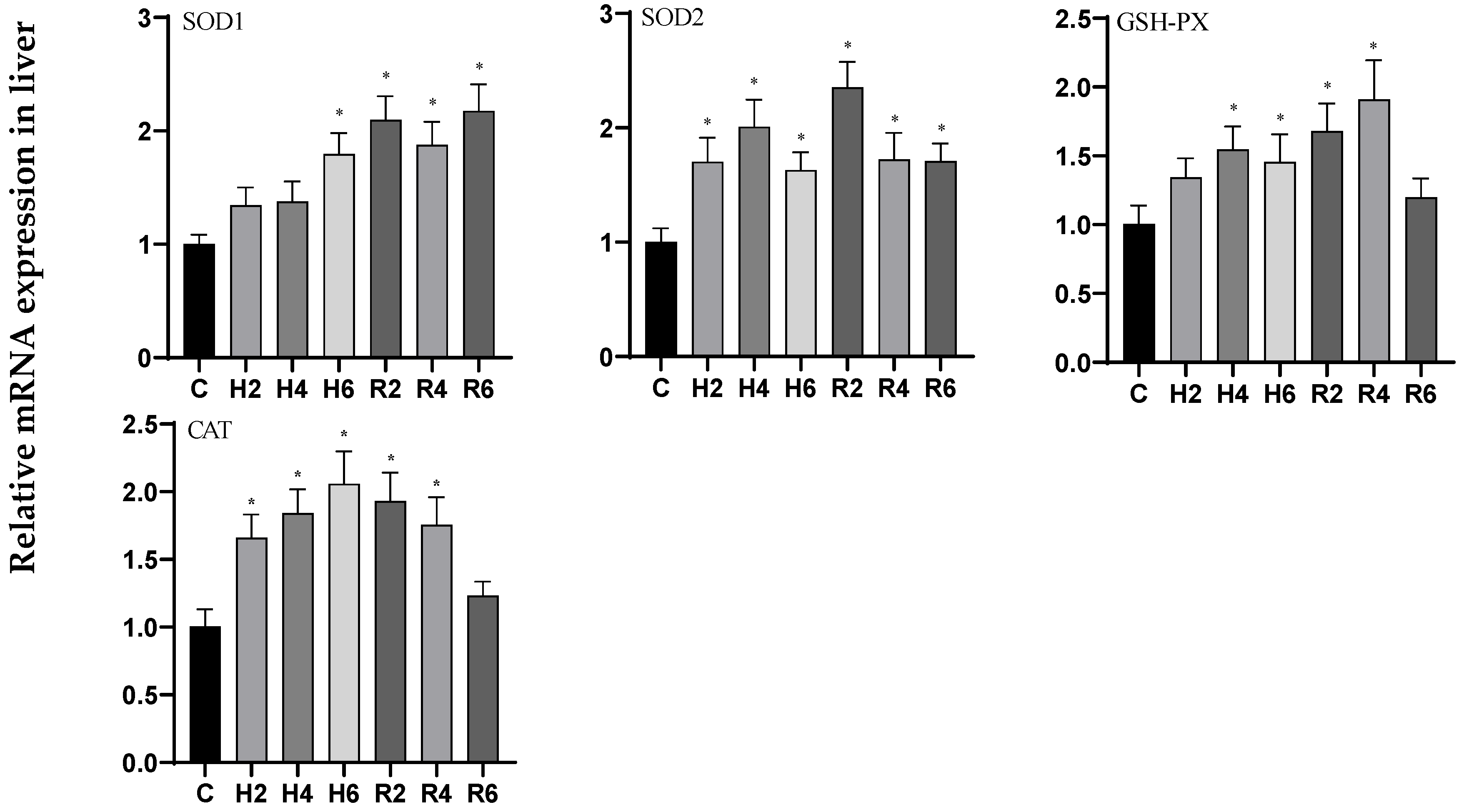
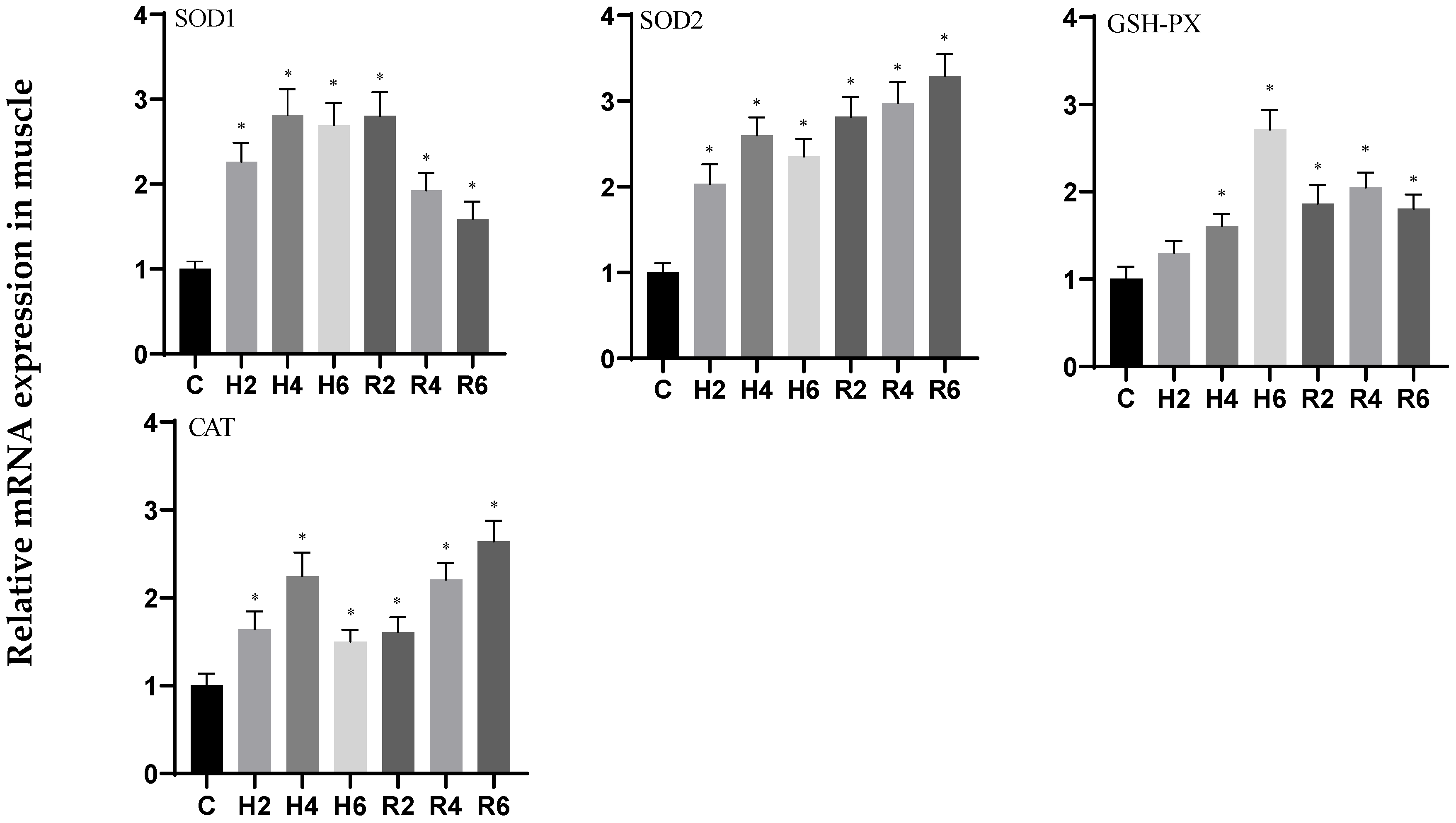
3.3. Effects of Hypoxia and Reoxygenation on Oxidative Stress-Related Genes in Hybrid Catfish

Under acute hypoxia, the expression levels of SOD1 and GSH-PX genes in brain tissue exhibited a biphasic response, showing an initial decrease followed by an increase, with minimum levels observed at H4 (p < 0.05), corresponding to 0.51- and 0.69-fold changes relative to the control group (C), respectively. Following reoxygenation, both genes demonstrated peak expression at R4, with statistically significant differences compared to the control group (p < 0.05). The expression pattern of the SOD2 gene displayed a progressive increase, whereas CAT gene expression demonstrated a decreasing trend. Both genes attained their maximum expression levels at R2 post-reoxygenation, with values significantly elevated compared to the control group (p < 0.05) (Figure 7).
In liver tissue under acute hypoxia, the expression levels of SOD1 and CAT genes showed an increasing trend, reaching peak levels at H6 (p < 0.05), with 1.79- and 2.05-fold increases relative to the control group (C), respectively. During the reoxygenation phase, SOD1 expression remained significantly elevated compared to control levels, while CAT expression peaked at R2, showing a significant increase over control values (p < 0.05), before gradually returning to baseline levels. The expression profiles of SOD2 and GSH-PX genes exhibited an initial increase followed by a decrease, with maximum levels observed at H4 (p < 0.05), corresponding to 2.0- and 1.46-fold changes relative to control levels, respectively. Following reoxygenation, these genes demonstrated peak expression at R2 and R4, respectively, showing statistically significant differences from control values (p < 0.05), before gradually returning to baseline levels (Figure 8).
In muscle tissue subjected to acute hypoxia, the expression levels of SOD1, SOD2, and CAT genes displayed an initial increase followed by a decrease, reaching peak levels at H4 (p < 0.05), with 2.80-, 2.59-, and 2.32-fold increases relative to control values, respectively. During the reoxygenation phase, SOD1 expression showed a gradual decline while maintaining significantly higher levels than the control group, whereas SOD2 and CAT expression levels progressively increased, peaking at R6 and showing statistically significant differences from control values (p < 0.05). The GSH-PX gene expression profile demonstrated a continuous upward trend, reaching maximum levels at H6 (p < 0.05), representing a 2.70-fold increase over control values, and remained significantly elevated compared to control levels throughout the reoxygenation period (p < 0.05) (Figure 9).

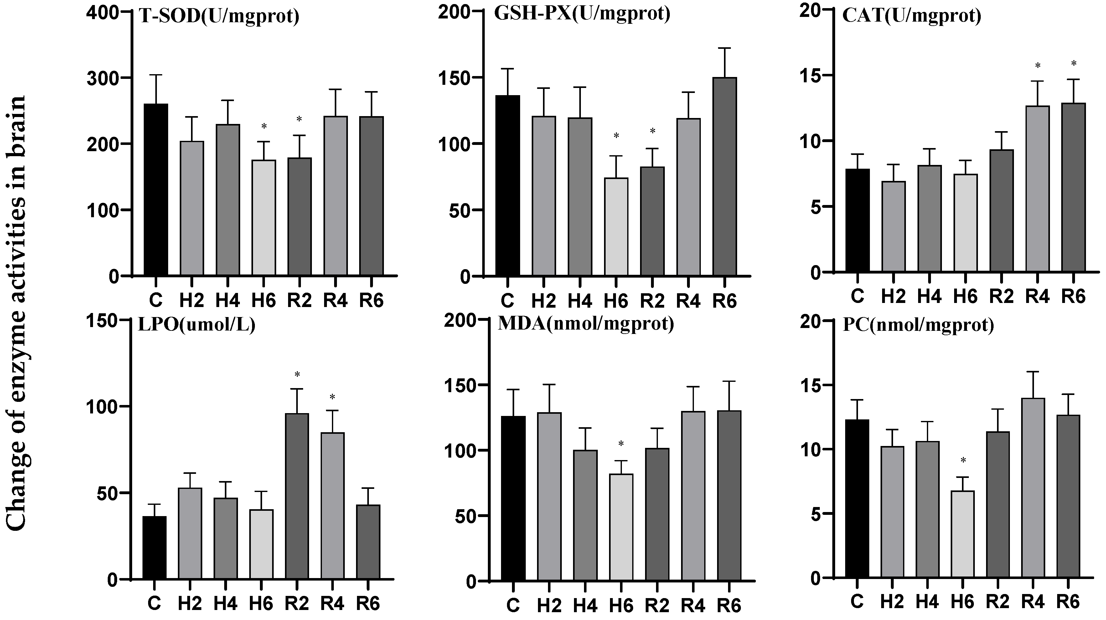
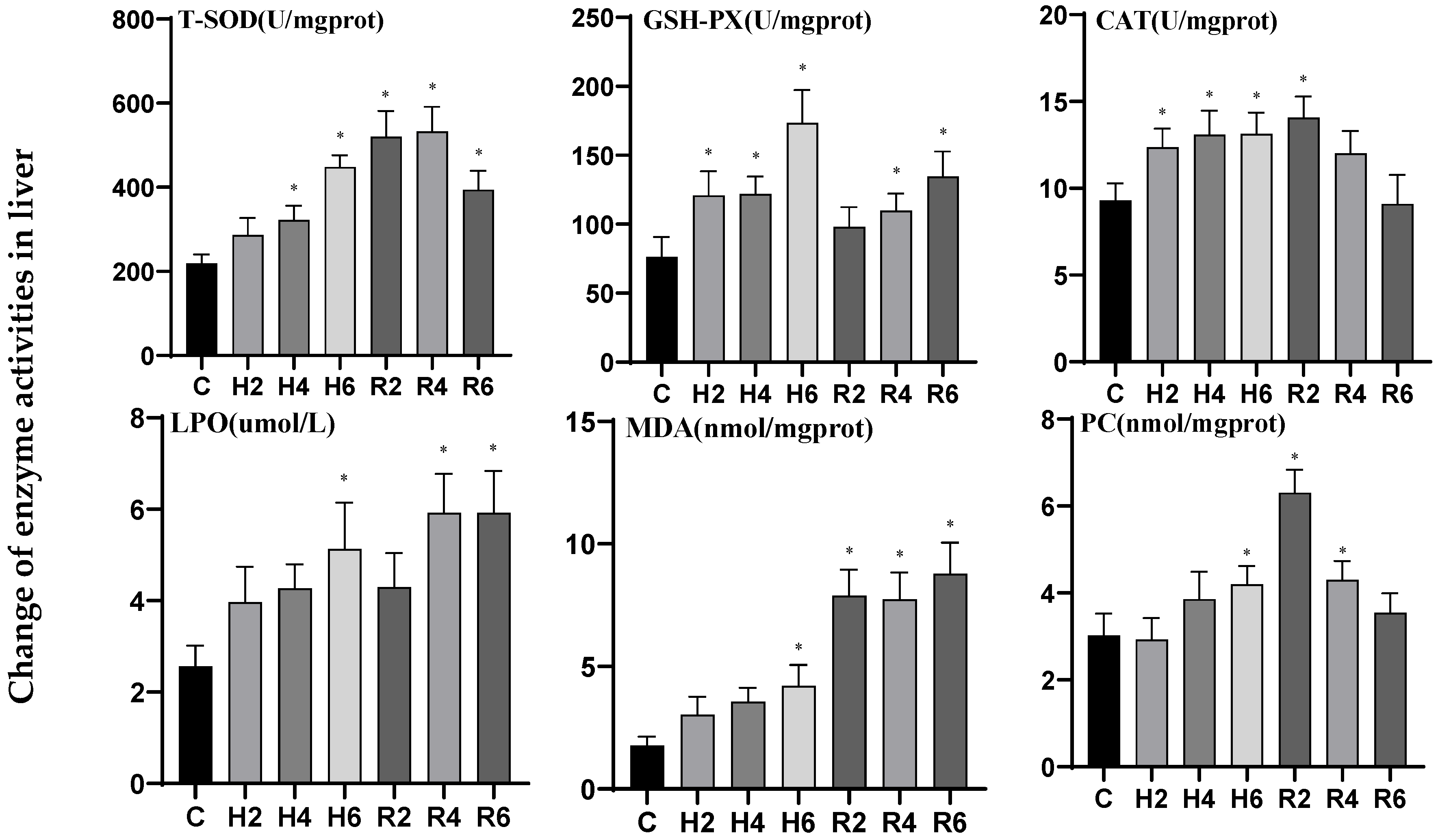
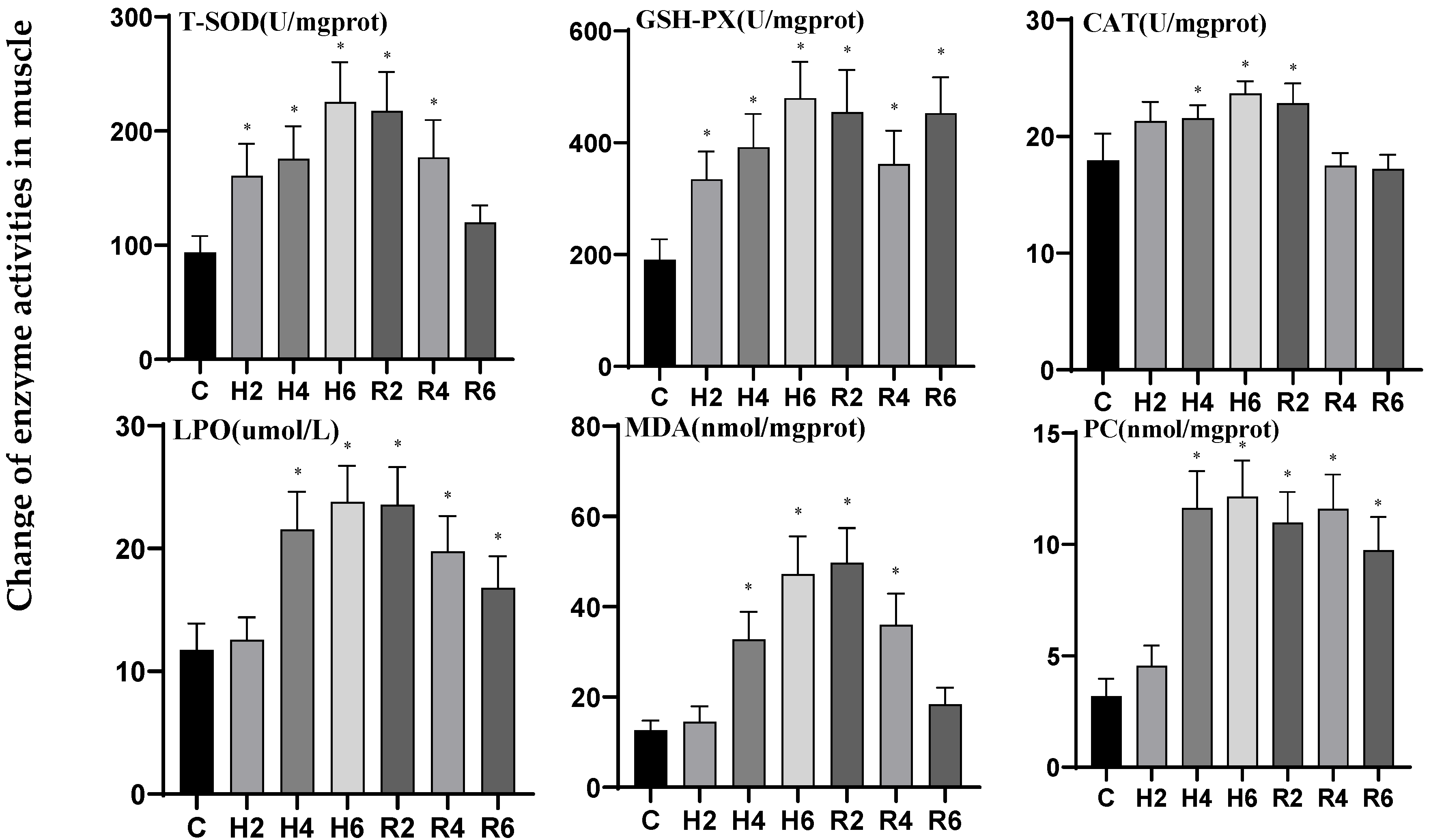
3.4. Effects of Hypoxia and Reoxygenation on Oxidative Stress Enzyme Activities and Parameters in Hybrid Catfish

Under acute hypoxia, the activities of T-SOD and GSH-PX in brain tissue decreased significantly, reaching their lowest levels at H6 (p < 0.05), representing 0.66 and 0.54 times the levels of the control group (C), respectively. After reoxygenation, these activities remained significantly lower than the control group at R2, after which they gradually increased and returned to control levels. The activities of CAT and PCO declined with fluctuations, with only PCO activity significantly reduced at H6 (p < 0.05), reaching 0.55 times the control level (C). After reoxygenation, only CAT activity showed a gradual increase, peaking at R6 and demonstrating a significant difference from the control group (p < 0.05). The activities of LPO and MDA initially rose and then declined, with only MDA activity significantly reduced at H6 (p < 0.05), reaching 0.65 times the control level (C). After reoxygenation, only LPO activity reached its peak at R2, after which it gradually decreased and returned to control levels (Figure 10).
In liver tissue under acute hypoxia, the activities of T-SOD, CAT, LPO, and MDA increased significantly, peaking at H6 (p < 0.05), reaching 2.05, 1.41, 2.0, and 2.38 times the levels of the control group (C), respectively. After reoxygenation, the activities of T-SOD, CAT, LPO, and MDA reached their peaks at R4, R2, R4, and R6, respectively. While CAT activity gradually returned to control levels, the activities of the other enzymes remained significantly elevated compared to the control group (p < 0.05). The activities of GSH-PX and PC exhibited an increasing trend, both reaching their peak values at H6 (p < 0.05), which were 2.27-fold and 1.39-fold of the control group (C), respectively. After reoxygenation, GSH-PX activity showed a gradual increase, peaking at R6 and demonstrating a significant difference from the control group (p < 0.05), while PCO activity reached its peak at R2, significantly elevated compared to the control group (p < 0.05), before gradually declining and returning to control levels (Figure 11).
In muscle tissue under acute hypoxia, the activities of T-SOD, CAT, LPO, MDA, and PCO increased significantly, peaking at H6 (p < 0.05), reaching 2.41, 1.32, 2.03, 3.74, and 3.81 times the levels of the control group (C), respectively. After reoxygenation, the activities of T-SOD, CAT, and MDA reached their peaks at R2, demonstrating significant differences from the control group (p < 0.05), before gradually declining and returning to control levels. In contrast, the activities of LPO and PCO remained significantly elevated compared to the control group after reoxygenation (p < 0.05). The activity of glutathione peroxidase (GSH-PX) demonstrated a significant upward trend, peaking at H6 (p < 0.05) with levels reaching 2.52-fold of the control group (C). Notably, during the reoxygenation phase, GSH-PX activity remained significantly elevated compared to control values (p < 0.05) (Figure 12).

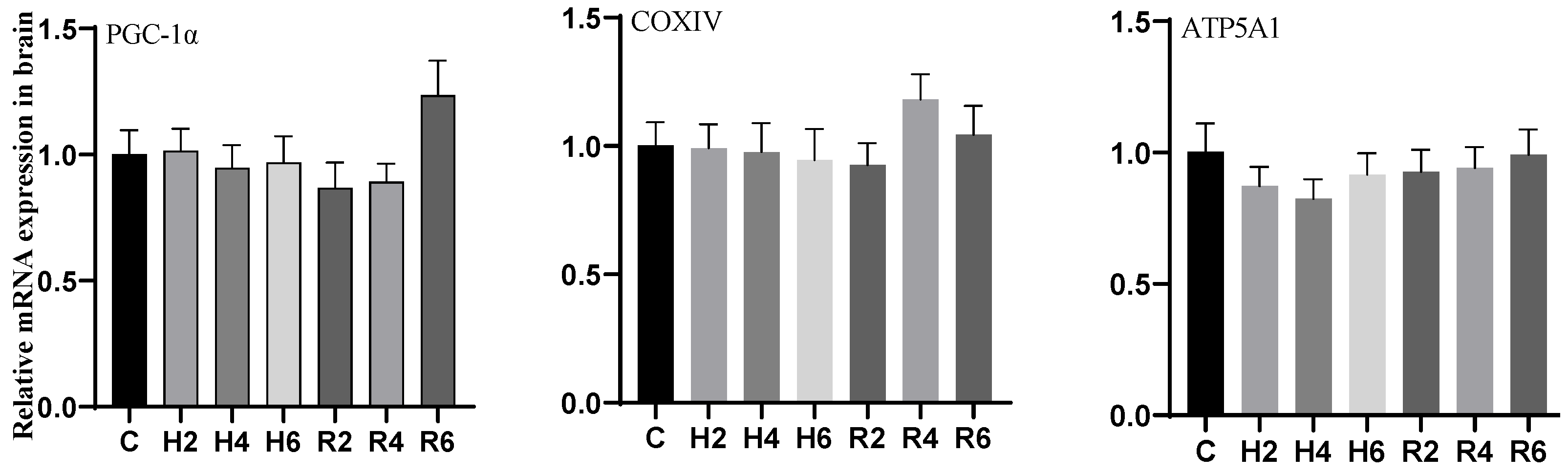
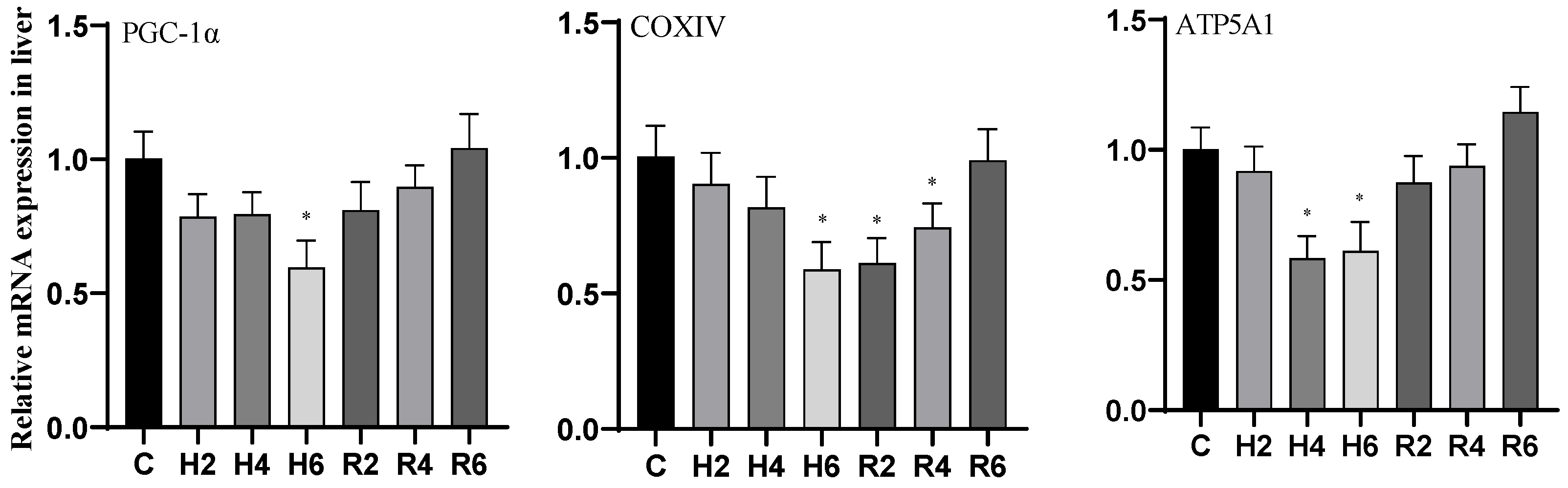
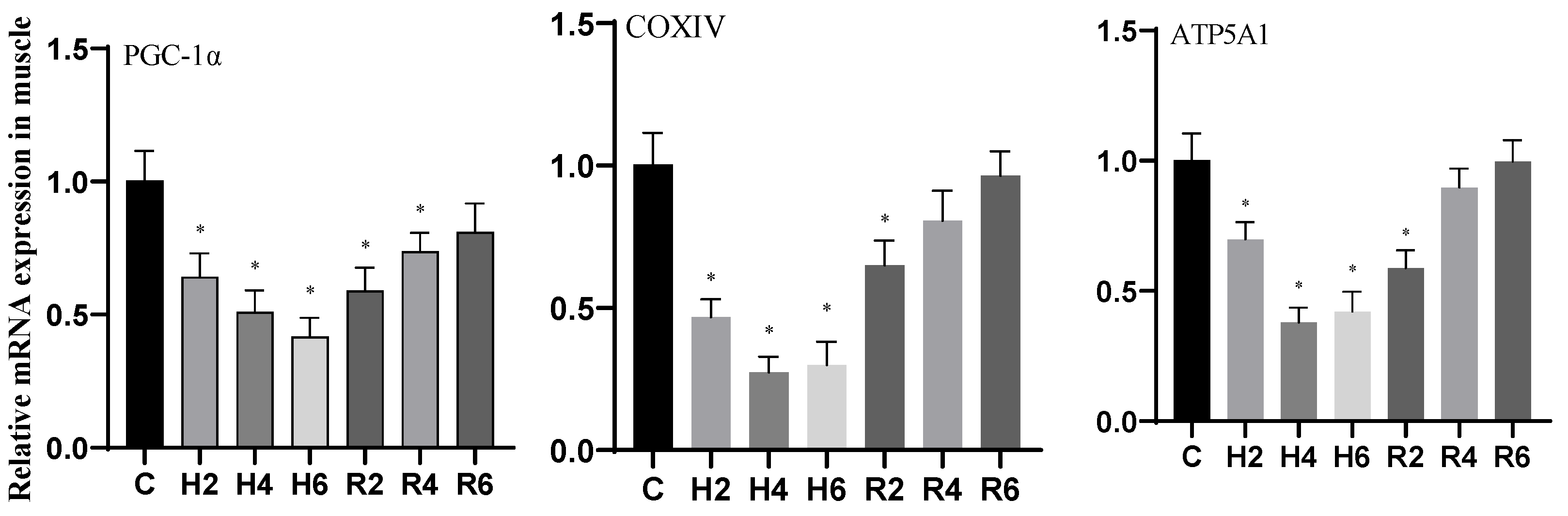
3.5. Effects of Hypoxia and Reoxygenation on Mitochondrial Damage Genes in Hybrid Catfish

During acute hypoxia and subsequent reoxygenation, the expression levels of PGC-1α, COXIV, and ATP5A1 genes in brain tissue remained unchanged compared to the control group (Figure 13).
Under acute hypoxia, the expression of PGC-1α and COXIV genes in liver tissue decreased significantly, reaching their minimum values at H6 (p < 0.05), which were 0.59 and 0.58 times that of the control group, respectively. After reoxygenation, the expression of PGC-1α and COXIV genes recovered gradually to control levels, although COXIV expression remained significantly lower than the control group at R2 (p < 0.05). The ATP5A1 gene expression initially decreased, reaching its minimum value at H4 (p < 0.05), which was 0.58 times that of the control group, and subsequently increased to control levels after reoxygenation (Figure 14).
Under acute hypoxia, the expression of the PGC-1α gene in muscle tissue decreased significantly, reaching its minimum value at H6 (p < 0.05), which was 0.42 times that of the control group. After reoxygenation, PGC-1α expression remained significantly lower than the control group at R2 (p < 0.05) but gradually recovered to control levels thereafter. The expression of COXIV and ATP5A1 genes initially decreased, reaching their minimum values at H4 (p < 0.05), which were 0.27 and 0.38 times that of the control group (C), respectively. After reoxygenation, their expression remained significantly lower than the control group at R2 (p < 0.05) but gradually recovered to control levels (Figure 15).

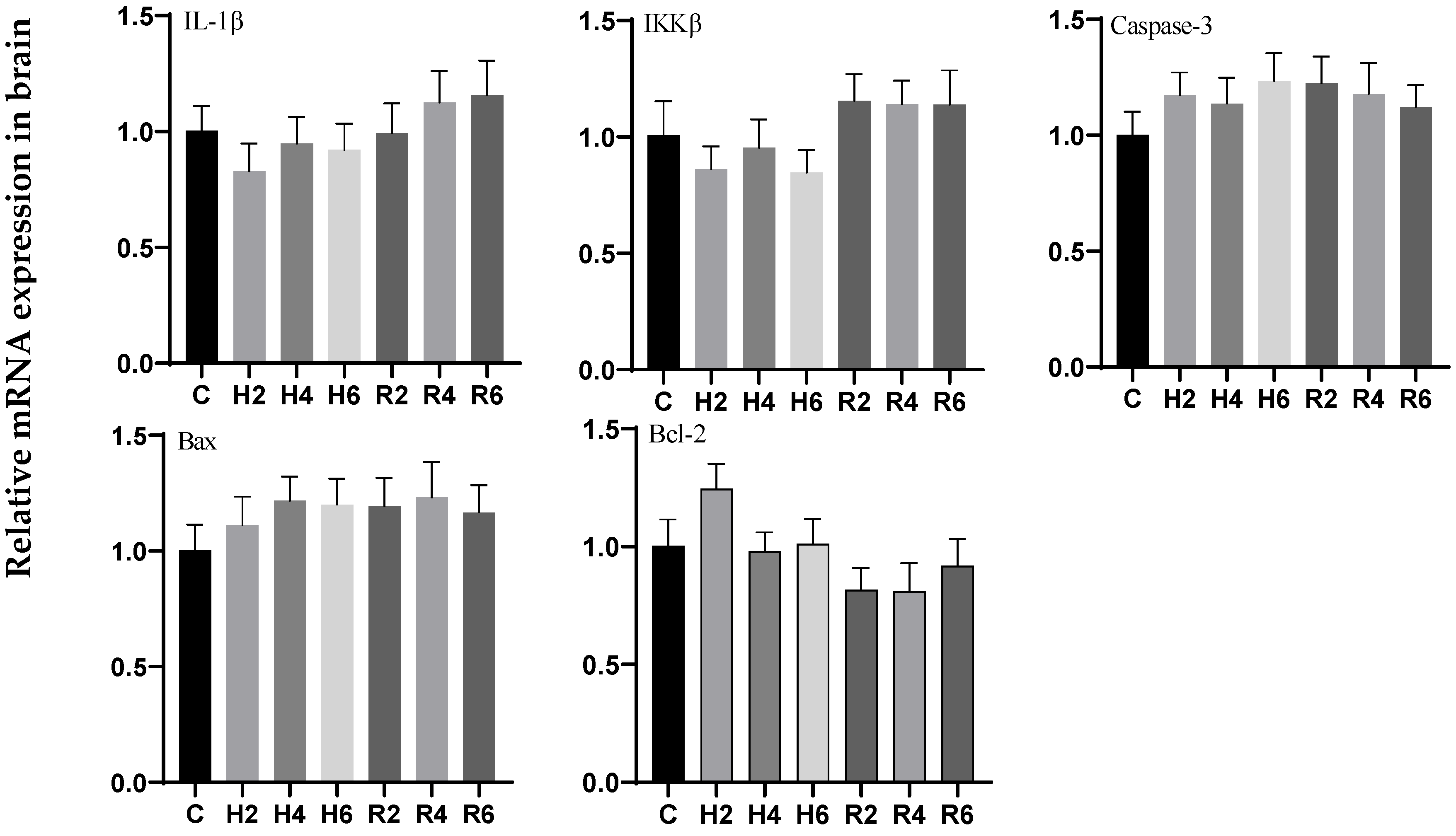
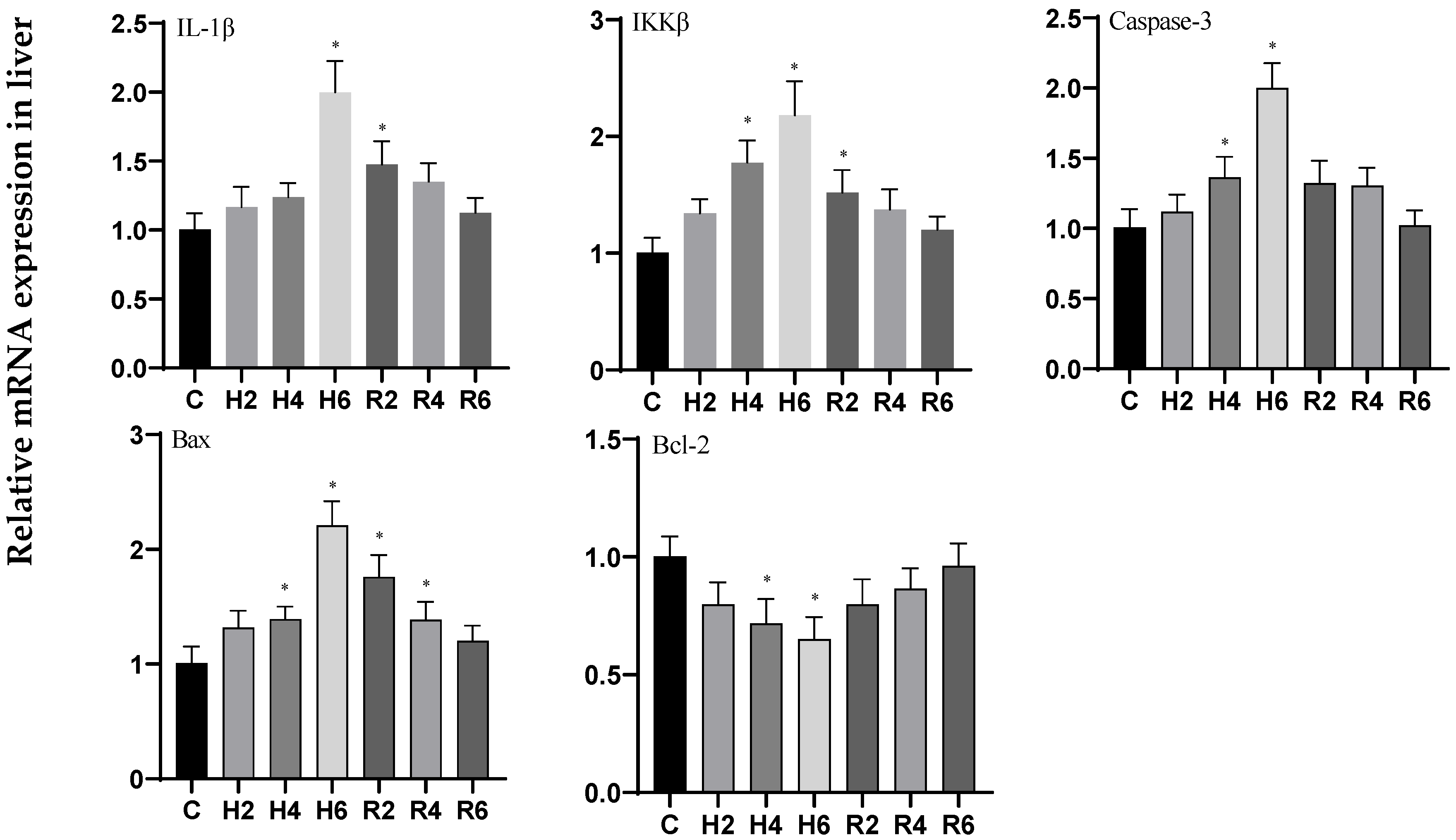
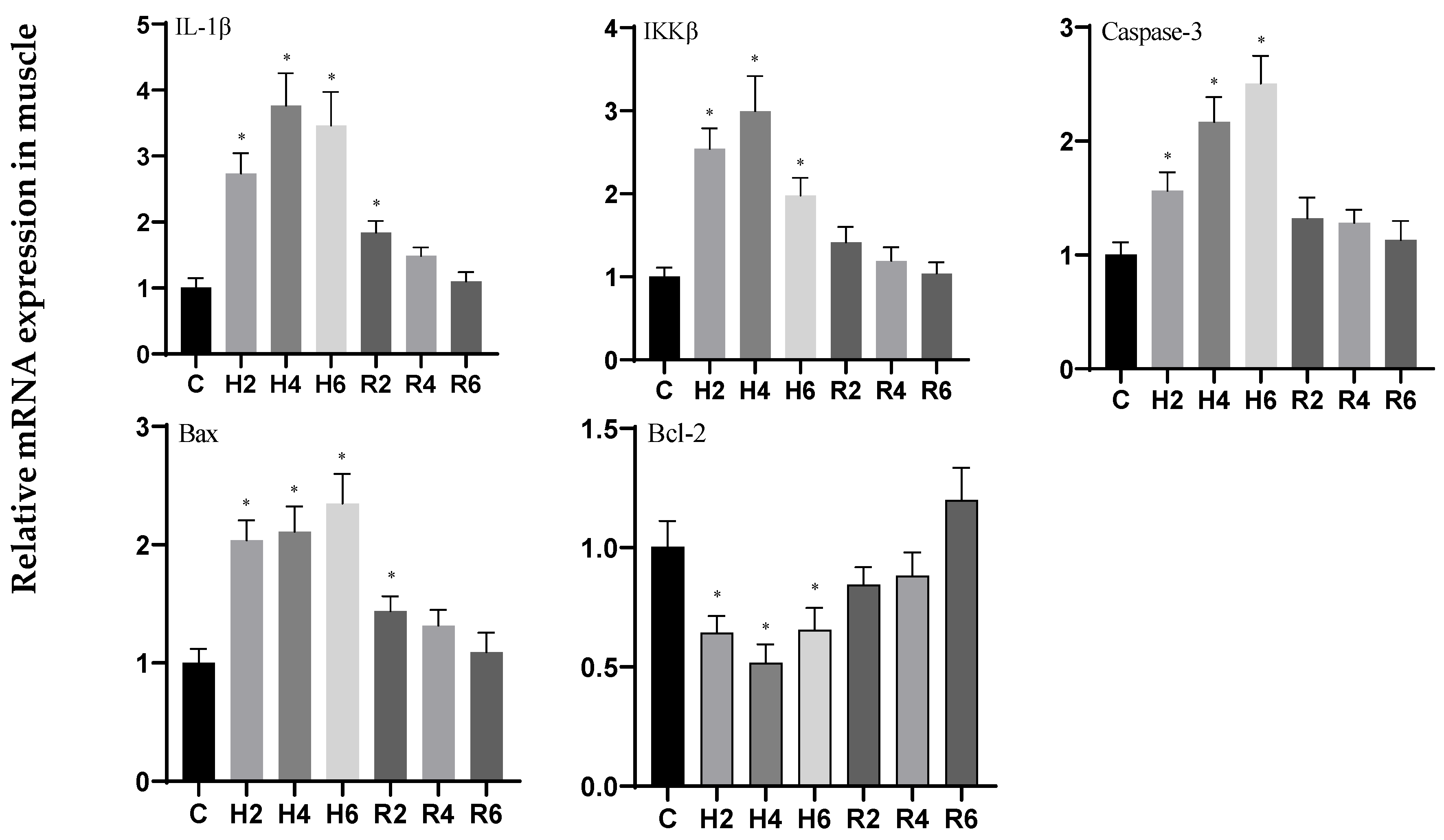
3.6. Effects of Hypoxia and Reoxygenation on Inflammation and Apoptosis-Related Genes in Hybrid Catfish

During acute hypoxia and subsequent reoxygenation, no significant differences were observed in the expression levels of IL-1β, IKKβ, Caspase 3, Bax, and Bcl-2 genes in brain tissue when compared to the control group (Figure 16).
Under acute hypoxia, the expression levels of IL-1β, IKKβ, Caspase 3, and Bax genes in liver tissue significantly increased, reaching their maximum values at H6 (p < 0.05), with expression levels 1.95-, 1.96-, 2.00-, and 2.13-fold higher than those of the control group (C), respectively. After reoxygenation, the expression levels of these genes gradually decreased, returning to control levels, although IL-1β, IKKβ, and Bax remained significantly elevated compared to the control group at R2 (p < 0.05). In contrast, the expression of the Bcl-2 gene significantly decreased, reaching its minimum value at H6 (p < 0.05), with expression levels 0.66-fold lower than those of the control group (C), and subsequently increased to control levels following reoxygenation (Figure 17).
Under acute hypoxia, the expression of IL-1β and IKKβ genes in muscle tissue initially increased, peaking at H4 (p < 0.05), with expression levels 3.26- and 2.84-fold higher than those of the control group (C), respectively, before subsequently decreasing. After reoxygenation, the expression levels of these genes gradually declined, returning to control levels, although IL-1β remained significantly elevated compared to the control group at R2 (p < 0.05). The expression of the Caspase 3 gene significantly increased, reaching its maximum value at H6 (p < 0.05), with expression levels 2.44-fold higher than those of the control group (C), and subsequently decreased to control levels following reoxygenation. The expression of the Bax gene fluctuated but overall increased, peaking at H6 (p < 0.05), with expression levels 2.30-fold higher than those of the control group (C), and remained significantly elevated compared to the control group at R2 (p < 0.05) before gradually declining to control levels. The expression of the Bcl-2 gene initially decreased, reaching its minimum value at H4 (p < 0.05), with expression levels 0.54-fold lower than those of the control group (C), and subsequently increased to control levels following reoxygenation (Figure 18).

4. Discussion

4.1. Effects of Hypoxia and Reoxygenation on Oxygen-Sensing Protein HIF-1α and Respiratory Metabolism in Hybrid Catfish

The survival of aquatic organisms is primarily dependent on the stability of their aquatic environment, with fluctuations in dissolved oxygen (DO) levels being of particular significance for fish [19]. To cope with significant changes in DO, fish have evolved a series of complex physiological adaptation mechanisms, among which the activation of the HIF-1 signaling pathway is of paramount importance [20]. This pathway, mediated by the binding of HIF heterodimers to hypoxia response elements (HREs), activates a cascade of genes involved in angiogenesis, glucose and iron transport, glycolysis, and cell cycle regulation, thereby enabling fish to maintain viability under hypoxic conditions [21]. The results of this study indicate that the expression of the HIF-1α gene in the brain, liver, and muscle tissues of hybrid catfish was significantly upregulated during hypoxia and subsequently returned to control levels after reoxygenation. These findings are consistent with studies on other fish species, such as Sebastes schlegelii [22], Leiostomus xanthurus [23], and Oreochromis niloticus [24], thereby reinforcing HIF-1α as a key oxygen-sensing protein that is significantly activated under hypoxic conditions.
Research demonstrates that the HIF-1 signaling pathway regulates key genes such as PFKL, HK1, PK, LDHA, and CS, which are integral to glycolysis, anaerobic respiration, and the tricarboxylic acid (TCA) cycle [25,26,27,28]. Under hypoxia, organisms typically adapt by lowering basal metabolic rates and transitioning between aerobic and anaerobic metabolic pathways [29]. Although this adaptation may result in physiological burdens such as lactate accumulation, it is critical for survival [11]. In this study, glycolysis-related (PFKL, HK1, PK) and anaerobic respiration-related (LDHA) genes and enzyme activities in the liver of hybrid catfish were significantly upregulated under hypoxia and subsequently returned to normal levels after reoxygenation. These results align with findings in P. fulvidraco [30], P. vachelli [31], and hybrid yellow catfish (P. fulvidraco ♀ × P. vachelli ♂) [32], indicating that liver tissue adapts to hypoxia by enhancing glycolysis and anaerobic respiration to ensure energy supply. Additionally, in the brain tissue, the HK gene and PK enzyme activity were significantly upregulated at H2, suggesting the activation of glycolysis during the early stages of hypoxia. Notably, LDHA was significantly downregulated at H6, potentially representing a protective mechanism to prevent lactate accumulation. In contrast, some glycolysis-, CS-, and anaerobic respiration-related genes and enzyme activities in muscle tissue exhibited a downward trend, consistent with findings in P. vachelli [33,34] and Trachinotus blochii [35]. This implies that muscle tissue may experience more severe hypoxia-induced metabolic disturbances compared to other tissues.

4.2. Effects of Hypoxia and Reoxygenation on Oxidative Stress in Hybrid Catfish

Under physiological homeostasis, the generation and clearance of reactive oxygen species (ROS) maintain a delicate dynamic equilibrium [36]. However, exposure to environmental stressors can disrupt this equilibrium, resulting in oxidative stress [37]. Numerous studies have demonstrated that various aquatic organisms, including Carassius auratus [38], Sinonovacula constricta [39], Megalobrama amblycephala [40], and Neohelice granulata [41], exhibit significant upregulation of antioxidant enzyme activities under hypoxic conditions, a phenomenon termed “oxidative stress preparation” to mitigate potential oxidative stress during reoxygenation [42]. Similarly, hybrid catfish exhibited analogous alterations in antioxidant enzyme activities under hypoxic conditions. Our findings revealed that antioxidant enzymes (e.g., SOD, CAT, GSH-PX) in hepatic and muscular tissues exhibited significant increases during the initial hypoxic phase (H2–H4), whereas oxidative stress markers (e.g., LPO, MDA, PCO) demonstrated substantial elevation during H4-H6. These observations suggest that hybrid catfish may possess an anticipatory antioxidant defense mechanism to counteract potential oxidative stress induced by hypoxic conditions [43]. In contrast, both antioxidant enzymes and oxidative stress markers in cerebral tissue remained stable or exhibited minimal declines, maintaining relatively consistent levels. Comparable findings have been documented in P. glenii [44] and P. vachelli [31], suggesting that cerebral tissue, as a vital organ, may receive preferential oxygen supply during acute hypoxic conditions, thereby minimizing oxidative stress damage [45,46]. Notably, oxidative stress markers in hepatic and muscular tissues demonstrated significant elevation during the advanced hypoxic phase, with muscular tissue displaying higher oxidative stress levels compared to hepatic tissue, indicating more pronounced oxidative damage in muscular tissue.
Reoxygenation has been demonstrated to induce rapid ROS accumulation, resulting in secondary oxidative stress responses. For instance, Cyprinus carpio var qingtianensis [47] and Mugil cephalus [48] demonstrated significant oxidative stress during reoxygenation. Similarly, the hybrid catfish exhibited ROS accumulation during reoxygenation. The present study revealed that antioxidant enzyme activities and oxidative stress parameters in brain, liver, and muscle tissues were significantly elevated following reoxygenation, suggesting that the antioxidant enzyme system maintains high activity to mitigate reoxygenation-induced oxidative stress.

4.3. Effects of Hypoxia and Reoxygenation on Mitochondrial Damage, Inflammation, and Apoptosis in Hybrid Catfish

Mitochondria, serving as the central organelles for cellular energy metabolism, predominantly depend on oxidative phosphorylation for energy conversion [49]. Numerous studies have demonstrated that hypoxia or ischemia significantly impairs mitochondrial oxidative phosphorylation, resulting in diminished mitochondrial biogenesis and functional impairments [50]. Key regulatory genes including PGC-1α, COXIV, and ATP5A1 are intrinsically linked to mitochondrial function, orchestrating mitochondrial biogenesis and maintenance, while their expression profiles serve as reliable biomarkers for mitochondrial dysfunction [51,52,53,54]. These genes play pivotal roles in regulating oxidative phosphorylation, thereby directly modulating cellular energy metabolism and maintaining functional homeostasis [55]. Experimental studies conducted on rat models [56,57] and P. vachelli [34] have consistently demonstrated that hypoxia significantly downregulates the expression of PGC-1α, COXIV, and ATP5A1 genes, highlighting the profound detrimental effects of hypoxia on mitochondrial functionality. In the current investigation, we observed significant downregulation of mitochondrial-related genes (PGC-1α, COXIV, ATP5A1) in the liver and muscle tissues of hybrid catfish under hypoxic conditions, suggesting substantial impairment in mitochondrial respiration and biogenesis processes. However, in brain tissue, these genes maintained stable expression levels, suggesting the presence of robust protective mechanisms and adaptive responses that preserve mitochondrial function under hypoxic stress.
The NF-κB signaling pathway plays a pivotal role in regulating inflammatory signaling transduction and mediating the expression of inflammatory factors [58]. This signaling cascade modulates the transcriptional activation of specific inflammatory factors, including TNF-α and IL-1β, in response to various cellular stress stimuli [59]. In P. vachelli [45], hypoxia-induced mitochondrial dysfunction was found to activate the NF-κB signaling pathway, subsequently triggering the release of pro-inflammatory cytokines and initiating apoptotic processes. Our experimental results demonstrated that the expression levels of IKKβ, IL-1β, Caspase-3, and Bax genes in muscle tissue were significantly upregulated during the initial phase of hypoxia exposure, showing more pronounced elevation compared to hepatic tissue, indicating that muscle cells might exhibit enhanced susceptibility to hypoxia-induced apoptosis. Furthermore, the elevated expression of IKKβ, a pivotal regulatory component of the NF-κB signaling cascade, was observed in both hepatic and muscular tissues, providing additional evidence for the pathway’s essential involvement in hypoxia-mediated cellular stress responses. Notably, the pronounced upregulation of IKKβ in muscular tissue suggests a potentially heightened cellular sensitivity to hypoxic conditions. Comparable observations have been documented in Rachycentron canadum [60], L. crocea [61], and Epinephelus coioides [62], where hypoxia-triggered reactive oxygen species (ROS) and peroxide accumulation were shown to activate the NF-κB signaling cascade [63], resulting in the upregulation of inflammatory mediators (TNF-α, IL-1β) and pro-apoptotic factors (Caspase-3, Bax), concomitant with downregulation of anti-apoptotic Bcl-2 expression. In the current investigation, we observed significant upregulation of IL-1β, Caspase-3, and Bax gene expression in both hepatic and muscular tissues under hypoxic conditions, accompanied by downregulation of Bcl-2 expression, which is presumably attributable to excessive ROS generation and subsequent NF-κB pathway activation. Conversely, the expression profiles of these inflammatory and apoptotic markers remained relatively stable in cerebral tissue, in agreement with previous findings in P. vachelli [33], implying that neural tissue might possess specialized metabolic adaptations and enhanced antioxidant defense mechanisms to counteract hypoxia-mediated cellular injury.

5. Conclusions

This study systematically investigated the effects of hypoxia–reoxygenation on oxygen-sensing proteins, respiratory metabolism, oxidative stress, mitochondrial function, inflammatory responses, and apoptosis in the brain, liver, and muscle tissues of hybrid catfish. Hypoxia significantly activated HIF-1α expression and induced metabolic reprogramming, with the liver shifting toward anaerobic metabolism, while the brain exhibited suppressed glycolysis and the muscle displayed concurrent inhibition of glycolysis and aerobic respiration. Notably, muscle tissue demonstrated heightened oxidative stress, mitochondrial dysfunction, inflammatory cascades, and apoptosis during hypoxia–reoxygenation. These findings elucidate the multi-tissue adaptive mechanisms in hybrid catfish under oxygen fluctuations. Future research should explore interspecific variations in hypoxia adaptation and genetic strategies to enhance hypoxia tolerance, providing insights for sustainable aquaculture development.

Supplementary Materials

The following supporting information can be downloaded at https://www.mdpi.com/article/10.3390/biology14080915/s1. Table S1: Enzyme activity determination method.

Author Contributions

Conceptualization, J.Y. and F.Z.; methodology, C.Z.; software, J.Y.; validation, F.Z.; formal analysis, J.Y.; investigation, J.Y. and F.Z.; data curation, F.Z.; writing—original draft preparation, J.Y.; writing—review and editing, F.L. and G.Z.; visualization, C.Z.; supervision, S.Y.; project administration, F.L., G.Z. and S.Y.; funding acquisition, G.Z. All authors have read and agreed to the published version of the manuscript.

Funding

This research was funded by the National Natural Science Foundation of China (No. 32402983), Qingchuang science and technology support program of Shandong provincial college (2024KJG006) and the “JBGS” Project of Seed Industry Revitalization in Jiangsu Province (JBGS [2021] 034).

Institutional Review Board Statement

This study was approved by the Nanjing Normal University Animal Ethics Committee (approval no. SYXK [Jiangsu] 2015-0028).

Informed Consent Statement

Not applicable.

Data Availability Statement

The data that support the findings of this study are available in [Mendeley Data] at [https://data.mendeley.com/preview/wn9fp3xfbm?a=597e50bd-e39e-47a3-bb16-ad66f914bda1], accessed on 23 May 2025.

Acknowledgments

We cordially thank the anonymous referees and the editors for their helpful suggestions on the earlier draft of our paper.

Conflicts of Interest

The authors declare no conflict of interest.

References

  1. Hammarlund, E.U.; Flashman, E.; Mohlin, S.; Licausi, F. Oxygen-sensing mechanisms across eukaryotic kingdoms and their roles in complex multicellularity. Science 2020, 370, eaba3512. [Google Scholar] [CrossRef] [PubMed]
  2. Wu, C.-B.; Zheng, G.-D.; Zhao, X.-Y.; Zhou, S.; Zou, S.-M. Hypoxia tolerance in a selectively bred F4 population of blunt snout bream (Megalobrama amblycephala) under hypoxic stress. Aquaculture 2020, 518, 734484. [Google Scholar] [CrossRef]
  3. Jenny, J.P.; Francus, P.; Normandeau, A.; Lapointe, F.; Perga, M.E.; Ojala, A.; Schimmelmann, A.; Zolitschka, B. Global spread of hypoxia in freshwater ecosystems during the last three centuries is caused by rising local human pressure. Glob. Change Biol. 2016, 22, 1481–1489. [Google Scholar] [CrossRef] [PubMed]
  4. Wang, Z.; Pu, D.; Zheng, J.; Li, P.; Lü, H.; Wei, X.; Li, M.; Li, D.; Gao, L. Hypoxia-induced physiological responses in fish: From organism to tissue to molecular levels. Ecotoxicol. Environ. Saf. 2023, 267, 115609. [Google Scholar] [CrossRef] [PubMed]
  5. Xie, R.; Guo, H.; Luo, Y.; Huang, W.; Ruan, Z.; Liu, W. New Insights into the Mechanism by Which the Pituitary Gland Copes with Hypoxia Stress Based on a Transcriptomic Analysis of Megalobrama amblycephala. Genes 2024, 15, 987. [Google Scholar] [CrossRef] [PubMed]
  6. Zhu, C.-D.; Wang, Z.-H.; Yan, B. Strategies for hypoxia adaptation in fish species: A review. J. Comp. Physiol. B 2013, 183, 1005–1013. [Google Scholar] [CrossRef] [PubMed]
  7. Wang, X.-J.; Si, L.-B. Advances on hypoxia inducible factor-1. Chin. Med. J. 2013, 126, 3567–3571. [Google Scholar] [CrossRef] [PubMed]
  8. Kumar, H.; Choi, D.-K. Hypoxia inducible factor pathway and physiological adaptation: A cell survival pathway? Mediat. Inflamm. 2015, 2015, 584758. [Google Scholar] [CrossRef] [PubMed]
  9. Wu, R.; Chao, Y.; Zhao, Y.; Chen, Q.; Zheng, Z.; Xia, M.; Qi, D. Molecular evolution and hypoxia-induced expression of Hif-α gene in Schizothoracinae fishes. Acta Lab. Anim. Sci. Sin. 2019, 27, 433–443. [Google Scholar]
  10. Zhan, Y.; Qi, X.; Wu, Y.; Gao, D.; Zhao, L.; Cao, S.; Xue, Z.; Wang, W. Hypoxia-inducible factor-1α as a biomarker for individuals under hypoxia duration and pattern in fat greenling Hexagrammos otakii. Aquac. Rep. 2024, 39, 102459. [Google Scholar] [CrossRef]
  11. Omlin, T.; Weber, J.-M. Hypoxia stimulates lactate disposal in rainbow trout. J. Exp. Biol. 2010, 213, 3802–3809. [Google Scholar] [CrossRef] [PubMed]
  12. Hoseini, S.M.; Taheri Mirghaed, A.; Pagheh, E.; Hoseinifar, S.H.; Van Doan, H. Anesthesia of rainbow trout with citronellal: Efficacy and biochemical effects. J. Exp. Zool. Part A Ecol. Integr. Physiol. 2022, 337, 227–237. [Google Scholar] [CrossRef] [PubMed]
  13. Chowdhury, S.; Saikia, S. Oxidative stress in fish: A review. J. Sci. Res. 2020, 12, 145–160. [Google Scholar] [CrossRef]
  14. Birnie-Gauvin, K.; Costantini, D.; Cooke, S.J.; Willmore, W.G. A comparative and evolutionary approach to oxidative stress in fish: A review. Fish Fish. 2017, 18, 928–942. [Google Scholar] [CrossRef]
  15. Wang, Q.-F.; Shen, W.-L.; Hou, C.-C.; Liu, C.; Wu, X.-F.; Zhu, J.-Q. Physiological responses and changes in gene expression in the large yellow croaker Larimichthys crocea following exposure to hypoxia. Chemosphere 2017, 169, 418–427. [Google Scholar] [CrossRef] [PubMed]
  16. Chen, F.; Ling, X.; Zhao, Y.; Fu, S. Hypoxia-induced oxidative stress and apoptosis in gills of scaleless carp (Gymnocypris przewalskii). Fish Physiol. Biochem. 2022, 48, 911–924. [Google Scholar] [CrossRef] [PubMed]
  17. Yang, S.; Yan, T.; Wu, H.; Xiao, Q.; Fu, H.; Luo, J.; Zhou, J.; Zhao, L.; Wang, Y.; Yang, S. Acute hypoxic stress: Effect on blood parameters, antioxidant enzymes, and expression of HIF-1alpha and GLUT-1 genes in largemouth bass (Micropterus salmoides). Fish Shellfish Immunol. 2017, 67, 449–458. [Google Scholar] [CrossRef] [PubMed]
  18. Qian, L. Sex Determination in Ictalurus punctatus and Genetic Identification of Pelteobagrus Vachelli, Leiocassis Longirostris, and their Hybrids. Master’s Thesis, Huazhong Agricultural University, Wuhan, China, 2023. [Google Scholar]
  19. Bulbul Ali, A.; Mishra, A. Effects of dissolved oxygen concentration on freshwater fish: A review. Int. J. Fish. Aquat. Stud. 2022, 10, 113–127. [Google Scholar] [CrossRef]
  20. Babin, C.H.; Leiva, F.P.; Verberk, W.C.; Rees, B.B. Evolution of key oxygen-sensing genes is associated with hypoxia tolerance in fishes. Genome Biol. Evol. 2024, 16, evae183. [Google Scholar] [CrossRef] [PubMed]
  21. Corrado, C.; Fontana, S. Hypoxia and HIF signaling: One axis with divergent effects. Int. J. Mol. Sci. 2020, 21, 5611. [Google Scholar] [CrossRef] [PubMed]
  22. Qin, H.; Zhang, X.; Xie, T.; Gao, Y.; Li, J.; Jia, Y. Hepatic transcriptomic analysis reveals that Hif1α/ldha signal is involved in the regulation of hypoxia stress in black rockfish Sebastes schlegelii. Comp. Biochem. Physiol. Part D Genom. Proteom. 2023, 47, 101098. [Google Scholar] [CrossRef] [PubMed]
  23. Smith, M.J.; Gelsleichter, J.; Smith, K.J. Protein expression of hypoxia-inducible factor 1-alpha (HIF-1α) in spot (Leiostomus xanthurus) exposed to constant and diel-cycling hypoxia. J. Exp. Mar. Biol. Ecol. 2012, 424, 1–4. [Google Scholar] [CrossRef]
  24. Li, M. Study on the Effects of Hypoxic Stress on Glycolipid Metabolism in Nile Tilapia and the Regulatory Role of Salidroside. Master’s Thesis, East China Normal University, Shanghai, China, 2018. [Google Scholar]
  25. Lynch, E.M.; Hansen, H.; Salay, L.; Cooper, M.; Timr, S.; Kollman, J.M.; Webb, B.A. Structural basis for allosteric regulation of human phosphofructokinase-1. bioRxiv 2024. [Google Scholar] [CrossRef] [PubMed]
  26. Li, Z.-L.; Ding, L.; Ma, R.-X.; Zhang, Y.; Zhang, Y.-L.; Ni, W.-J.; Tang, T.-T.; Wang, G.-H.; Wang, B.; Lv, L.-L. Activation of HIF-1α C-terminal transactivation domain protects against hypoxia-induced kidney injury through hexokinase 2-mediated mitophagy. Cell Death Dis. 2023, 14, 339. [Google Scholar] [CrossRef] [PubMed]
  27. Lantushenko, A.; Kohan, A.; Soldatov, A.; Degtyar, I.; Andreeva, A.Y. Expression of pyruvate kinase, malate and octopine dehydrogenase genes in the gills of the Mediterranean mussel Mytilus galloprovincialis (Lamarck, 1819) under conditions of hypoxia and reoxygenation. Biol. Bull. 2024, 51, 1206–1211. [Google Scholar] [CrossRef]
  28. Dong, M.; Tang, R.; Wang, W.; Xu, J.; Liu, J.; Liang, C.; Hua, J.; Meng, Q.; Yu, X.; Zhang, B. Integrated analysis revealed hypoxia signatures and LDHA related to tumor cell dedifferentiation and unfavorable prognosis in pancreatic adenocarcinoma: Hypoxia in PDAC. Transl. Oncol. 2023, 33, 101692. [Google Scholar] [CrossRef] [PubMed]
  29. Qi, R.; Zhang, X.; Shao, G.; Qi, R.; Zhang, X.; Shao, G. Advances in adaptive mechanisms to oxygen and research on hypoxic preconditioning. Sci. Technol. Rev. 2020, 38, 86–91. [Google Scholar]
  30. Wang, J.; Wang, M.; Li, B.; Guo, H.; Zhu, X.; Zhang, L. The combined effect of acute hypoxic stress and feeding status on the metabolism of yellow catfish (Pelteobagrus fulvidraco). Aquaculture 2022, 560, 738605. [Google Scholar] [CrossRef]
  31. Zhang, G. Molecular Mechanisms of Hypoxia Stress Response in Pelteobagrus Vachelli. Master’s Thesis, Nanjing Normal University, Nanjing, China, 2017. [Google Scholar]
  32. Sun, J.; Han, G.; Liu, Y.; Li, M.; Yuan, Y.; Fan, Q.; Wang, Y.; Yang, H. Comparison of growth performance and hypoxia tolerance between hybrid yellow catfish and common yellow catfish juveniles. Acta Hydrobiol. Sin. 2019, 43, 1271–1279. [Google Scholar]
  33. Li, J.; Yang, Z.; Yan, J.; Zhang, K.; Ning, X.; Wang, T.; Ji, J.; Zhang, G.; Yin, S.; Zhao, C. Multi-omics analysis revealed the brain dysfunction induced by energy metabolism in Pelteobagrus vachelli under hypoxia stress. Ecotoxicol. Environ. Saf. 2023, 254, 114749. [Google Scholar] [CrossRef] [PubMed]
  34. Li, J.; Zhang, G.; Yin, D.; Li, Y.; Zhang, Y.; Cheng, J.; Zhang, K.; Ji, J.; Wang, T.; Jia, Y. Integrated application of multiomics strategies provides insights into the environmental hypoxia response in Pelteobagrus vachelli muscle. Mol. Cell. Proteom. 2022, 21, 100196. [Google Scholar] [CrossRef] [PubMed]
  35. Sun, J.L.; Liu, Y.F.; Jiang, T.; Li, Y.Q.; Song, F.B.; Wen, X.; Luo, J. Golden pompano (Trachinotus blochii) adapts to acute hypoxic stress by altering the preferred mode of energy metabolism. Aquaculture 2021, 542, 736842. [Google Scholar] [CrossRef]
  36. Lushchak, V.I.; Lushchak, L.P.; Mota, A.A.; Hermes-Lima, M. Oxidative stress and antioxidant defenses in goldfish Carassius auratus during anoxia and reoxygenation. Am. J. Physiol.-Regul. Integr. Comp. Physiol. 2001, 280, R100–R107. [Google Scholar] [CrossRef] [PubMed]
  37. Lushchak, V.I. Adaptive response to oxidative stress: Bacteria, fungi, plants and animals. Comp. Biochem. Physiol. Part C Toxicol. Pharmacol. 2011, 153, 175–190. [Google Scholar] [CrossRef] [PubMed]
  38. Hermes-Lima, M.; Moreira, D.C.; Rivera-Ingraham, G.A.; Giraud-Billoud, M.; Genaro-Mattos, T.C.; Campos, É.G. Preparation for oxidative stress under hypoxia and metabolic depression: Revisiting the proposal two decades later. Free Radic. Biol. Med. 2015, 89, 1122–1143. [Google Scholar] [CrossRef] [PubMed]
  39. Liu, T.; Lu, Y.; Sun, M.; Shen, H.; Niu, D. Effects of acute hypoxia and reoxygenation on histological structure, antioxidant response, and apoptosis in razor clam Sinonovacula constricta. Fish Shellfish Immunol. 2024, 145, 109310. [Google Scholar] [CrossRef] [PubMed]
  40. Shuang, L.; Chen, S.-L.; Ren, C.; Su, X.-L.; Xu, X.-N.; Zheng, G.-D.; Zou, S.-M. Effects of hypoxia and reoxygenation on oxidative stress, histological structure, and apoptosis in a new hypoxia-tolerant variety of blunt snout bream (Megalobrama amblycephala). Comp. Biochem. Physiol. Part A Mol. Integr. Physiol. 2023, 278, 111358. [Google Scholar] [CrossRef] [PubMed]
  41. Geihs, M.A.; Vargas, M.A.; Maciel, F.E.; Vakkuri, O.; Meyer-Rochow, V.B.; Allodi, S.; Nery, L.E.M. Effects of hypoxia and reoxygenation on the antioxidant defense system of the locomotor muscle of the crab Neohelice granulata (Decapoda, Varunidae). J. Comp. Physiol. B 2016, 186, 569–579. [Google Scholar] [CrossRef] [PubMed]
  42. Hermes-Lima, M.; Zenteno-Savın, T. Animal response to drastic changes in oxygen availability and physiological oxidative stress. Comp. Biochem. Physiol. Part C Toxicol. Pharmacol. 2002, 133, 537–556. [Google Scholar] [CrossRef] [PubMed]
  43. Welker, A.F.; Moreira, D.C.; Campos, É.G.; Hermes-Lima, M. Role of redox metabolism for adaptation of aquatic animals to drastic changes in oxygen availability. Comp. Biochem. Physiol. Part A Mol. Integr. Physiol. 2013, 165, 384–404. [Google Scholar] [CrossRef] [PubMed]
  44. Lushchak, V.I.; Bagnyukova, T.V. Hypoxia induces oxidative stress in tissues of a goby, the rotan Perccottus glenii. Comp. Biochem. Physiol. Part B Biochem. Mol. Biol. 2007, 148, 390–397. [Google Scholar] [CrossRef] [PubMed]
  45. Li, J.; Li, Y.; Liang, X.; Yang, Z.; Peng, Y.; Zhang, Y.; Ning, X.; Zhang, K.; Ji, J.; Wang, T. Blood redistribution preferentially protects vital organs under hypoxic stress in Pelteobagrus vachelli. Aquat. Toxicol. 2023, 258, 106498. [Google Scholar] [CrossRef] [PubMed]
  46. Chapman, C.L.; Hess, H.W.; Worley, M.L. Heterogeneous redistribution of cerebral oxygen delivery to combined thermal and hypoxic exposure. J. Physiol. 2020, 598, 443–445. [Google Scholar] [CrossRef] [PubMed]
  47. Qi, M.; Hou, Y.; Liu, T.; Miao, Y.; Zhuang, S.; Qu, X.; Liu, Q. Effects of acute hypoxia stress and reoxygenation recovery on oxidative stress and energy metabolism in juvenile Qingtian paddy fish. Freshw. Fish 2020, 50, 92–98. [Google Scholar] [CrossRef]
  48. Xiong, X.; Huang, G.; Peng, Y.; Liu, X. Effects of hypoxia stress on growth, energy metabolism, and oxidative stress in juvenile mullet (Mugil cephalus). J. Fish. China 2016, 40, 73–82. [Google Scholar]
  49. Fuhrmann, D.C.; Brüne, B. Mitochondrial composition and function under the control of hypoxia. Redox Biol. 2017, 12, 208–215. [Google Scholar] [CrossRef] [PubMed]
  50. Cerretelli, P.; Gelfi, C. Energy metabolism in hypoxia: Reinterpreting some features of muscle physiology on molecular grounds. Eur. J. Appl. Physiol. 2011, 111, 421–432. [Google Scholar] [CrossRef] [PubMed]
  51. Casanova, A.; Wevers, A.; Navarro-Ledesma, S.; Pruimboom, L. Mitochondria: It is all about energy. Front. Physiol. 2023, 14, 1114231. [Google Scholar] [CrossRef] [PubMed]
  52. McMeekin, L.J.; Fox, S.N.; Boas, S.M.; Cowell, R.M. Dysregulation of PGC-1α-dependent transcriptional programs in neurological and developmental disorders: Therapeutic challenges and opportunities. Cells 2021, 10, 352. [Google Scholar] [CrossRef] [PubMed]
  53. Tang, J.X.; Thompson, K.; Taylor, R.W.; Oláhová, M. Mitochondrial OXPHOS biogenesis: Co-regulation of protein synthesis, import, and assembly pathways. Int. J. Mol. Sci. 2020, 21, 3820. [Google Scholar] [CrossRef] [PubMed]
  54. Kim, H.J.; Saikia, J.M.; Monte, K.M.A.; Ha, E.; Romaus-Sanjurjo, D.; Sanchez, J.J.; Moore, A.X.; Hernaiz-Llorens, M.; Chavez-Martinez, C.L.; Agba, C.K. Deep scRNA sequencing reveals a broadly applicable Regeneration Classifier and implicates antioxidant response in corticospinal axon regeneration. Neuron 2023, 111, 3953–3969.e3955. [Google Scholar] [CrossRef] [PubMed]
  55. Wu, N.; Liu, X. The effect of L-arginine supplementation on mitochondrial biogenesis in skeletal muscle induced by endurance exercise in rats. In Proceedings of the Chinese Physiological Society, Annual Meeting of the Exercise Physiology Committee of Chinese Physiological Society and the Academic Seminar on “Exercise and Health”, Guangzhou, China, 8 November 2013; Capital Institute of Physical Education: Beijing, China, 2013; pp. 101–102. [Google Scholar]
  56. Bo, H.; Peng, P.; Qin, Y.; Zhang, Y. Effects of hypoxia combined with exercise on mitochondrial content in rat skeletal muscle. Chin. J. Pathophysiol. 2014, 30, 1461–1466. [Google Scholar]
  57. Wu, J.; Ju, L.; Weng, X.; Xu, G.; Lin, W. Research progress on the effect of hypoxia stimulation on PGC-1α expression. Chem. Life 2016, 36, 81–87. [Google Scholar] [CrossRef]
  58. Chen, Y.; Chen, R.; Yuan, R.; Huo, L.; Gao, H.; Zhuo, Y.; Chen, X.; Zhang, C.; Yang, S. Discovery of New Heterocyclic/Benzofuran Hybrids as Potential Anti-Inflammatory Agents: Design, Synthesis, and Evaluation of the Inhibitory Activity of Their Related Inflammatory Factors Based on NF-κB and MAPK Signaling Pathways. Int. J. Mol. Sci. 2023, 24, 3575. [Google Scholar] [CrossRef] [PubMed]
  59. Ullah, S.; Feng, F.; Zhao, M.; Zhang, J.; Shao, Q. Comparative Effects of Dietary Supplementations with Microencapsulated Sodium Butyrate, Glycerol Monolaurate and Tributyrin on Growth, Immunity, and Gut Health in Black Sea Bream. Animals 2025, 15, 810. [Google Scholar] [CrossRef] [PubMed]
  60. Wang, W.; Zeng, Z.; Huang, J.; Guo, Z.; Li, H.; Chen, G. Effects of hypoxia stress on growth, serum biochemistry, and non-specific immune indices of juvenile cobia (Rachycentron canadum). Acta Ocean. Sin 2021, 43, 49–58. [Google Scholar]
  61. Mu, Y.; Li, W.; Wu, B.; Chen, J.; Chen, X. Transcriptome analysis reveals new insights into immune response to hypoxia challenge of large yellow croaker (Larimichthys crocea). Fish Shellfish Immunol. 2020, 98, 738–747. [Google Scholar] [CrossRef] [PubMed]
  62. Lai, X.-X.; Zhang, C.-P.; Wu, Y.-X.; Yang, Y.; Zhang, M.-Q.; Qin, W.-J.; Wang, R.-X.; Shu, H. Comparative transcriptome analysis reveals physiological responses in liver tissues of Epinephelus coioides under acute hypoxia stress. Comp. Biochem. Physiol. Part D Genom. Proteom. 2022, 43, 101005. [Google Scholar] [CrossRef] [PubMed]
  63. Takahashi, Y.; Ganster, R.W.; Gambotto, A.; Shao, L.; Kaizu, T.; Wu, T.; Yagnik, G.P.; Nakao, A.; Tsoulfas, G.; Ishikawa, T. Role of NF-κB on liver cold ischemia-reperfusion injury. Am. J. Physiol.-Gastrointest. Liver Physiol. 2002, 283, G1175–G1184. [Google Scholar] [CrossRef] [PubMed]
Figure 1. Effects of hypoxia (2 h, 4 h, and 6 h) and reoxygenation (2 h, 4 h, and 6 h) on brain oxygen-sensing protein and respiratory metabolism-related genes of hybrid catfish. Note: * at p < 0.05.
Biology 14 00915 g001
Figure 2. Effects of hypoxia (2 h, 4 h, 6 h) and reoxygenation (2 h, 4 h, 6 h) on liver oxygen-sensing protein and respiratory metabolism-related genes of hybrid catfish. Note: * at p < 0.05.
Biology 14 00915 g002
Figure 3. Effects of hypoxia (2 h, 4 h, 6 h) and reoxygenation (2 h, 4 h, 6 h) on muscle oxygen-sensing protein and respiratory metabolism-related genes of hybrid catfish. Note: * at p < 0.05.
Biology 14 00915 g003
Figure 4. Effects of hypoxia (2 h, 4 h, 6 h) and reoxygenation (2 h, 4 h, 6 h) on brain respiratory metabolism-related enzyme activities of hybrid catfish. Note: * at p < 0.05.
Biology 14 00915 g004
Figure 5. Effects of hypoxia (2 h, 4 h, 6 h) and reoxygenation (2 h, 4 h, 6 h) on liver respiratory metabolism-related enzyme activities of hybrid catfish. Note: * at p < 0.05.
Biology 14 00915 g005
Figure 6. Effects of hypoxia (2 h, 4 h, 6 h) and reoxygenation (2 h, 4 h, 6 h) on muscle respiratory metabolism-related enzyme activities of hybrid catfish. Note: * at p < 0.05.
Biology 14 00915 g006
Figure 7. Effects of hypoxia (2 h, 4 h, 6 h) and reoxygenation (2 h, 4 h, 6 h) on oxidative stress-related genes in brain of hybrid catfish. Note: * at p < 0.05.
Biology 14 00915 g007
Figure 8. Effects of hypoxia (2 h, 4 h, 6 h) and reoxygenation (2 h, 4 h, 6 h) on oxidative stress-related genes in liver of hybrid catfish. Note: * at p < 0.05.
Biology 14 00915 g008
Figure 9. Effects of hypoxia (2 h, 4 h, 6 h) and reoxygenation (2 h, 4 h, 6 h) on oxidative stress-related genes in muscle of hybrid catfish. Note: * at p < 0.05.
Biology 14 00915 g009
Figure 10. Effects of hypoxia (2 h, 4 h, 6 h) and reoxygenation (2 h, 4 h, 6 h) on brain oxidative kinase activity and parameters of hybrid catfish. Note: * at p < 0.05.
Biology 14 00915 g010
Figure 11. Effects of hypoxia (2 h, 4 h, 6 h) and reoxygenation (2 h, 4 h, 6 h) on liver oxidative kinase activity and parameters of hybrid catfish. Note: * at p < 0.05.
Biology 14 00915 g011
Figure 12. Effects of hypoxia (2 h, 4 h, 6 h) and reoxygenation (2 h, 4 h, 6 h) on muscle oxidative kinase activity and parameters of hybrid catfish. Note: * at p < 0.05.
Biology 14 00915 g012
Figure 13. Effects of hypoxia (2 h, 4 h, 6 h) and reoxygenation (2 h, 4 h, 6 h) on brain mitochondrial damage-related genes of hybrid catfish.
Biology 14 00915 g013
Figure 14. Effects of hypoxia (2 h, 4 h, 6 h) and reoxygenation (2 h, 4 h, 6 h) on liver mitochondrial damage-related genes of hybrid catfish. Note: * at p < 0.05.
Biology 14 00915 g014
Figure 15. Effects of hypoxia (2 h, 4 h, 6 h) and reoxygenation (2 h, 4 h, 6 h) on muscle mitochondrial damage-related genes of hybrid catfish. Note: * at p < 0.05.
Biology 14 00915 g015
Figure 16. Effects of hypoxia (2 h, 4 h, 6 h) and reoxygenation (2 h, 4 h, 6 h) on apoptosis and inflammation-related genes in brain cells of hybrid catfish.
Biology 14 00915 g016
Figure 17. Effects of hypoxia (2 h, 4 h, 6 h) and reoxygenation (2 h, 4 h, 6 h) on apoptosis and inflammation-related genes in liver cells of hybrid catfish. Note: * at p < 0.05.
Biology 14 00915 g017
Figure 18. Effects of hypoxia (2 h, 4 h, 6 h) and reoxygenation (2 h, 4 h, 6 h) on apoptosis and inflammation-related genes in muscle cells of hybrid catfish. Note: * at p < 0.05.
Biology 14 00915 g018
Table 1. Description of the primers used in this study.
GenePrimer Sequence (5′-3′)Product Size (bp)Accession No. Or PublicationAmplification Efficiency
HIF-1αForward: CTGGAAAGAGGGCTAAGGTG151XP_027023235.198.26
Reverse: AGTGACGGTCCTGAATAGGG
PFKLForward: TTGTGGATACCTGGCAACCA188XP_027018682.198.81
Reverse: AGTCGGTGCTGTATTGTGGA
PKForward: GGCTAATGCTGTTCTGGATG166XP_027029118.197.74
Reverse: GGGTCGGTAGAGTAGGCTGT
HK1Forward: CGAGACTGTGGACGGAGACG141XP_027030735.198.12
Reverse: TTTGCCAGGGTTGAGGGAGA
CSForward: GATGGGTGAAGTGGGGAAGA154XP_027016864.198.63
Reverse: GGTGTTGAGCAGGAAGTTGG
LDHAForward: ATCTGGACTCTGCTCGGTTC112XP_027028964.198.73
Reverse: CATACAGGCACGCTTGAGTC
SOD1Forward: GTAATGTGACTGCCGGTTCC106AOQ25512.197.72
Reverse: TGAATCACCATGGTCCTCCC
SOD2Forward: CCAAAGGTGACGTGACAACA153AOQ25511.197.95
Reverse: AATCACGCTTAATGGCCTCC
GSH-PXForward: GAATGGGAAAGACGCTCACC118XP_026771571.198.25
Reverse: GCACACAGGACTCCAGATGA
CATForward: TCCCACACCTTCAAGCTGAT156XP_027019602.198.17
Reverse: GGAGTTGTACAGGTCACGGA
Bcl-2Forward: CTTGTACCGACCCGACTTTG122XM_027142465.297.88
Reverse: GTCCCCAGTTGATCCCGTC
BaxForward: TGGAGATGAACTGGACAGCA128XP_027009035.197.52
Reverse: TGCCCCAGTTAAACTTCCCA
IL-1βForward: TTAGGCATAGAGGAGGTAAAAGAC123XP_027008231.197.92
Reverse: TTCACCGACTCGAAGGTGTT
IKKβForward: GCGAGAGATGGAGCAAACTG183XM_047823116.198.59
Reverse: TTCTCTCTCAGCTTGCGGAA
Caspase 3Forward: AGACCTGGACCCTGGTATTG138XP_026990614.197.64
GAGTAGTAACCTGGTGCTGTAGAA
PGC-1αForward: TTGACCACCACAGGCATAGT112XM_027150337.298.87
Reverse: GTCCTCTCTTCTGGTGGCAT
COXIVForward: ACTGAATCCGGAGCAGAAGT165XM_027177342.298.45
Reverse: AACAACATTCCAGCGACCAC
ATP5A1Forward: ACTGATACCGGTGCCATGAA137XM_027133276.297.29
Reverse: ATAAGTGCGTGTTTGCCGTT
β-actinForward: CACTGCTGCCTCTTCCTC181XM_027148463.298.62
Reverse: ATCCACATCGCACTTCAT
Disclaimer/Publisher’s Note: The statements, opinions and data contained in all publications are solely those of the individual author(s) and contributor(s) and not of MDPI and/or the editor(s). MDPI and/or the editor(s) disclaim responsibility for any injury to people or property resulting from any ideas, methods, instructions or products referred to in the content.

