Article

Isolation, Identification, Biological Characterization, and Pathogenicity of Entomopathogenic Fungus from the Larvae of the Evergestis extimalis (Scopoli) (Lepidoptera: Pyralidae)

1
Key Laboratory of Agricultural Integrated Pest Management of Qinghai Province, Qinghai Academy of Agricultural and Forestry Sciences, Qinghai University, Xining 810016, China
2
College of Agriculture and Animal Husbandry, Qinghai University, Xining 810016, China
*
Authors to whom correspondence should be addressed.
Biology 2025, 14(5), 467; https://doi.org/10.3390/biology14050467
Submission received: 25 March 2025 / Revised: 16 April 2025 / Accepted: 22 April 2025 / Published: 25 April 2025
(This article belongs to the Section Infection Biology)

Simple Summary

Evergestis extimalis is a significant pest of rapeseed crops. During our study on the biology of E. extimalis, we discovered a strain of pathogenic fungus that infected its larvae. Through morphological and molecular biological analyses, the strain was identified as Mucor hiemalis and named M. hiemalis QH01. Afterwards, the optimal growth conditions for strain QH01 were determined by evaluating mycelial growth rate and sporulation yield as indicators. We evaluated the pathogenicity of M. hiemalis against the larvae of the E. extimalis under laboratory conditions. The results indicated that the strain was highly pathogenic to the younger larvae, followed by the older larvae. Subsequently, the pathogenicity of the strain against the second instar larvae of the E. extimalis was further evaluated under greenhouse conditions. The results showed that the survival rate of the second instar larvae decreased significantly. Our findings will contribute to the development of M. hiemalis for the biological control of E. extimalis.

Abstract

At Qinghai agriculture districts, Evergestis extimalis poses a significant threat to spring rapeseed cultivation through its larvae burring into the rapeseed kernels and feeding seeds. To protect the ecological environment of the Qinghai–Tibet Plateau, it is essential to research and develop biological control technologies for pest management. In this study, we isolated and purified a new entomopathogenic fungus from the carcasses of E. extimalis larvae, which was identified as Mucor hiemalis based on morphological characteristics combined with ITS rDNA and 18S rDNA sequence analyses. Subsequently, the optimal growth conditions for the strain were determined as follows: SDAY medium, fructose as the carbon source, peptone as the nitrogen source, 25 °C, pH 6.0–7.0, and a 0:24 (light:dark) photoperiod.However, UV can significantly reduce fungal spore production. The bioassay result shows its pathogenicity was a concentration-dependent effect on E. extimalis, and younger larvae were more susceptible. With 1 × 10⁸ spores/mL inoculated, survival of second instar larvae decreased by the greenhouse pot experiment. In conclusion, M. hiemalis exhibits a significant biocontrol potential against E. extimalis.

1. Introduction

Evergestis extimalis Scopoli (Lepidoptera: Pyralidae), a destructive pest, is mainly distributed in China, Korea, Japan, and the United States. It primarily infests rapeseed, fennel, cabbage, kale, and sugar beet crops. Its larvae burring into the rapeseed kernels and eating seeds cause serious harm to spring rapeseed, while the insect harms other host plants by leaf-rolling and eating core leaves and seed buds [1,2]. The spring rapeseed crop has the largest planting area in Qinghai Province, China, which was the primary income resource for local famers. However, the occurrence of E. extimalis has seriously threatened rapeseed production in Qinghai, causing great economic losses in recent years [1]. At present, E. extimalis is mainly controlled by chemical pesticides, which is limited by the emergence of strong resistance, environmental pollution, and concerns for human and animal health. Therefore, it is urgent to explore new alternatives for the management of E. extimalis, including a novel high-efficiency, low-toxicity, and low-residue microbial insecticide.
Entomopathogenic fungi (EPF) widely exist in nature and can infest insects, causing disease or even death [3]. They are considered highly effective in regulating the population of many Lepidoptera insect pests [4,5,6]. Shehzad et al. [7] reported strong pathogenicity of Beauveria bassiana and Metarhizium anisopliae against the second and third instar of Plutella xylostella under both direct spraying of inoculum and leaf dipping treatments. M. anisoplariae CQMa128 was shown to have a good control effect against the fourth instar larvae of Pyrausta nubilalis and Pieris rapae, with up to 95% and 76.7% mortality rates, respectively [8]. EPF of E. extimalis has rarely been reported in the literature. EPF may rapidly germinate and grow as a biological control agent [9]. However, the germination, growth, pathogenicity, and persistence of EPF can vary in different hosts and may be influenced by the strain types and environmental conditions [10]. For example, the growth and sporulation of most EPF usually reach their maximum at about 25 °C [11,12,13], and the maximum virulence is generally achieved at 20–25 °C [14].
Mucor spp. are widely distributed in nature, parasitizing plants and animals or exhibiting saprophytism in soil, plant, and animal residues, as well as in other substrates [15,16]. Some mucor species can infect certain Lepidoptera, Coleoptera, and Diptera insects [17]. Heitor et al. [18] isolated a virulent strain of M. hiemalis from Mamestra brassicae larvae. Reiss et al. [19] found M. hiemalis in Artemia salina. Konstantopoulou and Mazomenos [20] isolated four EPF from diseased pupae of Bactrocera oleae and larvae of Sesamia nonagrioides, and two strains of M. hiemalis were reported to have a good controlling effect on the adult of Ceratitis capitata. Konstantopoulou et al. [21] reported that the metabolic extract of M. hiemalis SMU-21 has insecticidal activity.
In this study, we identified a fungus infecting the larvae of E. extimalis and evaluated its pathogenicity. Our results show that it could be a potential biological control agent for the control of E. extimalis. Meanwhile, to protect the ecological environment of the Qinghai–Tibet Plateau, it is essential to research and develop biological control technologies for pest management.

2. Materials and Methods

2.1. Isolation of Entomopathogenic Fungus

The EPF strain was isolated from the carcasses of E. extimalis larvae, which were collected from Datong County (36°43′ N/100°51′ E), Qinghai Province, China. Briefly, the carcasses were soaked in 0.3% sodium hypochlorite for 1 min, washed with distilled water thrice, and then placed in a disposable Petri dish lined with sterile filter paper for moisturizing the culture [22]. When mycelia or spores were grown, a few were isolated in a sterile environment to inoculate on PDA (Potato Dextrose Agar) medium and cultured in an incubator at 23 ± 1 °C in the dark for 3 days. The next generation of mycelium was transferred into a new PDA plate and cultured five times to obtain the final isolate.

2.2. Morphological Characterization and Molecular Identification of the Isolated Fungus

The fungal isolate was inoculated on PDA medium, with a sterile and obliquely inserted coverslip, at 25 °C for 3 days. Subsequently, the grown mycelium was stained with lactic acid phenol cotton blue staining solution for morphological observation under an optical microscope. The pathogen was imaged and morphologically identified according to the Fungal Identification Manual [23].
A fungal genomic DNA extraction kit (Sangon Biotech, Shanghai, China) was used to extract the fungal DNA. Universal primers ITS1 (5′-TCCGTAGGTGAACCTGCGG-3′) and ITS4 (5′-TCCTCCGCTTATTGATATGC-3′) were used to amplify the ribosomal DNA internal transcriptional spacer (ITS) of the isolate. The small subunit 18S ribosomal DNA was amplified with NS1 (5′-GTAGTCATATGCTTGTCTC-3′) and NS6 (5′-GCATCACAGACCTGTTATTGCCTC-3′) primers. The primers were obtained from Shanghai Sangon Biological Co., Ltd., Shanghai, China. The PCR amplification reaction system consisted of 1 μL template DNA, 1 μL primers, 12.5 μL PCR MasterMix, and 9.5 μL dd H2O. The reaction conditions were as follows: pre-denaturation at 95 °C for 5 min, 30 cycles of denaturation at 94 °C for 30 s, annealing at 57 °C for 30 s, extension at 72 °C for 90 s, and a final extension at 72 °C for 10 min. The amplified products were detected and recovered by gel electrophoresis and sent to Shanghai Sangon Biological Co., Ltd., Shanghai, China, for sequencing. The sequencing results of the isolate were submitted to the GenBank nucleic acid sequence database for BLAST (https://ftp.ncbi.nlm.nih.gov/blast/) comparison, and sequences with higher homology were selected to construct a Neighbor-joining (NJ) phylogenetic tree with 1000 bootstrap calculations using MEGA 7.0 [24].

2.3. Optimization of the Culture Conditions of the Isolated Fungus

2.3.1. Influence of Medium on Fungal Growth and Sporulation

Fungal growth rate and spore production were evaluated in various media (n = 8) as described in Table S1. The 8 mm diameter agar plug containing mycelia was inoculated in the plate center of the different test media, and the culture was performed in the dark at 25 °C for 3 days. The colony diameter was measured by the cross-crossing method every day to estimate the fungal growth rate. Each test was repeated three times. After 6 d of culture, the spores were eluted in 10 mL of sterile water, and the solution was filtered through four layers of gauze to make a spore suspension. The spores were estimated by counting using a blood cell counting plate under an optical microscope.

2.3.2. Optimizing the Carbon and Nitrogen Sources for Fungal Growth and Sporulation

Using PDA as the base medium, glucose was replaced with fructose, sucrose, inositol, and maltose in equal amounts. The control group had no carbon source. Organic peptone was used as a nitrogen source, which was then replaced with equal amounts of sodium nitrate, ammonium sulfate, ammonium nitrate, and potassium nitrate. The control group contained no nitrogen source. Agar plugs containing mycelia (d = 8 mm) were cultured on media with different carbon and nitrogen sources at 25 °C in the dark for 3 days. All tests were repeated three times. The fungal growth rate and sporulation were calculated as described in Section 2.3.1.

2.3.3. Optimization of Temperature for Fungal Growth and Sporulation

Agar plugs containing mycelia (d = 8 mm) were inoculated on optimized PDA plates at 15, 20, 25, 30, and 35 °C, as described in Section 2.3.1, and the fungal growth rate and sporulation were calculated. All tests had three repeats.

2.3.4. Effect of pH on Fungal Growth and Sporulation

The pH values were adjusted to 5.0, 6.0, 7.0, 8.0, 9.0, and 10.0 with 1 mol/L HCl or 1 mol/L NaOH. Agar plugs containing mycelia (d = 8 mm) were inoculated on PDA media with different pH values, as described in Section 2.3.1. Average fungal growth rate and sporulation were calculated from three repeats at different pH values.

2.3.5. Optimization of Photoperiod for Fungal Growth and Sporulation

The incubator was programmed with four light/dark (L:D) cycles: 24:0, 16:8, 12:12, and 0:24, while the rest of the fungal culture conditions were the same as in Section 2.3.1. Average fungal growth rate and sporulation were estimated from three repeats at different photoperiods.

2.3.6. Effect of Ultraviolet (UV) Irradiation on Fungal Growth and Sporulation

The 8 mm diameter agar plugs containing mycelium were inoculated on PDA plates, which were placed at a vertical distance of 40 cm from a 30 W UV-C lamp in an ultra-clean bench. The lids of the Petri dishes were removed, and the plates were exposed to UV-C radiation for 5, 10, 15, 20, and 25 min. Each treatment was repeated 3 times. After the UV-C treatment, plates were cultured as described in Section 2.3.1, and the fungal growth and spore production rates were calculated.

2.4. Pathogenicity of EPF

2.4.1. Rearing of Insects and EPF Treatment

The insect eggs and larvae were collected from Datong County, Xining City, Qinghai Province, China, and brought back to the laboratory in a self-made incubator (8 cm × 14 cm × 5 cm). The larvae were fed with fresh cabbage (Brassica rapa) in an artificial climate incubator (temperature 22 ± 1 °C, relative humidity 70 ± 5%). Fungal isolates were cultured on SDAY medium for 8 days at 25 °C, after which they were used to prepare 1 × 10⁵, 1 × 10⁶, 1 × 10⁷, and 1 × 10⁸ spores/mL aqueous spore solution (containing 0.05% Tween 80) under aseptic conditions. An aqueous solution containing 0.05% Tween 80 was used as a blank control.

2.4.2. Bioassay Test

The dipping method was utilized for bioassay treated with the 1st to 5th instar larvae. The larvae (n = 15) were immersed in different spore suspensions for 20 s and then picked out to dry naturally. Subsequently, the larvae were transferred to an incubator (25 ± 1 °C, RH 70 ± 5%) and fed with fresh cabbage. Each treatment was repeated four times, and the mortality of larvae was observed every day.

2.4.3. Greenhouse Pot Experiment

Several pots of rapeseed (Qingza No. 5) were planted in the greenhouse of the Qinghai Academy of Agriculture and Forestry, China. Finally, 16 healthy well-grown rapeseed pots were selected at the seedling stage. Healthy 2nd instar larvae of E. extimalis were carefully transferred to the plants using a soft brush, 15 larvae per pot. Spore suspensions were sprayed on the potted plants in low to high concentrations (1 × 10⁵, 1 × 10⁶, 1 × 10⁷, and 1 × 10⁸ spores/mL), ensuring coverage of larval cuticle. There were four replicates for each spore concentration, and the sterile aqueous solution containing 0.05% Tween 80 was used as a blank control. Survival rate was calculated on the third and fifth days of treatment.

2.5. Data Analysis

Equations (1)–(3) were used to calculate E. extimalis larval cumulative mortality, survival, and cumulative corrected mortality. SPSS 26.0 and GraphPad Prism 8.0 were used for statistical analysis. Analysis of variance (ANOVA) and least significant difference (LSD) tests were performed for fungal growth, spore production, and insect mortality rates. LC50, LT50, and their 95% confidence limits (CI) were calculated by probit probability analysis.
C u m u l a t i v e   m o r t a l i t y = Number   of   dead   insects Total   number   of   treated   insects × 100
S u r v i v a l   r a t e = Number   of   survival   insects Total   number   of   treated   insects × 100
C u m u l a t i v e   c o r r e c t e d   m o r t a l i t y = Cumulative   mortality   of   treatment   group     Cumulative   mortality   of   control   group 100     Cumulative   mortality   of   control   group × 100

3. Results

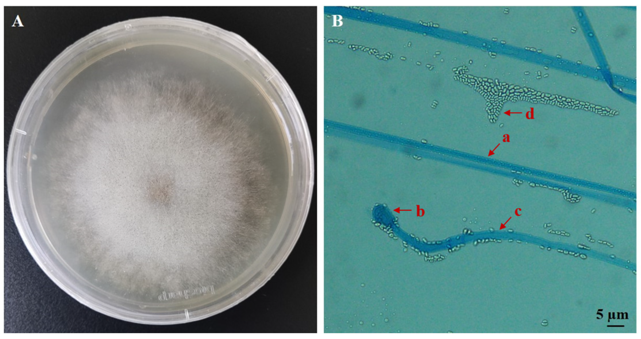
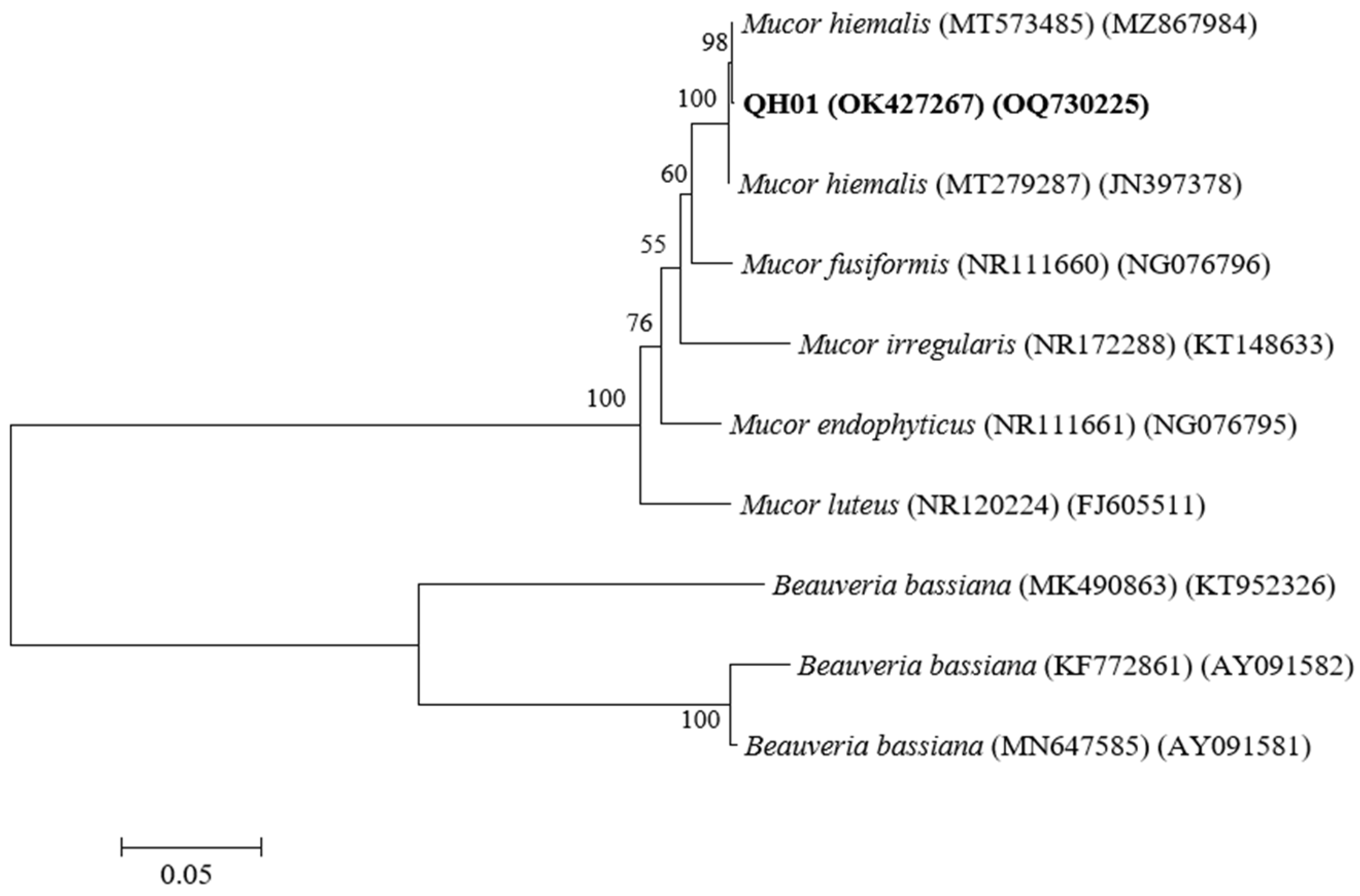
3.1. Identification of the EPF Strain

The fungus was cultured on a PDA medium for 3 days. The grown mycelium was white and feathery, with a dense center and sparse edges. Black sporangia could be observed at the top of the mycelium (Figure 1A). The fungus has 3.2–8.0 µm wide aseptate hyphae, and upon maturation of the sporangia releases ellipsoidal sporangiospores measuring 1.26–3.0 × 1.1–2.2 µm (Figure 1B).
The 535 bp ITS sequence of the fungal isolate shares 100% homology with M. hiemalis (such as MT573485 and MT279287). Additionally, the 1353 bp 18S sequence shares more than 99% homology with M. hiemalis. We named the strain M. hiemalis QH01 (GenBank accession numbers OK427267 and OQ730225) (Figure 2).

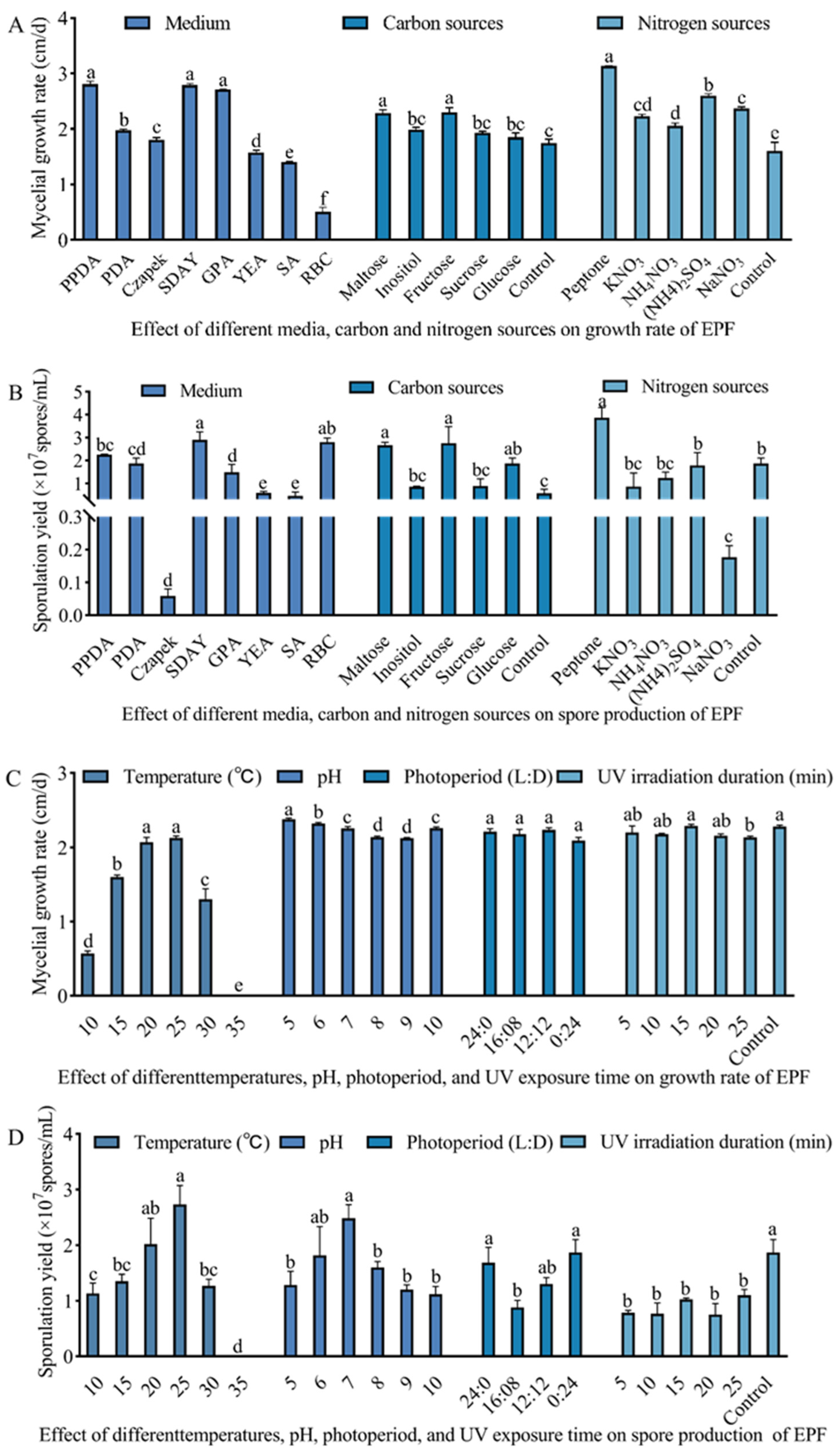
3.2. Culture Condition Optimization of M. hiemalis

We found marked differences in the growth and sporulation quantities of the M. hiemalis on different media (Figure 3A,B). The fastest growth rate of 2.32 cm/d was observed in the Peptone Potato Dextrose Agar (PPDA) medium, followed by the SDAY and Glucose Peptone Agar (GPA) media, which were not significantly different from each other. The slowest mycelial growth was 0.02 cm/d in the Bengal red medium (F7,16 = 25.85; p < 0.05). The highest sporulation quantity of 2.9 × 10⁷ spores/mL was in the SDAY medium, followed by Bengal red and PPDA media after 6 d of incubation. The lowest spore production of 0.06 × 10⁷ spores/mL was observed in the Czapek–Dox medium, which was significantly less than in other media (F7,16 = 358.15; p < 0.05). Thus, the SDAY medium was screened as the most suitable medium for the growth and spore production of M. hiemalis QH01.
As for the carbon source, strain QH01 grew fastest on medium with fructose, with a growth rate of up to 2.30 cm/d (Figure 3A,B). Mycelial growth was also enhanced with maltose and glucose, with a significant increase compared to the control group (F5,12 = 13.23; p < 0.05). The fungus produced the most spores on a fructose-containing medium, followed by media containing glucose or maltose, both of which were significantly better than the control (F5,12 = 7.73; p < 0.05). The growth and sporulation quantity of the fungus on the sucrose- or inositol-supplemented medium were not significantly different from the control medium.
Regarding the nitrogen sources, the fungal mycelial growth rate on the peptone-supplemented medium was 3.13 cm/d, which was significantly higher than for the other nitrogen sources (such as KNO3, NaNO3, (NH4)2SO4, and NH4NO3, F5,12 = 51.37; p < 0.05). The fungus produced the most spores on a peptone-containing medium, while the medium containing KNO3, (NH4)2SO4, or NH4NO3 was not significantly different from the control group. Notably, the spore production was severely inhibited and lower than the control when the fungus was grown with NaNO3 as the nitrogen source (F5,11 = 11.53; p < 0.05). Thus, fructose and peptone were selected as the optimum carbon and nitrogen sources for the growth and spore production of strain QH01 (Figure 3A,B).
Strain QH01 was able to grow from 10 to 30 °C, but the difference was significant (Figure 3C,D). At 25 °C, the fastest growth rate was 2.13 cm/d (F5,12 = 156.02; p < 0.05). The slowest fungal growth was at 10 °C. Under optimal temperature of 25 °C, the fungus produced 2.73 × 10⁷ spores/mL (F5,12 = 12.72; p < 0.05). No mycelial growth or spore production was observed at 35 °C.
Medium pH exerted a significant influence on both fungal mycelial growth and sporulation. The maximum mycelial growth rate of 2.38 cm/d was recorded at pH 5.0 (F5,12 = 35.18; p < 0.05). The optimum pH for fungal spore production was 6–7, but a too-acidic or alkaline medium reduced spore production (F5,12 = 3.70; p < 0.05). No significant differences in fungal mycelial growth rates were observed across the tested photoperiods (F3,8 = 2.02; p > 0.05). In contrast, sporulation quantity was affected under different treatments (F3,8 = 4.76; p < 0.05). The highest spore production (0.87 × 10⁷ spores/mL) was in the dark (24 h dark), and the lowest (0.88 × 10⁷ spores/mL) was at 16L:8D. The mycelial growth rate remained unaffected, showing no significant difference from the control treatment after the fungus was irradiated with UV-C light for 5–20 min (F5,9 = 1.91; p > 0.05). Compared to the control treatment, however, the sporulation quantity was obviously reduced, and there was little difference between different time treatments (F5,10 = 7.47; p < 0.05) (Figure 3C,D).

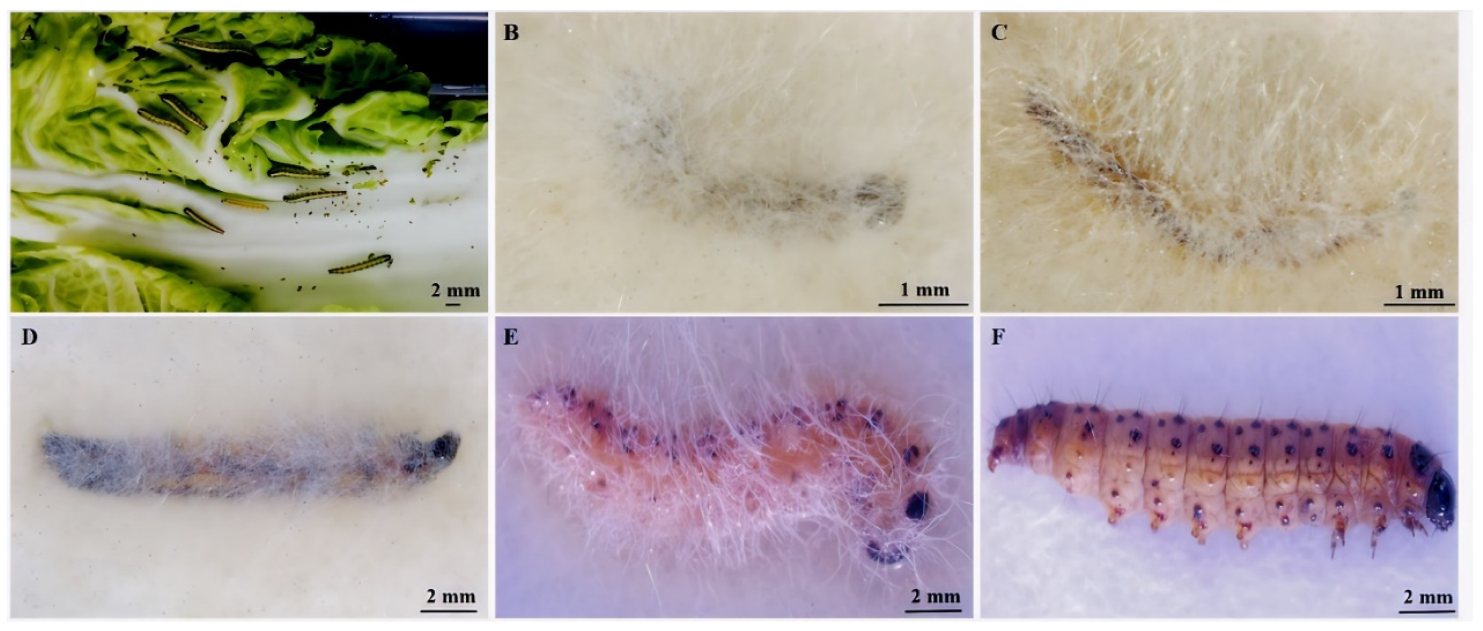
3.3. E. extimalis Larvae Infestation with M. hiemalis QH01

Since E. extimalis larvae were infected by M. hiemalis QH01, their feeding rate gradually decreased. The dead larvae turned brown or black-red, and the body softened and finally dried up. The larvae body surface became covered with white mycelium and black sporangia after 2–5 days of moisturized incubation at 25 °C (Figure 4).

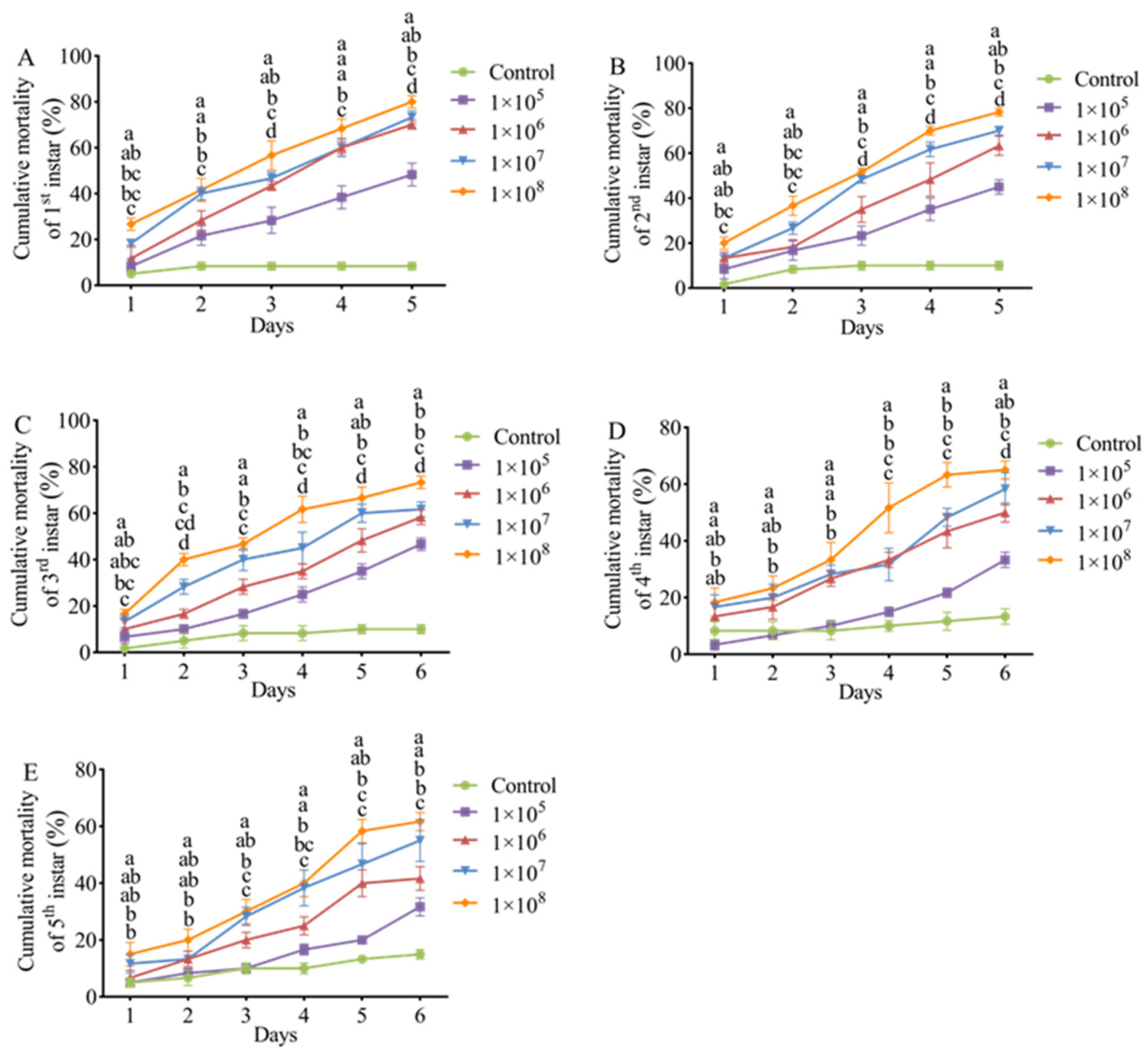
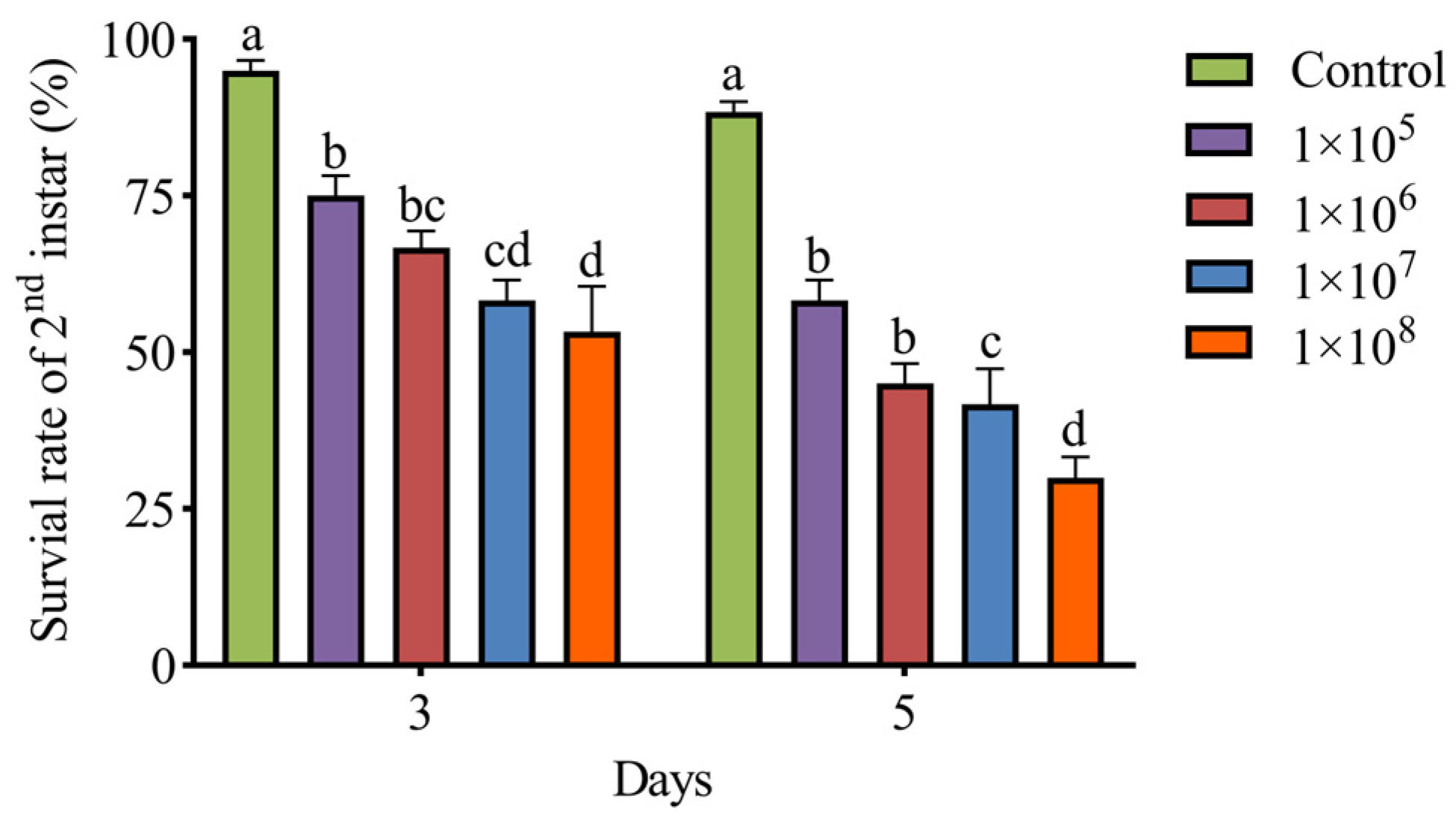
3.4. Pathogenicity of M. hiemalis QH01 Against E. extimalis Larvae

E. extimalis larvae infected with M. hiemalis spore suspensions exhibited significant dose- and time-dependent mortality compared to the control group (Figure 5). The spores induced mortality in all ages of insect larvae.
The LC50 values of M. hiemalis QH01 spores against E. extimalis larvae of various ages are listed in Table 1. The LC50 exhibited a time-dependent decrease, with larval susceptibility showing an inverse relationship to developmental maturity (first instar > second instar > third instar > fourth instar > fifth instar). Basically, the younger larvae were more sensitive to M. hiemalis QH01.
The LT50 values of M. hiemalis QH01 against E. extimalis larvae progressively decreased with increasing inoculum concentration. The fungus exhibited the fastest lethal effect against the 1st–5th instar larvae treated with 1 × 10⁸ spores/mL suspensions, and LT50 values were 2.99, 3.01, 3.37, 4.04, and 4.40 days, respectively. In contrast, under 1 × 10⁵ spores/mL concentration, the fungus had the slowest lethal effect, and LT50 values were 5.15, 5.38, and 5.29 days for 1st–3rd instar larvae. With this spore concentration, the mortality rate of 4th-5th instar larvae was lower than 50% even after 6 days of exposure, and it was not possible to estimate the LT50 (Table 2). These results indicated that M. hiemalis QH01 is highly virulent to young larvae.

3.5. Greenhouse Experiments

The results of greenhouse pot experiments of M. hiemalis QH01 on second instar larvae of E. extimalis are shown in Figure 6. By spraying M. hiemalis suspensions of 1 × 10⁵, 1 × 10⁶, 1 × 10⁷, and 1 × 10⁸ spores/mL, the survival rates of second instar larvae at 5 days were 58.33%, 45%, 41.67%, and 30%, respectively, which were significantly lower than control group (F4,15 = 37.47; p < 0.05). The suspension containing 1 × 10⁸ spores/mL resulted in the lowest survival rate, indicating a concentration-dependent survival.

4. Discussion

The composition of the culture medium can affect the growth characteristics of EPF [25]. We found that the SDAY medium was the most suitable for the growth and spore production of M. hiemalis QH01, which was consistent with previous studies [26]. The spore yield of other tested media, such as PDA, GPA, Yeast extract agar (YEA), etc., was relatively lower than that of SDAY, which might have been due to the difference in selective absorption of nutrients by the same strain because of the different types, contents, and solubility states of nutrients in different media. In addition, carbon and nitrogen sources can also affect the growth and spore production of EPF [27]. This study demonstrated that fructose favored the growth and spore production of M. hiemalis QH01, while the absence of a carbon source inhibited it. The best growth and spore production of strain QH01 were observed with peptone as a nitrogen source, which was consistent with Huang et al. [28].
The optimal growth and reproduction of EPF cannot be achieved without appropriate environmental conditions [29]. Temperature affects the metabolic activity of entomopathogenic fungi through key physiological processes including enzyme biosynthesis, toxin production, spore germination, and germ tube penetration [30]. We found that M. hiemalis QH01 exhibited a growth and sporulation temperature range of 10–30 °C, with an optimum at 25 °C. In contrast, M. hiemalis BO-1 grew the fastest at 23 °C and produced the highest sporulation at 18 °C [31]. This is likely attributable to QH01’s specific adaptations to the extreme climatic conditions of its native Tibetan Plateau environment. The medium pH can significantly impact the growth and pathogenicity of EPF [32]. For example, pH 5.5–6.5 is best suited for the sporulation of B. bassiana, and the same is also good for its pathogenicity [33,34]. M. hiemalis QH01 can grow and produce spores in a wide range of pH (5–10), which is consistent with the results of Wang et al. [35]. In addition to temperature and pH, external conditions such as natural light have a certain impact on EPF. In this study, M. hiemalis had the highest sporulation in total darkness. This may be attributed to Mucor spp.’s natural adaptations to light-limited environments, given their predominant colonization of soil and decaying organic substrates [16]. Their spore production mechanism may have evolved a dependence on darkness. UV light usually inhibits the growth, sporulation, and pathogenicity of EPF. We found that UV irradiation decreased spore production in M. hiemalis. Yao et al. [36] reported that 312 and 365 nm UV light exposure decreased the pathogenicity of M. anisopliae against tobacco aphids. The influence of ultraviolet light on the pathogenicity of M. hiemalis had to be considered when applying it to pest control in the field.
So far, there have been few reports on the biological control of E. extimalis. Wang et al. [37] conducted a field investigation and reported that the Cloth beetle and B. bassiana were the natural enemies of the insect in the larval stage, while the pupal stage could be parasitized by mites. Here, we show the pathogenicity of M. hiemalis against E. extimalis. Particularly, the young larvae of E. extimalis were more susceptible to M. hiemalis QH01, while the older larvae exhibited relatively a lower mortality rate. This difference can be attributed to the different body wall structures and immune capacities of young and old larvae. A thinner body wall in younger larvae makes them more susceptible to EPF infection. Older larvae are more resistant to disease due to hardening and thickening of the cuticle, enhanced immunity and metabolism [38]. Moreover, with the increase in the developmental stage of E. extimalis, the corresponding LT50 and LC50 values increased for the same concentration of M. hiemalis. This indicated that M. hiemalis QH01 is more virulent to lower-stage larvae than the developed larvae. This result is in agreement with the findings of Tozlu et al. [39]. Zhu et al. [40] reported 46.25% mortality in Bradysia impatiens second instar larvae following 5 days of exposure to M. hiemalis BO-1 at 1 × 10⁶ spores/mL, whereas our experimental data demonstrated 63.33% mortality in E. extimalis larvae under identical spore concentration with M. hiemalis QH01. This may be the result of host specificity, environmental adaptation, etc., which led to significant differences in the pathogenicity of strains of the same species. Notably, surviving larvae exhibited significantly reduced feeding activity, progressed to the prepupal stage with sporadic pupation, but ultimately failed to complete adult eclosion. It is possible that infection by M. hiemalis QH01 induces a sublethal effect on life history traits including developmental processes and reproduction in E. extimalis [41]. EPF can target all developmental stages of insects, such as eggs, larvae, and adults. Wang et al. [42] isolated a strain of M. hiemalis from diseased Bradysia odoriphaga larvae and found its pathogenicity varied against different stages of insect; strong pathogenicity was against larvae and relatively weak pathogenicity was against eggs and pupae. In the study, the pathogenicity of M. hiemalis QH01 was determined only against the larvae of E. extimalis, and further studies are needed to determine the pathogenicity against other insect forms.
Most EPF performs remarkably well under controlled conditions, such as in the laboratory, exhibiting high mortality against the target insects. However, abiotic factors such as changes in temperature, humidity, and UV exposure can significantly affect the biocontrol effect of EPF in field applications [43]. In greenhouse environments, after 7 days of treatment with 2 × 10⁶ spores/mL of Bea111 (B. bassiana) and 9 × 10⁶ spores/mL of Isa340 (Isaria javanica), third instar larvae of Duponchelia fovealis exhibited 52% and 45% mortality rates, respectively [44]. The larval stage of E. extimalis causes the most damage and economic loss in rapeseed fields. First instar larvae exhibit higher mortality rates when attempting to bore into rapeseed pods for feeding. From the third instar onward, larvae exhibit migratory behavior, transitioning from pod-boring to stem-tunneling and inter-plant movement, while their food consumption surges dramatically [45]. Thus, we evaluated the biocontrol potential of M. hiemalis QH01 against the second instar larvae of E. extimalis under greenhouse conditions and found it significantly reduced their survival rates.
In this study, the effectiveness of strain QH01 against E. extimalis second instar larvae in greenhouse pots differed from the results of the indoor virulence assay, which may be due to different bioassay methods [46] and external environmental conditions. To sum up, when preparing fungal powder, ultraviolet protectants, thermal stabilizers, and ionic dispersants should be added to the spore powder to enhance environmental adaptability. Greenhouse and field applications should be carried out after sunset or on cloudy days when the temperature is around 20–25 °C.

5. Conclusions

In the present study, a fungal strain isolated from E. extimalis larvae was identified as M. hiemalis QH01 based on morphological characteristics, ITS, and 18S rDNA sequence analyses. To our knowledge, this is the first report assessing the biocontrol effects of an entomopathogenic fungus against E. extimalis. Our results lay the foundation for the subsequent research on designing a green prevention and control program for E. extimalis using EPF to benefit the spring rapeseed industry.

Supplementary Materials

The following supporting information can be downloaded at: https://www.mdpi.com/article/10.3390/biology14050467/s1. Table S1: Test medium composition.

Author Contributions

Conceptualization, Y.M. and Y.L.; methodology, Y.M.; software, Y.M. and M.Q.; formal analysis, Y.Z. and Y.S.; investigation, Y.M. and Y.L.; resources, Y.L.; data curation, Y.Z. and Y.S.; writing—original draft preparation, Y.M. and M.Q.; writing—review and editing, Y.L. and G.L.; visualization, Y.M. and M.Q.; supervision, G.L.; project administration, Y.L.; funding acquisition, Y.L. and G.L. All authors have read and agreed to the published version of the manuscript.

Funding

This research was funded by the Applied Fundamental Research Fund of Qinghai Provincial Department of Science and Technology (Grant No. 2023ZJ739) and the Major Project of Qinghai Academy of Agriculture and Forestry (Grant No. 2018NKY003).

Institutional Review Board Statement

Not applicable.

Informed Consent Statement

Not applicable.

Data Availability Statement

Data are contained within the article.

Acknowledgments

The authors would like to thank the students of the Insect Research Group of the Key Laboratory of Integrated Control of Agricultural Pests in Qinghai Province for their help in collecting and rearing the insects.

Conflicts of Interest

The authors declare no conflicts of interest.

References

  1. Shao, H.N.; Liu, Y.X.; Liu, Y.J.; Lai, Y.P. The Effect of Ice-Nucleation-Active Bacteria on Metabolic Regulation in Evergestis extimalis (Scopoli) (Lepidoptera: Pyralidae) Overwintering Larvae on the Qinghai-Tibet Plateau. Insects 2022, 13, 909. [Google Scholar] [CrossRef] [PubMed]
  2. Lai, Y.P.; Tao, K.; Hou, T.P. Preliminary analysis of geographical distribution based on cold hardiness for Evergestis extimalis (Scopoli) (Lepidoptera: Pyralidae) on Qinghai-Tibet Plateau. Entomol. Res. 2019, 49, 13–20. [Google Scholar] [CrossRef]
  3. Shah, P.A.; Pell, J.K. Entomopathogenic fungi as biological control agents. Appl. Microbiol. Biotechnol. 2003, 61, 413–423. [Google Scholar] [CrossRef] [PubMed]
  4. Samuels, R.I.; Coracini, D.; Martins dos Santos, C.A.; Gava, C.A.T. Infection of Blissus antillus (Hemiptera: Lygaeidae) eggs by the entomopathogenic fungi Metarhizium anisopliae and Beauveria bassiana. Biol. Control 2002, 23, 269–273. [Google Scholar] [CrossRef]
  5. Yao, H.Q.; Xie, Y.P.; Wang, E.H. Morphologic observation of Apocheima cinerarius Erschoff (Lepidoptera: Geometridae) pupae and comparison of virulence of Beauveria bassiana infected on them. J. Environ. Entomol. 2015, 37, 348–354. [Google Scholar]
  6. Cruz-Avalos, A.M.; Bivián-Hernández, M.L.Á.; Ibarra, J.E.; Del Rincón-Castro, M. High virulence of mexican entomopathogenic fungi against fall armyworm, (Lepidoptera: Noctuidae). J. Econ. Entomol. 2019, 112, 99–107. [Google Scholar] [CrossRef]
  7. Shehzad, M.; Tariq, M.; Mukhtar, T.; Gulzar, A. On the virulence of the entomopathogenic fungi, Beauveria bassiana and Metarhizium anisopliae (Ascomycota: Hypocreales), against the diamondback moth, Plutella xylostella (L.) (Lepidoptera: Plutellidae). Egypt. J. Biol. Pest Control 2021, 31, 86–92. [Google Scholar] [CrossRef]
  8. Xie, N.; Yin, Y.P.; Zhang, J.W.; Shen, J.F.; Wang, Z.K. Characterization of the Metarhizium anisopliae CQMa128 and its virulence measurements against Lepidoptera insects. In Proceedings of the 2009 Annual Meeting of the Mycological Society of China, Beijing, China, 23–25 August 2009. [Google Scholar]
  9. Varela, A.; Morales, E. Characterization of some Beauveria bassiana isolates and their virulence toward the Coffee Berry Borer, Hypothenemus hampei. J. Invertebr. Pathol. 1996, 67, 147–152. [Google Scholar] [CrossRef]
  10. Wraight, S.P.; Inglis, G.D.; Goettel, M.S. Fungi. In Field Manual of Techniques in Invertebrate Pathology: Application and Evaluation of Pathogens for Control of Insects and other Invertebrate Pests; Springer: Dordrecht, The Netherlands, 2007; pp. 223–248. [Google Scholar]
  11. Steinkraus, D.C. Factors affecting transmission of fungal pathogens of aphids. J. Invertebr. Pathol. 2006, 92, 125–131. [Google Scholar] [CrossRef]
  12. Alali, S.; Mereghetti, V.; Faoro, F.; Bocchi, S.; Al Azmeh, F.; Montagna, M. Thermotolerant isolates of Beauveria bassiana as potential control agent of insect pest in subtropical climates. PLoS ONE 2019, 14, e0211457. [Google Scholar] [CrossRef]
  13. Jaronski, S.T. Ecological factors in the inundative use of fungal entomopathogens. BioControl 2010, 55, 159–185. [Google Scholar] [CrossRef]
  14. Mann, A.J.; Davis, T.S. Plant secondary metabolites and low temperature are the major limiting factors for Beauveria bassiana (Bals.–Criv.) Vuill. (Ascomycota: Hypocreales) growth and virulence in a bark beetle system. BioControl 2020, 141, 104–130. [Google Scholar] [CrossRef]
  15. Lyu, M.L.; Liu, Z.; Song, Z.; Wang, Y.N.; Liu, X.Y. Diversity and distribution of culturable Mucoromycota fungi in the Greater Khinggan Mountains. J. Biodivers. Sci. 2019, 27, 821–832. [Google Scholar]
  16. Morrissey, C.O.; Kim, H.Y.; Garnham, K.; Dao, A.; Chakrabarti, A.; Perfect, J.R.; Alastruey-Izquierdo, A.; Harrison, T.S.; Bongomin, F.; Galas, M.; et al. Mucorales: A systematic review to inform the World Health Organization priority list of fungal pathogens. Med. Mycol. 2024, 62, myad130. [Google Scholar] [CrossRef]
  17. Nentwig, W.; Prillinger, H. A zygomycetous fungus as a mortality factor in a laboratory stock of spiders. J. Arachnol. 1990, 18, 118–121. [Google Scholar]
  18. Heitor, F. Wound parasitism by the fungus Mucor hiemalis Wehmer in insects. Ann. Epiphyt. 1962, 13, 179–203. [Google Scholar]
  19. Reiss, J. Biotoxic activity in the Mucorales. Mycopathologia 1993, 121, 123–127. [Google Scholar] [CrossRef]
  20. Konstantopoulou, M.A.; Mazomenos, B.E. Evaluation of Beauveria bassiana and B. brongniartii strains and four wildtype fungal species against adults of Bactrocera oleae and Ceratitis capitata. BioControl 2005, 50, 293–305. [Google Scholar] [CrossRef]
  21. Konstantopoulou, M.A.; Milonas, P.; Mazomenos, B.E. Partial purification and insecticidal activity of toxic metabolites secreted by a Mucor hiemalis strain (SMU-21) against adults of Bactrocera oleae and Ceratitis capitata (Diptera: Tephritidae). J. Econ. Entomol. 2006, 99, 1657–1664. [Google Scholar] [CrossRef]
  22. Hallouti, A.; Ait Hamza, M.; Zahidi, A.; Ait Hammou, R.; Bouharroud, R.; Ait Ben Aoumar, A.; Boubaker, H. Diversity of entomopathogenic fungi associated with Mediterranean fruit fly (Ceratitis capitata (Diptera: Tephritidae)) in Moroccan Argan forests and nearby area: Impact of soil factors on their distribution. BMC Ecol. 2020, 20, 64. [Google Scholar] [CrossRef]
  23. Wei, J.C. Fungi Identification Manual; Shanghai Scientific & Technical Publishers: Shanghai, China, 1979; pp. 70–74. [Google Scholar]
  24. Kumar, S.; Nei, M.; Dudley, J.; Tamura, K. MEGA: A biologist-centric software for evolutionary analysis of DNA and protein sequences. Brief. Bioinform. 2008, 9, 299–306. [Google Scholar] [CrossRef] [PubMed]
  25. Fargues, J.F.; Robert, P.H. Effect of passaging through scarabeid hosts on the virulence and host specificity of two strains of the entomopathogenic hyphomycete Metarhizium anisopliae. Can. J. Microbiol. 1983, 29, 576–583. [Google Scholar] [CrossRef]
  26. Banu, J.G.; Rajalakshmi, S. Standardisation of media for mass multiplication of entomopathogenic fungi. Indian J. Plant Prot. 2014, 42, 91–93. [Google Scholar]
  27. Moore, D.; Douro-Kpindou, O.K.; Jenkins, N.E.; Lomer, C.J. Effects of moisture content and temperature on storage of Metarhizium flavoviride conidia. Biocontrol Sci. Technol. 1996, 6, 51–61. [Google Scholar] [CrossRef]
  28. Huang, P.; Yao, J.A.; Yu, D.Y. Biological Characteristics of Metarhizium anisopliae FM−03 and Its Infection against Planococcus citri. Chin. J. Biol. Control 2018, 34, 858–865. [Google Scholar]
  29. Hou, Y.; Xia, Y.F.; Xu, J.Q. Identification and Biological Characteristics of a Metarhizium Strain and Its Virulence against Oriental Migratory Locust. Chin. J. Biol. Control 2015, 31, 333–339. [Google Scholar]
  30. Anwar, A.U.; Xia, S.; Meng, L.; Fahim, R.M.; Zhang, Z.Y.; Zhang, H.Y. Isolation, characterization, culturing, and formulation of a new Beauveria bassiana fungus against Diaphorina citri. BioControl 2021, 158, 104586. [Google Scholar]
  31. Zhu, G.D.; Ding, W.J.; Xue, M.; Li, Z.; Li, M.; Zhao, Y. Identification and Pathogenicity of a New Entomopathogenic Fungus, Mucor hiemalis (Mucorales: Mucorales), on the Root Maggot, Bradysia odoriphaga (Diptera: Sciaridae). J. Insect Sci. 2022, 22, 2. [Google Scholar] [CrossRef]
  32. St leger, R.; Nelson, J.O.; Screen, S.E. The entomopathogenic fungus Metarhizium anisopliae alters ambient pH, allowing extracellular protease production and activity. Microbiology 1999, 145, 2691–2699. [Google Scholar] [CrossRef]
  33. Mishra, S.; Kumar, P.; Malik, A. Effect of temperature and humidity on pathogenicity of native Beauveria bassiana isolate against Musca domestica L. J. Parasit. Dis. 2015, 39, 697–704. [Google Scholar] [CrossRef]
  34. Cheng, G.H.; Shu, J.; Ding, K.J. Study on Nutrition Demand and Culture Condition of Beauveria bassiana. Chin. Agric. Sci. Bull. 2006, 22, 365–368. [Google Scholar]
  35. Wang, B.; Fan, M.Z.; Li, Z.Z. Sieving of Selective Media for Beauveria bassiana. J. Anhui Agric. Univ. 2000, 27, 23–28. [Google Scholar]
  36. Yao, S.L.; Ying, S.H.; Feng, M.G.; Hatting, J.L. In vitro and in vivo responses of fungal biocontrol agents to gradient doses of UV-B and UV-A irradiation. BioControl 2010, 55, 413–422. [Google Scholar] [CrossRef]
  37. Wang, L.Y.; Sun, Q.; Lin, Z.W.; Wang, H.Z.; Wu, Q.S.; Xin, H.P.; Zhong, X.Z. Occurrence and control of Evergestis extimalis. Chin. Bull. Entomol. 1998, 35, 331–332. [Google Scholar]
  38. Strak, J.D.; Sherman, M. Toxicity, penetration, and metabolism of acephate in three fruit fly species (Diptera: Tephritidae). J. Econ. Entomol. 1989, 82, 1564–1571. [Google Scholar] [CrossRef]
  39. Tozlu, E.; Tozlu, G.; Kotan, R. Biocontrol of Cymbalophora rivularis (Menetries) (Lepidoptera: Erebidae) larvae by entomopathogenic bacteria and fungi. Egypt. J. Biol. Pest Control 2022, 32, 66. [Google Scholar] [CrossRef]
  40. Zhu, G.D.; Ding, W.J.; QIU, J.Y.; Wang, Y.; Xue, M.; Zhao, H.P.; Zhang, G.F. Pathogenicity and field control efficiency of Mucor hiemalis BO-1 against Bradysia odoriphaga Larvae. Chin. J. Biol. Control. 2024, 40, 52–60. [Google Scholar]
  41. Latifian, M.; Soleimannejadian, E.; Ghazavi, M.; Mosadegh, M.S.; Hayati, J. Effects of sublethal concentrations of fungus Beauveria bassiana on the reproductive potentials of sawtoothed beetle Oryzaephilus surinamensis on commercial date cultivars. Plant Prot. J. 2010, 227–290. [Google Scholar]
  42. Wang, Y. Poisoning Activity of a New Strain of Mucor hiemalis to Bradysia odoriphaga Yang et Zhang. Master’s Thesis, Shandong Agricultural University, Taian, China, 2021. [Google Scholar]
  43. Beris, E.; Papachristos, D.; Ponchon, M.; Caca, D.; Kontodimas, D.; Reineke, A. The effects of temperature on pathogenicity of entomopathogenic fungi for controlling larval populations of the European grapevine moth (Lobesia botrana) (Lepidoptera: Tortricidae). J. Crop Prot. 2024, 177, 106542. [Google Scholar] [CrossRef]
  44. Amatuzzi, R.F.; Poitevin, C.G.; Poltronieri, A.S.; Zawadneak, M.A.C.; Pimentel, I.C. Susceptibility of Duponchelia fovealis Zeller (Lepidoptera: Crambidae) to Soil-Borne Entomopathogenic Fungi. Insects 2018, 9, 70. [Google Scholar] [CrossRef]
  45. Xie, C.H. Study on Occurrence and Control Countermeasures of Harmful Organisms with Rapeseed in Huangzhong County of Qinghai. Master’s Thesis, Lanzhou University, Lanzhou, China, 2019. [Google Scholar]
  46. Liu, H.; Skinner, M.; Parker, B.L. Bioassay method for assessing the virulence of Beauveria bassiana against tarnished plant bug, Lygus lineolaris (Hem., Miridae). J. J Appl Entomol. 2003, 127, 299–304. [Google Scholar] [CrossRef]
Figure 1. Morphological characteristics of the fungus on PDA medium. (A) Colony morphology after 3 days of growth on PDA at 25 °C. (B) Mycelia (a), sporangia (b), sporangiophore (c) and sporangiospores (d) under an optical microscope.
Biology 14 00467 g001
Figure 2. A NJ phylogenetic tree was constructed based on ITS and 18S gene sequences. Each isolate is marked with a GenBank accession number and the species name. Posterior probabilities are presented above the branches. The M. hiemalis isolated from E. extimalis in this study is marked in bold.
Biology 14 00467 g002
Figure 3. Effect of different media, carbon, and nitrogen sources on (A) growth rate and (B) spore production of EPF. Effect of different temperatures, pH, photoperiod, and UV exposure time on (C) fungal growth rate and (D) spore production. Data are mean ± SE. Letters on the error bars indicate significant differences analyzed by ANOVA with the LSD test (p < 0.05).
Biology 14 00467 g003
Figure 4. Symptoms of E. extimalis larvae infested with M. hiemalis QH01. Images were taken after 5 days of moisturized incubation. (A) Healthy, (B) 1st instar, (C) 2nd instar, (D) 3rd instar, (E) 4th instar, and (F) 5th instar larvae.
Biology 14 00467 g004
Figure 5. Pathogenicity of M. hiemalis QH01 against E. extimalis larvae. (AE) The mortality rate trend over time (in days) after insect treatment with four different concentrations of M. hiemalis. Data are mean ± SE. Letters on the error bars indicate significant differences analyzed using ANOVA with the LSD test (p < 0.05).
Biology 14 00467 g005
Figure 6. Greenhouse pot tests were performed to determine the survival rate of second instar larvae of E. extimalis against M. hiemalis. Data are mean ± SE. Letters on the error bars indicate significant differences analyzed using ANOVA with the LSD test (p < 0.05).
Biology 14 00467 g006
Table 1. Virulence regression equations for LC50 values of M. hiemalis QH01 against E. extimalis larvae.
DaysDevelopmental StageVirulence Regression EquationLC50 (Spores/mL)95% CI (Spores/mL)
31stY = 0.229X − 1.6501.63 × 1073.21 × 106–6.85 × 10⁸
2ndY = 0.262X − 1.9823.57 × 1078.00 × 106–1.30 × 10⁹
3rdY = 0.253X − 1.9173.88 × 1078.16 × 106–2.11 × 10⁹
4thY = 0.238X − 2.2593.13 × 1091.63 × 108–3.89 × 10¹⁵
5thY = 0.241X − 2.3626.53 × 1092.59 × 108–5.93 × 10¹⁶
41stY = 0.233X − 1.3405.71 × 1051.14 × 104–2.82 × 10⁶
2ndY = 0.312X − 1.9181.40 × 1062.69 × 105–4.73 × 10⁶
3rdY = 0.275X − 1.7201.83 × 1062.89 × 105–7.72 × 10⁶
4thY = 0.312X − 2.4991.01 × 1082.27 × 107–3.40 × 10⁹
5thY = 0.288X − 2.3871.93 × 1083.38 × 107–2.33 × 10¹⁰
51stY = 0.289X − 1.4127.77 × 1041.07 × 103–4.00 × 10⁵
2ndY = 0.294X − 1.5281.58 × 1055.15 × 103–6.83 × 10⁵
3rdY = 0.267X − 1.5255.23 × 1052.92 × 104–2.14 × 10⁶
4thY = 0.344X − 2.3979.13 × 1063.05 × 106–4.22 × 10⁷
5thY = 0.326X − 2.3551.69 × 1075.24 × 106–1.21 × 10⁸
Table 2. Virulence regression equations for LT50 values of M. hiemalis QH01 against E. extimalis larvae.
Concentration
(Spores/mL)
Developmental StageVirulence Regression EquationLT50 (Days)95% CI (Days)
1 × 1051stY = 0.265X − 1.3655.154.38–12.86
2ndY = 0.300X − 1.6125.384.61–10.39
3rdY = 0.296X − 1.5645.294.54–10.20
4th###
5th###
1 × 1061stY = 0.347X − 1.1863.412.00–3.95
2ndY = 0.363X − 1.4814.083.46–4.80
3rdY = 0.298X − 1.2624.243.51–5.63
4th###
5th###
1 × 1071stY = 0.353X − 1.1483.25 1.63–3.80
2ndY = 0.299X − 1.0683.57 1.71–4.21
3rdY = 0.275X − 1.0723.902.46–4.90
4th###
5th###
1 × 1081stY = 0.421X − 1.2612.991.60–3.51
2ndY = 0.391X − 1.1763.011.39–3.55
3rdY = 0.373X − 1.2583.372.12–3.88
4thY = 0.385X − 1.5574.043.45–4.66
5thY = 0.367X − 1.6144.40 3.88–5.40
Note: # indicates that the mortality rate of E. extimalis larvae was below 50%, and LT50 values cannot be estimated.
Disclaimer/Publisher’s Note: The statements, opinions and data contained in all publications are solely those of the individual author(s) and contributor(s) and not of MDPI and/or the editor(s). MDPI and/or the editor(s) disclaim responsibility for any injury to people or property resulting from any ideas, methods, instructions or products referred to in the content.

