Article

Influence of Variation in Hind Leg Structure of Auchenorrhyncha on Their Jumping Performance

1
Key Laboratory of Plant Protection Resources and Pest Management, Ministry of Education, College of Plant Protection, Northwest A&F University, Yangling 712100, China
2
Illinois Natural History Survey, Prairie Research Institute, University of Illinois, Champaign, IL 61820, USA
*
Author to whom correspondence should be addressed.
Biology 2025, 14(4), 418; https://doi.org/10.3390/biology14040418
Submission received: 16 February 2025 / Revised: 6 April 2025 / Accepted: 10 April 2025 / Published: 13 April 2025

Simple Summary

Although small insects generally have similar jumping mechanisms, their jumping performances exhibit significant variations. By conducting a comparative analysis of the external morphology of the hind legs using scanning electron microscopy (SEM) and recording jumping motions using high-speed capture technology, this study quantitatively evaluated the physical parameters generated during the jumping process. The results indicate that differences in the morphological structure of the hind legs significantly affect jumping performance, specifically manifested as notable divergence in key metrics, such as jumping height, distance, and initial velocity among different species. This finding provides important experimental evidence for the further understanding of the biomechanical mechanisms of insect jumping behavior.

Abstract

Four species representing four different families of the hemipteran insect suborder Auchenorrhyncha, Lepyronia coleoptrata (Aphrophoridae), Euricania ocellus (Ricaniidae), Kolla sp. (Cicadellidae) and Tricentrus sp. (Membracidae) were investigated using high-speed photography and scanning electron microscopy to identify hind leg structures that may influence jumping performance. The coxa–trochanteral joint, femur and tibia were found to have distinct structural adaptations that vary among these jumping insects. Froghoppers and planthoppers possess a coxal protrusion which is absent in leafhoppers and treehoppers, the latter featuring a more recessed coxal fossa. The medial coxae of these insects exhibit fields of microtrichia that vary in density and fine structure. Medial gears on the trochanters of Tricentrus sp. are implicated in the storage of energy prior to their jumps. These structural differences manifest in the insects’ jumping performance. The study demonstrated a correlation between the robustness of the microtrichia field interaction and the insect’s jumping capability. Specifically, leafhoppers, equipped with a pair of rivet-like structures connecting the hind coxae, were observed to achieve quicker and more stable take-offs. The study reveals that structural variations in the hind legs of Auchenorrhyncha species significantly influence their jumping performance, with implications for both efficiency and stability.

1. Introduction

Insects renowned for their extraordinary jumping abilities have evolved intricate morphological adaptations across their locomotor systems, including specialized limbs, joints, muscles and movement patterns, with particular specialization in their propulsive hind legs. The evolutionary development of jumping mechanisms across insect taxa reflects selective pressures for predator evasion through enhanced acceleration capabilities and biomechanical optimization of jump performance. Biomechanically, leg-driven jumping insects are categorized by their primary torque-generating joints [1,2]: (1) The Femur–Tibia (FT) mechanism utilizes energy storage and rapid joint extension [3], enabling swift movement through fast muscle contraction, as observed in locusts [4]; (2) The Trochanter-driven system (exemplified by froghoppers, planthoppers, leafhoppers, treehoppers, fleas) employs pleural arch spring recoil to generate torque at the coxa–trochanter joint (PADT mechanism: Pleural Arch Depression of the Trochanter) [5,6]. While some orthopterans, like crickets, achieve jumping through direct muscular torque [7], most high-performance jumpers supplement muscle action with exoskeletal elastic energy storage through catapult mechanisms. These systems combine specialized cuticular structures with mechanical advantage from elongated legs, achieving power outputs exceeding direct muscle capacity by three orders of magnitude [8,9,10,11,12,13,14], as demonstrated in grasshoppers [4], flea beetles [10], and all PADT jumpers. The rapid release of stored elastic energy produces greater propulsive forces and accelerations than muscular action alone [11,12], achieving remarkable velocities within millisecond timescales through energy release rates surpassing typical muscle contraction limits by 1000-fold [13,14].
Auchenorrhyncha (Insecta: Hemiptera), comprising Cercopoidea, Fulgoroidea, Membracidae and Cicadellidae, represent the pinnacle of insect jumping performance through extraordinary morphological adaptations [15,16]. Kinematic analyses reveal a conserved jumping sequence across these lineages: initial synchronized hind leg retraction positions the tibiae medially under the body, followed by simultaneous femur–tibia extension and coxa–trochanter depression [17,18]. Notably, while sharing this fundamental jumping mechanism, substantial performance divergences exist among families, correlating with ultrastructural specialization of metathoracic appendages. Planthoppers and froghoppers employ an elastomeric catapult mechanism through thoracic skeletal composites comprising resilin-rich elastic pads sandwiched between sclerotized cuticular layers [19,20]. This system features a coxal protrusion engaging with specific femoral regions for energy storage, complemented by microtrichia fields on coxal surfaces providing traction through cross–mesh interactions [17,19]. In contrast, leafhoppers (Cicadellidae), lacking both the energy-storing protrusions and resilin-containing coxal structures, achieve comparable performance through a unique evolutionary innovation: intermeshing microtrichia field with embedded rivet-like structures within the coxae [21]. This convergence highlights the alternative evolutionary pathways for achieving high-performance jumps.
The structure–function paradigm is further illustrated by specialized hind leg adaptations across diverse taxa, tailored to their environments for efficient locomotion. The Papuan weevil Trigonopterus oblongus (Pascoe) demonstrates a functional screw-and-nut system in its coxa–trochanteral joints, enabling substrate anchoring during feeding through chrysomeloid joint modification [22]. Nymphs of Issus planthoppers employ meshed gears in their femoro–trochanteral joints to synchronize leg movements with microsecond precision [23]. These biological innovations have inspired biomimetic designs, including froghopper-inspired jumping robots with symmetrical legs and synchronized gear systems [24].
This study investigates the specialized hind leg mechanisms underlying the jumping supremacy of Auchenorrhyncha. We analyze the functional morphology of the coxo–trochanter joint complex and its performance implications, with particular focus on a novel femur–coxa locking mechanism that enables sequential energy storage through gradual muscular tensioning followed by rapid elastic release. This mechanism potentially represents an evolutionary optimization for explosive power generation, providing new insights into the biomechanical diversification of insect jumping systems.

2. Materials and Methods

Adult specimens of Lepyronia coleoptrata (Linnaeus, 1758), Euricania ocellus (Walker, 1851), Kolla sp. and Tricentrus sp. were collected from low-growing shrubs in Yangling, Shaanxi Province. China (34°27′ N, 108°9′ E, elev440m). From June to September each year between 2021 and 2023, the numbers for each group were as follows: 17, 13, 14, and 9, respectively. Specimens were preserved in 95% ethanol and stored at 4 °C. The adult samples studied included both females and males.

2.1. Samples for SEM

Adult specimens were initially immersed in 95% ethanol. Their legs were excised using dissecting needles under a stereomicroscope (Olympus SZX10, Tokyo, Japan) and then dehydrated in 100% ethanol for two intervals of 30 min each. Subsequently, they were gradually transitioned through a series of tert-butyl ethanol (TBA) solutions at concentrations of 25%, 50%, and 75%, with ethanol to TBA ratios of 3:1, 1:1 and 1:3, respectively, for 15 min per concentration, followed by a final immersion in 100% TBA for 30 min. Dehydrated samples were then freeze-dried (VFD-21S, SHINKKU VD, Tokyo, Japan) for 3 h. The dried specimens were mounted on aluminum stubs with double-sided copper sticky tape and sputter-coated with a 40/60 gold/palladium alloy using a high-resolution coater (MSP-1S, SHINKKU VD, Tokyo, Japan). The SEM observations were conducted employing a T-3400 SEM (Hitachi, Tokyo, Japan) at an operating voltage of 15 kV or a Nova Nano SEM-450 (FEI, Hillsboro, OR, USA) at 5–10 kV.

2.2. High-Speed Photography

Sequential images of the insects’ jumps were captured using a high-speed camera at a frame rate of 10,000 s 1 with an exposure time of 1 μs. The imaging system utilized was the X213 model (Revealer, Hefei, China), equipped with a 105 mm micro lens.
Jumps were either spontaneous or induced by gentle mechanical stimulation using a fine paintbrush within a fully enclosed room that allowed insects to jump freely. Due to the limited capture field of the camera, there was a risk of insects exiting the frame and insects were placed on a 10 mm × 10 mm × 30 mm cube platform, the surface of which was wrapped with a wet paper towel to provide traction for the insects at take-off. The high-speed camera was positioned to capture the side view of the jumping process, focusing on the edge of the platform. The body angle was defined as the angle formed by the longitudinal body axis in relation to the horizontal plane. The peak velocity during take-off was calculated by tracking a selected mass point on the insect’s body using the processing software of the high-speed camera. The first movement of the hind legs occurred at the capture moment point. The moment when the hind legs lost contact with the ground, marking the insect as airborne, was designated as time t = 0 ms. This reference point allowed for the alignment and comparison of different jumps. The initial movement of the hind leg was marked out, and the time elapsed between this movement and take-off was used to calculate the acceleration of the body during the jump. The tergum of Kolla sp. was affixed to a transparent glass background using adhesive tape. A high-speed camera positioned vertically below the insects captured the interaction of their hind legs during the jump, providing footage of the ventral surface movement.

2.3. Image Processing and Measurement

The imported photographs were observed and measured utilizing Adobe Photoshop CS6 (Adobe Systems, San Jose, CA, USA). All characters are expressed as mean ± SEM. Statistical analyses were conducted using SPSS 19.0 (SPSS, Chicago, IL, USA). Graphs were produced using Microsoft Office Excel 2007.
Body length measurements were obtained from scanning electron microscopy (SEM) images using Photoshop CS6 software (Adobe Systems, San Jose, CA, USA). Adult insect mass was quantified immediately post-mortem using an analytical balance (LS220ASCS, Precisa, Zurich, Switzerland), following high-speed video recordings of their jumping behavior.

3. Results

3.1. Morphological Structure

Significant variations in body shape were observed among the insects analyzed, despite similar body lengths, accompanied by a 5-fold variation in body mass, ranging from 3.9 to 18.6 mg (Table 1). The tibia, being the longest segment on the insects’ hind legs, plays a crucial role in their jumping ability. The spindly hind legs of these insects enable even the smallest species to achieve superior jumping performance. Moreover, different structural features of these legs also influence their jumping capability and stability.
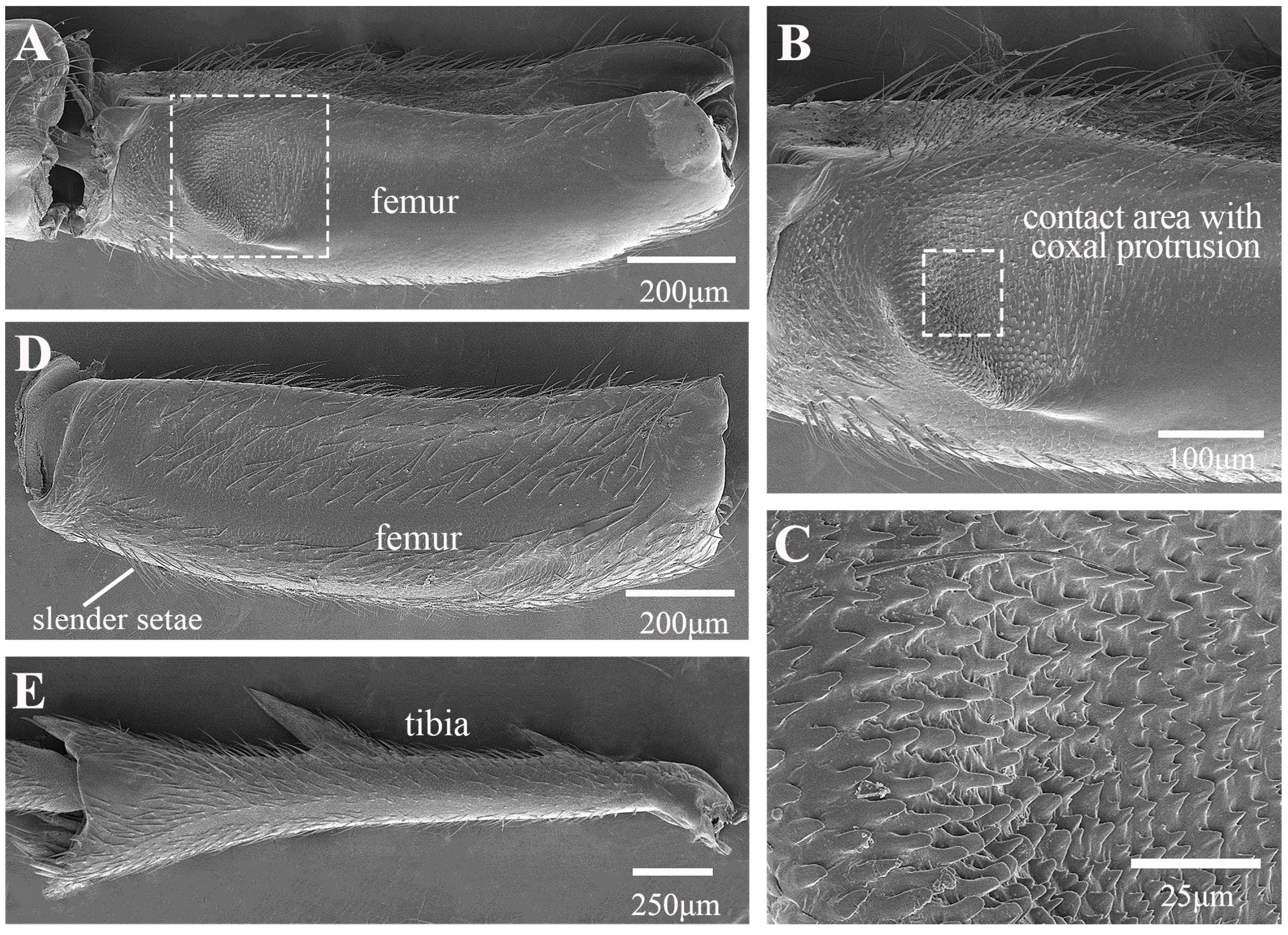
In the froghopper, Lepyronia coleoptrata, the folded forewings extend posteriorly to cover most of the hind legs when viewed from the side. The tibia is slightly more than twice as long as the femur and the total length of the hind leg is 84% of the total body length (Table 1). The coxae and trochanters, along with the interface between the trochantin and prosternum, are adorned with setae (Figure 1A–C). Notably, the posterior section of the coxa, which connects to the trochanter, features a concentration of longer setae (Figure 1C). The broad coxae have prominent bulbous protrusions on their lateral sides (Figure 1B). Spike-like microtrichia on protrusions are grouped into an oval, domed-shaped structure in the middle, with height gradually decreasing from the periphery to the center until it forms a smooth plane (Figure 1F). When the leg is retracted prior to jumping, this structure is in contact with a specific location on the femur of the hind legs (Figure 1D,E). A microtrichia field on the inner (medial) side of the coxae, where the coxae meet closely (Figure 1G), features microtrichia with raised edges and a depressed middle, forming a sucker shape (Figure 1G–I). The ventral surface of the femur is flat and smooth, except at the apex, where there is a depression significantly lower than the surrounding area oriented toward the connection with the coxal protrusion (Figure 2A) and featuring spiral inwardly oriented spikes that eventually become horizontally lamellar (Figure 2B). When the hind leg is retracted and elevated prior to jumping, the depression on the femur is pressed against the coxal protrusion (Figure 1D and Figure 2C). This action brings the microtrichia on the two surfaces directly into contact with each other (Figure 1D and Figure 2C). The femurs are relatively stout, bearing a few rows of slender setae on the dorsal side (Figure 2D). The two large spines protruding laterally from the tibia are an important distinguishing feature of this species and some other froghoppers, but their function remains to be determined. A row of stiff spines is present ventrally at the apex of the tibia adjacent to its junction with the tarsi (Figure 2E).
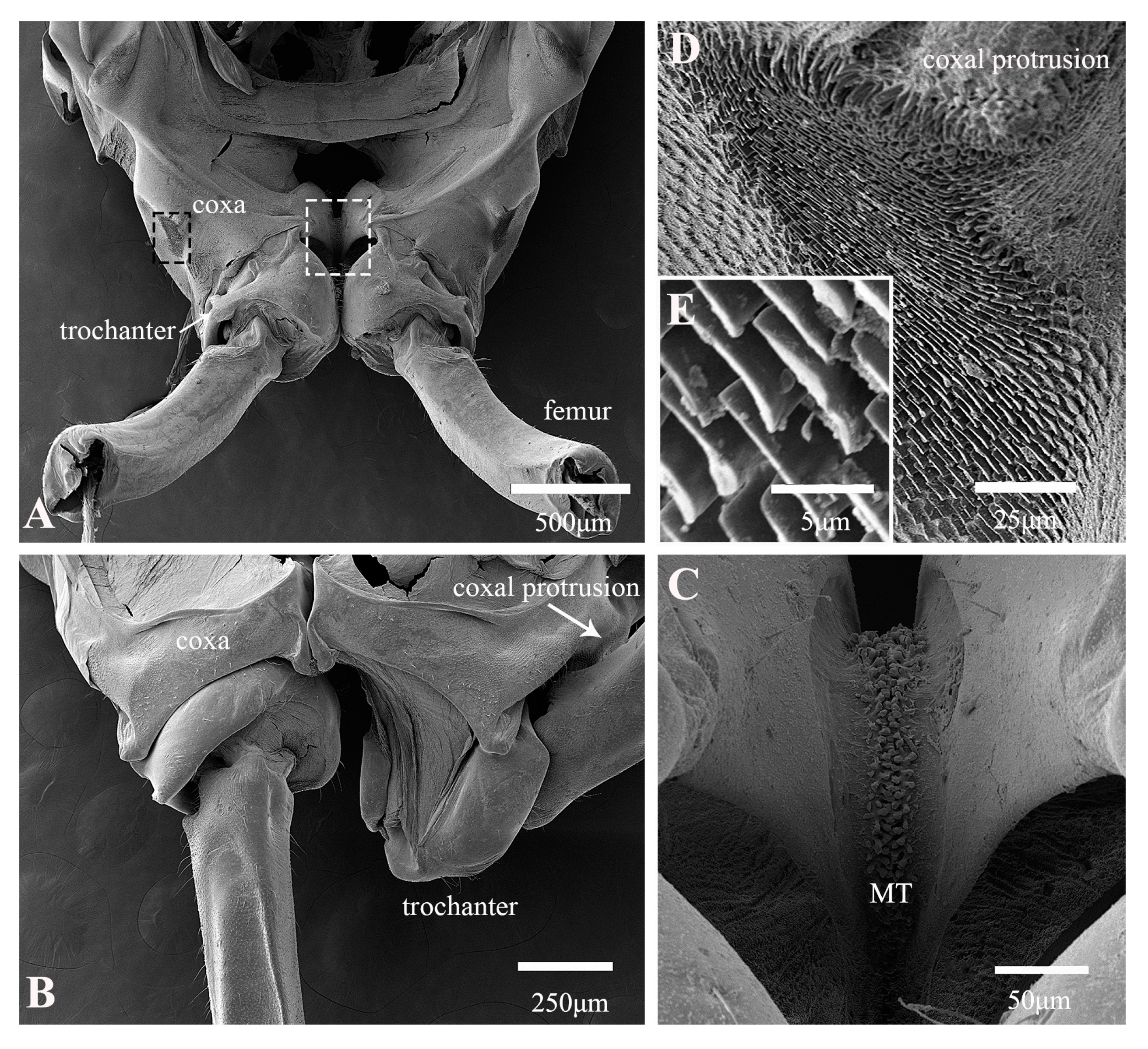
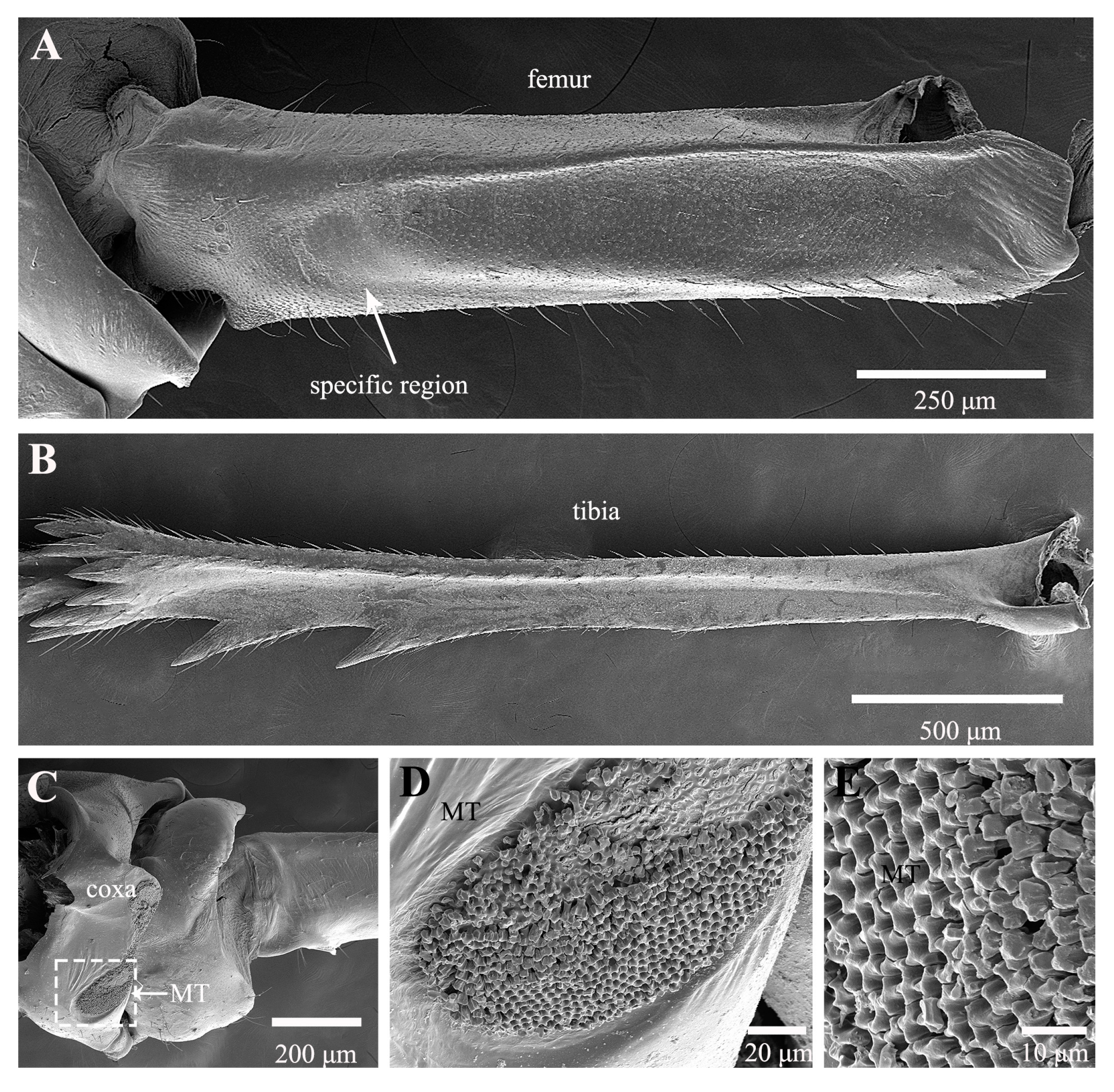
The forewings of ricaniid planthoppers are broadly triangular and are held tent-like over the body when at rest, forming a distinctive ridge along the dorsal body surface. In Euricania ocellus, the hind legs span an impressive 86% of the body length (Table 1). The tibia is only about 1.5× the length of the femur. The coxae of the two legs are closely adjacent, yet the trochanters possess the remarkable ability to rotate outward in preparation for a jump (Figure 3A,B). The rotation elevates the femora upward until they make contact with the coxal protrusions, which are densely covered with a lamellar array of microtrichia (Figure 3D,E). This critical juncture is not only where the hind leg can perform its lift and squeeze motion but also serves as the pivotal point for initiating a jump, contingent upon engagement with a specific region of the femur (Figure 4A). The medial surfaces of the coxae together form an hourglass-shaped structure (Figure 3C). Wavy structures project from the surface, intertwining in an irregular and complex manner and, thus, increasing the area of contact (Figure 4C–E). This unique architecture helps synchronize the movement of the hind legs to a certain degree (Figure 3C). It ensures the stability of the jump and is instrumental in storing a portion of the energy required. The tibiae are slender and robust, equipped laterally with two large spines, and also have a cluster of multiple spines at their junction with the tarsi (Figure 4B).
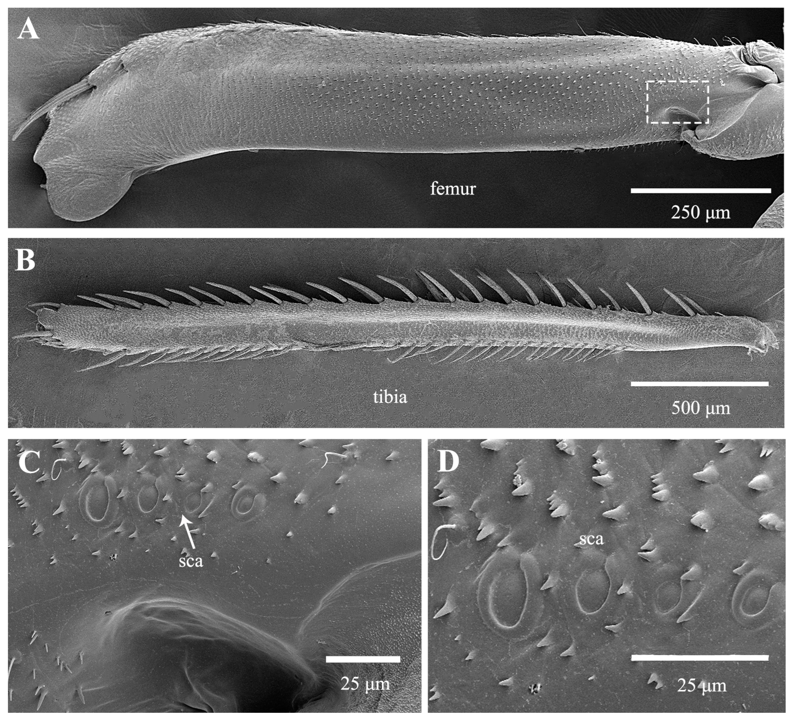
The leafhopper Kolla sp., possesses a uniform and smooth silhouette, marked by a streamlined form that presumably optimizes its aerodynamic efficiency. Although lighter in body mass compared to the studied froghopper, planthopper and treehopper, the hind legs are exceptionally long, constituting 88% of total body length (Table 1). The coxae have a dorsal depression that accommodates the femur just before a jump (Figure 5A). Within this depression lies a cluster of long hair plates, while a few shorter, thicker setae are situated on the side of the protrusion, adjacent to the hair plates (Figure 5C). Prior to jumping, the femora are lifted forward until they rest in these depressions, where they are squeezed against the hair plates and rows. The coxae of the two hind legs are intimately connected, and when pried open along the midline, they are seen to be linked by a protrusion from the medial wall of one coxa that fits into a socket of the other coxa (Figure 5B,D–H). This structure, in both shape and function, resembles a rivet. Two arrays of microtrichia adjacent to the protrusion and socket naturally engage with each other when the coxae are interlocked (Figure 5H). The femora are elongated and gradually taper from their junction with the tibia to the trochanters (Figure 6A). A row of four sensilla campaniformia is located near the trochanters (Figure 6C,D). The longitudinal rows of spine-like macrosetae on the tibia are the most distinctive features of leafhoppers (Figure 6B). Only a few relatively small spines are present at the apex of the tibia near its junction with the tarsi.
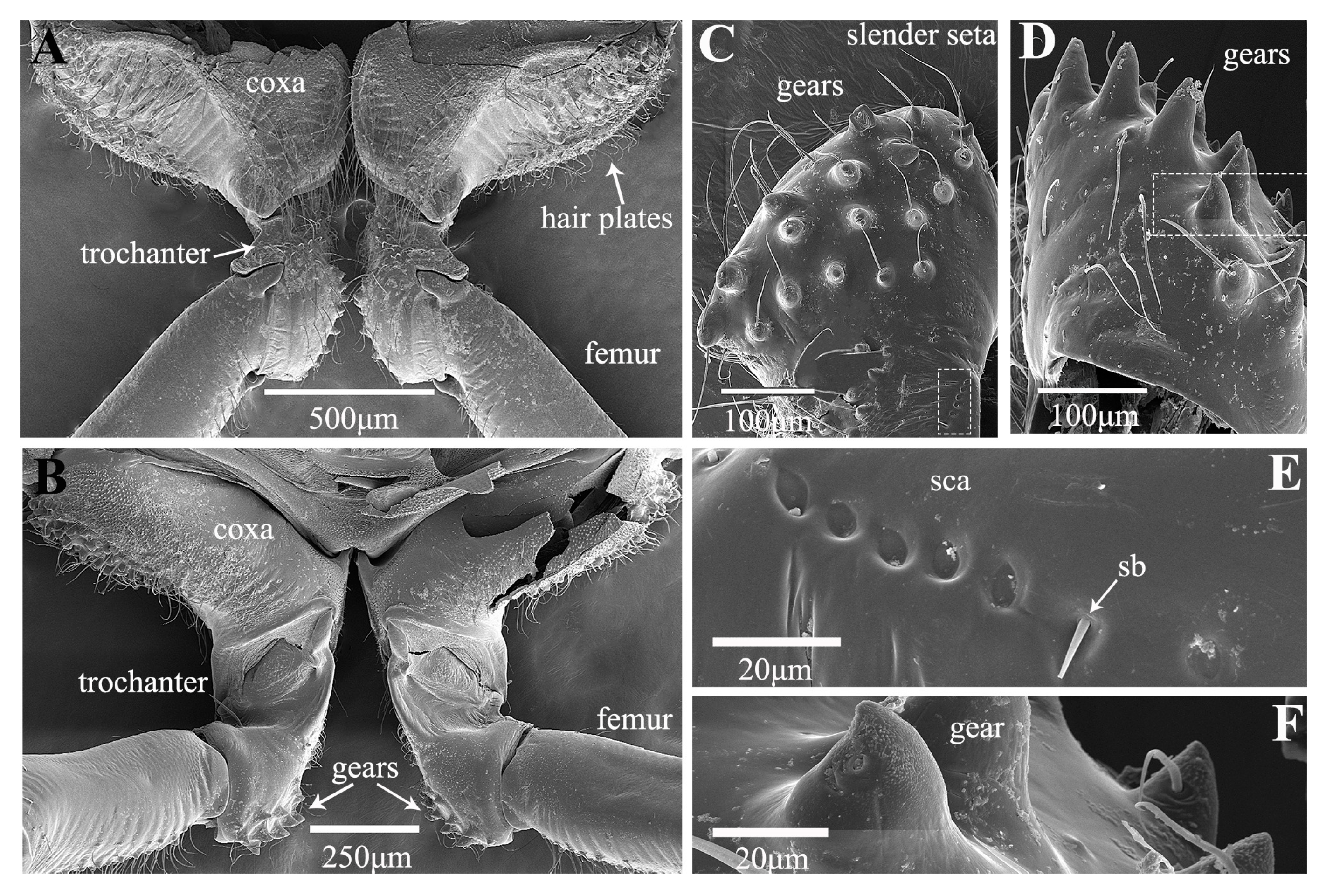
Treehoppers are noteworthy for their enlarged and often highly modified pronotum, but their hind leg structure is similar to that of leafhoppers. In Tricentrus sp., the hind legs are notably long, constituting 85% of the body length (Table 1). When viewed dorsally, the coxae resemble those of the leafhopper, featuring a concave surface equipped with hair plates (Figure 7A,B). The trochanters of Tricentrus sp. possess an array of gears with curved tips that point toward the coxa. These gears each bear a slender apical seta (Figure 7C,D,F). Adjacent to the gears, a row of six sensilla campaniformia (Sca) and a sensillum basiconicum (Sb) are present (Figure 7E), serving as crucial mechanical sensors that monitor leg movements. The ventral aspect of the femur is smooth and has only a few fine setae (Figure 8A). The distal end connecting with the tibia is adorned with several cucullate setae (Figure 8A,E). The tibia is slender, with longitudinal rows of cucullate setae, and terminates in an enlarged end equipped with a transverse row of spurs (Figure 8B–E).

3.2. Kinematics of the Jump

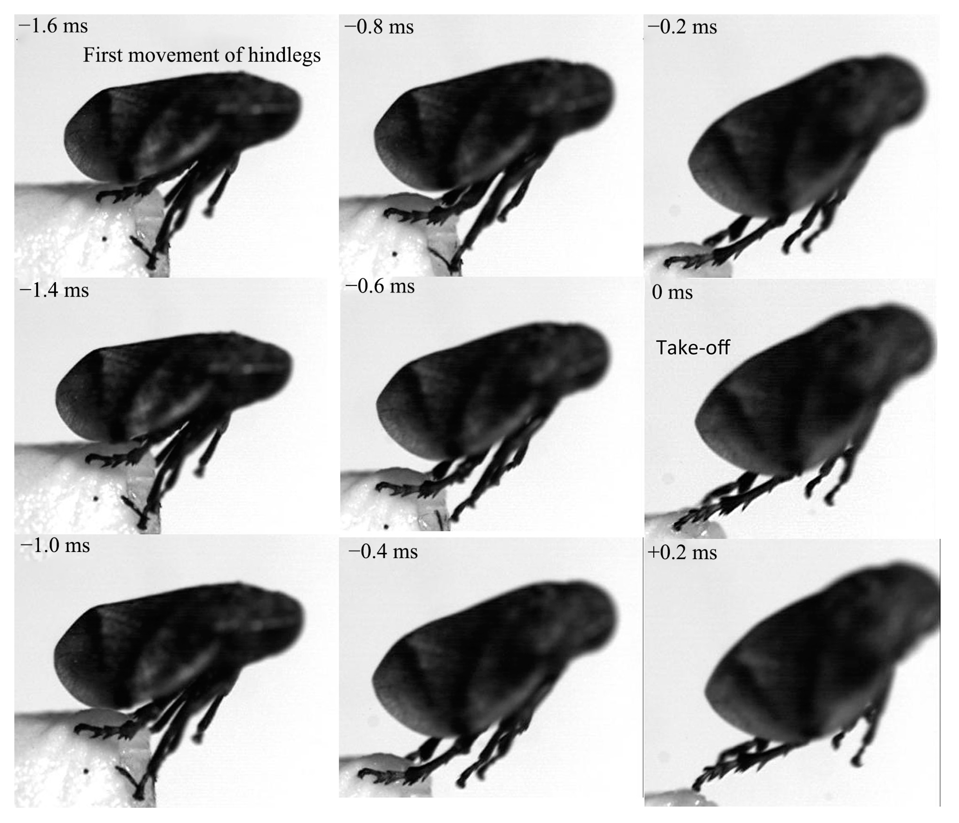
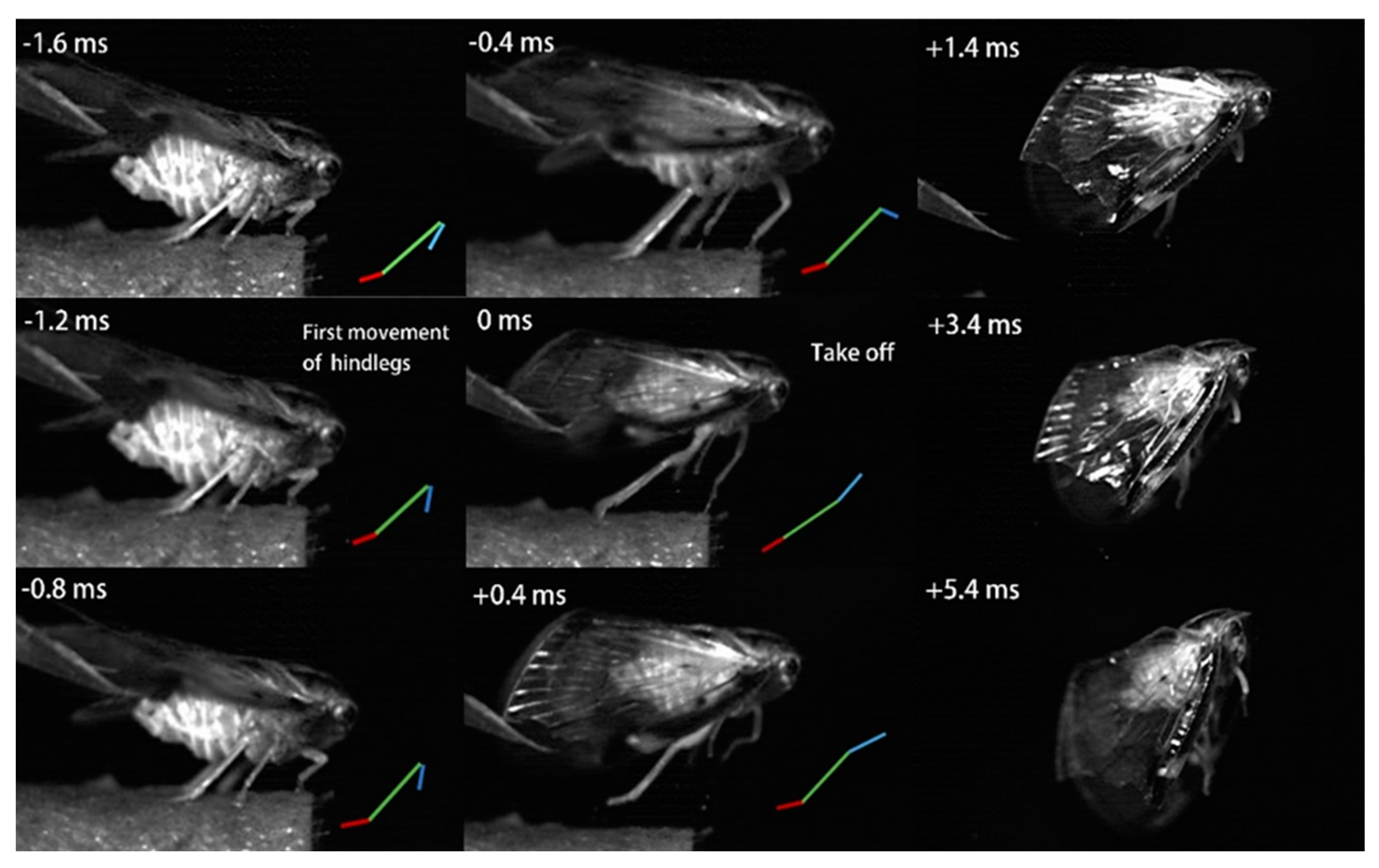
Jumping movements of the four species were analyzed using high-speed video recordings captured from a lateral view (Figure 9, Figure 10, Figure 11, Figure 12, Figure 13 and Figure 14). The side views of insects jumping in the horizontal plane provided detailed insights into the movement of the different legs, particularly the initial movement of the hind leg and the moment at which contact with the surface was lost (Figure 9, Figure 10, Figure 11 and Figure 13). A ventral view of the jumping process of the leafhopper clearly illustrated the motion of each hind leg segment and the synergy in movement between the two hind legs (Figure 12).
The initial movement of the hind legs in the jumping preparation phase involves pressing down on the body and simultaneously rotating the coxa–trochanteral joints to lift. This action facilitates the forward rotation of the trochanters and the extrusion of the femora towards the coxae to make contact with the coxal protrusion, which is crucial for triggering the jump. As the femora are rotated forward, the tibiae follow suit, positioning themselves alongside the body with the tarsi in contact with the surface (Figure 9, Figure 10, Figure 11 and Figure 13). This preparatory motion is usually sustained for a brief period, allowing for adjustment in the positions of the front and middle legs and elevating the body angle relative to the substrate. The take-off angle (relative to horizontal) varied as follows: 37° in Lepyronia coleoptrata, 48° in Euricania ocellus, 30° in Kolla sp. and 29° in Tricentrus sp. (Table 2).
Following the preparation period, a swift downward press and extension of the hind legs marks the initiation of the jump, propelling the body forward in a rapid take-off. The initial phase of this backward movement is identifiable by the depression of the coxa–trochanteral joint, best observed from the ventral view (Figure 11). When viewed from the side, the coxa–trochanteral joint is partially obscured by the wing; however, the first movement discernible is of the femur, connected to the tibia, moving downward and backward, which causes the tarsus to maintain contact with the ground. This coordinated motion quickly thrusts the body forward and upward, lifting it off the surface. The middle leg is the first to lose contact with the surface, followed by the front leg. In the jump’s final stage, only the pretarsus of the hind leg remains in contact with the surface, providing the last burst of propulsion (Figure 11). Throughout the jump, the body pushes forward, its speed increasing. At the moment of take-off, when the hind leg leaves the ground, the body’s speed peaks. Once airborne, the speed gradually decreases due to air resistance. At or immediately after take-off, the tarsi of the two hind legs converge at the body’s midline and, in some instances, cross each other. During the initial flight trajectory of the jump, the hind legs remain fully extended downward (Figure 12).

3.3. Motion Physics

Although the four species studied had similar hind leg movements during the preparatory phase for take-off, their trajectories after take-off varied significantly. All four species are comparable in body length, but Lepyronia coleoptrata is the heaviest, with a mass five times that of Kolla sp. This substantial body weight somewhat hinders its jumping capability, resulting in a maximum velocity of 2 m s 1 (Table 2). Lepyronia coleoptrata tends to tilt and rotate to one side upon take-off, which impedes smooth flight and often impedes a smooth landing. In contrast, Euricania ocellus, which has a similarly broad thorax, takes less time (1.2 ms) in the preparatory stage before jumping and can achieve a maximum speed of 2.8 m s 1 (Table 2). Its aerial trajectory is smoother, with greater vertical acceleration. Kolla sp., the lightest of the species studied, has the longest jumping preparation phase (t = 4 ms) and can reach the highest take-off speed (v = 3.9 m s 1 ) (Table 2). Its jumping trajectory closely resembles an ideal smooth parabolic arc. Tricentrus sp., the smallest of the four species, shares this superior jumping ability, but its jumping performance is erratic, with the body often deflecting to one side and tumbling. The energy expenditure of the four species’ jumps ranged from 24 to 45 μJ, with a power output of 16 to 38 mW and exerted forces between 3 and 27 mN.
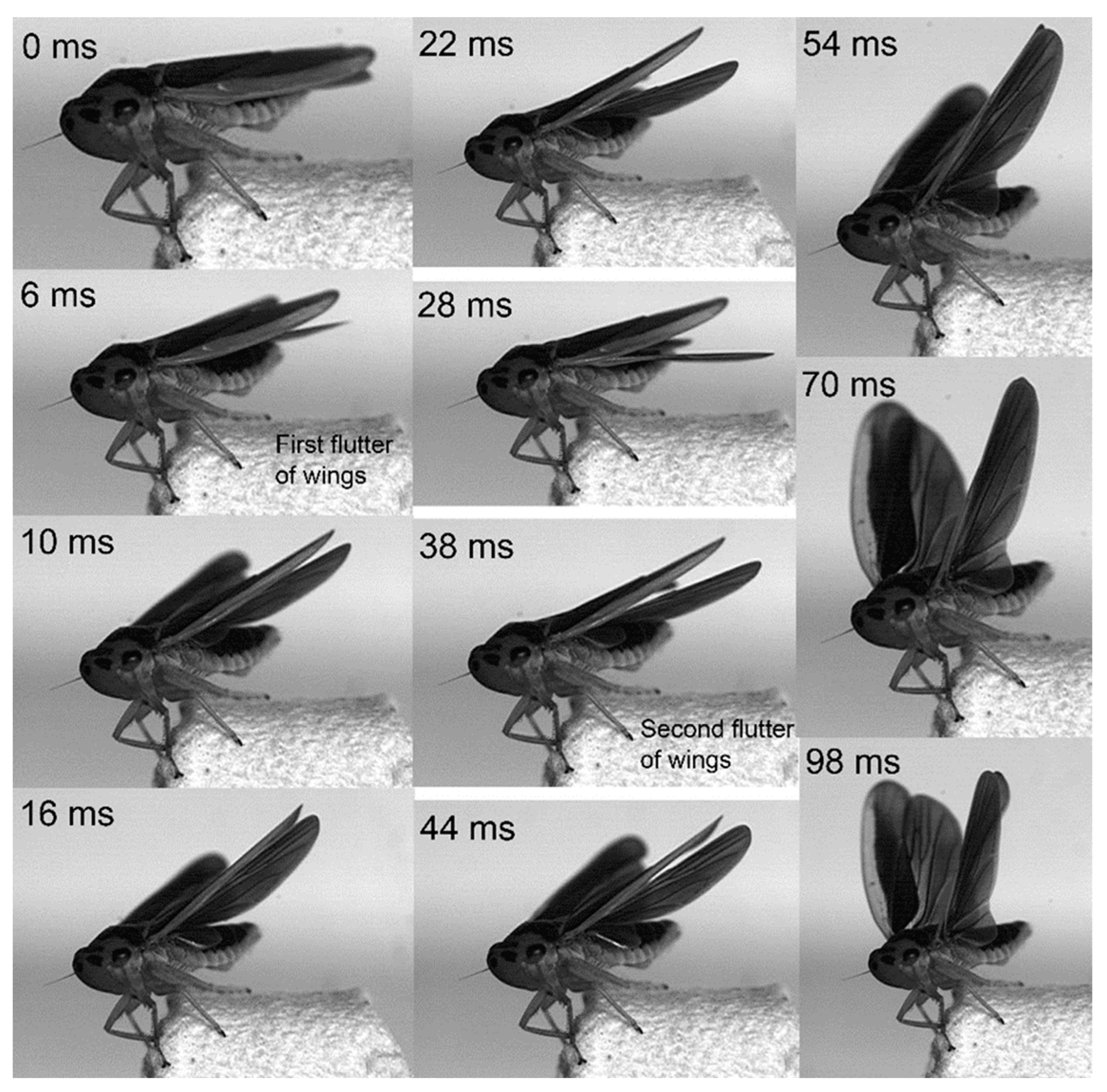
In the jumps previously described, the wings remained folded, indicating that they did not contribute to jumping performance. However, in certain instances, the wings were extended and flapped prior to take-off, facilitating a smooth transition into sustained winged flight (Figure 14). It is noteworthy that, in cases when the wings were extended prior to a jump, the timing of take-off, marked by the moment the hind legs left the surface, varied depending on the phase (ascent and descent) of the wing flapping cycle. In the above examples, neither Lepyronia coleoptrata nor Euricania ocellus exhibited wing flapping during preparation for jumping, at the moment of take-off, or after take-off. In contrast, Kolla sp. was observed to flap its wings before take-off. However, in some other taxa, wing flapping may occur during the preparation phase or while ascending.
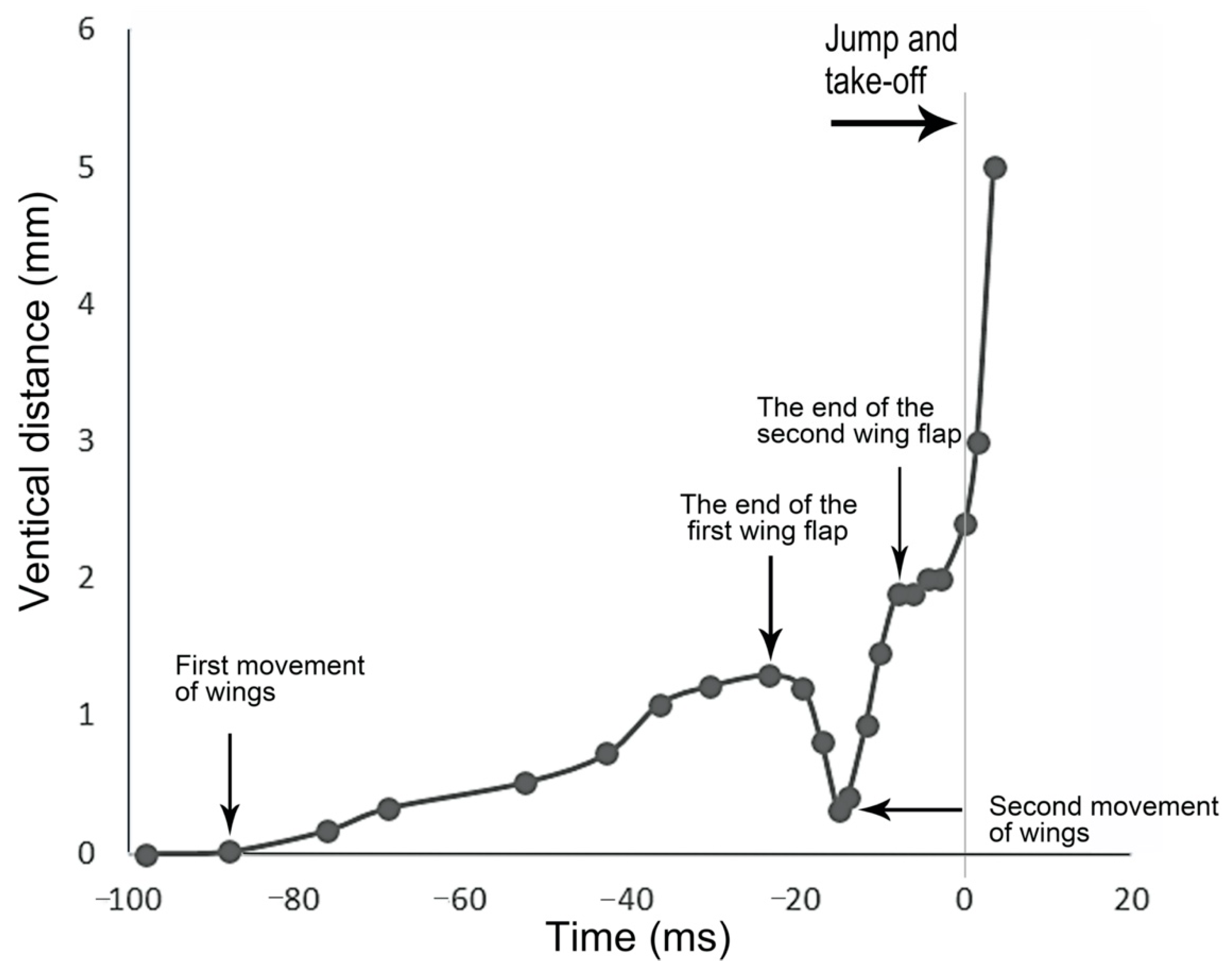
Wing flapping during take-off occurred over more than 100 ms, with the wings elevated and held in this position for some time, then accompanied by the extension of the hind legs to propel the body forward. This flapping action constituted two distinct cycles, each contributing to the insect’s ascent to a certain height. In the initial cycle, the wings were elevated for an extended period but did not immediately reach their zenith. Subsequently, the wings were swiftly retracted. The second cycle involved a rapid lift of the wings to their highest point, which was maintained for a brief interval before take-off. During this second cycle, the wings were held at their maximum height, creating a period in which any vertical movement was attributable to the hind legs performing the preparatory phase for lift-off (Figure 15).

4. Discussion

The present study confirms that Cercopoidea, Fulgoridae, Membracidae and Cicadellidae, despite sharing a common set of jumping mechanisms, have each evolved distinct strategies to enhance their leaping capabilities. Cercopoidea utilize a robust catapult mechanism known as PADT (pleural arch + depressor trochanter), which includes a mechanical locking mechanism, to generate the necessary propulsion force for their jump [25]. Fulgoridae, on the other hand, rely on their hind legs in conjunction with an elastic amplifier to interact with the surface, facilitating the completion of their jump [26]. Membracidae are believed to employ a power amplification mechanism that operates in a catapult-like fashion [27]. Cicadellidae, with proportionately longer hind legs, require a longer take-off time. Despite this, they exhibit remarkable jumping performance, facilitated by the storage of energy during the preparatory phase. Although Cicadellidae exhibit stronger jumping performance compared to Cercopoidea and Fulgoridae, their take-off time is longer, which somewhat reduces their jumping efficiency.
All four studied insects depend on their long, slender hind legs to execute their jumps, with the femur and tibia accounting for the majority of the hind leg’s length. In Lepyronia coleoptrata, three distinct arrays of setae on the coxa and trochanter are strategically positioned to serve as proprioceptors, signaling key movements during the jumping process. Prior to a jump, the femur presses to the coxa, making contact with a bulbous coxal protrusion that acts as a trigger for the jump. Surrounding this area, spike-like microtrichia form an oval, domed-shaped structure [28]. A substantial field of microtrichia (MT) on the hind coxae increases the contact area, ensuring synergy and a certain interlocking effect that stores energy prior to the jump [29]. Euricania ocellus, unlike Lepyronia coleoptrata, possesses a coxal protrusion covered with interlaced lamellar microtrichia that interact with the smooth end of the femur, also serving as a trigger for the jump [15,16]. Additionally, the coxae of the hind legs have a field of hourglass-shaped microtrichia on the mesal surfaces, which interlock more effectively than the toothlike microtricha of Lepyronia coleoptrata. Kolla sp. lacks a coxal protrusion but has hair plates and rows within the coxa’s depression that make contact with the femur. Notably, besides the two fields of pillar-shaped microtrichia on the interior surfaces of the hind coxae, Kolla sp. also possesses a pair of interlocking structures on the coxae resembling rivets in shape and function [21,30]. The coxa of Tricentrus sp. is similar to that of Kolla sp., with a concave surface that contacts the femur. However, unlike the other three species, Tricentrus sp. is distinguished by the presence of gears on the trochanters, marking it as the sole species among the four with this feature.
The jumping mechanisms of the insects studied here are similar to those of other previously studied small Auchenorrhyncha species [19,30,31]. Despite the similar body lengths of the four studied species, differences in the hind leg length itself do not significantly explain differences in jumping ability [6]. The increased contact area of the coxal microtrichia of Lepyronia coleoptrata resulted in less effective synergism between the hind legs compared to the intermeshed microtrichia of Euricania ocellus and Kolla sp. This difference may explain why Lepyronia coleoptrata tends to lean during take-off and struggles to maintain stable flight. In contrast, the meshing microtrichia in Euricania ocellus ensure synchronized hind leg movement during take-off and stability in gliding. Kolla sp., with its intermeshing microtrichia and a pair of rivet-like structures on the coxae, has a unique jumping mechanism. Among the four insects, Tricentrus sp. is notable for its opposable gears on the trochanters. However, these gears apparently do not contribute to hind leg synergy, leading to a tendency for Tricentrus sp. to tilt to one side during jumping. In the studied insects, the wings did not contribute to the energy required for jumping either at take-off or during the gliding phase. However, in Kolla sp., wing extension occurred 100 ms before take-off, with repeated wing beats culminating in an elevated position during take-off, although no wing flapping occurred during gliding. Leafhoppers’ preparatory movements for take-off include swaying their bodies from side to side, possibly allowing both hind legs to build up energy and adjust the jump direction. The irregular and unstable jumping of Auchenorrhyncha provides good adaptability and survival activity to avoid adverse environmental factors, such as natural enemies.
While direct observation of the distance and vertical height of jumps was not feasible in the studied insects. The use of a high-speed camera at a specific angle allowed for the tracking of individual leg movements, but using this method, it was not possible to capture the complete trajectory of the insects’ jumps. To overcome this limitation, standard equations for the motion of an inert body were employed [32] (Equations (1) and (2) below) to estimate the horizontal distance and vertical height achieved during the jump. These calculations were made without considering the contribution of the wings and under the assumption of negligible aerodynamic drag on the body:
s = v cos θ ( 2 v sin θ / 9.81 ) ,
h = ( v sin θ ) 2 / ( 2 × 9.81 ) ,
where s is the distance jumped, h is the maximum height reached, v is the instantaneous velocity at take-off, θ is the take-off angle (Table 2) and g is the acceleration due to gravity (9.81 m s 2 ).
The jumping motion of insects is not a standard curvilinear trajectory. The horizontal distances and vertical heights we calculated are theoretical estimates based on the simplification of the insect as a particle and the assumption of negligible aerodynamic drag [33,34]. These calculations suggest that these insects can cover remarkable distances, given their small body sizes (5.2 to 7.6 mm): Kolla sp. can leap to over 1.3 m, Euricania ocellus to nearly 0.8 m, Lepyronia coleoptrata to about 0.4 m and Tricentrus sp. to almost 0.5 m (Table 3). Vogel’s research suggested that the actual jumping range can be significantly reduced due to drag; for example, up to 25% in the froghopper Philaenus spumarius [34]. Smaller insects, such as flea beetles, may lose up to 40% of their jumping capabilities, and even smaller fleas (Siphonaptera) could lose as much as 80% of their jumping capabilities due to drag forces [32]. Despite these potential losses, the rapid acceleration, take-off velocity, and the projected distance of the upward and forward jumps of the studied species rank these insects among the most proficient leapers on par with the previously studied spittlebug, Philaenus spumarius [8].

5. Conclusions

In summary, the present results highlight that while the jumping principle remained consistent across the four insect families studied, the ultrastructural variations in their hind legs resulted in significant differences in the jumping mechanisms. Specifically, a higher degree of intermeshing in the coxa–trochanter correlated with increased jumping stability. Furthermore, the meshing degree of the coxa was superior to that of the trochanter.

Author Contributions

Y.X. and W.D. conceived the concept, designed the study, performed the experiments, analyzed most of the data and performed writing—original draft preparation; C.H.D. analyzed the morphological data and revised the manuscript. All authors have read and agreed to the published version of the manuscript.

Funding

This research was funded by the National Natural Science Foundation of China (Nos. 32270497, 32070479).

Institutional Review Board Statement

The study did not involve human participants or interfere in the welfare of animals. The authors did not use endangered species or collect animals in protected areas. All applicable international, national, and/or institutional guidelines for the care and use of animals were followed.

Informed Consent Statement

Not applicable.

Data Availability Statement

Most data analyzed and modeled during this study are included in this published article. The rest are available from the corresponding author upon reasonable request.

Conflicts of Interest

The authors declare no conflicts of interest.

References

  1. Sutton, G.P.; Burrows, M. The mechanics of elevation control in locust jumping. J. Comp. Physiol. A 2008, 194, 557–563. [Google Scholar] [CrossRef]
  2. Sutton, G.P.; Burrows, M. The mechanics of azimuth control in jumping by froghopper insects. J. Exp. Biol. 2010, 213, 1406–1416. [Google Scholar] [CrossRef]
  3. Burrows, M.; Picker, M.D. Jumping mechanisms and performance of pygmy mole crickets (Orthoptera, Tridactylidae). J. Exp. Biol. 2010, 213, 2386–2398. [Google Scholar] [CrossRef] [PubMed]
  4. Bennet-Clark, H.C. The energetics of the jump of the locust Schistocerca gregaria. J. Exp. Biol. 1975, 63, 53–83. [Google Scholar] [CrossRef] [PubMed]
  5. Burrows, M.; Ghosh, A.; Sutton, G.P.; Yeshwanth, H.M.; Rogers, S.M.; Sane, S.P. Jumping in lantern bugs (Hemiptera, Fulgoridae). J. Exp. Biol. 2021, 224, jeb243361. [Google Scholar] [CrossRef] [PubMed]
  6. Bennet-Clark, H.C.; Lucey, E.C.A. The jump of the flea: A study of the energetics and a model of the mechanism. J. Exp. Biol. 1967, 47, 59–76. [Google Scholar] [CrossRef]
  7. Burrows, M.; Morris, O. Jumping and kicking in bush crickets. J. Exp. Biol. 2003, 206, 1035–1049. [Google Scholar] [CrossRef]
  8. Burrows, M. Froghopper insects leap to new heights. Nature 2003, 424, 509. [Google Scholar] [CrossRef]
  9. Sutton, G.P.; Mendoza, E.; Azizi, E.; Longo, S.J.; Olberding, J.P.; Ilton, M.; Patek, S.N. Why do large animals never actuate their jumps with latchmediated springs? Because they can jump higher without them. Integr. Comp. Biol. 2019, 59, 1609–1618. [Google Scholar] [CrossRef]
  10. Nadein, K.; Betz, O. Jumping mechanisms and performance in beetles. I. Flea beetles (Coleoptera: Chrysomelidae: Alticini). J. Exp. Biol. 2016, 219, 2015–2027. [Google Scholar] [CrossRef]
  11. Ilton, M.; Bhamla, M.S.; Ma, X.; Cox, S.M.; Fitchett, L.L.; Kim, Y.; Koh, J.S.; Krishnamurthy, D.; Kuo, C.Y.; Temel, F.Z.; et al. The principles of cascading power limits in small, fast biological and engineered systems. Science 2018, 360, eaao1082. [Google Scholar] [CrossRef] [PubMed]
  12. Longo, S.J.; Cox, S.M.; Azizi, E.; Ilton, M.; Olberding, J.P.; St Pierre, R.; Patek, S.N. Beyond power amplification: Latch-mediated spring actuation is an emerging framework for the study of diverse elastic systems. J. Exp. Biol. 2019, 222, jeb197889. [Google Scholar] [CrossRef] [PubMed]
  13. Askew, G.N.; Marsh, R.L. Muscle designed for maximum short-term power output: Quail flight muscle. J. Exp. Biol. 2002, 205, 2153–2160. [Google Scholar] [CrossRef]
  14. Josephson, R.K. Contraction dynamics and power output of skeletal muscle. Annu. Rev. Physiol. 1993, 55, 527–546. [Google Scholar] [CrossRef]
  15. Burrows, M. Jumping performance of planthoppers (Hemiptera, Issidae). J. Exp. Biol. 2009, 212, 2844–2855. [Google Scholar] [CrossRef]
  16. Burrows, M. Jumping strategies and performance in shore bugs (Hemiptera, Heteroptera, Saldidae). J. Exp. Biol. 2009, 212, 106–115. [Google Scholar] [CrossRef]
  17. Burrows, M. Neural control and co-ordination of jumping in froghopper insects. J. Neurophysiol. 2007, 97, 320–330. [Google Scholar] [CrossRef] [PubMed]
  18. Burrows, M. Jumping mechanisms in flatid planthoppers (Hemiptera, Flatidae). J. Exp. Biol. 2014, 217, 2590–2600. [Google Scholar] [CrossRef]
  19. Burrows, M.; Bräunig, P. Actions of motor neurons and leg muscles in jumping by planthopper insects (Hemiptera, Issidae). J. Comp. Neurol. 2010, 518, 1349–1369. [Google Scholar] [CrossRef]
  20. Siwanowicz, I.; Burrows, M. Three dimensional reconstruction of energy stores for jumping in planthoppers and froghoppers from confocal laser scanning microscopy. eLife 2017, 6, e23824. [Google Scholar] [CrossRef]
  21. Burrows, M. Anatomy of the hind legs and actions of their muscles during jumping in leafhopper insects. J. Exp. Biol. 2007, 210, 3590–3600. [Google Scholar] [CrossRef] [PubMed]
  22. van de Kamp, T.; Vagovič, P.; Baumbach, T.; Riedel, A. A biological screw in a beetle’s leg. Science 2011, 333, 52. [Google Scholar] [CrossRef]
  23. Burrows, M.; Sutton, G. Interacting gears synchronize propulsive leg movements in a jumping insect. Science 2013, 341, 1254–1256. [Google Scholar] [CrossRef]
  24. Jung, G.P.; Cho, K.J. Froghopper-inspired direction-changing concept for miniature jumping robots. Bioinspir. Biomim. 2016, 11, 056015. [Google Scholar] [CrossRef]
  25. Burrows, M.; Shaw, S.R.; Sutton, G.P. Resilin and chitinous cuticle form a composite structure for energy storage in jumping by froghopper insects. BMC Biol. 2008, 6, 41. [Google Scholar] [CrossRef] [PubMed]
  26. Burrows, M. Jumping mechanisms in dictyopharid planthoppers (Hemiptera, Dicytyopharidae). J. Exp. Biol. 2014, 217, 402–413. [Google Scholar] [CrossRef] [PubMed]
  27. Burrows, M. Jumping mechanisms of treehopper insects (Hemiptera, Auchenorrhyncha, Membracidae). J. Exp. Biol. 2013, 216, 788–799. [Google Scholar] [CrossRef]
  28. Burrows, M. Morphology and action of the hind leg joints controlling jumping in froghopper insects. J. Exp. Biol. 2006, 209, 4622–4637. [Google Scholar] [CrossRef]
  29. Gorb, S.N. The jumping mechanism of cicada Cercopis vulnerata (Auchenorrhyncha, Cercopidae): Skeleton–muscle organisation, frictional surfaces, and inverse-kinematic model of leg movements. Arthropod Struct. Dev. 2004, 33, 201–220. [Google Scholar] [CrossRef]
  30. Gorb, S.N. Attachment Devices of Insect Cuticle; Kluwer Academic Publishers: Dordrecht, The Netherlands, 2001; pp. 1–305. [Google Scholar] [CrossRef]
  31. Burrows, M. Kinematics of jumping in leafhopper insects (Hemiptera, Auchenorrhyncha, Cicadellidae). J. Exp. Biol. 2007, 210, 3579–3589. [Google Scholar] [CrossRef]
  32. Alexander, R.M. Animal Mechanics; Blackwell Science: Oxford, UK, 1983; pp. 1–312. [Google Scholar]
  33. Bennet-Clark, H.C.; Alder, G.M. The effect of air resistance on the jumping performance of insects. J. Exp. Biol. 1979, 82, 105–121. [Google Scholar] [CrossRef] [PubMed]
  34. Vogel, S. Living in a physical world II. The bio-ballistics of small projectiles. J. Biosci. 2005, 30, 167–175. [Google Scholar] [CrossRef] [PubMed]
Figure 1. SEM of the proximal hind leg joints of Lepyronia coleoptrata: (A) dorsal view of coxae and trochanters; (B) enlarged view of (A); (C) setae on coxo–trochanteral joints; (D) ventral view of coxae and trochanters; (E) enlarged view of white outlined box in (D), showing coxal protrusion in contact with the specific location on the femur; (F) coxal protrusion; (G) medial view of coxa and trochanter; (H) enlarged view of (G), showing Microtrichia (MT); (I,J) enlarged view of Microtrichia (MT).
Biology 14 00418 g001
Figure 2. SEM of femur and tibia of Lepyronia coleoptrata: (A) depression on ventral surface of femur; (B) enlarged view of white outlined box in (A), showing contact area with coxal protrusion; (C) enlarged view of white outlined box in (B), microtrichia on the femur protrusion; (D) dorsal view of femur; (E) whole tibia showing spines.
Biology 14 00418 g002
Figure 3. SEM of the proximal hind leg joints of Euricania ocellus: (A) dorsal view of coxae and trochanters; (B) ventral view of coxae and trochanters; (C) enlarged view of white outlined box in (A), showing interlocked microtrichia (MF) fields of coxae; (D) enlarged view of black outlined box in (A), showing field of lamellar microtrichia; (E) enlarged view of lamellar microtrichia.
Biology 14 00418 g003
Figure 4. SEM of hind femur and tibia of Euricania ocellus: (A) ventral view of femur showing sunken part contacted and depressed with coxal protrusion; (B) tibia showing lateral spines; (C) medial aspect of the coxa with the microtrichia field; (D) enlarged view of white outlined box in (C), showing the microtrichia field covered in wavy microtrichia; (E) enlarged view of microtrichia.
Biology 14 00418 g004
Figure 5. SEM of the proximal hind leg joints of Kolla sp.: (A) dorsal view of coxae and trochanters; (B) ventral view of coxae and trochanters; (C) enlarged view of white outlined box in (A), showing hair plate and hair row; (D) enlarged view of white outlined box in (B), showing interlocked microtrichia fields of coxae; (E) medial aspect of the coxa, showing protrusion; (F) medial aspect of the coxa, showing socket; (G) enlarged view of the coxa from medial view, showing protrusion; (H) enlarged view of the coxa from medial view, showing socket and microtrichia.
Biology 14 00418 g005
Figure 6. SEM of femur and tibia of Kolla sp.: (A) dorsal view of femur; (B) spines of the tibia; (C) enlarged view of white outlined box in (A), showing sensilla campaniformia (Sca); (D) enlarged view of sensilla campaniformia (Sca).
Biology 14 00418 g006
Figure 7. SEM of the proximal hind leg joints of Tricentrus sp.: (A) dorsal view of coxae and trochanters, showing hair plates; (B) ventral view of coxae and trochanters, showing gears; (C) medial view of gears, showing slender setae; (D) lateral view of gears; (E) enlarged view of white outlined box in (C), showing sensilla campaniformia (Sca) and sensilla basiconica (Sb); (F) enlarged view of gear.
Biology 14 00418 g007
Figure 8. SEM of femur and tibia of Tricentrus sp.: (A) ventral view of femur; (B) tibia; (C) apex of tibia; (D) enlarged view of outlined box in (C), showing spurs; (E) cucullate seta.
Biology 14 00418 g008
Figure 9. Side view of Lepyronia coleoptrata jumping from a horizontal surface, captured at 10,000 images s 1 and exposure time of 1 μs, take-off was designated as t = 0 ms.
Biology 14 00418 g009
Figure 10. Side view of Euricania ocellus jumping from a horizontal surface, captured at 10,000 images s 1 and exposure time of 1 μs, take-off was designated as t = 0 ms. Positions of the different segments of the right hind leg are indicated by color-coded lines: femur in blue, tibia in green and tarsus in red.
Biology 14 00418 g010
Figure 11. Side view of Kolla sp. jumping from a horizontal surface, captured at 10,000 images s 1 and exposure time of 1 μs. Take-off was achieved in 0 ms.
Biology 14 00418 g011
Figure 12. Ventral view of jumping motion in Kolla sp., captured at 10,000 images s 1 and exposure time of 1 μs. The positions of the different segments of the right and left hind legs are indicated by the color-coded lines: femur in red, tibia in blue and tarsus in green, with the midline body axis in black.
Biology 14 00418 g012
Figure 13. Side view of Tricentrus sp. jumping from a horizontal surface, captured at 10,000 images s 1 and exposure time of 1 μs. Take-off was achieved in 1.7 ms. The positions of the different segments of the right and left hindlegs are indicated by the color-coded lines: femur in red, tibia in blue and tarsus in green, with the midline body axis in black.
Biology 14 00418 g013
Figure 14. Side view of Kolla sp. flapping wings before jump, captured at 10,000 images s 1 and exposure time of 1 μs.
Biology 14 00418 g014
Figure 15. The hind leg and wing movements of Kolla sp. during the jump are shown in Figure 14. The vertical distance of the wing flapping action relative to the resting state was recorded for 100 ms before take-off, using the wing resting parallel on the back of the body as the baseline. The gray vertical line represents the moment of take-off.
Biology 14 00418 g015
Table 1. Body length and mass, and lengths of the hind leg femora and tibiae in four species.
Body Mass
(mg)
Body Length
(mm)
Femur
(mm)
Tibia
(mm)
Hind Leg Length (% Body Length)
Lepyronia coleoptrata
(N = 17)
18.6 ± 3.47.6 ± 0.31.3 ± 0.13.1 ± 0.184
Euricania ocellus
(N = 13)
11.8 ± 0.36.1 ± 0.051.5 ± 0.12.2 ± 0.186
Kolla sp.
(N = 14)
3.9 ± 0.46.7 ± 0.11.4 ± 0.052.4 ± 0.188
Tricentrus sp.
(N = 9)
8.25.20.72.485
N, the number of insects analyzed in this table.
Table 2. The jumping performance of the four species.
Body
Mass
(mg)
Time to
Take-Off
(ms)
Take-Off
Velocity
( m s 1 )
Take-Off
Angle
(deg)
Acceleration
( m s 2 )
g-Force
(g)
Energy
(μJ)
Power
(mW)
Force
(mN)
Formula/symbolm v f = v/tg = f/9.81e = 0.5 m v 2 P = e/tF = mf
Lepyronia coleoptrata 2
mean
(N = 17, n = 3)
18.6 ± 3.41.7 ± 0.137 ± 41118114331921
best19.11.6 1250127382324
Euricania ocellus 2.8
mean
(N = 13, n = 7)
11.8 ± 0.31.3 ± 0.148 ± 22077212433325
best11.61.2 2333238453827
Kolla sp. 3.9
mean
(N = 14, n = 2)
3.9 ± 0.44.2 ± 0.230 ± 6857872563
best4.14 97599.38104
Tricentrus sp.
mean
(N = 9, n = 1)
best8.21.72.4291412144241412
The values in the five columns on the right were calculated from the mean values given in the four columns on the left. The best performance of a particular individual for each species is also given. Only data for the jumps that were viewed from the side are presented here. N, the number of insects analyzed in this table; n, the number of jumps.
Table 3. The jumping distances and heights of the four species.
SpeciesTake-Off
Velocity
( m s 1 )
Take-Off
Angle
(deg)
Body Length
(mm)
Horizontal Distance (mm)Horizontal Distance (Body Lengths)Vertical Height
(mm)
Vertical Height
(Body Lengths)
Lepyronia coleoptrata2377.6392527410
Euricania ocellus2.8486.179513022136
Kolla sp.3.9306.71343200737110
Tricentrus sp.2.4295.2498969318
Disclaimer/Publisher’s Note: The statements, opinions and data contained in all publications are solely those of the individual author(s) and contributor(s) and not of MDPI and/or the editor(s). MDPI and/or the editor(s) disclaim responsibility for any injury to people or property resulting from any ideas, methods, instructions or products referred to in the content.