Review

Disrupted Endoplasmic Reticulum Ca2+ Handling: A Harβinger of β-Cell Failure

by
Jordyn R. Dobson
and
David A. Jacobson
*
Department of Molecular Physiology and Biophysics, Vanderbilt University, Nashville, TN 37232, USA
*
Author to whom correspondence should be addressed.
Biology 2024, 13(6), 379; https://doi.org/10.3390/biology13060379
Submission received: 3 April 2024 / Revised: 17 May 2024 / Accepted: 17 May 2024 / Published: 25 May 2024
(This article belongs to the Special Issue β-Cells at the Center of Type 1 and Type 2 Diabetes)

Abstract

:

Simple Summary

Diabetes results from insufficient insulin production and secretion. Insulin accounts for almost half of the protein produced in the pancreatic β-cell, which occurs in the endoplasmic reticulum (ER). Ca2+ is a key signal within the ER that controls insulin production and processing. β-cell ER Ca2+ (Ca2+ER) depletion during the pathogenesis of type 1 and type 2 diabetes leads to reduced insulin production and secretion. Mutations in insulin that cause monogenic diabetes also reduce Ca2+ER and result in β-cell dysfunction. Thus, the mechanisms that tune β-cell Ca2+ER and become disrupted in diabetes are potential targets for enhancing insulin production and reducing hyperglycemia. This review focuses on β-cell Ca2+ER and how it impacts β-cell function and dysfunction.

Abstract

The β-cell workload increases in the setting of insulin resistance and reduced β-cell mass, which occurs in type 2 and type 1 diabetes, respectively. The prolonged elevation of insulin production and secretion during the pathogenesis of diabetes results in β-cell ER stress. The depletion of β-cell Ca2+ER during ER stress activates the unfolded protein response, leading to β-cell dysfunction. Ca2+ER is involved in many pathways that are critical to β-cell function, such as protein processing, tuning organelle and cytosolic Ca2+ handling, and modulating lipid homeostasis. Mutations that promote β-cell ER stress and deplete Ca2+ER stores are associated with or cause diabetes (e.g., mutations in ryanodine receptors and insulin). Thus, improving β-cell Ca2+ER handling and reducing ER stress under diabetogenic conditions could preserve β-cell function and delay or prevent the onset of diabetes. This review focuses on how mechanisms that control β-cell Ca2+ER are perturbed during the pathogenesis of diabetes and contribute to β-cell failure.

1. Introduction

Disruption in the production and/or secretion of insulin contribute(s) to hyperglycemia during the pathogenesis of diabetes. The pancreatic β-cell Ca2+ serves critical roles not only in stimulating insulin secretion but also in endoplasmic reticulum (ER) production and the processing of insulin. Due to the constant production and high daily requirement of insulin, the ER of the β-cell has a finely tuned quality control system that is modulated by Ca2+. The total concentration of Ca2+ER is ~2 mM, with the majority buffered by Ca2+ER binding proteins such as chaperones. The free Ca2+ER ranges between 50 and 500 µM (compared to 0.1–2 µM in the cytosol), which sets up a large driving force for Ca2+ER release [1,2,3]. Stressful conditions associated with diabetes reduce Ca2+ER stores; this elevates ER stress and initiates the unfolded protein response (UPR), which initially protects β-cells from diabetogenic stress but also limits insulin production and secretion [4,5]. The prolonged disruption of Ca2+ER can eventually lead to β-cell failure and/or senescence [6]. Importantly, alterations in the levels and/or function of the proteins involved in controlling Ca2+ER homeostasis can lead to diabetic phenotypes due to β-cell dysfunction. Moreover, mutations that disrupt insulin folding and promote accumulation in the ER lead to reduced Ca2+ER storage, UPR, and β-cell failure [2,7,8]. Therefore, elevating Ca2+ER under diabetic conditions helps to restore β-cell function and glucose homeostasis [9,10]. This review focuses on how β-cell Ca2+ER becomes disrupted and contributes to diabetes pathogenesis by impairing insulin production, secretion, and quality control (Figure 1).

1.1. Ca2+ER in β-Cell Survival and Health

The ER serves a critical role in β-cell pro-survival mechanisms for recovering from the stress associated with the high production of insulin. The necessity of Ca2+ER for β-cell survival is demonstrated by increased apoptosis following the prolonged inhibition of Ca2+ER uptake [11]. However, the activation of the adaptive UPR following the acute depletion of β-cell Ca2+ER stores enables protection from stress, which is critical to β-cell survival and longevity. This adaptive response includes the upregulation of IRE1, PERK, and ATF6 signaling, which limit translation, increase chaperone expression, and eliminate misfolded proteins [12]. The prolonged depletion of Ca2+ER stores leads to terminal UPR activation and subsequent pro-apoptotic signaling. This includes the degradation of ER-localized mRNAs by IRE1α, the increased expression of C/EBP homologous protein (CHOP), and the activation of caspase 2-mediated apoptosis [13]. Thus, Ca2+ER homeostasis serves critical roles in β-cell survival and adaptation to stress. Interestingly, β-cells from nondiabetic donors transition between different states of UPR activation, which are associated with alterations in insulin biosynthesis. Transient elevations in UPR likely allow for β-cell rest and recovery from stress associated with insulin production and secretion [14]. Therefore, the Ca2+ER control of UPR provides a pro-survival mechanism that allows for the β-cell to maintain health under stressful conditions, which can be recurrent due to high levels of insulin production and secretion.

1.2. Ca2+ER Control of Insulin Secretion

Insulin secretion is finely tuned by Ca2+ER uptake and release. Ca2+ER oscillates in phase with glucose-induced cytosolic Ca2+ (Ca2+C) oscillations, contributing to the regulation of Ca2+C and pulsatile insulin secretion [15]. Ca2+-induced Ca2+ release (CICR) from the ER amplifies insulin secretion. This occurs via the Ca2+ activation of ryanodine receptors (RyRs) as well as phospholipase C-mediated IP3 production and IP3R activation [16]. Ca2+ER handling not only tunes glucose-stimulated insulin secretion (GSIS) but also stimulates insulin secretion in response to Gq-G protein coupled receptor (Gq-GPCR) signaling via Ca2+ER release through IP3Rs [17]. Thus, alterations in Ca2+ER perturb insulin secretion. For example, the acute inhibition of Ca2+ER uptake increases glucose-stimulated Ca2+C and insulin secretion, whereas the sustained depletion of Ca2+ER stores upregulates the UPR, limiting insulin production and secretion. The machinery that controls Ca2+ER and how it modulates insulin secretion and contributes to β-cell dysfunction are discussed in detail below.

2. Ca2+ER Handling Proteins and Their β-Cell Function(s)

The ER contains specialized Ca2+ pumps, ion channels, and Ca2+ sensing/regulated proteins that control Ca2+ER sequestration, uptake, and release. Ca2+ER handling helps shape Ca2+C oscillations and pulsatile insulin secretion [15]. In addition, the release of Ca2+ER generates a negative charge buildup on the ER membrane. Therefore, ion channels and pumps serve to limit the generation of ER membrane potential, promote the driving force of Ca2+ER release, and provide a setpoint for Ca2+ER store homeostasis through balanced Ca2+ release and refilling. Dysfunction and/or changes in the expression of proteins that modulate Ca2+ER impair proper insulin production and secretion, which, when prolonged, induces β-cell failure. The primary mediators of β-cell Ca2+ER handling are detailed below.

2.1. Sarco/Endoplasmic Reticulum Calcium ATPase (SERCA)

Function: The uptake of Ca2+ into the ER occurs via SERCA, a P-type ATPase, which pumps 2 Ca2+ ions into the ER lumen and extrudes two to three protons per ATP hydrolyzed. While SERCA is critical for keeping Ca2+ER stores elevated, changes in its activity also help fine-tune Ca2+C handling (Figure 2). β-cell glucose metabolism leads to the ATP energization of SERCA and Ca2+ER uptake, which results in a drop in Ca2+C [18,19]. Interestingly, Ca2+ER oscillates in phase with glucose-induced Ca2+C oscillations [3]. Therefore, Ca2+ER uptake occurs during the upstroke of a Ca2+ oscillation and Ca2+ER release occurs during the termination [3,15,18]. This could be due, in part, to ATP oscillating out of phase with Ca2+ oscillations, thus energizing SERCA during each Ca2+ oscillation. Furthermore, elevated intraluminal Ca2+ER inhibits SERCA and reduces its activity, as Ca2+ER stores increase during the Ca2+ oscillation, which increases the relative contribution of Ca2+ER leak. This is supported by the pharmacological inhibition of SERCA, which causes each Ca2+ oscillation to have an accelerated rising phase, increased amplitude, and more rapid termination phase [20]. Ca2+ handling is also influenced by the different properties of SERCA2b (ATP2A2) and SERCA3a-c (ATP2A3) isoforms expressed in β-cells [10,21]. SERCA2b is the most abundant β-cell isoform and has a low affinity for Ca2+ (K Ca2+ = 0.2 µM), whereas SERCA3 has a lower expression (~50% compared to SERCA2b) and a higher affinity for Ca2+ (K Ca2+ = 1.2 µM) [22,23]. SERCA2b actively pumps Ca2+ into the ER lumen at basal and stimulatory glucose concentrations [18]. On the other hand, SERCA3 does not significantly alter basal Ca2+ER uptake due to its higher affinity for Ca2+ but contributes to Ca2+ER uptake during glucose-induced Ca2+ influx [18,24,25]. Taken together, small changes in SERCA activity play an important role in tuning β-cell Ca2+ handling. The maintenance of Ca2+ER storage by SERCA is also critical for protecting β-cells from stress; thus, prolonged SERCA inhibition leads to β-cell death.
SERCA expression and activity are also regulated by insulin signaling through insulin receptor substrate-1 and -2 proteins (IRS-1 and IRS-2). IRS-1 forms a complex with SERCA3 that can be enhanced with insulin [26]. The overexpression of IRS-1 in β-cells inhibits SERCA3, leading to reduced Ca2+ER storage and elevated Ca2+C [27,28]. In addition, IRS-1 and IRS-2 signaling enhances the expression of the genes encoding SERCA2 and SERCA3 (Atp2a2 and Atp2a3), which are reduced in IRS-1 or IRS-2 knockout mice [29,30]. IRS-1 knockout mice also show reduced glucose-induced Ca2+ influx and elevated Ca2+ER stores [29,30]. However, as SERCA inhibitors cause elevated glucose-induced Ca2+ influx and reduced Ca2+ER storage, the loss of SERCA abundance in IRS knockout models would not be expected to elevate Ca2+ER stores. Thus, the loss of IRS interaction and the inhibition of SERCA could potentially cause the observed elevation in Ca2+ER stores in islets from either IRS-1 or IRS-2 knockouts [29,30]. The insulin control of Ca2+ER through the IRS-mediated modulation of SERCA expression and activity could serve as a positive feedback circuit to enhance Ca2+C and insulin secretion [31,32,33].
SERCA activity is further regulated by post-translational modification such as O-GlcNAcylation. The O-GlcNAcylation of islet proteins including SERCA2 is increased under diet-induced hyperlipidemia [34]; this results in the potentiation of insulin secretion. Furthermore, the O-GlcNAcylation of SERCA2 increases its abundance and likely increases its function [34]. Importantly, defects in insulin secretion from O-linked N-acetylglycosamine transferase (OGT) ablation can be rescued by SERCA activation, which suggests that impaired SERCA2 activity may contribute to blunted Ca2+ influx in islets with OGT ablation [34,35]. The role of SERCA2 O-GlcNAcylation in Ca2+ER storage under physiological and diabetic conditions remains to be determined.
SERCA can also be activated post-translationally in response to glutathionylation. For most proteins that undergo glutathionylation, this increases in response to elevations in reactive oxygen and nitrogen species (ROS/RNS) [36]. Thus, elevations in the aortic smooth muscle NO activate SERCA2 via Cys674 glutathionylation and increase sarcoplasmic reticulum Ca2+ [37]. Despite elevations in β-cell ROS and nitric oxide (NO) during glucose and lipid metabolism, the glutathionylation of the β-cell proteome is decreased in response to glucose stimulation [38]. This corresponds with an increase in the level of reduced glutathione (GSH) to oxidized glutathione (GSSG). While the mechanism for the glucose reduction in glutathiolynation remains to be determined, the low β-cell antioxidative capacity and the reduction in subcellular ROS by glucose have been proposed [38,39,40]. The impact of the glucose control of β-cell SERCA activity through reversible glutathionylation remains to be determined.
Dysfunction: Given the critical role of SERCA in Ca2+ER storage and Ca2+C handling, perturbations in SERCA activity and/or expression cause β-cell dysfunction (Figure 2). Islet SERCA3 expression is significantly reduced in rodent models of T2D (Goto-Kakizaki rats) [21] and T1D (non-obese diabetic (NOD)) [41]. Although SERCA3 ablation does not result in islet ER stress, how alterations in SERCA3 levels or activity impact islet function under diabetogenic stress remains to be determined. Islet SERCA2b mRNA and protein are also reduced in response to diabetogenic conditions, including (1) cytokine-treatment [11,42], (2) rodent models of T1D (NOD) or T2D (db/db) [19,21,41], and (3) humans with T2D [22]. This has been modeled with β-cell specific SERCA2 knockout (βS2KO) islets that show reduced Ca2+ER uptake, depleted Ca2+ER stores, an increased duration of the first-phase glucose-induced Ca2+ influx, and reduced second phase Ca2+ influx [43]. Furthermore, βS2KO islets display elevated ER stress and impaired proinsulin processing [43]. Under diabetic conditions resulting from exposure to a high-fat diet (HFD), diminished Ca2+ER storage in islets with SERCA2b haploinsufficiency causes elevated ER stress, leading to impaired insulin processing and production, decreased GSIS, and increased β-cell death [10]. Importantly, the treatment of mouse models of T2D (e.g., ob/ob and db/db) with CDN1163, an SERCA2 agonist, improves glucose tolerance and reduces insulin demand [9,44]. Moreover, SERCA2 activation in β-cells (e.g., INS-1 and/or MIN6) prevents Ca2+ER depletion and diminishes ER stress under diabetic-like conditions [10,45]. Therefore, diminished SERCA expression detrimentally affects β-cell function by disrupting Ca2+ER handling, whereas activating SERCA holds promise for restoring Ca2+ER stores and preserving β-cell function under diabetogenic stress.
Not only does reduced SERCA expression contribute to β-cell dysfunction, but alterations in SERCA activity also contribute to changes in β-cell Ca2+ER handling under diabetic conditions. Elevations in O-GlcNAcylation during hyperlipidemia have been proposed to activate SERCA2 and amplify insulin secretion, which may serve as a protective measure for initially increasing Ca2+ER stores during insulin resistance [34]. Importantly, O-GlcNAcylation-deficient islets display impaired palmitate-induced insulin secretion, which can be restored with SERCA2 activation (via CDN1163) [34]. Elevated oxidative stress during the progression of β-cell failure and diabetes onset could potentially lead to the irreversible oxidation of SERCA; this would prevent the glutathionylation-mediated activation of SERCA [46]. SERCA inhibition also occurs in response to dyslipidemia [47,48]. The exact mechanism for dyslipidemia-mediated β-cell SERCA inhibition remains to be determined; however, palmitate metabolism and changes in ER membrane lipid composition may be involved [12,49,50]. For example, palmitate-treated β-cells have increased ceramide biosynthesis, and ceramide has been shown to inhibit SERCA activity in a carcinoma cell line [49]. In addition, elevations in ER phosphatidylcholine (PC, a major component of the ER membrane) in obese mice inhibited hepatocyte SERCA activity, which can be partially restored by reducing PC [50]. Finally, insulin resistance and the associated loss of IRS-1 interaction with SERCA3 would be predicted to increase SERCA3 activation. Indeed, this is supported by IRS-1 knockout β-cells that have elevated Ca2+ER stores and are also resistant to palmitate-induced ER stress [30]. Taken together, SERCA activity is adjusted in response to many signaling modalities that change during the pathogenesis of diabetes; the activation of SERCA supports β-cell health by increasing Ca2+ER stores, whereas the inhibition of SERCA acutely promotes insulin secretion but chronically leads to ER stress and β-cell destruction.

2.2. Ryanodine Receptor (RyR)

Function: There are three RyR subunits (RyR1-3) that form functional channels in a tetrameric conformation [51]. RyRs are activated by Ca2+, leading to Ca2+ER release and the activation of neighboring RyRs, which shapes the upstroke of Ca2+-induced Ca2+ release (CICR). CICR serves important roles in tuning islet hormone secretion and is therefore tightly controlled not only by the Ca2+-induced activation of RyRs but also by the Ca2+-induced closure of these receptors; Ca2+ binding to a high-affinity site activates RyRs, whereas Ca2+ binding to a low-affinity site inhibits RyRs [52]. Furthermore, the binding of certain nucleotides (e.g., cyclic AMP and ATP) allosterically increases RyRs’ open probability [53,54,55]. The tightly tuned activation and closure of RyRs play a role in orchestrating physiological β-cell Ca2+ signaling, which tunes insulin secretion and the β-cell ER stress response [56,57]. Although transcriptome studies show a low transcript abundance of RyRs in mouse and human β-cells, RyR1 and RyR2 expression is found in human and mouse β-cells via qRT-PCR and western blot [25,56,57,58,59,60]. Also, the activation of RyRs with ryanodine under subthreshold glucose concentrations (6 mM) resulted in Ca2+C bursting in mouse β-cells within pancreatic slices, whereas at stimulatory glucose concentrations (8mM), the inhibition of RyRs reduced mouse β-cell Ca2+ oscillations, suggesting that CICR contributes to islet Ca2+ handling [59]. β-cell RyR2-mediated Ca2+ER release may also amplify store-operated Ca2+ entry (SOCE); this was demonstrated in an RyR2-deficient β-cell line that showed reduced SOCE following SERCA inhibition. Therefore, it is important to investigate human β-cell-specific RyR functions under physiological and diabetogenic conditions, as they likely serve a role in Ca2+ER release and potentially the ER stress response.
Dysfunction: Perturbations in RyR expression and/or function during the pathogenesis of diabetes have been shown to play a role in disrupting Ca2+ER and cytoplasmic Ca2+ handling. Under conditions of protein misfolding and associated ER stress, RyR1 expression is significantly increased in INS-1, leading to Ca2+ER release, enhanced β-cell UPR, and increased apoptosis [56,61] (Figure 2). Thus, the inhibition of RyRs in INS-1 cells and mouse islets prevents tunicamycin-induced Ca2+ER depletion in β-cells [56]. While RyRs promote Ca2+ER release under tunicamycin-induced ER stress, RyRs do not modulate Ca2+ER in response to inflammatory cytokines [56]. In islets from T2D donors and ob/ob mice, RyR2 activity is increased by post-translational nitrosylation and oxidation [57]. Furthermore, palmitate reduces human islet sorcin expression, resulting in less of an inhibitory interaction of sorcin with RyRs [56,62]. Additionally, diabetic mouse and human islets show reduced RyR2 interaction with calstabin2, which increases the open probability by destabilizing the closed state [57]. This enhances Ca2+ER leak and islet ER stress in ob/ob mice, which can be reduced with Rycal S107, a small-molecule stabilizer of calstabin2-RyR interactions [57]. Due to the longevity of β-cells, the RyR-mediated enhancement of β-cell UPR may also help protect β-cells from destruction. Future studies with human islets under diabetogenic stress are important in elucidating the contribution of elevated RyR activity to β-cell UPR and associated dysfunction and/or protection from destruction.

2.3. Inositol Trisphosphate Receptor (IP3R)

Function: The primary Ca2+ER release channels of the β-cell are mediated by IP3Rs, which include three subfamily members (IP3R1-3; Figure 2). IP3R activity is modulated by several ligands, the most important of which are IP3 and Ca2+. IP3 increases the open probability of IP3Rs and modulates channel sensitivity to intracellular Ca2+; low Ca2+ levels augment IP3-mediated IP3R activation, whereas high Ca2+ levels promote IP3-mediated IP3R inactivation [63]. Phospholipase C (PLC) produces IP3 and is activated by Gq-GPCRs (e.g., primarily muscarinic M3 receptor and free fatty acid receptor 1 (FFAR1) in β-cells) [64,65]. Interestingly, β-cell specific muscarinic M3 receptor knockout mice show blunted GSIS as well as muscarinic M3 receptor agonist stimulated insulin secretion [66], whereas a mouse model with the β-cell specific overexpression of muscarinic M3 receptors shows enhanced plasma insulin without any effects on isolated islet GSIS but enhanced muscarinic M3 receptor agonist stimulated insulin secretion [66]. How the loss of muscarinic M3 receptors impacts β-cell GSIS without ligand activation remains to be determined [66]. Similarly, other Gq-GPCRs such as FFAR1 stimulate insulin secretion in an IP3R-dependent mechanism. The potentiation of Ca2+C induced by FFAR1 activation was significantly reduced in MIN6 cells with the knockdown of IP3R [17]. Furthermore, a decrease in insulin secretion was observed in isolated mouse islets treated with both the IP3R inhibitor xestospongin C and the FFAR1 activator fasiglifam [17]. These findings indicate that IP3R activity is necessary for Gq-GPCR signaling. Furthermore, IP3Rs undergo post-translational modifications including phosphorylation that stimulates Ca2+ER release. The cyclic AMP activation of protein kinase A (PKA) phosphorylates and enhances β-cell IP3Rs, leading to the amplification of CICR independently of RyRs [67]. IP3 levels are also elevated in response to the activation of specific PLCs by Ca2+, which may further enhance CICR. Indeed, elevated β-cell Ca2+ increases PLC activation, such as during 1) KCl-mediated Ca2+ influx [68] and 2) PLC activation occurring in phase with glucose-induced Ca2+ oscillations in mouse islets [69]. Taken together, the complexity and tight regulation of IP3Rs support its important roles in Ca2+ER release and β-cell signaling.
Dysfunction: Changes in the expression of Gq-GPCRs and/or IP3Rs under diabetogenic stress significantly impact insulin secretion via changes in Ca2+ER handling. The expression of muscarinic M3 receptors and, consequently, IP3R-mediated insulin secretion is significantly reduced in models of diabetogenic stress including ob/ob mice as well as mouse and human islets incubated under hyperglycemic conditions [70]. β-cell-specific muscarinic M3 receptor overexpressing mice are protected against HFD-induced hyperglycemia and maintain a normal glucose tolerance [66]. Furthermore, the treatment of islets from HFD-treated mice with a positive allosteric modulator (PAM) of the muscarinic M3 receptor significantly improved insulin secretion [71]. Interestingly, it has been reported that mouse and human islets under diabetogenic stress can induce a receptor switch from Gs to Gq-GPCR signaling pathways [72]. This implies that IP3R-mediated Ca2+ER release is upregulated under diabetic conditions, despite a reduction in muscarinic M3 receptors, to enhance insulin secretion via Gq-GPCR signaling. However, it is important to consider the various mechanisms of IP3R regulation (e.g., PLC and phosphorylation) and how they are altered during diabetes pathogenesis. Enhanced IP3R activity and/or expression would reduce Ca2+ER stores and may contribute to β-cell dysfunction. Indeed, ROS activates IP3Rs and is increased under diabetogenic conditions [73]. For example, dyslipidemia promotes oxidative stress, leading to the activation of IP3Rs and Ca2+ER depletion [12,74]. IP3R expression has also tentatively been shown to be upregulated in T2D human islets, db/db mice, and HFD-fed mice [75] (Figure 2). Moreover, the treatment of INS-1 cells with cytokines and high glucose significantly reduced Ca2+ER release stimulated by IP3 due to depleted Ca2+ER stores [56]. Therefore, enhanced IP3R-mediated Ca2+ER release promotes β-cell dysfunction under diabetogenic stress by diminishing Ca2+ER stores, promoting ER stress, and blunting insulin secretion.

2.4. Ca2+ER Leak Channels

Ca2+ homeostasis is not only modulated by ligand-gated Ca2+ER release (e.g., RyR and IP3R) but also by constitutive Ca2+ER leak. Ca2+ER levels are set by the amount of Ca2+ER leak and SERCA-mediated Ca2+ER uptake; thus, Ca2+ER storage is depleted via Ca2+ER leak when SERCA is inhibited. Ca2+ER leak plays an important role in tuning β-cell Ca2+ handling. For example, during glucose-induced Ca2+ oscillations that are responsible for pulsatile insulin secretion, Ca2+ER leak occurs following plasma membrane hyperpolarization between slow waves of depolarization; this results in a reduced rate of Ca2+C decay at the termination of each Ca2+ oscillation [15]. Thus, the blockade of SERCA and the depletion of Ca2+ER stores cause a significantly accelerated return to the baseline for each glucose-induced Ca2+ oscillation. [15]. β-cell Ca2+ leak channels remain to be conclusively determined; however, two potential candidates include Sec61 of the translocon complex [76] and presenilin-1 [77,78] (Figure 2).
During the transport of proteins across the ER membrane through Sec61, Ca2+ER leak also occurs. Specifically, Ca2+ER leak can occur through the Sec61 channel when it is attached to a ribosome and when it is transiently open after a peptide chain and ribosome have been released [79,80]. Sec61-mediated Ca2+ER leak is an important mechanism in regulating β-cell Ca2+ER. This is supported in human islets treated with the Sec61 agonist puromycin, which displayed significantly reduced Ca2+ER stores [76]. Moreover, the anisomycin inhibition of Sec61 in palmitate-treated human islets enhanced Ca2+ER storage, reduced ER stress, and improved insulin secretion [76]. Sec61-mediated Ca2+ER leak is also modulated by the binding of Sec61 with GRP78 (also referred to as immunoglobulin heavy chain binding protein (BiP)) and calmodulin (intracellular Ca2+ sensing protein). In HeLa cells, the silencing of GRP78 promoted Ca2+ER leak, and the combined silencing of GRP78 and Sec61 abolished this effect [81]. Therefore, GRP78 limits Ca2+ER leak and promotes Ca2+ER storage in part due to its interaction with Sec61. This is further supported by a mutation in the Sec61α subunit (Y344H) that prevents GRP78 binding and increases Ca2+ER leak through Sec61 [81,82]. Interestingly, when this mutation is introduced in mice, diabetes develops as a result of exacerbated ER stress and β-cell apoptosis. The expression of wild-type Sec61α in the mutant mice prevented HFD-induced hyperglycemia and reduced ER stress [83]. Thus, it is likely that a disruption of Sec61 interaction with GRP78 contributes to Ca2+ER store depletion, ER stress, apoptosis, and diabetes. Sec61 also contains an IQ calmodulin binding motif in its N-terminus. Calmodulin inhibited Sec61 currents in a Ca2+-dependent manner in rough microsome vesicles [84]. When calmodulin was inhibited in HeLa cells, an increase in Ca2+ER leak was observed, and this was prevented by the knockdown of Sec61α [84]. Taken together, these studies support Sec61 as a Ca2+ER leak channel that is regulated in a Ca2+-dependent manner. The reduced Ca2+-dependent inhibition of Sec61 by GRP78 and/or calmodulin under conditions of ER stress enhances Ca2+ leak and promotes β-cell failure.
Presenilin-1 and presenilin-2 have also been shown to form Ca2+ER leak channels. For example, mouse embryonic fibroblasts (MEFs) with the ablation of presenilin-1 and presenilin-2 displayed significantly reduced Ca2+ER leak (~80%), which was restored with the expression of either presenilin-1 or presenilin-2 [85]. However, this is controversial, and another group has also shown that presenilin-1 and presenilin-2 ablated MEFs display no changes in Ca2+ER [86]. In β-cells, presenilin-1 has been identified as a component in Ca2+ER handling and hypoxia-induced cell death. Presenilin-1’s Ca2+ER leak function is activated by glycogen synthase kinase 3 β (GSK3β) phosphorylation. The knockdown of presenilin-1 or the inhibition of GSK3β significantly reduced glucose-induced Ca2+ oscillations, first-phase insulin secretion, mitochondrial ATP production, and respiration in β-cells [77,78]. Although these findings suggest that presenilin-1-mediated Ca2+ER leak plays a role in the concerted regulation of β-cell function, future studies with ER-localized Ca2+ indicators are required to confirm if and how presenilins are involved in Ca2+ER leak and storage [86,87].

2.5. Two-Pore Domain K+ Channel (K2P)

ER Function: A subset of K2P channels are localized and functional on the ER membrane, where they provide a K+ countercurrent during Ca2+ER release [88,89] (Figure 2). ER K2P currents prevent negative charge buildup on the intraluminal membrane following Ca2+ER release. β-cells express two ER-localized K2P channels including TALK-1 (KCNK16) and TASK-1 (KCNK3). KCNK16 is the most highly expressed K+ channel in human β-cells. The knockout of mouse β-cell kcnk16 or the dominant negative inhibition of human β-cell TALK-1 channels result in increased Ca2+ER storage due to reduced Ca2+ER release [88]. ER TALK-1 channel K+ flux helps to dissipate ER membrane potential from being generated during Ca2+ER release. If the ER membrane potential were to become hyperpolarized, it would move towards the equilibrium potential of Ca2+ and thus reduce the electrical driving force for Ca2+ across the ER membrane. The TALK-1 augmentation of Ca2+ER release also activates Ca2+-activated K+ currents (termed Kslow) that hyperpolarize the membrane potential between Ca2+ oscillations [88]. Thus, the TALK-1 enhancement of Ca2+ER release slows glucose-induced Ca2+ oscillation frequency. Altogether, β-cell K2P channels play a role in tuning Ca2+ER handling, Ca2+C, and insulin secretion. Importantly, a single-nucleotide polymorphism (SNP) that results in a gain-of-function (GOF) in TALK-1 is associated with an increased risk for developing T2D and may impair β-cell Ca2+ER handling during the pathogenesis of diabetes [90].
Dysfunction: The β-cell ablation of TALK-1 not only reduces the ER stress response under diabetogenic conditions but also limits glucose intolerance in response to a diabetogenic HFD [88,90]. This predicts that overactive TALK-1 channels may exacerbate ER stress and contribute to glucose intolerance during the pathogenesis of diabetes. To date, there are two coding sequence (CDS) changes in TALK-1 that causes GOF; these include the T2D-associated nonsynonymous SNP (rs1535500) in KCNK16 as well as a monogenic mutation in TALK-1 (L114P) that causes maturity onset diabetes of the young (MODY) [91,92]. GOF TALK-1 channels localized to the ER would be predicted to increase Ca2+ER release and diminish Ca2+ER storage. Indeed, there is significant reduction in Ca2+ER storage following the expression of TALK-1 L114P and increased IP3-induced Ca2+ER release in β-cells from a mouse model containing the TALK-1 L114P mutation [92]. How these alterations of Ca2+ER handling caused by TALK-1 L114P contribute to β-cell dysfunction remains to be determined. However, TALK-1 L114P also localizes to the plasma membrane, where it prevents glucose-induced Ca2+ influx by hyperpolarizing the membrane potential; this is primarily responsible for blunted GSIS and severe glucose intolerance in TALK-1 L114P mice [92]. The expression of the other TALK-1 GOF channel in β-cells (resulting from rs1535500, TALK-1 A277E) caused a reduction in Ca2+ER stores and elevated the ER stress response to tunicamycin [88]. Not only does rs1535500 lead to a GOF polymorphism in TALK-1, but it is also in strong linkage disequilibrium with SNPs in the promoter and coding regions of the downstream gene KCNK17 (TALK-2), resulting in increased expression and GOF [93]. In addition to high extracellular pH, TALK-1 and, to a greater extent, TALK-2 are activated by long-chain CoAs, ROS, and nitric oxide in heterologous systems [94,95], all of which are elevated in diabetes. Interestingly, TALK-1 and TALK-2 form functional heterodimers [96]. Moreover, TALK-1/TALK-2 heterodimers have been detected in the human β-cell line Endo-Cβh5 [96]. The modulation of β-cell Ca2+ER handling by TALK-2 and/or TALK-1/TALK-2 heterodimers remains to be determined. Taken together, the increased activity of TALK channels may contribute to β-cell dysfunction under diabetogenic stress by enhancing Ca2+ER depletion and the associated ER stress response.

2.6. Store-Operated Ca2+ Entry (SOCE)

Function: When Ca2+ER levels decrease, a mechanism is activated to help replenish Ca2+ER stores, which is termed store-operated Ca2+ entry (SOCE). SOCE primarily involves the Ca2+ sensing stromal interaction molecule 1 (STIM1), Orai1 Ca2+ channels, and transient receptor potential canonical channel 1 (TRPC1) [97,98]. When Ca2+ER levels are at homeostasis under basal conditions, STIM1 EF hands bind to Ca2+, leading to dimerization and diffuse distribution throughout the ER membrane. A reduction in Ca2+ER leads to the oligomerization of STIM1 dimers at ER/plasmalemmal junctions, where STIM1 interacts with and activates Orai1 and TRPC1 channels, stimulating extracellular Ca2+ influx, which is then pumped into the ER via SERCA [98,99]. SOCE was significantly impaired in INS-1 cells that expressed dominant negative mutants of either Orai1 or TRPC1 and the knockout of STIM1; SOCE was measured following Ca2+ER depletion by SERCA inhibition (with thapsigargin) or muscarinic M3 receptor-mediated IP3R activation [100,101]. GSIS was reduced in mouse and rat islets treated with pharmacological SOCE inhibitors, which reduce glucose-induced Ca2+ influx [100,101]. Long-chain free fatty acids also induce β-cell SOCE following FFAR1-mediated IP3R activation [17]. MIN6 cells treated with the FFAR1 agonist fasiglifam displayed enhanced intracellular Ca2+ and subsequent insulin secretion that was blunted when STIM1 or Orai1 was silenced or when IP3R was inhibited [17]. In mice with conditional β-cell specific STIM1 knockdown, SOCE induced by SERCA inhibition or FFAR1 activation was significantly impaired [17]. Furthermore, in vivo studies show that insulin secretion was not altered without STIM1 during FFAR1 activation, whereas an increase in insulin secretion was observed in the controls. This supports that STIM1, and thus SOCE, is necessary for the FFAR1 potentiation of insulin secretion [17]. Altogether, β-cell SOCE contributes to glucose and free fatty acid stimulated insulin secretion; thus, dysfunction in any of the SOCE components would be expected to impact β-cell Ca2+ handling and ER stress.
Dysfunction: SOCE is impaired in diabetic settings and promotes β-cell dysfunction. STIM1 mRNA and protein levels are significantly reduced in human islets from T2D donors, islets from hyperglycemic mice (low-dose streptozotocin-treated), and cytokine and palmitate-treated INS-1 cells [101]. This suggests the reduced STIM1-mediated activation of SOCE and Ca2+ depletion under diabetogenic stress. Indeed, chronic hyperglycemic (72 h) conditions reduced SOCE in INS-1 cells [100]. However, tunicamycin-induced ER stress resulted in mouse islet Ca2+ oscillations at subthreshold glucose concentrations, which was abolished using SOCE inhibitors [102]. T2D donor islets with STIM1 overexpression show enhanced GSIS, likely due to enhanced SOCE [101]. Because most conditions of β-cell stress reduce Ca2+ER stores, the resulting SOCE may play an important role in the initial β-cell response to stress and eventual dysfunction.

3. Mutations That Affect Ca2+ER Function and Ca2+ Handling
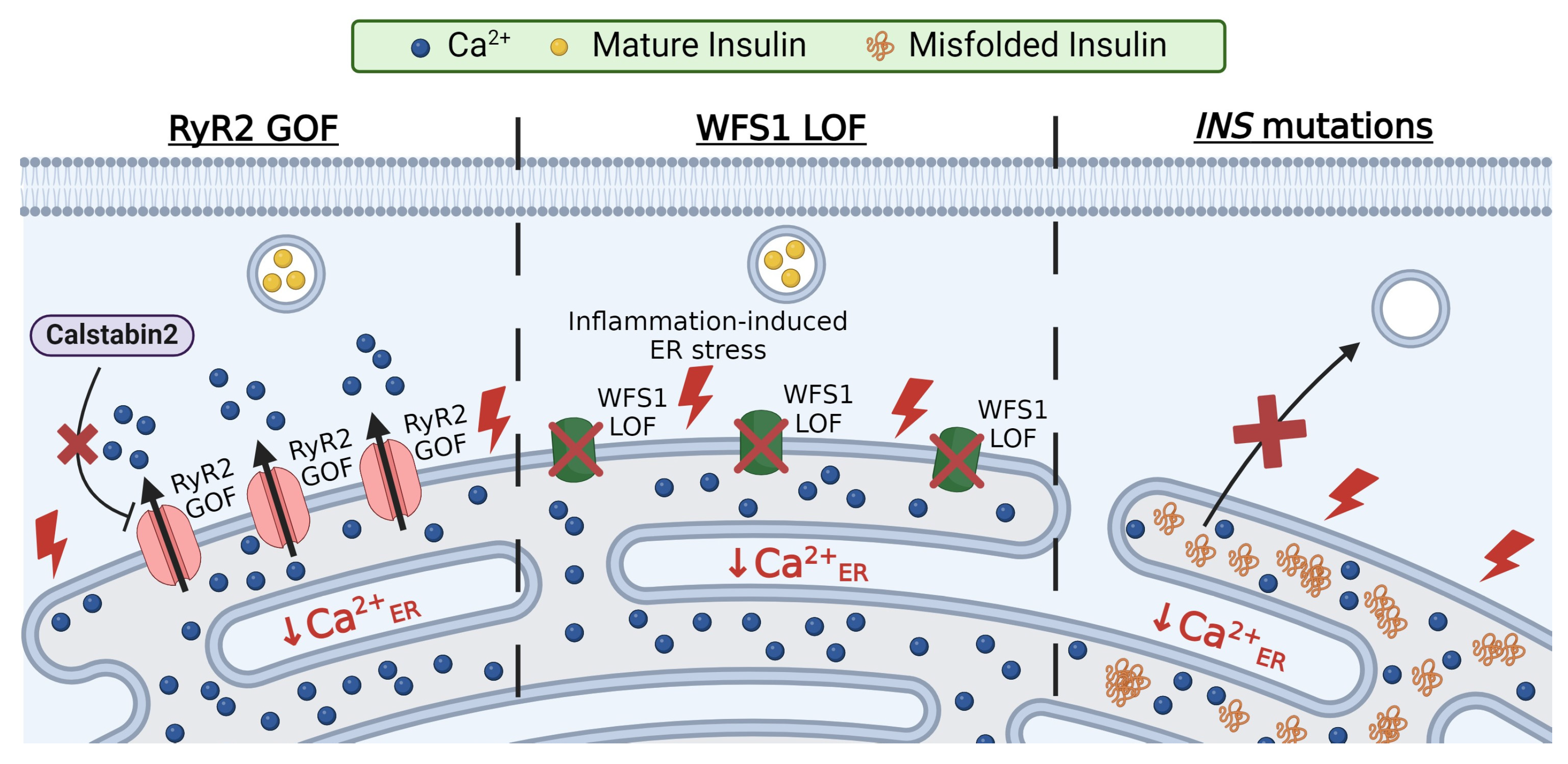
3.1. RyR2 Mutations

GOF mutations in RyR2 cause β-cell dysfunction and glucose intolerance. For example, RyR2 mutations (e.g., R2474S or N2386I) that disrupt the binding of calstabin2 result in leaky RyR2 channels [103] (Figure 3). These RyR2 mutations result in catecholaminergic polymorphic ventricular tachycardia (CPVT), glucose intolerance, and impaired insulin secretion [57]. Furthermore, mouse models with leaky RyR2 channels (with R2474S, N2386I, or S2814D mutations) displayed increased β-cell Ca2+ER release with a concomitant elevation in basal Ca2+C [57,104]. Leaky RyRs reduce Ca2+ER storage, causing ER stress and reduced GSIS. Ca2+ER release is also coupled with mitochondrial Ca2+ uptake, which is essential for several Ca2+-dependent energy processes such as ATP production (discussed in the next section). Thus, β-cells from mice expressing RyR R2474S or N2386I show defects in the mitochondrial structure and upregulation in markers of mitochondrial dysfunction, including uncoupling protein 2 and peroxisome proliferator-activator receptor γ coactivator 1a [57]. Importantly, reducing leaky RyR2s (R2474S or N2386I) with Rycal S107 restored Ca2+ER levels, enhanced insulin secretion, and improved mitochondria abnormalities [57]. Altogether, GOF mutations in RyR2 disrupt β-cell function by reducing Ca2+ER stores, which causes ER and mitochondrial stress, resulting in impaired insulin secretion and glucose intolerance.

3.2. Wolfram Syndrome 1 (WFS1) Mutations

Wolfram syndrome 1 (WFS1) is a rare autosomal recessive genetic disorder characterized by diabetes insipidus, diabetes mellites, optic atrophy, and deafness [105,106,107]. The WFS1 gene encodes the protein wolframin, an ER localized glycoprotein that has been linked to β-cell ER stress, Ca2+ER handling, and proteostasis. To date, there are over 200 mutations in WSF1 that result in a loss of function (LOF) and cause WFS1 [105,108]. Patients with two LOF WSF1 mutations show diabetic phenotypes, and carriers with one WSF1 LOF allele show an increased risk for developing T2D [109]. Within the islet, WFS1 is abundant in β-cells and has little to no expression in α, δ, or pancreatic polypeptide cells [110]. To mimic WFS1 LOF, Wfs1 knockout mouse models were utilized. Wfs1-deficient mice displayed reduced glucose-induced Ca2+ influx and impaired insulin secretion; GSIS was restored with the overexpression of Wfs1 [110]. The knockdown of Wfs1 in INS-1 cells caused Ca2+ER depletion [111]. Additionally, Wfs1-deficient β-cells promote cytokine-induced ER stress by upregulating proinflammatory cytokines and chemokines (Figure 3). Hyperglycemia-induced ER stress in Wfs1 knockout INS-1 cells involves PERK upregulation [112]. This suggests that Wfs1 is critical for limiting ER stress in β-cells under inflammatory or hyperglycemic conditions. Defects in Ca2+ER handling can be restored in β-cells with WFS1 deficiency following calpain inhibition (calpain initiates apoptosis) [113]. Taken together, WFS1 plays a crucial role in maintaining β-cell Ca2+ER and reducing ER stress; thus, diabetes observed in patients with WFS1 LOF mutations likely involves β-cell dysfunction due to reduced Ca2+ER and ER stress.

3.3. Insulin Mutations

Insulin biosynthesis accounts for up to 50% of glucose-stimulated β-cell protein production. Conditions of increased insulin demand (e.g., insulin resistance) and elevated insulin production result in ER stress, protein misfolding, and activation of the UPR [14]. The UPR is an important mechanism that protects β-cells from ER stress by reducing protein production, removing misfolded proteins, and increasing chaperone abundance. However, several insulin mutations lead to unresolvable ER stress; this occurs due to the buildup of insulin molecules in the ER by impairing insulin processing or via elevated insulin production due to reduced insulin receptor activation (Figure 3). To date, over 70 INS gene mutations have been identified; these mutations result in various diabetic phenotypes such as neonatal diabetes and mutant insulin-gene-induced diabetes of the youth (MIDY) [114,115,116,117]. Many INS mutations expressed in MIN6 cells show ER retention and cause ER stress, monitored by Chop upregulation [118]. INS mutations have been shown to disrupt Ca2+ handling. For example, glucose-induced Ca2+ influx was nearly abolished in islets from a mouse model of the C96Y insulin mutation (Akita mouse model); this mutation disrupts the disulfide bond formation between the A and B chains of insulin and proinsulin folding and causes insulin retention in the ER [56,119]. Interestingly, the ryanodine inhibition of RyR partially restored glucose-induced Ca2+ oscillations in islets from the Akita mouse, which suggests that C96Y leads to overactive RyRs and Ca2+ER depletion [56]. Another ER-retained INS mutation A24D also reduced Ca2+ER in a dose-dependent manner, and treatment with either translation inhibitors or the chemical chaperone TUDCA reduced Ca2+ER depletion [111,118,120]. Therefore, pathogenic INS mutations elevate ER stress and impair Ca2+ER handling, which causes β-cell dysfunction and, ultimately, failure.

4. Ca2+ER in the Context of Cellular Function

4.1. Chaperone-Mediated Ca2+ Binding

The majority of Ca2+ER is bound to chaperone proteins including calnexin, calreticulin, protein disulfide isomerase (PDI), glucose-regulated protein 78 (GRP78, also known as BiP), and glucose-regulated protein 94 (GRP94). Most of these chaperones have a high capacity for Ca2+ due to multiple low-affinity Ca2+ binding sites and thus tune Ca2+ER homeostasis [121]. As part of the ER stress response, these chaperones are upregulated to improve protein folding and maintain Ca2+ER levels. The roles of β-cell chaperone function have begun to emerge and have been well established in other cells. The C-terminal domain of calreticulin is responsible for buffering up to 50% of Ca2+ER, and thus, the overexpression of calreticulin increases Ca2+ER stores [121,122,123]. SOCE is also reduced in calreticulin overexpressing mouse L-fibroblasts due to increased Ca2+ER storage [124]. β-cell Ca2+ER depletion during NO-mediated apoptosis was reduced through the overexpression of calreticulin [125]. Therefore, the calreticulin upregulation observed in β-cells under diet-induced stress may serve to enhance Ca2+ER storage, reduce ER stress, and delay β-cell failure [126]. The glucose-regulated proteins were first characterized by their upregulation in cells following glucose starvation [127]. Two of the most abundant GRPs in β-cells are GRP78 and GRP94. GRP78 has been shown to sequester up to 25% of Ca2+ER [121,128]. Owing in part to its Ca2+ER buffering capacity, β-cell GRP78 overexpression protected mice from HFD-induced insulin resistance and ER stress [129]. Increasing levels of Ca2+ER alter the conformation of GRP94 to increase peptide binding [130]. GRP94-deficient cells show greater ER stress following Ca2+ER depletion, but the loss of GRP94 itself did not induce ER stress or disrupt Ca2+ER homeostasis [131]. The Ca2+ER buffering capabilities of ER chaperones help maintain Ca2+ER homeostasis and prevent Ca2+ER depletion under stress.

4.2. Protein Folding

Ca2+ is also necessary for chaperone-mediated protein folding [132]. Thus, a reduction in Ca2+ER impairs the protein folding capabilities of GRP78, GRP94, and PDIs; this leads to protein misfolding and the initiation of the UPR [133]. The knockdown of β-cell GRP78 or GRP94 increased UPR, reduced insulin content, and limited GSIS [134,135]. GRP78 and GRP94 are upregulated during β-cell ER stress to promote protein folding and Ca2+ER storage [135,136]. Thus, increasing the INS-1 levels of GRP78 reduced hyperglycemia-mediated UPR activation [134]. GRP94 interacts with proinsulin, promoting its processing, which increases the granule content of insulin and amplifies GSIS [135]. As mature insulin includes three disulfide bonds, the formation and cleavage of these disulfide bonds by PDIs also play an important role in insulin processing. A mouse model harboring the biallelic LOF mutation in Pdia6 encoding PDIA6 showed decreased β-cell insulin content, resulting in hyperglycemia [137], whereas the overexpression of P4HB encoding PDI promoted ER stress and impaired GSIS, which is likely due to the accumulation of proinsulin in the ER resulting from the PDI cleavage of proinsulin disulfide bonds [134]. Indeed, the knockdown of PDI in INS-1 cells elevated insulin disulfide bond formation and exit from the ER [138]. The activity of certain PDIs can be modulated by interactions with other chaperones. For example, the localization of PDI1A is dependent on Ca2+ER; the depletion of Ca2+ER promotes the interaction of PDI1A with calreticulin and the inhibition of its chaperone function [139]. Taken together, many chaperones efficiently orchestrate the multifaceted aspects of insulin processing. These chaperones also help amplify insulin production in response to increased insulin demand and reduce ER stress by limiting insulin misfolding and/or increasing Ca2+ER storage.

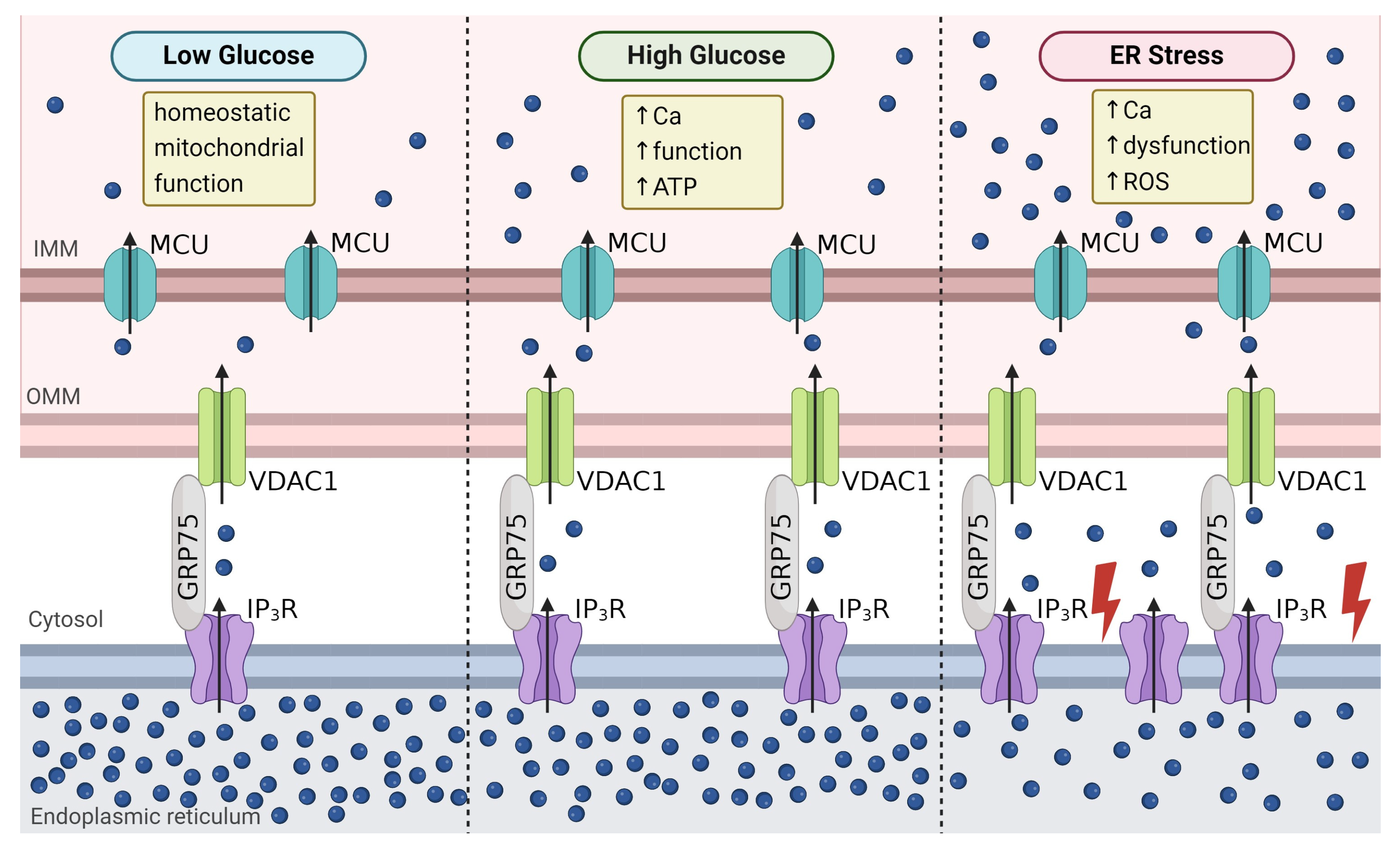
4.3. Mitochondrial Function

The ER and mitochondria interact at mitochondrial-associated membranes (MAMs), which enables the transfer of lipids and Ca2+ between these organelles. The MAM proteins involved in Ca2+ flux include GRP75, IP3R, and voltage-dependent anion channel (VDAC). GRP75 connects IP3Rs on the ER membrane with VDAC1 on the outer mitochondrial membrane [140]. Acute glucose stimulation increases IP3R2/VDAC1 interactions in INS-1 cells, elevating both mitochondrial Ca2+ content and the Gq-GPCR-mediated uptake of mitochondrial Ca2+ during IP3R2 Ca2+ER release [141] (Figure 4). The knockdown of Grp75 reduced IP3R2/VDAC1 interactions, limited mitochondrial Ca2+ uptake during IP3R-mediated Ca2+ER release, and blunted GSIS [141]. In T2D patients, β-cell IP3R2/VDAC1 interactions are reduced, which likely limits mitochondrial Ca2+ influx and contributes to β-cell dysfunction [142].
Both ER-stress-mediated Ca2+ER depletion and increased Ca2+ER storage can increase mitochondrial Ca2+. During chronic hyperglycemia, Ca2+ER depletion leads to increased basal mitochondrial Ca2+ uptake, which disrupts human islet glucose metabolism and GSIS [141]. Thus, ER stress initiates a greater transfer of Ca2+ER to the mitochondria; this leads to mitochondrial dysfunction, due in part to decreased ATP synthase activity, elevated ROS generation, and increased fission [141] (Figure 4). Alternatively, increasing Ca2+ER stores via SERCA activation increased mitochondrial Ca2+ uptake and ATP synthesis as well as prevented mitochondrial dysfunction in palmitate-treated β-cells [45]. The β-cell specific knockdown of TALK-1 channels also enhanced Ca2+ER storage, which led to increased mitochondrial Ca2+, ATP synthesis, and insulin secretion [143]. Therefore, alterations in Ca2+ER directly influence mitochondrial Ca2+, which either impairs β-cell function via mitochondrial Ca2+ overload or improves β-cell function via enhanced ATP production and subsequent insulin secretion.

5. Conclusions

Ca2+ER plays an important role in modulating β-cell function and the response to stress. During insulin resistance and β-cell depletion preceding type 2 and type 1 diabetes, respectively, increased insulin production leads to Ca2+ER depletion. Reduced Ca2+ER enhances β-cell ER stress and promotes the UPR. As β-cells are long-lived, the Ca2+ER regulation of the stress response also serves to modulate the workload and prevent or delay β-cell failure [144]. An integrated signaling network precisely orchestrates Ca2+ER handling. The altered expression and/or activity of proteins involved in Ca2+ER handling modifies insulin production and secretion, in part through changes in the Ca2+ER release into cytosol and/or mitochondria. Moreover, changes in Ca2+ER are sensed by many proteins (e.g., STIMs and chaperones) that modulate many aspects of β-cell function, including Ca2+ER refilling, transcription, translation, and protein processing. Therefore, mutations that perturb Ca2+ER result in β-cell dysfunction and monogenic diabetes. Due to the critical role that Ca2+ER depletion plays in β-cell dysfunction, increasing Ca2+ER could be used to restore β-cell function and health in diabetes. However, Ca2+ER homeostasis is important to most cells, and many of the proteins that control Ca2+ER handling are ubiquitously expressed. Thus, future studies are required to identify the β-cell-specific aspects of Ca2+ER handling that can be targeted without deleterious effects on other tissues.

Author Contributions

Conceptualization, J.R.D. and D.A.J.; writing—original draft preparation, J.R.D. and D.A.J.; writing—review and editing, J.R.D. and D.A.J. All authors have read and agreed to the published version of the manuscript.

Funding

This research was funded by the National Institutes of Health, grant numbers DK129340-S1, DK129340, and DK136768.

Institutional Review Board Statement

Not applicable.

Informed Consent Statement

Not applicable.

Data Availability Statement

Not applicable.

Conflicts of Interest

The authors declare no conflicts of interest.

References

  1. Rutter, G.A.; Hodson, D.J.; Chabosseau, P.; Haythorne, E.; Pullen, T.J.; Leclerc, I. Local and regional control of calcium dynamics in the pancreatic islet. Diabetes Obes. Metab. 2017, 19, 30–41. [Google Scholar] [CrossRef] [PubMed]
  2. Fonseca, S.G.; Gromada, J.; Urano, F. Endoplasmic Reticulum Stress and Pancreatic Beta-Cell Death. Trends Endocrinol. Metab. 2011, 22, 266–274. [Google Scholar]
  3. Gilon, P.; Chae, H.Y.; Rutter, G.A.; Ravier, M.A. Calcium Signaling in Pancreatic Beta-Cells in Health and in Type 2 Diabetes. Cell Calcium 2014, 56, 340–361. [Google Scholar] [CrossRef]
  4. Shrestha, N.; De Franco, E.; Arvan, P.; Cnop, M. Pathological Beta-Cell Endoplasmic Reticulum Stress in Type 2 Diabetes: Current Evidence. Front. Endocrinol. 2021, 12, 650158. [Google Scholar] [CrossRef]
  5. Scheuner, D.; Kaufman, R.J. The Unfolded Protein Response: A Pathway That Links Insulin Demand with Beta-Cell Failure and Diabetes. Endocr. Rev. 2008, 29, 317–333. [Google Scholar] [CrossRef] [PubMed]
  6. Cha, J.; Aguayo-Mazzucato, C.; Thompson, P.J. Pancreatic Beta-Cell Senescence in Diabetes: Mechanisms, Markers and Therapies. Front. Endocrinol. 2023, 14, 1212716. [Google Scholar] [CrossRef] [PubMed]
  7. Liu, M.; Haataja, L.; Wright, J.; Wickramasinghe, N.P.; Hua, Q.-X.; Phillips, N.F.; Barbetti, F.; Weiss, M.A.; Arvan, P. Mutant Ins-Gene Induced Diabetes of Youth: Proinsulin Cysteine Residues Impose Dominant-Negative Inhibition on Wild-Type Proinsulin Transport. PLoS ONE 2010, 5, e13333. [Google Scholar] [CrossRef]
  8. Hodish, I.; Liu, M.; Rajpal, G.; Larkin, D.; Holz, R.W.; Adams, A.; Liu, L. Arvan Misfolded Proinsulin Affects Bystander Proinsulin in Neonatal Diabetes. J. Biol. Chem. 2010, 285, 685–694. [Google Scholar] [CrossRef]
  9. Kang, S.; Dahl, R.; Hsieh, W.; Shin, A.; Zsebo, K.M.; Buettner, C.; Hajjar, R.J.; Lebeche, D. Small Molecular Allosteric Activator of the Sarco/Endoplasmic Reticulum Ca2+-ATPase (SERCA) Attenuates Diabetes and Metabolic Disorders. J. Biol. Chem. 2016, 291, 5185–5198. [Google Scholar] [CrossRef]
  10. Tong, X.T.; Kono, E.K.; Anderson-Baucum, W.; Yamamoto, P.; Gilon, D.; Lebeche, R.N.; Day, R.; Shull, G.E.; Evans-Molina, C. Serca2 Deficiency Impairs Pancreatic Beta-Cell Function in Response to Diet-Induced Obesity. Diabetes 2016, 65, 3039–3052. [Google Scholar] [CrossRef]
  11. Cardozo, A.K.F.; Ortis, J.; Storling, Y.M.; Feng, J.; Rasschaert, M.; Van Eylen, F.T.; Mandrup-Poulsen, T.; Herchuelz, A.; Eizirik, D.L. Cytokines Downregulate the Sarcoendoplasmic Reticulum Pump Ca2+ Atpase 2b and Deplete Endoplasmic Reticulum Ca2+, Leading to Induction of Endoplasmic Reticulum Stress in Pancreatic Beta-Cells. Diabetes 2005, 54, 452–461. [Google Scholar] [CrossRef] [PubMed]
  12. Ly, L.D.; Xu, S.; Choi, S.-K.; Ha, C.-M.; Thoudam, T.; Cha, S.-K.; Wiederkehr, A.; Wollheim, C.B.; Lee, I.-K.; Park, K.-S. Oxidative stress and calcium dysregulation by palmitate in type 2 diabetes. Exp. Mol. Med. 2017, 49, e291. [Google Scholar] [CrossRef]
  13. Zhang, I.X.; Raghavan, M.; Satin, L.S. The Endoplasmic Reticulum and Calcium Homeostasis in Pancreatic Beta Cells. Endocrinology 2020, 161, bqz028. [Google Scholar] [CrossRef] [PubMed]
  14. Xin, Y.; Gutierrez, G.D.; Okamoto, H.; Kim, J.; Lee, A.H.; Adler, C.; Ni, M.; Yancopoulos, G.D.; Murphy, A.J.; Gromada, J. Pseudotime Ordering of Single Human Beta-Cells Reveals States of Insulin Production and Unfolded Protein Response. Diabetes 2018, 67, 1783–1794. [Google Scholar] [CrossRef] [PubMed]
  15. Gilon, P.; Arredouani, A.; Gailly, P.; Gromada, J.; Henquin, J.-C. Uptake and Release of Ca2+ by the Endoplasmic Reticulum Contribute to the Oscillations of the Cytosolic Ca2+ Concentration Triggered by Ca2+ Influx in the Electrically Excitable Pancreatic B-cell. J. Biol. Chem. 1999, 274, 20197–20205. [Google Scholar] [CrossRef] [PubMed]
  16. Llanos, P.; Contreras-Ferrat, A.; Barrientos, G.; Valencia, M.; Mears, D.; Hidalgo, C. Glucose-Dependent Insulin Secretion in Pancreatic Beta-Cell Islets from Male Rats Requires Ca2+ Release Via Ros-Stimulated Ryanodine Receptors. PLoS ONE 2015, 10, e0129238. [Google Scholar]
  17. Usui, R.; Yabe, D.; Fauzi, M.; Goto, H.; Botagarova, A.; Tokumoto, S.; Tatsuoka, H.; Tahara, Y.; Kobayashi, S.; Manabe, T.; et al. Gpr40 Activation Initiates Store-Operated Ca2+ Entry and Potentiates Insulin Secretion Via the Ip3r1/Stim1/Orai1 Pathway in Pancreatic Beta-Cells. Sci. Rep. 2019, 9, 15562. [Google Scholar] [CrossRef] [PubMed]
  18. Arredouani, A.; Guiot, Y.; Jonas, J.C.; Liu, L.H.; Nenquin, M.; Pertusa, J.A.; Rahier, J.; Rolland, J.F.; Shull, G.E.; Stevens, M.; et al. Serca3 Ablation Does Not Impair Insulin Secretion but Suggests Distinct Roles of Different Sarcoendoplasmic Reticulum Ca2+ Pumps for Ca2+ Homeostasis in Pancreatic Beta-Cells. Diabetes 2002, 51, 3245–3253. [Google Scholar] [CrossRef] [PubMed]
  19. Roe, M.W.; Philipson, L.H.; Frangakis, C.J.; Kuznetsov, A.; Mertz, R.J.; Lancaster, M.E.; Spencer, B.; Worley, J.F., 3rd; Dukes, I.D. Defective Glucose-Dependent Endoplasmic Reticulum Ca2+ Sequestration in Diabetic Mouse Islets of Langerhans. J. Biol. Chem. 1994, 269, 18279–18282. [Google Scholar] [CrossRef]
  20. Roe, M.W.; Mertz, R.J.; Lancaster, M.E., 3rd; Worley, J.F.; Dukes, I.D. Thapsigargin Inhibits the Glucose-Induced Decrease of Intracellular Ca2+ in Mouse Islets of Langerhans. Am. J. Physiol. Endocrinol. Metab. 1994, 266, E852–E862. [Google Scholar] [CrossRef]
  21. Váradi, A.; Molnár, E.; Östenson, C.-G.; Ashcroft, S.J.H. Isoforms of endoplasmic reticulum Ca2+-ATPase are differentially expressed in normal and diabetic islets of Langerhans. Biochem. J. 1996, 319, 521–527. [Google Scholar] [CrossRef] [PubMed]
  22. Kono, T.; Ahn, G.; Moss, D.R.; Gann, L.; Zarain-Herzberg, A.; Nishiki, Y.; Fueger, P.T.; Ogihara, T.; Evans-Molina, C. Ppar-Gamma Activation Restores Pancreatic Islet Serca2 Levels and Prevents Beta-Cell Dysfunction under Conditions of Hyperglycemic and Cytokine Stress. Mol. Endocrinol. 2012, 26, 257–271. [Google Scholar] [CrossRef] [PubMed]
  23. Zarain-Herzberg, A.; García-Rivas, G.; Estrada-Avilés, R. Regulation of SERCA pumps expression in diabetes. Cell Calcium 2014, 56, 302–310. [Google Scholar] [CrossRef] [PubMed]
  24. Beauvois, M.C.; Merezak, C.; Jonas, J.C.; Ravier, M.A.; Henquin, J.C.; Gilon, P. Glucose-Induced Mixed [Ca2+]C Oscillations in Mouse Beta-Cells Are Controlled by the Membrane Potential and the Serca3 Ca2+-Atpase of the Endoplasmic Reticulum. Am. J. Physiol. Cell Physiol. 2006, 290, C1503–C1511. [Google Scholar] [CrossRef]
  25. Varadi, A.; Rutter, G.A. Dynamic Imaging of Endoplasmic Reticulum Ca2+ Concentration in Insulin-Secreting Min6 Cells Using Recombinant Targeted Cameleons: Roles of Sarco(Endo)Plasmic Reticulum Ca2+-Atpase (Serca)-2 and Ryanodine Receptors. Diabetes 2002, 51, S190–S201. [Google Scholar] [CrossRef] [PubMed]
  26. Gao, Z.Y.; Borge, P.D., Jr.; Jegier, P.A.; Young, R.A.; Wolf, B.A. Insulin Regulation of Beta-Cell Function Involves a Feedback Loop on Serca Gene Expression, Ca2+ Homeostasis, and Insulin Expression and Secretion. Biochemistry 2000, 39, 14912–14919. [Google Scholar]
  27. Gao, Z.Y.; Borge, P.D., Jr.; Wolf, B.A. Insulin Receptor Substrate 1-Induced Inhibition of Endoplasmic Reticulum Ca2+ Uptake in Beta-Cells. Autocrine Regulation of Intracellular Ca2+ Homeostasis and Insulin Secretion. J. Biol. Chem. 1999, 274, 18067–18074. [Google Scholar]
  28. Borge, P.D.; Wolf, B.A., Jr. Insulin Receptor Substrate 1 Regulation of Sarco-Endoplasmic Reticulum Calcium Atpase 3 in Insulin-Secreting Beta-Cells. J. Biol. Chem. 2003, 278, 11359–11368. [Google Scholar] [CrossRef] [PubMed]
  29. Kulkarni, R.N.; Roper, M.G.; Dahlgren, G.; Shih, D.Q.; Kauri, L.M.; Peters, J.L.; Stoffel, M.; Kennedy, R.T. Islet Secretory Defect in Insulin Receptor Substrate 1 Null Mice Is Linked With Reduced Calcium Signaling and Expression of Sarco(endo)plasmic Reticulum Ca2+-ATPase (SERCA)-2b and -3. Diabetes 2004, 53, 1517–1525. [Google Scholar] [CrossRef]
  30. Takatani, T.; Shirakawa, J.; Roe, M.W.; Leech, C.A.; Maier, B.F.; Mirmira, R.G.; Kulkarni, R.N. Irs1 Deficiency Protects Beta-Cells against Er Stress-Induced Apoptosis by Modulating Sxbp-1 Stability and Protein Translation. Sci. Rep. 2016, 6, 28177. [Google Scholar] [CrossRef]
  31. Borge, P.D.; Moibi, J.; Greene, S.R.; Trucco, M.; Young, R.A.; Gao, Z.; Wolf, B.A. Insulin Receptor Signaling and Sarco/Endoplasmic Reticulum Calcium Atpase in Beta-Cells. Diabetes 2002, 51 (Suppl. 3), S427–S433. [Google Scholar] [CrossRef] [PubMed]
  32. Withers, D.J.; Gutierrez, J.S.; Towery, H.; Burks, D.J.; Ren, J.-M.; Previs, S.; Zhang, Y.; Bernal, D.; Pons, S.; Shulman, G.I.; et al. Disruption of IRS-2 causes type 2 diabetes in mice. Nature 1998, 391, 900–904. [Google Scholar] [CrossRef] [PubMed]
  33. Tamemoto, H.; Kadowaki, T.; Tobe, K.; Yagi, T.; Sakura, H.; Hayakawa, T.; Terauchi, Y.; Ueki, K.; Kaburagi, Y.; Satoh, S.; et al. Insulin resistance and growth retardation in mice lacking insulin receptor substrate-1. Nature 1994, 372, 182–186. [Google Scholar] [CrossRef]
  34. Lockridge, A.; Jo, S.; Gustafson, E.; Damberg, N.; Mohan, R.; Olson, M.; Abrahante, J.E.; Alejandro, E.U. Islet O-Glcnacylation Is Required for Lipid Potentiation of Insulin Secretion through Serca2. Cell Rep. 2020, 31, 107609. [Google Scholar] [CrossRef] [PubMed]
  35. Alejandro, E.U.; Bozadjieva, N.; Kumusoglu, D.; Abdulhamid, S.; Levine, H.; Haataja, L.; Vadrevu, S.; Satin, L.S.; Arvan, P.; Bernal-Mizrachi, E. Disruption of O-Linked N-Acetylglucosamine Signaling Induces Er Stress and Beta Cell Failure. Cell Rep. 2015, 13, 2527–2538. [Google Scholar] [CrossRef]
  36. Sánchez-Gómez, F.J.; Espinosa-Díez, C.; Dubey, M.; Dikshit, M.; Lamas, S. S-glutathionylation: Relevance in diabetes and potential role as a biomarker. Biol. Chem. 2013, 394, 1263–1280. [Google Scholar] [CrossRef] [PubMed]
  37. Adachi, T.; Weisbrod, R.M.; Pimentel, D.R.; Ying, J.; Sharov, V.S.; Schöneich, C.; Cohen, R.A. S-Glutathiolation by peroxynitrite activates SERCA during arterial relaxation by nitric oxide. Nat. Med. 2004, 10, 1200–1207. [Google Scholar] [CrossRef] [PubMed]
  38. Mailloux, R.J.; Fu, A.; Robson-Doucette, C.; Allister, E.M.; Wheeler, M.B.; Screaton, R.; Harper, M.-E. Glutathionylation State of Uncoupling Protein-2 and the Control of Glucose-stimulated Insulin Secretion. J. Biol. Chem. 2012, 287, 39673–39685. [Google Scholar] [CrossRef] [PubMed]
  39. Ammon, H.P.; Grimm, A.; Lutz, S.; Wagner-Teschner, D.; Handel, M.; Hagenloh, I. Islet Glutathione and Insulin Release. Diabetes 1980, 29, 830–834. [Google Scholar] [CrossRef]
  40. Rebelato, E.; Abdulkader, F.; Curi, R.; Carpinelli, A.R. Control of the Intracellular Redox State by Glucose Participates in the Insulin Secretion Mechanism. PLoS ONE 2011, 6, e24507. [Google Scholar] [CrossRef]
  41. Liang, K.; Du, W.; Zhu, W.; Liu, S.; Cui, Y.; Sun, H.; Luo, B.; Xue, Y.; Yang, L.; Chen, L.; et al. Contribution of Different Mechanisms to Pancreatic Beta-cell Hyper-secretion in Non-obese Diabetic (NOD) Mice during Pre-diabetes. J. Biol. Chem. 2011, 286, 39537–39545. [Google Scholar] [CrossRef] [PubMed]
  42. Dickerson, M.T.; Bogart, A.M.; Altman, M.K.; Milian, S.C.; Jordan, K.L.; Dadi, P.K.; Jacobson, D.A. Cytokine-Mediated Changes in K+ Channel Activity Promotes an Adaptive Ca2+ Response That Sustains Beta-Cell Insulin Secretion During Inflammation. Sci. Rep. 2018, 8, 1158. [Google Scholar] [CrossRef] [PubMed]
  43. Iida, H.; Kono, T.; Lee, C.-C.; Krishnan, P.; Arvin, M.C.; Weaver, S.A.; Jarvela, T.S.; Branco, R.C.S.; McLaughlin, M.R.; Bone, R.N.; et al. SERCA2 regulates proinsulin processing and processing enzyme maturation in pancreatic beta cells. Diabetologia 2023, 66, 2042–2061. [Google Scholar] [CrossRef] [PubMed]
  44. Kimura, T.; Kagami, K.; Sato, A.; Osaki, A.; Ito, K.; Horii, S.; Toya, T.; Masaki, N.; Yasuda, R.; Nagatomo, Y.; et al. Sarco/Endoplasmic Reticulum Ca2+ ATPase 2 Activator Ameliorates Endothelial Dysfunction; Insulin Resistance in Diabetic Mice. Cells 2022, 11, 1488. [Google Scholar] [CrossRef] [PubMed]
  45. Nguyen, H.T.; Polo, C.N.; Wiederkehr, A.; Wollheim, C.B.; Park, K.S. Cdn1163, an Activator of Sarco/Endoplasmic Reticulum Ca2+ Atpase, up-Regulates Mitochondrial Functions and Protects against Lipotoxicity in Pancreatic Beta-Cells. Br. J. Pharmacol. 2023, 180, 2762–2776. [Google Scholar] [CrossRef] [PubMed]
  46. Xu, H.; Van Remmen, H. The Sarcoendoplasmic Reticulum Calcium Atpase (Serca) Pump: A Potential Target for Intervention in Aging and Skeletal Muscle Pathologies. Skelet. Muscle 2021, 11, 25. [Google Scholar] [CrossRef] [PubMed]
  47. Gwiazda, K.S.; Yang, T.L.; Lin, Y.; Johnson, J.D. Effects of Palmitate on Er and Cytosolic Ca2+ Homeostasis in Beta-Cells. Am. J. Physiol. Endocrinol. Metab. 2009, 296, E690–E701. [Google Scholar] [CrossRef] [PubMed]
  48. Cunha, D.A.; Hekerman, P.; Ladriere, L.; Bazarra-Castro, A.; Ortis, F.; Wakeham, M.C.; Moore, F.; Rasschaert, J.; Cardozo, A.K.; Bellomo, E.; et al. Initiation and Execution of Lipotoxic Er Stress in Pancreatic Beta-Cells. J. Cell Sci. 2008, 121, 2308–2318. [Google Scholar] [CrossRef] [PubMed]
  49. Liu, Z.; Xia, Y.; Li, B.; Xu, H.; Wang, C.; Liu, Y.; Li, Y.; Li, C.; Gao, N.; Li, L. Induction of ER stress-mediated apoptosis by ceramide via disruption of ER Ca2+ homeostasis in human adenoid cystic carcinoma cells. Cell Biosci. 2014, 4, 1–11. [Google Scholar] [CrossRef]
  50. Fu, S.; Yang, L.; Li, P.; Hofmann, O.; Dicker, L.; Hide, W.; Lin, X.; Watkins, S.M.; Ivanov, A.R.; Hotamisligil, G.S. Aberrant lipid metabolism disrupts calcium homeostasis causing liver endoplasmic reticulum stress in obesity. Nature 2011, 473, 528–531. [Google Scholar] [CrossRef]
  51. Lanner, J.T.; Georgiou, D.K.; Joshi, A.D.; Hamilton, S.L. Ryanodine Receptors: Structure, Expression, Molecular Details, and Function in Calcium Release. Cold Spring Harb. Perspect. Biol. 2010, 2, a003996. [Google Scholar] [CrossRef] [PubMed]
  52. Meissner, G.; Rios, E.; Tripathy, A.; Pasek, D.A. Regulation of Skeletal Muscle Ca2+ Release Channel (Ryanodine Receptor) by Ca2+ and Monovalent Cations and Anions. J. Biol. Chem. 1997, 272, 1628–1638. [Google Scholar] [CrossRef] [PubMed]
  53. Islam, M.S. The Ryanodine Receptor Calcium Channel of Beta-Cells: Molecular Regulation and Physiological Significance. Diabetes 2002, 51, 1299–1309. [Google Scholar] [CrossRef] [PubMed]
  54. Lee, H.C. Cyclic ADP-ribose and Nicotinic Acid Adenine Dinucleotide Phosphate (NAADP) as Messengers for Calcium Mobilization. J. Biol. Chem. 2012, 287, 31633–31640. [Google Scholar] [CrossRef] [PubMed]
  55. Mu-U-Min, R.B.A.; Diane, A.; Allouch, A.; Al-Siddiqi, H.H. Ca2+-Mediated Signaling Pathways: A Promising Target for the Successful Generation of Mature and Functional Stem Cell-Derived Pancreatic Beta Cells In Vitro. Biomedicines 2023, 11, 1577. [Google Scholar] [CrossRef] [PubMed]
  56. Yamamoto, W.R.; Bone, R.N.; Sohn, P.; Syed, F.; Reissaus, C.A.; Mosley, A.L.; Wijeratne, A.B.; True, J.D.; Tong, X.; Kono, T.; et al. Endoplasmic Reticulum Stress Alters Ryanodine Receptor Function in the Murine Pancreatic Beta Cell. J. Biol. Chem. 2019, 294, 168–181. [Google Scholar] [CrossRef] [PubMed]
  57. Santulli, G.; Pagano, G.; Sardu, C.; Xie, W.; Reiken, S.; D’Ascia, S.L.; Cannone, M.; Marziliano, N.; Trimarco, B.; Guise, T.A.; et al. Calcium Release Channel Ryr2 Regulates Insulin Release and Glucose Homeostasis. J. Clin. Investig. 2015, 125, 4316. [Google Scholar] [CrossRef] [PubMed]
  58. Takasawa, S.; Akiyama, T.; Nata, K.; Kuroki, M.; Tohgo, A.; Noguchi, N.; Kobayashi, S.; Kato, I.; Katada, T.; Okamoto, H. Cyclic Adp-Ribose and Inositol 1,4,5-Trisphosphate as Alternate Second Messengers for Intracellular Ca2+ Mobilization in Normal and Diabetic Beta-Cells. J. Biol. Chem. 1998, 273, 2497–2500. [Google Scholar] [CrossRef] [PubMed]
  59. Postic, S.; Sarikas, S.; Pfabe, J.; Pohorec, V.; Bombek, L.K.; Sluga, N.; Klemen, M.S.; Dolensek, J.; Korosak, D.; Stozer, A.; et al. High-Resolution Analysis of the Cytosolic Ca2+ Events in Beta Cell Collectives in Situ. Am. J. Physiol. Endocrinol. Metab. 2023, 324, E42–E55. [Google Scholar] [CrossRef]
  60. Dror, V.; Kalynyak, T.B.; Bychkivska, Y.; Frey, M.H.; Tee, M.; Jeffrey, K.D.; Nguyen, V.; Luciani, D.S.; Johnson, J.D. Glucose and Endoplasmic Reticulum Calcium Channels Regulate Hif-1beta Via Presenilin in Pancreatic Beta-Cells. J. Biol. Chem. 2018, 283, 9909–9916. [Google Scholar] [CrossRef]
  61. Zhang, I.X.; Herrmann, A.; Leon, J.; Jeyarajan, S.; Arunagiri, A.; Arvan, P.; Gilon, P.; Satin, L.S. ER stress increases expression of intracellular calcium channel RyR1 to modify Ca2+ homeostasis in pancreatic beta cells. J. Biol. Chem. 2023, 299, 105065. [Google Scholar] [CrossRef] [PubMed]
  62. Marmugi, A.; Parnis, J.; Chen, X.; Carmichael, L.; Hardy, J.; Mannan, N.; Marchetti, P.; Piemonti, L.; Bosco, D.; Johnson, P.; et al. Sorcin Links Pancreatic Beta-Cell Lipotoxicity to Er Ca2+ Stores. Diabetes 2016, 65, 1009–1021. [Google Scholar] [CrossRef] [PubMed]
  63. Foskett, J.K.; Mak, D.O.D. Regulation of Ip(3)R Channel Gating by Ca2+ and Ca2+ Binding Proteins. Curr. Top. Membr. 2010, 66, 235–272. [Google Scholar] [PubMed]
  64. Stutzmann, G.E.; Mattson, M.P. Endoplasmic Reticulum Ca2+Handling in Excitable Cells in Health and Disease. Pharmacol. Rev. 2011, 63, 700–727. [Google Scholar] [CrossRef] [PubMed]
  65. Berridge, M.J. The Inositol Trisphosphate/Calcium Signaling Pathway in Health and Disease. Physiol. Rev. 2016, 96, 1261–1296. [Google Scholar] [CrossRef] [PubMed]
  66. Gautam, D.; Han, S.J.; Hamdan, F.F.; Jeon, J.; Li, B.; Li, J.H.; Cui, Y.; Mears, D.; Lu, H.; Deng, C.; et al. A Critical Role for Beta Cell M3 Muscarinic Acetylcholine Receptors in Regulating Insulin Release and Blood Glucose Homeostasis in Vivo. Cell Metab. 2006, 3, 449–461. [Google Scholar] [CrossRef] [PubMed]
  67. Dyachok, O.; Gylfe, E. Ca2+-Induced Ca2+ Release Via Inositol 1,4,5-Trisphosphate Receptors Is Amplified by Protein Kinase a and Triggers Exocytosis in Pancreatic Beta-Cells. J. Biol. Chem. 2004, 279, 45455–45461. [Google Scholar] [CrossRef]
  68. Thore, S.; Dyachok, O.; Tengholm, A. Oscillations of Phospholipase C Activity Triggered by Depolarization and Ca2+ Influx in Insulin-secreting Cells. J. Biol. Chem. 2004, 279, 19396–19400. [Google Scholar] [CrossRef] [PubMed]
  69. Tamarina, N.A.; Kuznetsov, A.; Rhodes, C.J.; Bindokas, V.P.; Philipson, L.H. Inositol (1,4,5)-Trisphosphate Dynamics and Intracellular Calcium Oscillations in Pancreatic Beta-Cells. Diabetes 2005, 54, 3073–3081. [Google Scholar] [CrossRef]
  70. Hauge-Evans, A.C.; Reers, C.; Kerby, A.; Franklin, Z.; Amisten, S.; King, A.J.; Hassan, Z.; Vilches-Flores, A.; Tippu, Z.; Persaud, S.J.; et al. Effect of hyperglycaemia on muscarinic M3 receptor expression and secretory sensitivity to cholinergic receptor activation in islets. Diabetes, Obes. Metab. 2014, 16, 947–956. [Google Scholar] [CrossRef]
  71. Zhu, L.; Rossi, M.; Cohen, A.; Pham, J.; Zheng, H.; Dattaroy, D.; Mukaibo, T.; Melvin, J.E.; Langel, J.L.; Hattar, S.; et al. Allosteric Modulation of Beta-Cell M(3) Muscarinic Acetylcholine Receptors Greatly Improves Glucose Homeostasis in Lean and Obese Mice. Proc. Natl. Acad. Sci. USA 2019, 116, 18684–18690. [Google Scholar] [CrossRef] [PubMed]
  72. Oduori, O.S.; Murao, N.; Shimomura, K.; Takahashi, H.; Zhang, Q.; Dou, H.; Sakai, S.; Minami, K.; Chanclon, B.; Guida, C.; et al. Gs/Gq Signaling Switch in Beta Cells Defines Incretin Effectiveness in Diabetes. J. Clin. Investig. 2020, 130, 6639–6655. [Google Scholar] [CrossRef] [PubMed]
  73. Görlach, A.; Bertram, K.; Hudecova, S.; Krizanova, O. Calcium and ROS: A mutual interplay. Redox Biol. 2015, 6, 260–271. [Google Scholar] [CrossRef] [PubMed]
  74. Vilas-Boas, E.A.; Almeida, D.C.; Roma, L.P.; Ortis, F.; Carpinelli, A.R. Lipotoxicity and Beta-Cell Failure in Type 2 Diabetes: Oxidative Stress Linked to Nadph Oxidase and Er Stress. Cells 2021, 10, 3328. [Google Scholar] [CrossRef] [PubMed]
  75. Shu, J.; Gambardella, J.; Sorriento, D.; Santulli, G. Mechanistic Role of IP3R Calcium Release Channel in Pancreatic Beta-Cell Function. Diabetes 2018, 67, 313. [Google Scholar] [CrossRef]
  76. Cassel, R.; Ducreux, S.; Alam, M.R.; Dingreville, F.; Berlé, C.; Burda-Jacob, K.; Chauvin, M.A.; Chikh, K.; Païta, L.; Al-Mawla, R.; et al. Protection of Human Pancreatic Islets from Lipotoxicity by Modulation of the Translocon. PLoS ONE 2016, 11, e0148686. [Google Scholar] [CrossRef] [PubMed]
  77. Klec, C.; Madreiter-Sokolowski, C.T.; Stryeck, S.; Sachdev, V.; Duta-Mare, M.; Gottschalk, B.; Depaoli, M.R.; Rost, R.; Hay, J.; Waldeck-Weiermair, M.; et al. Glycogen Synthase Kinase 3 Beta Controls Presenilin-1-Mediated Endoplasmic Reticulum Ca2+ Leak Directed to Mitochondria in Pancreatic Islets and beta-Cells. Cell. Physiol. Biochem. 2019, 52, 57–75. [Google Scholar] [CrossRef] [PubMed]
  78. Klec, C.; Madreiter-Sokolowski, C.T.; Ziomek, G.; Stryeck, S.; Sachdev, V.; Duta-Mare, M.; Gottschalk, B.; Depaoli, M.R.; Rost, R.; Hay, J.; et al. Presenilin-1 Established Er-Ca2+ Leak: A Follow up on Its Importance for the Initial Insulin Secretion in Pancreatic Islets and Beta-Cells Upon Elevated Glucose. Cell Physiol. Biochem. 2019, 53, 573–586. [Google Scholar] [PubMed]
  79. Parys, J.B.; Van Coppenolle, F. Sec61 complex/translocon: The role of an atypical ER Ca2+-leak channel in health and disease. Front. Physiol. 2022, 13, 991149. [Google Scholar] [CrossRef]
  80. Bhadra, P.; Dos Santos, S.; Gamayun, I.; Pick, T.; Neumann, C.; Ogbechi, J.; Hall, B.S.; Zimmermann, R.; Helms, V.; Simmonds, R.E.; et al. Mycolactone enhances the Ca2+ leak from endoplasmic reticulum by trapping Sec61 translocons in a Ca2+ permeable state. Biochem. J. 2021, 478, 4005–4024. [Google Scholar] [CrossRef]
  81. Schäuble, N.; Sven, L.; Martin, J.; Cappel, S.; Schorr, S.; Ulucan, Ö.; Linxweiler, J.; Dudek, J.; Blum, R.; Helms, V.; et al. Bip-Mediated Closing of the Sec61 Channel Limits Ca2+ Leakage from the Er. EMBO J. 2012, 31, 3282–3296. [Google Scholar] [CrossRef]
  82. Lemos, F.O.; Bultynck, G.; Parys, J.B. A comprehensive overview of the complex world of the endo- and sarcoplasmic reticulum Ca2+-leak channels. Biochim. Biophys. Acta BBA Mol. Cell Res. 2021, 1868, 119020. [Google Scholar] [CrossRef]
  83. Lloyd, D.J.; Wheeler, M.C.; Gekakis, N. A Point Mutation in Sec61α1 Leads to Diabetes and Hepatosteatosis in Mice. Diabetes 2009, 59, 460–470. [Google Scholar] [CrossRef]
  84. Erdmann, F.; Schäuble, N.; Lang, S.; Jung, M.; Honigmann, A.; Ahmad, M.; Dudek, J.; Benedix, J.; Harsman, A.; Kopp, A.; et al. Interaction of calmodulin with Sec61α limits Ca2+leakage from the endoplasmic reticulum. EMBO J. 2010, 30, 17–31. [Google Scholar] [CrossRef]
  85. Tu, H.; Nelson, O.; Bezprozvanny, A.; Wang, Z.; Lee, S.F.; Hao, Y.H.; Serneels, L.; De Strooper, B.; Yu, G.; Bezprozvanny, I. Presenilins Form Er Ca2+ Leak Channels, a Function Disrupted by Familial Alzheimer’s Disease-Linked Mutations. Cell 2006, 126, 981–993. [Google Scholar] [CrossRef]
  86. Shilling, D.; Mak, D.-O.D.; Kang, D.E.; Foskett, J.K. Lack of Evidence for Presenilins as Endoplasmic Reticulum Ca2+ Leak Channels. J. Biol. Chem. 2012, 287, 10933–10944. [Google Scholar] [CrossRef]
  87. Kasri, N.N.; Kocks, S.L.; Verbert, L.; Hébert, S.S.; Callewaert, G.; Parys, J.B.; Missiaen, L.; De Smedt, H. Up-regulation of inositol 1,4,5-trisphosphate receptor type 1 is responsible for a decreased endoplasmic-reticulum Ca2+ content in presenilin double knock-out cells. Cell Calcium 2006, 40, 41–51. [Google Scholar] [CrossRef]
  88. Vierra, N.C.; Dadi, P.K.; Milian, S.C.; Dickerson, M.T.; Jordan, K.L.; Gilon, P.; Jacobson, D.A. Talk-1 Channels Control Beta Cell Endoplasmic Reticulum Ca2+ Homeostasis. Sci. Signal 2017, 10, eaan2883. [Google Scholar] [CrossRef]
  89. Yazawa, M.; Ferrante, C.; Feng, J.; Mio, K.; Ogura, T.; Zhang, M.; Lin, P.-H.; Pan, Z.; Komazaki, S.; Kato, K.; et al. TRIC channels are essential for Ca2+ handling in intracellular stores. Nature 2007, 448, 78–82. [Google Scholar] [CrossRef]
  90. Vierra, N.C.; Dadi, P.K.; Jeong, I.; Dickerson, M.; Powell, D.R.; Jacobson, D.A. Type 2 Diabetes-Associated K+ Channel Talk-1 Modulates Beta-Cell Electrical Excitability, Second-Phase Insulin Secretion, and Glucose Homeostasis. Diabetes 2015, 64, 3818–3828. [Google Scholar] [CrossRef]
  91. Graff, S.M.; Johnson, S.R.; Leo, P.J.; Dadi, P.K.; Dickerson, M.T.; Nakhe, A.Y.; McInerney-Leo, A.M.; Marshall, M.; Zaborska, K.E.; Schaub, C.M.; et al. A KCNK16 mutation causing TALK-1 gain of function is associated with maturity-onset diabetes of the young. J. Clin. Investig. 2021, 6, e138057. [Google Scholar] [CrossRef]
  92. Nakhe, A.; Prasanna, Y.; Dadi, K.; Kim, J.; Shrestha, S.; Cartailler, J.-P.; Sampson, L.; Magnuson, M.A.; Jacobson, D.A. The Mody-Associated Talk-1 L114p Mutation Causes Islet A-Cell Overactivity and Β-Cell Inactivity Resulting in Transient Neonatal Diabetes and Glucose Dyshomeostasis in Adults; eLife Sciences Publications, Ltd.: Cambridge, UK, 2023; 114p. [Google Scholar]
  93. Varshney, A.; Scott, L.J.; Welch, R.P.; Erdos, M.R.; Chines, P.S.; Narisu, N.; Albanus, R.D.; Orchard, P.; Wolford, B.N.; Kursawe, R.; et al. Genetic regulatory signatures underlying islet gene expression and type 2 diabetes. Proc. Natl. Acad. Sci. USA 2017, 114, 2301–2306. [Google Scholar] [CrossRef] [PubMed]
  94. Duprat, F.; Girard, C.; Jarretou, G.; Lazdunski, M. Pancreatic Two P Domain K+ Channels Talk-1 and Talk-2 Are Activated by Nitric Oxide and Reactive Oxygen Species. J. Physiol. 2005, 562, 235–244. [Google Scholar] [CrossRef]
  95. Riel, E.B.; Jürs, B.C.; Cordeiro, S.; Musinszki, M.; Schewe, M.; Baukrowitz, T. The versatile regulation of K2P channels by polyanionic lipids of the phosphoinositide and fatty acid metabolism. J. Gen. Physiol. 2021, 154, e202112989. [Google Scholar] [CrossRef]
  96. Khoubza, L.; Gilbert, N.; Kim, E.J.; Chatelain, F.C.; Feliciangeli, S.; Abelanet, S.; Kang, D.; Lesage, F.; Bichet, D. Alkaline-Sensitive Two-Pore Domain Potassium Channels Form Functional Heteromers in Pancreatic Beta-Cells. J. Biol. Chem. 2022, 298, 102447. [Google Scholar] [CrossRef] [PubMed]
  97. Putney, J.W., Jr. A model for receptor-regulated calcium entry. Cell Calcium 1986, 7, 1–12. [Google Scholar] [CrossRef] [PubMed]
  98. Sabourin, J.; Allagnat, F. Store-operated Ca2+ entry: A key component of the insulin secretion machinery. J. Mol. Endocrinol. 2016, 57, F35–F39. [Google Scholar] [CrossRef]
  99. Klec, C.; Ziomek, G.; Pichler, M.; Malli, R.; Graier, W.F. Calcium Signaling in ß-cell Physiology and Pathology: A Revisit. Int. J. Mol. Sci. 2019, 20, 6110. [Google Scholar] [CrossRef]
  100. Sabourin, J.; Le Gal, L.; Saurwein, L.; Haefliger, J.A.; Raddatz, E.; Allagnat, F. Store-Operated Ca2+ Entry Mediated by Orai1 and Trpc1 Participates to Insulin Secretion in Rat Beta-Cells. J. Biol. Chem. 2015, 290, 30530–30539. [Google Scholar] [CrossRef]
  101. Kono, T.; Tong, X.; Taleb, S.; Bone, R.N.; Iida, H.; Lee, C.C.; Sohn, P.; Gilon, P.; Roe, M.W.; Evans-Molina, C. Impaired Store-Operated Calcium Entry and Stim1 Loss Lead to Reduced Insulin Secretion and Increased Endoplasmic Reticulum Stress in the Diabetic Beta-Cell. Diabetes 2018, 67, 2293–2304. [Google Scholar] [CrossRef]
  102. Zhang, I.X.; Ren, J.; Vadrevu, S.; Raghavan, M.; Satin, L.S. ER stress increases store-operated Ca2+ entry (SOCE) and augments basal insulin secretion in pancreatic beta cells. J. Biol. Chem. 2020, 295, 5685–5700. [Google Scholar] [CrossRef]
  103. Lehnart, S.E.; Wehrens, X.H.; Marks, A.R. Calstabin Deficiency, Ryanodine Receptors, and Sudden Cardiac Death. Biochem. Biophys. Res. Commun. 2004, 322, 1267–1279. [Google Scholar] [CrossRef]
  104. Dixit, S.S.; Wang, T.; Manzano, E.J.; Yoo, S.; Lee, J.; Chiang, D.Y.; Ryan, N.; Respress, J.L.; Yechoor, V.K.; Wehrens, X.H. Effects of Camkii-Mediated Phosphorylation of Ryanodine Receptor Type 2 on Islet Calcium Handling, Insulin Secretion, and Glucose Tolerance. PLoS ONE 2013, 8, e58655. [Google Scholar] [CrossRef]
  105. Kõks, S. Genomics of Wolfram Syndrome 1 (WFS1). Biomolecules 2023, 13, 1346. [Google Scholar] [CrossRef]
  106. Didmoad (Wolfram) Syndrome. Lancet 1986, 1, 1075–1076.
  107. Cremers, C.W.; Wijdeveld, P.G.; Pinckers, A.J. Juvenile Diabetes Mellitus, Optic Atrophy, Hearing Loss, Diabetes Insipidus, Atonia of the Urinary Tract and Bladder, and Other Abnormalities (Wolfram Syndrome). A Review of 88 Cases from the Literature with Personal Observations on 3 New Patients. Acta Paediatr. Scand. Suppl. 1977, 264, 1–16. [Google Scholar] [CrossRef]
  108. Gong, Y.; Xiong, L.; Li, X.; Su, L.; Xiao, H. A novel mutation of WFS1 gene leading to increase ER stress and cell apoptosis is associated an autosomal dominant form of Wolfram syndrome type 1. BMC Endocr. Disord. 2021, 21, 1–13. [Google Scholar] [CrossRef]
  109. Minton, J.A.; Hattersley, A.T.; Owen, K.; McCarthy, M.I.; Walker, M.; Latif, F.; Barrett, T.; Frayling, T.M. Association Studies of Genetic Variation in the Wfs1 Gene and Type 2 Diabetes in U.K. Populations. Diabetes 2002, 51, 1287–1290. [Google Scholar] [CrossRef]
  110. Ishihara, H.; Takeda, S.; Tamura, A.; Takahashi, R.; Yamaguchi, S.; Takei, D.; Yamada, T.; Inoue, H.; Soga, H.; Katagiri, H.; et al. Disruption of the Wfs1 Gene in Mice Causes Progressive Beta-Cell Loss and Impaired Stimulus-Secretion Coupling in Insulin Secretion. Hum. Mol. Genet. 2004, 13, 1159–1170. [Google Scholar] [CrossRef]
  111. Hara, T.; Mahadevan, J.; Kanekura, K.; Hara, M.; Lu, S.; Urano, F. Calcium Efflux from the Endoplasmic Reticulum Leads to Beta-Cell Death. Endocrinology 2014, 155, 758–768. [Google Scholar] [CrossRef]
  112. Morikawa, S.; Blacher, L.; Onwumere, C.; Urano, F. Loss of Function of WFS1 Causes ER Stress-Mediated Inflammation in Pancreatic Beta-Cells. Front. Endocrinol. 2022, 13, 849204. [Google Scholar] [CrossRef]
  113. Nguyen, L.D.; Fischer, T.T.; Abreu, D.; Arroyo, A.; Urano, F.; Ehrlich, B.E. Calpain Inhibitor and Ibudilast Rescue Beta Cell Functions in a Cellular Model of Wolfram Syndrome. Proc. Natl. Acad. Sci. USA 2020, 117, 17389–17398. [Google Scholar] [CrossRef]
  114. Liu, M.; Sun, J.; Cui, J.; Chen, W.; Guo, H.; Barbetti, F.; Arvan, P. INS-gene mutations: From genetics and beta cell biology to clinical disease. Mol. Asp. Med. 2015, 42, 3–18. [Google Scholar] [CrossRef]
  115. Støy, J.; Edghill, E.L.; Flanagan, S.E.; Ye, H.; Paz, V.P.; Pluzhnikov, A.; Below, J.E.; Hayes, M.G.; Cox, N.J.; Lipkind, G.M.; et al. Insulin gene mutations as a cause of permanent neonatal diabetes. Proc. Natl. Acad. Sci. USA 2007, 104, 15040–15044. [Google Scholar] [CrossRef]
  116. Ataie-Ashtiani, S.; Forbes, B. A Review of the Biosynthesis and Structural Implications of Insulin Gene Mutations Linked to Human Disease. Cells 2023, 12, 1008. [Google Scholar] [CrossRef]
  117. Liu, M.; Hodish, I.; Haataja, L.; Lara-Lemus, R.; Rajpal, G.; Wright, J.; Arvan, P. Proinsulin misfolding and diabetes: Mutant INS gene-induced diabetes of youth. Trends Endocrinol. Metab. 2010, 21, 652–659. [Google Scholar] [CrossRef]
  118. Rajan, S.; Eames, S.C.; Park, S.-Y.; Labno, C.; Bell, G.I.; Prince, V.E.; Philipson, L.H. In vitro processing and secretion of mutant insulin proteins that cause permanent neonatal diabetes. Am. J. Physiol. Metab. 2010, 298, E403–E410. [Google Scholar] [CrossRef]
  119. Wang, J.; Takeuchi, T.; Tanaka, S.; Kubo, S.K.; Kayo, T.; Lu, D.; Takata, K.; Koizumi, A.; Izumi, T. A Mutation in the Insulin 2 Gene Induces Diabetes with Severe Pancreatic Beta-Cell Dysfunction in the Mody Mouse. J. Clin. Investig. 1999, 103, 27–37. [Google Scholar] [CrossRef]
  120. Liu, M.; Lara-Lemus, R.; Shan, S.-O.; Wright, J.; Haataja, L.; Barbetti, F.; Guo, H.; Larkin, D.; Arvan, P. Impaired Cleavage of Preproinsulin Signal Peptide Linked to Autosomal-Dominant Diabetes. Diabetes 2012, 61, 828–837. [Google Scholar] [CrossRef]
  121. Wang, W.-A.; Agellon, L.B.; Michalak, M. Organellar Calcium Handling in the Cellular Reticular Network. Cold Spring Harb. Perspect. Biol. 2019, 11, a038265. [Google Scholar] [CrossRef]
  122. Michalak, M. Calreticulin: Endoplasmic Reticulum Ca2+ Gatekeeper. J. Cell Mol. Med. 2023, 28, e17839. [Google Scholar] [CrossRef]
  123. Gelebart, P.; Opas, M.; Michalak, M. Calreticulin, a Ca2+-binding chaperone of the endoplasmic reticulum. Int. J. Biochem. Cell Biol. 2005, 37, 260–266. [Google Scholar] [CrossRef]
  124. Mery, L.; Mesaeli, N.; Michalak, M.; Opas, M.; Lew, D.P.; Krause, K.-H. Overexpression of Calreticulin Increases Intracellular Ca2+ Storage and Decreases Store-operated Ca2+ Influx. J. Biol. Chem. 1996, 271, 9332–9339. [Google Scholar] [CrossRef]
  125. Oyadomari, S.; Takeda, K.; Takiguchi, M.; Gotoh, T.; Matsumoto, M.; Wada, I.; Akira, S.; Araki, E.; Mori, M. Nitric Oxide-Induced Apoptosis in Pancreatic Beta Cells Is Mediated by the Endoplasmic Reticulum Stress Pathway. Proc. Natl. Acad. Sci. USA 2001, 98, 10845–10850. [Google Scholar] [CrossRef]
  126. Gupta, D.; Jetton, T.L.; LaRock, K.; Monga, N.; Satish, B.; Lausier, J.; Peshavaria, M.; Leahy, J.L. Temporal Characterization of Beta Cell-Adaptive and -Maladaptive Mechanisms During Chronic High-Fat Feeding in C57bl/6ntac Mice. J. Biol. Chem. 2017, 292, 12449–12459. [Google Scholar] [CrossRef]
  127. Franklin, J.L.; Amsler, M.O.; Messina, J.L. Regulation of glucose responsive protein (GRP) gene expression by insulin. Cell Stress Chaperon 2022, 27, 27–35. [Google Scholar] [CrossRef]
  128. Lièvremont, J.-P.; Rizzuto, R.; Hendershot, L.; Meldolesi, J. BiP, a Major Chaperone Protein of the Endoplasmic Reticulum Lumen, Plays a Direct and Important Role in the Storage of the Rapidly Exchanging Pool of Ca2+. J. Biol. Chem. 1997, 272, 30873–30879. [Google Scholar] [CrossRef]
  129. Teodoro-Morrison, T.; Schuiki, I.; Zhang, L.; Belsham, D.D.; Volchuk, A. GRP78 overproduction in pancreatic beta cells protects against high-fat-diet-induced diabetes in mice. Diabetologia 2013, 56, 1057–1067. [Google Scholar] [CrossRef]
  130. Biswas, C.; Ostrovsky, O.; Makarewich, C.A.; Wanderling, S.; Gidalevitz, T.; Argon, Y. The peptide-binding activity of GRP94 is regulated by calcium. Biochem. J. 2007, 405, 233–241. [Google Scholar] [CrossRef]
  131. Poirier, S.; Mamarbachi, M.; Chen, W.-T.; Lee, A.S.; Mayer, G. GRP94 Regulates Circulating Cholesterol Levels through Blockade of PCSK9-Induced LDLR Degradation. Cell Rep. 2015, 13, 2064–2071. [Google Scholar] [CrossRef]
  132. Mekahli, D.; Bultynck, G.; Parys, J.B.; De Smedt, H.; Missiaen, L. Endoplasmic-Reticulum Calcium Depletion and Disease. Cold Spring Harb. Perspect. Biol. 2011, 3, a004317. [Google Scholar] [CrossRef] [PubMed]
  133. Daverkausen-Fischer, L.; Prols, F. Regulation of Calcium Homeostasis and Flux between the Endoplasmic Reticulum and the Cytosol. J. Biol. Chem. 2022, 298, 102061. [Google Scholar] [CrossRef] [PubMed]
  134. Zhang, L.; Lai, E.; Teodoro, T.; Volchuk, A. Grp78, but Not Protein-Disulfide Isomerase, Partially Reverses Hyperglycemia-Induced Inhibition of Insulin Synthesis and Secretion in Pancreatic Beta-Cells. J. Biol. Chem. 2009, 284, 5289–5298. [Google Scholar] [CrossRef] [PubMed]
  135. Ghiasi, S.M.; Dahlby, T.; Andersen, C.H.; Haataja, L.; Petersen, S.; Omar-Hmeadi, M.; Yang, M.; Pihl, C.; Bresson, S.E.; Khilji, M.S.; et al. Endoplasmic Reticulum Chaperone Glucose-Regulated Protein 94 Is Essential for Proinsulin Handling. Diabetes 2019, 68, 747–760. [Google Scholar] [CrossRef] [PubMed]
  136. Marchetti, P.; Bugliani, M.; Lupi, R.; Marselli, L.; Masini, M.; Boggi, U.; Filipponi, F.; Weir, G.C.; Eizirik, D.L.; Cnop, M. The endoplasmic reticulum in pancreatic beta cells of type 2 diabetes patients. Diabetologia 2007, 50, 2486–2494. [Google Scholar] [CrossRef] [PubMed]
  137. Chhabra, N.F.; Amend, A.L.; Bastidas-Ponce, A.; Sabrautzki, S.; Tarquis-Medina, M.; Sachs, S.; Rubey, M.; Lorenz-Depiereux, B.; Feuchtinger, A.; Bakhti, M.; et al. A Point Mutation in the Pdia6 Gene Results in Loss of Pancreatic Beta-Cell Identity Causing Overt Diabetes. Mol. Metab. 2021, 54, 101334. [Google Scholar] [CrossRef] [PubMed]
  138. Rajpal, G.; Schuiki, I.; Liu, M.; Volchuk, A.; Arvan, P. Action of Protein Disulfide Isomerase on Proinsulin Exit from Endoplasmic Reticulum of Pancreatic Beta-Cells. J. Biol. Chem. 2012, 287, 43–47. [Google Scholar] [CrossRef] [PubMed]
  139. Avezov, E.; Konno, T.; Zyryanova, A.; Chen, W.; Laine, R.; Crespillo-Casado, A.; Melo, E.P.; Ushioda, R.; Nagata, K.; Kaminski, C.F.; et al. Retarded PDI diffusion and a reductive shift in poise of the calcium depleted endoplasmic reticulum. BMC Biol. 2015, 13, 1–15. [Google Scholar] [CrossRef]
  140. Szabadkai, G.; Bianchi, K.; Várnai, P.; De Stefani, D.; Wieckowski, M.R.; Cavagna, D.; Nagy, A.I.; Balla, T.; Rizzuto, R. Chaperone-mediated coupling of endoplasmic reticulum and mitochondrial Ca2+ channels. J. Cell Biol. 2006, 175, 901–911. [Google Scholar] [CrossRef]
  141. Dingreville, F.; Panthu, B.; Thivolet, C.; Ducreux, S.; Gouriou, Y.; Pesenti, S.; Chauvin, M.A.; Chikh, K.; Errazuriz-Cerda, E.; Van Coppenolle, F.; et al. Differential Effect of Glucose on Er-Mitochondria Ca2+ Exchange Participates in Insulin Secretion and Glucotoxicity-Mediated Dysfunction of Beta-Cells. Diabetes 2019, 68, 1778–1794. [Google Scholar] [CrossRef]
  142. Thivolet, C.; Vial, G.; Cassel, R.; Rieusset, J.; Madec, A.-M. Reduction of endoplasmic reticulum- mitochondria interactions in beta cells from patients with type 2 diabetes. PLoS ONE 2017, 12, e0182027. [Google Scholar] [CrossRef] [PubMed]
  143. Graff, S.M.; Nakhe, A.Y.; Dadi, P.K.; Dickerson, M.T.; Dobson, J.R.; Zaborska, K.E.; Ibsen, C.E.; Butterworth, R.B.; Vierra, N.C.; Jacobson, D.A. Talk-1-Mediated Alterations of Beta-Cell Mitochondrial Function and Insulin Secretion Impair Glucose Homeostasis on a Diabetogenic Diet. Cell Rep. 2024, 43, 113673. [Google Scholar] [CrossRef] [PubMed]
  144. Shrestha, S.; Erikson, G.; Lyon, J.; Spigelman, A.F.; Bautista, A.; Fox, J.E.M.; dos Santos, C.; Shokhirev, M.; Cartailler, J.-P.; Hetzer, M.W.; et al. Aging compromises human islet beta cell function and identity by decreasing transcription factor activity and inducing ER stress. Sci. Adv. 2022, 8, eabo3932. [Google Scholar] [CrossRef] [PubMed]
Figure 1. Ca2+ER is essential for β-cell function and becomes disrupted under diabetic conditions. The precise level and release of Ca2+ from the ER modulates chaperone function, promotes protein folding, augments insulin secretion, and contributes to mitochondrial ATP generation via Ca2+ transfer (upper panels, green). The depletion of Ca2+ER stores observed in type 1 and type 2 diabetes is detrimental to β-cells, initiating blunted insulin secretion, reduced translation, protein misfolding, and mitochondrial Ca2+ oversaturation, resulting in dysfunction (lower panels, red). Arrows denote the direction of change with physiological or diabetic conditions. Created with BioRender.com (accessed on 28 March 2024).
Biology 13 00379 g001
Figure 2. β-cell Ca2+ER handling proteins under physiological and diabetogenic stress. (A) Ca2+ER handling proteins maintain the high concentration of Ca2+ER to support ER function. SERCA pumps Ca2+ against its concentration gradient to fill Ca2+ER stores. IP3R and RyR release Ca2+ER in response to various stimuli and contribute to increasing Ca2+C and insulin secretion. β-cell Ca2+ER is also modulated by Ca2+ leak channels that remain to be determined but may include Sec61 and/or presenilin-1. (B) Under diabetogenic stress, the activity and expression of Ca2+ER handling proteins are altered. The increased expression of IP3R and RyR promotes Ca2+ER depletion and ER stress. Additionally, SERCA pumps are downregulated, which limits Ca2+ER uptake. This depletion in Ca2+ER stores is observed in diabetes and contributes to β-cell dysfunction. The activity of RyR and IP3R is increased under diabetogenic conditions and contributes to Ca2+ER depletion. Although SERCA expression is reduced with diabetogenic conditions, activity is likely increased to preserve Ca2+ER stores. Arrows denote movement of Ca2+ (blue) or K+ (orange) through indicated channels. Thicker arrows show increased channel activity. Created with BioRender.com (accessed on 28 March 2024).
Biology 13 00379 g002
Figure 3. Monogenic mutations that impair β-cell Ca2+ER. RyR2 mutations that disrupt the binding of its endogenous inhibitor, calstabin2, result in RyR2 GOF; this enhances Ca2+ER release and reduces GSIS (movement of Ca2+ through RyR2 denoted by arrows; left panel). WFS1 is critical for limiting β-cell ER stress under proinflammatory conditions; thus, the loss of WFS1 function results in inflammation-induced ER stress, diminished Ca2+ER stores, and blunted insulin secretion (middle panel). Mutations in the INS gene disrupt insulin trafficking and lead to insulin aggregation and accumulation in the ER; this results in β-cell dysfunction, in part due to chronic ER stress, UPR, and depleted Ca2+ER stores (right panel). Created with BioRender.com (accessed on 28 March 2024).
Biology 13 00379 g003
Figure 4. β-cell mitochondrial-associated membranes. Under low glucose conditions, VDAC1/IP3R/GRP75 interactions help maintain mitochondrial Ca2+ (left panel). Upon glucose stimulation, an increase in VDAC1/IP3R/GRP75 interactions raises mitochondrial Ca2+ levels. This transfer of Ca2+ into the mitochondria stimulates ATP synthesis and insulin secretion (middle panel). IP3R-mediated Ca2+ER depletion under ER stress increases the uptake of mitochondrial Ca2+. This Ca2+ overload in the mitochondrial enhances ROS generation and impairs function (right panel). Arrows indicate the movement of Ca2+ through channels. Abbreviations: IMM—inner mitochondrial membrane; OMM—outer mitochondrial membrane; MCU—mitochondria Ca2+ uniporter (teal); VDAC1- voltage-dependent anion channel (green); IP3R- inositol trisphosphate receptor (purple); ROS—reactive oxygen species. Created with BioRender.com (accessed on 28 March 2024).
Biology 13 00379 g004
Disclaimer/Publisher’s Note: The statements, opinions and data contained in all publications are solely those of the individual author(s) and contributor(s) and not of MDPI and/or the editor(s). MDPI and/or the editor(s) disclaim responsibility for any injury to people or property resulting from any ideas, methods, instructions or products referred to in the content.

