Article

Spectroscopic Investigation of Tomato Seed Germination Stimulated by Trichoderma spp.

1
Faculty of Ecological Agriculture, Educons University, Vojvode Putnika 87, 21208 Sremska Kamenica, Serbia
2
Institute of General and Physical Chemistry, Studentski trg 12/V, 11000 Belgrade, Serbia
3
Faculty of Agriculture, University of Belgrade, Nemanjina 6, 11080 Belgrade, Serbia
4
Institute for Multidisciplinary Research, University of Belgrade, Bulevar despota Stefana 142, 11060 Belgrade, Serbia
5
Julius Kuehn Institute, Institute for Resistance Research and Stress Tolerance, Erwin Baur Strasse 27, 06484 Quedlinburg, Germany
*
Author to whom correspondence should be addressed.
Biology 2024, 13(5), 340; https://doi.org/10.3390/biology13050340
Submission received: 29 March 2024 / Revised: 22 April 2024 / Accepted: 26 April 2024 / Published: 13 May 2024
(This article belongs to the Special Issue Beneficial Microorganisms for Plants)

Abstract

:

Simple Summary

Fungi of the genus Trichoderma have a number of positive effects on plants that are manifested in the stimulation of plant growth and tolerance to abiotic and biotic stress factors. Our findings provide evidence for the swift response of tomato seeds to Trichoderma spp. exposure under in vitro conditions. The response was examined after 48 h of germination, before any physical contact was established between Trichoderma spp. and tomato seeds. Spectroscopic methods were used to reveal the nature of tomato seed germination stimulation by Trichoderma. Induction of the higher synthesis of phenolic compounds through plant specific responses within seed radicles was indicated. Observed differences between treatments were predominantly based on modifications in the pectin content in the middle lamella, as well as alterations in hemicelluloses and xyloglucan within the primary cell wall. Significant alterations in the composition of tomato seed radicles treated with Trichoderma spp., characterized by changes in total protein and a concurrent reduction in pectin and/or xyloglucan levels were observed. Applied spectroscopic methods are valuable tools to investigate the stimulation of seed germination by symbiotic microorganisms. Characterized by low cost and high measurement speed, they could be used in large-scale selection of the best genotype-strain combinations for better germination.

Abstract

Seed germination is a complex process that can be negatively affected by numerous stresses. Trichoderma spp. are known as effective biocontrol agents as well as plant growth and germination stimulators. However, understanding of the early interactions between seeds and Trichoderma spp. remains limited. In the present paper, Fourier-transform infrared spectroscopy (FTIR) and Raman spectroscopy were used to reveal the nature of tomato seed germination as stimulated by Trichoderma. A rapid response of tomato seeds to Trichoderma spp. was observed within 48 h on Murashige and Skoog medium (MS) substrate, preceding any physical contact. Raman analysis indicated that both Trichoderma species stimulated phenolic compound synthesis by triggering plant-specific responses in seed radicles. The impact of T. harzianum and T. brevicompactum on two tomato cultivars resulted in alterations to the middle lamella pectin, cellulose, and xyloglucan in the primary cell wall. The Raman spectra indicated increased xylan content in NA with T9 treatment as well as increased hemicelluloses in GZ with T4 treatment. Moreover, T4 treatment resulted in elevated conjugated aldehydes in lignin in GZ, whereas the trend was reversed in NA. Additionally, FTIR analysis revealed significant changes in total protein levels in Trichoderma spp.-treated tomato seed radicles, with simultaneous decreases in pectin and/or xyloglucan. Our results indicate that two complementary spectroscopic methods, FTIR and Raman spectroscopy, can give valuable information on rapid changes in the plant cell wall structure of tomato radicles during germination stimulated by Trichoderma spp.

1. Introduction

The extensive application of chemical fertilizers and pesticides has led to a decrease in soil fertility, a reduction in soil biota, and the development of resistant pathogens [1]. Consequently, there has been a growing trend in agricultural practices of increasing reliance on biological agents [2]. These agents involve the use of microorganisms or their metabolites to protect seeds, enhance germination, promote plant growth, and effectively manage various pathogens and pests [3,4].
Tomato (Solanum lycopersicum L.) is one of the most economically significant vegetables globally, ranking as a major crop after the sweet potato and playing a crucial role in agriculture worldwide [5]. However, tomato seed dormancy frequently leads to germination failure in freshly harvested tomato seeds. The persistence of inactivity in seeds aged up to one year has been reported, thereby imparting significant challenges for tomato production on a global scale [6].
The genus Trichoderma is a cosmopolitan filamentous Plant Growth Promoting Fungi (PGPF), with the ability to trigger defense mechanisms in plants [7]. These mechanisms induce resistance against both biotic and abiotic stresses while concurrently facilitating plant growth. Trichoderma, as biocontrol agents against biotic stress, utilize antagonistic strategies such as competition [8], parasitism [9], and anti-biosis [10]. Trichoderma also help plants in alleviating abiotic stress effects such as drought [11,12,13], salinity [14,15], the presence of toxic metals [16,17], and low temperature [12,18]. The means through which Trichoderma stimulate plant growth and yield include the synthesis of phytohormones, its influence on nutrient availability and uptake, and the stimulation of plant growth through secondary metabolites [19,20].
Over the past decade, Trichoderma spp. have gained recognition not only as effective biocontrol agents and plant growth stimulators but also as seed germination enhancers [19]. Consequently, treatments applied to seeds can bring about enhancements in seed quality and subsequent plant performance, both in the short term and long term. However, understanding of the early interactions between seeds and Trichoderma spp. remains limited [21]. The germination stage is considered the most crucial phase in a plant’s life cycle, as plants are highly susceptible to disease, damage, and environmental stress [22]. During seed germination after prolonged storage or osmotic stress, a pronounced rise in respiratory activity occurs, accompanied by heightened generation of reactive oxygen species (ROS) [23], a common factor that negatively affects plants. Research conducted by [21] revealed that in seeds subjected to prolonged storage or exposed to osmotic stress, subsequent treatment with Trichoderma resulted in a reduction in the accumulation of lipid peroxides, which, in turn, had a positive effect on seed germination. The stimulation of seed germination by Trichoderma was shown to be closely associated with the production of phytohormones, including gibberellins, brassinosteroids, and indole acetic acid [24,25], as well as auxin accumulation in the roots by Volatile Organic Compounds (VOCs) [26].
It is well documented that Plant—Trichoderma interactions are genotype- and Trichoderma strain-dependent [27,28]. The aim of this work was to study the short-term effects of five Trichoderma species (T. harzianum, T. brevicompactum, T. virens, T. longibrachiatum, and T. citrinoviride) on the seed germination of two tomato cultivars (Narvik and Gruzanski zlatni). To determine the indicator of tomato seed stimulation by the two best strains, T. harzianum and T. brevicompactum, a study was conducted utilizing both Fourier-transform infrared spectroscopy (FTIR) and Raman spectroscopy techniques. Fourier-Transform Infrared (FTIR) and Raman spectroscopy are invaluable tools in analyzing data to provide detailed molecular information. Applied together, FTIR and Raman spectroscopy enable studying structural changes in the cell walls of two tomato cultivars in the presence of the two fungi strains. The Raman spectroscopy was applied to monitor cell wall constituents (polysaccharides and lignin); in addition, FTIR spectroscopy provided information on changes in protein content. Raman and FTIR spectroscopic methods are characterized by low cost and high measurement speed, with little or no need for sample preparation, in comparison to standard biochemical methods. Here, we show that they are valuable tools in contributing to a better understanding of plant–microbe interactions.

2. Materials and Methods

2.1. Fungal Strain and Growth Condition

The species used in this experiment, T. harzianum SZMC 20660 (T4), T. brevicompactum SZMC 22661 (T9), T. virens SZMC 22659 (T7), T. longibrachiatum SZMC 22664 (T11), and T. citrinoviride SZMC 22668 (T13) (deposited at Educons University, Faculty of Ecological Agriculture, in Serbia and the University of Szeged, Department of Microbiology, in Hungary) were isolated from the A horizon (5–30 cm) of agricultural soil (Table 1). Species identification was based on their internal transcribed spacer (ITS) and TEF1-α sequences [29,30]. Pure cultures of five Trichoderma isolates were transferred from 20% glycerol solution stored at −20 °C to potato dextrose broth (PDA) medium and were incubated at 25 °C in the dark. Four-day-old Trichoderma isolates were transplanted from PDA (5 mm diameter cut discs) to the center of a 9 cm diameter Petri dish containing Murashige and Skoog medium (MS) [31] (MS Basal Salts Mixture, M5524 Sigma Aldrich, St. Louis, MI, USA). Also, 30 seeds were distributed around the rim of each Petri dish for each treatment.

2.2. Plant Material and Growth Conditions

Two medium-late tomato cultivars, Gruzanski zlatni (GZ) and Narvik (NA), often used in industrial production due to the good quality of the fruit and resistance to diseases, were investigated. GZ is resistant to diseases Fusarium oxysporum f. sp. Lycopersici, Verticillium albo-atrum, and Phytophthora infestans, while NA is resistant to Fusarium oxysporum f. sp. Lycopersici and Verticillium albo-atrum [32,33]. The seeds of the tomato varieties were obtained from the Institute for Vegetable Crops, Smederevska Palanka, Serbia, while the germination of seeds and the collection of plant material was conducted at Educons University, Sremska Kamenica, Serbia.
Tomato seeds were sterilized in 2% NaClO for 20 min and then washed with distilled water. The seeds were germinated in a Petri dish (Ø 15 cm) on a 0.5 × MS medium for 48 h in darkness at 25 °C. Tomato seeds of GZ and NA were co-cultured with five Trichoderma species. The impact of Trichoderma spp. on the germination process of two tomato cultivars was investigated, over 48 h, before direct physical contact between the Trichoderma and the tomato seed was established (Figure 1). Research was conducted on over 10,000 tomato seeds, comprising approximately 5000 GZ and 5000 NA seeds.

2.3. Scanning Electron Microscope (SEM) Measurements

According to a study [34], SEM is a suitable method for imaging fungi on a plant sample. Surface imaging of the epidermis and cross-sectional imaging of the radicle of tomato seeds were performed using a scanning electron microscope (SEM) precisely 48 h after sowing the seeds and Trichoderma on the MS medium, before establishing physical contact between the fungus and the seed. Additional recordings were taken after 48 h to illustrate the appearance of the contact between the fungus and the plant. The SEM (Tescan Vega TS 5130 MM, Brno, Czech Republic) had a BSE detector (back-scattered electrons) and EDS system (energy dispersive spectroscopy) (“INCAPentaFETx-3” detector, Oxford Instruments, Boston, MA, USA). The prepared samples were observed under magnifications of 200×, 500×, 1000×, 2000×, and 5000×, at a beam voltage of 20 kV.

2.4. Raman Spectroscopy Measurements

This method was used to monitor hemicelluloses, xylan, pectin, and lignin as the cell wall constituents. Emerging radicles of tomato seeds (length ~4 mm), co-cultured with two strains that are most effective in stimulating the germination of seeds, either T. harzianum or T. brevicompactum, were cut longitudinally (thickness of 70 µm) at room temperature and were recorded using an XploRA Raman spectrometer from Horiba Jobin Yvon. For the Raman spectra, there were 4–6 biological replicates for the NA sample type and 9–10 replicates for GZ samples. Raman spectra were recorded using the laser at a wavelength of 785 nm (maximum output power of 125 mW) equipped with 600 line/mm grating. Spectra were acquired by applying an exposure time of 10 s and scanning the sample 10 times, using a 100% filter. The spectral resolution was 3 cm−1, and calibration was checked by a 520.47 cm−1 line of silicon. The spectral range in the interval from 800 to 1800 cm−1 was analyzed. Four to ten spectra were collected and averaged for each sample.

2.5. PCA of Raman Spectra

The outcomes of Raman spectroscopy were subjected to Principal Component Analysis (PCA). PCA was carried out on data smoothed, baseline-corrected, and normalized by the highest intensity band and using spectral regions from 800 to 1800 cm−1. The spectra preprocessing was realized using Spectragryph software (Oberstdorf, Germany, v1.2.8.) [35]. Spectra were baseline-corrected using Savitzky–Golay filters with 7 points, and a second-order polynomial function was used for spectra smoothing. PCA was performed using PAST software (Oslo, Norway, v4.03) [36]. PCA was performed using 4 to 10 samples of radicles per treatment.

2.6. Fourier-Transform Infrared Spectroscopy (FTIR) Measurements

This method was used to monitor the protein content, carbohydrates, and cell wall constituents. The influence of Trichoderma spp. on the germination process of tomato seeds was determined using an FTIR spectrophotometer (IRAffinity-1, Shimadzu, Japan). Radicles of tomato cultivars GZ and NA, grown under control conditions as well as in co-culture with the two most perspective strains, either T. harzianum or T. brevicompactum, were collected from MS media. Radicles (length ~4 mm) were dried at 60 °C for five days, after which they were homogenized. From each treatment, 250 mg of dry material (around 1530 radicles) was collected. Sample analysis was conducted utilizing the KBr tablet technique [37] (1 mg of sample in 200 mg of KBr), encompassing a wavelength range of 4000–600 cm−1, 100 scans per spectrum, and a resolution of 4 cm−1. Several independent experiments of seed germination and Trichoderma growth were performed to prepare three pooled samples per treatment, which were recorded.

2.7. Statistical Analysis

To determine statistically significant differences (p < 0.05) in the germination rate of tomato seeds under cocultivation with Trichoderma spp., a one-way analysis of variance (ANOVA) employing Tukey’s test was conducted. The analysis was executed utilizing GraphPad Prism software version 6.01 (GraphPad Software, San Diego, CA, USA).

3. Results

3.1. Scanning Electron Micrography (SEM)

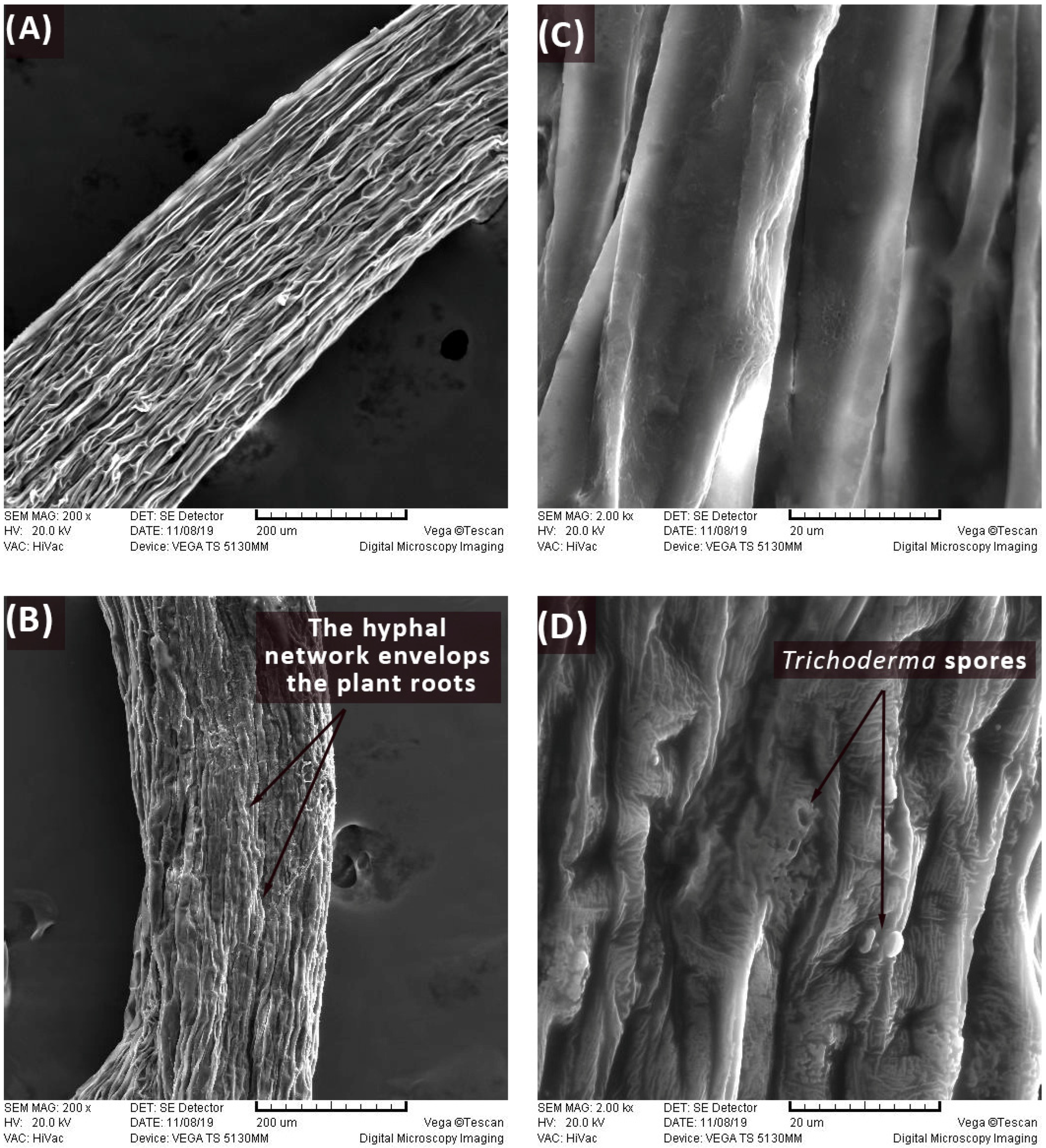
Scanning electron micrography was conducted to check the presence of physical contact between the Trichoderma spp. and the tomato seed radicle during the first 48 h of the co-culture. When radicle samples were collected for Raman and FTIR analysis (Figure 2B), besides the absence of physical contact, it was observed that the radicles in the co-cultures of T. harzianum and T. brevicompactum were larger and that the zone of the primary bark seen in the longitudinal section was less wrinkled (Figure 2A,C). After 48 h in the co-culture, physical contact was established between Trichoderma spp. and the tomato seed radicle. This interaction was visually evidenced in the micrographs, which distinctly depict the emergence of a branched network of hyphal cells accompanied by the presence of spores (Figure 2B,D).

3.2. Stimulating In Vitro Tomato Seed Germination with Trichoderma spp.

The influence of five Trichoderma species on the germination of two tomato varieties, GZ and NA, was investigated in vitro. Tomato seeds were germinated for 48 h in co-culture with T. harzianum, T. brevicompactum, T. virens, T. longibrachiatum, and T. citrinoviride on MS media. The highest stimulation of germination observed for the T. harzianum and T. longibrachiatum treatment of GZ was 15.7% and 15.4%, respectively. T. brevicompactum and T. citrinoviride stimulated germination for 13.6% and 10.2%, respectively, in comparison to the control. Only the T. virens exhibited no statistically significant enhancement in seed germination compared to the control (Figure 3A).
In comparison to control values, the highest stimulation of NA seed germination was observed for T. brevicompactum (15%). The stimulation with other Trichoderma species ranged from 11.62% (T. harzianum) to 14.31% (T. citrinoviride) (Figure 3B).

3.3. Determination of Changes in the Cell Wall Using Raman Spectroscopy

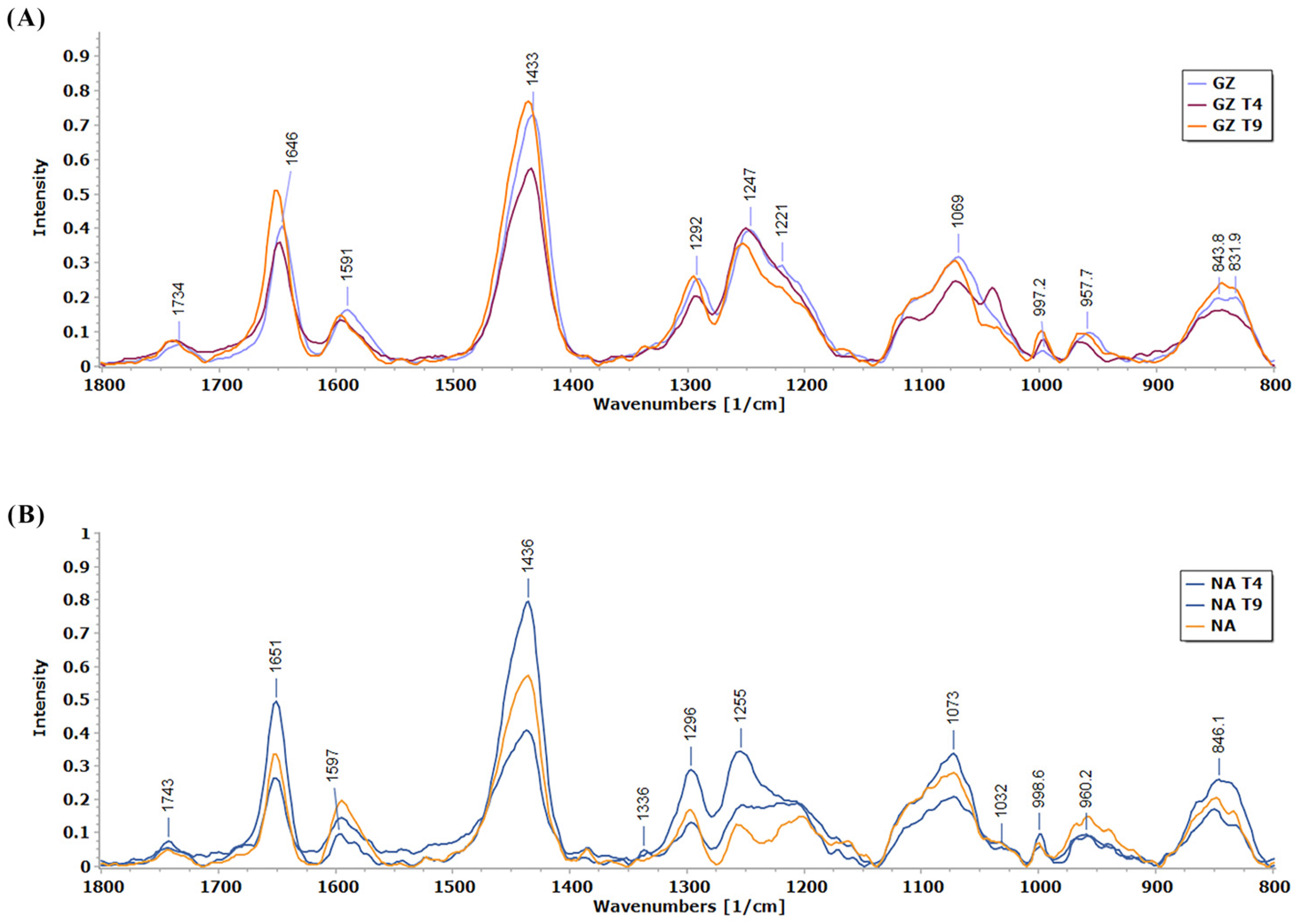
The effect of T4 treatment on the cell wall constituents was less pronounced in the NA tomato variety compared to the effects of the T9 treatment (Figure 4). The most pronounced was the effect of the T9 treatment on the spectral region 1200–1260 cm−1, indicating that this treatment affected content of hemicelluloses, mostly based on xylan. Also, T9 induced an increase of the protein band at around 1550 cm−1, which is not well pronounced in the Raman spectra but is pronounced in the FTIR spectra (Figure 4).
Based on the Raman spectra, it is obvious that Trichoderma T4 had a more pronounced effect on the cell wall structural components as compared to the effect of T9 (Figure 4). This influence is most expressed in the three spectral regions. The band 830–850 cm−1 related to CH in the lignin monomers’ ring is more pronounced in the T4 treatment and has a different structure compared to the corresponding bands in the control and T9 treatment. The bands at 1040 and 1070 cm−1 originate from arabinan-based polysaccharides (arabinan, arabinoglucuronoxylan+galactoglucomannan), while the 1070 cm−1 band may originate from xyloglucan. Their ratio is opposite in the spectra of the T4 treatment compared to the control and T9. These differences indicate that arabinan-based hemicelluloses and xyloglucan are significantly targeted in the T4 treatment. The region 1100–1150 cm−1 is more expressed and differently structured in the T4 treatment compared to the control and T9. Some smaller changes are observed in the region 1200–1300 cm−1, indicating the influence of both T4 and T9 on xylan-based hemicelluloses. The band with the highest intensity in all samples is in the spectral region from 1433 to 1437 cm−1 and might be related to the phenolic compounds in tomato radicles (Figure 4). The region 1594–1596 cm−1 is characteristic of C=O stretch in the conjugated aldehydic structures in lignin. This band is most pronounced in T4-treated plants, indicating the highest content of such type of structure in lignin of this sample. The T4 had some effect on the 1650 cm−1 band, most possibly related to proteins (Table 2).

3.4. PCA Analysis of Raman Spectra of Two Tomato Cultivar Radicles in Co-Culture with Either T. harzianum or T. brevicompactum

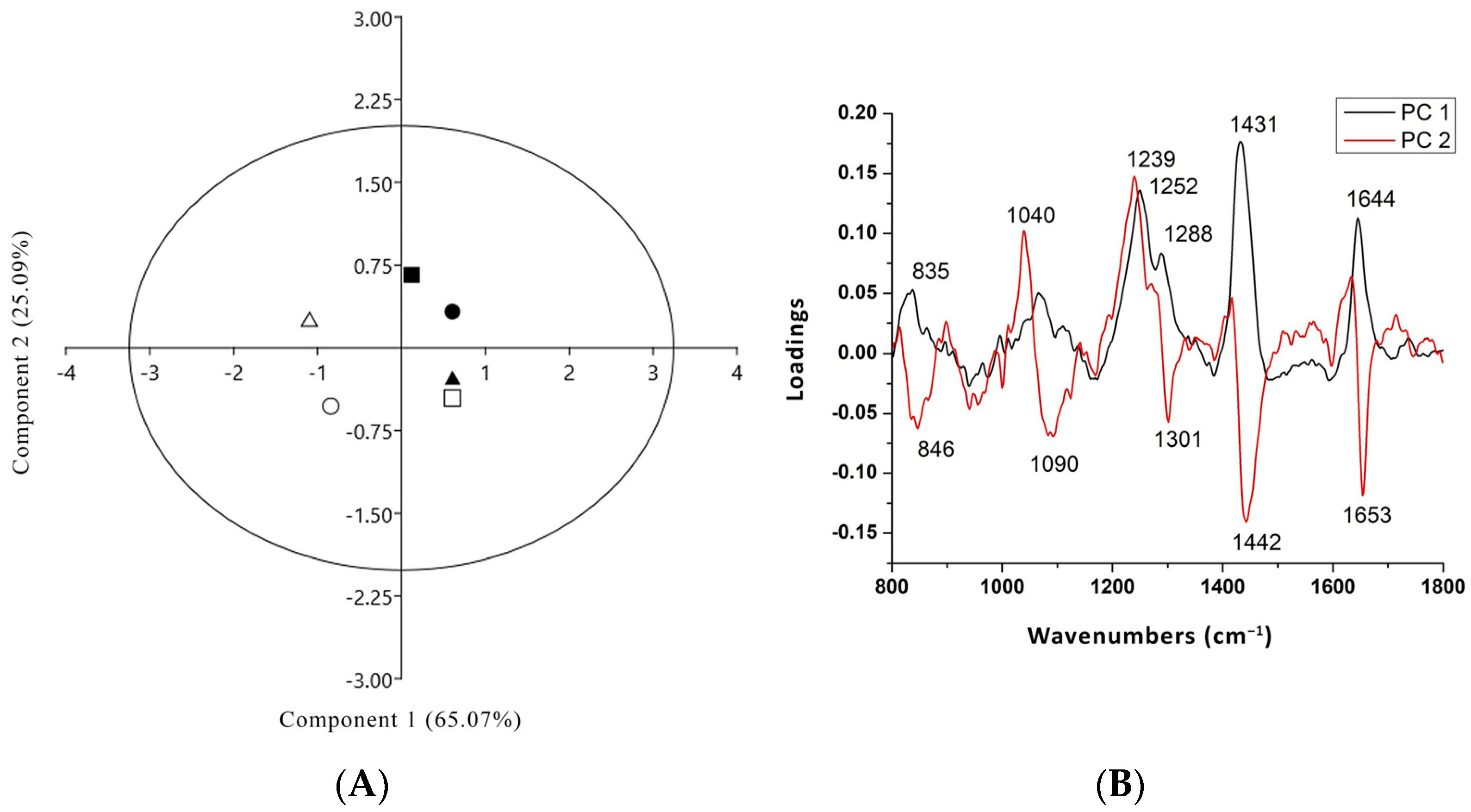
Multivariate analysis, based on PCA, was applied for analysis of the Raman spectra of the radicles from two tomato cultivars in co-culture with T. harzianum or T. brevicompactum. Figure 5 presents the scores and loading plots of the PCA. Figure 5A highlights the good separation into two different groups, where the first and second principal components described 90.17% of data variance. The score plot of PC1 versus PC2 (Figure 5) shows a good separation between the tomato radicle of cultivar Narvik in the control condition and that treated with T. harzianum and T. brevicompactum (NAT4 and NAT9), and the tomato cultivar Gruzanski zlatni radicle treated with T. harzianum (GZT4) was clearly separated from the tomato radicle in the control cultivar (GZ) and the cultivar treated with T. brevicompactum (GZT9). For this purpose, loadings for each PC were analyzed to identify which bands had the most influence, such that it was possible to determine which main chemical constituent had the most influence, since each main chemical constituent was represented by multiple bands in the Raman spectra.
The loading plot of the PC1 (Figure 5B) displays the loadings responsible for the separation in the tomato cultivar GZ in co-culture with T. brevicompactum (GZT9) and T. harzianum (GZT4) from the control tomato radicles (NA) and those in co-culture with T. harzianum and T. brevicompactum (NAT4, NAT9) as well as GZ. PC1 typically explains the majority of variability (65.08%); therefore, the bands in the spectral range 800–1800 cm−1 are primarily assigned to lignin, hemicelluloses including xylan (e.g., broad bands at 835, 1040, 1239–1280 cm−1), phenols (1434 cm−1), and Amide I of proteins (the loadings at 1644 cm−1). The most prominent band, which is responsible for the differentiation among the samples, comes from a broad band position at 1252 cm−1, possibly due to the C-O stretch, C-H, and/or C-O-H bending in hemicellulose or pectic acid, as well as the band for the (O-C-O) skeletal mode of Arabian polysaccharides at 1040 cm−1.
The results show that the GZ samples are well separated along PC1, while the N samples are separated along PC2. The loading plot of the PC2 (Figure 5B) displays the bands responsible for differences between different NA and GZ samples. The most prominent bands responsible for the differentiation among the N samples come from hemicelluloses including xylan (1040, 1239–1280 cm−1), phenols (1442 cm−1), and Amide I (1653 cm−1). This suggests that in both GZT4 and NAT4, there is a higher content of hemi-celluloses, xylan, and phenolics than in the control and T9 treatment.

3.5. FTIR Characterization of Cell Wall Modifications

Within the polysaccharide-associated spectral regions, substantial distinctions between the control and treatment samples are generally absent, except for a notable deviation at 1155 cm−1 (Figure 6). This particular peak at 1155 cm−1 corresponds to the glycosidic vibration in polysaccharides, most probably in pectin (Table 2). The intensity of the peak at 1155 cm−1 is the highest in the control, which indicates that T. harzianum and T. brevicompactum contributed to the reduction in pectin content in the seed radicles. The FTIR spectra of radicles of GZ show that the largest changes occur in the protein part of the spectrum (Figure 6). Prominent protein bands are observed, namely Amide I (max. 1650 cm−1) and Amide II (max. 1520 cm−1) (Table 2), in both tomato varieties (Figure 6). Relative to the control group, the radicles of GZ seeds treated with T. harzianum and T. brevicompactum exhibit increased peak intensity, suggesting an increase in protein content, while the decrease in protein peak intensities for the radicles of NA seeds indicate a decrease in protein content in this variety. This result shows a different effect of the fungi on the protein metabolism in the two varieties.

4. Discussion

The influence of Trichoderma on the growth and development of plants, in the absence of physical contact between these entities, was also examined in Arabidopsis plants [26]. The authors investigated in vitro stimulation of T. atrovirides and T. virens on growth in the plants over a period of 3 to 5 days. It was considered that the stimulation occurs as a result of elevated root auxin content, primarily induced by volatile organic compounds (VOCs) dominated by sesquiterpenes. However, in most of the studies, Trichoderma was directly applied on plants. The stimulation of the in vitro germination of tomato (Arka Meghali) seeds by eight Trichoderma species ranged from 5% to 9% [38]. In a study of the germination and growth response to T. virens treatment in numerous plant species, stimulations were connected with improved nutrient uptake and the induction of indole acetic acid (IAA). Similar to our results, three T. harzianum isolates where shown to stimulate the germination rate of wheat by about 15% after 48 h in vitro [39]. When the experiment was conducted in soil-filled pots, seed germination increased by 45%.
Plant cell walls are very complex and highly variable structures. Their basic structural elements are crystalline cellulose [(1-4)-β-linked glucose] microfibrils embedded in matrix polymers [40]. In the primary cell wall built during early growth, a network is formed together with cellulose, hemicellulose, and pectin [41], with the addition of non-polysaccharide components like proteins, lipids, enzymes, and aromatic compounds [42]. All the different polysaccharides present in the cell walls give rise to the Raman signature of plant cell wall spectra, with partly broadening and overlapping of characteristic bands, but some marker bands for functional groups of pure polysaccharides still exist [41].
The cell walls have a role in seed germination control. It was shown that disruption of genes involved in pectin maturation and hemicellulose deposition strongly influence germination dynamics [43]. In the primary cell wall, the existence of covalently linked pectin to hemicelluloses, especially xylan, is of crucial importance for the cell wall structure [44]. Xyloglucan, a hemicellulose polymer in the primary cell walls, contributes to loosening or stiffening of the wall during cell elongation [45], and its synthesis is under control of a set of enzymes. Based on the vibrational bands characteristic of the glycosidic vibrations of polysaccharide pectin and hemicelluloses xyloglucan and xylan, it can be concluded that in the radicles of Trichoderma-treated seed, the content of pectin decreased in both tomato genotypes. The content of xylan increased in Narvik treated with T9, based on vibration at 1230–1270 cm−1. The content of hemicelluloses increased in GZ in the presence of T4, based on the band at 1040 cm−1. Based on the band at 1596 cm−1, in GZ the conjugated aldehydes in lignin increased in the presence of T4, while in Narvik, the trend was opposite. Characteristic bands for cell wall constituents in the spectral region 1730–1750 cm−1 (C=O stretching vibration of the ester carbonyl group) are due to the presence of pectin and hemicellulose [46,47,48] and are higher in NA treated with T9. The assignments of the characteristic bands in the vibrational spectra of the seedlings are shown in Table 2.
Table 2. Characteristic bands in the vibrational spectra of the tomato seed radicles.
Peak Position (cm−1)AssignmentReferences
817–832C-H out of plane in position 2, 5, and 6 of G units (lignin)[49]
833ring, pectin[46]
854C-H out of plane in position 2, 5, and 6 of G units, aromatic [49]
921C-H out of plane; aromatic [49]
940–960Pectic polysaccharides[50]
966-CH=CH- out-of-plane deformation [49]
1000–1300hemicellulose region[46,47]
1040 + 1070arabinan polysaccharides (hemicellulose) (arabinan, arabinoglucuronoxylan+galactoglucomannan)[47]
1070Xyloglucan[47,50]
1230–1270C–O stretchingvibration in O––C–O, in hemicellulose (xylan), pectin[47,50,51]
1433–1437C-H in phenols[50]
1520Amide II- stretching bands of protein[40]
1596C=O stretching (conjugated aldehydes) in lignin[49]
1650Amide I- stretching bands of protein[52]
Studies involving the root systems of spring and winter wheat cultivars in association with T. cremeum and T. atroviride reveal discernible modifications in the composition of the cell wall [53]. The accumulation of lignin and reorganization of pectin were observed, suggesting the use of these species to protect against pathogens. In the primary cell wall of plants that are in the development phase, there is an increase in the content of cellulose, hemicellulose, and pectin [41]. According to the FTIR bands at 1500–1700 cm−1, in GZ, the protein content increased in the presence of T4, while in Narvik, the protein content decreased, especially in the T9 treatment. It was shown that seeds and roots of corn plants treated with T. lixii had increased soluble protein content as determined by the Bradford method [54]. Similar results were observed in rapeseed seedlings treated with T. reesei using a nitrogen/protein analyzer [55]. During the colonization of plants by Trichoderma spp., proteomic changes occurred in the roots and aerial parts of the plant [56]. However, the molecular mechanisms of action of Trichoderma on plant root protein content when physical contact is not established are still insufficiently investigated.
T. reesei was shown to secrete enzymes like esterase, xyloglucanase, and β-1,4 endoglucanase, capable of breaking down xyloglucan structures [57]. We also showed previously that Trichoderma spp. secrete various extracellular enzymes. It is possible that some of these enzymes may migrate through the medium and may affect seed germination [27,30,57]. According to the API-ZIM test, all tested Trichoderma spp. isolates exhibited similar high activity in three enzymes: acid phosphatase, naphthol-AS-BI-phosphohydrolase, and N-acetyl-β-glucosaminidase [27,30,58]. Data in the literature indicate that two of the three mentioned enzymes are active during the seed germination process. Acid phosphatases are significant in the metabolic processes of plant germination and maturation [59]. They exhibit heightened activity during germination and facilitate the release of reserve materials for the growing embryo [60]. N-acetyl-β-glucosaminidase exhibits increased activity during the germination of cotton seeds [61] and radishes [62]. Recent research has also suggested its use in agriculture to promote seed germination and provide plant protection [63].
Our results indicate that two complementary spectroscopic methods, FTIR and Raman spectroscopy, can give valuable information on rapid changes in the plant cell wall structure of tomato radicles during germination stimulated by Trichoderma spp.

5. Conclusions

To elucidate the early indicators of tomato seed stimulation by Trichoderma spp., a study was carried out using both FTIR and Raman spectroscopy techniques. The results of this study provide evidence for a rapid response of tomato seeds to Trichoderma spp. exposure under in vitro conditions after 48 h on MS substrates, before any physical contact between Trichoderma spp. and tomato seeds was established.
Raman analysis reveals that T. harzianum and T. brevicompactum induce higher synthesis of phenolic compounds through genotype-specific responses within seed radicles. The observed differences between treatments are predominantly based on modifications in the pectin content in the middle lamella, as well as alterations in hemicelluloses and xyloglucan within the primary cell wall. The Raman spectra indicate that the content of xylan increased in NA treated with T9. The content of hemicelluloses increased in GZ in the presence of T4. In GZ, the conjugated aldehydes in lignin increased in the presence of T4, while in NA the trend was opposite. Analysis of the FTIR spectra revealed significant alterations in the protein content of tomato seed radicles treated with Trichoderma spp. in both cultivars. The obtained results provide valuable insights into the molecular mechanisms underlying the interaction between tomato seeds and Trichoderma spp., contributing to a better understanding of plant–microbe interactions. Further investigation should aim to identify the specific compounds responsible for stimulating seed germination. FTIR combined with proteomics should be used to define early changes in proteins in seed radicles and/or small proteins excreted by Trichoderma.

Author Contributions

Conceptualization, I.V., D.R. and D.P.; methodology, I.V., D.R. and D.P.; software, I.V., I.P., S.L., K.R. and D.D; FTIR and Raman spectra assignments, K.R., I.P., S.L. and D.D.; FTIR spectra deconvolution, K.R. and D.D.; formal analysis, I.V. and D.P.; investigation, I.V.; resources, I.V., D.R. and D.P.; data curation, I.V. and D.P.; writing—original draft preparation, I.V.; writing—review and editing D.R., I.P., S.L., D.D., K.R. and D.P.; visualization, I.V., I.P., S.L., D.D. and K.R.; supervision, D.P. All authors have read and agreed to the published version of the manuscript.

Funding

This research was funded by the Provincial Secretary for Higher Education and Scientific Research, Autonomous Province of Vojvodina project, “Use of Trichoderma spp. in sustainable agriculture”, No. 142-451-3172/2022-01/01, Ministry of Science, Technological Development and Innovation of the Republic of Serbia, contract No. 451-03-47/2023-01/200053 with the Institute for Multidisciplinary Research University of Belgrade, and contract No. 451-03-66/2024-03/200051 with the Institute of General and Physical Chemistry, and European Union’s Horizon Europe Project GREENLand—Twinning Microplastic-free Environment under grant agreement number 101079267.

Institutional Review Board Statement

Not applicable.

Informed Consent Statement

Not applicable.

Data Availability Statement

Data available on request.

Conflicts of Interest

The authors declare no conflicts of interest.

References

  1. Pahalvi, H.N.; Rafiya, L.; Rashid, S.; Nisar, B.; Kamili, A.N. Chemical Fertilizers and Their Impact on Soil Health. In Microbiota and Biofertilizers; Dar, G.H., Bhat, R.A., Mehmood, M.A., Hakeem, K.R., Eds.; Springer: Cham, Switzerland, 2021; Volume 2, pp. 1–20. [Google Scholar]
  2. Usigbe, M.J.; Asem-Hiablie, S.; Uyeh, D.D.; Iyiola, O.; Park, T.; Mallipeddi, R. Enhancing resilience in agricultural production systems with AI-based technologies. Environ. Dev. Sustain. 2023, 2, 1–29. [Google Scholar] [CrossRef]
  3. Andrade-Hoyos, P.; Silva-Rojas, H.V.; Romero-Arenas, O. Endophytic Trichoderma species isolated from Persea americana and Cinnamomum verum roots reduce symptoms caused by Phytophthora cinnamomi in avocado. Plants 2020, 9, 1220. [Google Scholar] [CrossRef]
  4. Silletti, S.; Di Stasio, E.; Van Oosten, M.J.; Ventorino, V.; Pepe, O.; Napolitano, M.; Marra, R.; Woo, S.L.; Cirillo, V.; Maggio, A. Biostimulant activity of Azotobacter chroococcum and Trichoderma harzianum in Durum wheat under water and nitrogen deficiency. Agronomy 2021, 11, 380. [Google Scholar] [CrossRef]
  5. Quinet, M.; Angosto, T.; Yuste-Lisbona, F.J.; Blanchard-Gros, R.; Bigot, S.; Martinez, J.P.; Lutts, S. Tomato fruit development and metabolism. Front. Plant Sci. 2019, 10, 1554. [Google Scholar] [CrossRef]
  6. Farooq, M.S.M.A.; Basra, S.M.A.; Saleem, B.A.; Nafees, M.; Chishti, S.A. Enhancement of tomato seed germination and seedling vigor by osmopriming. Pak. J. Agric. Sci. 2005, 42, 3–4. [Google Scholar]
  7. Sharma, S.; Kour, D.; Rana, K.L.; Dhiman, A.; Thakur, S.; Thakur, P.; Thakur, S.; Thakur, N.; Sudheer, S.; Yadav, N. Trichoderma: Biodiversity, ecological significances, and industrial applications. In Recent Advancement in White Biotechnology through Fungi; Springer: Berlin/Heidelberg, Germany, 2019; pp. 85–120. [Google Scholar]
  8. Ahluwalia, V.; Kumar, J.; Rana, V.S.; Sati, O.P.; Walia, S. Comparative evaluation of two Trichoderma harzianum strains for major secondary metabolite production and antifungal activity. Nat. Prod. Res. 2015, 29, 914–920. [Google Scholar] [CrossRef]
  9. Karuppiah, V.; Li, T.; Vallikkannu, M.; Chen, J. Co-cultivation of Trichoderma asperellum GDFS1009 and Bacillus amyloliquefaciens 1841 causes differential gene expression and improvement in the wheat growth and biocontrol activity. Front. Microbiol. 2019, 10, 1068. [Google Scholar] [CrossRef]
  10. Juliatti, F.C.; Rezende, A.A.; Juliatti, B.C.M.; Morais, T.P. Trichoderma—The Most Widely Used Fungicide. IntechOpen: London, UK, 2019. [Google Scholar]
  11. Racić, G.; Vukelić, I.; Prokić, L.; Ćurčić, N.; Zorić, M.; Jovanović, L.; Panković, D. The influence of Trichoderma brevicompactum treatment and drought on physiological parameters, abscisic acid content and signalling pathway marker gene expression in leaves and roots of tomato. Ann. Appl. Biol. 2018, 173, 213–221. [Google Scholar] [CrossRef]
  12. Cornejo-Ríos, K.; Osorno-Suárez, M.d.P.; Hernández-León, S.; Reyes-Santamaría, M.I.; Juárez-Díaz, J.A.; Pérez-España, V.H.; Peláez-Acero, A.; Madariaga-Navarrete, A.; Saucedo-García, M. Impact of Trichoderma asperellum on chilling and drought stress in tomato (Solanum lycopersicum). Horticulturae 2021, 7, 385. [Google Scholar] [CrossRef]
  13. Bashyal, B.M.; Parmar, P.; Zaidi, N.W.; Aggarwal, R. Molecular programming of drought-challenged Trichoderma harzianum-bioprimed rice (Oryza sativa L.). Front. Microbiol. 2021, 12, 655165. [Google Scholar] [CrossRef]
  14. Sánchez-Montesinos, B.; Diánez, F.; Moreno-Gavira, A.; Gea, F.J.; Santos, M. Plant growth promotion and biocontrol of Pythium ultimum by saline tolerant Trichoderma isolates under salinity stress. Int. J. Environ. Res. Public Health 2019, 16, 2053. [Google Scholar] [CrossRef]
  15. Brotman, Y.; Landau, U.; Cuadros-Inostroza, Á.; Takayuki, T.; Fernie, A.R.; Chet, I.; Viterbo, A.; Willmitzer, L. Trichoderma-plant root colonization: Escaping early plant defense responses and activation of the antioxidant machinery for saline stress tolerance. PLoS Pathog. 2013, 9, e1003221. [Google Scholar] [CrossRef]
  16. Maurya, S.; Rashk-E-Eram; Naik, S.K.; Choudhary, J.S.; Kumar, S. Heavy metals scavenging potential of Trichoderma asperellum and Hypocrea nigricans isolated from acid soil of Jharkhand. Indian J. Microbiol. 2019, 59, 27–38. [Google Scholar] [CrossRef]
  17. Sun, H.; Wu, L.; Hao, Y.; Liu, C.; Pan, L.; Zhu, Z. Tolerance mechanism of Trichoderma asperellum to Pb2+: Response changes of related active ingredients under Pb2+ stress. RSC Adv. 2020, 10, 5202–5211. [Google Scholar] [CrossRef]
  18. Ghorbanpour, A.; Salimi, A.; Ghanbary, M.A.T.; Pirdashti, H.; Dehestani, A. The effect of Trichoderma harzianum in mitigating low temperature stress in tomato (Solanum lycopersicum L.) plants. Sci. Hortic. 2018, 230, 134–141. [Google Scholar] [CrossRef]
  19. Contreras-Cornejo, H.A.; Macías-Rodríguez, L.; del-Val, E.; Larsen, J. Ecological functions of Trichoderma spp. and their secondary metabolites in the rhizosphere: Interactions with plants. FEMS Microbiol. Ecol. 2016, 92, 036. [Google Scholar] [CrossRef]
  20. Kubicek, C.P.; Steindorff, A.S.; Chenthamara, K.; Manganiello, G.; Henrissat, B.; Zhang, J. Evolution and comparative genomics of the most common Trichoderma species. BMC Genom. 2019, 20, 485. [Google Scholar] [CrossRef]
  21. Mastouri, F.; Björkman, T.; Harman, G.E. Seed treatment with Trichoderma harzianum alleviates biotic, abiotic, and physiological stresses in germinating seeds and seedlings. Phytopathology 2010, 100, 1213–1221. [Google Scholar] [CrossRef]
  22. Rajjou, L.; Duval, M.; Gallardo, K.; Catusse, J.; Bally, J.; Job, C.; Job, D. Seed germination and vigor. Annu. Rev. Plant Biol. 2012, 63, 507–533. [Google Scholar] [CrossRef]
  23. Li, W.; Niu, Y.; Zheng, Y.; Wang, Z. Advances in the understanding of reactive oxygen species-dependent regulation on seed dormancy, germination, and deterioration in crops. Front. Plant Sci. 2022, 13, 826809. [Google Scholar] [CrossRef]
  24. You, J.; Zhang, J.; Wu, M.; Yang, L.; Chen, W.; Li, G. Multiple criteria-based screening of Trichoderma isolates for biological control of Botrytis cinerea on tomato. Biol. Control 2016, 101, 31–38. [Google Scholar] [CrossRef]
  25. Jogaiah, S.; Abdelrahman, M.; Tran, L.S.P.; Ito, S.I. Different mechanisms of Trichoderma virens—Mediated resistance in tomato against Fusarium wilt involve the jasmonic and salicylic acid pathways. Mol. Plant Pathol. 2018, 19, 870–882. [Google Scholar] [CrossRef]
  26. González-Pérez, E.; Ortega-Amaro, M.A.; Salazar-Badillo, F.B.; Bautista, E.; Douterlungne, D.; Jiménez-Bremont, J.F. The Arabidopsis-Trichoderma interaction reveals that the fungal growth medium is an important factor in plant growth induction. Sci. Rep. 2018, 8, 16427. [Google Scholar] [CrossRef]
  27. Vukelić, I.D.; Prokić, L.T.; Racić, G.M.; Pešić, M.B.; Bojović, M.M.; Sierka, E.M.; Panković, D.M. Effects of Trichoderma harzianum on photosynthetic characteristics and fruit quality of tomato plants. Int. J. Mol. Sci. 2021, 22, 6961. [Google Scholar] [CrossRef]
  28. Tucci, M.; Ruocco, M.; De Masi, L.; De Palma, M.; Lorito, M. The beneficial effect of Trichoderma spp. on tomato is modulated by the plant genotype. Mol. Plant Pathol. 2011, 12, 341–354. [Google Scholar] [CrossRef]
  29. Racić, G.; Körmöczi, P.; Kredics, L.; Raičević, V.; Mutavdžić, B.; Vrvić, M.M.; Panković, D. Effect of the edaphic factors and metal content in soil on the diversity of Trichoderma spp. Environ. Sci. Pollut. Res. 2017, 24, 3375–3386. [Google Scholar] [CrossRef]
  30. Racić, G. Variability of Indigenous Trichoderma spp. in Different Soil Types and Their Ecological and Biochemical Characterization. Ph.D. Thesis, University of Belgrade, Belgrade, Sebia, 2017. [Google Scholar]
  31. Murashige, T.; Skoog, F. A revised medium for rapid growth and bio assays with tobacco tissue cultures. Physiol. Plant. 1962, 15, 473–497. [Google Scholar] [CrossRef]
  32. Institute for Vegetable Crops, Smederevska Palanka. Available online: https://institut-palanka.rs/ (accessed on 17 January 2018).
  33. Đorđević, M.; Vatchev, T.; Girek, Z.; Šević, M.; Zečević, B.; Zdravković, J.; Ivanović, M. Reaction of different tomato cultivars toward race 1 of Fusarium oxysporum f. sp. Lycopersici. Genetika 2012, 44, 109–118. [Google Scholar]
  34. Clearya, M.R.; Danielb, G.; Stenlid, J. Light and scanning electron microscopy studies of the early infection stages of Hymenoscyphus pseudoalbiduson Fraxinus excelsior. Molecules 2022, 27, 4616. [Google Scholar]
  35. Menges, F. Spectragryph—Optical Spectroscopy Software, Version 1.2.8; Spectragryph: Oberstdorf, Germany, 2018. Available online: http://www.effemm2.de/spectragryph/(accessed on 12 March 2019).
  36. Hammer, Ø.; Harper, D.A.T.; Ryan, P.D. PAST: Paleontological statistics software package for education and data analysis. Palaeont. Electr. 2001, 4, 9. [Google Scholar]
  37. Global Analytical and Measuring Instruments. Available online: https://www.shimadzu.com/an/service-support/technical-support/analysisbasics/ftirtalk/talk8.html (accessed on 17 January 2018).
  38. Konappa, N.; Krishnamurthy, S.; Arakere, U.C.; Chowdappa, S.; Ramachandrappa, N.S. Efficacy of indigenous plant growth-promoting rhizobacteria and Trichoderma strains in eliciting resistance against bacterial wilt in a tomato. Egypt. J. Biol. Pest Control 2020, 30, 30. [Google Scholar] [CrossRef]
  39. Anjum, Z.A.; Hayat, S.; Ghazanfar, M.U.; Ahmad, S.; Adnan, M.; Hussian, I. Does seed priming with Trichoderma isolates have any impact on germination and seedling vigor of wheat. Int. J. Bot. Stud. 2020, 5, 65–68. [Google Scholar]
  40. Gierlinger, N. Revealing changes in molecular composition of plant cell walls on the micron-level by Raman mapping and vertex component analysis (VCA). Front. Plant Sci. 2014, 5, 306. [Google Scholar] [CrossRef]
  41. Keegstra, K. Plant cell walls. Plant Physiol. 2010, 154, 483–486. [Google Scholar] [CrossRef]
  42. Carpita, N.C.; Gibeaut, D.M. Structural models of primary cell walls in flowering plants: Consistency of molecular structure with the physical properties of the walls during growth. Plant J. 1993, 3, 1–30. [Google Scholar] [CrossRef]
  43. Di Marzo, M.; Babolin, N.; Viana, V.E.; de Oliveira, A.C.; Gugi, B.; Caporali, E.; Herrera-Ubaldo, H.; Martínez-Estrada, E.; Driouich, A.; de Folter, S.; et al. The Genetic Control of SEEDSTICK and LEUNIG-HOMOLOG in Seed and Fruit Development: New Insights into Cell Wall Control. Plants 2022, 11, 3146. [Google Scholar] [CrossRef]
  44. Broxterman, S.E.; Schols, H.A. Characterisation of pectin-xylan complexes in tomato primary plant cell walls. Carbohydr. Polym. 2018, 197, 269–276. [Google Scholar] [CrossRef]
  45. Cosgrove, D.J. Growth of the plant cell wall. Nat. Rev. Mol. Cell Biol. 2005, 6, 850–861. [Google Scholar] [CrossRef]
  46. Kacuráková, M.; Smith, A.C.; Gidley, M.J.; Wilson, R.H. Molecular interactions in bacterial cellulose composites studied by 1D FT-IR and dynamic 2D FT-IR spectroscopy. Carbohydr Res. 2002, 337, 1145–1153. [Google Scholar] [CrossRef]
  47. Kačuráková, M.; Capek, P.; Sasinková, V.; Wellner, N.; Ebringerová, A. FT-IR study of plant cell wall model compounds: Pectic polysaccharides and hemicelluloses. Carbohydr. Polym. 2000, 43, 195–203. [Google Scholar] [CrossRef]
  48. Schulz, H.; Baranska, M. Identification and Quantification of Valuable Plant Substances by IR and Raman Spectroscopy. Vib. Spectrosc. 2007, 43, 13–25. [Google Scholar] [CrossRef]
  49. Faix, O. Classification of Lignins from Different Botanical Origins by FT-IR Spectroscopy. Holzforschung 1991, 45, 21–28. [Google Scholar] [CrossRef]
  50. Alonso-Simón, A.; García-Angulo, P.; Mélida, H.; Encina, A.; Álvarez, J.M.; Acebes, J.L. The use of FTIR spectroscopy to monitor modifications in plant cell wall architecture caused by cellulose biosynthesis inhibitors. Plant Signal Behav. 2011, 6, 1104–1110. [Google Scholar] [CrossRef]
  51. Chowdappa, P.; Kumar, S.M.; Lakshmi, M.J.; Upreti, K.K. Growth stimulation and induction of systemic resistance in tomato against early and late blight by Bacillus subtilis OTPB1 or Trichoderma harzianum OTPB3. Biol. Control 2013, 65, 109–117. [Google Scholar] [CrossRef]
  52. McCann, M.C.; Hammouri, M.; Wilson, R.; Belton, P.; Roberts, K. Fourier transform infrared microspectroscopy is a new way to look at plant cell walls. Plant Physiol. 1992, 100, 1940–1947. [Google Scholar] [CrossRef]
  53. Basińska-Barczak, A.; Błaszczyk, L.; Szentner, K. Plant cell wall changes in common wheat roots as a result of their interaction with beneficial fungi of Trichoderma. Cells 2020, 9, 2319. [Google Scholar] [CrossRef]
  54. Pehlivan, N.; Yesilyurt, A.M.; Durmus, N.; Karaoglu, S.A. Trichoderma lixii ID11D seed biopriming mitigates dose dependent salt toxicity in maize. Acta Physiol. Plant. 2017, 39, 79. [Google Scholar] [CrossRef]
  55. Alhomodi, A.F.; Zavadil, A.; Berhow, M.; Gibbons, W.R.; Karki, B. Composition of canola seed sprouts fermented by Aureobasidium pullulans, Neurospora crassa, and Trichoderma reesei under submerged-state fermentation. Food Bioprod. Process. 2021, 126, 256–264. [Google Scholar] [CrossRef]
  56. Zhang, B.; Gao, Y.; Zhang, L.; Zhou, Y. The plant cell wall: Biosynthesis, construction, and functions. J. Integr. Plant Biol. 2021, 63, 251–272. [Google Scholar] [CrossRef]
  57. Rangel, P.N.; Tovborg, M.; Soleimani, F.A.; Della, P.E.A. Multicomponent carbohydrase system from Trichoderma reesei: A toolbox to address complexity of cell walls of plant substrates in animal feed. PLoS ONE 2021, 16, e0251556. [Google Scholar]
  58. Danilovic, G.; Radic, D.; Raicevic, V.; Jovanovic, L.; Kredics, L.; Pankovic, D. Extracellular enzyme activity of Trichoderma strains isolated from different soil types. In Proceedings of the 2nd International Symposium for Agriculture and Food, Ohrid, Republic of Macedonia, 7–9 October 2015; pp. 323–327. [Google Scholar]
  59. dos Prazeres, J.N.; Ferreira, C.V.; Aoyama, H. Acid phosphatase activities during the germination of Glycine max seeds. Plant Physiol. Biochem. 2004, 42, 15–20. [Google Scholar] [CrossRef]
  60. Thomas, T.L. Gene expression during plant embryogenesis and germination: An overview. Plant Cell 1993, 5, 1401. [Google Scholar]
  61. Yi, C.K. Increase in β-N-acetylglucosaminidase activity during germination of cotton seeds. Plant Physiol. 1981, 67, 68–73. [Google Scholar] [CrossRef]
  62. Berger, S.; Menudier, A.; Julien, R.; Karamanos, Y. Endo-N-acetyl-β-D-glucosaminidase and peptide-N4-(N-acetyl-glucosaminyl) asparagine amidase activities during germination of Raphanus sativus. Phytochemistry 1995, 39, 481–487. [Google Scholar] [CrossRef]
  63. Cao, S.; Liu, Y.; Shi, L.; Zhu, W.; Wang, H. N-Acetylglucosamine as a platform chemical produced from renewable resources: Opportunity, challenge, and future prospects. Green Chem. 2022, 24, 493–509. [Google Scholar] [CrossRef]
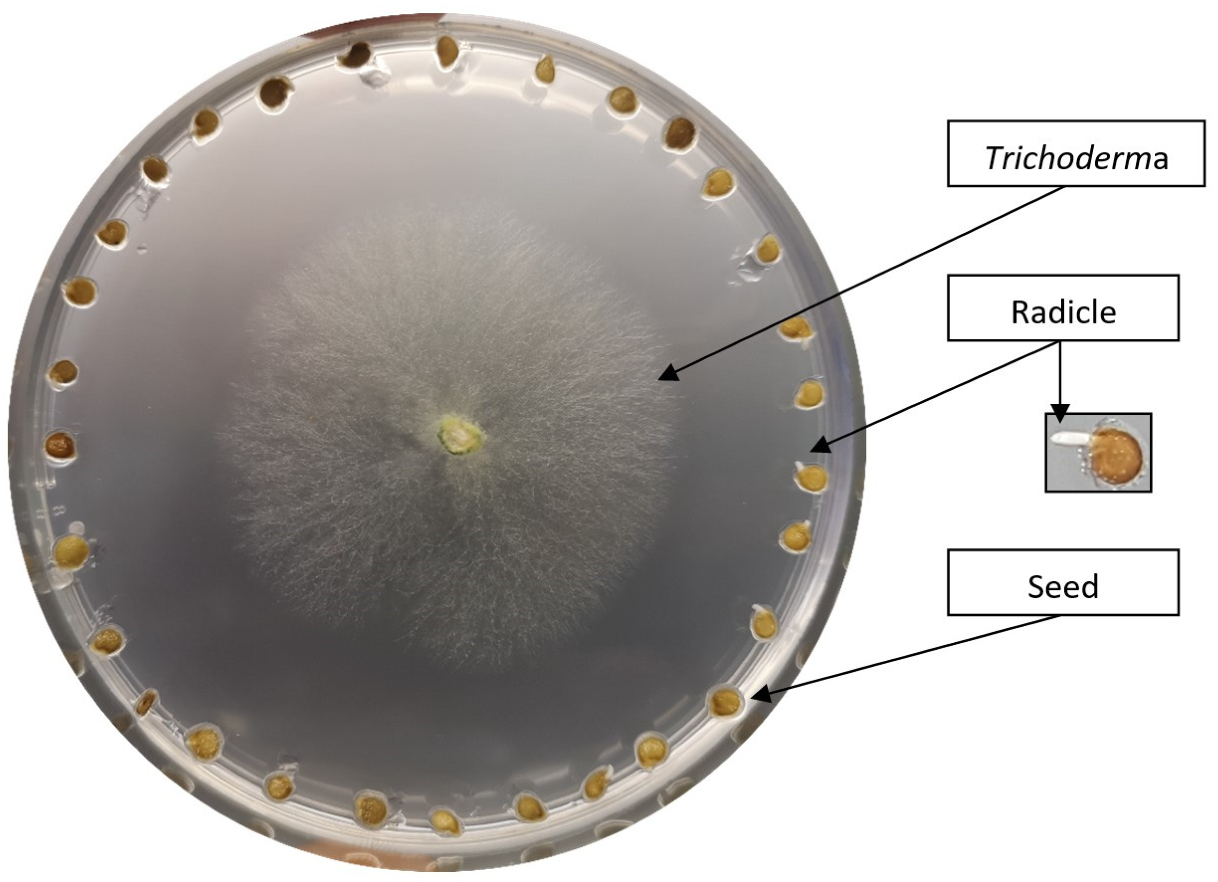
Figure 1. Tomato seed (GZ) cultivated in the presence of T. harzianum on MS medium.
Biology 13 00340 g001
Figure 2. Plant radicles imaged with a scanning electron microscope. GZ tomato radicle epidermis surface in control condition at 200× magnification (A) and at 2000× magnification (C); GZ tomato radicle epidermis surface after contact with T. brevicompactum at 200× magnification (B) and at 2000× magnification (D). Similar results were obtained for another tomato variety Narvik (not presented).
Biology 13 00340 g002
Figure 3. Percentage of germinated tomato seeds, after 48 h on MS media under different treatments: control (C), T. harzianum (T4), T. virens (T7), T. brevicompactum (T9), T. longibrachiatum (T11), and T. citrinovirides (T13). (A) Percentage of germinated GZ seeds; (B) Percentage of germinated NA seeds. One-way ANOVA and post hoc Tukey’s test were used for statistical analysis of data. Values marked with the same letter are not statistically significantly different according to Tukey’s test (p < 0.05). The error lines represent ± standard deviation of the mean. Different letters (a, b and c) indicate statistically significant differences according to Tukey’s test (p < 0.05). For each treatment, 28 Petri dishes of 30 seeds each were analyzed (n = 28).
Biology 13 00340 g003
Figure 4. Averages of normalized Raman spectra of two tomato cultivar radicles GZ and NA in control conditions or in co-culture with either T. harzianum or T. brevicompactum. (A) Spectra of the Gruzanski zlatni cultivar radicles (GZ—control; GZT4—T. harzianum; GZT9—T. brevicompactum); (B) Spectra of the Narvik cultivar radicles (NA—control; NAT4—T. harzianum; NAT9—T. brevicompactum).
Biology 13 00340 g004
Figure 5. (A) Score plot of the first principal component (PC1) versus the second principal component (PC2) of the tomato radicle from two cultivars (GZ and NA) treated with T. harzianum (T4) and T. brevicompactum (T9). (B) Loading plot corresponding to PC1 and PC2 responsible for the differences between the cultivars. N—closed cycle, NAT4—closed square, NAT9—closed triangle, GZ—open cycle, GZT4—open square, and GZT9—open triangle.
Biology 13 00340 g005
Figure 6. FTIR spectra of homogenized Gruzanski zlatni (A) and Narvik (B) radicles under different treatments: control (GZ, NA), seeds treated with T. harzianum (GZT4, NAT4), seeds treated with T. brevicompactum (GZT9, NAT9).
Biology 13 00340 g006
Table 1. Trichoderma species selected for the experiment.
GenusSpeciesCodeNo. of Isolates in the SZMC CollectionAccession Number
TrichodermaharzianumT422660KP316448.1
TrichodermabrevicompactumT922661KP316440.1
TrichodermavirensT722659KP316449.1
TrichodermalongibrachiatumT1122664KP316444.1
TrichodermacitrinovirideT1322668KP316445.1
Disclaimer/Publisher’s Note: The statements, opinions and data contained in all publications are solely those of the individual author(s) and contributor(s) and not of MDPI and/or the editor(s). MDPI and/or the editor(s) disclaim responsibility for any injury to people or property resulting from any ideas, methods, instructions or products referred to in the content.