Article

N6-Methyladenosine Directly Regulates CD40L Expression in CD4+ T Lymphocytes

by
Ellen C. N. van Vroonhoven
1,
Lucas W. Picavet
1,
Rianne C. Scholman
1,
Noortje A. M. van den Dungen
2,
Michal Mokry
2,
Anouk Evers
3,
Robert J. Lebbink
3,
Jorg J. A. Calis
1,
Sebastiaan J. Vastert
1,4 and
Jorg van Loosdregt
1,*
1
Center for Translational Immunology, University Medical Center Utrecht, 3584 CX Utrecht, The Netherlands
2
Department of Experimental Cardiology, University Medical Center Utrecht, 3584 CX Utrecht, The Netherlands
3
Department of Medical Microbiology, University Medical Center Utrecht, 3584 CX Utrecht, The Netherlands
4
Department of Pediatric Rheumatology and Immunology, University Medical Center Utrecht, 3584 CX Utrecht, The Netherlands
*
Author to whom correspondence should be addressed.
Biology 2023, 12(7), 1004; https://doi.org/10.3390/biology12071004
Submission received: 11 May 2023 / Revised: 29 June 2023 / Accepted: 5 July 2023 / Published: 14 July 2023

Abstract

:

Simple Summary

The tight regulation of the expression of cytokines, surface receptors and co-stimulatory and co-inhibitory molecules is necessary for a well-functioning immune system. There are various cellular processes that regulate the expression of these immune regulatory proteins, for instance at the RNA transcript level. Epitranscriptomic regulation, involving RNA binding proteins and modifications, can determine the turnover and translation of mRNA transcripts. N6-methyladenosine (m6A) is the most abundant RNA modification in eukaryotic cells and it was demonstrated that m6A is involved in the early differentiation of CD4+ T lymphocytes. However, the function of m6A in CD4+ T cell activation and function is still incompletely understood. Here, we demonstrate that m6A regulates the activation of CD4+ T lymphocytes via the regulation of CD40 ligand expression, a key co-stimulatory molecule expressed on the cell surface of CD4+ T cells. The discovery of this novel function of m6A and its regulatory proteins contributes to our general understanding of CD4+ T cell activation, gene expression regulation and autoimmune disease pathogenesis.

Abstract

T cell activation is a highly regulated process, modulated via the expression of various immune regulatory proteins including cytokines, surface receptors and co-stimulatory proteins. N6-methyladenosine (m6A) is an RNA modification that can directly regulate RNA expression levels and it is associated with various biological processes. However, the function of m6A in T cell activation remains incompletely understood. We identify m6A as a novel regulator of the expression of the CD40 ligand (CD40L) in human CD4+ lymphocytes. Manipulation of the m6A ‘eraser’ fat mass and obesity-associated protein (FTO) and m6A ‘writer’ protein methyltransferase-like 3 (METTL3) directly affects the expression of CD40L. The m6A ‘reader’ protein YT521-B homology domain family-2 (YTHDF2) is hypothesized to be able to recognize and bind m6A specific sequences on the CD40L mRNA and promotes its degradation. This study demonstrates that CD40L expression in human primary CD4+ T lymphocytes is regulated via m6A modifications, elucidating a new regulatory mechanism in CD4+ T cell activation that could possibly be leveraged in the future to modulate T cell responses in patients with immune-related diseases.

1. Introduction

CD4+ T cell activation is achieved upon the binding of the peptide major histocompatibility complex (pMHC) on the surface of antigen-presenting cells (APCs) to the T cell receptor (TCR). Upon the pMHC–TCR interaction, co-stimulatory molecules such as CD28, inducible T cell co-stimulator (ICOS) and CD40 ligand (CD40L) are translocated to the CD4+ T cell surface. Together, these co-stimulatory and co-inhibitory molecules modulate the duration and intensity of TCR-mediated signaling, thereby facilitating an optimal immune response. Given the complexity of the immune response, precise regulation of the expression of co-stimulatory molecules amongst other processes is essential for a well-orchestrated T cell response.
Post-transcriptional regulation, involving RNA binding proteins and RNA modifications, plays a crucial role in the regulation of protein expression. One of these modifications is N6-methyladenosine (m6A), which was discovered in the 1970s and is the most prevalent RNA modification in eukaryotes [1,2,3]. m6A is a dynamic modification that is co- and post-transcriptionally installed on specific mRNA sequences, mostly located in the 3′-untranslated region (UTR), near the STOP codon and in the 5′-UTR of the processed mRNA [3]. m6A is increased in so-called ‘DRACH’-motif sequences (D = A, G, or U; R = G or A; H = A, C or U) [4] and is installed by a complex of ‘writer’ proteins that include METTL3 [5,6], methyltransferase-like 14 (METTL14) and Wilms’ tumor 1-associating protein (WTAP) [6]. ‘Reader’ proteins from the YT521-B homology domain-containing (YTH) family selectively bind to the m6A modification, thereby determining the fate of the transcript [7,8,9]. These readers have been described to regulate RNA stability and therefore RNA degradation (YTHDF1, YTHDF2 and YTHDF3) [9,10] by promoting transport towards processing bodies, or to be involved in splicing regulation and the nuclear export of mRNA transcripts (YTHDC1) [11]. m6A can be removed from the RNA by ‘eraser’ proteins FTO [12,13] or AlkB homologue 5 (ALKBH5) [14].
Recently, it has been demonstrated that m6A modifications are involved in early CD4+ T cell differentiation. For instance, METTL3 was identified as an important stabilizer of T follicular helper (TFH) cell signature gene Tcf7, indicating a role for m6A in TFH cell differentiation [15]. Additionally, METTL3 was found to regulate naïve T lymphocyte proliferation and differentiation [16]. Naïve T cells from Mettl3-KO mice showed increased expression of Socs1, Socs3 and Cish, thereby suppressing IL-7/STAT5 signaling and enhancing ERK and AKT signaling. The eraser protein ALKBH5 was demonstrated to have a checkpoint role in the early differentiation of γδ T cells and αβ T cells by targeting Jagged1/Notch2 signaling [17]. Despite recent advances in understanding the implications of m6A in CD4+ T cell regulation, the full implications of its involvement remain incompletely understood.
To obtain further insight into the role of m6A in T cell activation, we assessed the changes in the m6A modification of TCR signaling and T cell activation-associated RNA transcripts in human primary CD4+ T lymphocytes and identified many changes upon activation. m6A enrichment was found to be increased on co-stimulatory molecule CD40 ligand (CD40L) mRNA, primarily in the 3′ UTR of the transcript. Promoting m6A levels in primary CD4+ T cells resulted in the decreased expression of CD40L. YTHDF2, a m6A reader associated with RNA degradation, is hypothesized to directly bind to m6A-specific sequences in the 5′ and 3′-UTR of the CD40L mRNA. Taken together, our data demonstrate that the expression of CD40L in CD4+ T lymphocytes is directly regulated via m6A and its regulatory proteins.

2. Materials and Methods

2.1. Cell Culture

Peripheral blood mononuclear cells (PBMCs) were obtained from healthy donor blood using Ficoll-Paque Plus (Cytiva, Marlborough, MA, USA) density gradient media. Primary human CD4+ T lymphocytes were isolated via magnetic-activated cell sorting (MACS) using the human CD4+ T cell isolation kit (Miltenyi Biotec, Bergisch Gladbach, Germany). Cells were cultured in a Roswell Park Memorial Institute (RPMI) 1640 medium (Fisher Scientific, Hampton, NH, USA) containing 10% heat-inactivated human AB serum, 1% L-glutamine (Life Technologies, Carlsbad, CA, USA), 100 U/mL of penicillin and 100 mg/mL of streptomycin (Gibco, New York, NY, USA) supplemented with 50 U/mL of IL-2. Jurkat cells (Clone E6.1, ATCC, Manassas, VA, USA) were cultured in Dulbecco’s modified Eagle medium (DMEM) with GlutaMax (Gibco), supplemented with 10% heat-inactivated fetal calf serum (Sigma-Aldrich, St. Louis, MI, USA), 100 U/mL of penicillin and 100 mg/mL of streptomycin (Gibco). All cells were cultured at 37 °C in 5% CO2. For the manipulation of FTO and METTL3, 50 µM entacapone (Sigma, SML0654-10MG) and 30 µM STM2457 (MedChemExpress, Monmouth Junction, NJ, USA, 2499663-01-1) were used. For CRISPR/Cas9 genome engineering, single-guide RNAs (sgRNAs) were introduced in the lentiviral pSicoR-CRISPR-PuroR vector (RP-557) [18] as described previously. The used crRNA sequence for FTO knockout was GGCTGCTTATTTCGGGACC. CRISPR/Cas9 knockout of FTO in Jurkat cells was performed via lentiviral transduction. Second-generation lentiviral particles were produced in HEK293T cells using polyethylenimine (PEI) MAX (Polysciences Europe GmbH, Hirschberg, Germany), and Jurkat cells were transduced using 5 µg/mL of polybrene (Santa Cruz, Santa Cruz, CA, USA). Transduced cells were single-cell-cultured and selected with 2 µg/mL of puromycin (Merck, Rahway, NJ, USA).

2.2. meRIP Sequencing and qPCR

CD4+ T lymphocytes were isolated from healthy donor PBMCs using MACS. The cells were either activated for 4 h with PMA (20 ng/mL, Sigma) and ionomycin (1 ug/mL, Calbiochem), or stimulated overnight with CD3/CD28 Dynabeads (Invitrogen, Waltham, MA, USA). Cells were lysed in Trizol LS reagent (Fisher Scientific) and RNA was isolated according to the manufacturer’s protocol. For meRIPseq only, mRNA was isolated and subsequently fragmented by using the NEXTflex Rapid directional mRNA-seq bundle (5138-10). Subsequently, Protein A/G magnetic beads (Fisher Scientific) were coated with mouse anti-m6A (Synaptic Systems Gmbh, Göttingen, Germany, 202-111) or mouse anti-IgG2b,k isotype control (BD, 556577). mRNA was added to the coated beads and IP was performed. mRNA was washed using a high-salt–low-salt washing method. To elute the bound RNA, the beads were mixed with RLT lysis buffer from the RNeasy mini-kit (Qiagen, Hilden, Germany). This kit was subsequently used for the purification of the mRNA. Sequences from the Epimark N6-Methyladenosine Enrichment kit (New England Biolabs, Ipswich, MA, USA) were added to the samples. meRIPseq samples were single-end sequenced at 50 bp on the Illumina NextSeq500 sequencer (Utrecht DNA Sequencing Facility, Utrecht, The Netherlands). Sequencing reads were aligned using STAR (version 2.7.1) [19] to the human genome (version GRCh38) with transcriptomic information from ENSEMBL (version GRCh38.v100) supplemented with sequences from the Epimark N6-Methyladenosine Enrichment kit (New England Biolabs). Unique reads were selected, and expression was quantified at the gene level using htseq-count (version 0.11.4) [20]. Differential expression was analyzed with edgeR [21], and per sample expression was normalized using TMM normalization. cDNA synthesis was performed on the meRIP qPCR samples using the iScript cDNA synthesis kit (Bio-Rad, Hercules, CA, USA). qPCR was performed with SYBR Select mastermix (Life Technologies) in a Quantstudio 12K Flex (Thermo Fisher Scientific, Waltham, MA, USA) in accordance with the manufacturer’s protocol. A list of primers used in this study can be found in Table S1.

2.3. Flow Cytometry

Cells were washed in FACS buffer (PBS with 2% FBS, and 0.1% NaN3), fixated and permeabilized with a fixation/permeabilization solution kit (BD) and stained with antibodies (Table S2). Measurements were performed using the BD FACSCanto™ II flow cytometer, and FlowJo v10 was used for data analysis.

2.4. Western Blot

Cells were lysed in Laemmli buffer (0.12 M Tris-HCl, pH 6.8, 4% SDS, 20% glycerol, 0.05 µg/µL of bromophenol blue, and 35 mM β-mercaptoethanol). Samples were separated using SDS-PAGE on 10% gel and transferred to a polyvinylidene difluoride (PVDF) membrane (Merck). After blocking with 5% milk powder in 1% tris-buffered saline and Tween 20 (TBST), the membrane was probed with the antibodies indicated in Table S3 and analyzed using enhanced chemiluminescence (Thermo Fisher Scientific).

2.5. Statistics

Statistical analysis was performed using Graphpad Prism 9. The statistical tests used to test significance are specified in the figure legends.

3. Results

3.1. m6A Enrichment Is Increased on CD40L mRNA in Activated CD4+ T Lymphocytes

To assess the transcriptome-wide m6A landscape in activated human CD4+ lymphocytes, meRIP sequencing was performed on primary CD4+ T lymphocytes that were stimulated overnight with CD3/CD28 Dynabeads. m6A enrichment was increased on the mRNA transcripts of 687 genes, indicated by a log2 fold change value equal to or higher than 1.5. m6A methylated transcripts including many transcripts associated with T cell activation, in particular mRNA transcripts encoding for co-stimulatory proteins (Figure 1A). We specifically focused on CD40L for further assessment, as CD40L was highly expressed in the sequencing data and CD40L mRNA was highly enriched after the stimulation of T cells. Furthermore, CD40L was selected as it is an important co-stimulatory protein for TCR signaling and T cell activation, and there is evidence that CD40L is implicated in autoimmune disease pathogenesis [22,23,24]. In the meRIP-seq data, CD40L was highly expressed and methylated, and m6A methylation was increased mostly at the 5′- and 3′ UTR upon T cell activation (Figure 1B). To further evaluate CD40L and to confirm these results, meRIP-qPCR was performed. Again, m6A was found to be significantly enriched on the CD40L mRNA of stimulated CD4+ T cells compared to that on unstimulated cells (Figure 1C). To further validate these data, we assessed the m6A methylation of CD40L in the mouse CD4+ T cell m6A CLIP-seq dataset that was made available by Ito-Kureha et al. [25]. In line with our observations in human CD4+ T cells, CD40L was shown to be methylated at the 3′-UTR (Figure 1D). Taken together, these data demonstrate that CD40L mRNA can be m6A methylated and that the methylation of these transcripts increases upon CD4+ T cell activation.

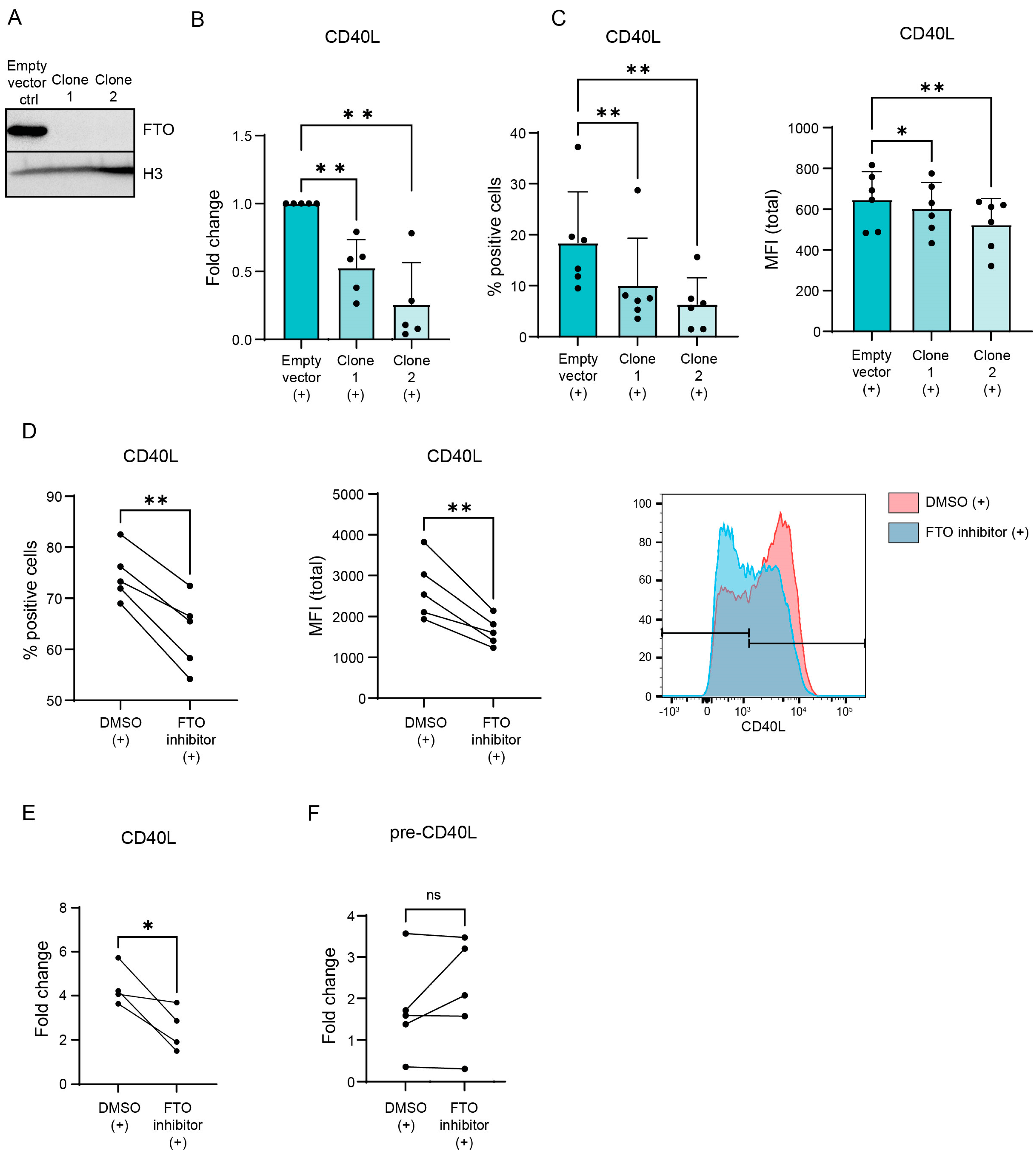
3.2. CD40L Expression Is Regulated via m6A ‘Eraser’ FTO in Activated CD4+ T Lymphocytes

m6A is often associated with (increased) mRNA degradation [9]. To determine whether or not m6A regulates CD40L expression, m6A levels were manipulated via the CRISPR/Cas9-mediated knockout of the demethylase FTO in a Jurkat cell line (Figure 2A). After knockout, cells were stimulated and CD40L mRNA expression was assessed using qPCR. FTO knockout reduced CD40L mRNA expression levels indicating that the increased m6A methylation of CD40L promotes its degradation (Figure 2B). Subsequently, CD40L protein expression was assessed using flow cytometry upon stimulation (Figure 2C and Supplemental Figure S1A). Both the percentage of cells expressing CD40L and the amount of CD40L expressed per cell was decreased upon the knockout of FTO. To assess whether or not m6A also regulates CD40L expression in primary cells, healthy control CD4+ T lymphocytes were treated with an FTO inhibitor [26] and CD40L protein expression was assessed (Figure 2D and Figure S1). While TCR stimulation increased CD40L expression, both the amount of CD40L-expressing cells as well as the levels of CD40L expressed by each cell was decreased after incubation with the FTO inhibitor. Again, these observations were reflected by similar changes induced in mRNA levels, as the amount of CD40L mRNA was also decreased upon FTO inhibition (Figure 2E). The expression of CD40L pre-mRNA did not change upon FTO inhibition, indicating that the observed changes in CD40L expression are the result of the direct degradation of CD40L and not the result of decreased CD40L transcription (Figure 2F). Together, these results indicate that CD40L expression in activated CD4+ T lymphocytes is directly regulated by m6A and its eraser protein FTO.

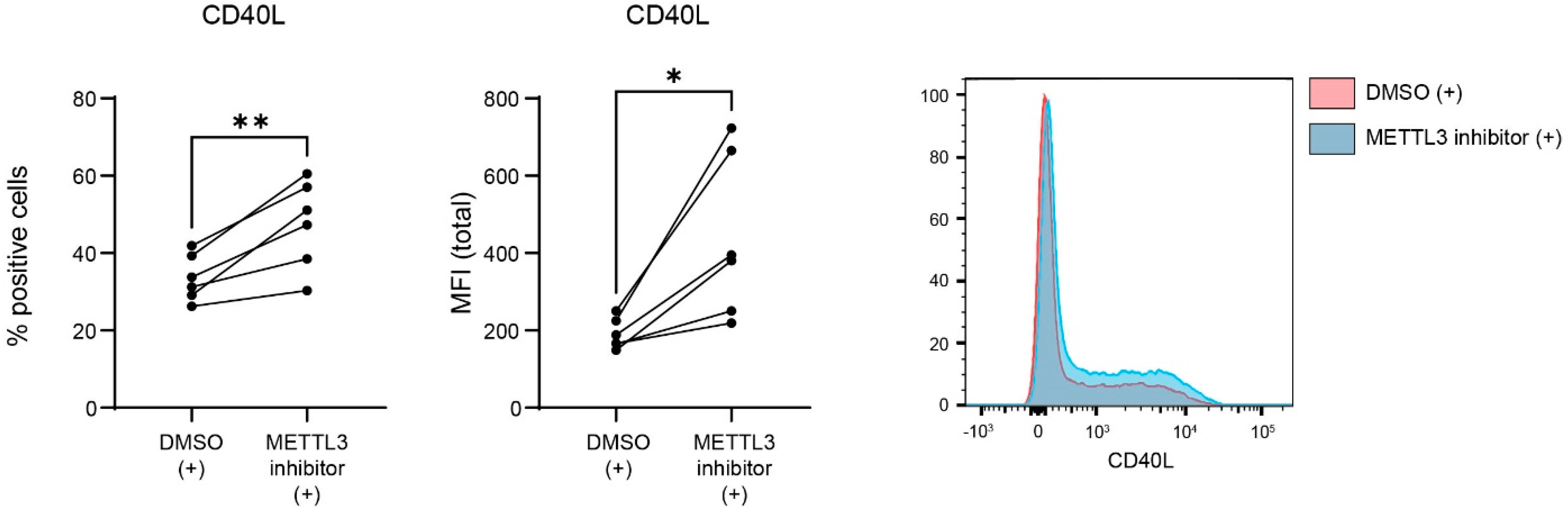
3.3. METTL3 Regulates CD40L Expression in Activated CD4+ T Lymphocytes

To further validate that m6A is involved in the regulation of CD40L expression, human CD4+ T lymphocytes were treated with an inhibitor of METTL3 [27,28,29,30], thereby reducing m6A levels. Flow cytometry was performed to assess CD40L protein expression (Figure 3). As observed previously, CD40L expression was increased upon the stimulation of the CD4+ T lymphocytes. Incubation with the METTL3 inhibitor resulted in an increased expression of both the amount of CD40L-expressing cells and the expression of CD40L per cell. Taken together, these results indicate that CD40L expression in stimulated CD4+ T lymphocytes is regulated via m6A.

3.4. m6A Reader YTHDF2 Possiblybinds to m6A Sequences on Cd40lg mRNA

The YTHDF2 ‘reader’ protein has been demonstrated to increase RNA degradation via the direct binding of m6A-methylated transcripts and promotion of their transfer to p-bodies for degradation [31]. Our results suggests that CD40L mRNA degradation is mediated by m6A methylation. Next, we wanted to determine whether or not YTHDF2 is involved in the destabilization of methylated CD40L mRNA, so we analyzed m6A CLIP sequencing data available online [25] from mouse CD4+ T cell mRNA. As hypothesized based on the data presented in Figure 4, YTHDF2 can associate with Cd40lg mRNA at the 3′ UTR at the location that is also m6A-methylated. These data suggest that YTHDF2 can directly bind to m6A sequences on CD40L mRNA, thereby promoting the degradation of CD40L mRNA via YTHDF2-mediated mRNA destabilization.

4. Discussion

Despite the increasing understanding of the role that m6A RNA modifications play in various biological processes, the implications of m6A in CD4+ T cell regulation remain incompletely understood. In this study, we provide evidence for a novel role of m6A in CD4+ T lymphocyte activation and function. We observed the m6A enrichment of many transcripts involved in TCR activation upon CD4+ T cell activation. Specifically, m6A enrichment on CD40L mRNA is increased upon the activation of CD4+ T lymphocytes. CD40L is an important co-stimulatory protein expressed on the cell surface of CD4+ T cells. The CD40–CD40L interaction is directly involved in autoimmune disease pathogenesis [24], mainly in diseases that are driven by autoantibodies such as rheumatoid arthritis (RA) [32], systemic lupus erythematosus (SLE) [33] and Sjögren’s syndrome [33]. Several studies have implicated m6A and its regulatory proteins to be involved in autoimmune disease pathogenesis [34,35,36,37,38,39,40]. However, the expression of CD40L and a possibly compromised CD40–CD40L interaction were not evaluated in these studies. Increasing our understanding of the regulation of CD40L expression could contribute to a better understanding of the disease mechanisms underlying these autoimmune diseases. Here, we demonstrate that the manipulation of m6A in T cells, either by inhibiting the activity of FTO or METTL3 directly affects CD40L expression levels. This research also suggests that m6A ‘reader’ YTHDF2 is probably able to bind to m6A-specific sequences on CD40L mRNA to regulate its expression. Further research needs to be performed to find the exact mechanism behind this regulatory mechanism. In this study, meRIP sequencing was used to map m6A enrichment in CD4+ T lymphocytes. There are some drawbacks to this approach. First, our experiment was a single experimental replicate, and therefore only provides a rough overview of the methylated transcript that needs to be validated, as we did for CD40L. In addition, the resolution of meRIP sequencing is limited [41]. Therefore, it is not possible to exactly determine the localization of detected m6A modifications and to quantitatively measure the fraction of transcripts that contain this modification. Also, the m6A antibodies that are used for meRIP sequencing do not exclusively detect m6A, but can also detect the N6,2′-O-dimethyladenosine (m6Am) modification that is located at the 5′ UTR of mRNA transcripts [4,42]. Since m6Am is present in a lower abundance compared to m6A, and considering the known location of m6Am at the 5′ UTR, the presence of these modifications can still be distinguished. Due to the limited resolution with meRIP-seq, it is not possible to determine the exact adenosine(s) in the CD40L transcript that is/ are methylated. It would be valuable to exactly determine the location of the m6A modification(s) on CD40L mRNA in the future with higher-resolution sequencing approaches such as m6A-CLIP [4]. The additional experiments can be performed where the DRACH-sequences containing the methylated adenosine(s) can be mutated to further explore this process and determine the location of the adenosine that is most dominant for the observed phenotype.
Transcriptome-wide m6A mapping in previous studies revealed that roughly one of every three mRNA transcripts in human cells contain m6A modifications [1,3]. These transcripts contain three to five m6A-specific DRACH sequences on average, situated at various locations within the transcript. For the vast majority of transcripts, only a part of the DRACH sequences are methylated [1]. Using SRAMP [43], a computational predictor of mammalian m6A sites, six high-confidence m6A consensus sequences were identified in human CD40L mRNA. It would therefore be interesting to establish a direct correlation between a specific m6A modification on the CD40L transcript, mRNA faith and therefore CD4+ T cell activation. This could be achieved via point mutations of specific adenosines in the DRACH sequences of CD40L mRNA, thereby specifically reducing the amount of m6A modifications and assessing the effects on the abundance of expressed CD40L protein.
CD40L is not the only co-stimulatory molecule expressed on activated CD4+ T cells, and co-inhibitory molecules contribute to the regulation of an immune response. Therefore, it is plausible that the expression of more of these molecules would be regulated in similar ways. In our meRIP-seq data, many m6A enrichment changes were observed upon the activation of CD4+ T lymphocytes. Methylation changes could not only be observed for co-stimulatory molecule CD40L, but also for CD28 and inducible T cell co-stimulators (ICOS). Co-inhibitory proteins that showed m6A enrichment changes are programmed cell death-1 (PD-1), cytotoxic T- lymphocyte antigen-4 (CTLA-4) and CD45. Although we did not examine the methylation of these transcripts in detail, these observations indicate that the regulation of co-stimulatory/inhibitory protein expression via m6A is not an exclusive regulatory mechanism for CD40L.
We hypothesize that the m6A-mediated regulation of CD40L expression is a constant negative feedback mechanism in order to regulate the intensity and duration of an immune response. In the early immune response, CD40L expression is upregulated to enable co-stimulation and TCR activation. In later stages of inflammation, CD40L expression is reduced via m6A-mediated regulation to resolve the immune response. This proposed regulatory mechanism would prevent the chronic overactivation of CD4+ T lymphocytes, potentially leading to tissue damage and severe immunopathology.

5. Conclusions

Our data increase the general understanding of the role of m6A in CD4+ T lymphocyte activation and function. As CD40L is a co-stimulatory protein expressed by CD4+ T cells and the CD40–CD40L interaction is implicated in the pathogenesis of several diseases, this study contributes to our general understanding of autoimmune disease pathology.

Supplementary Materials

The following supporting information can be downloaded at https://www.mdpi.com/article/10.3390/biology12071004/s1. Figure S1: CD40L expression is regulated via m6A ‘eraser’ FTO in activated CD4+ T lymphocytes; Table S1: qPCR primers; Table S2: Flow cytometry antibodies; Table S3: Western blot antibodies.

Author Contributions

Conceptualization, J.v.L. and E.C.N.v.V.; methodology, J.v.L. and E.C.N.v.V.; validation, E.C.N.v.V., L.W.P., R.C.S. and J.J.A.C.; formal analysis, E.C.N.v.V., L.W.P., R.C.S. and J.J.A.C.; investigation, E.C.N.v.V., L.W.P., R.C.S., N.A.M.v.d.D., M.M., A.E. and R.J.L.; resources, S.J.V. and J.v.L.; data curation, E.C.N.v.V., R.C.S. and J.J.A.C.; writing—original draft preparation, E.C.N.v.V.; writing—review and editing, E.C.N.v.V., L.W.P., J.J.A.C., S.J.V. and J.v.L.; visualization, E.C.N.v.V.; supervision, S.J.V. and J.v.L.; project administration, S.J.V. and J.v.L.; funding acquisition, S.J.V. and J.v.L. All authors have read and agreed to the published version of the manuscript.

Funding

This research was funded by the Dutch Arthritis Foundation (ReumaNederland), funding number: LLP10.

Institutional Review Board Statement

Not applicable.

Informed Consent Statement

Informed consent was obtained from all subjects involved in the study.

Data Availability Statement

The meRIP sequencing data have been deposited online. Read count data and per gene TMM normalized log2CPM expression values from meRIP RNA-seq are accessible at 10.5281/zenodo.8016876. Not all raw RNA sequencing data used in this study are publicly available due to research participant privacy/consent. Therefore, these data are only available upon request and after signing a Data Sharing Agreement within a specially designed UMC Utrecht-provided environment.

Acknowledgments

We want to thank the Utrecht Sequencing Facility.

Conflicts of Interest

The authors declare no conflict of interest. The funders had no role in the design of the study; in the collection, analyses, or interpretation of data; in the writing of the manuscript; or in the decision to publish the results.

References

  1. Dominissini, D.; Moshitch-Moshkovitz, S.; Schwartz, S.; Salmon-Divon, M.; Ungar, L.; Osenberg, S.; Cesarkas, K.; Jacob-Hirsch, J.; Amariglio, N.; Kupiec, M.; et al. Topology of the human and mouse m6A RNA methylomes revealed by m6A-seq. Nature 2012, 485, 201–206. [Google Scholar] [CrossRef] [PubMed]
  2. Chen, K.; Luo, G.-Z.; He, C. High-Resolution Mapping of N6-Methyladenosine in Transcriptome and Genome Using a Photo-Crosslinking-Assisted Strategy, 1st ed.; Elsevier Inc.: Amsterdam, The Netherlands, 2015; Volume 560. [Google Scholar] [CrossRef]
  3. Meyer, K.D.; Saletore, Y.; Zumbo, P.; Elemento, O.; Mason, C.E.; Jaffrey, S.R. Comprehensive Analysis of mRNA Methylation Reveals Enrichment in 3′ UTRs and near Stop Codons. Cell 2012, 149, 1635–1646. [Google Scholar] [CrossRef] [PubMed] [Green Version]
  4. Linder, B.; Grozhik, A.V.; Olarerin-George, A.O.; Meydan, C.; Mason, C.E.; Jaffrey, S.R. Single-nucleotide-resolution mapping of m6A and m6Am throughout the transcriptome. Nat. Methods 2015, 12, 767–772. [Google Scholar] [CrossRef] [PubMed]
  5. Bokar, J.; Rath-Shambaugh, M.; Ludwiczak, R.; Narayan, P.; Rottman, F. Characterization and partial purification of mRNA N6-adenosine methyltransferase from HeLa cell nuclei. Internal mRNA methylation requires a multisubunit complex. J. Biol. Chem. 1994, 269, 17697–17704. [Google Scholar] [CrossRef] [PubMed]
  6. Liu, J.; Yue, Y.; Han, D.; Wang, X.; Fu, Y.; Zhang, L.; Jia, G.; Yu, M.; Lu, Z.; Deng, X.; et al. A METTL3–METTL14 complex mediates mammalian nuclear RNA N6-adenosine methylation. Nat. Chem. Biol. 2013, 10, 93–95. [Google Scholar] [CrossRef] [Green Version]
  7. Meyer, K.D.; Jaffrey, S.R. Rethinking m6A Readers, Writers, and Erasers. Annu. Rev. Cell Dev. Biol. 2017, 33, 319–342. [Google Scholar] [CrossRef] [Green Version]
  8. Zaccara, S.; Ries, R.J.; Jaffrey, S.R. Reading, writing and erasing mRNA methylation. Nat. Rev. Mol. Cell Biol. 2019, 20, 608–624. [Google Scholar] [CrossRef] [PubMed]
  9. Zaccara, S.; Jaffrey, S.R. A Unified Model for the Function of YTHDF Proteins in Regulating m6A-Modified mRNA. Cell 2020, 181, 1582–1595.e18. [Google Scholar] [CrossRef]
  10. Patil, D.P.; Pickering, B.F.; Jaffrey, S.R. Reading m6A in the Transcriptome: m6A-Binding Proteins. Trends Cell Biol. 2017, 28, 113–127. [Google Scholar] [CrossRef]
  11. Xiao, W.; Adhikari, S.; Dahal, U.; Chen, Y.-S.; Hao, Y.-J.; Sun, B.-F.; Sun, H.-Y.; Li, A.; Ping, X.-L.; Lai, W.-Y.; et al. Nuclear m 6 A Reader YTHDC1 Regulates mRNA Splicing. Mol. Cell 2016, 61, 507–519. [Google Scholar] [CrossRef] [Green Version]
  12. Jia, G.; Fu, Y.; Zhao, X.; Dai, Q.; Zheng, G.; Yang, Y.; Yi, C.; Lindahl, T.; Pan, T.; Yang, Y.-G.; et al. N6-Methyladenosine in nuclear RNA is a major substrate of the obesity-associated FTO. Nat. Chem. Biol. 2011, 7, 885–887. [Google Scholar] [CrossRef]
  13. Wei, J.; Liu, F.; Lu, Z.; Fei, Q.; Ai, Y.; He, P.C.; Shi, H.; Cui, X.; Su, R.; Klungland, A.; et al. Differential m6A, m6Am, and m1A Demethylation Mediated by FTO in the Cell Nucleus and Cytoplasm. Mol. Cell 2018, 71, 973–985.e5. [Google Scholar] [CrossRef] [PubMed] [Green Version]
  14. Zheng, G.; Dahl, J.A.; Niu, Y.; Fedorcsak, P.; Huang, C.-M.; Li, C.J.; Vågbø, C.B.; Shi, Y.; Wang, W.-L.; Song, S.-H.; et al. ALKBH5 Is a Mammalian RNA Demethylase that Impacts RNA Metabolism and Mouse Fertility. Mol. Cell 2013, 49, 18–29. [Google Scholar] [CrossRef] [PubMed] [Green Version]
  15. Yao, Y.; Yang, Y.; Guo, W.; Xu, L.; You, M.; Zhang, Y.-C.; Sun, Z.; Cui, X.; Yu, G.; Qi, Z.; et al. METTL3-dependent m6A modification programs T follicular helper cell differentiation. Nat. Commun. 2021, 12, 1333. [Google Scholar] [CrossRef] [PubMed]
  16. Li, H.-B.; Tong, J.; Zhu, S.; Batista, P.J.; Duffy, E.E.; Zhao, J.; Bailis, W.; Cao, G.; Kroehling, L.; Chen, Y.; et al. m6A mRNA methylation controls T cell homeostasis by targeting the IL-7/STAT5/SOCS pathways. Nature 2017, 548, 338–342. [Google Scholar] [CrossRef] [Green Version]
  17. Ding, C.; Xu, H.; Yu, Z.; Roulis, M.; Qu, R.; Zhou, J.; Oh, J.; Crawford, J.; Gao, Y.; Jackson, R.; et al. RNA m6A demethylase ALKBH5 regulates the development of γδ T cells. Proc. Natl. Acad. Sci. USA 2022, 119, e2203318119. [Google Scholar] [CrossRef]
  18. van Diemen, F.R.; Kruse, E.M.; Hooykaas, M.J.G.; Bruggeling, C.E.; Schürch, A.C.; van Ham, P.M.; Imhof, S.M.; Nijhuis, M.; Wiertz, E.J.H.J.; Lebbink, R.J. CRISPR/Cas9-Mediated Genome Editing of Herpesviruses Limits Productive and Latent Infections. PLoS Pathog. 2016, 12, e1005701. [Google Scholar] [CrossRef] [Green Version]
  19. Dobin, A.; Davis, C.A.; Schlesinger, F.; Drenkow, J.; Zaleski, C.; Jha, S.; Batut, P.; Chaisson, M.; Gingeras, T.R. STAR: Ultrafast universal RNA-seq aligner. Bioinformatics 2013, 29, 15–21. [Google Scholar] [CrossRef]
  20. Anders, S.; Pyl, P.T.; Huber, W. HTSeq—A Python framework to work with high-throughput sequencing data. Bioinformatics 2015, 31, 166–169. [Google Scholar] [CrossRef] [Green Version]
  21. Robinson, M.D.; McCarthy, D.J.; Smyth, G.K. EdgeR: A Bioconductor package for differential expression analysis of digital gene expression data. Bioinformatics 2010, 26, 139–140. [Google Scholar] [CrossRef] [Green Version]
  22. Chatzigeorgiou, A.; Lyberi, M.; Chatzilymperis, G.; Nezos, A.; Kamper, E. CD40/CD40L signaling and its implication in health and disease. Biofactors 2009, 35, 474–483. [Google Scholar] [CrossRef] [PubMed]
  23. Tang, T.; Cheng, X.; Truong, B.; Sun, L.; Yang, X.; Wang, H. Molecular basis and therapeutic implications of CD40/CD40L immune checkpoint. Pharmacol. Ther. 2020, 219, 107709. [Google Scholar] [CrossRef] [PubMed]
  24. Karnell, J.L.; Rieder, S.A.; Ettinger, R.; Kolbeck, R. Targeting the CD40-CD40L pathway in autoimmune diseases: Humoral immunity and beyond. Adv. Drug Deliv. Rev. 2018, 141, 92–103. [Google Scholar] [CrossRef] [PubMed]
  25. Ito-Kureha, T.; Leoni, C.; Borland, K.; Cantini, G.; Bataclan, M.; Metzger, R.N.; Ammann, G.; Krug, A.B.; Marsico, A.; Kaiser, S.; et al. The function of Wtap in N6-adenosine methylation of mRNAs controls T cell receptor signaling and survival of T cells. Nat. Immunol. 2022, 23, 1208–1221. [Google Scholar] [CrossRef]
  26. Peng, S.; Xiao, W.; Ju, D.; Sun, B.; Hou, N.; Liu, Q.; Wang, Y.; Zhao, H.; Gao, C.; Zhang, S.; et al. Identification of entacapone as a chemical inhibitor of FTO mediating metabolic regulation through FOXO1. Sci. Transl. Med. 2019, 11, eaau7116. [Google Scholar] [CrossRef] [PubMed]
  27. Yankova, E.; Blackaby, W.; Albertella, M.; Rak, J.; De Braekeleer, E.; Tsagkogeorga, G.; Pilka, E.S.; Aspris, D.; Leggate, D.; Hendrick, A.G.; et al. Small-molecule inhibition of METTL3 as a strategy against myeloid leukaemia. Nature 2021, 593, 597–601. [Google Scholar] [CrossRef]
  28. Li, J.; Gregory, R.I. Mining for METTL3 inhibitors to suppress cancer. Nat. Struct. Mol. Biol. 2021, 28, 460–462. [Google Scholar] [CrossRef]
  29. Zhang, Z.-W.; Teng, X.; Zhao, F.; Ma, C.; Zhang, J.; Xiao, L.-F.; Wang, Y.; Chang, M.; Tian, Y.; Li, C.; et al. METTL3 regulates m6A methylation of PTCH1 and GLI2 in Sonic hedgehog signaling to promote tumor progression in SHH-medulloblastoma. Cell Rep. 2022, 41, 111530. [Google Scholar] [CrossRef]
  30. Li, S.; Zou, D.; Chen, W.; Britz, G.W.; Liu, Z.; Weng, Y. METTL3 inhibition reduces N6-methyladenosine levels and prevents allogeneic CD4 + T-cell responses. Immunol. Cell Biol. 2022, 100, 718–730. [Google Scholar] [CrossRef]
  31. Du, H.; Zhao, Y.; He, J.; Zhang, Y.; Xi, H.; Liu, M.; Ma, J.; Wu, L. YTHDF2 destabilizes m6A-containing RNA through direct recruitment of the CCR4–NOT deadenylase complex. Nat. Commun. 2016, 7, 12626. [Google Scholar] [CrossRef]
  32. Kitagawa, M.; Suzuki, H.; Adachi, Y.; Nakamura, H.; Yoshino, S.; Sumida, T. Interferon-γ enhances interleukin 12 production in rheumatoid synovial cells via CD40-CD154 dependent and independent pathways. J. Rheumatol. 2001, 28, 1764–1771. [Google Scholar] [PubMed]
  33. Goules, A.; Tzioufas, A.G.; Manousakis, M.N.; Kirou, K.A.; Crow, M.K.; Routsias, J.G. Elevated levels of soluble CD40 ligand (sCD40L) in serum of patients with systemic autoimmune diseases. J. Autoimmun. 2006, 26, 165–171. [Google Scholar] [CrossRef]
  34. Zhang, S.; Qin, J.; Zhao, Y.; Wang, J.; Tian, Z. Identification of molecular patterns and diagnostic biomarkers in juvenile idiopathic arthritis based on the gene expression of m6A regulators. Front. Pediatr. 2022, 10, 930119b. [Google Scholar] [CrossRef]
  35. Luo, Q.; Fu, B.; Zhang, L.; Guo, Y.; Huang, Z.; Li, J. Decreased Peripheral Blood ALKBH5 Correlates with Markers of Autoimmune Response in Systemic Lupus Erythematosus. Dis. Markers 2020, 2020, 8193895. [Google Scholar] [CrossRef] [PubMed]
  36. Luo, Q.; Rao, J.; Zhang, L.; Fu, B.; Guo, Y.; Huang, Z.; Li, J. The study of METTL14, ALKBH5, and YTHDF2 in peripheral blood mononuclear cells from systemic lupus erythematosus. Mol. Genet. Genom. Med. 2020, 8, e1298. [Google Scholar] [CrossRef] [PubMed]
  37. Mo, X.; Zhang, Y.-H.; Lei, S.-F. Genome-Wide Identification of N6-Methyladenosine (m6A) SNPs Associated With Rheumatoid Arthritis. Front. Genet. 2018, 9, 299. [Google Scholar] [CrossRef] [Green Version]
  38. Wang, J.; Yan, S.; Lu, H.; Wang, S.; Xu, D. METTL3 Attenuates LPS-Induced Inflammatory Response in Macrophages via NF-κB Signaling Pathway. Mediat. Inflamm. 2019, 2019, 3120391. [Google Scholar] [CrossRef] [Green Version]
  39. Mo, X.-B.; Lei, S.-F.; Qian, Q.-Y.; Guo, Y.-F.; Zhang, Y.-H.; Zhang, H. Integrative analysis revealed potential causal genetic and epigenetic factors for multiple sclerosis. J. Neurol. 2019, 266, 2699–2709. [Google Scholar] [CrossRef]
  40. Lu, T.X.; Zheng, Z.; Zhang, L.; Sun, H.-L.; Bissonnette, M.; Huang, H.; He, C. A New Model of Spontaneous Colitis in Mice Induced by Deletion of an RNA m6A Methyltransferase Component METTL14 in T Cells. Cell. Mol. Gastroenterol. Hepatol. 2020, 10, 747–761. [Google Scholar] [CrossRef]
  41. McIntyre, A.B.R.; Gokhale, N.S.; Cerchietti, L.; Jaffrey, S.R.; Horner, S.M.; Mason, C.E. Limits in the detection of m6A changes using MeRIP/m6A-seq. Sci. Rep. 2020, 10, 6590. [Google Scholar] [CrossRef] [Green Version]
  42. Mauer, J.; Jaffrey, S.R. FTO, m6Am, and the hypothesis of reversible epitranscriptomic mRNA modifications. FEBS Lett. 2018, 592, 2012–2022. [Google Scholar] [CrossRef] [PubMed] [Green Version]
  43. Zhou, Y.; Zeng, P.; Li, Y.-H.; Zhang, Z.; Cui, Q. SRAMP: Prediction of mammalian N6-methyladenosine (m6A) sites based on sequence-derived features. Nucleic Acids Res. 2016, 44, e91. [Google Scholar] [CrossRef] [PubMed] [Green Version]
Figure 1. Increase in m6A enrichment on CD40L mRNA in activated CD4+ T lymphocytes. (A) Heat map of m6A enrichment changes in mRNA transcripts of co-stimulatory molecules, expressed in Log2CPM. meRIP sequencing data on unstimulated CD4+ T lymphocytes and CD4+ T lymphocytes stimulated with CD3/CD28 beads overnight (n = 1). (B) meRIP sequencing in healthy control CD4+ T lymphocytes. Cells were activated with CD3/CD28 beads (n = 1). (C) meRIP qPCR in healthy control CD4+ T lymphocytes. Cells were activated with either 4 h PMA and ionomycin or overnight with CD3/CD28 beads. p values were calculated using a Mann–Whitney t-test (* p < 0.05) (n = 4). (D) Integrative Genomics Viewer tracks of m6A CLIP read distribution in naïve mouse T lymphocytes provided by Ito-Kureha et al. [25]. Data obtained from three biological replicates.
Biology 12 01004 g001
Figure 2. Regulation of CD40L expression via m6A ‘eraser’ FTO in activated CD4+ T lymphocytes. (A) CRISPR/Cas9 knockout of FTO in Jurkat cells compared to control. Western blot for FTO in two single-cell clones. (B) Jurkat cells with knockout of FTO activated with 4 h of PMA and ionomycin. qPCR on CD40L mRNA relative to housekeeping gene GUSB. (C) Jurkat cells with CRISPR/Cas9 knockout of FTO activated with 4 h PMA and ionomycin. Flow cytometry on CD40L protein expression. (D) Healthy control CD4+ T lymphocytes pre-incubated for 48 h with 50 µM entacapone. The cells were subsequently activated with CD3/CD28 beads overnight. Flow cytometry was performed on the CD40L protein. Right: representative histograms of CD40L protein reduction after entacapone treatment. (E) Healthy control CD4+ T lymphocytes pre-incubated for 48 h with 50 µM entacapone. The cells were subsequently activated with CD3/CD28 beads overnight. qPCR on CD40L mRNA. (F) Healthy control CD4+ T lymphocytes pre-incubated for 48 h with 50 µM entacapone. The cells were subsequently activated with CD3/CD28 beads overnight. qPCR on pre-CD40L mRNA. p values were calculated using a paired t-test; not significant (ns), * p < 0.05, and ** p < 0.01.
Biology 12 01004 g002
Figure 3. Regulation of CD40L expression by METTL3 in activated CD4+ T lymphocytes. Healthy control CD4+ T lymphocytes were incubated for 48 h with 30 µM STM2457, and subsequently activated with CD3/CD28 beads overnight. Flow cytometry on the CD40L protein and representative histograms of CD40L protein increase after STM2457 incubation. p values were calculated using a paired t-test; * p < 0.05, and ** p < 0.01.
Biology 12 01004 g003
Figure 4. Possible binding of the m6A reader YTHDF2 to m6A sequences on Cd40lg mRNA. Integrative Genomics Viewer tracks of m6A CLIP and Ythdf2 iCLIP read distribution in naïve mouse T lymphocytes. Data combined from three biological replicates of the m6A CLIP and two replicates for the YTHDF2 iCLIP.
Biology 12 01004 g004
Disclaimer/Publisher’s Note: The statements, opinions and data contained in all publications are solely those of the individual author(s) and contributor(s) and not of MDPI and/or the editor(s). MDPI and/or the editor(s) disclaim responsibility for any injury to people or property resulting from any ideas, methods, instructions or products referred to in the content.

