Article

Epigenetic Regulation of miR-25 and Lnc107153 on Expression of Seasonal Estrus Key Gene CHGA in Sheep

1
Key Laboratory of Animal Genetics, Breeding and Reproduction of Ministry of Agriculture and Rural Affairs, Institute of Animal Science, Chinese Academy of Agricultural Sciences, Beijing 100193, China
2
Institute of Genetics and Developmental Biology, Chinese Academy of Sciences, Beijing 100101, China
3
Small Ruminant Genomics, International Center for Agricultural Research in the Dry Areas (ICARDA), Addis Ababa P.O. Box 5689, Ethiopia
4
Animal and Veterinary Sciences, SRUC and Centre for Tropical Livestock Genetics and Health (CTLGH), The Roslin Institute Building, Midlothian EH25 9RG, UK
*
Authors to whom correspondence should be addressed.
These authors contributed equally to this work.
Biology 2023, 12(2), 250; https://doi.org/10.3390/biology12020250
Submission received: 30 December 2022 / Revised: 29 January 2023 / Accepted: 2 February 2023 / Published: 4 February 2023
(This article belongs to the Special Issue New Advances and Insights in Animal Genetics and Breeding)

Abstract

:

Simple Summary

In sheep pituitary pars tuberalis tissue, EYA3, TSHβ and CHGA are key genes regulating seasonal reproduction. Their high expression is the characteristic signal under long photoperiods (EYA3, TSHβ) and short photoperiods (CHGA). The epigenetic regulation of microRNAs and LncRNAs on their expression remains unknown. In this study, our results indicated that miR-25 could inhibit the expression of CHGA by specifically binding to its 3′UTR region in pituitary cells. Lnc107153 played the role of CeRNA, which can adsorb miR-25 and weaken the inhibitory effect of miR-25 on CHGA expression. Together, these results suggested the epigenetic regulative network of Lnc107153-miR-25-CHGA was involved in sheep seasonal reproduction.

Abstract

Pituitary pars tuberalis (PT) plays an important role as the transmission center in the seasonal reproduction of animals. It helps convert external photoperiod signals into intrinsic seasonal reproduction signals. In sheep PT, specific expression patterns of several genes (including short photoperiod-induced gene CHGA and long photoperiod genes EYA3 and TSHβ) under different photoperiods are crucial characteristics during this signal transduction. Recent studies have revealed the role of epigenetics in regulating the expression of seasonal reproductive key genes. Therefore, we explored whether microRNAs and LncRNAs regulated the expressions of the above key genes. Firstly, the expression of miR-25 and CHGA showed a significant negative correlation in sheep PT. Results of the dual luciferase reporter assay and miR-25 overexpression indicated that miR-25 could inhibit the expression of CHGA by specifically binding to its 3′UTR region in pituitary cells. Then, expression negative correlation and dual luciferase reporter analyses were used to screen and identify the candidate LncRNA (Lnc107153) targeted by miR-25. Finally, the results of fluorescence in situ hybridization and Lnc107153 overexpression suggested that Lnc107153 and miR-25 were involved in the epigenetic regulation of CHGA expression. However, the expressions of EYA3 and TSHβ were not regulated by miRNAs. These results will provide new insights into the epigenetic regulatory network of key genes in sheep seasonal reproduction.

1. Introduction

For many animal species, seasonal reproduction is the result of natural selection for increasing the survival rate of their offspring, thus their offspring can be born during the most appropriate stage of the annual climate and food supply [1]. However, for the sheep industry, seasonal reproduction has become an important factor limiting production efficiency [2,3]. The global demand for mutton is gradually increasing, and the current reproduction efficiency of sheep needs to be greatly improved [3]. Therefore, research on the molecular basis of the seasonal reproduction of sheep is crucial for possible artificial regulation of this trait in the future. Furthermore, the molecular regulatory network of seasonal reproduction remains to be improved in depth.
Pituitary pars tuberalis (PT) plays an important role as the transmission center in the seasonal reproduction of animals. It helps convert external photoperiod signals into intrinsic seasonal reproduction signals by the MEL-TSH-TH axis [4,5,6,7,8], which regulates the seasonal estrus of animals. In sheep PT tissue, specific expression patterns of several genes under different photoperiods are crucial characteristics during this signal transduction. For example, a short photoperiod (SP) induced a high expression of the CHGA gene [9], and a long photoperiod (LP) initiated the high expression of EYA3 and TSHβ [5,6,7,8].
Recent studies have revealed the role of epigenetics in regulating the expression of seasonal reproductive key genes [10,11,12,13,14,15]. For example, it was reported that a short photoperiod inhibited hypothalamic DNA methyltransferase expression and reduced dio3 promoter DNA methylation, which up-regulated dio3 expression and induced gonadal regression [13]. In addition to DNA methylation, previous studies implied that microRNAs and LncRNAs were also important epigenetic regulators for seasonal reproduction [14,16,17,18,19]. It is worth exploring whether the specific expression patterns of the above key genes (CHGA, EYA3 and TSHβ) are regulated by miRNAs and LncRNAs, which will expand our understanding of the epigenetic regulation of the key genes in sheep seasonal reproduction.
Sunite sheep is one typical seasonal-reproductive breed in Inner Mongolia, China, which rest from April to July every year [14]. Therefore, in this study, Sunite sheep were used and experienced the process from SP to LP after removing the ovaries and embedding estrogen. First, miRNAs targeting the above three key genes (EYA3, TSHβ and CHGA) were predicted with a sequence complementary relationship, and miRNA–gene target combinations with negative correlation in expression were screened using quantitative PCR results of miRNAs and genes under SP and LP in sheep PT. Then, the target binding relationship between miRNA and gene was verified with the dual luciferase reporter system, and the regulatory effect of miRNA on gene expression was analyzed using an overexpression experiment of miRNA in pituitary cells. Next, expression correlation and dual luciferase reporter analysis were used to screen and identify the candidate LncRNA targeted by miR-25. Finally, the regulatory network of LncRNA-miRNA-gene was revealed using expression correlation analysis, the dual luciferase reporter system and an intracellular LncRNA overexpression experiment. These results will provide new insights into the epigenetic regulatory network of the key genes in sheep seasonal reproduction.

2. Materials and Methods

2.1. Gene Expression Assay

Determination of gene expression in tissues was conducted in six adult Sunite ewes (3 years old; weight 40–45 kg). They were collected from a farm in Urat Middle Banner (40°75′ north latitude), Inner Mongolia Autonomous Region, China. All ewes were raised under the same conditions, with free access to water and feed. The surgery of ovariectomized (OVX), estradiol-implantation and the light control experiment referred to the methods previously reported [8,19,20,21,22]. Estradiol (E2, Sigma Chemical Co., St. Louis, MO, USA) was implanted to maintain plasma estradiol levels of 3–5 pg/mL [8,14,17,19]. After the surgery of OVX and estradiol-implantation, the ewes recovered for 30 days before artificial light control. Then, all the ewes were brought indoors with a light program simulating the outdoor photoperiodic condition using a time-control switch. Firstly, all ewes were kept in artificial SP (8 h light: 16 h dark) for 21 days and switched to LP (16 h light: 8 h dark) for 42 days, with free access to water and food. Ewes were euthanized (intravenous pentobarbital: 100 mg/kg) at ZT4 (4 h after lights on) of SP21 and LP42 [14]. Consistent with the specific location described by Wood et al. [9] and Lomet et al. [8], the PT tissue (the specific location is shown in Figure S1) of each ewe was immediately collected and stored at −80 °C.
Total RNA was extracted from PT tissues with TRIzol Reagent (Invitrogen, Carlsbad, CA, USA) according to the manufacturer’s instructions. The PrimeScriptTM reagent RT kit (Takara-Bio, Dalian, China) was used for genomic DNA removal and reverse transcription to synthesize the first strand of cDNA. After searching EYA3, TSH, CHGA and RPL19 genes and Lnc20518, Lnc107153 and Lnc72519 sequences in our previous transcriptome sequencing results [14], the qPCR primers for the above four genes and three LncRNAs were designed using Premier 5 software (Table 1). RPL19 was used as an internal reference to normalize target gene expression. The qPCR was performed using the SYBR premix ExTaq II kit (Takara-Bio, Dalian, China). The 20 μL qPCR reaction system included 1.0 μL cDNA template, 10 μL SYBR Premix Ex Taq, 0.5 μL forward and reverse primers and 8 μL ddH2O. The qPCR reaction program was as follows: 95 °C for 15 min, followed by 40 cycles of 95 °C for 10 s and 60 °C for 30 s. The expression levels were then calculated using the 2−ΔΔCt method [23,24] and processed using SPSS 20.0 with a one-way analysis of variance.
The miRNAs which target CHGA, TSHB and EYA3 genes were predicted using the miRBase database [25]. The first strand of miRNA cDNA was synthesized using the miRcute enhanced miRNA cDNA first strand synthesis kit (Tiangen biotech, Beijing, China) with total RNA as a template. MiRprimer2 was used to design the upstream primer sequences for miR-25, miR-30d and miR-22-3p (Table 2). Downstream primer was provided by the kit. The 20 μL reaction system included 2.0 μL miRNA cDNA template, 10 μL 2 × miRcute Plus miRNA premix, 0.4 μL forward and reverse primers and 7.2 μL ddH2O. The procedures for miRNA qPCR are shown in Table S1. The correlation of expression between genes (or LncRNAs) and miRNAs was analyzed using SPSS 20.0, including EYA3-miR-22-3p, TSH-miR-30d, CHGA-miR-25, Lnc20518-miR-25-CHGA, Lnc107153-miR-25-CHGA and Lnc72519-miR-25-CHGA.

2.2. Identification of the Targeting Relationship between miR-25 and CHGA

To verify whether there is a targeted binding relationship between miR-25 and CHGA, the CHGA 3′UTR sequence (Table S2) was inserted downstream of luc2 in the pmir-Glo vector and named as CHGA 3′UTR-WT. In order to ensure the accuracy of the test results, the binding site of miR-25 in CHGA 3′UTR was mutated, and the mutated CHGA 3′UTR was inserted downstream of luc2 in the pmir-Glo vector, which was named CHGA 3′UTR-MUT. The miR-25 mature sequence (AUUGCACUUGUCUCGUCUGA) was provided to Guangzhou Ruibo Biotechnology Co., Ltd. for miR-25 mimics synthesis. The micrON mimic NC with a base length of 21 nt was selected as the control. One of the two pmir-Glo vectors and one of the two mimics (Specific combinations are shown in the following experimental groups) were co-transfected into HEK 293T cells line using Lipofectamine 2000. Specifically, there were four experimental groups: ① CHGA 3′UTR-WT + miR-25 mimics; ② CHGA 3′UTR-MUT + miR-25 mimics; ③ CHGA 3′UTR-WT + mimics NC; and ④ CHGA 3′UTR-MUT + mimics NC. The luciferase activity of luc2/hRluc in transfected cells would be significantly reduced when the miRNA targeted the 3′UTR of the candidate gene.
RC-4B/C cells (rat adenohypophysoma cells) were purchased from American Type Culture Collection (San Diego, CA, USA). Cells were cultured in strict accordance with the instructions. Cells were cultured in 6-well plates with the same number of cells (106) per well. Three biological replicates were set for each experimental group. When the cell density reached 80%, the experimental group was transfected with miR-25 mimics and the control group was transfected with mimic NC and the medium was replaced with OPTI-MEM. After 6 h, OPTI-MEM was replaced with a complete medium. After 48 h, cells were collected for qPCR and Western blot. The primers and detection methods of qPCR were the same as described above. Total cellular protein was extracted for concentration determination and Western blot. Anti-Chromogranin A Antibody (ab283265) was purchased from Abcam Company. GAPDH Mouse Monoclonal Antibody was purchased from Proteintech Company. HRP-conjugated Goat Anti-Mouse IgG (H+L) was purchased from Proteintech Company. The results of the Western blot were analyzed with Image J software. The difference significance test for the ratio of the target protein to GAPDH was performed using the chi-square test in SPSS 20.0.

2.3. Screening and Identification of LncRNA Targeted by miR-25

Another Sunite ewe was euthanized at ZT4 under the short photoperiod (SP21). PT tissue was immediately removed and fixed in 4 % paraformaldehyde (PFA). After washing, it was dehydrated and embedded with OTC. The tissue was cut into 10 μm sections using a freezing microtome and sections were pasted on slides. They were stored at −80 °C before fluorescence in situ hybridization. An Lnc107153 probe (sequence: 5′-cy3-AUAGAUCCUCCAGCUACCACCAUCAGACC-3′) labeled with CY3 was designed and synthesized by Bioengineering (Shanghai, China) Co., Ltd. The cell nuclei were stained with DAPI. When the fluorescence in situ hybridization ended, the images were collected using an upright fluorescence microscope.
The 3′UTR sequence (Table S2) of Lnc107153 was inserted downstream of luc2 in the pmir-Glo vector and named Lnc107153 3′UTR-WT. In order to ensure the accuracy of the test results, the binding site of miR-25 in the 3′UTR of Lnc107153 was mutated, and the mutated 3′UTR of Lnc107153 was inserted downstream of luc2 in the pmir-Glo vector, which was named Lnc107153 3′UTR-MUT. Four experimental groups were set up: Lnc107153-WT + miR-25 mimics; Lnc107153-WT + mimics NC; Lnc107153-MUT + miR-25 mimics; and Lnc107153-MUT + mimics NC. The pmir-Glo vector and mimics were co-transfected into the HEK293T cells line using Lipofectamine 2000. The luciferase activity of luc2/hRluc in transfected cells was detected.
In order to verify the role of Lnc107153 in pituitary cells, the Lnc107153 sequence (Table S3) was inserted into the vector pIRES2-EGFP to construct an overexpression plasmid (Lnc107153 + pIRES2-EGFP). Similar to miR-25 experiments, transfection was performed when the RC-4B/C cell culture density reached 80%. The experimental group was transfected with the Lnc107153 overexpression plasmid, and the control group was transfected with an overexpression empty vector. After 48 h, cells were collected, and total RNA and total protein were extracted for qPCR and Western blot, respectively.

3. Results

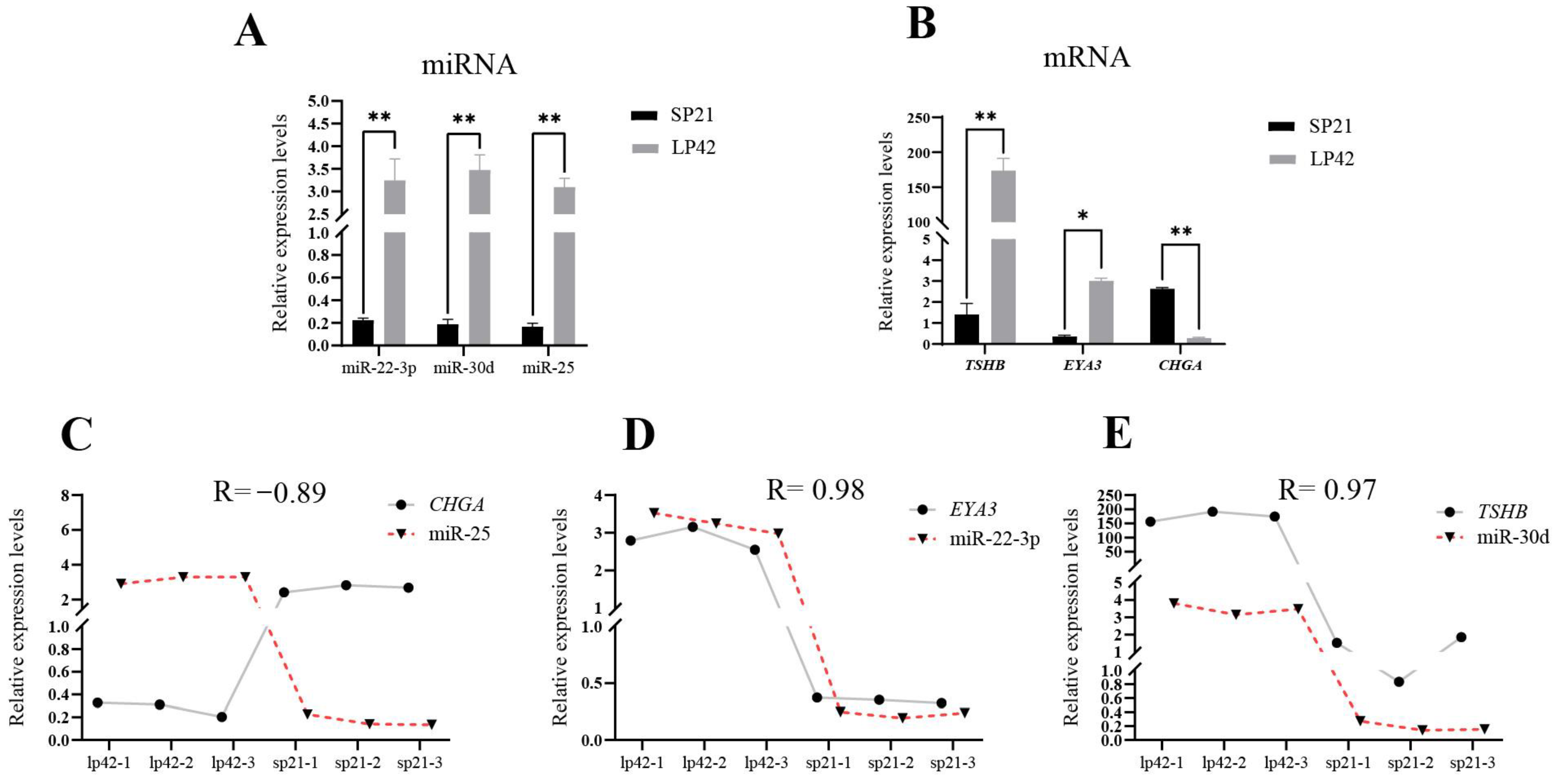
3.1. Prediction of the Targeting Relationship between the Key Genes and miRNA in Sheep PT

In sheep PT tissue, EYA3, TSHβ and CHGA are key genes involved in the regulation of seasonal reproduction. Using information from the miRBase database, the top-ranking miRNAs targeting these three genes were predicted. They were miR-22-3p targeting EYA3, miR-30d targeting TSHβ and miR-25 targeting CHGA, respectively. The fluorescence quantitative PCR results showed that the three miRNAs had significantly higher expression levels during the long photoperiod compared to the short photoperiod in sheep PT (Figure 1A), and the expression of miR-25 was highly negatively correlated with the expression of CHGA (correlation coefficient: −0.89) (Figure 1A–C). Therefore, miR-25 was predicted to be a candidate miRNA negatively regulating the CHGA gene. However, for genes EYA3 and TSHβ, there were no miRNAs that negatively regulated their expression (Figure 1D,E). Thus, the expressions of EYA3 and TSHβ may be regulated by factors other than miRNAs.

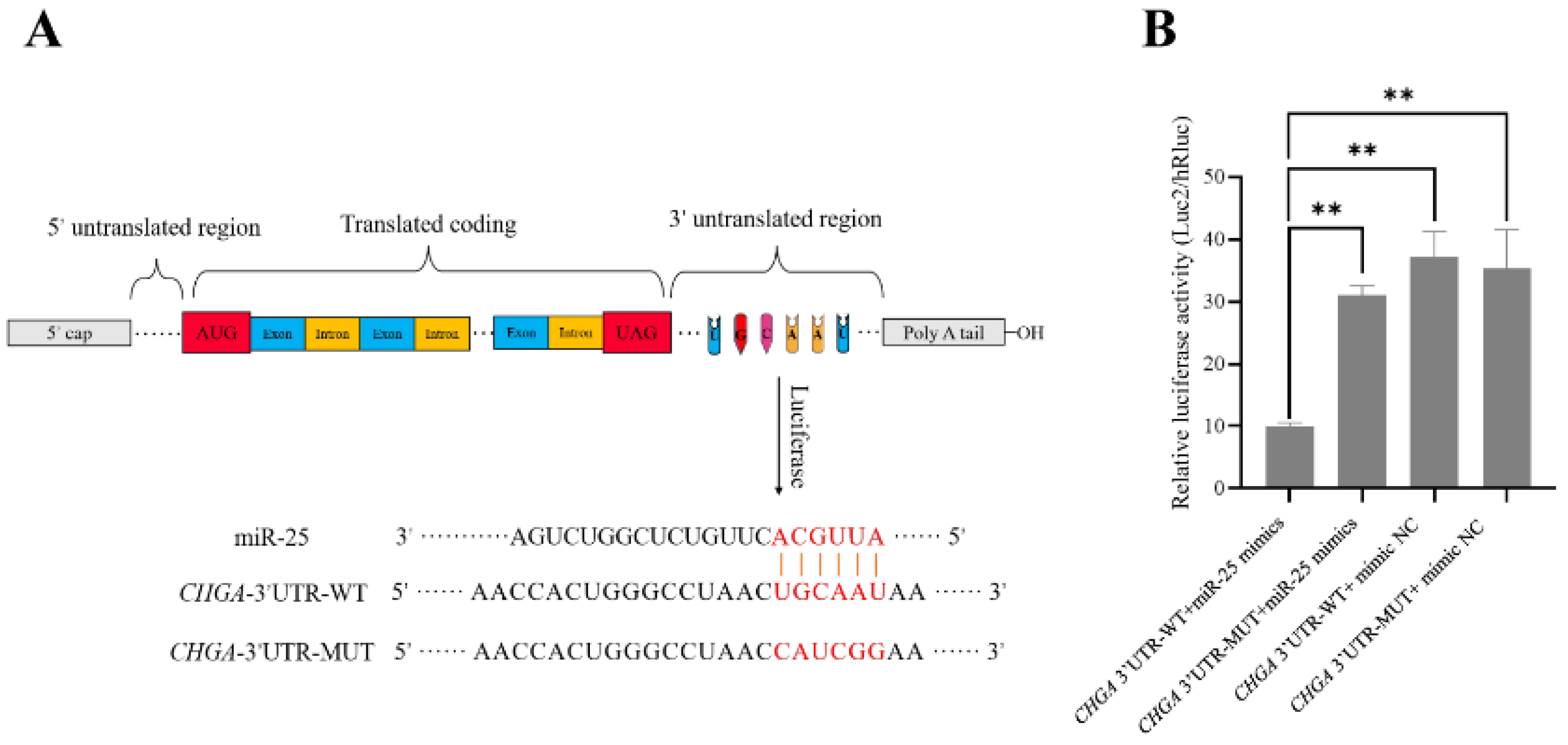
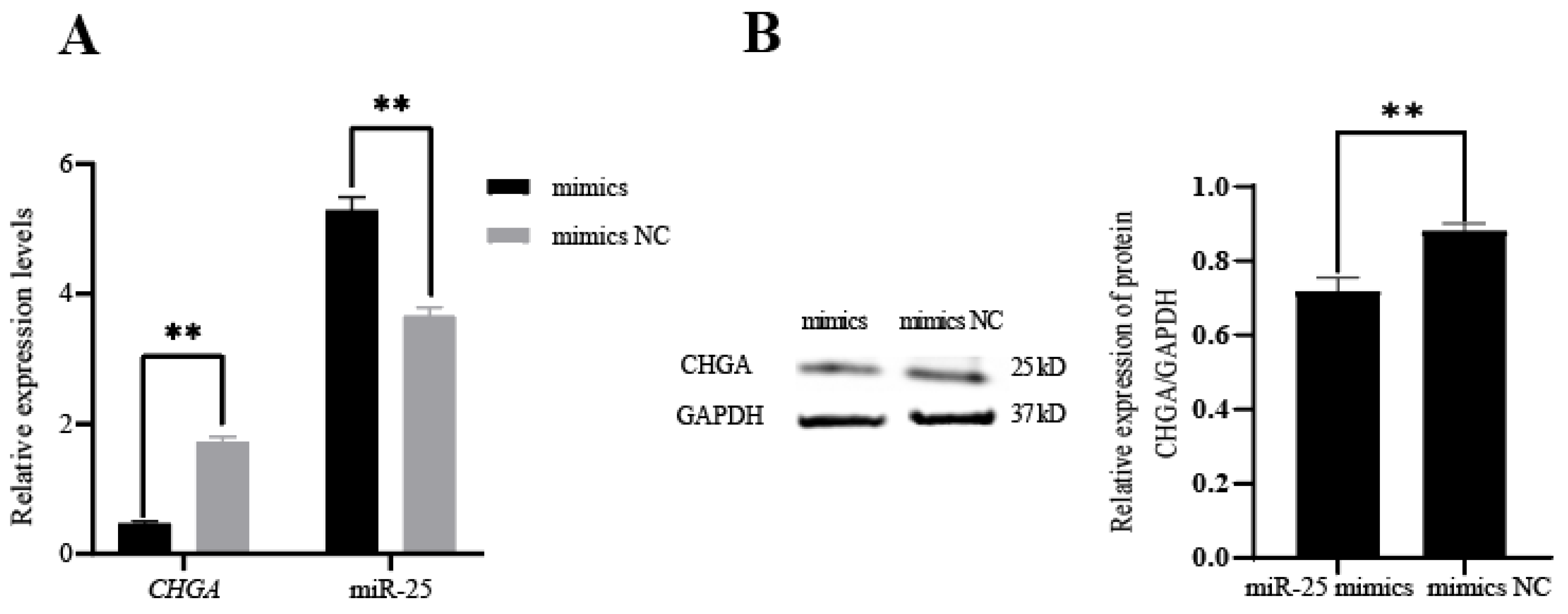
3.2. Identification of the Targeting Relationship between miR-25 and CHGA

Using the dual luciferase reporter system, we found that the relative fluorescence value of the co-transfected CHGA 3′UTR wild-type and miR-25 mimics group was significantly lower (p < 0.01) than those of the other three groups (CHGA 3′UTR mutant + miR-25 mimics; CHGA 3′UTR-WT + mimics NC; CHGA 3′UTR-MUT + mimics NC) (Figure 2), and there was no significant difference among the other three groups (p > 0.05). This result indicated that miR-25 can specifically bind to the 3′UTR region of CHGA through base complementation. Furthermore, the miR-25 mimics and mimic NC were transfected into pituitary cells (RC-4B/C cells) that can express CHGA, respectively. The qPCR results showed that the expression of miR-25 was significantly higher in the miR-25 mimics overexpression group than that in the mimic NC group (p < 0.01), which indicated that miR-25 was successfully transfected. After transfection of the miR-25 mimics, the transcription and translation levels of CHGA were significantly decreased (p < 0.01) (Figure 3A,B). In summary, miR-25 can inhibit the expression of CHGA in pituitary cells by specifically binding to the 3′UTR region of CHGA.

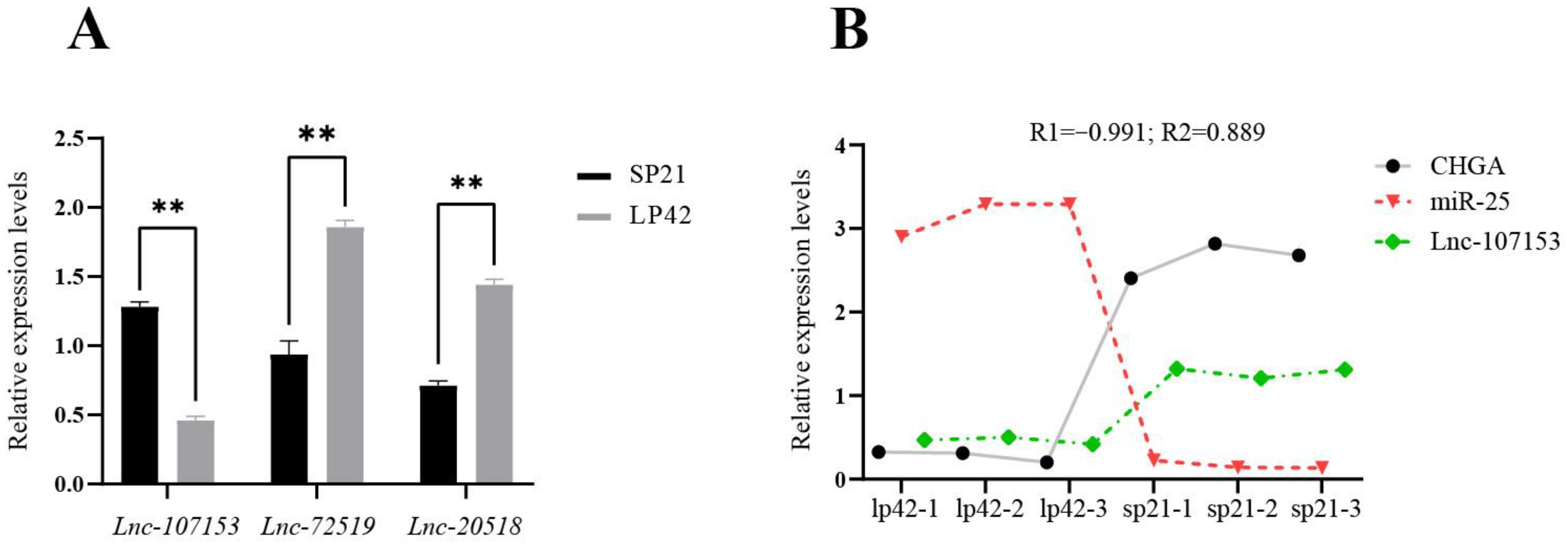
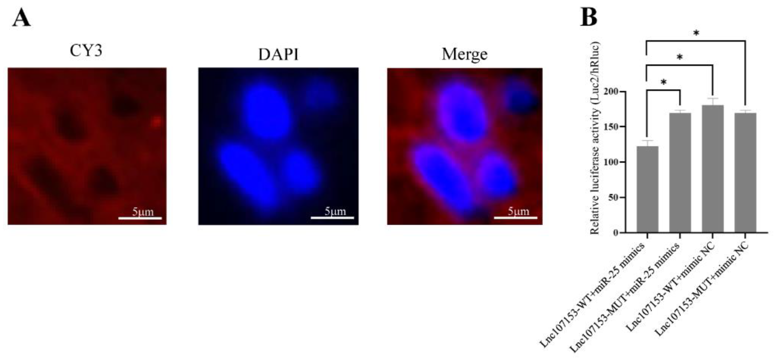
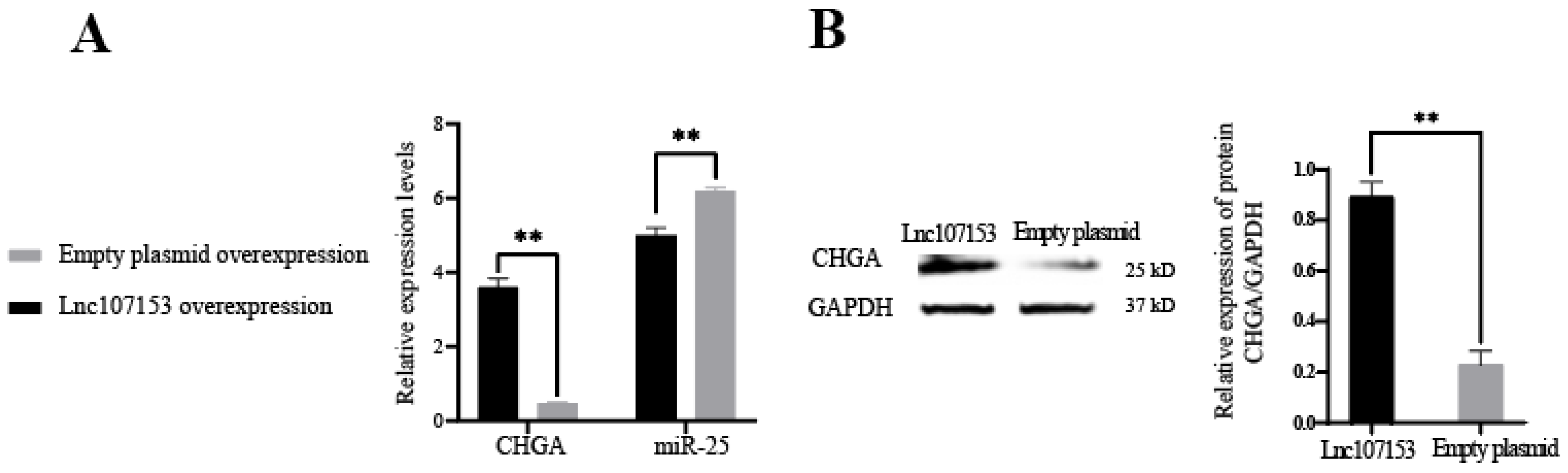
3.3. Screening and Identification of LncRNA Targeted by miR-25

Based on our previous transcriptome sequencing data [14], we predicted that targeted complementary relationships existed between miR-25 and three LncRNAs (Lnc107153, Lnc20518 and Lnc72519). Next, the results of the qPCR in sheep PT under different photoperiods showed that the expression of Lnc107153 during the short photoperiod was significantly higher than that during the long photoperiod (p < 0.01), and the expression of Lnc107153 was highly negatively correlated with the expression of miR-25 (Figure 4, correlation coefficient: −0.991). For the expression of the other two LncRNAs (Lnc20518 and Lnc72519), there was no negative correlation found with miR-25 (Table S4). Notably, a high positive correlation of expression existed between Lnc107153 and CHGA (Figure 4B, the correlation coefficient is 0.889). Therefore, the regulative network Lnc107153-miR-25-CHGA was screened for subsequent validation.
Firstly, Lnc107153 was localized in the cytoplasm of sheep PT cells with fluorescence in situ hybridization (Figure 5A, Lnc107153 with CY3 red marker), suggesting that Lnc107153 has the potential to competitively bind miRNA as a CeRNA. Next, the results of the dual luciferase reporter assay (Figure 5B) showed that the relative fluorescence value of the transfected Lnc107153-WT + miR-25 mimics group was significantly lower than those of the other three groups (Lnc107153-WT + mimics NC, Lnc107153-MUT + miR-25 mimics, Lnc107153-MUT + mimics NC), indicating that miR-25 can specifically target the 3′UTR region of Lnc107153.
In order to further verify the CeRNA function of Lnc107153, the Lnc107153 overexpression plasmid and the control empty overexpression plasmid were transfected into RC-4B/C cells, respectively. Together, the results of the qPCR and Western blot showed that the overexpression of Lnc107153 significantly reduced the level of miR-25 and increased the transcription and translation of the CHGA gene (Figure 6). Combined with the above result of this study that miR-25 can inhibit the expression of the CHGA gene, it can be speculated that Lnc107153 might play the role of CeRNA to adsorb miR-25 and weaken the inhibitory effect of miR-25 on CHGA expression in pituitary cells. Therefore, the regulative network Lnc107153-miR-25-CHGA might be summarized as (Figure 7): during the long photoperiod, miR-25 was highly expressed in sheep PT which could specifically bind to the 3′UTR region of CHGA to inhibit CHGA protein expression; during the short photoperiod, the expression of Lnc107153 increased, which could bind to miR-25 to weaken the inhibitory effect of miR-25 on CHGA expression, and consequently, the expression level of CHGA protein increased significantly.

4. Discussion

In sheep, the retina receives light signals of different seasons and day lengths, then the signals are transmitted to the pineal gland via suprachiasmatic nuclei (SCN) [26]. In the pineal gland, the continuous secretion duration of melatonin (MEL) changes accordingly [27,28,29]. After MEL receptors in the pituitary pars tuberalis perceive the dynamic changes in MEL, the expression levels of a series of photoperiod-induced genes (such as the SP-induced gene CHGA [9], LP factor EYA3 [6], TSHβ [30] and BMAL2 [31]) will undergo a landmark alteration. These photoperiodic-induced genes can help convert photoperiodic signals into intrinsic seasonal reproduction signals by the MEL-TSH-TH axis, then they directly or indirectly affect the release of gonadotropin-releasing hormone (GnRH) in the hypothalamus and further regulate the reproductive phenotype through the hypothalamic–pituitary–gonadal axis [4,32].
In this study, the results of the qPCR in sheep PT showed that the expression of EYA3 and TSH under the long photoperiod was significantly higher than that of the short photoperiod, and the expression of CHGA exhibited the opposite trend. These results were consistent with previous reports [5,6,9] and proved the reliability of transcriptome sequencing results. EYA3 is involved in the regulation of the seasonal reproduction of animals as a long photoperiod signal. A study on quail revealed that EYA3 is the first gene to be activated and induced to express in PT on the first day of the long photoperiod [33]. Studies on sheep have shown that the LP initiates the high expression of EYA3 [6] and further activates TSHβ transcription mainly through the TEF/SIX1/EYA3 complex [34]. Wood et al. [9] revealed that CHGA was involved in the regulation of the seasonal reproduction of sheep as an SP signal using RNA-seq analysis and fluorescence immunoassay. Their results showed that the transcription level of CHGA under SP was three times that under the LP in sheep PT, and the level of CHGA protein was also significantly higher than that in the LP. High levels of CHGA could make thyroid-stimulating cells surrounded by stellate follicular cells [9], resulting in a decrease in T3 concentration in the hypothalamus. A low concentration of T3 relieved the encirclement of ependymal cells on GnRH neuronal terminals, thus the release of GnRH increased [6,35], which would promote animal estrus. CHGA is a signal factor secreted by chromaffin granules of neurosecretory cells. Its biological functions in cells are mainly concentrated in two aspects: one is to selectively regulate the accumulation of target peptide hormones and neurotransmitters on the Golgi network membrane, and the other is that the CHGA protein can be hydrolyzed into angiostatin-I, angiostatin-II and catechins to promote the permeability of the Golgi network and participate in Ca2+ and catecholamine metabolism [36].
MicroRNA can bind to the 3′UTR region of the target gene to inhibit protein translation. It has been previously reported that other miRNAs inhibited CHGA expression by binding to its 3′UTR. For example, in the study of the CHGA 3′UTR variant C + 87T (rs7610), Zhang et al. [37] found that miR-107 has a sequence complementary to CHGA 3′UTR and the expression level of CHGA in human neuroblastoma cells is opposite to the expression level of miR-107, and then verified that miR-107 can inhibit CHGA expression by targeting its 3′UTR using double luciferase reporter system. When Fu et al. [38] screened differentially expressed genes and miRNAs in gastric cancer cells from the expression profile in the GEO database, they found that miR-125b-5p and miR-199a-5p had a targeting relationship with CHGA. These two miRNAs were also found in our sequencing results, but their expression levels did not show significant differences between different photoperiods (not shown), suggesting that miRNAs function in a tissue-specific manner. Current research on miR-25 focuses on diseases of fat, blood vessels and the pancreas [39,40], but less on brain tissue and reproductive tissues. In this study, the targeted relationship between miR-25 and CHGA was verified with a dual luciferase report, overexpression, qPCR and Western blot experiments, which provided new insights into the regulatory role of miR-25 in the animal pituitary.
LncRNA is a kind of long non-coding RNA with a similar base structure as mRNA. This feature enables LncRNA as a sponge to bind a variety of miRNAs, thereby relieving the inhibition of miRNAs on their target genes and increasing the expression level of target genes. This effect is also known as the competitive endogenous RNA (CeRNA) mechanism [41,42], which exists in the cytoplasm [43,44]. In sheep, there were also reports on the function of LncRNA as CeRNA. For example, Yao et al. [45] found that LncRNA FDNCR as a molecular sponge can absorb miR-543-3p in granulosa cells of Hu sheep, thus it prevented miR-543-3p from binding to 3′UTR of the target gene DCN, consequently activated DCN and finally promoted granulosa cell apoptosis. Combining the results of fluorescence in situ hybridization, the dual luciferase reporter assay, overexpression, qPCR and the Western blot, our study revealed that Lnc107153 could act as a CeRNA to regulate the expression of CHGA by competitively binding to miR-25. This result will help us further understand the epigenetic regulation mechanism of photoperiod-induced key genes in sheep seasonal reproduction. In addition, these results suggest that it is possible in the future that, under long photoperiods, Lnc107153 can be used to increase the expression of CHGA, and then the estrous season of sheep will be extended through the TSH-TH axis. In this way, the results also suggest that microRNAs and LncRNAs are involved in the regulation of seasonal estrus in sheep. The molecular regulatory network of animal seasonal reproduction can be explored from the perspective of epigenetics.

5. Conclusions

In sheep PT tissue, EYA3, TSHβ and CHGA are key genes regulating seasonal reproduction. The epigenetic regulation of microRNAs and LncRNAs on their expression remains unknown. In this study, our results indicated that miR-25 could inhibit the expression of CHGA by specifically binding to its 3′UTR region in pituitary cells. Lnc107153 played the role of CeRNA, which can adsorb miR-25 and weaken the inhibitory effect of miR-25 on CHGA expression. Together, these results suggested the epigenetic regulative network of Lnc107153-miR-25-CHGA was involved in sheep seasonal reproduction.

Supplementary Materials

The following supporting information can be downloaded at: https://www.mdpi.com/article/10.3390/biology12020250/s1. Figure S1: The location where the sheep pituitary pars tuberalis was collected in this study; Table S1: qPCR procedure for miRNAs in this study; Table S2: Sequence information of wild-type and mutant-type 3′UTR of CHGA and Lnc107153; Table S3: Sequence information of Lnc107153 in sheep PT; Table S4: Pearson correlation coefficient of expression pattern of LncRNA-miRNA-mRNA.

Author Contributions

Conceptualization, R.D. and M.C.; validation, Y.F.; methodology, X.H. and Q.L.; software, X.W.; resources, C.W.; investigation, J.M.M.; review and editing, Y.L.; visualization, Y.G.; project administration, R.D. and M.C. All authors have read and agreed to the published version of the manuscript.

Funding

This research was funded by the National Natural Science Foundation of China, grant number 31861143012; the Earmarked Fund for China Agriculture Research System of MOF and MARA, grant number CARS-38; and the Agricultural Science and Technology Innovation Program of China, grant numbers CAAS-ZDRW202106 and ASTIP-IAS13.

Institutional Review Board Statement

This study was conducted according to the guidelines of the Declaration of Helsinki, and approved by the Ethics Committee of the Institute of Animal Science, Chinese Academy of Agricultural Sciences (IAS-CAAS) (Beijing, China) protocol code IAS2018-3 and date of approval 10 April 2018.

Informed Consent Statement

Not applicable.

Data Availability Statement

All the data in this study were shown in figures, tables and supplementary material files.

Acknowledgments

Thanks to Zhang Xiaosheng and Zhang Jinlong for their help in the light control experiment.

Conflicts of Interest

The authors declare no conflict of interest. The funders had no role in the design of the study; in the collection, analyses, or interpretation of data; in the writing of the manuscript; or in the decision to publish the results.

References

  1. Ebling, F.J.; Foster, D.L. Photoperiod requirements for puberty differ from those for the onset of the adult breeding season in female sheep. J. Reprod. Fertil. 1988, 84, 283–293. [Google Scholar] [CrossRef] [PubMed]
  2. Casellas, J.; Id-Lahoucine, S.; Martin de Hijas-Villalba, M. Analysis of lambing distribution in the Ripollesa sheep breed. II. Environmental and genetic sources of variation. Animal 2019, 13, 2140–2145. [Google Scholar] [CrossRef] [PubMed]
  3. Notter, D.R. Genetic improvement of reproductive efficiency of sheep and goats. Anim. Reprod. Sci. 2012, 130, 147–151. [Google Scholar] [CrossRef] [PubMed]
  4. Dardente, H. Melatonin-dependent timing of seasonal reproduction by the pars tuberalis: Pivotal roles for long daylengths and thyroid hormones. J. Neuroendocrinol. 2012, 24, 249–266. [Google Scholar] [CrossRef] [PubMed]
  5. Dardente, H.; Wyse, C.A.; Birnie, M.J.; Dupre, S.M.; Loudon, A.S.; Lincoln, G.A.; Hazlerigg, D.G. A molecular switch for photoperiod responsiveness in mammals. Curr. Biol. 2010, 20, 2193–2198. [Google Scholar] [CrossRef]
  6. Dupre, S.M.; Miedzinska, K.; Duval, C.V.; Yu, L.; Goodman, R.L.; Lincoln, G.A.; Davis, J.R.; McNeilly, A.S.; Burt, D.D.; Loudon, A.S. Identification of Eya3 and TAC1 as long-day signals in the sheep pituitary. Curr. Biol. 2010, 20, 829–835. [Google Scholar] [CrossRef]
  7. Hanon, E.A.; Lincoln, G.A.; Fustin, J.M.; Dardente, H.; Masson-Pevet, M.; Morgan, P.J.; Hazlerigg, D.G. Ancestral TSH mechanism signals summer in a photoperiodic mammal. Curr. Biol. 2008, 18, 1147–1152. [Google Scholar] [CrossRef]
  8. Lomet, D.; Cognie, J.; Chesneau, D.; Dubois, E.; Hazlerigg, D.; Dardente, H. The impact of thyroid hormone in seasonal breeding has a restricted transcriptional signature. Cell Mol. Life Sci. 2018, 75, 905–919. [Google Scholar] [CrossRef]
  9. Wood, S.H.; Christian, H.C.; Miedzinska, K.; Saer, B.R.; Johnson, M.; Paton, B.; Yu, L.; McNeilly, J.; Davis, J.R.; McNeilly, A.S.; et al. Binary Switching of Calendar Cells in the Pituitary Defines the Phase of the Circannual Cycle in Mammals. Curr. Biol. 2015, 25, 2651–2662. [Google Scholar] [CrossRef]
  10. Borah, B.K.; Renthlei, Z.; Tripathi, A.; Trivedi, A.K. Molecular and epigenetic regulation of seasonal reproduction in Terai tree frog (Polypedates teraiensis). Photochem. Photobiol. Sci. 2022, 21, 1067–1076. [Google Scholar] [CrossRef]
  11. He, X.; Liu, Q.; Li, X.; Guo, X.; Wang, X.; Hu, W.; Di, R.; Chu, M. Molecular cloning and epigenetic change detection of Kiss1 during seasonal reproduction in Chinese indigenous sheep. Reprod. Fertil. Dev. 2018, 30, 734–743. [Google Scholar] [CrossRef] [PubMed]
  12. Stevenson, T.J. Circannual and circadian rhythms of hypothalamic DNA methyltransferase and histone deacetylase expression in male Siberian hamsters (Phodopus sungorus). Gen. Comp. Endocrinol. 2017, 243, 130–137. [Google Scholar] [CrossRef] [PubMed]
  13. Stevenson, T.J.; Prendergast, B.J. Reversible DNA methylation regulates seasonal photoperiodic time measurement. Proc. Natl. Acad. Sci. USA 2013, 110, 16651–16656. [Google Scholar] [CrossRef]
  14. Xia, Q.; Chu, M.; He, X.; Liu, Q.; Zhang, X.; Zhang, J.; Guo, X.; Di, R. Identification of Photoperiod-Induced LncRNAs and mRNAs in Pituitary Pars Tuberalis of Sheep. Front. Vet. Sci. 2021, 8, 644474. [Google Scholar] [CrossRef]
  15. Lindner, M.; Laine, V.N.; Verhagen, I.; Viitaniemi, H.M.; Visser, M.E.; van Oers, K.; Husby, A. Rapid changes in DNA methylation associated with the initiation of reproduction in a small songbird. Mol. Ecol. 2021, 30, 3645–3659. [Google Scholar] [CrossRef] [PubMed]
  16. Huo, H.; Wei, S.; Bradford, K.J. Delay of germination1 (DOG1) regulates both seed dormancy and flowering time through microRNA pathways. Proc. Natl. Acad. Sci. USA 2016, 113, E2199–E2206. [Google Scholar] [CrossRef]
  17. Longpre, K.M.; Kinstlinger, N.S.; Mead, E.A.; Wang, Y.; Thekkumthala, A.P.; Carreno, K.A.; Hot, A.; Keefer, J.M.; Tully, L.; Katz, L.S.; et al. Seasonal variation of urinary microRNA expression in male goats (Capra hircus) as assessed by next generation sequencing. Gen. Comp. Endocrinol. 2014, 199, 1–15. [Google Scholar] [CrossRef]
  18. Yang, H.; Fu, L.; Luo, Q.; Li, L.; Zheng, F.; Liu, X.; Zhao, Z.; Wang, Z.; Xu, H. Comparative analysis and identification of differentially expressed micrornas in the hypothalamus of Kazakh sheep exposed to different photoperiod conditions. Biochemistry 2021, 86, 1315–1325. [Google Scholar] [CrossRef]
  19. He, X.Y.; Di, R.; Guo, X.F.; Cao, X.H.; Zhou, M.; Li, X.Y.; Xia, Q.; Wang, X.Y.; Zhang, J.L.; Zhang, X.S.; et al. Transcriptomic Changes of Photoperiodic Response in the Hypothalamus Were Identified in Ovariectomized and Estradiol-Treated Sheep. Front. Mol. Biosci. 2022, 9, 848144. [Google Scholar] [CrossRef]
  20. He, X.Y.; Tao, L.; Zhong, Y.J.; Di, R.; Xia, Q.; Wang, X.Y.; Guo, X.F.; Gan, S.Q.; Zhang, X.S.; Zhang, J.L.; et al. Photoperiod induced the pituitary differential regulation of lncRNAs and mRNAs related to reproduction in sheep. PeerJ 2021, 9, e10953. [Google Scholar] [CrossRef]
  21. Karsch, F.J.; Bittman, E.L.; Foster, D.L.; Goodman, R.L.; Legan, S.J.; Robinson, J.E. Neuroendocrine basis of seasonal reproduction. Recent Prog. Horm. Res. 1984, 40, 185–232. [Google Scholar] [CrossRef] [PubMed]
  22. Smith, J.T.; Clay, C.M.; Caraty, A.; Clarke, I.J. KiSS-1 messenger ribonucleic acid expression in the hypothalamus of the ewe is regulated by sex steroids and season. Endocrinology 2007, 148, 1150–1157. [Google Scholar] [CrossRef] [PubMed]
  23. Livak, K.J.; Schmittgen, T.D. Analysis of relative gene expression data using real-time quantitative PCR and the 2(-Delta Delta C(T)) Method. Methods 2001, 25, 402–408. [Google Scholar] [CrossRef] [PubMed]
  24. Schmittgen, T.D.; Livak, K.J. Analyzing real-time PCR data by the comparative C(T) method. Nat. Protoc. 2008, 3, 1101–1108. [Google Scholar] [CrossRef] [PubMed]
  25. Marin, F.R.; Davalos, A.; Kiltschewskij, D.; Crespo, M.C.; Cairns, M.; Andres-Leon, E.; Soler-Rivas, C. RNA-Seq, Bioinformatic Identification of Potential MicroRNA-like Small RNAs in the Edible Mushroom Agaricus bisporus and Experimental Approach for Their Validation. Int. J. Mol. Sci. 2022, 23, 4923. [Google Scholar] [CrossRef]
  26. de Reviers, M.M.; Tillet, Y.; Pelletier, J. Melatonin binding sites in the brain of sheep exposed to light or pinealectomized. Neurosci. Lett. 1991, 121, 17–20. [Google Scholar] [CrossRef]
  27. Misztal, T.; Romanowicz, K.; Barcikowski, B. Melatonin—A modulator of the GnRH/LH axis in sheep. Reprod. Biol. 2002, 2, 267–275. [Google Scholar]
  28. Ortavant, R.; Bocquier, F.; Pelletier, J.; Ravault, J.P.; Thimonier, J.; Volland-Nail, P. Seasonality of reproduction in sheep and its control by photoperiod. Aust. J. Biol. Sci. 1988, 41, 69–85. [Google Scholar] [CrossRef]
  29. Woodfill, C.J.; Wayne, N.L.; Moenter, S.M.; Karsch, F.J. Photoperiodic synchronization of a circannual reproductive rhythm in sheep: Identification of season-specific time cues. Biol. Reprod. 1994, 50, 965–976. [Google Scholar] [CrossRef]
  30. Saenz de Miera, C.; Monecke, S.; Bartzen-Sprauer, J.; Laran-Chich, M.P.; Pevet, P.; Hazlerigg, D.G.; Simonneaux, V. A circannual clock drives expression of genes central for seasonal reproduction. Curr. Biol. 2014, 24, 1500–1506. [Google Scholar] [CrossRef]
  31. Wood, S.H.; Hindle, M.M.; Mizoro, Y.; Cheng, Y.; Saer, B.R.C.; Miedzinska, K.; Christian, H.C.; Begley, N.; McNeilly, J.; McNeilly, A.S.; et al. Circadian clock mechanism driving mammalian photoperiodism. Nat. Commun. 2020, 11, 4291. [Google Scholar] [CrossRef]
  32. Saenz de Miera, C.; Hanon, E.A.; Dardente, H.; Birnie, M.; Simonneaux, V.; Lincoln, G.A.; Hazlerigg, D.G. Circannual variation in thyroid hormone deiodinases in a short-day breeder. J. Neuroendocrinol. 2013, 25, 412–421. [Google Scholar] [CrossRef]
  33. Nakao, N.; Ono, H.; Yamamura, T.; Anraku, T.; Takagi, T.; Higashi, K.; Yasuo, S.; Katou, Y.; Kageyama, S.; Uno, Y.; et al. Thyrotrophin in the pars tuberalis triggers photoperiodic response. Nature 2008, 452, 317–322. [Google Scholar] [CrossRef] [PubMed]
  34. Haugg, E.; Borner, J.; Diedrich, V.; Herwig, A. Comparative transcriptomics of the Djungarian hamster hypothalamus during short photoperiod acclimation and spontaneous torpor. FEBS Open Bio. 2022, 12, 443–459. [Google Scholar] [CrossRef] [PubMed]
  35. Dardente, H.; Hazlerigg, D.G.; Ebling, F.J. Thyroid hormone and seasonal rhythmicity. Front. Endocrinol. 2014, 5, 19. [Google Scholar] [CrossRef]
  36. Li, R.; Zhao, J.; Huang, L.; Yi, Y.; Li, A.; Li, D.; Tao, M.; Liu, Y. Antimicrobial peptide CGA-N12 decreases the Candida tropicalis mitochondrial membrane potential via mitochondrial permeability transition pore. Biosci. Rep. 2020, 40, BSR20201007. [Google Scholar] [CrossRef] [PubMed]
  37. Zhang, K.; Mir, S.A.; Hightower, C.M.; Miramontes-Gonzalez, J.P.; Maihofer, A.X.; Chen, Y.; Mahata, S.K.; Nievergelt, C.M.; Schork, N.J.; Freedman, B.I.; et al. Molecular Mechanism for Hypertensive Renal Disease: Differential Regulation of Chromogranin A Expression at 3′-Untranslated Region Polymorphism C+87T by MicroRNA-107. J. Am. Soc. Nephrol. 2015, 26, 1816–1825. [Google Scholar] [CrossRef] [PubMed]
  38. Fu, Z.; Xu, Y.; Chen, Y.; Lv, H.; Chen, G.; Chen, Y. Construction of miRNA-mRNA-TF Regulatory Network for Diagnosis of Gastric Cancer. Biomed. Res. Int. 2021, 2021, 9121478. [Google Scholar] [CrossRef] [PubMed]
  39. Kuang, Y.; Zheng, X.; Zhang, L.; Ai, X.; Venkataramani, V.; Kilic, E.; Hermann, D.M.; Majid, A.; Bahr, M.; Doeppner, T.R. Adipose-derived mesenchymal stem cells reduce autophagy in stroke mice by extracellular vesicle transfer of miR-25. J. Extracell. Vesicles. 2020, 10, e12024. [Google Scholar] [CrossRef]
  40. Zhang, J.; Bai, R.; Li, M.; Ye, H.; Wu, C.; Wang, C.; Li, S.; Tan, L.; Mai, D.; Li, G.; et al. Excessive miR-25-3p maturation via N(6)-methyladenosine stimulated by cigarette smoke promotes pancreatic cancer progression. Nat. Commun. 2019, 10, 1858. [Google Scholar] [CrossRef]
  41. Chang, Z.; Huang, R.; Fu, W.; Li, J.; Ji, G.; Huang, J.; Shi, W.; Yin, H.; Wang, W.; Meng, T.; et al. The Construction and Analysis of ceRNA Network and Patterns of Immune Infiltration in Colon Adenocarcinoma Metastasis. Front. Cell Dev. Biol. 2020, 8, 688. [Google Scholar] [CrossRef] [PubMed]
  42. Sadeghi, M.; Bahrami, A.; Hasankhani, A.; Kioumarsi, H.; Nouralizadeh, R.; Abdulkareem, S.A.; Ghafouri, F.; Barkema, H.W. lncRNA-miRNA-mRNA ceRNA Network Involved in Sheep Prolificacy: An Integrated Approach. Genes 2022, 13, 1295. [Google Scholar] [CrossRef] [PubMed]
  43. Hu, T.; Wang, F.; Han, G. LncRNA PSMB8-AS1 acts as ceRNA of miR-22-3p to regulate DDIT4 expression in glioblastoma. Neurosci. Lett. 2020, 728, 134896. [Google Scholar] [CrossRef] [PubMed]
  44. Liu, G.; Ye, Z.; Zhao, X.; Ji, Z. SP1-induced up-regulation of lncRNA SNHG14 as a ceRNA promotes migration and invasion of clear cell renal cell carcinoma by regulating N-WASP. Am. J. Cancer Res. 2017, 7, 2515–2525. [Google Scholar]
  45. Yao, X.; Gao, X.; Bao, Y.; El-Samahy, M.A.; Yang, J.; Wang, Z.; Li, X.; Zhang, G.; Zhang, Y.; Liu, W.; et al. lncRNA FDNCR promotes apoptosis of granulosa cells by targeting the miR-543-3p/DCN/TGF-beta signaling pathway in Hu sheep. Mol. Ther. Nucleic. Acids. 2021, 24, 223–240. [Google Scholar] [CrossRef]
Figure 1. Expression level (A,B) and correlation analysis (CE) of miRNAs and mRNAs in pars tuberalis of sheep under short and long photoperiods. * p < 0.05; ** p < 0.01.
Biology 12 00250 g001
Figure 2. Complementary relationship between miR-25 and CHGA 3′UTR sequence (A) and results of dual luciferase reporter assay (B). ** p < 0.01.
Biology 12 00250 g002
Figure 3. Expression of CHGA and miR-25 in RC-4B/C cells transfected with miR-25 mimics and mimic NC. (A) Transcriptional level; (B) translational level. ** p < 0.01.
Biology 12 00250 g003
Figure 4. Expression of three LncRNAs in sheep PT (A) and correlation analysis of CHGA-miR-25-Lnc107153 expression under short and long photoperiods (B). R1 represents the correlation coefficient between Lnc107153 and miR-25; R2 represents the correlation coefficient between Lnc107153 and CHGA. ** p < 0.01.
Biology 12 00250 g004
Figure 5. Fluorescence in situ hybridization results of Lnc107153 in sheep PT (A) and dual luciferase activity detection of miR-25 binding to Lnc107153 3′UTR (B). Lnc107153 was labeled with CY3 (red fluorescence); the nucleus was labeled with DAPI (blue fluorescence). * p < 0.05.
Biology 12 00250 g005
Figure 6. Expression levels of CHGA and mir-25 in RC-4B/C cells after overexpression of Lnc107153 and empty plasmid vector. (A) Transcriptional level of CHGA and mir-25; (B) translational level of CHGA. ** p < 0.01.
Biology 12 00250 g006
Figure 7. Epigenetic regulative network of Lnc107153-miR-25-CHGA involved in sheep seasonal reproduction.
Biology 12 00250 g007
Table 1. Primer sequences for qPCR of four genes and three LncRNAs.
Genes/LncRNAsPrimer Sequences (5′-3′)Length (bp)Amplification Efficiency
EYA3F: ACCCTTCTCCAAGCCCATC23297%
R: ACTGTTGGGTCCTTTCCATACTT
TSHβF: CCTAACCATCAACACCACCAT21198%
R: ATTACACTTGCCACACTTACAGC
CHGAF: GAATAAAGGGGACACTGAGGTGAT11296%
R: TCCTCGGAGCGTCTCAAAAC
RPL19F: TCTAAGAAGATTGACCGCCACAT24099%
R: CTCCTTGGACAGAGTCTTGATGATT
Lnc107153F: AATATGGGTCTGATGGTGGTAGC18798%
R: TGCCTTTGCCATATAACGTAGC
Lnc72519F: ATGAAGTGATGGGACCAGATG14997%
R: GATGAAGCACAAGCTGGAATC
Lnc20518F: CCACAAGGGAAGCCCATAGT24497%
R: AGGTGACTTCCAAATCTTGGCT
Table 2. Mature sequences and forward primer sequences for qPCR of three miRNAs.
miRNAsMature Sequences (5′-3′)Forward Primer Sequences (5′-3″)
miR-25AUUGCACUUGUCUCGGUCUGAF: GATTGCACTTGTCTCGGT
miR-30dUGUAAACAUCCCCGACUGGF: GCAGTGTAAACATCCCCGA
miR-22-3pAAGCUGCCAGUUGAAGAACUGF: GAAGCTGCCAGTTGAAGA
u6-F: CTCGCTTCGGCAGCACA
Disclaimer/Publisher’s Note: The statements, opinions and data contained in all publications are solely those of the individual author(s) and contributor(s) and not of MDPI and/or the editor(s). MDPI and/or the editor(s) disclaim responsibility for any injury to people or property resulting from any ideas, methods, instructions or products referred to in the content.

