Article

Trematodes of Genera Gyrabascus and Parabascus from Bats in European Russia: Morphology and Molecular Phylogeny

by
Nadezhda Yu. Kirillova
1,
Sergei V. Shchenkov
2,
Alexander A. Kirillov
1 and
Alexander B. Ruchin
3,*
1
Samara Federal Research Scientific Center RAS, Institute of Ecology of Volga River Basin RAS, Togliatti 445003, Russia
2
Department of Invertebrate Zoology, Saint Petersburg State University, St. Petersburg 199034, Russia
3
Joint Directorate of the Mordovia State Nature Reserve, National Park “Smolny”, Saransk 430005, Russia
*
Author to whom correspondence should be addressed.
Biology 2022, 11(6), 878; https://doi.org/10.3390/biology11060878
Submission received: 25 April 2022 / Revised: 28 May 2022 / Accepted: 1 June 2022 / Published: 8 June 2022
(This article belongs to the Section Zoology)

Abstract

:

Simple Summary

The ecology of bats determines their unique parasitic fauna. Most species of worms from bats are highly specialized parasites. We studied parasitic worms of bats that died of natural causes, using classical morphological and molecular phylogenetic approaches. Original drawings, descriptions, and results of molecular phylogenetic analysis for five species of trematodes were provided. We established a broad morphological variability in the studied trematode species, which means that the identification of closely related species may be problematic for researchers. We proposed a taxonomic key for the reliable identification of the studied trematode species. The results of our study contribute to the knowledge of bat helminths and host-parasite relationships in general.

Abstract

Morphological variability of trematodes from bats (Chiroptera) is poorly studied. Since the variability of adult digenean specimens may be rather high, morphological features are often insufficient for the identification of closely related species, and confirmation with the use of molecular data is required. The aim of our study was to combine the morphological and molecular phylogenetic analyses of several bat trematodes from the genera Gyrabascus and Parabascus (Pleurogenidae): Gyrabascus amphoraeformis, Gyrabascus oppositus, Parabascus lepidotus, Parabascus duboisi, and Parabascus semisquamosus, of which G. amphoraeformis and G. oppositus are little known in European Russia. We made detailed morphological descriptions of these trematodes from several definitive hosts, analyzed morphometric features, and generated new partial sequences of the 28S rRNA gene. A broad variability of trematodes of the genera Gyrabascus and Parabascus was revealed both from various host species and from specimens of the same host species. We propose a new taxonomic key for the identification of the studied species. Certain host specificity of these trematodes was revealed.

Graphical Abstract

1. Introduction

Bats (Chiroptera) are the only mammals capable of active flight which strongly affects their food chains. Chiropterans harbor a unique helminth fauna, and the host specificity of their parasites is generally high [1,2].
Twenty-seven species of bats are found in European Russia and sixteen of them are known from the Volga Upland [3,4,5,6]. In recent decades, studies of bats in Russia have been devoted to various aspects of ecology, distribution, taxonomy, and parasite fauna [6,7,8,9,10,11,12,13,14].
Presently, 35 species of helminths are known for bats in the Middle Volga region, including 23 trematodes [15,16,17,18,19,20,21,22,23]. The study of trematodes, as well as of other parasitic worms, is necessary due to their epidemiological and epizootic importance. Some species of trematodes are known as causative agents of dangerous helminthiasis. The parasitological potential of the Middle Volga region is very high. In total, 34 species of trematodes of the 216 found in terrestrial vertebrates of the region may pose a serious threat to humans and wild and domesticated animals [16,17,18,19]. Yet bats, due to their high degree of ecological specialization and isolation, have not listed dangerous trematode species.
The variability of morphological features in trematodes from bats has seldom been studied. Skvortsov [24] noted that bat trematodes are characterized by a broad morphological variability, indicating the variability of a number of diagnostic morphological features, such as the location of the ovary, testes, cirrus sac, etc. Data on the morphological variability of Plagiorchis species from bats have been provided by Sharpilo and Tkach [25] and Tkach et al. [26]. Odening [27] has given measurements of Parabascus duboisi (Hurkova, 1961) from M. daubentonii, indicating a high variability of this parasite, as well as drawings of P. duboisi from various hosts with clearly visible differences in general morphology.
Zdzitowiecki [28] has provided detailed descriptions of Gyrabascus (former Allassogonoporus) amphoraeformis (Mödlinger, 1930), Parabascus lepidotus Looss, 1907, and P. duboisi from various bat species, showing the host-induced variability of these parasites. The morphological variability of bat trematodes of the genus Parabascus Looss, 1907 was discussed in the papers of Khotenovsky [29] and Sokolov et al. [30]. The variability of morphometric features of the trematode Prosthodendrium ascidia (Beneden, 1873) from various hosts is shown in Kirillov et al. [17].
Trematodes from the genera Parabascus and Gyrabascus Macy, 1935 are Holarctic species [29,31,32]. The distribution of most trematode species {G. amphoraeformis, Gyrabascus oppositus (Zdzitowiecki 1969), P. duboisi, Parabascus semisquamosus (Braun, 1900)} used in this study is limited to Europe. Only P. lepidotus has a Western Palearctic distribution [28,30,33].
Parabascus semisquamosus was described by Braun [34] from the bats in Germany as Distomum semisquamosum, Parabascus lepidotus was described by Looss [35] from a bat in Egypt, and Parabascus duboisi was found by Hurkova [36,37] in M. daubentonii in former Czechoslovakia (as Parabascus sp.).
Gyrabascus amphoraeformis was described by Modlinger [38] in Hungary from Myotis blythii Tomes, 1857 as Distomum amphoraeformis. Lotz and Font [31] synonymize the family Alassogonoporidae Skarbilovich, 1947 and the genus Alassogonoporus Olivier, 1938 with Gyrabascidae Macy, 1935 and Gyrabascus, respectively.
Gyrabascus oppositus was described as Parabascus oppositus by Zdzitowiecki [28] from bats in Poland. Earlier, Mituch [39] found G. oppositus in Miniopterus schreibersii Kuhl, 1817 in Hungary, but defined it as P. semisquamosus. Skvortsov [24] pointed out a significant similarity between the descriptions and drawings of G. amphoraeformis and G. oppositus given in Zdzitowiecki [28] and listed the latter species as a synonym of G. amphoraeformis. Khotenovsky [29] considers G. oppositus as a synonym for P. lepidotus without any explanation. A recent study of G. oppositus from Pipistrellus kuhlii (Kuhl, 1817) by Sokolov et al. [30] confirmed its validity and contributed to the revision of the generic affiliation of this species and its transfer to the genus Gyrabascus.
Tkach et al. [40,41] have repeatedly emphasized that morphological features are not always sufficient for the identification of trematode species and genera and that confirmation by molecular data is required. Only a few studies have been conducted on DNA sequencing of bat helminths [26,30,40,41,42,43,44].
Here, we studied five species of pleurogonid trematodes—Gyrabascus amphoraeformis, Gyrabascus oppositus, Parabascus duboisi, Parabascus lepidotus, and Parabascus semisquamosus—parasitizing bats in the Middle Volga region (Russia). The aim of our research was to provide reliable identification of these trematodes by combining morphological and morphometric analyses with the newly obtained molecular phylogenetic data.

2. Materials and Methods

2.1. Parasite Collection and Examination

Adult trematode specimens were collected from dead bats in 2019–2021 in two localities in the Republic of Mordovia (Russia): Pushta village (54°42′56″ N, 43°13′31″ E) and Smolny village (54°43′23″ N, 45°17′03″ E). No animals were killed intentionally for our research. Some dead specimens of bats were kindly provided by the staff of the Mordovia Nature Reserve and National Park “Smolny”. Several dead bats were provided by rural residents. Animals died of natural causes or were killed by domestic cats. Necropsy was performed on bats within approximately 1–9 h of their death. Only alive motile adult trematodes were collected for further investigations.
In total, we examined 16 bat specimens that belonged to six species: three Myotis brandtii Eversmann, 1845, one Myotis daubentonii Kuhl, 1817, five Nyctalus noctula Schreber, 1774, one Nyctalus leisleri (Kuhl, 1817), two Pipistrellus nathusii Keyserling et Blasius, 1839, and four Vespertilio murinus Linnaeus, 1758.
For the morphological examination, the trematodes were recovered from the intestine and killed by careful heating in water under identical conditions. The trematodes were stained with aceto-carmine, dehydrated in an ethanol series (70–96%), cleared in clove oil, and mounted in Canada balsam [45,46]. Trematode specimens for molecular phylogenetic analysis were fixed in 96% ethanol and stored at +4 °C.
In total, 109 specimens of trematodes were studied: 25 Gyrabascus oppositus, 1 Gyrabascus amphoraeformis, 28 Parabascus duboisi, 25 Parabascus lepidotus, and 30 Parabascus semisquamosus. Drawings were made using an MBI-9 light microscope with the Levenhuk M500 BASE Digital Camera and drawing tube RA-7. All the measurements are given in micrometers. For a comparative analysis of the morphology and measurements of trematodes, we used only those works that contained morphological drawings and morphometric data on the trematodes species of interest.
The taxonomic identification and morphological examination of the helminths were carried out in the Laboratory of Population Ecology of the Institute of Ecology of the Volga Basin of the Russian Academy of Sciences (Togliatti, Russia). The trematodes were identified according to Zdzietowiecki [28], Khotenovsky [29,47,48], Skvortsov [49], Sharpilo, Iskova [33], Odening [27], Kirillov et al. [17], and Sokolov et al. [30]. The voucher specimens of trematodes are stored in the parasitological collection of the Institute of Ecology of Volga Basin of RAS (IEVB RAS), a branch of the Samara Federal Research Center of the Russian Academy of Sciences.
The helminth taxonomy is given according to Lotz and Font [31,32], Fauna Europaea (https://fauna-eu.org/, accessed on 22 February 2022) [50], and Sokolov et al. [30].

2.2. DNA Extraction, Amplification, Sequencing, and Phylogenetic Analysis

In order to obtain 28S rDNA and sequences, the specimens were dried on ethanol in a dry block heater for 1.5 h at 35 °C, digested with a mixture of 49 μL 0.1% Chelex-100 and 1 μL Proteinase K (concentration 10 mg/mL), and incubated for 1 h at 55 °C and 25 min at 95 °C. After that, the water solution of the total DNA was placed into a sterile 500 μL tube and frozen. All DNA was extracted from single worms.
The D1-D3 domain of LSU rDNA (approximately 1000–1300 bp long) was amplified using several primers. The thermal cycle parameters are shown in Supplementary Table S1. The newly obtained sequences from both forward and reverse primers were assembled using Chromas Pro 1.7.4. (Technelysium Pty Ltd., Brisbane, Australia). After assembling and trimming low-quality parts of contigs, the sequences were mounted in general alignment. All specimens were used for PCR after the preliminary morphological examination.
Sequences for general alignment were downloaded from the GenBank database with a custom script based on the “ape” package in the “R studio” [51,52]. Newly obtained sequences were aligned together with others using the “Muscle” algorithm as implemented in the “R studio” “msa” package [53,54]. Information on sequences is given in Supplementary Table S2. The alignment was then trimmed manually in SeaView software to a length of approximately 90% of sequences [55]. The final length of alignment was 1260 bp.
The evolutionary model for Maximum likelihood and Bayesian inference analysis was chosen with MrModeltest v. 2.4 [56]. The best-fitted model was GTR + G + I. Maximum likelihood analysis was performed through the Cipres portal [57] with non-parametric bootstrap with 1000 pseudoreplicates. Bayesian analysis was performed using MrBayes 3.2.7 with computational resources provided by Resource Center “Computer Center of SpbU” [58]. Trees were run as two separate chains (default heating parameters) for 15 million generations, by which time they ceased converging. The quality of the chains was estimated using built-in MrBayes tools and additionally with the Tracer 1.6 package [59]. Based on the estimates by Tracer, the first 25,000 generations were discarded for burn-in.

3. Results

3.1. Molecular Phylogenetic Analysis

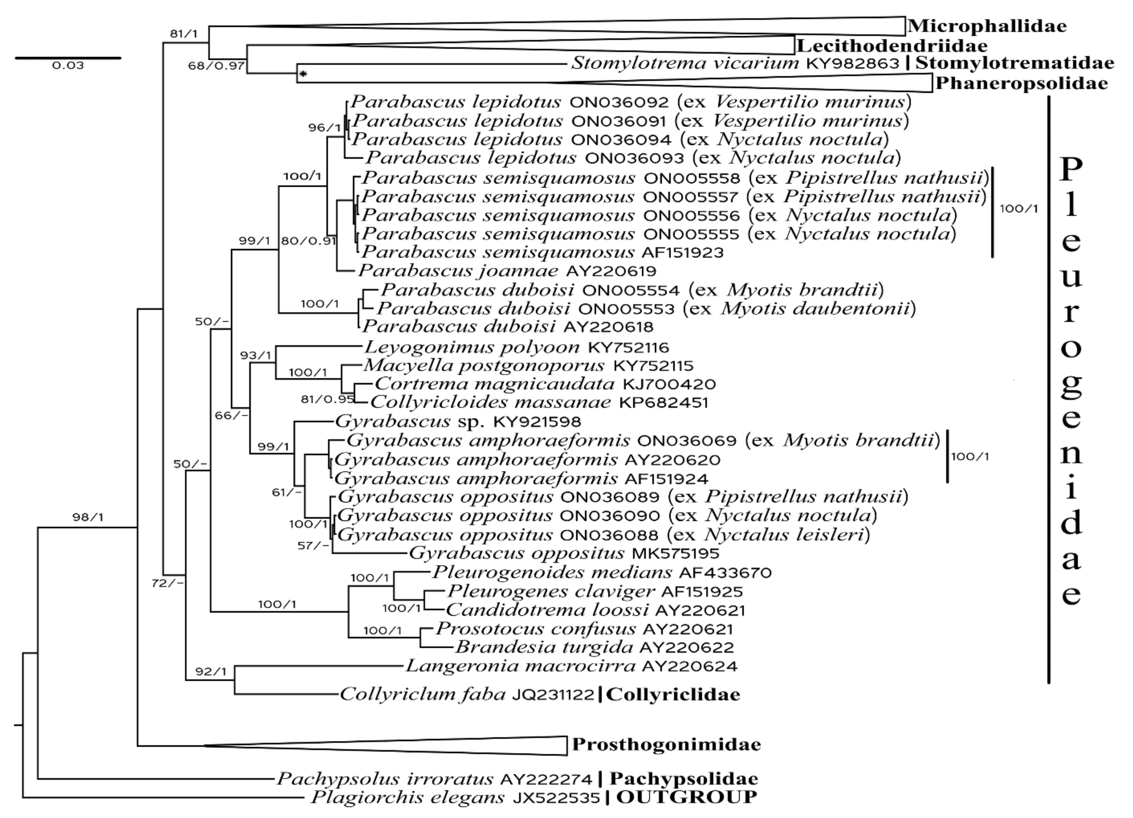
We generated 14 new sequences of partial 28S rRNA genes for five trematode species and distinguished their relationships with closely related digeneans (Figure 1). We did not discuss the relationships among species of the families Microphallidae, Lecithodendriidae, Phaneropsolidae, and Prosthogonimidae in detail.
In our analysis, Microphallidae is the sister taxa to Lecithodendriidae + Phaneropsolidae + Stomylotrematidae. Stomylotrema vicarium (Stomylotrematidae) forms a sister clade to Phaneropsolidae with a relatively low nodal support in Bayesian Inference analysis. In Maximum likelihood analysis, Stomylotrematidae is a separate clade sister to Lecithodendriidae. Pleurogenidae is a sister to the previously described taxa with a relatively low posterior probability. Prosthogonimidae are a basal clade to Microphallidae + Lecithodendriidae + Phaneropsolidae + Stomylotrematidae, and Pleurogenidae. Pachypsolus irroratus (Pachypsolidae) is basal to the other trematodes under consideration.
Among pleurogenids, Gyrabascus spp. form a sister clade to Parabascus spp. with a relatively high Bayesian probability. Three newly generated sequences of G. oppositus (ex N. leisleri, N. noctula, and P. nathusii) cluster together with the previously obtained sequence of G. oppositus (GenBank No MK575195, ex P. kuhlii). Gyrabascus amphoraephormis ex M. brandtii clustered with other previously obtained specimens of these species. Two specimens of P. duboisi (both ex M. daubentonii and M. brandtii) were found to be closely related to the previously known P. duboisi sequence (GenBank No AY220618). Four newly obtained sequences of P. semisquamosus (ex N. noctula and P. nathusii) formed a compact clade without any clear correlation with the host species. Parabascus joannae formed a sister clade to P. semisquamosus. Four newly obtained specimens of P. lepidotus (ex N. noctula and V. murinus) clustered together without any clear correlation with the host species and form a sister clade to P. joannae + P. semisquamosus. Collyriclum and Loxogenes were found to be closely related. Both genera are sister groups to the other pleurogenids.

3.2. Systematics and Morphological Characteristics

  • Superfamily Microphalloidea Ward, 1901
  • Family Pleurogenidae Looss, 1899
  • Genus Gyrabascus Macy 1935
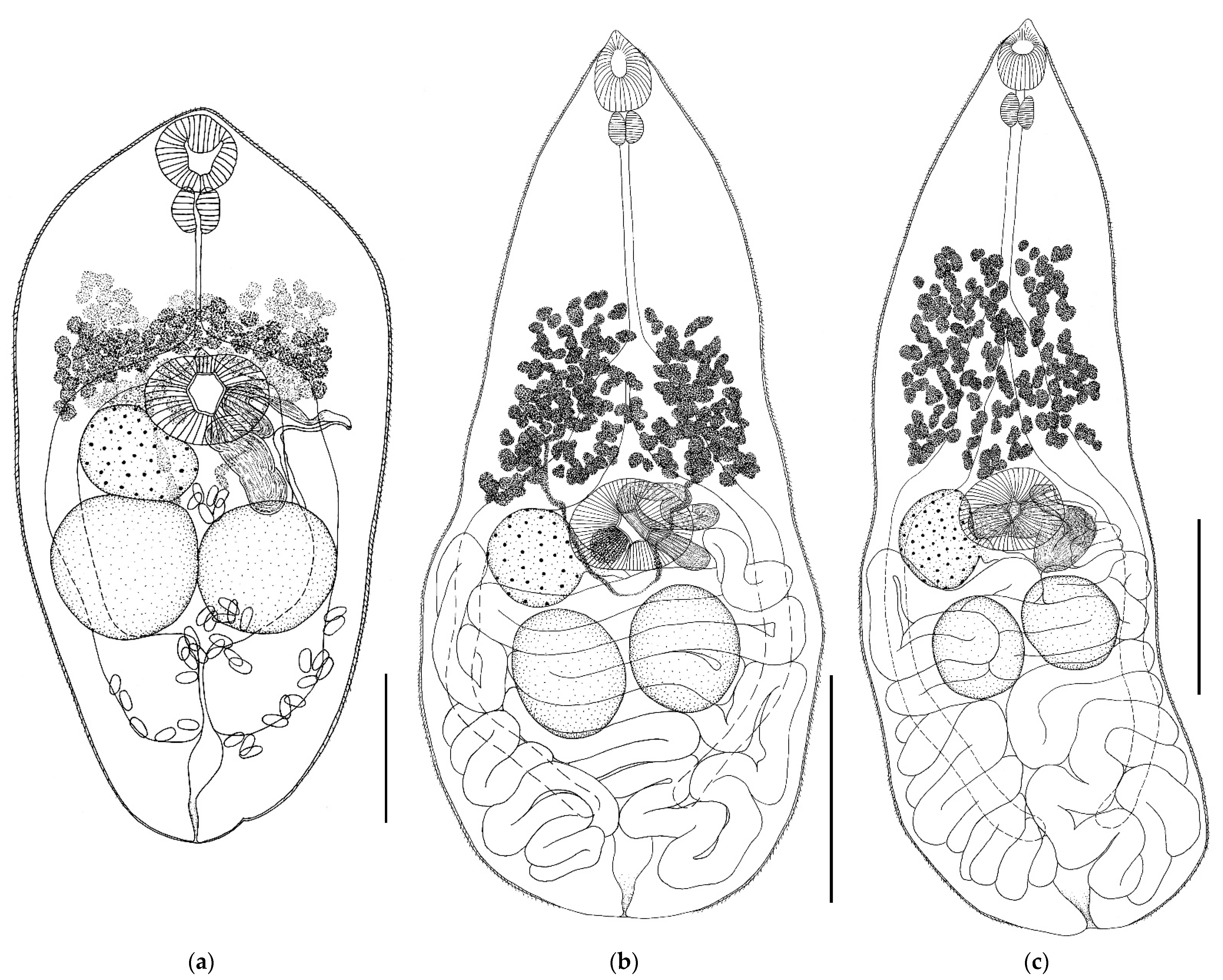
  • Gyrabascus amphoraeformis (Mödlinger, 1930) (Figure 2)
  • Host: Myotis brandtii.
  • Geographical locality: Smolny village (Republic of Mordovia, Russia).
  • Availability: GenBank No ON036069.
  • Accession numbers in collection of IEVB RAS: No 2101.
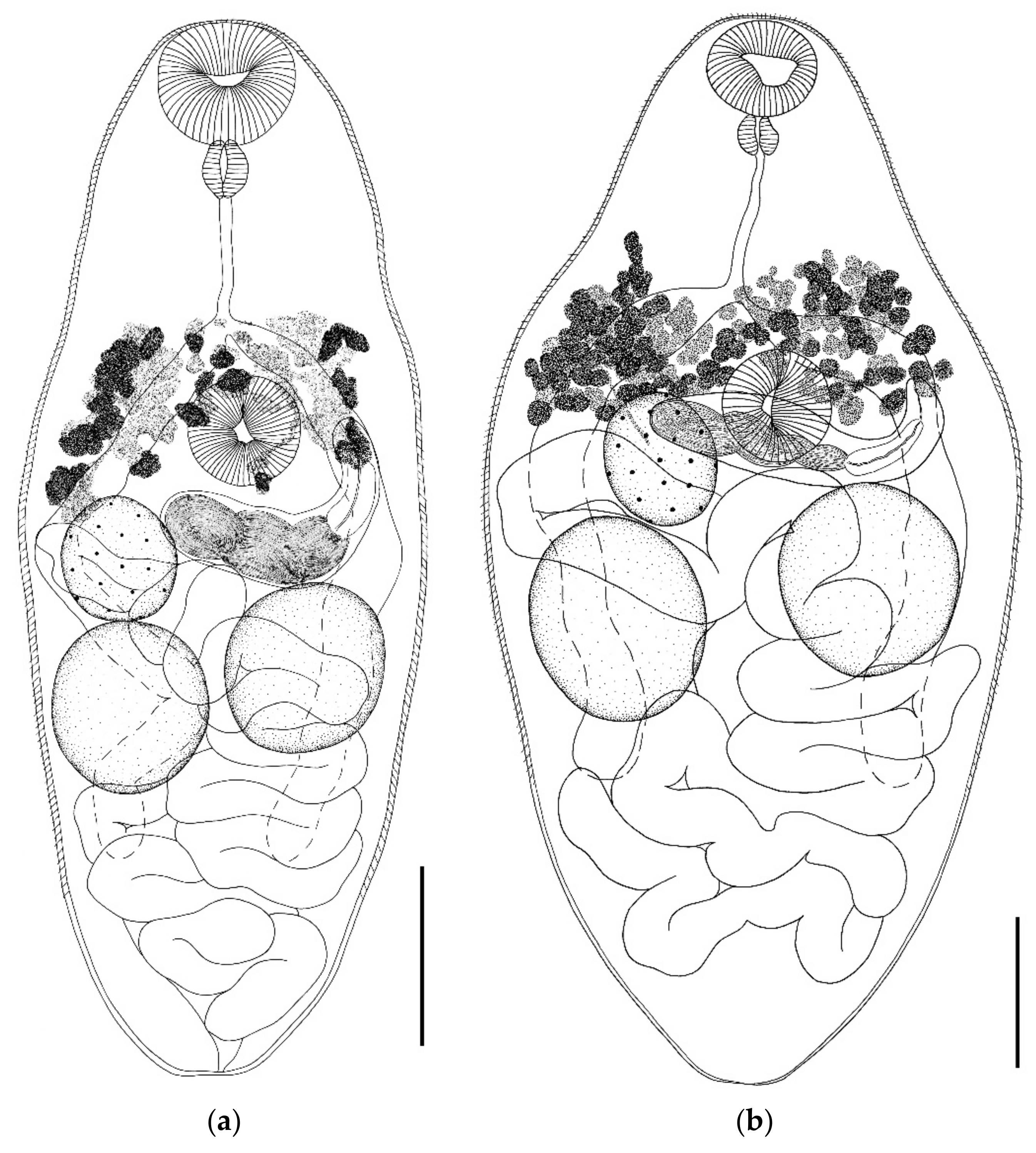
General description (based on single adult specimen, measurements are given in text directly): Body oval, somewhat narrowed anteriorly and posteriorly; length 494, maximum width 255 at level of intestinal bifurcation. Body length to width ratio 1.9:1. Body covered with spines except at posterior end. Oral sucker subterminal, 53 × 55. Ventral sucker pre-equatorial, transversally elongated, 60 × 85. Oral sucker to ventral sucker width ratio 0.7:1. Pharynx 30 × 32. Prepharynx not visible. Oral sucker to pharynx width ratio 1.7:1. esophagus narrow and long, length 65. Intestinal bifurcation placed before ventral sucker. Intestinal branches end blindly behind testes, not reaching hind body. Testes rounded, post-equatorial; 0.089–0.096 × 0.094–0.099. Cirrus sac absent. Seminal vesicle large, convoluted, located dorsally of ventral sucker. Ejaculatory duct departs from the distal end of seminal vesicle and opens into genital atrium. Genital pore at level of ventral sucker, in the middle between lateral edge of ventral sucker and body margin. Ovary rounded, submedial, located at posterolateral edge of ventral sucker, 62 × 82. Vitellarium consists of numerous irregular-shaped follicles and extends from about mid-esophagus level to anterior ovary edge. Egg size 14–17 × 7–9. Excretory pore terminal.
  • Gyrabascus oppositus (Zdzitowiecki, 1969) (Figure 2)
  • Host: Nyctalus noctula, Nyctalus leisleri, Pipistrellus nathusii.
  • Geographical locality: Smolny village (Republic of Mordovia, Russia).
  • Availability: GenBank No ON036088—ON036090.
  • Accession numbers in collection of IEVB RAS: No 2102–2105.
General description (based on 25 adult specimens): Body pear-shaped, elongated, with maximum body width at testes level. Body narrows toward conical anterior end of body and widens toward rounded posterior end. Body densely covered with spines except at posterior end. Oral sucker subterminal, drop-shaped, with elongated anterodorsal side. Prepharynx short. Esophagus long. Intestinal bifurcation approximately at border of anterior and middle thirds of body. Intestinal branches long, extending beyond testes level but not reaching posterior body end. Ventral sucker equatorial, transversely elongated, always larger than oral sucker. Testes oval, lying behind ventral sucker in hind body, approximately at the same level or one slightly behind the other. Cirrus sac absent. Seminal vesicle convoluted, lying freely in parenchyma approximately at level of ventral sucker, more or less overlapping with it. Genital pore submedial, opens at ventral sucker level on opposite side from ovary. Ovary round or oval, submedially located at lateral edge of ventral sucker, may be partially overlapped by it. Vitellarium consists of numerous oval or irregularly shaped follicles. Follicles located between level of intestinal bifurcation and ventral sucker, or slightly in front of it. Vitelline fields do not extend below ventral sucker level. Uterus forms numerous transverse loops, occupies all space in hind body, behind level of ventral sucker, and completely overlaps testes. Excretory pore terminal.
Remarks. Trematodes G. oppositus from N. noctula and from P. nathusii are morphologically similar and differ in body size only (Table 1 and Figure 2). The body size varied both in specimens from different hosts and in specimens from the same host. Trematodes from P. nathusii had the same width as those from N. noctula but a smaller body length. So, trematodes ex P. nathusii had a body length from 652 to 948 (in N. noctula 941–1230) and a width of 348 to 460 (in N. noctula 326–450). Correspondingly, the body length to width ratio in trematodes ex P. nathusii is 1.8–2.2:1 (average 2.0:1), while for trematodes ex N. noctula it is 2.6–3.1:1 (average 2.8:1) (Table 1). Specimens from P. nathusii are wider in the posterior half of the body and less elongated, while those from N. noctula are more elongated. The size of oral and ventral suckers varied both in specimens from different host species and in specimens from the same host species. Thus, the oral sucker width of trematodes from N. noctula varied from 54 to 75, while that of trematodes from P. nathusii varied from 49 to 69. The width of the ventral sucker of specimens from N. noctula varied from 91 to 114, while that in specimens from P. nathusii, from 102 to 126. Accordingly, the oral sucker width of trematodes G. oppositus from different hosts ranged from 49 to 75, while the ventral sucker width ranged from 91 up to 126. The width ratio of the oral sucker to ventral sucker remained relatively constant. Larger suckers were noted in specimens from N. noctula. The pharynx size varied in G. oppositus from the two hosts, being 39–51 in specimens from N. noctula and 30–37 in specimens from P. nathusii (Table 1). This feature changed only slightly in specimens from one host species. Considerable differences were noted in the esophagus length of trematodes G. oppositus both from the two host species and from the same host species. A longer esophagus was noted in G. oppositus from N. noctula, 148–217 (from P. nathusii 126–177) (Table 1). Variability was noted in the size of testes and ovaries in trematodes both from the two host species and from the same host species. The reproductive organs were larger in G. oppositus from N. noctula. The size of eggs in trematodes varied slightly (Table 1).
  • Genus Parabascus Looss, 1907
  • Parabascus duboisi (Hurkova, 1961) (Figure 3 and Figure 4)
  • Host: Myotis brandtii, Myotis daubentonii.
  • Geographical locality: Smolny and Pushta villages (Republic of Mordovia, Russia).
  • Availability: GenBank No ON005553, ON005554.
  • Accession numbers in collection of IEVB RAS: No 2106–2108.
General description (based on 28 adult specimens): Body fusiform, ovoid, lanceolate, or oval. Body densely covered with spines except at posterior end. Oral sucker round, subterminal. Prepharynx not visible. Ventral sucker pre-equatorial; equal to or somewhat smaller than oral one. Intestinal bifurcation located at anterior edge of ventral sucker. Intestinal branches long, extending beyond testes level, not reaching hind body. Testes round or oval, located below ovary level or at some distance from it. Testes approximately at same level or one somewhat behind the other. Cirrus sac elongated, club-shaped, located in ventral sucker region or directly behind it, lies across the body, or at an angle to longitudinal axis of body. Proximal part of cirrus sac may touch the ovary, more or less overlap it. Distal part of cirrus sac in most cases reaches the anterior edge of testes or, less often (in three cases), proximal end of cirrus sac touches the testes. Both testes are at some distance from cirrus sac. Convoluted seminal vesicle occupies all proximal parts of cirrus sac. Genital pore submedial at level of ventral sucker or somewhat behind it. Ovary round or oval, located at ventral sucker level or somewhat behind it. Vitellarium consists of numerous irregularly shaped follicles, begins at level of intestinal bifurcation or slightly above it, reaches level of posterior edge of ventral sucker or goes somewhat beyond it. Uterus forms numerous loops, fills all the space below level of ventral sucker, partially overlaps testes. Eggs oval. Excretory pore terminal.
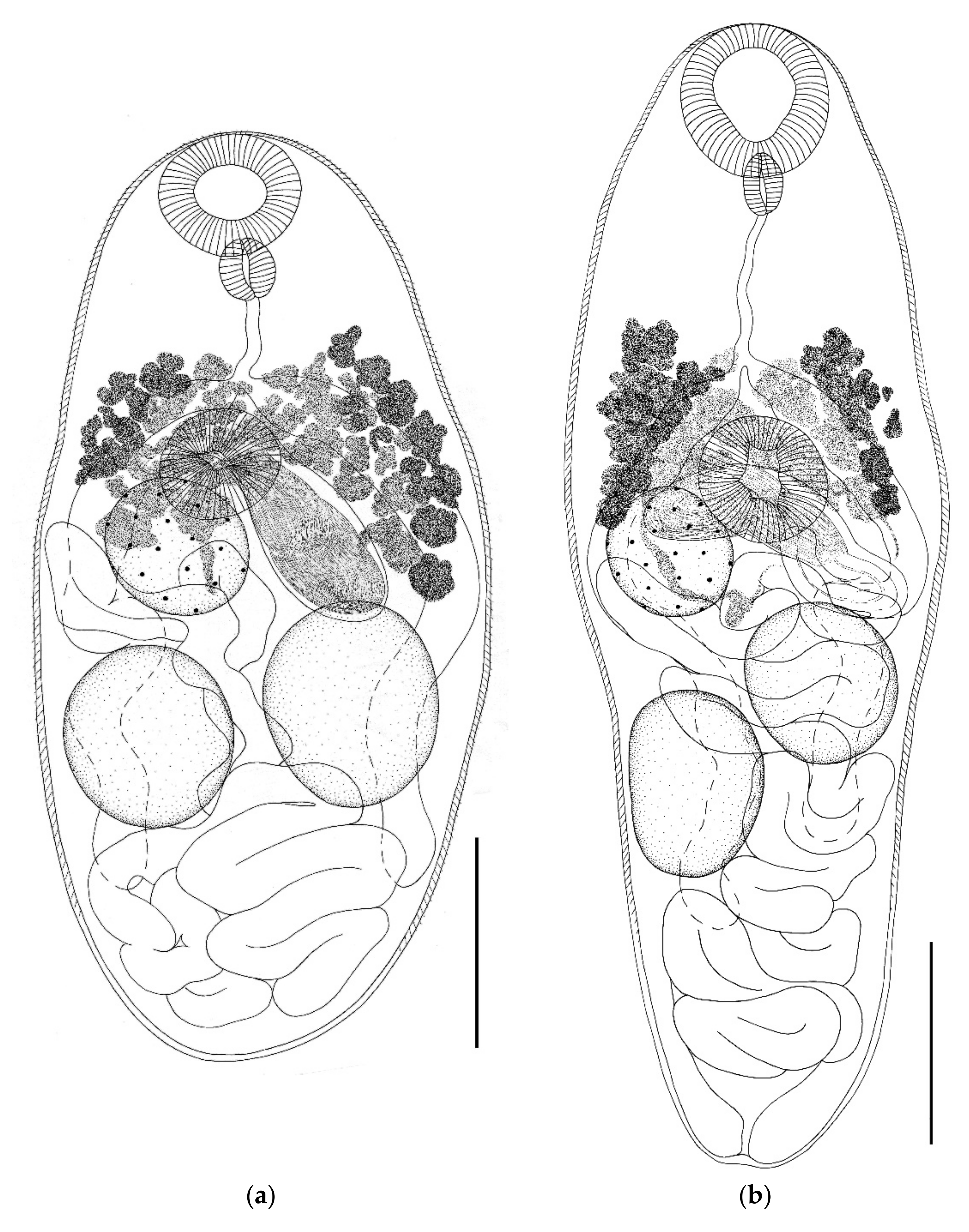
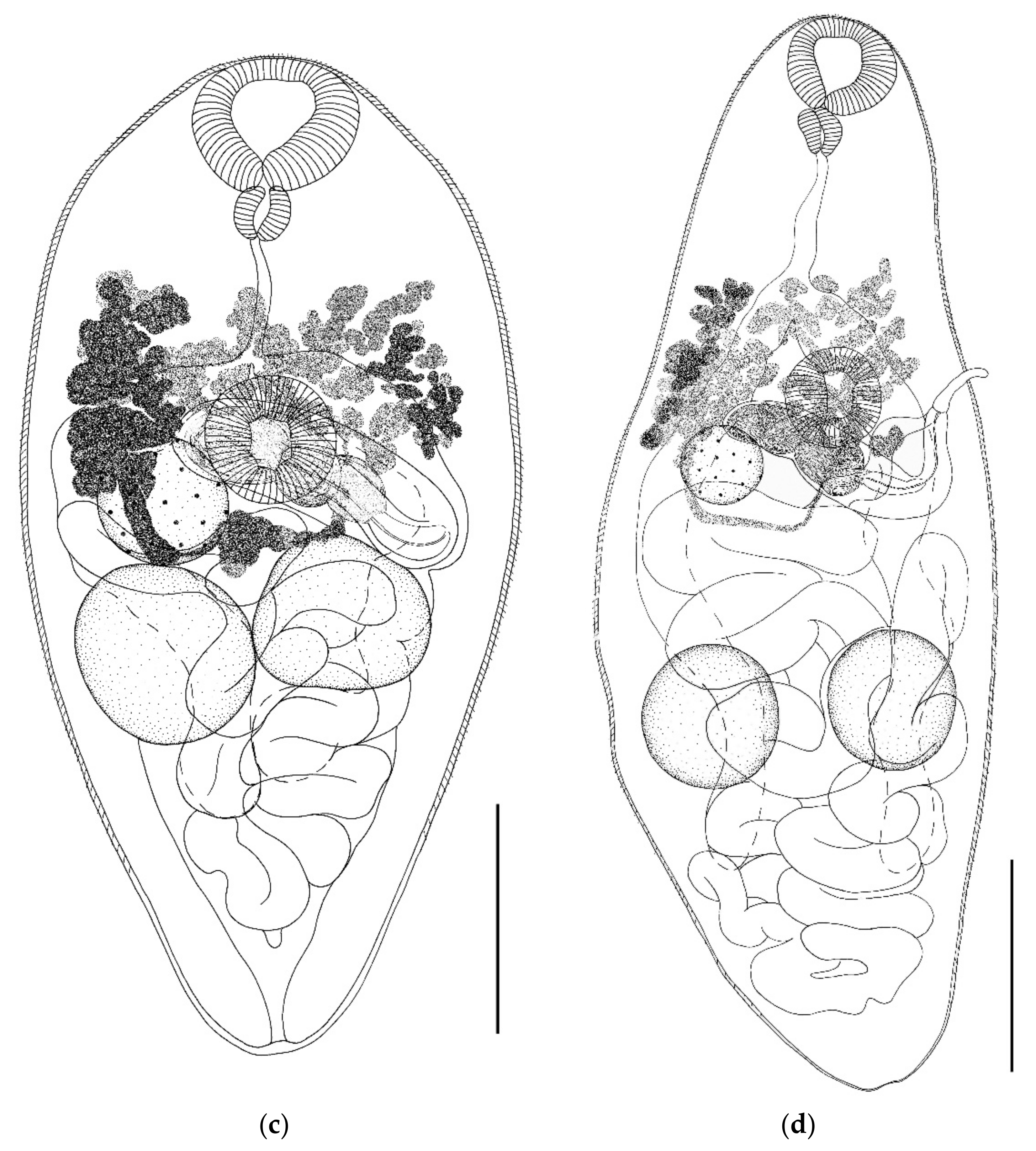
Remarks. Specimens of Parabascus duboisi from M. daubentonii and M. brandtii differ in morphology and size of individual organs (Table 1, Figure 3 and Figure 4). The largest specimens of this trematode as well as the smallest ones were noted in M. daubentonii. Trematodes from M. brandtii were intermediate in size (Table 1). The body length to width ratio varied both in specimens from different hosts and in specimens from one host species. This value varied in trematodes from the two host species in the range from 1.5 to 3.0 (Table 1). The greatest variability in body size was noted in parasites from M. daubentonii. Body length varied from 394–1020 (in trematodes from M. brandtii, 520–852), and the body width varied from 187–415 (in trematodes from M. brandtii, 217–422). The sizes of suckers varied in trematodes regardless of the host species. Thus, the oral sucker width in trematodes from M. daubentonii was 63–98 (in trematodes from M. brandtii 59–87), while the ventral sucker width in trematodes from M. daubentonii was 49–95 (from M. brandtii 49–79) (Table 1). The width of the oral sucker to ventral sucker ratio remained relatively constant in specimens from both host species, varying within 1.0–1.4: 1. The pharynx size and the width of the oral sucker to pharynx ratio in P. duboisi differed insignificantly. A greater variability was observed in trematodes from one host and even from one host specimen. This was also noted in the case of the length of the esophagus and ovary size. The variability in the size of testes in trematodes from different host species was more marked. The largest testes were noted in trematodes from M. brandtii. This also applies to the size of the cirrus sac (Table 1). The body shape changed significantly, both in specimens from different host species and in specimens from the same host species. Variability in the position of the cirrus sac and the interposition of the ovary and testes was noted. Testes could be located directly behind the ovary. In this case, one of the testes could touch the ovary, or the testes were located at some distance from it. The variability of the length of vitelline fields was small (Figure 3 and Figure 4). The egg size in P. duboisi was constant and did not depend on the host species.
  • Parabascus semisquamosus (Braun, 1900) (Figure 4)
  • Host: Nyctalus noctula, Pipistrellus nathusii.
  • Geographical locality: Smolny and Pushta villages (Republic of Mordovia, Russia).
  • Availability: GenBank No ON005555—ON005558.
  • Accession numbers in collection of IEVB RAS: No 2109–2112.
General description (based on 30 adult specimens): Body elongate, tapering at anterior and posterior ends. Body covered with spines, except at posterior end. Oral sucker subterminal. Prepharynx not visible. Esophagus narrow and long. Intestinal bifurcation approximately in middle part of anterior third of body. Intestinal branches long, extending well beyond testes level, but do not reach hind body. Ventral sucker pre-equatorial, larger than oral sucker, located in posterior part of anterior third of body. Testes rounded or oval, some distance behind ventral sucker, approximately at same level or one slightly behind the other. Cirrus sac elongate and club-shaped, situated immediately behind ventral sucker at an angle to longitudinal axis of body. Convoluted seminal vesicle located in proximal part of cirrus sac. Proximal part of cirrus sac located at lateral or posterolateral edge of ovary and may overlap it to a greater or lesser extent. Convoluted seminal vesicle occupies whole proximal part of cirrus sac. Genital pore is submedial at level of ventral sucker on opposite side from ovary. Ovary rounded or oval, located submedially behind ventral sucker. Vitellarium consists of numerous oval, rounded, and pear-shaped follicles located between intestinal bifurcation and ventral sucker. Vitelline fields do not extend beyond level of posterior edge of ventral sucker. Uterus forms numerous loops, occupies all space behind ovary. Terminal part of uterus with well-defined metraterm. Excretory pore terminal.
Remarks. The specimens of P. semisquamosus from N. noctula and P. nathusii are morphologically similar (Figure 4) but differ in the size of the body and individual organs. The body size of specimens from different host species varied widely. The largest trematodes were noted in N. noctula: body length 1354–1892 with a width of 277–460; the smallest ones were from P. nathusii: body length 1031–1523, with a width of 246–415. Parasites from P. nathusii had approximately the same width as those from N. noctula, with a smaller body length (Table 1). Accordingly, specimens from P. nathusii were wider in the mid body, and those from N. noctula were narrower. The variability of the body size was less marked in specimens from the same host species. The size of oral and ventral suckers of P. semisquamosus from various hosts was approximately the same, as was their ratio. Considerable differences were noted in the size of oral and ventral suckers from the same host. Thus, the oral sucker width in trematodes from N. noctula was 46–67 (from P. nathusii 40–68), and the ventral sucker width was 75–106 (from P. nathusii 73–110). The same applies to the size of the pharynx (Table 1). The esophagus length of P. semisquamosus varied in specimens from the two host species. A longer esophagus was noted in specimens from N. noctula: 187–335, as compared with 150–268 in specimens from P. nathusii. High variability was observed in the size of reproductive organs of trematodes, regardless of the host species. Larger testes, cirrus sac, and ovary were noted in trematodes from N. noctula. Egg size was a constant characteristic in trematodes from both host species.
  • Parabascus lepidotus Looss, 1907 (Figure 5)
  • Host: Nyctalus noctula, Vespertilio murinus.
  • Geographical locality: Smolny and Pushta villages (Republic of Mordovia, Russia).
  • Availability: GenBank No ON036091—ON036094.
  • Accession numbers in collection of IEVB RAS: No 2113–2119.
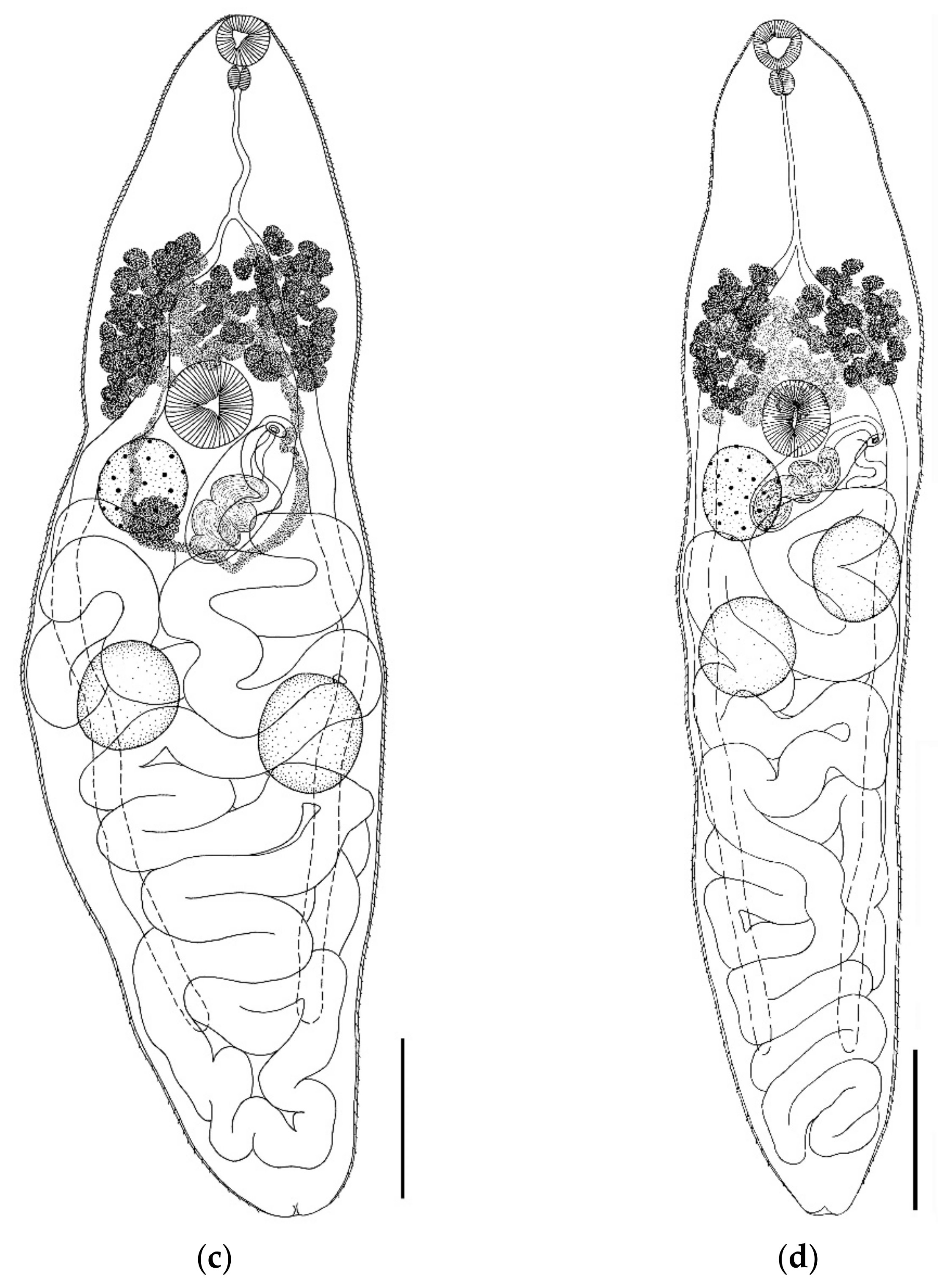
General description (based on 25 adult specimens): Body pear-shaped or spindle-shaped, covered with spines, except at posterior end. Round or oval oral sucker subterminal. Prepharynx very short and not always visible. Esophagus long. Ventral sucker equatorial, somewhat larger than oral sucker or equal to it. Intestinal bifurcation close to ventral sucker. Intestinal branches long, go beyond testes level, do not reach posterior body end. Testes rounded, located one somewhat behind the other or symmetrically. Cirrus sac elongated, located in ventral sucker region and partially overlapped by it. Convoluted seminal vesicle located in proximal part of cirrus sac. Genital pore submedial, at level of ventral sucker. Ovary round to oval, located at ventral sucker level or somewhat behind it. Vitellarium composed of numerous, irregularly shaped follicles; begins at level below mid-esophagus or intestinal bifurcation, does not go beyond the level of posterior edge of ventral sucker. Uterus forms numerous loops and fills all space in hind body behind the ventral sucker, partially overlapping the testes. Excretory pore terminal.
Remarks. Significant variability in size and morphology was noted in P. lepidotus from V. murinus and N. noctula (Figure 5). The largest specimens were noted in N. noctula: body length 844–1160 with a width of 385–619; the smallest one, in V. murinus: body length 441–770 with a width of 272–519. The variability in body size was especially pronounced in P. lepidotus from V. murinus. In this host, trematode specimens strikingly different in width were noted. The body length to width ratio in specimens ex V. murinus was 1.1–1.8:1 (average 1.6:1), and that in specimens ex N. noctula was 1.5–2.5:1 (average 2.1:1). (Table 1). The size of the oral and ventral suckers from the two host species varied. The oral sucker width of specimens from N. noctula varied from 51 to 83, while that of specimens from V. murinus varied from 43 to 67. The width of the ventral sucker of specimens from N. noctula varied from 63 to 98, and that in specimens from V. murinus, from 45 to 75. In P. lepidotus from V. murinus, the oral sucker could be smaller than the ventral sucker, but more often the suckers were equal in size. In trematodes from N. noctula, the oral sucker was always smaller than the ventral one. The oral sucker to ventral sucker ratio in specimens from different hosts was constant. This ratio was more variable in trematodes from the same host species. The pharynx of P. lepidotus specimens from N. noctula was slightly larger than that of the specimens from V. murinus. The width of the oral sucker to pharynx ratio was approximately the same in trematodes from various hosts. Considerable variability in the esophagus length was noted in P. lepidotus specimens from both host species. The esophagus of specimens from N. noctula (217–293) was much longer than that of specimens from V. murinus (102–181). The size of the testes, ovary, and cirrus sac varied greatly in P. lepidotus specimens from the two host species. These features were less variable in trematodes from the same host species. The largest organs were noted in specimens from N. noctula. Differences in the body shape of parasites from different host species were noted. In P. lepidotus specimens ex V. murinus, the body is pear-shaped with a tapering anterior end and a rounded, widened posterior one. In specimens from N. noctula, the body is more spindle-shaped with tapering anterior and posterior ends. Differences in the location of the ventral sucker were also noted. The position of the cirrus sac was different in specimens from various host species. In P. lepidotus specimens from V. murinus, the cirrus sac was always located submedially at the ventral sucker level, partly overlapped by it. In specimens from N. noctula, the cirrus sac was located medially and directly behind ventral sucker. Variability was noted in the length of vitelline fields in specimens from various hosts. In P. lepidotus specimens from V. murinus, the posterior edge of the vitelline follicles did not extend beyond the ventral sucker level. In P. lepidotus specimens from N. noctula, the vitelline fields sometimes extended beyond the level of the lower edge of the ventral sucker. The anterior edge of the vitelline fields in P. lepidotus specimens from both host species was at the level of intestinal bifurcation or slightly above it. Egg size was constant regardless of the host species.

4. Discussion

In this study, we presented morphological descriptions of five species of trematodes from the genera Gyrabascus and Parabascus from various species of bats from Mordovia (Russia) and novel molecular phylogenetic data on these parasites. The combined use of molecular and morphological methods made it possible to perform a reliable identification of Gyrabascus amphoraeformis, Gyrabascus oppositus, Parabascus duboisi, Parabascus lepidotus, and Parabascus semisquamosus.
The general topology of the tree obtained in this study is in good agreement with previous publications [30,60,61]. The two exceptions are the families Stomylotrematidae and Phaneropsolidae. In Shchenkov et al. [60] and Sokolov et al. [30], Stomylotrematidae was the closest to Lecithodendriidae, while Phaneropsolidae formed a sister clade to Lecithodendriidae + Stomylotrematidae branch. In Dellagnola et al. [62], Stomylotrematidae and Phaneropsolidae were sister clades to each other and close to Microphallidae. In Dellagnola et al. [62], Lecithodendriidae appeared to be the basal clade to other microphallids. In Fernandes et al. [61], Phaneropsolidae is a sister clade to Lecithodendriidae, while Stomylotrematidae was left out of the analysis. In our analysis, the phylogenetic position of Stomylotrematidae was unstable between ML and BI analyses. All digenean specimens incorporated into our molecular phylogenetic analysis belonged to five distinct species, which were difficult to distinguish based on morphological features only. No impact of host species on the clusterization of the species under consideration was revealed.
In general, the trematode specimens examined in this study corresponded to the earlier morphological descriptions of the species under consideration [17,24,27,28,29,30,33,35,36,37,38,39,47,48,63,64,65,66,67,68]. However, we noted some differences in the topology of the inner organs and the morphometric features. Specimens of Gyrabascus spp. and Parabascus spp. from various host species and from specimens of the same host showed a broad morphometric variability. Morphometric features such as the body length and width, and the size of the reproductive organs varied broadly, apparently depending to a large extent on the age of the parasites. The dependence of morphometric changes on the trematode age has been noted by several authors [69,70,71,72,73,74] and is especially evident in trematodes from the same host species [73].
Morphological variability of the body shape and the position of the cirrus sac noted in our study also depended on the degree of development of the trematode specimen. The variability of the length of the vitelline fields, the body length to width ratio, and the oral sucker to pharynx ratio were less pronounced. Features such as the oral sucker to the ventral sucker width ratio and the size of eggs were relatively stable (Table 1).
The observation that the variability of parasites depends on the host species has been made in a number of studies [70,71,73,74,75,76,77,78,79]. We also found that the morphological variability of trematodes depended on the species of the host. A host-induced variability was recorded in P. oppositus, P. duboisi, P. semisquamosus, and P. lepidotus in respect of the body size, body shape, and the size of inner organs (Table 1 and Figure 2, Figure 3, Figure 4 and Figure 5).
We gave the first complete description of G. amphoraeformis from M. brandtii, including morphometric data. The comparison of our specimen of G. amphoraeformis with the descriptions of other authors [27,28,33,36,38,48,63,64,65,67] demonstrated a good agreement in the main morphological and morphometric characteristics, except for the egg size. In our specimen, the eggs were somewhat smaller than in descriptions by other authors (Section 3.2 and Table 2).
The specimens of G. oppositus described by us ex N. noctula differed from the previously described specimens from other host species in being longer (Table 3). It should be noted that this species has been previously recorded in P. kuhlii, M. schreibersi, and Eptesicus serotinus Schreber, 1774 [28,30,39]. Pipistrellus nathusii and N. noctula are new hosts for G. oppositus.
Specimens of P. semisquamosus examined in our study fully corresponded to the descriptions available in the literature [17,24,27,28,29,33,34,64,80]. Specimens of P. duboisi examined here differed in body length, size of the oral sucker, and cirrus sac (Table 1 and Table 4) from those described by other authors [17,24,27,28,29,63,66,67]. The difference was mostly due to the fact that our specimens from M. daubentonii were relatively larger than the parasites described earlier. Our specimens of P. duboisi from M. brandtii fully corresponded to the literature data in respect of the measurements (Table 4).
The specimens of P. lepidotus from V. murinus obtained in our study were smaller in respect of body size than those described in earlier studies [17,24,28,33,35,37,38,47,63]. Accordingly, their organs were also smaller (Table 1). Specimens of P. lepidotus from the N. noctula mainly corresponded to the morphological and morphometric characteristics given earlier (Table 5).
Related species of trematodes can be identified erroneously, therefore, it is necessary to take into account the morphological and morphometric variability in their diagnosis. Zdzitowiecki [28] noted that P. duboisi and P. lepidotus are morphologically very similar. The results of our study confirm this. It was often difficult to distinguish these species. For example, such a diagnostic morphometric feature as the size of the oral and the ventral sucker was not always applicable. In our material, there were trematode specimens (both P. duboisi and P. lepidotus) in which the size of the oral sucker was equal to that of the ventral sucker. Another diagnostic feature that could not be used in these two species was the esophagus length. There were specimens in which the values of this feature were the same. We conclude that the following morphological and morphometric features should be taken into account when identifying these species: the body length to width ratio, the oral sucker to pharynx width ratio, the oral sucker to the ventral sucker width ratio, and the location of the ventral sucker relative to the mid body.
Zdzitowiecki [28] has also noted that G. oppositus is similar in many morphological features to P. lepidotus and that many early descriptions of the latter [36,37,38,63] could also apply to G. oppositus. This is probably why Khotenovsky [29] reduced G. oppositus to synonyms of P. lepidotus. In addition, these species of trematodes have a common host, N. noctula. Therefore, when identifying these trematode species, one should take into account the following morphological features: the body shape, the oral sucker shape, the size and position of the ventral sucker relative to the mid body, and the presence/absence of the cirrus sac.
In turn, G. oppositus is similar to G. amphoraeformis. Skvortsov [49] considered the former as a synonym of the latter. Distinctive morphological features for these two species are the body shape, the oral sucker shape, the position of the ventral sucker relative to the mid body and the location of the genital pore.
It is much easier to distinguish P. semisquamosus from P. lepidotus and P. duboisi. Here, the distinctive morphological and morphometric features are the body shape, the body length to width ratio, the oral sucker to the ventral sucker width ratio, the position of the ventral sucker relative to the mid body, and the length of the intestinal branches.
Based on our results, we propose a key for identifying Gyrabascus spp. and Parabascus spp. involved in our study. It should be noted, however, that the applicability of this key is somewhat limited due to a broad morphological variability of these trematodes, which is especially pronounced in immature specimens. Special care should also be taken during the identification of adults that could have been deformed during fixation and whole mount preparation.
  • 1(6) Cirrus sac absent.
  • 2(4) Genital pore marginal.
  • 3(2) Body oval with a maximum width at the intestinal bifurcation level. Ventral sucker pre-equatorial ...................... Gyrabascus amphoraeformis (Mödlinger, 1930).
  • 4(2) Genital pore submedial.
  • 5(4) Body pear-shaped with a maximum width at the testes level. Ventral sucker equatorial ………….………………….... Gyrabascus oppositus (Zdzitowiecki 1969).
  • 6(1) Cirrus sac present.
  • 7(12) Ventral sucker pre-equatorial.
  • 8(10) Ventral sucker larger than oral sucker.
  • 9(8) Body elongated, narrow. Intestinal branches extend far beyond the testes level. The body length to width ratio is 3.2–5.2: 1. The oral sucker to ventral sucker ratio is 0.6–0.7:1. The oral sucker to pharynx ratio is 1.6–2.3:1 ...................
    ...................................................................... Parabascus semisquamosus (Braun, 1900).
  • 10(8) Ventral sucker equal to or less than oral sucker.
  • 11(10) Body oval, shortened. Intestinal branches end directly behind the testes. The body length to width ratio is 1.5–3.0:1. The oral sucker to ventral sucker ratio is 1.0–1.4:1. The oral sucker to pharynx ratio is 2.0–3.0:1 ……………….…..……..
    ………………………………………………….. Parabascus duboisi (Hurkova, 1961).
  • 12(7) Ventral sucker equatorial.
  • 13(12) Body pear-shaped with a maximum width at the testes level. The body length to width ratio is 1.1–2.5:1. The oral sucker to ventral sucker ratio is 0.7–0.9:1. The oral sucker to pharynx ratio is 1.6–2.3:1 ....... Parabascus lepidotus (Looss, 1907).
An analysis of the literature data and the results of our own studies showed that trematodes of the genera Gyrabascus and Parabascus exhibit a certain host specificity. In our study, P. lepidotus was found only in N. noctula and V. murinus. P. semisquamosus and G. oppositus were noted only in P. nathusii, N. noctule, and N. leisleri. Parabascus duboisi was recorded only in Myotis spp. We found two specimens of G. amphoraeformis in M. brandtii.
Gyrabascus amphoraeformis mainly occurs in Myotis bats. Though several authors reported this species from N. noctula, P. pipistrellus, E. serotinus, and Barbastella barbastellus Schreber, 1774 [24,28,33], all of them provided drawings of specimens from Myotis spp., and so these reports should be treated with caution. However, the presence of G. amphoraeformis in P. kuhlii has been confirmed by molecular analysis [40], indicating that G. amphoraeformis may indeed parasitize bats other than Myotis spp.
Gyrabascus oppositus has been previously noted only by Mituch [39] (as P. semisquamosus), Zdzietowiecki [28], and Sokolov et al. [30] from P. kuhlii, M. schreibersii, and E. serotinus. In this study, we added three new species to the list of hosts of G. oppositus.
According to the literature data, the hosts of P. semisquamosus are N. noctula and P. pipistrellus (Table 6). Khotenovsky [29] is the only source where M. daubentonii is indicated as its host. The description and drawing of the parasite in this work match those of P. semisquamosus, but we believe that an error could have occurred in identifying the host. Sharpilo and Iskova [33] report P. semisquamosus from Myotis mystacinus Kuhl, 1817 (the drawing and description of the specimen from N. noctula). Skvortsov [24] doubts the findings of P. semisquamosus in Myotis bats since the trematode is a specific parasite of Nyctalus and Pipistrellus bats. We have never found P. semisquamosus in Myotis spp. in our long-term studies of bat helminths [15,16,17,18,19,20,21,22,23].
Parabascus lepidotus has previously been recorded mainly in E. serotinus. Other bats mentioned as its hosts are E. nilssoni, N. noctula, P. kuhlii, P. pipistrellus, P. auritus, V. murinus, M. blythii, and M. nattereri [17,24,28,29,33,35,37,38,47,63]. Findings of P. lepidotus in Myotis bats require confirmation, as the authors may have dealt with a closely related species, P. duboisi, which is a common parasite of Myotis spp. No drawings of P. lepidotus from Myotis bats are given in the studies cited above, except for Zdzietowiecki [28]. His work contains a drawing of a parasite from M. nattereri, which, though we cannot be certain, seems very similar to P. duboisi. We have critically reviewed whole mounts of trematodes from bats of the Samarskaya Luka (Russia) [16,17] and found that of all Parabascus spp. only P. duboisi parasitizes Myotis bats.
Parabascusduboisi is a specific parasite of Myotis spp. [17,24,27,28,29,63,66,67]. Morozov [66] notes M. daubentonii and P. pipistrellus as hosts of P. semisquamosus, but the description and the drawing of the trematodes from M. daubentonii match that of P. duboisi. The trematodes from P. pipistrellus were correctly identified as P. semisquamosus. Revision of whole mounts of trematodes from bats of the Samarskaya Luka showed that E. nilssoni is parasitized by P. lepidotus [16,17].

5. Conclusions

The combined use of molecular and morphological methods in our study made it possible to reliably identify closely related trematode species Gyrabascus amphoraeformis, Gyrabascus oppositus, Parabascus duboisi, Parabascus lepidotus, and Parabascus semisquamosus. A broad morphological variability of Gyrabascus spp. and Parabascus spp. was revealed, both from various host species and from various specimens of the same host species. We reevaluated morphological characters for a reliable identification of the closely related species of the genera Gyrabascus and Parabascus involved in our study and proposed a key for their identification.
Our data complement and expand the knowledge of bat parasites. We provided the first record of Gyrabascus amphoraeformis from bats in the Volga basin and the first record of Gyrabascus oppositus from bats in the Middle Volga region. We also established three new hosts of Gyrabascus oppositus: N. leisleri, N. noctula, and P. nathusii.

Supplementary Materials

The following supporting information can be downloaded at: https://www.mdpi.com/article/10.3390/biology11060878/s1, Table S1: PCR conditions used in the present study [40,81,82,83]; Table S2: Sequences used in molecular phylogenetic analysis [30,40,41,42,84,85,86,87,88,89,90,91,92,93,94,95,96,97,98,99,100,101,102,103].

Author Contributions

Conceptualization, N.Y.K. and S.V.S.; methodology, S.V.S. and N.Y.K.; software, A.B.R.; validation, N.Y.K., S.V.S. and A.B.R.; formal analysis, A.A.K. and S.V.S.; investigation, N.Y.K., S.V.S. and A.A.K.; resources, A.B.R. and S.V.S.; writing—original draft preparation, A.A.K., N.Y.K., S.V.S. and A.B.R.; writing—review and editing, A.B.R., A.A.K. and S.V.S.; supervision, A.B.R.; project administration, A.B.R.; funding acquisition, A.B.R. All authors have read and agreed to the published version of the manuscript.

Funding

This research received no external funding.

Institutional Review Board Statement

All applicable international, national, and institutional guidelines for the use and care of wild animals were followed. No animal was killed with the purpose of helminth sampling for our study. Our research was conducted in compliance with the ethical standards of humane treatment of animals according to the recommended standards of the Directive of the European Parliament and of the Council of the European Union of 22 September 2010 “On the protection of animals used for scientific purposes” (EU Directive 2010/63/EU). The material for parasitological research was obtained as a result of fieldwork on accounting for the number of wild animals. They are regularly held with the research topics of the Federal State Budgetary Institution “Reserved Mordovia” (“Zapovednaya Mordovia”) in 2018–2022. The research theme and handling procedures with wild animals were approved by the Ministry of Natural Resources and Ecology of Russia.

Informed Consent Statement

Not applicable.

Data Availability Statement

GenBank numbers are given in the relevant section of the manuscript. Any other data is available after a reasonable request.

Acknowledgments

We would like to kindly thank Thayane Fernandes and Tyler Achatz for providing their data. The work was performed using the equipment of the Resource Center “Culture Collection of Microorganisms” and “Centre for Molecular and Cell Technologies” at St. Petersburg State University. The study was carried out on the research topic of the Institute of Ecology of the Volga River Basin, a branch of the Samara Federal Research Center of the Russian Academy of Sciences—“Structure, dynamics and sustainable development of ecosystems in the Volga River Basin” No 1021060107217-0-1.6.19.

Conflicts of Interest

The authors declare no conflict of interest.

References

  1. Khotenovsky, I.A. On the evolution of trematodes from bats. Parazitologiia 1972, 6, 79–82. [Google Scholar]
  2. Thewissen, J.G.M.; Babcock, S.K. The origin of flight in bats. BioScience 1992, 42, 340–345. [Google Scholar] [CrossRef]
  3. Mammals of Russia. Available online: http://rusmam.ru/mammal/index?sort=sort (accessed on 20 January 2022).
  4. Il’in, V.Y.; Vekhnik, V.P.; Smirnov, D.G.; Kurmaeva, N.M.; Zolina, N.F.; Matrosova, O.M. Dynamics of abundance of bats (Chiroptera: Vespertilionidae) during hibernation in caves of the Samarian Luka over a 20-year period. Rus. J. Ecol. 1999, 30, 428–431. [Google Scholar]
  5. Artaev, O.N.; Smirnov, D.G. Bats (Chiroptera; Mammalia) of Mordovia: Specific structure and features of distribution. Nat. Conserv. Res. 2016, 1, 38–51. [Google Scholar] [CrossRef] [Green Version]
  6. Smirnov, D.G.; Vekhnik, V.P.; Kurmaeva, N.M.; Shepelev, A.A.; Il’in, V.Y. Species structure and dynamics of bat communities (Chiroptera: Vespertilionidae) hibernating in artificial caves of Samara Luka. Biol. Bull. 2007, 34, 507–516. [Google Scholar] [CrossRef]
  7. Smirnov, D.G.; Vekhnik, V.P. Ecology of nutrition and differentiation of the trophic niches of bats (Chiroptera: Vespertilionidae) in floodplain ecosystems of the Samara Bend. Biol. Bull. 2014, 41, 60–70. [Google Scholar] [CrossRef]
  8. Orlova, M.V.; Smirnov, D.G.; Vekhnik, V.P.; Lukyanenko, A.M.; Zabashta, A.V. Ectoparasites and pathogens of Kuhl’s pipistrelle Pipistrellus kuhlii (Kuhl, 1817) (Chiroptera: Vespertilionidae): Our own and published data review. Russ. J. Biol. Invasions 2020, 11, 348–362. [Google Scholar] [CrossRef]
  9. Orlova, M.V.; Klimov, P.B.; Moskvitina, N.S.; Orlov, O.L.; Zhigalin, A.V.; Smirnov, D.G.; Dzhamirzoyev, H.S.; Vekhnik, V.P.; Pavlov, A.V.; Emelyanova, A.A.; et al. New records of bat flies (Diptera: Nycteribiidae), with an updated checklist of the nycteribiids of Russia. Zootaxa 2021, 4927, 410–430. [Google Scholar] [CrossRef]
  10. Orlova, M.V.; Klimov, P.B.; Orlov, O.L.; Smirnov, D.G.; Zhigalin, A.V.; Budaeva, I.V.; Emelyanova, A.A.; Anisimov, N.V. A checklist of bat-associated macronyssid mites (Acari: Gamasina: Macronyssidae) of Russia, with new host and geographical records. Zootaxa 2021, 4974, 537–564. [Google Scholar] [CrossRef] [PubMed]
  11. Smirnov, D.G.; Vekhnik, V.P.; Dzhamirzoyev, G.S.; Titov, S.V. On the taxonomic status of species from the group Myotis nattereri» (Chiroptera, Vespertilionidae) in the Eastern Caucasus. Nat. Conserv. Res. 2020, 5, 30–42. [Google Scholar] [CrossRef]
  12. Bazhenov, Y.A. Ecology of bat species in the arid region of the Daurian steppe at the peak of drought. Nat. Conserv. Res. 2021, 6, 42–49. [Google Scholar] [CrossRef]
  13. Belkin, V.V.; Fyodorov, F.V.; Ilyukha, V.A.; Yakimova, A.E. Characteristics of the bat (Chiroptera) population in Protected Areas in the northern and middle taiga subzones of European Russia. Nat. Conserv. Res. 2021, 6 (Suppl. 1), 17–31. [Google Scholar] [CrossRef]
  14. Smirnov, D.G.; Bezrukov, V.A.; Kurmaeva, N.M. Use of habitat and foraging time by females of Eptesicus nilssonii (Chiroptera, Vespertilionidae). Russ. J. Theriol. 2021, 20, 1–10. [Google Scholar] [CrossRef]
  15. Kirillova, N.Y.; Kirillov, A.A.; Vekhnik, V.P. Nematodes (Nematoda) from bats (Chiroptera) of the Samarskaya Luka peninsula (Russia). Parazitologiia 2008, 42, 526–532. [Google Scholar] [PubMed]
  16. Kirillov, A.A.; Kirillov, N.Y.; Vekhnik, V.P. Trematodes (Trematoda) of bats (Chiroptera) from the Middle Volga region. Parazitologiia 2012, 46, 384–413. [Google Scholar] [PubMed]
  17. Kirillov, A.A.; Kirillova, N.Y.; Chikhlyaev, I.V. Trematodes of Terrestrial Vertebrates of the Middle Volga Region; Cassandra: Togliatti, Russia, 2012; pp. 3–329. [Google Scholar]
  18. Kirillov, A.A.; Ruchin, A.B.; Artaev, O.N. Helminths of bats (Chiroptera) from Mordovia. Bull. Vol. Univ. Tatishch. 2015, 4, 319–328. [Google Scholar]
  19. Kirillov, A.A.; Ruchin, A.B.; Artaev, O.N. To the study of helminths of bats (Chiroptera) from Mordovia. Proceed. Sam. Sci. Cent Rus. Acad. Sci. 2015, 17, 859–866. [Google Scholar]
  20. Ruchin, A.B.; Kirillov, A.A.; Chikhlyaev, I.V.; Kirillova, N.Y. Parasitic worms of land vertebrates of the Mordovia Nature Reserve. In Flora and Fauna of Reserves; Committee of RAS for the Conservation of Biological Diversity: Moscow, Russia, 2016; Volume 124, pp. 3–72. [Google Scholar]
  21. Kirillova, N.Y.; Kirillov, A.A. Overview of helminths in small mammals in the Zhiguli State Reserve. Nat. Conserv. Res. 2017, 2, 24–37. [Google Scholar] [CrossRef] [Green Version]
  22. Kirillov, A.A.; Kirillova, N.Y.; Krasnobayev, Y.P.; Vekhnik, V.P. Parasitic Worms of Small Mammals in Zhiguli State Nature Reserve; Committee of RAS for the Conservation of Biological Diversity, A.N. Severtzov Institute of Ecology and Evolution of RAS: Moscow, Russia, 2017; pp. 3–81. [Google Scholar]
  23. Kirillov, N.Y.; Kirillov, A.A.; Vekhnik, V.P.; Ruchin, A.B.; Grishutkin, G.F. Helminths of bats (Chiroptera) in “Smolny” National Park: Preliminary Data. Proceed. Mord. St. Nat. Res. 2018, 21, 223–230. [Google Scholar]
  24. Skvortsov, V.G. Trematodes of the family Lecithodendriidae from bats in Moldavia. Paras. Anim. Plant. Mold. 1970, 5, 17–36. [Google Scholar]
  25. Sharpilo, V.P.; Tkach, V.V. About type species of genus Plagiorchis Luhe, 1899 (Trematoda, Plagiorchiidae). Vest. Zool. 1992, 5, 8–15. [Google Scholar]
  26. Tkach, V.V.; Pawlowski, J.; Sharpilo, V.P. Molecular and morphological differentiation between species of the Plagiorchis vespertilionis group (Digenea, Plagiorchiidae) occurring in European bats, with a re-description of P. vespertilionis (Muller, 1780). Syst. Parasitol. 2000, 47, 9–22. [Google Scholar] [CrossRef]
  27. Odening, K. Exkretionssystem und systematische stellung exkretionssystem und systematische stellung einiger eledermaustrematoden aus Berlin und umgebung nebst bemerkungen zum Lecithodendrioiden komplex. Z. Parasitenkd. 1964, 24, 453–483. [Google Scholar] [CrossRef] [PubMed]
  28. Zdzietowiecki, K. Helminths of bats in Poland. III. Trematodes of the subfamily Lecithodendriidae, except for Lecithodendriinae. Acta Parasitol. Pol. 1969, 16, 227–237. [Google Scholar]
  29. Khotenovsky, I.A. Trematodes of the genus Parabascus (Trematoda, Pleurogenidae) from bats in Holarctic. Parasitol. Coll. Pap. 1985, 33, 125–133. [Google Scholar]
  30. Sokolov, S.G.; Shchenkov, S.V.; Kalmykov, A.P.; Smirnova, A.D. Morphology and phylogenetic position of two microphalloid trematode species, parasites of the vesper bat Pipistrellus kuhlii in the Lower Volga region of Russia. Zool. Zhurnal 2020, 99, 261–274. [Google Scholar] [CrossRef]
  31. Lotz, J.M.; Font, W.F. Family Gyrabascidae Macy, 1935. In Keys to Trematoda; Bray, R.A., Gibson, D.J., Jones, A., Eds.; CAB International and Natural History Museum: London, UK, 2008; Volume 3, pp. 523–525. [Google Scholar]
  32. Lotz, J.M.; Font, W.F. Family Phaneropsolidae Mehra, 1935. In Keys to Trematoda; Bray, R.A., Gibson, D.J., Jones, A., Eds.; CAB International and Natural History Museum: London, UK, 2008; Volume 3, pp. 545–562. [Google Scholar]
  33. Sharpilo, V.P.; Iskova, N.I. Trematodes. Plagiorchiata. Fauna of Ukraine; Naukova Dumka: Kiev, Ukraine, 1989; Volume 34, pp. 3–280. [Google Scholar]
  34. Braun, M. Trematoden der Chiroptera. Ann. Naturhist. Mus. Wien. 1900, 15, 215–236. [Google Scholar]
  35. Looss, A. Ueber einige neue Trematoden der aegyptischen fauna. Notizen zur Helminthologie Egyptens. Zbl. Bakteriol. Parasitenkd. 1907, 43, 478–490. [Google Scholar]
  36. Hurkova, J.A. A contribution to the knowledge of bat trematodes in Czechoslovakia. Acta Univ. Carol.-Biol. 1959, 1, 29–36. [Google Scholar]
  37. Hurkova, J.A. A contribution to the knowledge of bat trematodes of the g. Parabascus Looss and g. Limatulum Travassos (fam. Lecithodendriidae) with a description of a new species. Vest. Ceskosl. Zool. Spol. 1961, 25, 277–288. [Google Scholar]
  38. Modlinger, G. Trematoden ungarischer Chiropteren. Stud. Zool. Bud. 1930, 1, 191–203. [Google Scholar]
  39. Mituch, J. Beitrag zur Ercenntnis der Helminthenfauna von Miniopterus schreibersi (Kuhl, 1819) in der Slovakei (CSSR). Helminthologia 1965, 6, 109–119. [Google Scholar]
  40. Tkach, V.V.; Pawlowski, J.; Mariaux, J. Phylogenetic analysis of the suborder Plagiorchiata (Platyhelminthes, Digenea) based on partial lsrDNA sequences. Int. J. Parasitol. 2000, 30, 83–93. [Google Scholar] [CrossRef]
  41. Tkach, V.V.; Littlewood, D.T.J.; Olson, P.D.; Kinsella, J.M.; Swiderski, Z. Molecular phylogenetic analysis of the Microphalloidea Ward, 1901 (Trematoda: Digenea). Syst. Parasitol. 2003, 56, 1–15. [Google Scholar] [CrossRef]
  42. Lord, J.S.; Parker, S.; Parker, F.; Brooks, D.R. Gastrointestinal helminths of pipistrelle bats (Pipistrellus pipistrellus/Pipistrellus pygmaeus) (Chiroptera: Vespertilionidae) of England. Parasitology 2012, 139, 366–374. [Google Scholar] [CrossRef]
  43. Sümer, N.; Yildirimhan, H.S. DNA sequencing of Digenea nuclear lsrDNA of the whiskered brown bat, Myotis aurescens (Vespertilionidae: Chiroptera), from Turkey. Turk. J. Zool. 2017, 41, 64–66. [Google Scholar] [CrossRef]
  44. Sümer, N.; Yildirimhan, H.S. Intestinal Helminth Parasites of Two Bat Species of the Genus Myotis Kaup, 1829 (Chiroptera: Vespertilionidae) from Turkey, with DNA Sequencing of Helminth Nuclear LSR rDNA. Acta Zool. Bulg. 2019, 71, 273–278. [Google Scholar]
  45. Ivashkin, V.M.; Kontrimavichus, V.L.; Nasarova, N.S. Methods of the Collection and Studies of Helminths of Land Mammals; Nauka: Moscow, Russia, 1971; pp. 3–123. [Google Scholar]
  46. Anikanova, V.S.; Bugmyrin, S.V.; Ieshko, E.P. Methods of the Collection and Studies of Helminths of Small Mammals; Karelian Scientific Center of RAS: Petrozavodsk, Russia, 2007; pp. 3–145. [Google Scholar]
  47. Khotenovsky, I.A. Family Pleurogenidae Looss, 1899. In Trematodes of Animals and Man. Essentials of Trematodology; Skryabin, K.I., Ed.; Nauka: Moscow, Russia, 1970; Volume 23, pp. 135–306. [Google Scholar]
  48. Khotenovsky, I.A. Family Allassogonoporidae Skarbilovich, 1947. In Trematodes of Animals and Man. Essentials of Trematodology; Skryabin, K.I., Ed.; Nauka: Moscow, Russia, 1974; Volume 25, pp. 245–258. [Google Scholar]
  49. Skvortsov, V.G. Critical overview of the helminth fauna in bats of the USSR and European countries. Proceed. Acad. Mold. SSR. Biol. Chem. Ser. 1971, 6, 53–59. [Google Scholar]
  50. Fauna Europaea. Available online: https://fauna-eu.org/ (accessed on 22 February 2022).
  51. R Core Team. R: A Language and Environment for Statistical Computing. R Foundation for Statistical Computing. Available online: http://www.r-project.org/index.html (accessed on 22 February 2022).
  52. Paradis, E.; Schliep, K. Ape 5.0: An environment for modern phylogenetics and evolutionary analyses in R. Bioinformatics 2019, 35, 526–528. [Google Scholar] [CrossRef]
  53. Bodenhofer, U.; Bonatesta, E.; Horejs-Kainrath, C.; Hochreiter, S. Msa: An R package for multiple sequence alignment. Bioinformatics 2015, 31, 3997–3999. [Google Scholar] [CrossRef]
  54. Edgar, R.C. MUSCLE: Multiple sequence alignment with high accuracy and high throughput. Nucleic Acids Res. 2004, 32, 1792–1797. [Google Scholar] [CrossRef] [Green Version]
  55. Gouy, M.; Guindon, S.; Gascuel, O. SeaView version 4: A multiplatform graphical user interface for sequence alignment and phylogenetic tree building. Mol. Biol. Evol. 2010, 27, 221–224. [Google Scholar] [CrossRef] [PubMed] [Green Version]
  56. Nylander, J.A.A. MrModeltest v2. Program Distributed by the Author; Evolutionary Biology Centre, Uppsala University: Uppsala, Sweden, 2004. [Google Scholar]
  57. Miller, M.A.; Pfeiffer, W.; Schwartz, T. Creating the CIPRES Science Gateway for inference of large phylogenetic trees. In Proceedings of the Gateway Computing Environments Workshop, New Orleans, LA, USA, 14 November 2010; pp. 1–8. [Google Scholar]
  58. Resource Center “Computer Center of SpbU”. Available online: http://cc.spbu.ru (accessed on 23 February 2022).
  59. Rambaut, A.; Drummond, A.J.; Xie, D.; Baele, G.; Suchard, M.A. Posterior summarisation in Bayesian phylogenetics using Tracer 1.7. Syst. Biol. 2018, 67, 901–904. [Google Scholar] [CrossRef] [Green Version]
  60. Shchenkov, S.; Denisova, S.; Kremnev, G.; Dobrovolskij, A. Five new morphological types of virgulate and microcotylous xiphidiocercariae based on morphological and molecular phylogenetic analyses. J. Helminthol. 2020, 94, E94. [Google Scholar] [CrossRef]
  61. Fernandes, T.F.; dos Santos, J.N.; de Vasconcelos Melo, F.T.; Achatz, T.J.; McAllister, C.T.; Carrion Bonilla, C.; Tkach, V.V. Phylogenetic relationships of Ochoterenatrema Caballero, 1943 (Digenea: Lecithodendriidae) with descriptions of two new species. Parasitol. Int. 2022; in press. [Google Scholar] [CrossRef] [PubMed]
  62. Dellagnola, F.; Campoy-Diaz, A.; Vega, I. First morphological and molecular identification of the cercaria of Stomylotrema vicarium from the endemic apple snail Pomacea americanista. Parasitology 2022, 149, 95–104. [Google Scholar] [CrossRef]
  63. Hurkova, J.A. Bat trematodes in Czechoslovakia. I. A systematical review of occurring species. Vest. Cescosl. Spolec. Zool. 1963, 27, 250–276. [Google Scholar]
  64. Dubois, G. Contribution à l’étude des Trématodes de Chiroptères. Rev. Suiss. Zool. 1956, 63, 683–695. [Google Scholar] [CrossRef]
  65. Dubois, G. Contribution à l’étude des Trématodes de Chiroptères. Revision du genre Alassogonoporus Olivier, 1938 et note additionelle sur le sous-genre Prosthodendrium Dollfus, 1931. Rev. Suiss. Zool. 1963, 70, 103–125. [Google Scholar] [CrossRef]
  66. Morozov, Y.F. To the helminth fauna of bats in the Belovezhskaya Pushcha. Bull. Acad. Sci. Bel. SSR 1961, 2, 92–98. [Google Scholar]
  67. Matskasi, I. A Bakony–Hegyseg kisemloseinek metely (trematodes) faunaja I. Veszprem Megyel Muz. Közl. 1973, 12, 253–256. [Google Scholar]
  68. Matskasi, I. The systematico-faunistical survey of the trematode fauna of hungarian bats. I. Ann. Hist.-Nat. Musei Nat. Hung. 1967, 59, 217–238. [Google Scholar]
  69. Styczynska-Jurewicz, E. The life cycle of Plagiorchis elegans (Rud., 1802) and revision of the genus Plagiorchis Luhe, 1889. Acta Parasitol. Pol. 1962, 10, 419–445. [Google Scholar]
  70. Blankespoor, H.D. Host-induced variation in Plagiorchis noblei Park, 1936 (Plagiorchiidae, Trematoda). Am. Midl. Nat. 1974, 92, 415–433. [Google Scholar] [CrossRef]
  71. Krasnolobova, T.A. Biological features of trematode taxonomy of genus Plagiorchis: Development of the trematode P. laricola in final hosts. Proceed. Gel. Lab. USSR 1971, 22, 92–118. [Google Scholar]
  72. Krasnolobova, T.A. Principles of trematode taxonomy of genus Plagiorchis Luhe, 1899. Proceed. Gel. Lab. USSR 1977, 27, 65–110. [Google Scholar]
  73. Krasnolobova, T.A. Trematodes of Fauna of the USSR. Genus Plagiorchis; Nauka: Moscow, Russia, 1987; pp. 3–165. [Google Scholar]
  74. Genov, T.; Somnaliev, P. Biology, morphology and taxonomy of Plagiorchis elegans (Rud., 1802) (Plagiorchiidae) in Bulgaria. In Fauna, Taxonomy and Ecology of Helminths in Birds; Bulgarian Academy of Sciences Publish: Sofia, Bulgaria, 1984; pp. 75–114. [Google Scholar]
  75. Filimonova, L.V. Life circle of Plagiorchis obtusus (Trematoda, Plagiorchiidae). Zool. Zhurn. 1973, 52, 766–769. [Google Scholar]
  76. Machado-Silva, J.R.; Galvao, C.; Presgrave, O.A.F.; Rey, L.; Gomes, D.C. Host-induced morphological changes of Schistosoma mansoni Sambon, 1907 male worms. Mem. Inst. Oswaldo Cruz 1994, 89, 411–416. [Google Scholar] [CrossRef] [Green Version]
  77. Perez-Ponce de Leon, G. Host-induced morphological variability in adult Posthodiplostomum minimum (Digenea: Neodiplostomidae). J. Parasitol. 1995, 81, 818–820. [Google Scholar] [CrossRef]
  78. Popovic, E.J.; Bjelic-Cabrilo, O.N.; Micic, R.M. Host-induced morphological variability of Pleurogenoides medians (Olson, 1876) Travassos, 1921 (Trematoda: Lecithodendriidae). Zbor. Mat. Srpsk. Prirod. Nauk. 2000, 98, 79–84. [Google Scholar]
  79. Hildebrand, J.; Adamczyk, M.; Laskowski, Z.; Zalesny, G. Host-dependent morphology of Isthmiophora melis (Schrank, 1788) Luhe, 1909 (Digenea, Echinostomatinae)–morphological variation vs. molecular stability. Parasites Vectors 2015, 8, 481. [Google Scholar] [CrossRef] [Green Version]
  80. Soltys, A. The helminth fauna of bats (Chiroptera) of Lublin palatinate. Acta Parasitol. Pol. 1959, 7, 599–613. [Google Scholar]
  81. León-Règagnon, V.; Brooks, D.R. Molecular phylogeny of Haematoloechus Looss, 1899 (Digenea: Plagiorchiidae), with emphasis on north American species. J. Parasitol. 2003, 89, 1206–1211. [Google Scholar] [CrossRef] [PubMed] [Green Version]
  82. Littlewood, D.T.J. Molecular phylogenetics of cupped oysters based on partial 28S rRNA gene sequences. Mol. Phylogen. Evol. 1994, 3, 221–229. [Google Scholar] [CrossRef]
  83. Snyder, S.D.; Tkach, V.V. Phylogenetic and biogeographical relationships among some Holarctic frog lung flukes (Digenea: Haematoloechidae). J. Parasitol. 2001, 87, 1433–1440. [Google Scholar] [CrossRef]
  84. Kudlai, O.; Stunzenas, V.; Tkach, V. The taxonomic identity and phylogenetic relationships of Cercaria pugnax and C. helvetica XII (Digenea: Lecithodendriidae) based on morphological and molecular data. Fol. Parasitol. 2015, 62, 3. [Google Scholar] [CrossRef] [Green Version]
  85. Enabulele, E.E.; Lawton, S.P.; Walker, A.J.; Kirk, R.S. Molecular and morphological characterization of the cercariae of Lecithodendrium linstowi (Dollfus, 1931), a trematode of bats, and incrimination of the first intermediate snail host, Radix balthica. Parasitology 2018, 145, 307–312. [Google Scholar] [CrossRef] [PubMed] [Green Version]
  86. Nakao, M.; Sasaki, M. Trematode diversity in freshwater snails from a stopover point for migratory waterfowls in Hokkaido, Japan: An assessment by molecular phylogenetic and population genetic analyses. Parasitol. Int. 2021, 83, 102329. [Google Scholar] [CrossRef] [PubMed]
  87. Dellagnola, F.A.; Montes, M.M.; Martorelli, S.R.; Vega, I.A. Morphological characterization and molecular phylogeny of zoonotic trematodes in the freshwater snail Asolene platae. Parasitology 2019, 146, 839–848. [Google Scholar] [CrossRef]
  88. Kanarek, G.; Zalesny, G.; Sitko, J.; Tkach, V.V. Phylogenetic relationships and systematic position of the families Cortrematidae and Phaneropsolidae (Platyhelminthes: Digenea). Fol. Parasitol. 2014, 61, 523. [Google Scholar] [CrossRef] [Green Version]
  89. Kanarek, G.; Zaleśny, G.; Sitko, J.; Tkach, V.V. The systematic position and structure of the genus Leyogonimus Ginetsinskaya, 1948 (Platyhelminthes: Digenea) with comments on the taxonomy of the superfamily Microphalloidea Ward, 1901. Acta Parasitol. 2017, 62, 617–624. [Google Scholar] [CrossRef]
  90. Al-Kandari, W.Y.; Al-Bustan, S.A.; Alnaqeeb, M. Ribosomal DNA sequence characterization of Maritrema cf. eroliae Yamaguti, 1939 (Digenea: Microphallidae) and its life cycle. J. Parasitol. 2011, 97, 1067–1074. [Google Scholar] [CrossRef] [PubMed]
  91. Presswell, B.; Blasco-Costa, I.; Kostadinova, A. Two new species of Maritrema Nicoll, 1907 (Digenea: Microphallidae) from New Zealand: Morphological and molecular characterisation. Parasitol. Res. 2014, 113, 1641–1656. [Google Scholar] [CrossRef] [PubMed]
  92. Kudlai, O.; Cutmore, S.C.; Cribb, T.H. Morphological and molecular data for three species of the Microphallidae (Trematoda: Digenea) in Australia, including the first descriptions of the cercariae of Maritrema brevisacciferum Shimazu et Pearson, 1991 and Microphallus minutus Johnston, 1948. Fol. Parasitol. 2015, 62, 1. [Google Scholar] [CrossRef] [PubMed] [Green Version]
  93. Olson, P.D.; Cribb, T.H.; Tkach, V.V.; Bray, R.A.; Littlewood, D.T.J. Phylogeny and classification of the Digenea (Platyhelminthes: Trematoda). Int. J. Parasitol. 2003, 33, 733–755. [Google Scholar] [CrossRef]
  94. Galaktionov, K.V.; Blasco-Costa, I.; Olson, P.D. Life cycles, molecular phylogeny and historical biogeography of the ‘pygmaeus’ microphallids (Digenea: Microphallidae): Widespread parasites of marine and coastal birds in the Holarctic. Parasitology 2012, 139, 1346–1360. [Google Scholar] [CrossRef] [Green Version]
  95. Galaktionov, K.V.; Blasco-Costa, I. Microphallus ochotensis sp. nov. (Digenea, Microphallidae) and relative merits of two-host microphallid life cycles. Parasitol. Res. 2018, 117, 1051–1068. [Google Scholar] [CrossRef]
  96. Athokpam, V.; (North-Eastern Hill University, Shillong, India); Tandon, V.; (North-Eastern Hill University, Shillong, India). Personal communication, 2022. Direct Submission.
  97. Kakui, K. A novel transmission pathway: First report of a larval trematode in a tanaidacean crustacean. Fauna Ryukyuana 2014, 17, 13–22. [Google Scholar]
  98. Pina, S.; Russell-Pinto, F.; Rodrigues, P. Morphological and molecular study of Microphallus primas (Digenea: Microphallidae) metacercaria, infecting the shore crab Carcinus maenas from northern Portugal. Fol. Parasitol. 2011, 58, 48–54. [Google Scholar] [CrossRef] [Green Version]
  99. O’Dwyer, K.; Faltýnková, A.; Georgieva, S.; Kostadinova, A. An integrative taxonomic investigation of the diversity of digenean parasites infecting the intertidal snail Austrolittorina unifasciata Gray, 1826 (Gastropoda: Littorinidae) in Australia. Parasitol. Res. 2015, 114, 2381–2397. [Google Scholar] [CrossRef]
  100. Kudlai, O.; Cribb, T.H.; Cutmore, S.C. A new species of microphallid (Trematoda: Digenea) infecting a novel host family, the Muraenidae, on the northern Great Barrier Reef, Australia. Syst. Parasitol. 2016, 93, 863–876. [Google Scholar] [CrossRef] [PubMed]
  101. Bell, J.A.; González-Acuña, D.; Tkach, V.V. First record of Gyrabascus (Digenea, Pleurogenidae) from Dromiciops bozinovici D’Elia et al., 2016 (Marsupialia: Microbiotheriidae) in Chile and its phylogenetic relationships. Comp. Parasitol. 2018, 85, 58–65. [Google Scholar] [CrossRef]
  102. Kanarek, G.; Zaleśny, G.; Czujkowska, A.; Sitko, J.; Harris, P.D. On the systematic position of Collyricloides massanae Vaucher, 1969 (Platyhelminthes: Digenea) with notes on distribution of this trematode species. Parasitol. Res. 2015, 114, 1495–1501. [Google Scholar] [CrossRef] [PubMed] [Green Version]
  103. Boyce, K.; Hide, G.; Craig, P.S.; Reynolds, C.; Hussain, M.; Bodell, A.J.; Bradshaw, H.; Pickles, A.; Rogan, M.T. A molecular and ecological analysis of the trematode Plagiorchis elegans in the wood mouse Apodemus sylvaticus from a periaquatic ecosystem in the UK. J. Helminthol. 2014, 88, 310–320. [Google Scholar] [CrossRef] [Green Version]
Figure 1. Phylogenetic tree of Gyrabascus and Parabascus species under consideration based on 28S rRNA gene sequences. Nodal support: ML ≥ 50/BI ≥ 0.9. Node marked with “*” absent in ML analysis.
Biology 11 00878 g001
Figure 2. (a) Gyrabascus amphoraeformis from Myotis brandtii, whole view. Scale bar 0.1 mm; (b) Gyrabascus oppositus from Pipistrellus nathusii, whole view. Scale bars 0.2 mm; and (c) Gyrabascus oppositus from Nyctalus noctula, whole view. Scale bar 0.2 mm.
Biology 11 00878 g002
Figure 3. Parabascus duboisi from Myotis daubentonii, whole views; (ac) Scale bar 0.1 mm; (d) Scale bar 0.2 mm.
Biology 11 00878 g003aBiology 11 00878 g003b
Figure 4. (a,b) Parabascus duboisi from Myotis brandtii, whole views; (c) Parabascus semisquamosus from Pipistrellus nathusii; and (d) Parabascus semisquamosus from Nyctalus noctula. Scale bar 0.1 mm.
Biology 11 00878 g004aBiology 11 00878 g004b
Figure 5. (ac) Parabascus lepidotus from Vespertilio murinus, whole views. Scale bar 0.1 mm; (d) Parabascus lepidotus from Nyctalus noctula. Scale bar 0.2 mm.
Biology 11 00878 g005aBiology 11 00878 g005b
Table 1. Measurements of trematode species in our study.
Gyrabascus oppositusParabascus duboisiParabascus semisquamosusParabascus lepidotus
HostN. noctulaP. nathusiiM. daubentoniiM. brandtiiP. nathusiiN. noctulaV. murinusN. noctula
LocalitySmolnySmolnyPushtaSmolnySmolnyPushtaPushtaPushta
Body length941–1230 (1073)652–948 (776)394–1020 (611)520–852 (679)1031–1523 (1343)1354–1892 (1608)441–770 (599)844–1160 (967)
Body width326–450 (382)348–460 (394)187–415 (245)217–422 (348)246–415 (362)277–460 (354)272–519 (386)385–619 (479)
OS length79–110 (94)69–91 (81)56–95 (68)55–79 (67)40–63 (53)47–63 (55)43–63 (52)49–79 (64)
OS width54–75 (67)49–69 (60)63–98 (74)59–87 (75)40–68 (54)49–67 (59)43–67 (54)51–83 (68)
VS length91–114 (104)102–126 (110)49–95 (64)49–79 (67)73–110 (90)75–106 (92)45–75 (60)63–98 (77)
VS width110–138 (126)79–106 (91)51–91 (64)47–79 (66)79–113 (93)75–110 (98)43–69 (58)55–106 (78)
Pharynx length39–51 (44)30–37 (33)24–39 (31)24–35 (29)24–39 (31)30–39 (34)24–32 (27)24–43 (32)
Pharynx width39–51 (45)30–37 (34)24–39 (31)22–35 (31)24–39 (31)26–41 (33)24–34 (29)24–45 (37)
Esophagus 148–217 (178)126–177 (156)40–122 (71)51–118 (87)150–268 (189)187–335 (268)102–181 (135)217–293 (240)
Testes length100–158 (125)98–126 (114)63–138 (101)87–146 (114)118–165 (135)118–193 (151)71–130 (106)106–185 (143)
Testes width91–130 (109)86–106 (96)67–118 (86)87–138 (106)87–134 (115)91–165 (126)75–118 (103)106–169 (133)
Cirrus sac length158–275 (198)197–276 (234)138–226 (153)177–267 (215)154–205 (175)191–311 (248)
Cirrus sac width34–67 (46)37–55 (45)51–93 (60)59–110 (80)51–79 (64)67–85 (78)
Ovary length110–138 (120)86–118 (97)47–98 (66)51–102 (75)87–146 (113)96–157 (126)60–102 (91)102–165 (126)
Ovary width87–114 (103)78–110 (91)47–106 (68)55–98 (76)79–134 (102)83–139 (114)55–98 (82)104–165 (117)
Eggs length24–30 (27)23–26 (25)20–26 (23)20–26 (24)19–24 (21)19–24 (21)19–24 (20)20–24 (21)
Eggs width12–16 (14)11–14 (12)12–16 (13)12–16 (14)8–12 (10)8–12 (10)10–14 (12)11–14 (12)
Body length/width ratio2.6–3.1 (2.8)1.8–2.2 (2.0)1.5–3.0 (2.5)1.6–2.8 (2.0)3.2–4.9 (3.7)3.3–5.2 (4.6)1.1–1.8 (1.6)1.5–2.5 (2.1)
OS/VS width ratio0.5–0.6 (0.5)0.5–0.6 (0.6)1.0–1.4 (1.2)1.0–1.2 (1.1)0.6–0.7 (0.6)0.6–0.7 (0.7)0.7–0.9 (0.8)0.7–0.9 (0.8)
OS/pharynx width ratio 1.3–1.7 (1.5)1.5–1.9 (1.8)2.0–2.8 (2.5)2.1–3.0 (2.4)1.6–1.9 (1.7)1.6–2.3 (1.8)1.7–2.1 (1.9)1.6–2.3 (1.8)
Note: Here and in other tables, measurements in micrometers. Mean values are given in parentheses; OS—oral sucker, VS—ventral sucker.
Table 2. Measurements of Gyrabascus amphoraeformis from the original description and redescriptions.
Modlinger [38]Dubois [64,65]Hurkova
[37,63]
Odening [27]Zdzietowiecki [28]Matskasi [67]Khotenovsky [48]Sharpilo, Iskova
[33]
HostM. blythiiM. mystacinus, M. daubentoniiM. mystacinus,
M. daubentonii,
M. emarginatus, M. myotis,
P. austriacus
M. myotisM. myotis,
M. dasycneme,
M. daubentonii,
M. mystacinus,
B. barbastellus
M. daubentoniiM. daubentoniiM. bechsteinii,
M. myotis,
N. noctula,
E. serotinus
LocalityHungaryFrance, Switzerlandformer CzechoslovakiaGermanyPolandHungaryRussiaUkraine
Body size580–590 × 390600–720 × 370–540370–770 × 270–6101050–1210 × 650–820351–865 × 207–572555 × 475420–630 × 240–340820–950 × 550–680
Oral sucker50–6060–9040–7080–9039–94 × 44–8443×5040–7066–88 × 77–93
Ventral sucker140 1100–200 × 120–16080 × 140120–170 × 140–18063–129 × 80–17395 × 12560–80 × 80–100140–150 × 130–150
Pharynx50–6040–5030–4040–50 × 500–60023–55 × 26–5830–5033–44 × 38–49
Esophagus Very short60–100140–23044–17150–150110–160
Testes260–270 × 180–190100–160 × 160–200110–200 × 80–140260–340 × 150–21058–182 × 46–202125×8760–120 × 70–110170–270 × 140–170
Ovary130 160–100 × 90–13080–140 × 60–90110–220×100–23043–110 × 44–11080 × 6560–80 10.11–0.16 × 0.11–0.15
Eggs26 × 1121–26 × 10–1220–26 × 8–1319–23 × 9–1221–27 × 10–1420–28 × 11–1420–22 × 10–13
Note: 1—diameter.
Table 3. Measurements of Gyrabascus oppositus from the original description and redescriptions.
Sokolov et al. [30]Mituch [39]Zdzietowiecki [28]
HostPipistrellus kuhliiMiniopterus schreibersiiEptesicus serotinus
LocalityRussiaformer CzechoslovakiaPoland
Body size620–1010 × 330–4401077 × 385763–1070 × 370–547
Oral sucker89–108 × 57–7093 × 6663–72 × 71–73
Ventral sucker82–108 × 108–133146 × 106103–121 × 137–148
Pharynx38–51 × 38–5142–48 × 39–49
Esophagus 120–215266125–178
Testes90–152 × 76–138106 × 6694–158 × 90–142
Ovary82–114 × 95–152100 × 6069–105 × 77–87
Eggs24–26 × 13–1615 × 924–28 × 12–14
Table 4. Measurements of Parabascus duboisi from the original description and redescriptions.
Morozov [66]Hurkova [63]Odening [27]Zdzietowiecki [28]Skvortsov [24]Matskasi [67]Khotenovsky [29]Kirillov et al.
[17]
HostM. daubentonii,
P. pipistrellus
M. dasycneme,
M. dasycneme,
M. mystacinus
M. daubentonii,
M. myotis
M. daubentonii,
M. dasycneme,
M. emarginatus
M. daubentoniiM. daubentoniiM. daubentoniiM. dasycneme
LocalityBelarusformer CzechoslovakiaGermanyformer Czechoslovakia, PolandMoldovaHungaryRussiaRussia
Body size700–820 × 350–460550–910 × 220–370323–595 × 198–330478–984 × 191–432292–986 × 132–520950 × 580length 290–900667–815 × 274–414
Oral sucker70–80 × 60–9050–80 149–62 × 42–7254–86 × 65–9442–74 × 45–9362 × 7553–100 174–82 × 78–85
Ventral sucker70 × 60–7050–70 140–60 × 46–5854–78 × 56–8739–98 × 42–10163 × 10046–90 167–76 × 74–79
Pharynx30–40 120–40 123–26 × 21–3324–43 × 26–4321–39 × 26–4222–24 124–39 × 28–43
Esophagus80–11060–10040–9952–12534–13352–98
Testes100–170 × 80–14080–130 × 80–14067–107 × 56–9069–161 × 64–14258–172 × 79–207150 × 12572–93 × 76–122
Cirrus sac190–260 × 80–140120–220 × 50–10086–151 × 28–5138–8186–255 × 42–79Small143–212 × 47–58
Ovary90–110 × 80–14060–80 × 60–10037–81 × 33–7454–116 × 48–11953–138 × 58–15990 × 130Spherical62–86 × 71–94
Eggs22–25 × 11–1322–26 × 12–1619–23 × 9–1221–26 × 12–1521–26 × 18–2122–28 × 12–1618–24 × 8–12
Note: 1—diameter.
Table 5. Measurements of Parabascus lepidotus Looss, 1907 from the original description and redescriptions.
Looss [35]Modlinger [38]Hurkova [37,63]Zdzietowiecki [28]Skvortsov [24]Khotenovsky [47]Sharpilo,
Iskova [33]
Kirillov et al.
[17]
HostP. kuhliiE. serotinusN. noctula,
P. pipistrellus
E. serotinus,
M. nattereri
E. serotinusE. serotinusE. serotinusE. nilssoni
LocalityEgyptHungaryformer CzechoslovakiaPolandMoldovaUzbekistanUkraineRussia
Body size1100 × 430860 × 3501030–1300 × 460–650836–847 × 366–424854–1396 × 530–686790–1300 × 360–6001000–1400 × 670–780907–1277 × 461–675
Oral sucker80 170 170–80 166–68 × 56–7769–93 × 79–9860–90 170–75 × 82–11563–85 × 78–91
Ventral sucker150 1150 1130–140 182–101 × 74–9496–127 × 111–148100–150 1105–110 × 90–12083–94 × 86–98
Pharynx60 × 3050 140–50 132–41 × 28–4337–53 × 47–5830–60 138–43 132–36 × 35–49
Esophagus LongLong220–230141–170172–287110–230220–270220–272
TestesLarge200 1100–120 × 150–170117–198 × 101–157106–178 × 172–226120–220 × 130–210140–240 1114–137 × 128–143
Cirrus sac220–230 × 90Width 110212–411 × 85–118200–300 × 80–110270–300 × 80–120216–305 × 78–83
Ovary150 1130–140 × 90–10086–144 × 75–81118–145 × 122–178110–160 1130–150 × 110–13098–116 × 102–125
Eggs22–23 × 1329–30 × 1124–25 × 13–1518–23 × 11–1518–23 × 10–1320–25 × 11–1421–23 × 11–1320–24 × 11–14
Note: 1—diameter.
Table 6. Measurements of Parabascus semisquamosus from the original description and redescriptions.
Braun [34]Soltys [80]Dubois
[64]
Odening [27]Zdzietowiecki [28]Skvortsov [24]Khotenovsky [29]Sharpilo, Iskova [33]Kirillov et al. [17]
HostN. noctulaN. noctulaN. noctulaP. pipistrellusN. noctulaN. noctulaM. daubentoniiN. noctulaP. nathusii
LocalityGermanyPolandFranceGermanyPolandMoldovaRussiaUkraineRussia
Body size1500 × 2601000–1560 × 250–4001470–1960 × 310–3701210–1630 × 470–6301347 × 254266–1712 × 118–421900–26001300–1800 × 330–4701061–1617 × 215–446
Oral sucker70–90 150–80 170–80 × 8051–52 × 62–7263–66 × 65–7445–74 × 47–8240–90 155–77 × 60–8847–60 × 55–71
Ventral sucker110 × 9090–100 × 130–140106–110 × 109–11487–107 × 82–10539–106 × 47–11852–140 193–125 × 99–13059–88 × 67–90
Pharynx40 130 140–50 128–32 × 26–3832–35 × 33–3621–26 × 31–3919–38 130–40 125–31 × 30–35
Esophagus Medium length160–240260106–13128285–250220–360192–268
Testes150–200160–200 × 140–170144–228 × 104–172103–135 × 73–9163–140 × 87–17480–190 × 100–17090–118 × 74–87
Cirrus sac120–200 × 70310–350 × 90–100224–259 × 93–104Width 74–75106–258 × 47–98220–300 × 60–100 190–200 × 58–79
OvaryPostacetobular150–180160–170 × 130–140127–135 × 90–14396–166 × 78–9461–106 × 54–15965–160 × 66–14067–83 × 75–98
Eggs23–1820 × 1020–22 × 10–1418–21 × 9–1418–22 × 11–1318–23 × 13–1518–25 × 9–1519–22 × 1117–20 × 9–11
Note: 1—diameter.
Publisher’s Note: MDPI stays neutral with regard to jurisdictional claims in published maps and institutional affiliations.