Article

Isolation of Listeria ivanovii from Bulk-Tank Milk of Sheep and Goat Farms—From Clinical Work to Bioinformatics Studies: Prevalence, Association with Milk Quality, Antibiotic Susceptibility, Predictors, Whole Genome Sequence and Phylogenetic Relationships

by
Daphne T. Lianou
1,†,
Anargyros Skoulakis
2,†,
Charalambia K. Michael
1,
Eleni I. Katsarou
1,
Dimitris C. Chatzopoulos
3,
Nikolaos Solomakos
1,
Katerina Tsilipounidaki
4,
Zoe Florou
4,
Peter J. Cripps
1,
Angeliki I. Katsafadou
3,
Natalia G. C. Vasileiou
5,
Konstantina S. Dimoveli
1,
Maria V. Bourganou
3,
Dimitra V. Liagka
5,
Vasileios G. Papatsiros
1,
Panagiota I. Kontou
6,
Vasia S. Mavrogianni
1,
Mariangela Caroprese
7,
Efthymia Petinaki
4 and
George C. Fthenakis
1,*
1
Veterinary Faculty, University of Thessaly, 43100 Karditsa, Greece
2
Institute for Bioinnovation, BSRC Alexander Fleming, 16672 Vari, Greece
3
Faculty of Public and One Health, University of Thessaly, 43100 Karditsa, Greece
4
University Hospital of Larissa, 41110 Larissa, Greece
5
Faculty of Animal Science, University of Thessaly, 41110 Larissa, Greece
6
Department of Mathematics, University of Thessaly, 35131 Lamia, Greece
7
Department of Agriculture, Food, Natural Resources and Engineering (DAFNE), University of Foggia, 71122 Foggia, Italy
*
Author to whom correspondence should be addressed.
These authors have contributed equally and their names are listed alphabetically.
Biology 2022, 11(6), 871; https://doi.org/10.3390/biology11060871
Submission received: 2 May 2022 / Revised: 4 June 2022 / Accepted: 5 June 2022 / Published: 6 June 2022
(This article belongs to the Special Issue Microbial Diversity and Microbial Resistance)

Abstract

:

Simple Summary

An extensive countrywide study in Greece revealed that isolation of the zoonotic pathogens Listeria monocytogenes and Listeria ivanovii from the milk produced in sheep or goat farms was infrequent: 1.2% of farms sampled. The presence of pigs on the farms, low average relative humidity in the environment and a high number of animals on the farms were found to be associated with the isolations. Detailed assessment of the L. ivanovii strains, for which there is a paucity of information worldwide, revealed that the isolated strains belonged to the L. ivanovii subsp. ivanovii branch. All strains of the branch appeared to be very similar, with the distance between them being small, which suggests that global spread of this clonal branch is a recent evolutionary event or that the branch is characterized by a slow evolutionary rate.

Abstract

A cross-sectional study was performed in 325 sheep and 119 goat dairy farms in Greece. Samples of bulk-tank milk were examined by standard microbiological techniques for Listeria spp. Listeria monocytogenes was isolated from one (0.3%) and Listeria ivanovii from three (0.9%) sheep farms. No associations between the isolation of L. monocytogenes or L. ivanovii and milk quality were found. No resistance to antibiotics was identified. Three variables emerged as significant predictors of isolation of the organism: the presence of pigs, low average relative humidity and a high number of ewes on the farm. The three L. ivanovii isolates were assessed in silico for identification of plasmids, prophages, antibiotic resistance genes, virulence factors, CRISPRs and CAS genes. Phylogenetic analysis using the core genome revealed that the three strains belonged to the L. ivanovii subsp. ivanovii branch and were especially close to the PAM 55 strain. All strains of the branch appeared to be very similar, with the distance between them being small.

1. Introduction

Listeria ivanovii belongs to the family Listeriaceae and was first identified in 1984 [1]. The species includes two subspecies, namely L. ivanovii subsp. ivanovii and L. ivanovii subsp. londoniensis [2]. L. ivanovii is one of the two pathogenic Listeria species (the other being Listeria monocytogenes) and is included in the Listeria sensu stricto clade [3]. The whole genome sequence of L. ivanovii subsp. ivanovii was first reported in 2011 and referred to an isolate from an outbreak of abortion in sheep in Spain (reference strain: PAM 55; serotype 5) [4].
The relevant international literature on the organism is limited. A search in the Web of Science database under the term ‘Listeria ivanovii’ and the subsequent detailed evaluation of the returned records revealed a total of 355 relevant original articles that have been published up to the end of 2021 (Table S1). The organism is an infrequent but confirmed pathogen of ruminants. There are a few reports of isolation of the organism from clinical samples from sheep [5,6], including milk samples [7,8], as well as from goats [9,10], cattle [11,12] and buffaloes [13] In ruminants, the organism has been mainly associated with abortion [14]. Isolation of the organism from other animal species has rarely been reported. There are also some reports of isolation of the organism from clinical samples collected from humans (United Kingdom (n = 2), France (n = 2), Germany, Israel and Spain), mainly from immunocompromised patients [15,16]. Human infections are mainly of foodborne origin [17], which indicates a need for an evaluation of the presence of the pathogen in ruminants and their products, whence it may spread to animal products.
Greece has a high number of sheep and goats, around 8,400,000 and 3,600,000, respectively [18], which account for approximately 6.5% and 22.0%, respectively, of the total number of small ruminants in Europe [19]. The respective milk production from these animals amounts to 645,000 and 350,000 tons annually [19,20]. Milk from sheep and goats is of particular significance for the Greek agricultural sector because of its increased use in dairy products.
This study presents the results of an extensive countrywide investigation performed in 325 dairy sheep flocks and 119 goat herds throughout Greece. The objectives of the study were (a) to investigate the possible presence of Listeria spp. in the bulk-tank raw milk of small-ruminant farms in Greece; (b) to identify the factors potentially associated with the presence of the bacteria in bulk-tank raw milk; (c) to assess the antibiotic susceptibility patterns of the isolates obtained; (d) to study the whole genome sequence of the isolates obtained, with special reference to L. ivanovii strains, and define antibiotic resistance genes, virulence factors genes, mobile elements and CRISPR/CAS locus (clustered regularly interspaced short palindromic repeats/CRISPR-associated genes) present on the genome and (e) to reveal the phylogenetic relationships of the genomes of the strains of L. ivanovii obtained during the study in comparison with strains previously isolated.

2. Materials and Methods

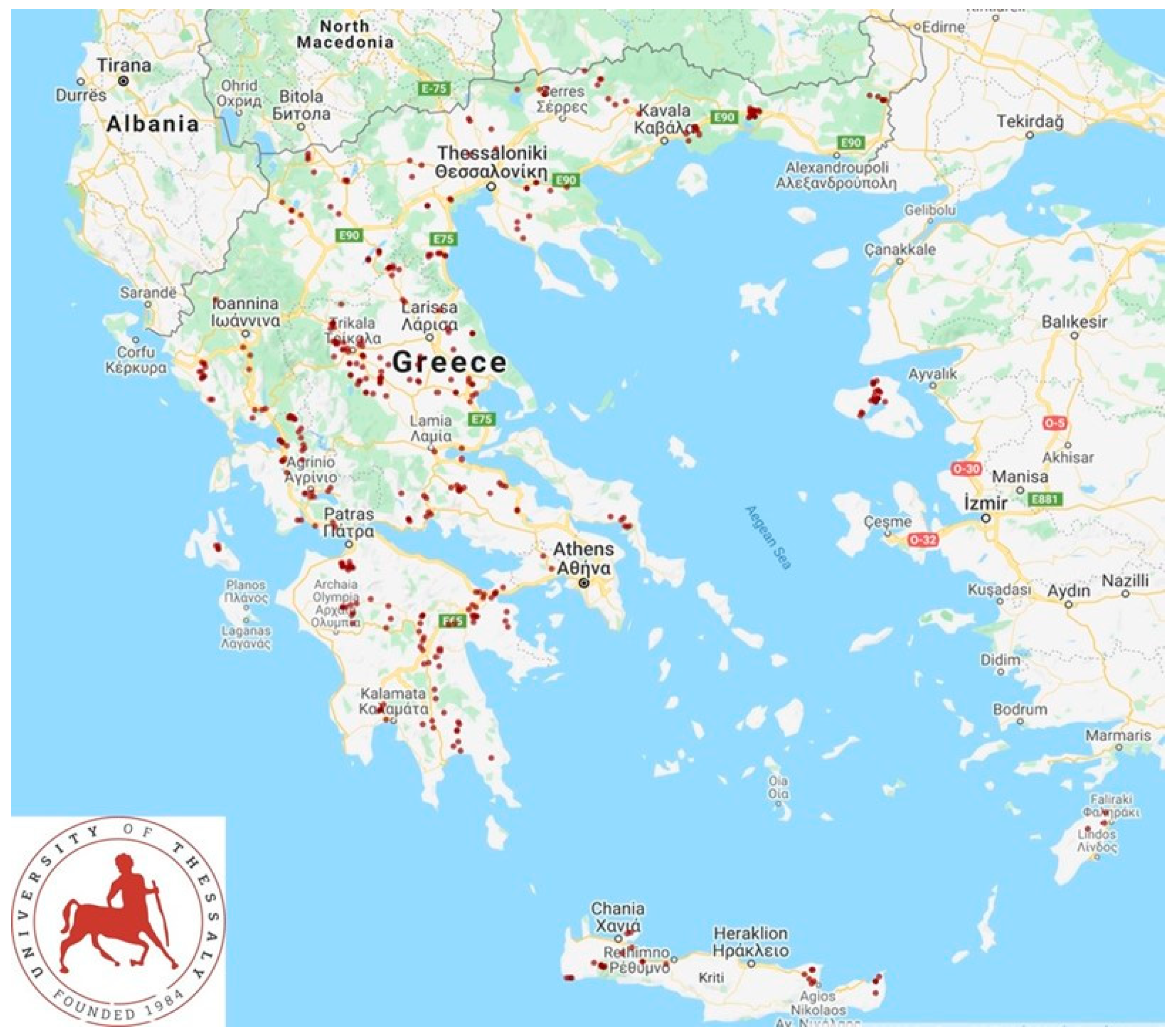
2.1. Farms and Sampling

A cross-sectional study was performed from April 2019 to July 2020. In total, 325 sheep flocks and 119 goat herds in the 13 administrative regions of Greece (Figure 1) were included in the study and visited to collect samples and information. Veterinarians active in small ruminant health management around Greece were contacted by telephone and asked if they wished to collaborate in the investigation; in total, 48 veterinarians were contacted and of these, 47 (97.9%) agreed to collaborate. Farms were selected by the collaborating veterinarians on a convenience basis (the willingness of farmers to accept a visit by university personnel for an interview and sample collection) [21,22]. Visits were scheduled to 327 flocks, but on two occasions (0.6%), although the investigators had already arrived at these farms, the respective farmers refused to collaborate [21,22]. Three investigators (authors D.T.L., C.K.M. and G.C.F.) visited all the farms for sample collection. During the visit to each farm, data on the farm’s location were collected using hand-held Garmin global positioning system units. The geo-references were resolved to the specific farm level.
At the start of each visit, an interview of the farmer was performed by using a detailed questionnaire [23] to record management practices used for the flock. Next, samples were collected from the bulk-tank raw milk of the farms. The samples were collected directly from the milk cooling tank on each farm; all tanks had one or more agitators which stirred the milk continuously until the cover of the tank was raised for sample collection. Milk samples were collected in sterile plastic single-use pipettes which were immersed into the tank to withdraw the samples. In total, four 20 mL samples were collected from the milk tank of each farm, and a new pipette was used for each sample. Immediately after collection, samples were transferred into sterile plastic Universal-type vials.
Samples were stored at 0.0 to 4.0 °C using ice packs in portable refrigerators. Somatic cell counting and milk composition measurements were performed on each of the samples within 4 h after sample collection. Transportation of samples to the laboratory was undertaken by the investigators.

2.2. Overview of Laboratory Examinations

Of the four milk samples collected from the milk tank of each flock, two samples were used for somatic cell counting and milk composition measurements; the other two were used for the bacteriological examinations. With regard to the samples for bacteriological examination, two equal subsamples (i.e., 10 mL each) were created from each sample, i.e., in total, 4 subsamples of 10 mL each were available for all culturing experiments.
Somatic cell counts and milk composition measurements were taken within 4 h of collection as described before [21,22]. Each measurement was performed four times (i.e., once in each subsample derived from each original milk sample).
Bacteriological examinations started within 24 h of sample collection and included total bacterial counts, performed by following the standardized procedures described by Laird et al. [24], and culturing for the detection of Listeria spp. After completion of sample aliquot withdrawal for the microbiological examination, the temperature of the respective samples was measured and was found to never exceed 3.8 °C.

2.3. Procedures for Listeria spp. Isolation and Identification

The currently valid standardized protocol for Listeria spp. isolation ISO 11290-1:2017 [25] (title: Microbiology of the food chain—Horizontal method for the detection and enumeration of Listeria monocytogenes and of Listeria spp.) was applied for culturing samples for the isolation of Listeria spp. from the samples. For this work, each of the four subsamples from the bulk-tank raw milk was cultured independently. In brief, the procedure followed was as follows.
A total volume of 25 mL from each milk subsample (in equal proportions from each) was inoculated into 225 mL of half-Fraser broth, as appropriate, for incubation at 30 °C for 24 h (primary enrichment stage; half-Fraser broth is a modification of Fraser broth containing half of the concentration of nalidixic acid and acriflavine hydrochloride). Next, a volume (0.1 mL) of the cultured half-Fraser broth was inoculated, as appropriate, into Fraser broth (10 mL) for incubation at 37 °C for 48 h (secondary enrichment stage). The contents of the half-Fraser broth and the Fraser broth were separately plated onto (a) plates with Listeria agar acc. Ottaviani and Agosti and (b) plates with PALCAM Listeria selective agar for incubation at 37 °C for 48 h (culturing stage). The colonies on each plate were counted; if confluent growth was evident, this was marked as ‘+’ or ‘++’. The presence of at least one colony in any of the plates inoculated with material from each bulk-tank sample sufficed to indicate a suspicion of the presence of Listeria spp. All the colonies were subcultured onto blood agar for incubation at 37 °C for 48 h (refining stage) and were submitted for final identification.
The identification of all the colonies isolated on solid media was first performed by using conventional techniques. The identification was verified by means of matrix-assisted laser desorption/ionization time-of-flight mass spectrometry (MALDI TOF MS) (VITEK MS; BioMerieux, Marcy-l’-Étoile, France). In brief, each isolate was smeared from Petri dishes onto target slides, and then 1 μL of the VITEK MS matrix was applied over the sample, air-dried and allowed to co-crystallize with the sample; target slides with all isolates thus prepared were loaded into the VITEK MS system. Next, the mass spectra of whole bacterial cell proteins were acquired and compared with the known mass spectra included in the database for each species [26,27].
When a colony was not identified as Listeria sp., then another four colonies from the same agar plate were tested for confirmation of the negative result (unless, of course, fewer colonies were isolated, in which case all the colonies obtained were tested). All isolates confirmed as Listeria spp. were stored in brain–heart infusion broth with 15% glycerol at −80 °C.

2.4. Testing for Susceptibility to Antibiotics

All Listeria spp. isolates were tested for their susceptibility to antibiotics by means of the disk diffusion method, according to EUCAST recommendations (www.eucast.org; accessed on 15 February 2022). Briefly, for each isolate, a bacterial suspension with 1.5 × 10⁸ colony-forming units mL−1, which corresponded to 0.5 McFarland turbidity, was inoculated on Mueller–Hinton agar with 5% defibrinated horse blood and 20 mg L−1 β-nicotinamide adenine dinucleotide. Disks with benzylpenicillin (1 unit), ampicillin (2 μg), meropenem (10 μg), erythromycin (15 μg) and trimethoprim/sulfamethoxazole (1.25 μg/23.75 µg) were used for testing. The strain Streptococcus pneumoniae ATCC 49619 was used for quality control. The media were incubated for 24 h at 35 °C with 5% CO2. The zones of inhibition were measured (mm) and the isolates were categorized as susceptible or resistant according to the criteria of EUCAST for Listeria monocytogenes.

2.5. Data Management and Analysis

Data were entered into Microsoft Excel (Microsoft Corporation, Redmond, WA, USA) and analysed using SPSS v. 21 (IBM Analytics, Armonk, NY, USA). A basic descriptive analysis was performed. Exact binomial confidence intervals (CI) were obtained.
Initially, the potential association of isolation of L. monocytogenes or L. ivanovii from bulk-tank raw milk with somatic cell counts, total bacterial counts, the composition of bulk-tank raw milk and milk production on the farm was assessed by using one-way analysis of variance.
Next, 36 management-related and six human resource-related variables were evaluated for potential association with the isolation of L. monocytogenes or L. ivanovii from the bulk-tank raw milk of the farms (Table S2); these were either taken directly from the answers to the interview performed at the start of the visit or calculated from these answers. For each of these variables, categories were created according to the answers of the farmers.
Subsequently, climatic variables were derived from ‘The POWER (Prediction of Worldwide Energy Resources) Project’ (NASA Langley Research Center (LaRC), Hampton, VA, USA), which provides meteorological datasets from NASA research for supporting agricultural needs. The following settings were used for obtaining the data: user community, agroclimatology; temporal average, daily; latitude/longitude, geo-references of each farm; time extent, start date D16 to end date D1 (D0: day of the visit to each farm); output file format, ASCII. Data for nine climatic parameters were extracted (Table S2). For the evaluations, the daily results provided by this platform for the 15 days prior to the visit were averaged.
The outcomes of the isolation of L. monocytogenes from bulk-tank raw milk or the isolation of L. ivanovii from bulk-tank raw milk were considered. Exact binomial CIs were obtained. In the case of management-related and human resource-related variables, the importance of predictors was assessed by using cross-tabulation with Pearson’s chi-squared test and with simple logistic regression. In the case of climatic variables, the importance of the predictors was assessed by using analysis of variance. Separate univariate analyses were performed for each of the two outcomes. Subsequently, a multivariate model was created, initially including in the model all variables which achieved a statistical significance with a p-value of <0.20 in either of the two univariate analyses, i.e., for either of the two outcomes (n = 13). Variables were removed from the initial model by backward elimination. The p-value of the removal of a variable was assessed by the likelihood ratio test, and for those with a statistical significance with a p-value of >0.20, the variable with the largest probability was removed. This process was repeated until no variable could be removed with a value of >0.20. The variables required for the final multivariable model (n = 6) were: (a) number of female animals on the farm, (b) the presence of pigs on the farm, (c) the temperature of the cleaning water in the milking parlour, (d) the season of the start of the lambing period on the farm, (e) quarantining of new animals entering the farm and (f) average relative humidity during the 15 days prior to sampling. Finally, among farms with pigs, the numbers of these animals on the farms where Listeria spp. had been isolated were compared by using the Mann–Whitney test.
In all analyses, statistical significance was defined at p < 0.05.

2.6. Whole Genome Analysis of L. ivanovii Isolates

Whole genome analysis was performed for all L. ivanovii isolates obtained in the study. Initially, libraries were prepared using Ion Torrent technology and Ion Chef workflows (Thermo Fisher Scientific, Waltham, MA, USA). Sequencing was performed in the S5XLS system, and analysis of the primary data was conducted with Ion Torrent Suite v.5.10.0 (Thermo Scientific). The quality of the reads was checked using FastQC software [28]. The reads for each sample were assembled using the SPAdes genome assembler v3.15. 4 [29] with the default parameters. The quality of the assembled genomes was assessed with the tool Quast version 5.0.2 [30]. Each genome was annotated using Prokka version 1.14 [31] with the default parameters. The average coverage for each genome was computed using the tool bbmap (https://sourceforge.net/projects/bbmap/; accessed on 26 March 2022).
Evaluation for the presence of antibiotic resistance genes in the assembled genomes was performed by using the online tool ResFinder-4.1 [32,33,34], with the ID threshold set to 90% and the minimum length set to 60%. Evaluation for the presence of virulence factor genes was performed by using the online tool VirulenceFinder 2.0 [32,35,36], with the ID threshold set to 90% and the minimum length set to 60%. Subsequently, proteins encoded by the detected virulence factor genes were retrieved from the database of the National Center for Biotechnology Information. The function of each of these proteins was recorded in the UniProt Knowledge Base database (UniProtKB/Swiss-Prot (release 2022_01)).
The presence of mobile elements was assessed by using the tool Mobile Element Finder v1.0.3 [37]. The presence of prophages was assessed by using the tool PHASTER [38,39]. The presence of plasmids in the assemblies was assessed by means of Plasmid Finder v2.1 [32,40], using the Gram-positive bacteria database, with the minimum identity threshold set to 95% and the minimum coverage set to 60%. Lastly, the online tool CRISPRCasFinder was used, with the default parameters, to evaluate presence of CRISPR/CAS loci [41,42,43,44,45]. In order to determine the origin of the sequence of the spacers from the isolates obtained, a BLAST analysis was performed. Only results with a high identity score (100% coverage and ≥90% identity) were considered.

2.7. Phylogenetic Relationships of L. ivanovii Isolates

The phylogenetic relationships of the genomes of all L. ivanovii isolates obtained in the study were compared with genomes of L. ivanovii publicly available up to 15 March 2022 in GenBank [46]. In order to obtain the best possible phylogenetic resolution, the phylogenetic tree was constructed using the core genome of L. ivanovii [47]. For finding the core genome, the assembled genomes of L. ivanovii stored in the GenBank database were downloaded. Among these, two genomes with a very low N50 (GCA_003261115.1 with N50: 9946 and GCA_000183925.1 with N50: 3514) were excluded from this analysis. Next, by using the tool Prokka, the assemblies were annotated and, by using the CD-HIT tool [48,49], their proteins were grouped into groups of orthologous proteins (clusters) based on the similarity and coverage rates.
Two proteins were grouped in the same orthologous cluster if the threshold for similarity was over 75% for at least 70% of their length. Clusters in which all the genomes used in the analysis were found were considered to be the clusters of the core genome. In each cluster of the core genome, the different genes found in this cluster were aligned using the tool Clustal Omega [50,51,52]. Using the aligned data, the pseudogenome of every assembly was created; the pseudogenome of an assembly consisted of the aligned sequences of all the genes of this assembly contained in the clusters of the core genome, joined in series. Through a comparison of the different pseudogenomes, the phylogenetic tree of all L. ivanovii isolates in GenBank and of the L. ivanovii isolates obtained in the present study was created using the partitioned analysis of RAxML version 8.2.11 [53] with the GTRGAMMA model. As bootstrapping is computationally intensive, the RAxML’s rapid bootstrapping algorithm was used. Hence, 100 rapid bootstrap searches and 20 ML searches were conducted in order to find the best ML tree with its support values.
The best ML tree was edited in iTol software v 6.5.6 [54,55,56,57,58]. For each biosample, relevant information about the geographic location was extracted from GenBank information or the relevant literature and incorporated inτο the phylogenetic tree. From the best ML tree, using the online platform T-REX, we created the distance matrix of the L. ivanovii genomes [59]. Lastly, the BLAST Ring Image Generator (BRIG) was used to generate a circular map to compare the genomes of the strains sequenced in the present study with the strain PAM 55, an outbreak-related strain [60].

3. Results

3.1. Isolation of Listeria monocytogenes and Listeria ivanovii from Bulk-Tank Raw Milk

L. monocytogenes was isolated from the bulk-tank raw milk of one sheep farm; the isolation rate was 0.3% (1/325) (95% CI: 0.1–1.7%). L. ivanovii was isolated from the bulk-tank raw milk of three sheep farms; the isolation rate was 0.9% (3/325) (95% CI: 0.3–2.7%) (Table S3). The organisms were not isolated from the milk of goat farms; the isolation rate was 0.0% (0/119) (95% CI: 0.0–3.1%). There were no significant differences in the isolation rate between sheep and goat farms (p > 0.54 for all comparisons). Listeria innocua, Listeria seeligeri and Listeria welshimeri were not isolated from any sample.
There were no spatial associations among the four farms from which L. monocytogenes or L. ivanovii were isolated. Specifically, two of these farms were located in North Greece (in different administrative regions), one in Central Greece and one in the islands. The average distance between the three farms on the mainland was 191 km.
During susceptibility testing, the L. monocytogenes and all three L. ivanovii isolates were found to be phenotypically susceptible to all the antibiotics tested. The full results of susceptibility testing are in Table S4.

3.2. Lack of an Association of Isolation of L. monocytogenes or L. ivanovii with Milk Quality

There was no difference in somatic cell counts (p = 0.39), total bacterial counts (p = 0.66), fat content (p = 0.26) and protein content (p = 0.53) in the bulk-tank raw milk of farms from which L. monocytogenes or L. ivanovii was or was not isolated (Table S5). Moreover, there was no difference in the average milk quantity produced annually from ewes in flocks from which L. monocytogenes or L. ivanovii was or was not isolated (120 L, 233.9 ± 20.6 L and 207.5 ± 5.0 L, respectively; p = 0.61).

3.3. Predictors of the Isolation of L. monocytogenes or L. ivanovii from Bulk-Tank Raw Milk

The details of the univariate analyses are in Tables S6–S8. In the multivariate analysis, three variables emerged as significant predictors for the isolation of L. monocytogenes or L. ivanovii from bulk-tank raw milk samples from sheep farms. These were: (a) the presence of pigs on the same farm (p < 0.0001), (b) low average relative humidity at 2 m (p = 0.006) and (c) a high number of female animals on the farm (p = 0.049) (Table 1, Figure 2).
Moreover, among farms with pigs, the farms where L. monocytogenes or L. ivanovii was isolated had a higher number of pigs (median: 10 (minimum, 10; maximum, 12)) than the farms where the bacteria were not isolated (two (1–20)) (p = 0.04) (Figure 1). An ad hoc inquiry with the veterinarians responsible for the three farms in which L. monocytogenes or L. ivanovii was isolated and where pigs were also present revealed that the pigs in these farms were fattening pigs (i.e., in anticipation for slaughter).

3.4. Whole Genome Analysis of L. ivanovii Isolates

The results of the whole genome analysis of the three L. ivanovii isolates are given in Table S9. No antibiotic resistance gene was found in any of the three genomes. Moreover, mobile elements, intact prophages and plasmids were also not found in any of the three assemblies (Table S10).
The following 15 virulence factor genes were detected in all three genomes: flaA, lap, oppA, rli55, rli60, rsbv, clpp, degU, fri, hfq, inlC, lhrC, lhrC, lsp, tcsA and tig (Tables S11 and S12). Finally, with regard to the CRISPR/CAS loci, four possible CRIPSRs and one CAS cluster were found in two assemblies (NGS01 and NGS03), and five CRISPRs and one CAS cluster in the third (NGS04) (Table S13). All three strains carried three identical CRISPR arrays (the same repeat and spacer sequences) and the same CAS cluster (CAS Type IB_1). In addition, the strains NGS03 and NGS04 shared a fourth common CRISPR array. BLASTn analysis showed that none of the spacers matched the phage genome.

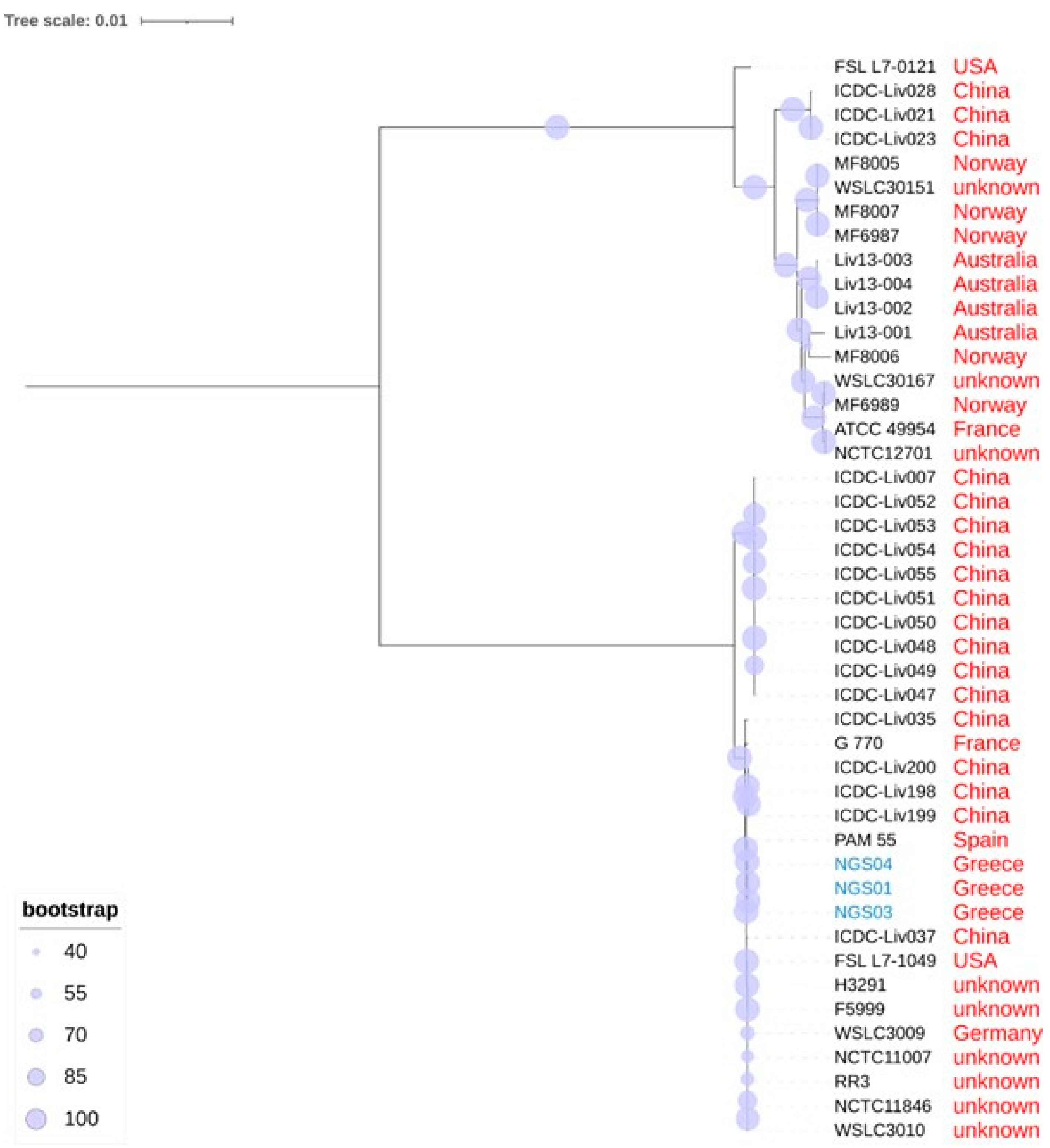
3.5. Phylogenetic relationships of L. ivanovii Isolates

The maximum likelihood tree (GTRGAMMA model) of 45 L. ivanovii strains with bootstrapping support values is shown in Figure 3. The tree was constructed by comparing 2042 genes, which form the core genome of L. ivanovii. The top branch of the tree includes 17 strains of L. ivanovii subsp. londoniensis; the lower branch includes 28 strains of L. ivanovii subsp. ivanovii. All three strains isolated in the present study were assigned to the L. ivanovii subsp. ivanovii branch (Figure 3).
The 28 L. ivanovii subsp. ivanovii strains form two clusters, one which includes only strains isolated in the People’s Republic of China and a second with strains from various other countries (including the three strains isolated in the present study) (Figure 3). The strains of that second cluster appear to be very similar to each other; the distance between them is small, despite the geographical diversity of the locations where these strains were isolated, which are from across various parts of the world (Table S14).
The three strains isolated in the present study were found to be phylogenetically close to the PAM 55 strain, which was isolated in 1997 in Spain from an outbreak of abortion in sheep (Figure 3, Table S14). Indeed, the three genomes were found to have many similarities to that of the PAM 55 strain, with only minor differences identified in a few regions, e.g., in the region near 1800 kbp (Figure 4).

4. Discussion

4.1. Presence and Identification of L. monocytogenes or L. ivanovii in Bulk-Tank Milk

The results provide evidence regarding the low prevalence of Listeria spp. in milk samples from dairy sheep and goat farms in Greece. In the present study, sheep and goat farms from all regions of Greece were included in the study; that way, conditions prevailing throughout the country were taken into account and factors of regional importance weighed less. In order to minimize possible bias, the study also used consistent methodologies and ensured that specific tasks were always performed by the same investigators.
We note that L. ivanovii was detected for the first time in Greece in a large collection of the milk of sheep and goats. During the whole genome sequencing, which was followed by a phylogenetic analysis, the strains were identified as L. ivanovii subspecies ivanovii. This could not be determined by the use of MALDI TOF MS alone, despite its high discriminatory capacity.
These findings are, in general, similar to those found in other countries of Europe [61]. For example, Amagliani et al. [62] and Condoleo et al. [63] did not isolate the pathogens from samples of raw sheep milk in Italy, and Bogdanovicova et al. [64] isolated only L. monocytogenes (<4% of samples) in a similar investigation in the Czech Republic; however, in sharp contrast, Artursson et al. [65] reported an isolation rate of 29% from raw sheep and goat milk samples in Sweden. Notably, in all previous relevant studies, the isolation rate of L. monocytogenes was generally higher than that of L. ivanovii [66,67].
Despite the low rate of isolation of the pathogens in the raw milk, there was a large variation in the isolation rates of Listeria spp. pathogens in samples from cheese of milk of sheep or goat origin in Greece. In recent studies, these were found to vary from 0% [68,69] to 39% [70] of samples tested. Potential sources of Listeria spp. in cheese, apart from the raw material (i.e., the milk), also include the environment of the processing plants [71,72]. Given the low rate of isolation of the pathogens in raw milk, close monitoring of cheese production plants is necessary to prevent contamination by the bacteria.
In any case, the mean annual notification rate of human listeriosis in Greece was 1.03 cases per 1,000,000 population for the period 2004–2017. Apparently, this is lower than the respective rate in EU countries (for 2016: 1.85 and 4.7 cases per 1,000,000 population) [73]. To our knowledge, only L. monocytogenes isolates have been identified from clinical samples from people in Greece.

4.2. Predictors of the Ιsolation of L. monocytogenes or L. ivanovii in Βulk-Τank Μilk

Reports of the isolation of Listeria spp. in clinical samples from pigs are infrequent for both L. monocytogenes [74,75] and L. ivanovii [76,77]. Hence, the identification of the presence of pigs in a farm as a predictor of the isolation of L. monocytogenes or L. ivanovii in bulk-tank milk was surprising.
Although clinical listeriosis is rare in pigs [78,79,80], domestic pigs could be a possible reservoir of Listeria spp. [81,82]. Listeria spp. has been occasionally isolated from faecal and skin samples of clinically healthy pigs [83]. The organism is considered to be harboured in the intestinal tract of these animals, and the prevalence of the isolation of L. monocytogenes from faecal samples ranges from 0% to 47% [84]. We hypothesise that, possibly, pigs might have been infected subclinically, excreting the pathogens in their faeces, thus contaminating the environment and contributing to an increased risk of infection of the grazing ewes, which subsequently excreted the organism in their faeces or the milk. However, the possibility that the teats of ewes might have been in touch with the contaminated ground of the farm and thus become contaminated with L. monocytogenes or L. ivanovii cannot be excluded. In the affected sheep farms, the pigs were maintained mainly to provide food for the family rather than for commercial purposes; in these circumstances, pigs are often allowed increased access to outdoor areas, which can contribute to contamination of the farm environment. In fact, in previous studies, the isolation of Listeria spp. from paddocks where pigs and cattle were co-grazing has been reported [76]. The association of the number of pigs on the farm with the isolation of the bacteria lends further support to this hypothesis, as a larger number of infected pigs would have contributed to higher levels of contamination of the grazing areas.
Moreover, the identification of the number of ewes in the flock as another predictor of the isolation of L. monocytogenes or L. ivanovii from milk may also be linked to the increased burden of the bacteria in the flock. Consequently, larger amounts of the bacteria are excreted in the milk, making the isolation easier.
The emergence of relative humidity as a predictor of the isolation of the organism can be possibly related to the growth characteristics of the organism. The results of previous studies regarding the potential effects of relative humidity on L. monocytogenes are conflicting, whilst no relevant studies for L. ivanovii are available. In food-related matrices, relative humidity in the range from 43% to 68% was found to adversely affect the viability of the bacteria [85,86] as the result of changes in the permeability of the bacterial membrane [86]. In contrast, in an experimental study that assessed viability of L. monocytogenes in the environment, this was found to be best at a relative humidity of around 50% (i.e., close to the mean relative humidity of the four farms where L. monocytogenes or L. ivanovii was isolated, which was 55.5%), irrespective of the temperature [87]. One should also consider that perhaps reasons unrelated to the bacteria themselves might have also played a role; for example, under conditions of lower relative humidity, sheep feel more comfortable and are more likely to be outdoors for grazing, which can contribute to spread of the bacteria within the flock.

4.3. Bioinformatics Assessment

The core genome of a bacterial species consists of the genes that are reserved in all the strains of the species, suggesting that these are probably essential for the survival and growth of the species [88]. There is no consensus on the procedure used to find the core genome’s genes, and different approaches exist in the literature. In the present analysis, we considered the core genome genes to be the protein coding genes, which were found to be present in all the strains studied. Phylogenetic analysis using the core genome genes can provide a deep and detailed phylogeny of the analysed bacteria. The core genome’s phylogenetic analysis outperformed phylogeny using 16S rRNA gene, as the core genome’s phylogeny acknowledges multiple conserved genes in the analysed species.
In the present analysis, the core genome phylogenetic tree correctly divided L. ivanovii into the two known subspecies, L. ivanovii subsp. ivanovii and L. ivanovii londoniensis, and also indicated the two distinct branches of L. ivanovii subsp. ivanovii. Gan et al. [89] indicated that strains from these two branches differed in motility, metabolic activity and virulence. The strains within the second branch, where the three strains of L. ivanovii isolated in the present study (NGS01, NGS03 and NGS04) have been assigned, exhibit a very small evolutionary distance, even though they are globally dispersed. This suggests either that the global spread of this specific clonal branch is a recent evolutionary event or that this branch is characterized by a relatively slow evolutionary rate.
Moreover, the three strains of L. ivanovii isolated in the present study have been analysed in silico for various genetic and phenotypic traits in order to fully assess them. Antibiotic resistance genes have been detected in none of these strains, which was consistent with the results of susceptibility testing. The strains also have not been found to have any plasmid or intact prophages. Incomplete prophages probably cannot perform the essential phage functions [90], which might have contributed to the lack of antibiotic resistance.
The results of the BLASTn analysis of the spacers in the CRISPR array may possibly explain the fact that L. ivanovii subsp. ivanovii strains are sensitive to bacteriophages, whereas the other subspecies seem to be resistant to infection by phages [91].
During the analysis, 15 virulence factor genes were also detected, and the three strains all contained the exact same genes. Among these, the presence of flaA and degU suggest the motility of these strains [89,92], whilst inlC and lap encode for factors that participate in the pathogenicity of L. monocytogenes or L. ivanovii strains [93,94]. Flagellin A, encoded by flaA, is considered to play a role in actin polymerization, which occurs at nucleation sites on the surface of the pathogen, to form the filaments of the flagella of Listeria spp. These are nucleated on the surface of the organism, possibly as branches from the surrounding cytoskeletal filaments in a way that, finally, the comet tail of the pathogen consists of a stiff scaffold of interconnected bundles [95]. Once the comet tail is longer than the organism itself, the pathogen can move while the comet tail remains stationary [96]. Nevertheless, none of the three strains harboured genes for Listeria pathogenicity island 1 and Listeria pathogenicity island 2, which, according to Gan et al. [89], are the predominant factors in the pathogenicity of L. ivanovii. This may possibly explain the lack of differences in the quality of milk produced from the farms from which the organism was isolated, as these strains lacked the increased pathogenicity causing mammary lesions in the affected ewes.
Despite the above, these three strains are evolutionarily close to the strains PAM 55 and G770, which were isolated from an outbreak of abortion in sheep in Spain [4] and a fatal case of aortic prosthesis infection in France [97], respectively, and thus should be monitored, given the importance of the small ruminant industry in the country and the increased production of cheese from the milk of these animals. The epidemiological surveillance of bacteria isolated from diverse environmental sources can help in preventing the emergence of hypervirulent and super-resistant isolates.

5. Conclusions

This work describes a comprehensive assessment of a little-studied zoonotic pathogen. The infrequent isolation of Listeria ivanovii from samples from small-ruminant farms, linked with the lack of isolation from dairy products, indicates a small risk for people consuming such products. The identification of predictors of the isolation of the organism should be taken into account in the health management of farms. The findings of the molecular studies, taken together, suggest that possibly, the reduced evolutionary diversity of the three strains is reflected in the reduced pathogenicity of the organism.

Supplementary Materials

The following supporting information can be downloaded at: https://www.mdpi.com/article/10.3390/biology11060871/s1. Table S1: Classification of articles found in the Web of Science presenting work related to Listeria ivanovii, according to the topic presented therein and the origin of the study (by country). Table S2: Variables evaluated for potential association with the isolation of Listeria monocytogenes and Listeria ivanovii from the bulk-tank raw milk of 444 small-ruminant farms during a countrywide investigation in Greece. Table S3: Details of the isolation of Listeria monocytogenes and Listeria ivanovii from the bulk-tank milk raw milk of 325 sheep farms during a countrywide investigation in Greece. Table S4: Details of the results of testing (diameters of the zones of inhibition) for susceptibility to antibiotics in Listeria monocytogenes and Listeria ivanovii from the bulk-tank raw milk of 325 sheep farms during a countrywide investigation in Greece. Table S5: Quality parameters of bulk-tank raw milk of 325 sheep farms from which Listeria monocytogenes and Listeria ivanovii were or were not isolated during a countrywide investigation in Greece. Table S6: Results of the univariate analysis for the association of husbandry-related variables with the isolation of Listeria monocytogenes or Listeria ivanovii from the bulk-tank raw milk of 325 sheep farms during a countrywide investigation in Greece. Table S7: Results of the univariate analysis for the association of human resource-related variables with the isolation of Listeria monocytogenes or Listeria ivanovii from the bulk-tank raw milk of 325 sheep farms during a countrywide investigation in Greece. Table S8: Results of univariate analysis for the association of climate-related variables with the isolation of Listeria monocytogenes or Listeria ivanovii from the bulk-tank raw milk of 325 sheep farms during a countrywide investigation in Greece. Table S9: Metrics of the assembled genome of three L. ivanovii isolates from the bulk-tank raw milk of sheep in Greece. Table S10: Summary findings of the assessment of genomes, using the tool PHASTER, of three Listeria ivanovii isolates obtained from the bulk-tank raw milk of sheep in Greece for the presence of prophages. Table S11: Virulence factor genes detected by using the tool VirulenceFinder 2.0 in the genome of three Listeria ivanovii isolates obtained from the bulk-tank raw milk of sheep in Greece. Table S12: Proteins encoded by the virulence factor genes (in alphabetical order) detected in the genome of three Listeria ivanovii isolates obtained from the bulk-tank raw milk of sheep in Greece. Table S13: The output of CRISPRCasFinder for the genome of three Listeria ivanovii isolates obtained from the bulk-tank raw milk of sheep in Greece. Table S14: Distance matrix between the 45 Listeria ivanovii strains included in the maximum likelihood tree.

Author Contributions

Conceptualization, D.T.L., A.S., E.P. and G.C.F.; methodology, D.T.L., A.S., C.K.M., E.I.K., D.C.C., N.S., P.J.C., E.P. and G.C.F.; validation, A.S., E.P. and P.J.C.; formal analysis, D.T.L., A.S. and P.J.C.; investigation, D.T.L., C.K.M., E.I.K., D.C.C., N.S., K.T., Z.F., A.I.K., N.G.C.V., K.S.D., M.V.B., D.V.L. and P.I.K.; resources, A.S., E.P. and G.C.F.; data curation, D.T.L. and A.S.; writing—original draft preparation, D.T.L., A.S. and G.C.F.; writing—review and editing, D.T.L., A.S., E.I.K., D.C.C., N.S., P.J.C., A.I.K., N.G.C.V., V.G.P., V.S.M., E.P. and G.C.F.; visualization, D.T.L. and A.S.; supervision, V.S.M., M.C., E.P. and G.C.F.; project administration, E.P. and G.C.F.; funding acquisition, E.P. and G.C.F. All authors have read and agreed to the published version of the manuscript.

Funding

This research received no external funding.

Institutional Review Board Statement

Not applicable; the study does not involve humans or animals. The protocols of the study were approved by the academic board of the Veterinary Faculty of the University of Thessaly, meeting 34/03.04.19.

Informed Consent Statement

Not applicable.

Data Availability Statement

Most data presented in this study are in the Supplementary Materials. The remaining data are available on request from the corresponding author. Some of the data are not publicly available as they form part of the PhD thesis of the first author, which has not yet been examined, approved and uploaded in the official depository of PhD theses from Greek universities. The accession number of the Bioproject deposited in the NCBI database is PRJNA812022. In the Bioproject record, there are links to the biosamples (SAMN26366399, SAMN26366413, SAMN26366412) and to the Sequence Read Archive (SRA) data (SRS12212767, SRS12212768, SRS12212769). The genomes of the L. ivanovii strains NGS01, NGS03 and NGS04 isolated in the present study were deposited in GenBank under accession numbers GCA_022815685.1, GCA_022815695.1 and GCA_022815665.1, respectively.

Conflicts of Interest

The authors declare no conflict of interest.

References

  1. Seeliger, H.P.R.; Rocourt, J.; Schretten-Brunner, A.; Grimont, P.A.D.; Jones, D. Listeria ivanovii sp. nov. Int. J. Syst. Bacteriol. 1984, 34, 336–337. [Google Scholar] [CrossRef]
  2. Parte, A.C.; Carbasse, J.S.; Meier-Kolthoff, J.P.; Reimer, L.C.; Goeker, M. List of Prokaryotic names with Standing in Nomenclature (LPSN) moves to the DSMZ. Int. J. Syst. Evol. Microbiol. 2020, 70, 5607–5612. [Google Scholar] [CrossRef] [PubMed]
  3. Chiara, M.; Caruso, M.; D’Erchia, A.M.; Manzari, C.; Fraccalvieri, R.; Goffredo, E.; Latorre, L.; Miccolupo, A.; Padalino, I.; Santagada, G.; et al. Comparative genomics of Listeria sensu lato: Genus-wide differences in evolutionary dynamics and the progressive gain of complex, potentially pathogenicity-related traits through lateral gene transfer. Genome Biol. Evol. 2015, 7, 2154–2172. [Google Scholar] [CrossRef] [PubMed] [Green Version]
  4. Buchrieser, C.; Rusniok, C.; Garrido, P.; Hain, T.; Scortti, M.; Lampidis, R.; Karst, U.; Chakraborty, T.; Cossart, P.; Kreft, J.; et al. Complete genome sequence of the animal pathogen Listeria ivanovii, which provides insights into host specificities and evolution of the genus Listeria. J. Bacteriol. 2011, 193, 6787–6788. [Google Scholar] [CrossRef] [PubMed] [Green Version]
  5. Sarangi, L.N.; Panda, H.K. Isolation, characterization and antibiotic sensitivity test of pathogenic Listeria species in livestock, poultry and farm environment of Odisha. Ind. J. Anim. Res. 2011, 46, 242–247. [Google Scholar]
  6. Dunnett, E.; Florea, L.; Thurston, L.; Floyd, T.; Collins, R.; Otter, A. Deaths of weaned lambs with visceral Listeria ivanovii infections. Vet. Rec. Case Rep. 2020, 8, e001254. [Google Scholar] [CrossRef]
  7. Rodriguez-Lazaro, D.; Lopez-Enriquez, L.; Hernandez, M. smcL as a novel diagnostic marker for quantitative detection of Listeria ivanovii in biological samples. J. Appl. Microbiol. 2010, 109, 863–872. [Google Scholar] [CrossRef]
  8. Osman, K.M.; Zolnikov, T.R.; Samir, A.; Orabi, A. Prevalence, pathogenic capability, virulence genes, biofilm formation, and antibiotic resistance of Listeria in goat and sheep milk confirms need of hygienic milking conditions. Pathog. Glob. Health 2014, 108, 21–29. [Google Scholar] [CrossRef] [Green Version]
  9. Soncini, G.; Valnegri, L. Analysis of bulk goats’ milk and milk-filters from Valtellina and Valchiavenna (Lombardy Prealps) for the presence of Listeria species. Small Rumin. Res. 2005, 58, 143–147. [Google Scholar] [CrossRef]
  10. Elezebeth, G.; Malik, S.V.S.; Chaudhari, S.P.; Barbuddhe, S.B. The occurrence of Listeria species and antibodies against listeriolysin-O in naturally infected goats. Small Rumin. Res. 2007, 67, 173–178. [Google Scholar] [CrossRef]
  11. Arimi, S.M.; Ryser, E.T.; Pritchard, T.J.; Donnelly, C.W. Diversity of Listeria ribotypes recovered from dairy cattle, silage, and dairy processing environments. J. Food Prot. 1997, 60, 811–816. [Google Scholar] [CrossRef] [PubMed]
  12. Terentjeva, M.; Steingolde, Z.; Meistere, I.; Elferts, D.; Avsejenko, J.; Streikisa, M.; Gradovska, S.; Alksne, L.; Kibilds, J.; Berzins, A. Prevalence, genetic diversity and factors associated with distribution of Listeria monocytogenes and other Listeria spp. in cattle farms in Latvia. Pathogens 2021, 10, 851. [Google Scholar] [CrossRef] [PubMed]
  13. Rawool, D.B.; Malik, S.V.S.; Shakuntala, I.; Sahare, A.M.; Barbuddhe, S.B. Detection of multiple virulence-associated genes in Listeria monocytogenes, isolated from bovine mastitis cases. Int. J. Food Microbiol. 2007, 113, 201–207. [Google Scholar] [CrossRef] [PubMed]
  14. Ramage, C.P.; Low, J.C.; McLauchlin, J.; Donachie, W. Characterisation of Listeria ivanovii isolates from the U.K. using pulsed-field gel electrophoresis. FEBS Microbiol. Lett. 1999, 170, 349–353. [Google Scholar] [CrossRef] [PubMed]
  15. Snapir, Y.M.; Vaisbein, E.; Nassar, F. Low virulence but potentially fatal outcome—Listeria ivanovii. Eur. J. Intern. Med. 2006, 17, 286–287. [Google Scholar] [CrossRef]
  16. Guillet, C.; Join-Lambert, O.; Le Monnier, A.; Leclercq, A.; Mechai, F.; Mamzer-Bruneel, M.F.; Bielecka, M.K.; Scortti, M.; Disson, O.; Berche, P.; et al. Human listeriosis caused by Listeria ivanovii. Emerg. Infect Dis. 2010, 16, 136–138. [Google Scholar] [CrossRef]
  17. Giacone, V.; Bertoja, G. Listeria and listeriosis in the milk production’s chain: An up-to-date. Large Anim. Rev. 2013, 19, 280–286. [Google Scholar]
  18. Hellenic Statistical Authority. Farm Structure Surveys. Available online: https://www.statistics.gr (accessed on 15 February 2022).
  19. Pulina, G.; Milan, M.J.; Lavin, M.P.; Theodoridis, A.; Morin, E.; Capote, J.; Thomas, D.L.; Francesconi, A.H.D.; Caja, G. Invited review: Current production trends, farm structures, and economics of the dairy sheep and goat sector. J. Dairy Sci. 2018, 101, 6715–6729. [Google Scholar] [CrossRef] [Green Version]
  20. Hellenic Agricultural Organisation—Demeter. Deliveries of Ovine and Caprine Milk by Region and Regional Authority and Average Milk Price—Calendar Year 2019. Available online: https://www.elgo.gr/images/ELOGAK_files/Statistics/2021/Milk21/6.%CE%95%CE%9B%CE%93%CE%9F_STATS_%CE%A0%CE%91%CE%A1%CE%91%CE%94_%CE%A0%CE%9F%CE%A3_%CE%A0%CE%A1%CE%9F%CE%92%CE%95%CE%99%CE%9F%CE%A5_%CE%93%CE%99%CE%94%CE%99%CE%9D%CE%9F%CE%A5_%CE%91%CE%9D%CE%91_%CE%A0%CE%95%CE%A1%CE%99%CE%A6_2019.pdf (accessed on 12 January 2022).
  21. Lianou, D.T.; Michael, C.K.; Vasileiou, N.G.C.; Petinaki, E.; Cripps, P.J.; Tsilipounidaki, K.; Katsafadou, A.I.; Politis, A.P.; Kordalis, N.G.; Ioannidi, K.S.; et al. Extensive countrywide field investigation of somatic cell counts and total bacterial counts in bulk-tank raw milk in sheep flocks in Greece. Foods 2021, 10, 268. [Google Scholar] [CrossRef]
  22. Lianou, D.T.; Michael, C.K.; Vasileiou, N.G.C.; Petinaki, E.; Cripps, P.J.; Tsilipounidaki, K.; Katsafadou, A.I.; Politis, A.P.; Kordalis, N.G.; Ioannidi, K.S.; et al. Extensive countrywide field investigation of somatic cell counts and total bacterial counts in bulk-tank raw milk in goat herds in Greece. J. Dairy Res. 2021, 88, 307–313. [Google Scholar] [CrossRef]
  23. Lianou, D.T.; Chatziprodromidou, I.P.; Vasileiou, N.G.C.; Michael, C.K.; Mavrogianni, V.S.; Politis, A.P.; Kordalis, N.G.; Billinis, C.; Giannakopoulos, A.; Papadopoulos, E.; et al. A detailed questionnaire for the evaluation of health management in dairy sheep and goats. Animals 2020, 10, 1489. [Google Scholar] [CrossRef] [PubMed]
  24. Laird, D.T.; Gambrel-Lenarz, S.A.; Scher, F.M.; Graham, T.E.; Reddy, R. Microbiological count methods. In Standard Methods for the Examination of Dairy Products, 17th ed.; Wehr, H.M., Frank, J.F., Eds.; APHA Press: Washington, DC, USA, 2004; pp. 153–186. [Google Scholar]
  25. ISO 11290-1:2017; Microbiology of the Food Chain—Horizontal Method for the Detection and Enumeration of Listeria Monocytogenes and of Listeria spp.—Part 1: Detection Method. 2nd ed. International Organization for Standardization: Geneva, Switzerland, 2017; 36p.
  26. Ojima-Kato, T.; Yamamoto, N.; Takahashi, H.; Tamura, H. Matrix-assisted Laser Desorption Ionization-Time of Flight Mass Spectrometry (MALDI-TOF MS) can precisely discriminate the lineages of Listeria monocytogenes and species of Listeria. PLoS ONE 2016, 11, e0159730. [Google Scholar] [CrossRef] [PubMed] [Green Version]
  27. Thouvenot, P.; Vales, G.; Bracq-Dieye, H.; Tessaud-Rita, N.; Maury, M.M.; Moura, A.; Lecuit, M.; Leclercq, A. MALDI-TOF mass spectrometry-based identification of Listeria species in surveillance: A prospective study. J. Microbiol. Methods 2018, 144, 29–32. [Google Scholar] [CrossRef] [PubMed] [Green Version]
  28. Andrews, S. FastQC: A quality Control Tool for High Throughput Sequence Data, 2015. Available online: https://www.bioinformatics.babraham.ac.uk/projects/fastqc/ (accessed on 15 January 2022).
  29. Bankevich, A.; Nurk, S.; Antipov, D.; Gurevich, A.A.; Dvorkin, M.; Kulikov, A.S.; Lesin, V.M.; Nikolenko, S.I.; Pham, S.; Prjibelski, A.D.; et al. SPAdes: A new genome assembly algorithm and its applications to single-cell sequencing. J. Comput. Biol. 2012, 19, 455. [Google Scholar] [CrossRef] [Green Version]
  30. Gurevich, A.; Saveliev, V.; Vyahhi, N.; Tesler, G. QUAST: Quality assessment tool for genome assemblies. Bioinformatics 2013, 29, 1072–1075. [Google Scholar] [CrossRef]
  31. Seemann, T. Prokka: Rapid prokaryotic genome annotation. Bioinformatics 2014, 30, 2068–2069. [Google Scholar] [CrossRef]
  32. Camacho, C.; Coulouris, G.; Avagyan, V.; Ma, N.; Papadopoulos, J.; Bealer, K.; Madden, T.L. BLAST+: Architecture and applications. BMC Bioinform. 2009, 10, 421. [Google Scholar] [CrossRef] [Green Version]
  33. Bortolaia, V.; Kaas, R.F.; Ruppe, E.; Roberts, M.C.; Schwarz, S.; Cattoir, V.; Philippon, A.; Allesoe, R.L.; Rebelo, A.R.; Florensa, A.R.; et al. ResFinder 4.0 for predictions of phenotypes from genotypes. J. Antimicrob. Chemother. 2020, 75, 3491–3500. [Google Scholar] [CrossRef]
  34. Zankari, E.; Allesøe, R.; Joensen, K.G.; Cavaco, L.M.; Lund, O.; Aarestrup, F.M. PointFinder: A novel web tool for WGS-based detection of antimicrobial resistance associated with chromosomal point mutations in bacterial pathogens. J. Antimicrob. Chemother. 2020, 72, 2764–2768. [Google Scholar] [CrossRef] [Green Version]
  35. Joensen, K.G.; Scheutz, F.; Lund, O.; Hasman, H.; Kaas, R.S.; Nielsen, E.M.; Aarestrup, F.M. Real-time whole-genome sequencing for routine typing, surveillance, and outbreak detection of verotoxigenic Escherichia coli. J. Clin. Microbiol. 2014, 52, 1501–1510. [Google Scholar] [CrossRef] [Green Version]
  36. Malberg Tetzschner, A.M.; Johnson, J.R.; Johnston, B.D.; Lund, O.; Scheutz, F. In Silico genotyping of Escherichia coli isolates for extraintestinal virulence genes by uUse of whole-genome sequencing data. J. Clin. Microbiol. 2020, 58, e01269-20. [Google Scholar] [CrossRef] [PubMed]
  37. Johansson, M.H.K.; Bortolaia, V.; Tansirichaiya, S.; Aarestrup, F.M.; Roberts, A.P.; Petersen, T.N. Detection of mobile genetic elements associated with antibiotic resistance in Salmonella enterica using a newly developed web tool: MobileElementFinder. J. Antimicrob. Chemother. 2021, 76, 101–109. [Google Scholar] [CrossRef] [PubMed]
  38. Zhou, Y.; Liang, Y.; Lynch, K.H.; Dennis, J.J.; Wishart, D.S. PHAST: A fast phage search tool. Nucleic Acids Res. 2011, 39, W347–W352. [Google Scholar] [CrossRef] [PubMed]
  39. Arndt, D.; Grant, J.; Marcu, A.; Sajed, T.; Pon, A.; Liang, Y.; Wishart, D.S. PHASTER: A better, faster version of the PHAST phage search tool. Nucleic Acids Res. 2016, 44, W16–W21. [Google Scholar] [CrossRef] [PubMed] [Green Version]
  40. Carattoli, A.; Zankari, E.; Garcia-Fernandez, A.; Voldby Larsen, M.; Lund, O.; Villa, L.; Aarestrup, F.M.; Hasman, H. PlasmidFinder and pMLST: In silico detection and typing of plasmids. Antimicrob. Agents Chemother. 2014, 58, 3895–3903. [Google Scholar] [CrossRef] [Green Version]
  41. Kurtz, S.; Schleiermacher, C. REPuter: Fast computation of maximal repeats in complete genomes. Bioinformatics 1999, 15, 426–427. [Google Scholar] [CrossRef] [Green Version]
  42. Abouelhoda, M.I.; Kurtz, S.; Ohlebusch, E. Replacing suffix trees with enhanced suffix arrays. J. Discret. Algorithms 2004, 2, 53–86. [Google Scholar] [CrossRef] [Green Version]
  43. Hyatt, D.; Chen, G.-L.; Locascio, P.F.; Land, M.L.; Larimer, F.W.; Hauser, L.J. Prodigal: Prokaryotic gene recognition and translation initiation site identification. BMC Bioinform. 2010, 11, 119. [Google Scholar] [CrossRef] [Green Version]
  44. Abby, S.S.; Neron, B.; Menager, H.; Touchon, M.; Rocha, E.P. MacSyFinder: A program to mine genomes for molecular systems with an application to CRISPR-Cas systems. PLoS ONE 2014, 9, e110726. [Google Scholar] [CrossRef]
  45. Biswas, A.; Fineran, P.C.; Brown, C.M. Accurate computational prediction of the transcribed strand of CRISPR non-coding R.N.As. Bioinformatics 2014, 30, 1805–1813. [Google Scholar] [CrossRef]
  46. Clark, K.; Karsch-Mizrachi, I.; Lipman, D.J.; Ostell, J.; Sayers, E.W. GenBank. Nucleic Acids Res. 2016, 44, D67–D72. [Google Scholar] [CrossRef] [PubMed] [Green Version]
  47. Tettelin, H.; Riley, D.; Cattuto, C.; Medini, D. Comparative genomics: The bacterial pan-genome. Curr. Opin. Microbiol. 2008, 11, 472–477. [Google Scholar] [CrossRef] [PubMed]
  48. Li, W.; Godzik, A. Cd-hit: A fast program for clustering and comparing large sets of protein or nucleotide sequences. Bioinformatics 2006, 22, 1658–1659. [Google Scholar] [CrossRef] [PubMed] [Green Version]
  49. Fu, L.; Niu, B.; Zhu, Z.; Wu, S.; Li, W. CD-HIT: Accelerated for clustering the next-generation sequencing data. Bioinformatics 2012, 28, 3150–3152. [Google Scholar] [CrossRef]
  50. Sievers, F.; Wilm, A.; Dineen, D.G.; Gibson, T.J.; Karplus, K.; Li, W.; Lopez, R.; McWilliam, H.; Remmert, M.; Söding, J.; et al. Fast, scalable generation of high-quality protein multiple sequence alignments using Clustal Omega. Mol. Syst. Biol. 2011, 7, 539. [Google Scholar] [CrossRef]
  51. Sievers, F.; Higgins, D.G. Clustal Omega for making accurate alignments of many protein sequences. Protein Sci. 2018, 27, 135–145. [Google Scholar] [CrossRef] [Green Version]
  52. Sievers, F.; Barton, G.J.; Higgins, D.G. Multiple Sequence Alignment. In Bioinformatics, 4th ed.; Baxevanis, A.D., Bader, G.D., Wishart, D.S., Eds.; Wiley: Hoboken, NJ, USA, 2020; pp. 227–250. [Google Scholar]
  53. Stamatakis, A. RAxML version 8: A tool for phylogenetic analysis and post-analysis of large phylogenies. Bioinformatics 2014, 30, 1312. [Google Scholar] [CrossRef]
  54. Letunic, I.; Bork, P. Interactive Tree of Life (iTOL): An online tool for phylogenetic tree display and annotation. Bioinformatics 2006, 23, 127–128. [Google Scholar] [CrossRef] [Green Version]
  55. Letunic, I.; Bork, P. Interactive Tree of Life v2: Online annotation and display of phylogenetic trees made easy. Nucleic Acids Res. 2011, 39, W475–W478. [Google Scholar] [CrossRef]
  56. Letunic, I.; Bork, P. Interactive Tree of Life (iTOL) v3: An online tool for the display and annotation of phylogenetic and othertrees. Nucleic Acids Res. 2016, 44, W242–W245. [Google Scholar] [CrossRef]
  57. Letunic, I.; Bork, P. Interactive Tree of Life (iTOL) v4: Recent updates and new developments. Nucleic Acids Res. 2019, 47, W256–W259. [Google Scholar] [CrossRef] [PubMed] [Green Version]
  58. Letunic, I.; Bork, P. Interactive Tree of Life (iTOL) v5: An online tool for phylogenetic tree display and annotation. Nucleic Acids Res. 2021, 49, W293–W296. [Google Scholar] [CrossRef] [PubMed]
  59. Boc, A.; Diallo, A.B.; Makarenkov, V. T-REX: A web server for inferring, validating and visualizing phylogenetic trees and networks. Nucleic Acids Res. 2012, 40, W573–W579. [Google Scholar] [CrossRef] [PubMed] [Green Version]
  60. Alikhan, N.F.; Petty, N.K.; Ben Zakour, N.L.; Beatson, S.A. BLAST Ring Image Generator (BRIG): Simple prokaryote genome comparisons. BMC Genom. 2011, 12, 402. [Google Scholar] [CrossRef] [PubMed] [Green Version]
  61. Gonzales-Barron, U.; Goncalves-Tenorio, A.; Rodrigues, V.; Cadavez, V. Foodborne pathogens in raw milk and cheese of sheep and goat origin: A meta-analysis approach. Curr. Opin. Food Sci. 2017, 18, 7–13. [Google Scholar] [CrossRef]
  62. Amagliani, G.; Petruzzelli, A.; Carloni, E.; Tonucci, F.; Foglini, M.; Micci, E.; Ricci, M.; Di Lullo, S.; Rotundo, L.; Brandi, G. Presence of Escherichia coli O157, Salmonella spp., and Listeria monocytogenes in raw ovine milk destined for cheese production and evaluation of the equivalence between the analytical methods applied. Foodborne Pathog. Dis. 2016, 13, 626–632. [Google Scholar] [CrossRef]
  63. Condoleo, R.; Giangolini, G.; Chiaverini, A.; Patriarca, D.; Scaramozzino, P.; Mezher, Z. Occurrence of Listeria monocytogenes and Escherichia coli in raw sheep’s milk from farm bulk tanks in Central Italy. J. Food Prot. 2020, 83, 1929–1933. [Google Scholar] [CrossRef]
  64. Bogdanovicova, K.; Vyletelova-Klimesova, M.; Babak, V.; Kalhotka, L.; Kolackova, I.; Karpiskova, R. Microbiological quality of raw milk in the Czech Republic. Czech J. Food Sci. 2016, 34, 189–196. [Google Scholar]
  65. Artursson, K.; Schelin, J.; Lambertz, S.T.; Hansson, I.; Engvall, E.O. Foodborne pathogens in unpasteurized milk in Sweden. Int. J. Food Microbiol. 2018, 284, 120–127. [Google Scholar] [CrossRef]
  66. Al-Tahiri, R.; Omar, S.; Rewashdeh, A. A study of the occurrence of Listeria species in raw sheep milk. Int. J. Dairy Technol. 2008, 61, 347–351. [Google Scholar] [CrossRef]
  67. Atil, E.; Ertas, H.B.; Ozbey, G. Isolation and molecular characterization of Listeria spp. from animals, food and environmental samples. Vet. Med. 2011, 56, 386–394. [Google Scholar] [CrossRef] [Green Version]
  68. Tzora, A.; Nelli, A.; Voidarou, C.; Fthenakis, G.; Rozos, G.; Theodorides, G.; Bonos, E.; Skoufos, I. Microbiota “fingerprint” of Greek feta cheese through ripening. Appl. Sci. 2021, 11, 5631. [Google Scholar] [CrossRef]
  69. Pappa, E.C.; Bontinis, T.G.; Samelis, J.; Sotirakoglou, K. Assessment of the microbiological quality and biochemical parameters of traditional hard xinotyri cheese made from raw or pasteurized goat milk. Fermentation 2022, 8, 20. [Google Scholar] [CrossRef]
  70. Rhoades, J.; Anastasiou, I.; Michailidou, S.; Koinidis, A.; Doulgerakis, C.; Alexa, E.A.; Alvarez-Ordonez, A.; Argiriou, A.; Likotrafiti, E. Microbiological analysis of Greek Protected Designation of Origin cheeses and characterisation of the isolated lactic acid bacteria. Int. Dairy J. 2021, 123, 105183. [Google Scholar] [CrossRef]
  71. Vazquez-Villanueva, J.; Orgaz, B.; Ortiz, S.; Lopez, V.; Martinez-Suarez, J.V.; San Jose, C. Predominance and persistence of a single clone of Listeria ivanovii in a manchego cheese factory over 6 months. Zoonoses Public Health 2010, 57, 402–410. [Google Scholar] [CrossRef]
  72. Spanu, C.; Scarano, C.; Ibba, M.; Spanu, V.; De Santis, E.P.L. Occurrence and traceability of Listeria monocytogenes strains isolated from sheep’s milk cheese-making plants environment. Food Control 2015, 47, 318–325. [Google Scholar] [CrossRef]
  73. Hellenic Center for Disease Control and Prevention. Epidemiological Data for Listeriosis in Greece 2004–2017. Available online: https://eody.gov.gr/wp-content/uploads/2019/01/Epidemiological_data_for_listeriosis_Greece_2004_2017.pdf (accessed on 18 February 2022).
  74. Farzan, A.; Friendship, R.M.; Cook, A.; Pollari, F. Occurrence of Salmonella, Campylobacter, Yersinia enterocolitica, Escherichia coli O157 and Listeria monocytogenes in swine. Zoonoses Public Health 2010, 57, 388–396. [Google Scholar] [CrossRef]
  75. Gomez-Laguna, J.; Cardoso-Toset, F.; Meza-Torres, J.; Pizarro-Cerda, J.; Quereda, J.J. Virulence potential of Listeria monocytogenes strains recovered from pigs in Spain. Vet. Rec. 2020, 187, 105945. [Google Scholar] [CrossRef]
  76. Ikeh, M.A.C.; Obi, S.K.C.; Ezeasor, D.N.; Ezeonu, I.M.; Moneke, A.N. Incidence and pathogenicity profile of Listeria spp. isolated from food and environmental samples in Nsukka, Nigeria. Afr. J. Biotechnol. 2010, 9, 4776–4782. [Google Scholar]
  77. Sarno, E.; Stephan, R.; Zweifel, C. Occurrence of Erysipelothrix spp., Salmonella spp., and Listeria spp. in tonsils of healthy Swiss pigs at slaughter. Arch. Lebensmittelhyg. 2012, 63, 11–15. [Google Scholar]
  78. Gray, M.L.; Killinger, A.H. Listeria monocytogenes and listeric infections. Bacteriol. Rev. 1966, 30, 309–382. [Google Scholar] [CrossRef] [PubMed]
  79. Moerner, T. Monitoring disease in wildlife—A review of diseases in the orders lagomorpha and rodentia in Sweden. Verhber. Erkg. Zootiere. 1999, 39, 255–262. [Google Scholar]
  80. Gyles, C.L.; Prescott, J.F.; Songer, G.; Thoen, C.O. Pathogenesis of Bacterial Infections in Animals, 4th ed.; Wiley-Blackwell: Ames, IA, USA, 2020. [Google Scholar]
  81. Wacheck, S.; Giezendanner, N.; König, M.; Fredriksson-Ahomaa, M.; Stephan, R. Phenotypical and genotypical traits of Listeria monocytogens strains isolated from tonsils of wild boars hunted in Switzerland. Int. J. Infect. Dis. 2010, 14, e162. [Google Scholar] [CrossRef] [Green Version]
  82. Sannö, A.; Rosendal, T.; Aspán, A.; Backhans, A.; Jacobson, M. Distribution of enteropathogenic Yersinia spp. and Salmonella spp. in the Swedish wild boar population, and assessment of risk factors that may affect their prevalence. Acta Vet. Scand. 2018, 60, 40. [Google Scholar] [CrossRef] [Green Version]
  83. Skovgaard, N.; Nørrung, B. The incidence of Listeria spp. in faeces of Danish pigs and in minced pork meat. Int. J. Food Microbiol. 1989, 8, 59–63. [Google Scholar] [CrossRef]
  84. Felon, D.R.; Wilson, J.; Donachie, W. The incidence and level of Listeria monocytogenes contamination of food sources at primary production and initial processing. J. Appl. Microbiol. 1996, 81, 641–650. [Google Scholar] [CrossRef]
  85. Zoz, F.; Iaconelli, C.; Lang, E.; Iddir, H.; Guyot, S.; Grandvalet, C.; Gervais, P.; Beney, L. Control of relative air humidity as a potential means to improve hygiene on surfaces: A preliminary approach with Listeria monocytogenes. PLoS ONE 2016, 11, e0148418. [Google Scholar] [CrossRef]
  86. Zoz, F.; Guyot, S.; Grandvalet, C.; Ragon, M.; Lesniewska, E.; Dupont, S.; Firmesse, O.; Carpentier, B.; Beney, L. Management of Listeria monocytogenes on surfaces via relative air humidity: Key role of cell envelope. Foods 2021, 10, 2002. [Google Scholar] [CrossRef]
  87. Redfern, J.; Verran, J. Effect of humidity and temperature on the survival of Listeria monocytogenes on surfaces. Appl. Microbiol. 2017, 64, 276–282. [Google Scholar] [CrossRef]
  88. Tettelin, H.; Masignani, V.; Cieslewicz, M.J.; Donati, C.; Medini, D.; Ward, N.L.; Angiuoli, S.V.; Crabtree, J.; Jones, A.L.; Scott Durkin, A.; et al. Genome analysis of multiple pathogenic isolates of Streptococcus agalactiae: Implications for the microbial “pan-genome”. Proc. Natl. Acad. Sci. USA 2005, 102, 13950–13955. [Google Scholar] [CrossRef] [Green Version]
  89. Gan, L.; Mao, P.; Jiang, H.; Zhang, L.; Liu, D.; Cao, X.; Wang, Y.; Wang, Y.; Sun, H.; Huang, Y.; et al. Two prevalent Listeria ivanovii subsp. ivanovii clonal strains with different virulence exist in wild rodents and pikas of China. Front. Veter Sci. 2020, 7, 88. [Google Scholar]
  90. De Sousa, J.A.M.; Buffet, A.; Haudiquet, M.; Rocha, E.P.C.; Rendueles, O. Modular prophage interactions driven by capsule serotype select for capsule loss under phage predation. ISME J. 2020, 14, 2980. [Google Scholar] [CrossRef] [PubMed]
  91. Hupfeld, M.; Fouts, D.E.; Loessner, M.J.; Klumpp, J. Genome Sequences of the Listeria ivanovii subsp. ivanovii type strain and two Listeria ivanovii subsp. londoniensis strains. Genome Announc. 2015, 3, e01440-14. [Google Scholar] [PubMed] [Green Version]
  92. Knudsen, G.M.; Olsen, J.E.; Dons, L. Characterization of DegU, a response regulator in Listeria monocytogenes, involved in regulation of motility and contributes to virulence. FEMS Microbiol. Lett. 2004, 15, 171–179. [Google Scholar] [CrossRef] [Green Version]
  93. Kim, H.; Bhunia, A.K. Secreted Listeria adhesion protein (Lap) influences Lap-mediated Listeria monocytogenes paracellular translocation through epithelial barrier. Gut Pathog. 2013, 5, 16. [Google Scholar] [CrossRef] [Green Version]
  94. Rajabian, T.; Gavicherla, B.; Heisig, M.; Müller-Altrock, S.; Goebel, W.; Gray-Owen, S.D.; Ireton, K. The bacterial virulence factor InlC perturbs apical cell junctions and promotes cell-to-cell spread of Listeria. Nat. Cell Biol. 2009, 11, 1212–1218. [Google Scholar] [CrossRef] [Green Version]
  95. Jasnin, M.; Asanoa, S.; Gouin, E.; Hegerla, R.; Plitzkoa, J.M.; Villa, E.; Cossart, P.; Baumeister, W. Three-dimensional architecture of actin filaments in Listeria monocytogenes comet tails. Proc. Natl Acad. Sci. USA 2013, 110, 20521–20526. [Google Scholar] [CrossRef] [Green Version]
  96. Plastino, J.; Sykes, C. The actin slingshot. Curr. Opin. Cell Biol. 2005, 17, 62–66. [Google Scholar] [CrossRef]
  97. Beye, M.; Gouriet, F.; Michelle, C.; Casalta, J.P.; Habib, G.; Raoult, D.; Fournier, P.E. Genome analysis of Listeria ivanovii strain G770 that caused a deadly aortic prosthesis infection. New Microbes New Infect. 2016, 10, 87–92. [Google Scholar] [CrossRef] [Green Version]
Figure 1. Location of 444 small-ruminant farms around Greece, which were visited for sampling of the bulk-tank raw milk for potential isolation of Listeria spp. (drawn with the use of GPS Visualizer (Adam Schneider; Portland, OR, USA)).
Biology 11 00871 g001
Figure 2. Scatterplot of predictors for the isolation of L. monocytogenes or L. ivanovii from the bulk-tank raw milk of sheep in Greece (red dots: farms from which no L. monocytogenes or L. ivanovii was isolated; blue dots: farms from which L. monocytogenes or L. ivanovii was isolated).
Biology 11 00871 g002
Figure 3. The maximum likelihood tree (GTRGAMMA model) of 45 L. ivanovii strains with bootstrapping support values (image created by Itol). Strains NGS01, NGS03 and NGS04, shown in blue typescript, were isolated from the bulk-tank raw milk of sheep during the present study.
Biology 11 00871 g003
Figure 4. Comparison of the whole genome of three L. ivanovii strains (NGS01, NGS03, NGS04) isolated from the bulk-tank raw milk of sheep in Greece during the present study with the strain PAM 55, isolated in 1997 in Spain from an outbreak of abortion in sheep, using the tool BRIG; the yellow asterisks indicate the regions where the similarities of the NGS01, NGS03, NGS04 strains to PAM 55 were below 70%.
Biology 11 00871 g004
Table 1. Results of the multivariate analysis for the isolation of L. monocytogenes or L. ivanovii from the bulk-tank raw milk of sheep farms during a countrywide investigation in Greece.
Variables (n = 3)Odds Ratios 1 (95% CI)p
Isolation of L. monocytogenes
Presence of pigs on the farm <0.0001
Yes (1/36 = 2.8%)24.46 (0.98–612.06)0.05
No (0/289 = 0.0%)reference-
Average relative humidity at 2 m 0.006
Per unit decrease6.09 (0.00–13.19)0.006
Number of female animals on the farm
≤165 (0/88 = 0.0%)1.36 (0.03–69.29)0.88
166–330 (0/120 = 0.0%)reference-
331–500 (0/66 = 0.0%)1.81 (0.04–92.38)0.77
>500 (1/51 = 2.0%)7.16 (0.2 9–178.71)0.23
Isolation of L. ivanovii
Presence of pigs on the farm <0.0001
Yes (2/36 = 5.6%)17.00 (1.50–192.46)0.022
No (1/289 = 0.3%)reference-
Average relative humidity at 2 m 0.006
Per unit decrease6.09 (0.00–13.19)0.006
Number of female animals on the farm 0.049
≤165 (0/88 = 0.0%)1.36 (0.03–69.29)0.88
166–330 (0/120 = 0.0%)reference-
331– 500 (1/66 = 1.5%)5.52 (0.22–137.41)0.30
>500 (2/51 = 3.9%)12.17 (0.57–258.14)0.11
1 Odds ratio calculated against the associations with the lowest prevalence of the variable.
Publisher’s Note: MDPI stays neutral with regard to jurisdictional claims in published maps and institutional affiliations.

