Article

Novel Glycosylation by Amylosucrase to Produce Glycoside Anomers

1
Department of Food Science, National Quemoy University, Kinmen County 892, Taiwan
2
Department of Cosmetic Science, Chia Nan University of Pharmacy and Science, No. 60 Erh-Jen Rd., Sec. 1, Jen-Te District, Tainan 71710, Taiwan
3
Department of Chemistry, National Chung Hsing University, Taichung 40227, Taiwan
4
Biodiversity Research Center, Academia Sinica, Taipei 11529, Taiwan
5
Department of Biological Sciences and Technology, National University of Tainan, Tainan 70005, Taiwan
*
Author to whom correspondence should be addressed.
These authors contributed equally to this work.
Biology 2022, 11(6), 822; https://doi.org/10.3390/biology11060822
Submission received: 8 May 2022 / Revised: 24 May 2022 / Accepted: 25 May 2022 / Published: 27 May 2022
(This article belongs to the Special Issue Bio-Based Chemicals Biosynthesis and Metabolic Regulation)

Abstract

:

Simple Summary

All livings are composed of cells, which contain lipid, proteins, nuclei acids, and saccharides. Saccharides include polysaccharides, oligo saccharides, disaccharides, which are linked by monosaccharides. Monosaccharides such as glucose exist in two forms, named α and β anomer, in solution. In addition, monosaccharides could be linked with lipid, proteins, nuclei acids or other saccharide to form glycosides through glycosylation. In nature, glycosylation is catalyzed by enzymes. Until now, all enzymes catalyzed glycosylation to form glycosides with either α or β form but not both. This study found an enzyme, amylosucrase from Deinococcus geothermalis (DgAS), could catalyze glycosylation of a kind of lipid named ganoderic acids triterpenoids from a medicinal fungus Ganoderma lucidum to form both α and β anomer of glycosides. This is the first report that enzymes could catalyze such glycosylation and a possible reaction mechanism was proposed.

Abstract

Glycosylation occurring at either lipids, proteins, or sugars plays important roles in many biological systems. In nature, enzymatic glycosylation is the formation of a glycosidic bond between the anomeric carbon of the donor sugar and the functional group of the sugar acceptor. This study found novel glycoside anomers without an anomeric carbon linkage of the sugar donor. A glycoside hydrolase (GH) enzyme, amylosucrase from Deinococcus geothermalis (DgAS), was evaluated to glycosylate ganoderic acid F (GAF), a lanostane triterpenoid from medicinal fungus Ganoderma lucidum, at different pH levels. The results showed that GAF was glycosylated by DgAS at acidic conditions pH 5 and pH 6, whereas the activity dramatically decreased to be undetectable at pH 7 or pH 8. The biotransformation product was purified by preparative high-performance liquid chromatography and identified as unusual α-glucosyl-(2→26)-GAF and β-glucosyl-(2→26)-GAF anomers by mass and nucleic magnetic resonance (NMR) spectroscopy. We further used DgAS to catalyze another six triterpenoids. Under the acidic conditions, two of six compounds, ganoderic acid A (GAA) and ganoderic acid G (GAG), could be converted to α–glucosyl-(2→26)-GAA and β–glucosyl-(2→26)-GAA anomers and α-glucosyl-(2→26)-GAG and β-glucosyl-(2→26)-GAG anomers, respectively. The glycosylation of triterpenoid aglycones was first confirmed to be converted via a GH enzyme, DgAS. The novel enzymatic glycosylation-formed glycoside anomers opens a new bioreaction in the pharmaceutical industry and in the biotechnology sector.

1. Introduction

Glycosylation is the reaction in which a carbohydrate (i.e., a glycosyl donor) attaches to a functional group of another molecule (a glycosyl acceptor) and forms a glycoconjugate or glycoside [1]. The glycosyl acceptor could be a lipid, a protein, or another sugar, and the produced glycoside was found to be linked by an O- (an O-glycoside), N- (a glycosylamine), S- (a thioglycoside), or C- (a C-glycoside) glycosidic bond. In nature, the attached sugar group is bonded through the anomeric carbon (the hemiacetal/hemiketal carbon) of the sugar donor to the functional group of the glycosyl acceptor via a glycosidic bond. According to the anomer type of the attached sugar, the typical enzymatic glycosides could be either an α-linkage or β-linkage anomer. Different anomers have different physical, chemical, and biological properties. Therefore, anomer glycosylation with different enzymes could be explored in the future.
In general, the glycosylation of natural products is catalyzed by glycosylatransferase (GT), which transfers the sugar moiety from activated sugar donors (sugar nucleotides), such as uridine diphosphate-glucose (UDP-G), to the sugar acceptor molecules [2]. According to a carbohydrate activating enzyme (CAZy) database, there are 117 subfamilies of GTs containing over 850,000 GTs that have been discovered so far [3]. The GT enzymes use the sugar nucleotides to synthesize complex carbohydrates and crucial intermediates in carbohydrate metabolism [4]. However, all of the sugar nucleotides must be linked with anomeric carbon.
Natural triterpenoids, which include more than 14,000 identified triterpenoids, are one of the largest subclasses of specialized metabolites, and usually form tetra- or pentacyclic structures. To date, more than 100 cyclical triterpene scaffolds have been identified from plants and fungus [5]. Triterpenoids exhibit various bioactivities and thus play important roles in developing new drugs. In plants, triterpenoids usually exist in the glycosidic forms, called saponins, with linkages of sugar moieties. Some saponins possess unique bioactivities while their aglycone forms do not. For example, ginseng saponin Rg1 has been reported to increase human immune responses; detox oxidized low-density-lipoproteins relax the endothelium-dependent porcine coronary arteries, improve insulin resistance in diabetes, suppress tumor cell growth in cancers and arrest melanoma cell cycle progression in the G1 phase. Animal model studies have also demonstrated that ginseng saponins Rb1, CK and Rg1 can prevent scopolamine-induced memory deficits. In contrast, the ginseng triterpenoid aglycones did not demonstrate any bioactivity [6]. Thus, the glycosylation of triterpenoids has become a derived area in biotechnology.
On the other hand, triterpenoids from microorganisms rarely occur in glycosidic form. For example, a well-known medicinal fungus, Ganoderma lucidum, called ‘Lingzhi’ in Chinese, also contains plenty of triterpenoids, of which over 300 have been identified [7,8,9]. Among the Ganoderma triterpenoids, ganoderic acid A (GAA) is a major one. All Ganoderma triterpenoids that belonged to the lanostane type were found to possess various bioactivities. In spite of the diversity of Ganoderma triterpenoids, only a few Ganoderma triterpenoid saponins were identified [7,8,9]. In previous studies, we found that several bacterial GTs are able to glycosylate Ganoderma triterpenoids to Ganoderma triterpenoid glycosides [10,11,12,13]. These discovered glycosides were all β-linkage aglycons. BsGT110 is the most specific bacterial GT among them [10]. BsGT110 showed glycosylation activity toward GAA specifically at pH 6 but lost most activity at pH 8. In contrast, BsGT110 showed glycosylation activity toward 8-hydroxydaidzein, specifically at pH 8, but lost most activity at pH 6.
Previous studies indicated that GT needs expansive sugar donors (eq: UDP-G) for glycosylation, whereas scientists later used glycoside hydrolases (GHs) to glycosylate molecules with cheap sugar donors, such as starch, maltodextrin, maltose, and sucrose. In addition to hydrolytic activity, several GHs were found to trans-glycosylate the sugar moiety from sugar donors to acceptor molecules [14]. According to the CAZy database, there are 171 GH families and over one million GHs. Several of these GH enzymes, such as GH family 13 (GH13) and GH family 68 (GH68), have been proven to glycosylate small molecules [15]. The precursor molecules include aliphatic alcohols, polyols, phenolic compounds, hydroxycinnamic acids, hydroxybenzoic acids, xanthonoids, flavonoids (glycosides), stilbenoids (glycosides), diterpenoid saponins (steviol), and triterpenoid saponins [14]. Thus, GHs could be enzymes with high potential to produce bioactive glycosylated molecules. However, there is no report that GH can glycosylate a triterpenoid aglycone, although over one million GHs have been identified.
This study found that an amylosucrase is capable of glycosylating the triterpenoid aglycone (ganoderic acid). Amylosucrase (AS, E.C. 2.4.1.4) is a versatile sucrose-hydrolyzing enzyme that belongs to GH13 [16,17]. Amylosucrase can synthesize α-1,4-glucans from sucrose as a sole substrate and can utilize various small molecules as acceptors. An amylosucrase from Deinococcus geothermalis (DgAS) is the most famous and has been proven to glycosylate many small molecules, including arbutin, baicalein, catechin, daidzin, hydroquinone, luteolin, rutin, salicin [16,17], and 8-hydroxydaidzein [18]. DgAS performed optimal sucrose hydrolysis activity at pH 8 and optimal transglycosylation activity with sucrose toward various small molecules between pH 7 to pH 8 [16,17]. However, the enzyme has never been reported to perform optimal transglycosylation activity to small molecules at acidic conditions. In contrast, our previous results revealed that GAA could be glycosylated via GTs under acidic conditions [10]. Accordingly, we are interesting in evaluating whether DgAS could glycosylate the triterpenoids under different pH.

2. Materials and Methods

2.1. Enzymes and Chemicals

GAA, ganoderic acid G (GAG), ganoderic acid F (GAF), and celastrol were purchased from Baoji Herbest Bio-Tech (Xi'an, Shaanxi, China). GAA-15-O-β-glucoside [11], GAA-26-O-β-glucoside [10], and antcin K [19] were obtained from our previous studies. Recombinant DgAS was prepared from our previous study, and the specific sucrose hydrolysis activity of the purified recombinant DgAS was determined to be 6.6 U/mg [20].

2.2. Biotransformation

Biotransformation was conducted according to a previous study [18] and briefly described as below: 1 mg/mL of triterpenoid was incubated with 25 µg/mL of DgAS, 100 mM or 1500 mM of sucrose, and 50 mM of different buffers: acetate buffer (pH 5), phosphate buffer (pH 6 and pH 7), and Tris buffer (pH 8) at 40 °C for 24 h. After reaction, the mixture was analyzed using high-performance liquid chromatography (HPLC).

2.3. HPLC Analysis

HPLC analysis was conducted according to a previous study [18] and briefly described as below: An Agilent® 1100 series HPLC system (Santa Clara, CA, USA) equipped with a gradient pump (Waters 600, Waters, Milford, MA, USA) was used and the stationary phase was a C18 column (Sharpsil H-C18, 5 μm, 4.6 i.d. × 250 mm, Sharpsil, Beijing, China), and the mobile phase was 1% acetic acid in water (A) and methanol (B). The elution condition was a linear gradient from 0 min with 40% B to 20 min with 70% B; isocratic from 20 min to 25 min with 70% B; a linear gradient from 25 min with 70% B to 28 min with 40% B; and isocratic from 28 min to 35 min with 40% B. The flow rate of the mobile phase was 1 mL/min. The sample volume was 10 µL. The detection intensity was set at 254 nm.

2.4. Purification and Identification

The purification process of the biotransformation metabolites was conducted according to a previous study [18] and briefly described as below: The biotransformation was scaled-up to 20 mL containing 1 mg/mL of ganoderic acid (GAF, GAA, or GAG), 25 µg/mL of DgAS, 1500 mM of sucrose, and 50 mM of acetate buffer (pH 5). After incubation t 40 °C for 24 h, the biotransformation products were purified by a preparative YoungLin HPLC system (YL9100, YL Instrument, Gyeonggi-do, Korea) [18]. The elution corresponding to the peak of the product in HPLC was collected, concentrated under vacuum, and then lyophilized. Finally, the structure of the purified compound was confirmed using mass and nucleic magnetic resonance (NMR) spectrometry.

3. Results and Discussion

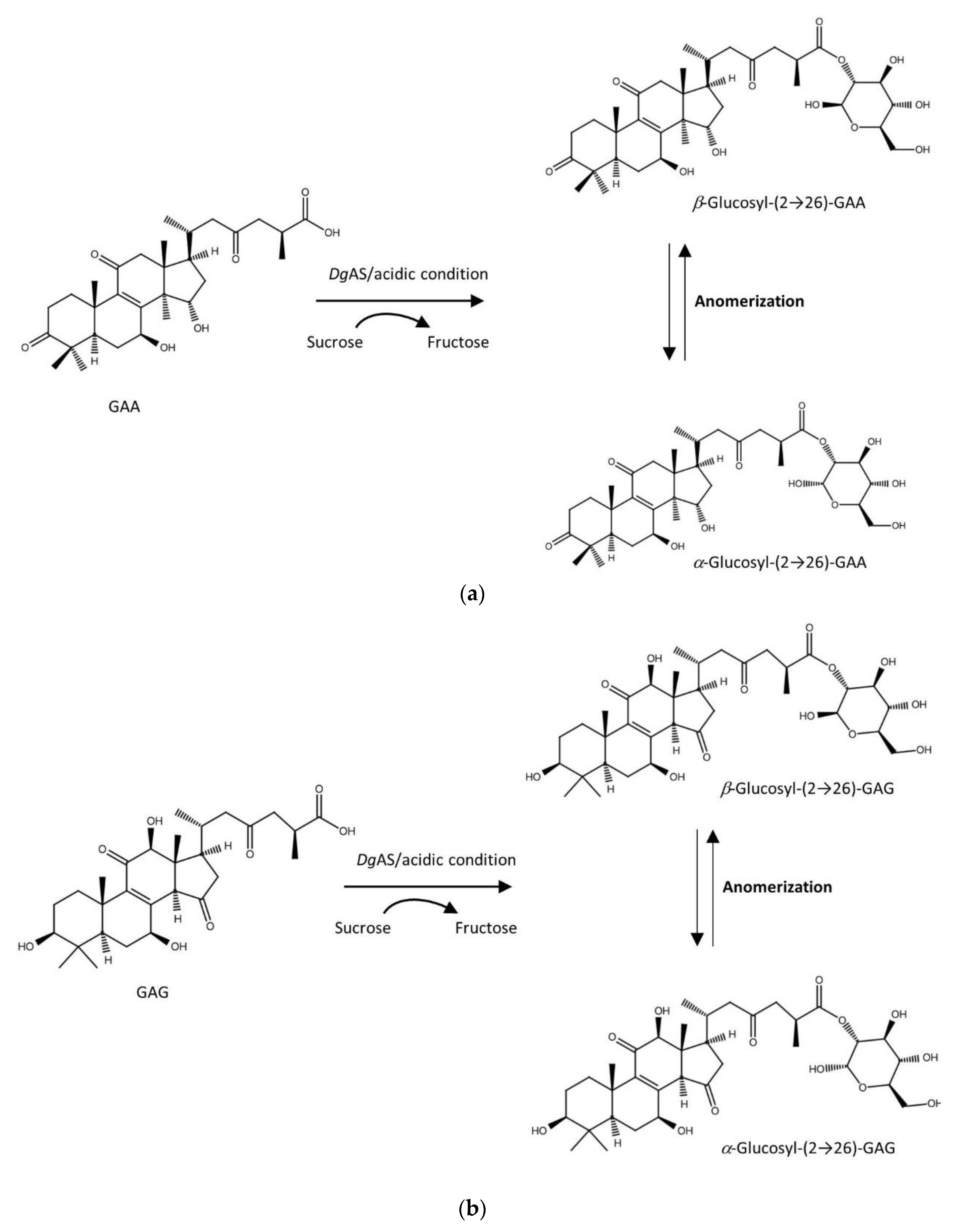
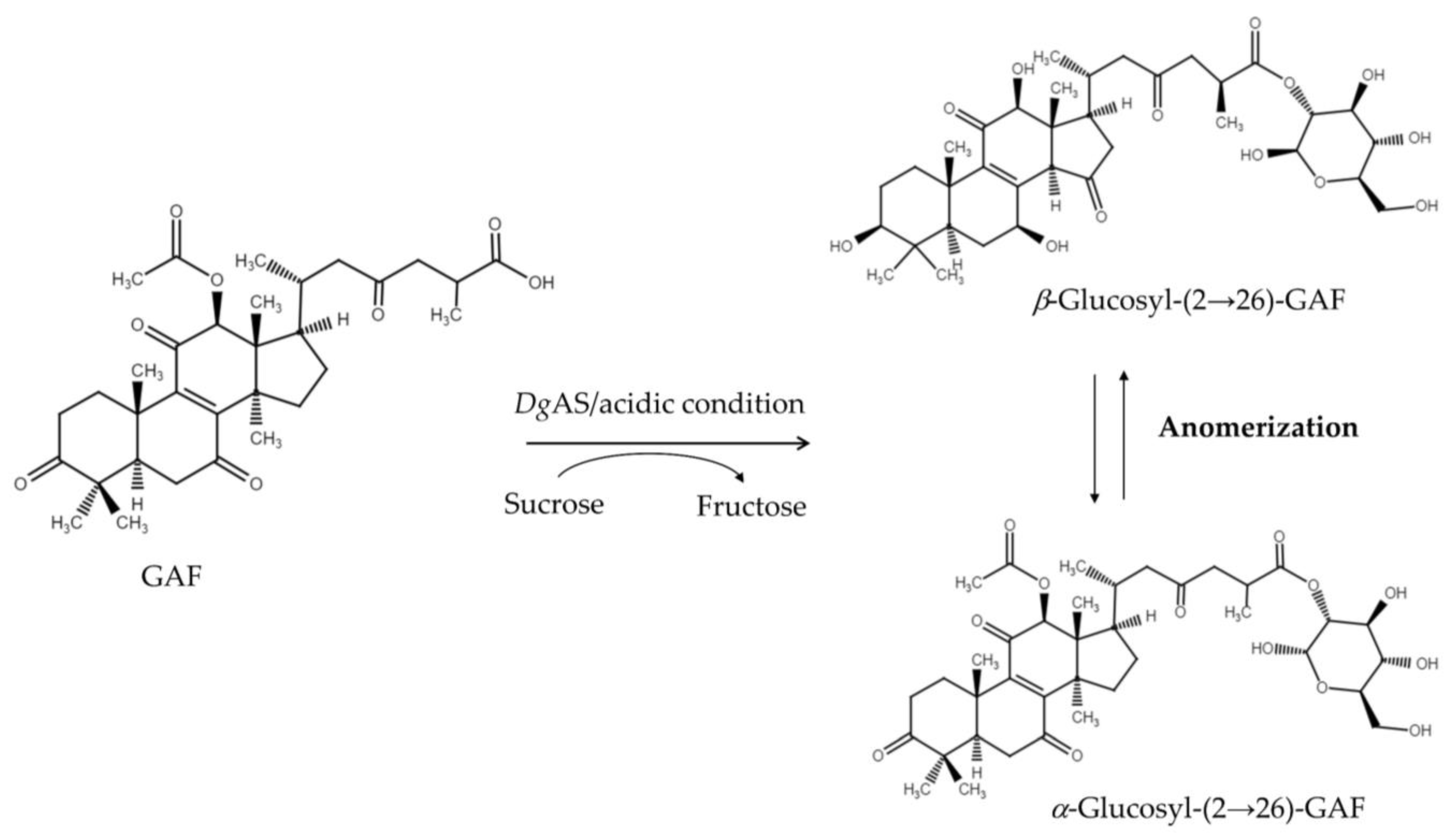
3.1. Biotransformation of GAF by DgAS

To make sure that DgAS could glycosylate triterpenoid aglycone, we first used triterpenoid aglycone with simple carboxyl group GAF as the starting point (Figure 1). This Ganoderma triterpenoid substrate contains a C-26 carboxyl group, which is the major functional group that exists in all ganoderic acids. GAF was then incubated with DgAS with 1500 mM of sucrose concentrations under different pH values, at 40 °C for 24 h. After incubation, the reaction mixture was analyzed by HPLC. The results indicated that DgAS could glycosylate GAF to compound (1) under acidic conditions of pH 6 and pH 5 (Figure S1), and pH 5 was the optimal condition (Figure 2a). However, the activity decreased extremely and became undetectable at pH 7 or pH 8. We performed three negative controls at pH 5: (1) reaction with the heat-denatured (inactivated) enzyme; (2) reaction without substrates (sucrose); and (3) reaction without GAF substrate. There are no targeted products in the three negative controls. The DgAS was first identified in a GH13 enzyme with novel acidic glycosylation of Ganoderma triterpenoids; its glycosylatic activities under different pH levels are similar to the enzymatic condition of BsGT110 glycosylation [10].
We further investigated the effects of temperature, sucrose concentration, and reaction time on the acidic glycosylation activity of DgAS toward GAF. The results showed that the optimal reaction conditions were 100 to 1500 mM of sucrose at 40 °C for 24 h (Figure 2). The maximum yield of compound (1) was 47%. However, the number of catalytic cycles by the enzyme before its inactivation was not evaluated. It has been reported that high concentrations of sugar donors would improve the glycosylation activity of GH enzymes [21]. However, sucrose concentrations between 100 mM and 1500 mM did not significantly affect the glycosylation activity of DgAS toward GAF.

3.2. Purification and Identification of the Biotransformation Product

Compound (1) (10.4 mg) was purified from 20 mL of biotransformation of GAF by DgAS by preparative HPLC. The chemical structure of compound (1) was identified using mass and NMR spectral analysis. The mass spectrometer showed an [M + H] ion peak at m/z: 731.4 in the electrospray ionization mass spectrum (ESI-MS) implying that compound (1) contained one glucosyl moiety attached to the GAF (molecular weight of 570) structure (Figure S2). The structure of compound (1) was elucidated by NMR in advance. Compound (1) showed the presence of a triterpenoid substituent and an α-glucose or a β-glucose unit, the hydrolyzed moiety of GAF [22,23]. In addition to the signals of the GAF, 14 proton signals (from 4.27 to 6.05 ppm) and 12 carbon signals (from 62.7 to 96.5 ppm) corresponding to two glucose moiety structures were observed. The attachment of the glucose moiety to C-26 of GAF was confirmed by the heteronuclear multiple bond connectivity (HMBC) correlation from the anomeric proton of Glu H-2′a (δH 5.50, d, J =3.5, 9.8 Hz) to C-26 (δC 176.2). The observation of coupling constants (J = 3.5 Hz) of the anomeric protons confirmed that the glycosidic bonds of compound (1) were in the α-configuration, demonstrating the structure of compound (1) as α-glucosyl-(2→26)-GAF. The large coupling constant (7.7, 9.1 Hz) of the anomeric proton H-2′β (5.65 ppm) indicated the β-configuration. The cross peak of H-2′β with C-26 (5.65/175.4 ppm) in the HMBC spectrum demonstrated that the structure of compound (1) is β-glucosyl-(2→26)-GAF. In addition, two sets of glucose signals, assignable to α and β, were found in the 1H-NMR and 13C-NMR spectrum, which could be grouped by distortionless enhancement by polarization transfer (DEPT), heteronuclear single quantum coherence (HSQC), HMBC, correlation spectroscopy (COSY), and nuclear Overhauser effect spectroscopy (NOESY) spectra (shown in Figures S3–S9). The NMR signals were fully identified, as shown in Table S1. Compound (1) thus confirmed the coexistence of α-glucosyl-(2→26)-GAF and β-glucosyl-(2→26)-GAF; In addition, according to the NMR analysis, the anomeric ratio (α:β) of the products was 3:2 (60%:40%). Figure 3 summarizes the biotransformation of GAF mediated by DgAS.

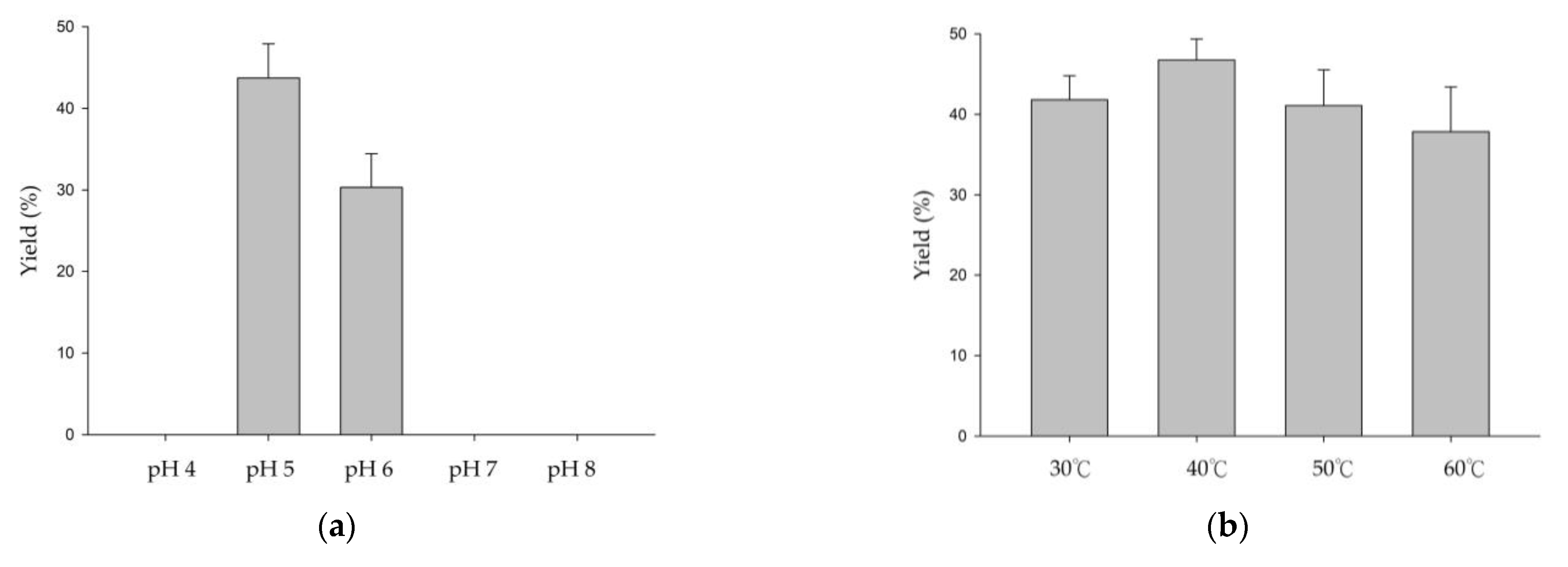
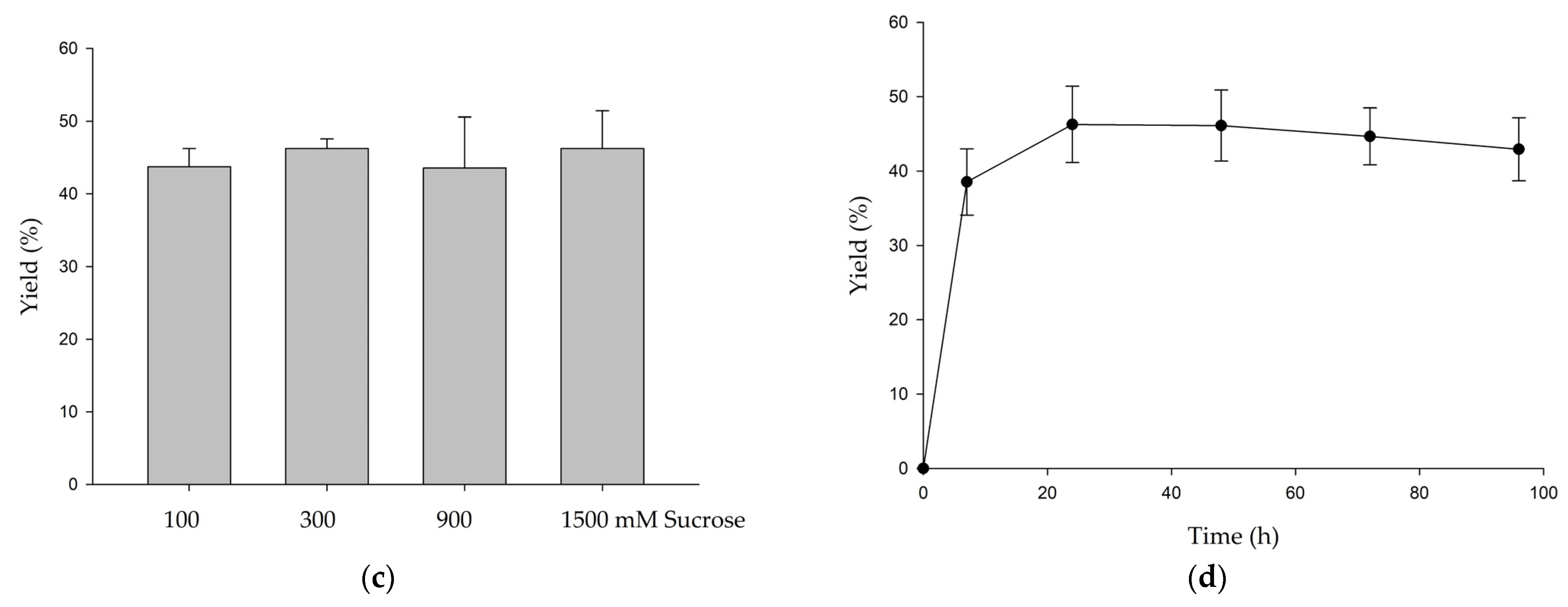
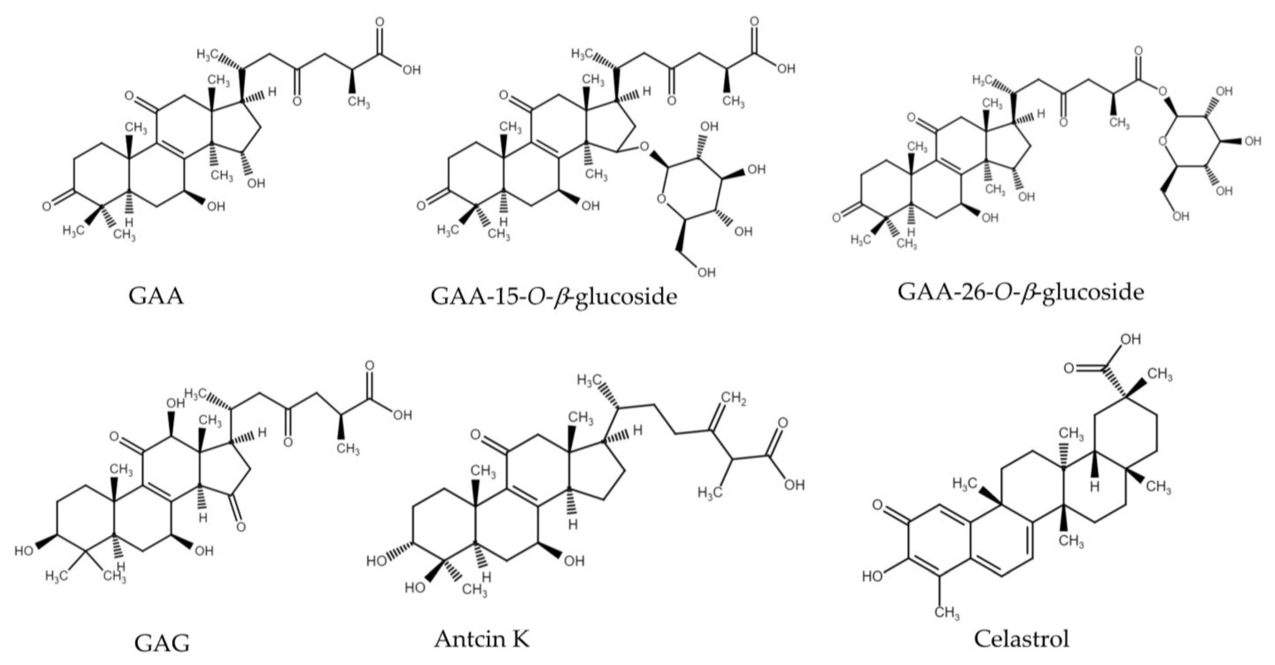
3.3. Evaluation of the Glycosylation Activity of DgAS toward Other Triterpenoids

Based on the novel glycosylation activity by DgAS toward the carboxyl group of the tetracyclic triterpenoid GAF, the other either tetracyclic or pentacyclic triterpenoids (antcin K, celastrol and 4 ganoderic acids in Figure 4) with carboxyl group were selected to evaluate the detailed acidic glycosylation activity of DgAS. Among the six triterpenoids, GAA and GAG are the tetracyclic lanostane type of Ganoderma triterpenoids with the same C-26 carboxyl group as that of GAF. GAA-15-O-β-glucoside and GAA-26-O-β-glucoside were previous biosynthetic glycosides from GAA by the bacterial GTs [10,11]. Antcin K also contains a C-26 carboxyl group. Antcin K is the most abundant tetracyclic ergostane type of triterpenoid isolated from the fruiting bodies of Antrodia cinnamomea, which is a parasitic fungus that only grows on the inner heartwood wall of the aromatic tree Cinnamomum kanehirai Hay (Lauraceae) [19]. In addition, celastrol contains a C-29 carboxyl group which is a pentacyclic triterpenoid isolated from the root of Tripterygium wilfordii (thunder god vine) [24].
The results showed that DgAS could glycosylate GAA and GAG into compound (2) and compound (3) but cannot glycosylate GAA-15-O-β-glucoside, GAA-26-O-β-glucoside, antcin K, or celastrol (Figure S10 and Table 1). The glycosylation condition of DgAS toward GAA and GAG was similar to that toward GAF, for which glycosylation activity was favored at pH 5 and pH 6 but dramatically decreased at pH 7 or pH 8, and the acidic activity was not significantly affected by sucrose concentration from 100 to 1500 mM. Although all the six tested triterpenoids contain a carboxyl group in their structures, it seems that the novel acidic glycosylation activity of DgAS was special toward Ganoderma triterpenoids.

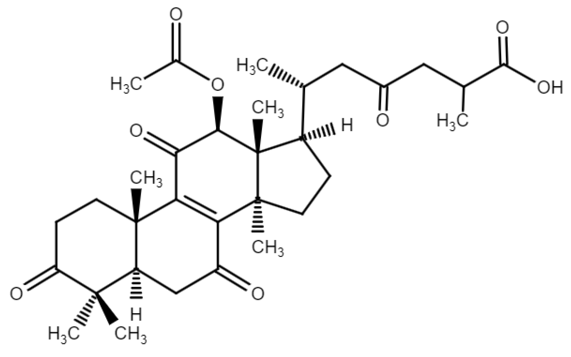
3.4. Purification and Identification of Biotransformation Products from GAA and GAG

To identify the chemical structure of compound (2) and compound (3), the biotransformation was scaled up to 20 mL, and the two compounds were purified by preparative HPLC. Finally, 8.8 mg of compound (2) and 9.3 mg of compound (3) were obtained. The chemical structures of compound (2) and compound (3) were identified using mass and NMR spectral analysis.
In the mass analysis of compound (2), the mass spectrometer showed an [M + H] ion peak at m/z: 677.6 in the ESI-MS corresponding to the molecular formula C36H54O12 (Figure S11). The mass data imply that compound (2) contained one glucosyl moiety attached to the GAA (molecular weight of 516) structure.
In the mass analysis of compound (3), the mass spectrometer showed an [M + H] ion peak at m/z: 693.5 in the electrospray ionization mass spectrum (ESI-MS) corresponding to the molecular formula C36H54O13 (Figure S12). The mass data imply that compound (3) contained one glucosyl moiety attached to the GAG (molecular weight of 532) structure.
To identify the structures of compound (2) and compound (3) in advance, the NMR spectroscopy method was then used. The full assignments of the 1H and 13C- NMR signals were further aided by DEPT, HSQC, HMBC, COSY, and NOESY spectra, as shown in Tables S2 and S3 and Figures S13–S26.
Compound (2) of the NMR spectra exhibited characteristic α-glucosyl and β-glucosyl signals. In addition to the signals of the GAA [25], 14 proton signals (from 4.28 to 6.04 ppm) and 12 carbon signals (from 62.7 to 96.5 ppm) corresponding to two glucose moiety structures were observed. The attachment of the glucose moiety to C-26 of GAA was confirmed by the HMBC correlation from the anomeric proton of Glu H-2′a (δH 5.48, d, J = 3.5, 9.8 Hz) to C-26 (δC 176.2). The observation of coupling constants (J = 3.5 Hz) of the anomeric protons confirmed that the glycosidic bonds of compound (2) were in the α-configuration, demonstrating the structure of compound (2) as α-glucosyl-(2→26)-GAA. The large coupling constant (8.4 Hz) of the anomeric proton H-2′β (5.64 ppm) indicated the β-configuration. The cross peak of H-2′βwith C-26 (5.64/175.5 ppm) in the HMBC spectrum demonstrated that the structure of compound (2) is β-glucosyl-(2→26)-GAA. In addition, two sets of glucose signals, assignable to α and β, full assignments of the 1H- and 13C-NMR signals were further aided by DEPT, HSQC, HMBC, COSY, and NOESY spectra, as shown in Table S2 and Figures S13–S19. Compound (2) thus confirmed the coexistence of α-glucosyl-(2→26)-GAA and β-glucosyl-(2→26)-GAA.
Compound (3) showed the presence of a triterpenoid GAG [26], and two sets of glucose signals, assignable to α and β, were found in the 1H-NMR and 13C-NMR spectra. In addition to the signals of the GAG, 14 proton signals (from 4.21 to 6.05 ppm) and 12 carbon signals (from 62.7 to 96.5 ppm) corresponding to two glucose moiety structures were observed, which could be grouped by HMBC correlations. An ether linkage between H-2′α of glucose and C-26 (5.47/176.3 ppm) of the GAG was proven by the HMBC spectrum. The coupling constant (3.5, 9.8 Hz) of the anomeric proton H-2′α (5.47 ppm) indicated that the α-configuration of the structure of compound (3) is α-glucosyl-(2→26)-GAG. The large coupling constant (8.4 Hz) of the anomeric proton H-2′β (5.64 ppm) indicated the β-configuration. The cross peak of H-2′β with C-26 (5.64/176.5 ppm) in the HMBC spectrum demonstrated that the structure of compound (3) is β-glucosyl-(2→26)-GAG. The NMR spectroscopic data are exhibited in Table S3 and Figures S20–S26. The results also showed the coexistence of α-glucosyl-(2→26)-GAG and β-glucosyl-(2→26)-GAG (3). In addition, according to the NMR analysis, the anomeric ratio (α:β) of the products was 3:2 (60%:40%).
Figure 5 summarizes the biotransformation of GAA and GAG mediated by DgAS.

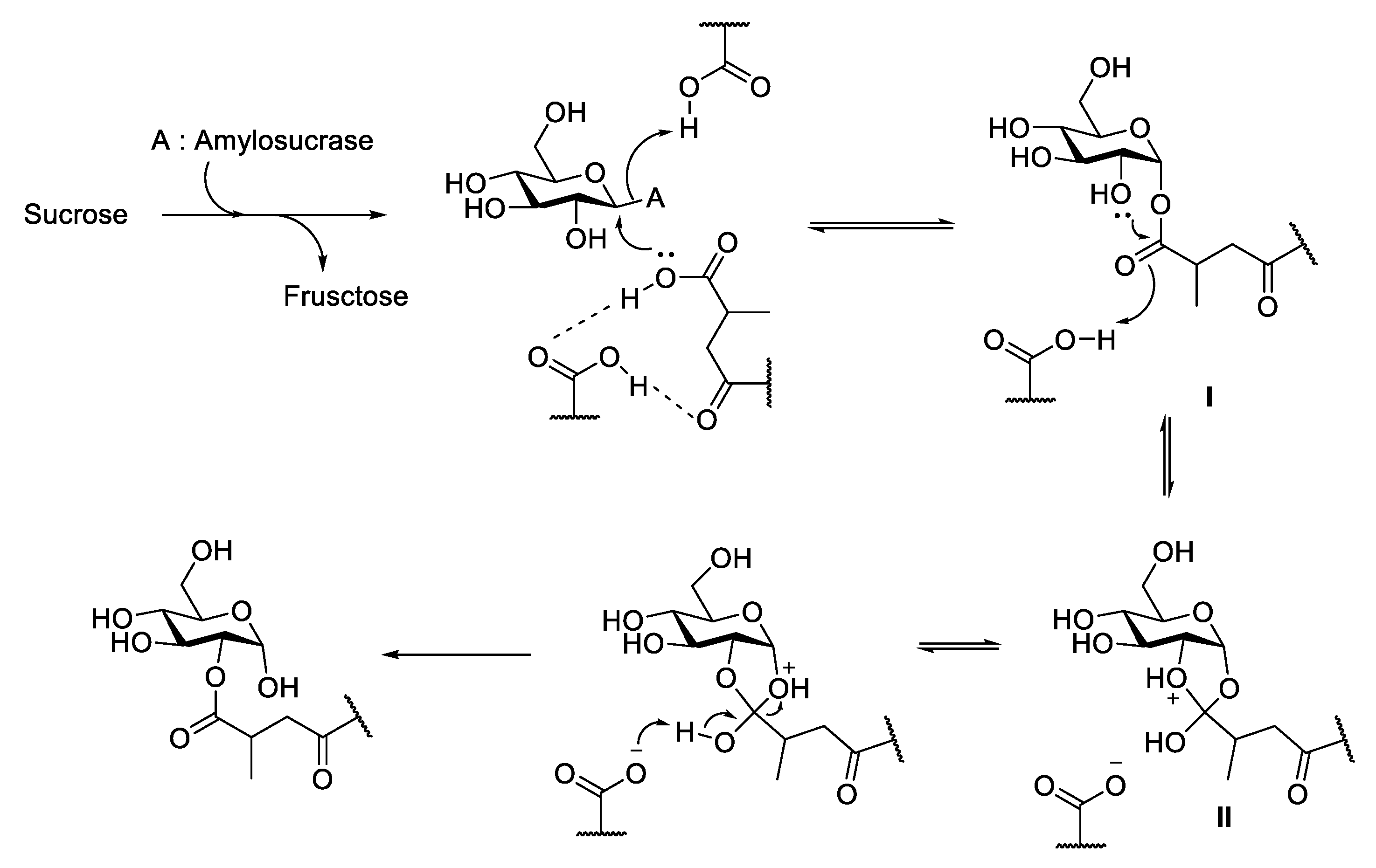
3.5. Proposed Reaction Mechanism of DgAS toward Ganoderic Acids

In nature, enzymatic glycosylation is always bonded through the anomeric carbon of the sugar donor (C1 of glucose) to the functional group of the glycosyl acceptor. However, this study discovered a new mechanism by which the DgAS catalyzed glycosylation through the non-anomeric carbon (C2) of the sugar donor linkage to the sugar acceptors (ganoderic acids). The novel linkage of the enzymatic glycosylation might occur through a carbon switch mechanism [27]. Figure 6 illustrates the putative transglycosylation mechanism of Ganoderma triterpenoids by the GH13 enzyme (DgAS). The glycosyl-enzyme intermediate approaches by ganoderic acid formed an intermediate I, which underwent formation of 1,2-cyclic intermediate II. The rearrangement of the 1,2-cyclic intermediate then formed the desired glycosylated product [27]. According to the proposed mechanism, the lack of free carboxyl group in GAA-26-O-β-glucoside explains why the molecule cannot be glycosylated by DgAS (Table 1). Also, an oxo group (C-23) near the glycosylated carboxyl group (C-26) is needed for the stabilization of the intermediate (Figure 6). Thus, neither antcin K nor celastrol is glycosylated by DgAS due to the lack of the oxo group near their carboxyl group. In addition, the bulky glucosyl group at C-15 of GAA-15-O-β-glucoside might form steric hindrance in the catalytic reaction by DgAS and block the catalytic activity toward the molecule by DgAS.

4. Conclusions

DgAS was the first confirmed GH to be able to glycosylate GAA, GAG, and GAF to form glucosyl-(2→26) ganoderic acid anomers under acidic conditions. This study proposed a new mechanism to form the glycoside anomers of triterpenoids by DgAS. The new mechanism by GHs, such as DgAS, to produce the Ganoderma triterpenoid saponin anomers could be applied for new triterpenoid derivatives in pharmaceutical biotechnology.

Supplementary Materials

The following supporting information can be downloaded at: https://www.mdpi.com/article/10.3390/biology11060822/s1. Table S1. 1H and 13C NMR assignments in pyridine-d5 at 700 and 175 MHz for compounds (1; α-glucosyl-(2→26)-GAF and β-glucosyl-(2→26)-GAF). (δ in ppm, J in Hz). Table S2. 1H and 13C NMR assignments in pyridine-d5 at 700 and 175 MHz for compounds (2; α-glucosyl-(2→26)-GAA and β-glucosyl--(2→26)-GAA). (δ in ppm, J in Hz). Table S3. 1H and 13C NMR assignments in pyridine-d5 at 700 and 175 MHz for compounds (3; α-glucosyl-(2→26)-GAG and β-glucosyl-(2→26)-GAG). (δ in ppm, J in Hz). Figure S1. High-performance liquid chromatography (HPLC) results of the biotransformation products of ganoderic acid F (GAF) using amylosucrase from Deinococcus geothermalis (DgAS) enzyme at different pH levels. Figure S2. The mass-mass analysis of compound (1) at the negative mode. Figure S3. 1D NMR spectrum (1H-NMR, 700 MHz, pyridine-d5) of the compound (1). Figure S4. 1D NMR spectrum (13C-NMR, 175 MHz, pyridine-d5) of the compound (1). Figure S5. 1D NMR spectrum (DEPT-135, 175 MHz, pyridine-d5) of the compound (1). Figure S6. 2D NMR spectrum ( 1H-13C HSQC, 700 MHz, pyridine-d5) of the compound (1). Figure S7. 2D NMR spectrum ( 1H-13C HMBC, 700 MHz, pyridine-d5) of the compound (1). Figure S8. 2D NMR spectrum (1H-1H COSY, 700 MHz, pyridine-d5) of the compound (1). Figure S9. 2D NMR spectrum (1H-1H NOESY, 700 MHz, pyridine-d5) of the compound (1). Figure S10. HPLC results of the biotransformation products of GAA (a) and GAG) (b) using DgAS enzyme at different pH levels. Figure S11. The mass-mass analysis of compound (2) at the negative mode. Figure S12. The mass-mass analysis of compound (3) at the negative mode. Figure S13. 1D NMR spectrum (1H-NMR, 700 MHz, pyridine-d5) of the compound (2). Figure S14. 1D NMR spectrum (13C-NMR, 175 MHz, pyridine-d5) of the compound (2). Figure S15. 1D NMR spectrum (DEPT-135, 175 MHz, pyridine-d5) of the compound (2). Figure S16. 2D NMR spectrum (1H-13C HSQC, 700 MHz, pyridine-d5) of the compound (2). Figure S17. 2D NMR spectrum (1H-13C HMBC, 700 MHz, pyridine-d5) of the compound (2). Figure S18. 2D NMR spectrum (1H-1H COSY, 700 MHz, pyridine-d5) of the compound (2). Figure S19. 2D NMR spectrum (1H-1H NOESY, 700 MHz, pyridine-d5) of the compound (2). Figure S20. 1D NMR spectrum (1H-NMR, 700 MHz, pyridine-d5) of the compound (3). Figure S21. 1D NMR spectrum (13C-NMR, 175 MHz, pyridine-d5) of the compound (3). Figure S22. 1D NMR spectrum (DEPT-135, 175 MHz, pyridine-d5) of the compound (3). Figure S23. 2D NMR spectrum (1H-13C HSQC, 700 MHz, pyridine-d5) of the compound (3). Figure S24. 2D NMR spectrum (1H-13C HMBC, 700 MHz, pyridine-d5) of the compound (3). Figure S25. 2D NMR spectrum (1H-1H COSY, 700 MHz, pyridine-d5) of the compound (3). Figure S26. 2D NMR spectrum (1H-1H NOESY, 700 MHz, pyridine-d5) of the compound (3).

Author Contributions

Conceptualization: T.-S.C.; data curation and methodology: S.-Y.L., Y.-L.T., T.-S.C. and H.-Y.D.; project administration: T.-S.C. and J.-Y.W.; writing—original draft, review, and editing: S.-Y.L., T.-S.C., T.-Y.W., J.-Y.W. and H.-Y.D. All authors have read and agreed to the published version of the manuscript.

Funding

This research was funded by the Ministry of Science and Technology of Taiwan under grant number MOST 110-2221-E-024-002 to T.-S.C. and grant number MOST 110-2221-E-507-002 to J.-Y.W.

Data Availability Statement

Not applicable.

Acknowledgments

We thank Chun-Cheng Lin (National Tsing Hua University, Hsinchu, Taiwan) for the advisement of the transglycosylation mechanism.

Conflicts of Interest

The authors declare that they have no conflict of interest.

References

  1. Benini, S. Carbohydrate-active enzymes: Structure, activity, and reaction products. Int. J. Mol. Sci. 2020, 21, 2727. [Google Scholar] [CrossRef] [PubMed] [Green Version]
  2. Mestrom, L.; Przypis, M.; Kowalczykiewicz, D.; Pollender, A.; Kumpf, A.; Marsden, S.R.; Bento, I.; Jarzębski, A.B.; Szymańska, K.; Chruściel, A.; et al. Leloir glycosyltransferases in applied biocatalysis: A multidisciplinary approach. Int. J. Mol. Sci. 2019, 20, 5263. [Google Scholar] [CrossRef] [PubMed] [Green Version]
  3. Cantarel, B.L.; Coutinho, P.M.; Rancurel, C.; Bernard, T.; Lombard, V.; Henrissat, B. The Carbohydrate-active EnZymes database (CAZy): An expert resource for glycogenomics. Nucleic Acids Res. 2008, 37, D233–D238. [Google Scholar] [CrossRef] [PubMed]
  4. Mikkola, S. Nucleotide sugars in chemistry and biology. Molecules 2020, 25, 5755. [Google Scholar] [CrossRef] [PubMed]
  5. Cárdenas, P.D.; Almeida, A.; Bak, S. Evolution of structural diversity of triterpenoids. Front. Plant Sci. 2019, 10, 1523. [Google Scholar] [CrossRef] [PubMed]
  6. Shi, Z.-Y.; Zeng, J.-Z.; Wong, A.S.T. Chemical structures and pharmacological profiles of ginseng saponins. Molecules 2019, 24, 2443. [Google Scholar] [CrossRef] [PubMed] [Green Version]
  7. Yang, Y.; Zhang, H.; Zuo, J.; Gong, X.; Yi, F.; Zhu, W.; Li, L. Advances in research on the active constituents and physiological effects of Ganoderma lucidum. Biomed. Dermatol. 2019, 3, 6. [Google Scholar] [CrossRef]
  8. Liang, C.; Tian, D.; Liu, Y.; Li, H.; Zhu, J.; Li, M.; Xin, M.; Xia, J. Review of the molecular mechanisms of Ganoderma lucidum triterpenoids: Ganoderic acids A, C2, D, F, DM, X and Y. Eur. J. Med. Chem. 2019, 174, 130–141. [Google Scholar] [CrossRef]
  9. Xia, Q.; Zhang, H.; Sun, X.; Zhao, H.; Wu, L.; Zhu, D.; Yang, G.; Shao, Y.; Zhang, X.; Mao, X.; et al. A comprehensive review of the structure elucidation and biological activity of triterpenoids from Ganoderma spp. Molecules 2014, 19, 17478–17535. [Google Scholar] [CrossRef]
  10. Chang, T.S.; Chiang, C.M.; Kao, Y.H.; Wu, J.Y.; Wu, Y.W.; Wang, T.Y. A new triterpenoid glucoside from a novel acidic glycosylation of ganoderic acid A via recombinant glycosyltransferase of Bacillus subtilis. Molecules 2019, 24, 3457. [Google Scholar] [CrossRef] [Green Version]
  11. Chang, T.S.; Wu, J.Y.; Wang, T.Y.; Wu, K.Y.; Chiang, C.M. Uridine diphosphate-dependent glycosyltransferases from Bacillus subtilis ATCC 6633 catalyze the 15-O-glycosylation of ganoderic acid A. Int. J. Mol. Sci. 2018, 19, 3469. [Google Scholar] [CrossRef] [PubMed] [Green Version]
  12. Chang, T.-S.; Wang, T.Y.; Hsueh, T.Y.; Lee, Y.W.; Chuang, H.M.; Cai, W.X.; Wu, J.Y.; Chiang, C.M.; Wu, Y.W. A genome-centric approach reveals a novel glycosyltransferase from the GA A07 strain of Bacillus thuringiensis responsible for catalyzing 15-O-glycosylation of ganoderic acid A. Int. J. Mol. Sci. 2019, 20, 5192. [Google Scholar] [CrossRef] [PubMed] [Green Version]
  13. Wu, J.Y.; Ding, H.Y.; Wang, T.Y.; Zhang, Y.R.; Chang, T.S. Glycosylation of ganoderic acid G by Bacillus glycosyltransferases. Int. J. Mol. Sci. 2021, 22, 9744. [Google Scholar] [CrossRef] [PubMed]
  14. Moulis, C.; Guieysse, D.; Morel, S.; Severac, E.; Remaud-Simeon, M. Natural and engineered transglycosylases: Green tools for the enzyme-based synthesis of glycoproducts. Curr. Opin. Chem. Biol. 2021, 61, 96–106. [Google Scholar] [CrossRef] [PubMed]
  15. Zhu, X.; Tian, Y.; Zhang, W.; Zhang, T.; Guang, C.; Mu, W. Recent progress on biological production of alpha-arbutin. Appl. Microbiol. Biotechnol. 2018, 102, 8145–8152. [Google Scholar] [CrossRef] [PubMed]
  16. Seo, D.H.; Yoo, S.H.; Choi, S.J.; Kim, Y.R.; Park, C.S. Versatile biotechnological applications of amylosucrase, a novel glucosyltransferase. Food Sci. Biotechnol. 2020, 29, 1–16. [Google Scholar] [CrossRef]
  17. Tian, Y.; Xu, W.; Zhang, W.; Zhang, T.; Guang, C.; Mu, W. Amylosucrase as a transglucosylation tool: From molecular features to bioengineering applications. Biotechnol. Adv. 2018, 36, 1540–1552. [Google Scholar] [CrossRef]
  18. Chang, T.S.; Wang, T.Y.; Yang, S.Y.; Kao, Y.H.; Wu, J.Y.; Chiang, C.M. Potential industrial production of a well-soluble, alkaline-stable, and anti-inflammatory isoflavone glucoside from 8-hydroxydaidzein glucosylated by recombinant amylosucrase of Deinococcus geothermalis. Molecules 2019, 24, 2236. [Google Scholar] [CrossRef] [Green Version]
  19. Chiang, C.-M.; Wang, T.-Y.; Ke, A.-N.; Chang, T.-S.; Wu, J.-Y. Biotransformation of ergostane triterpenoid antcin K from Antrodia cinnamomea by soil-isolated Psychrobacillus sp. AK 1817. Catalysts 2017, 7, 299. [Google Scholar] [CrossRef] [Green Version]
  20. Chiang, C.M.; Wang, T.Y.; Wu, J.Y.; Zhang, Y.R.; Lin, S.Y.; Chang, T.S. Production of new isoflavone diglucosides from glycosylation of 8-hydroxydaidzein by Deinococcus geothermalis amylosucrase. Fermentation 2021, 7, 232. [Google Scholar] [CrossRef]
  21. Rha, C.S.; Kim, H.G.; Baek, N.I.; Kim, D.O.; Park, C.S. Using amylosucrase for the controlled synthesis of novel isoquercitrin glycosides with different glycosidic linkages. J. Agric. Food Chem. 2020, 68, 13798–13805. [Google Scholar] [CrossRef] [PubMed]
  22. Kikuchi, T.; Matsuda, S.; Kodota, S. Ganoderic acid D, E, F, and H and lucidenic aicd D, E, and F, new triterpenoids from Ganoderma lucidum. Chem. Pharm. Bull. 1985, 33, 2624–2627. [Google Scholar] [CrossRef] [Green Version]
  23. Kimura, Y.; Taniguchi, M.; Baba, K. Antitumor and antimetastatic effects on liver triterpenoid fractions of Ganoderma lucidum: Mechanism of action and isolation of an active substance. Anticancer Res. 2002, 22, 3309–3318. [Google Scholar] [PubMed]
  24. Chang, T.S.; Wang, T.Y.; Chiang, C.M.; Lin, Y.J.; Chen, H.L.; Wu, Y.W.; Ting, H.J.; Wu, J.Y. Biotransformation of celastrol to a novel, well-soluble, low-toxic and anti-oxidative celastrol-29-O-β-glucoside by Bacillus glycosyltransferases. J. Biosci. Bioeng. 2021, 131, 176–182. [Google Scholar] [CrossRef] [PubMed]
  25. Kohda, H.; Tokumoto, W.; Sakamoto, K.; Fujii, M.; Hirai, Y.; Yamasaki, K.; Komoda, Y.; Nakamura, H.; Ishihara, S.; Uchida, M. The biologically active constituents of Ganoderma lucidum (FR.) Karst. histamine release-inhibitory triterpenes. Chem. Pharm. Bull. 1985, 33, 1367–1374. [Google Scholar] [CrossRef] [Green Version]
  26. Kikuchi, T.; Matsuda, S.; Murai, Y.; Ogita, Z. Ganoderic acid G and I and ganolucidic acid A and B, new triterpenoids from Ganoderma lucidum. Chem. Pharm. Bull. 1985, 33, 2628–2631. [Google Scholar] [CrossRef] [Green Version]
  27. Bassanini, I.; Kapešová, J.; Petrásková, L.; Pelantová, H.; Markošová, K.; Rebroš, M.; Valentová, K.; Kotik, M.; Káňová, K.; Bojarová, P.; et al. Glycosidase-catalyzed synthesis of glycosyl esters and phenolic glycosides of aromatic acids. Adv. Synth. Catal. 2019, 361, 2627–2637. [Google Scholar] [CrossRef] [Green Version]
Figure 1. The chemical structure of ganoderic acid F (GAF).
Biology 11 00822 g001
Figure 2. Effects of pH (a), temperature (b), sucrose concentration (c), and reaction time (d) on the yield of compound (1) from biotransformation of ganoderic acid F (GAF) by DgAS. The standard reaction condition was 1 mg/mL of GAF, 25 µg/mL of DgAS, and 1500 mM of sucrose at 50 mM of acetate buffer (pH 5) and 40 °C for 24 h. To determine suitable reaction conditions, different pH values, temperatures, sucrose concentrations, and reaction times were tested. After incubation, the biotransformation products were analyzed using HPLC. The detailed reaction conditions and the HPLC procedure are described in the Section 2.
Biology 11 00822 g002aBiology 11 00822 g002b
Figure 3. The biotransformation process of GAF to GAF glycoside anomers by DgAS.
Biology 11 00822 g003
Figure 4. The chemical structures of ganoderic acid A (GAA), ganoderic acid G (GAG), GAA-15-O-β-glucoside, GAA-26-O-β-glucoside, antcin K, and celastrol.
Biology 11 00822 g004
Figure 5. The biotransformation process of GAA (a) and GAG (b) to the corresponding glycosides by DgAS.
Biology 11 00822 g005
Figure 6. Proposed reaction mechanism of DgAS toward ganoderic acids.
Biology 11 00822 g006
Table 1. Glycosylation activity 1 of DgAS toward tested triterpenoids at different pH and sucrose concentrations.
TriterpenoidSucrose (mM)pH 4pH 5pH 6pH 7pH 8
GAA100N.D. 253.3 ± 4.731.1 ± 2.63.1 ± 0.0N.D.
1500N.D.50.7 ± 2.727.7 ± 0.93.2 ± 0.1N.D.
GAG100N.D.52.4 ± 3.617.2 ± 1.02.1 ± 0.0N.D.
1500N.D.47.6 ± 1.518.0 ± 0.80.8 ± 0.0N.D.
GAA-15-O-β-glucoside100N.D.N.D.N.D.N.D.N.D.
1500N.D.N.D.N.D.N.D.N.D.
GAA-26-O-β-glucoside100N.D.N.D.N.D.N.D.N.D.
1500N.D.N.D.N.D.N.D.N.D.
Antcin K100N.D.N.D.N.D.N.D.N.D.
1500N.D.N.D.N.D.N.D.N.D.
Celastrol100N.D.N.D.N.D.N.D.N.D.
1500N.D.N.D.N.D.N.D.N.D.
1 Activity was calculated by dividing the amount of the high-performance liquid chromatography (HPLC) area of the produced triterpenoid saponins in each reaction by the HPLC area of the peak of the triterpenoid without enzymes and expressed as a percentage. The mean (n = 3) is shown, and the standard deviations are represented by error bars. 2 N.D. means not detectable.
Publisher’s Note: MDPI stays neutral with regard to jurisdictional claims in published maps and institutional affiliations.

