Next Article in Journal
Paramecium bursaria as a Potential Tool for Evaluation of Microplastics Toxicity
Next Article in Special Issue
Identification of Potential Hub Genes Related to Aflatoxin B1, Liver Fibrosis and Hepatocellular Carcinoma via Integrated Bioinformatics Analysis
Previous Article in Journal
Oral Processing of Three Guenon Species in Taï National Park, Côte d’Ivoire
Previous Article in Special Issue
Integrating Expression Data-Based Deep Neural Network Models with Biological Networks to Identify Regulatory Modules for Lung Adenocarcinoma
 
 
Retraction published on 30 January 2024, see Biology 2024, 13(2), 86.
Article

RETRACTED: Identify Biomarkers and Design Effective Multi-Target Drugs in Ovarian Cancer: Hit Network-Target Sets Model Optimizing

by
Amir Abbas Esmaeilzadeh
1,
Mahdis Kashian
2,
Hayder Mahmood Salman
3,
Marwa Fadhil Alsaffar
4,
Mustafa Musa Jaber
5,6,
Siamak Soltani
7,
Danial Amiri Manjili
8,*,
Ahmet Ilhan
9,*,
Abolfazl Bahrami
10,11 and
John W. Kastelic
12
1
Salamat yar Behesht Dayan, Dayan Biotech Company, Tehran 14531, Iran
2
Department of Obstetrics and Gynecology, Medical College of Iran University, Tehran 14535, Iran
3
Department of Computer Science, Al-Turath University College Al Mansour, Baghdad 10011, Iraq
4
Medical Laboratory Techniques Department, AL-Mustaqbal University College, Hillah 51001, Iraq
5
Computer Techniques Engineering Department, Dijlah University College, Baghdad 00964, Iraq
6
Computer Techniques Engineering Department, Al-Farahidi University, Baghdad 10011, Iraq
7
Department of Forensic Medicine, School of Medicine, Iran University of Medical Sciences, Tehran 14535, Iran
8
Department of Infectious Disease, School of Medicine, Babol University of Medical Sciences, Babol 47414, Iran
9
Department of Medical Biochemistry, Faculty of Medicine, Cukurova University, Adana 01330, Turkey
10
Department of Animal Science, College of Agriculture and Natural Resources, University of Tehran, Karaj 1417643184, Iran
11
Biomedical Center for Systems Biology Science Munich, Ludwig-Maximilians-University, 80333 Munich, Germany
12
Department of Health, University of Calgary, Calgary, AB T2N 1N4, Canada
*
Authors to whom correspondence should be addressed.
Biology 2022, 11(12), 1851; https://doi.org/10.3390/biology11121851
Submission received: 14 November 2022 / Revised: 14 December 2022 / Accepted: 16 December 2022 / Published: 19 December 2022 / Retracted: 30 January 2024
(This article belongs to the Special Issue Bioinformatics and Machine Learning for Cancer Biology (Volume II))

Abstract

:

Simple Summary

We integrated three distinct methods (driver nodes, core module, and core nodes) to produce different HNSs for identifying hub genes involved in epithelial ovarian cancer (EOC). Immunohistochemical (IHC), qRT-PCR, and Western blotting were performed to validate the expression of hub genes and proteins. The results of the clinical experiment and the other data sets analyses confirmed the performance of the OHNS. Finally, the expression levels and diagnostic performance of OHNS showed statistical significance in evaluating external databases. This study also characterizes the critical genetic and transcriptomic features and their mutual regulatory relationships in EOC, providing valuable resources for identifying new molecular mechanisms and potential therapeutic targets for EOC.

Abstract

Epithelial ovarian cancer (EOC) is highly aggressive with poor patient outcomes, and a deeper understanding of ovarian cancer tumorigenesis could help guide future treatment development. We proposed an optimized hit network-target sets model to systematically characterize the underlying pathological mechanisms and intra-tumoral heterogeneity in human ovarian cancer. Using TCGA data, we constructed an epithelial ovarian cancer regulatory network in this study. We use three distinct methods to produce different HNSs for identification of the driver genes/nodes, core modules, and core genes/nodes. Following the creation of the optimized HNS (OHNS) by the integration of DN (driver nodes), CM (core module), and CN (core nodes), the effectiveness of various HNSs was assessed based on the significance of the network topology, control potential, and clinical value. Immunohistochemical (IHC), qRT-PCR, and Western blotting were adopted to measure the expression of hub genes and proteins involved in epithelial ovarian cancer (EOC). We discovered that the OHNS has two key advantages: the network’s central location and controllability. It also plays a significant role in the illness network due to its wide range of capabilities. The OHNS and clinical samples revealed the endometrial cancer signaling, and the PI3K/AKT, NER, and BMP pathways. MUC16, FOXA1, FBXL2, ARID1A, COX15, COX17, SCO1, SCO2, NDUFA4L2, NDUFA, and PTEN hub genes were predicted and may serve as potential candidates for new treatments and biomarkers for EOC. This research can aid in better capturing the disease progression, the creation of potent multi-target medications, and the direction of the therapeutic community in the optimization of effective treatment regimens by various research objectives in cancer treatment.

1. Introduction

The study of human disease has been transformed by the computational modeling of biological networks, which has also paved the path for the discovery of new therapeutic targets and personalized medicine. In addition to outlining the pattern of molecular signaling relationships, network-based research also shows transcriptional circuits, enrichment patterns, and system-wide characteristics [1,2]. Additionally, network-based approaches to biological research aid in our comprehension of the dynamics and control features of many complicated biochemical networks in conjunction with congruent experimental results. Pharmaceutical studies, especially those that offer substantial approaches in research and development, have been concerning in recent decades because of the declining efficacy of new medication designs [3]. The reductionist approach to medical research can only provide a limited understanding of the complicated etiology and multi-target pathologies of systemic diseases, and it has trouble defining the best strategies to address these complexities. Because systemic diseases, such as cancer, cardiovascular disease, and neurodegenerative disorders, are governed by complex biological networks and require multiple steps of genetic and environmental challenges to progress, they cannot be effectively treated with mono-target or bullet-based drug designs [4]. Due to the disease nature and pathway redundancy, numerous monotherapies have been shown in clinics to have limited effects or too many adverse effects when used in the long-term treatment of systemic disorders. For example, mono-target cancer therapy may allow cancer cells to evolve a resistance to the drug [5,6,7]; in contrast, multi-target therapeutics may be more effective or less prone to allowing adaptive drug resistance [8,9,10] due to the biological system’s decreased capacity to simultaneously compensate for multiple actions orchestrated by two or more drugs. On the other hand, when an illness strikes, the body loses homeostasis. The purpose of treating diseases is to correct this imbalance and bring the body back to a healthy state. How to effectively control the disease network and affect the capacity of biological systems to choose states by manipulating signals is one of the most appealing topics of research. Excellent control methods can help with the creation of therapeutic targets for viral diseases, the design of ideal molecules with desired effects, and the discovery of new applications for prescription drugs already on the market [4]. Disease development is significantly influenced by hit network-target sets (HNS), which are groups of multi-component units with network-controlling capabilities that occupy the network’s center nodes and are easily accessible. These components have the potential to be important system perturbation drivers for the network. However, understanding HNS is far from complete, which makes it difficult to use to control disease and find new medicines. Recent advances in network-based target research and prediction have been made possible by the constant accumulation of omics and big data. The effectiveness of network-based drug discovery will depend on the choice of pharmacological targets. It is now possible to locate the HNS in the network, but it can be difficult to condense the main traits of HNS even though numerous exploratory investigations have been conducted. For instance, driver nodes (DNs) based on structural control theory [11] and network core module nodes (CMs) for measuring the significance of node sets are used to identify the important nodes in complex networks as drug targets [12]. The module nodes allow automated prediction of the function of unidentified protein complexes from high-quality protein–protein interaction data. By using core nodes (CNs), often referred to as hub nodes, which rank elements in a network by the network properties that reflect their relevance in the network [13], we can discover important elements of biological networks. Because it enables researchers to manipulate targets in intricate networks, a full-fledged, abstract state-dependent dynamical model of diffusion dynamics in genomic networks is often employed to investigate cancer [14]. The computational methods for the identification of HNS based on network topologies have made tremendous progress, but there are still several obstacles that need to be overcome. For example, highly connected genes (hubs) have a major impact on the network’s structure, which is crucial for cell growth and survival due to their strong centrality [15]. The network can be significantly disrupted by using these nodes as targets; however, removing the related genes will render the organism lifeless and result in death or infertility. It is vital to find a method that can both avoid hubs and have some level of network control. Hu et al. [10] coined the phrase “driving nodes” to refer to important nodes having a strong capacity to affect the states of other nodes and a weak sensitivity to being affected by those states. Additionally, by administering control inputs (drugs, signals from the environment or within the organism, etc.) to critical and high-frequency driver nodes, the disease’s overall state could be controlled, suggesting that these nodes could serve as potential drug targets [16].
The precision and effectiveness of disease control must be improved as a result of these findings by choosing a strategy to reach a balance that can defy the disease and guarantee the survival of the organism. In this study, the multi-angle properties of network topology and structure control theory were used to build the regulatory network HNS. Regarding structural integrity and network information, we evaluated the developed ovarian cancer network. This study recommends a computational technique for simplifying the control of complex dynamical systems and biochemical regulation. In addition to in silico analyses, we aimed to investigate whether hub genes induce a change in biological functions in EOC. Furthermore, we used cancer cells to explore whether the hub high-traffic gene exerts effects on EOC. Finally, immunohistochemical (IHC) staining, qRT-PCR, and Western blotting were performed to examine and validate the expression of hub genes in ovary functions. Our results provide a new perspective on interactions and identify therapeutic targets for EOC. This approach will benefit logical drug design and aid in selecting appropriate drug targets.

2. Materials and Methods

2.1. Data Collection

Over 20,000 primary cancer and matched normal samples from 33 different cancer types are available in the Cancer Genome Atlas (TCGA) database, which can be accessed at http://cancergenome.nih.gov (accessed on 12 May 2022). In addition, Cancer-associated targets were obtained from the Comparative Toxicogenomics Database (CTD, http://ctdbase.org/ (accessed on 12 May 2022)) [17], Gene Cards (https://www.genecards.org/ (accessed on 14 July 2022)) [18], NCBI Gene (https://www.ncbi.nlm.nih.gov/ (accessed on 14 July 2022)) [19], Online Mendelian Inheritance in Man (OMIM, https://omim.org/ (accessed on 12 May 2022)) [20], Therapeutic Target Database (TTD, http://db.idrblab.net/ (accessed on 12 May 2022)) [21], PubChem (https://pubchem.ncbi.nlm.nih.gov/ (accessed on 23 May 2022)) [22], and the DisGeNET (http://www.disgenet.org/ (accessed on 23 May 2022)) database [23]. In addition, the top 1,235,000 targets from the CTD and the GeneCards database based on the “Inference score” were selected as the screening criteria. These databases are crucial tools for assessing the biological significance of cancer genomics discoveries. We used ovarian cancer tissues and normal tissues from 910 samples of the mRNA-seq data in this study. The batch effects of the RNA-seq raw data were removed by the ComBat-seq method using the R package sva [24].

2.2. Detection of Differentially Expressed Genes

The quality of the raw RNA sequences was evaluated using FastQC software (v0.11.9) [25]; after that, these sequences were pre-processed to remove adapters, low-quality reads, and PCR primers using Trimmomatic software (v0.38.1) [26]. The remaining reads were mapped to the reference genome using HISAT2 software (v2.2.1) [27]. Afterward, differences in transcript expression were detected using DESeq2 software (v2.11.40.7) [28]. The criteria of logFC (fold change < −1.5 and >1.5) and FDR < 0.05 (false discovery rate) were used to determine the threshold for statistical significance of the differential expression of each gene.

2.3. Gene Regulatory Network Construction

A gene regulatory network was constructed using Internet databases and gene interactions. These included MIPS (Mammalian Protein–Protein Interactions Database), BIND (Biomolecular Interaction Network Database) [29], PPI (Protein–Protein Interaction) [30], and BioGRID (Biological General Repository for Interaction Datasets) [31]. Additionally, interaction data were discovered by looking through related studies and research in interaction databases including Gene-MANIA and the STRING database [32,33], and cytoscape plugins were used to extract, integrate, visualize, and analyze interactive data [34].

2.4. Core Nodes/Genes Identification and Identification of Driver Nodes

More essential proteins are included in the top-ranked list of both high-degree and low-degree genes according to maximal clique centrality (MCC). MCC (v) equals the degree of node v if there is no edge between its neighbors. A node’s MCC is defined as MCC(v) = ∑C∈S(v)(|C| − 1)!, where S(v) is the set of all maximal cliques that contain v, and (|C| − 1)! is the sum of all positive integers less than |C| [35]. In a bipartite graph corresponding to the original network, the identification of driver nodes can be formulated as a maximum cardinality bipartite matching problem [36], which contains information about the identification algorithm and the driver nodes in detail. The maximum cardinality matching problem was resolved in this work using the HopcroftCKarp algorithm [37]. The concept of control centrality was created to quantify a node’s capacity for network control, which equates to the dimension of the controllable subspace and is calculated using an algorithm proposed by Liu et al. [12].

2.5. Modular Screening and Stability

The Markov cluster algorithm (MCL; parameters: number of iterations = 16) [38] and the molecular complex detection (MCODE; parameters: degree cutoff = 2, K-core = 2, and node score threshold = 0.2) [39] were two module-screening techniques that were contrasted. Simple connectivity-based divisions make up the connected components. Gene families are assigned using the MCL algorithm based on information about pre-calculated sequence similarity.
A column stochastic matrix is a non-negative matrix with the property that each of its columns sums to 1. Given such a matrix M and a real number r > 1, the column stochastic matrix resulting from inflating each of the columns of M with power coefficient r is written Γr (M), and Γr is called the inflation operator with power coefficient r. Write Σr,j (M) for the summation of all the entries in column j of M raised to the power r (sum after taking powers). Then, Γr (M) is defined in an entrywise manner by setting:
Γr(Mij) = Mijr/Σr,j(M)
where each column j of a stochastic matrix M corresponds with node j of the stochastic graph associated with M. Row entry i in column j (i.e., the matrix entry Mij) corresponds to the probability of going from node j to node i.
In order to isolate dense regions that meet the specified criteria, MCODE relies on vertex weighting by local neighborhood density and outward traversal from locally dense seed genes [39]. To counteract the selective speculation, their network structure entropies were calculated. The following is a definition of network structure entropy:
E   =   i = 1 N IiIn   Ii
where N is the number of nodes in the network, and Ii is the importance of node i. A smaller entropy value means a higher similarity between modular nodes, thereby determining the module stability.

2.6. Core Module Identification

The module was considered important, and the weighted edges between these modules were derived from the intermolecular relations across modules. The methods of multiple modular characteristic fusing (MMCF) were employed for this search process [40]. These methods included weighted degree, betweenness centrality, and PageRank. We removed each core module from the whole network and the module network, respectively, and then observed the rate change of the characteristic path length L22 to validate the results of identification:
L   =   1 n i N Li   =   1 n i N i N . i j dij n 1
where i and j are the different nodes in the network, Li is the average distance between node i and all other nodes, and dij is the distance between node i and j.

2.7. Performance Assessment of the OHNS

We removed each CN, CM, and DN from the whole network and observed changes in the characteristic path length and giant component. We randomly selected genes from CN, DN, CM, and RN (random nodes in the entire network) and OHNS (the combination of CM and the top 50 control centrality of DN) to better compare the change trends of the characteristic path length and giant component. Since the cardinal number starts with 5, 10 random copies of 5 genes were added. We measured the size of the remaining giant component in the graph and the characteristic path length for each one removed.
(a)
Characteristic path length (L) [41] based on Equation (3);
(b)
Giant component (GC): The giant component is the most significant connected component in each network. The fraction was calculated by dividing the number of nodes in the giant component by the total number of nodes in each network [10];
(c)
Calculation of the F-measure: To assess the F-measure, taking into account the precision and recall of the predicted HNS using the following formula, the key cancer genes are annotated in the list of drug targets and biomarker genes (Supplementary Table S1) for ovarian cancer (Comparative Toxicogenomics Database, http://ctdbase.org/ (accessed on 23 May 2022)) were chosen:
Fi = 2 × (Precision × Recall)/(Precision + Recall)
where precision is a measure of how many of the positive predictions made are correct (true positives), recall is a measure of how many of the positive cases the classifier correctly predicted, over all the positive cases in the data.
(d)
Perturbation effects: Cancer Dependency Map’s genome-scale CRISPR-Cas9 knockout data were used (https://depmap.org/portal/ (accessed on 23 May 2022)). In CM, CN, and DN, the required genes for perturbing 178 cancer cell lines were gathered, respectively. A lower Chronos score suggests a higher probability that the target gene is crucial in a particular cell line. A gene with a score of 0 is not considered influential; a score of −1 is comparable to the median of all genes considered necessary.

2.8. Gene Ontology and Functional Enrichment Analysis

Functional enrichment analysis of the differential expressed genes was performed by the online programs the KEGG database (release 71.0), DAVID [42] (Database for Annotation, Visualization and Integrated Discovery; https://david.ncifcrf.gov/ (accessed on 27 July 2022)), and g:Profiler [43] (https://biit.cs.ut.ee/gprofiler/gost/ (accessed on 24 July 2022)). We used the ClueGO plugin for Cytoscape (v2.5.9) in order to identify biological processes, molecular functions, and the involvement of cellular components [44]. Hochberg (p < 0.05) [45].

2.9. Prognostic Risk Assessment

The TCGA dataset (https://portal.gdc.com/ (accessed on 12 May 2022)) was used to download the RNA-sequencing expression (Level 3) profiles and associated clinical data for ovarian cancer. P-values and hazard ratios with 95% confidence intervals for Kaplan–Meier curves were calculated using log-rank tests and univariate cox proportional hazards regression; p < 0.05 was regarded as statistically significant, and R version 4.0.3 was used to implement all the analysis techniques and R packages.

2.10. Tissue-Specific Enrichment and Correlation Analyses of Hub Genes

We carried out Spearman’s correlation analysis to identify the correlations between hub genes in multiple tissues through the “Cor. Test” function in R, and results were presented by a scatter plot [46].

2.11. Replication and Validation Analyses

Validation and replication of the OHNS were performed using datasets (GSE211669) in the Gene Expression Omnibus (GEO) related to 131 samples of ovarian cancer [47]. RNA-seq analyses were performed as mentioned in Section 2.2.

2.12. Samples Collection

All tissue samples were obtained from patients enrolled in a national ongoing cohort initiated in 2008. Patients were diagnosed and surgically treated for EOC between October 2014 and January 2020. We received approval of the experimental protocol from the Experimental Ethics Committee of the Tehran Specialist Hospital and Research Center (No. 2022–0260). A specialized pathologist in gynecology revised histologic diagnoses for all tissue samples. A total of 59 biopsy attempts were made; 54 (92%) resulted in obtaining an ovary tissue specimen at the first attempt. The average size of the obtained biopsy core was 1 mm in diameter and 1.8 mm in length (range, 0.5 to 3 mm). Samples were instantly frozen in liquid nitrogen and stored at −80 °C.

2.13. Sample Preparation

The samples were transferred to the laboratory as fast as possible. Cells were separated by means of a stereo microscope. After washing in the PBS containing 1% Bovine serum Album (Bio-Rad Laboratories, CA, USA), the cells of the ovaries were cultivated in the balanced M16 medium under 5% CO2 at 37 °C. Samples were processed in cells with a healthy named normal group and EOC group. After 24 h or 72 h, the cells of the tissues were observed, and we calculated the developmental rate, respectively.

2.14. Validation of RNA-Seq Results Using qRT-PCR

Pooled samples were dissolved using a GenElute RNA Extraction Kit (Sigma) to isolate total RNA. The total RNA of the cell sample was extracted using the Steady Pure Universal RNA Extraction Kits (Accurate Biology, AG21017, Changsha, China). Total RNA was utilized for the synthesis of single-stranded complementary DNA using Evo M-MLV Mix Kits (Accurate Biology, AG11728). Real-time PCR was conducted with SYBR green premix pro-Taq qPCR Kits (Accurate Biology, AG11701). Next, quantitative re-verse-transcription-PCR was carried out for six genes, including SCO2, FBXL2, COX15, MUC16, NDUFA, and FOXA1. The following PCR cycling conditions were used: 30 s at 95 °C; 40 cycles of 5 s at 95 °C and 30 s at 60 °C. Melting-curve analysis was used to check product identities. PCR was carried out in triplicates, and the values of the mean threshold cycle (Ct) were normalized to GAPDH expression. Finally, the relative mRNA expression levels were analyzed. Forward and reverse primer sequences and accession numbers of selected genes are given in Supplementary Table S2.

2.15. Western Blotting

Total proteins from tissues were extracted using RIPA lysis buffer containing a mixture of protease inhibitors. Protein concentrations were measured using the BCA Protein Assay Kit according to the manufacturer’s protocol. After boiling the total protein, the proteins (20 μL per well) were separated in 10% SDS-PAGE and then transferred to PVDF membranes. To seal the proteins, the PVDF membranes were incubated with 5% fat-free milk for 90 min. Then, the membranes were incubated overnight at 4 °C using primary antibodies MUC16 (1:500, Abcam, UK), FOXA1 (1:1000, Abcam, UK), NDUFA (1:1000, Abcam, UK), COX15 (1:1000, Abcam, UK), β-actin (1:3000, Abcam, UK). After washing, the membrane was incubated with the appropriate secondary antibody for 2 h. After the detection of the signal using digital imaging equipment, the protein bands were quantified by ImageJ software.

2.16. Immunohistochemistry

The ovary tissues were fixed in 4% paraformaldehyde, paraffin-embedded, and then sliced into 5-μm sections. After deparaffinization, rehydration, and antigen retrieval, nonspecific binding of sections was blocked with goat serum. Subsequently, the sections were incubated with antibody MUC16 (1:500), NDUFA (1:100), FOXA1 (1:1000), and COX15 (1:500) primary antibody overnight at 4 °C. The tissues were then incubated with a secondary antibody at 37 °C for 40 min, followed by incubation with diaminobenzidine as a chromogen. Images were assessed using an Olympus optical microscope (Japan). The immunohistochemistry (IHC) scores were evaluated by multiplying the 2 scores. Five 200-fold visual fields for each sample were randomly selected, and the average score of the five visual fields is the final score of the sample.

2.17. Statistical Analysis

SPSS 22.0 was used for the statistical analyses, and a p value of 0.05 was regarded as statistically significant. The degree (in and out) distribution ratio of various groups in the network was examined using the Chi-square test. The data are presented as the mean ± SEM. We used the package ggplot2 in R, and results were presented by a bar and violin plot. The degree (in and out), characteristic path length, and giant components of the HNS were correlated using the Pearson correlation coefficient.

3. Results

3.1. Hit Network-Target Sets Identification

We obtained 12,659 differentially expressed genes from the TCGA, with 10,102 being upregulated and 2557 being downregulated (Supplementary Table S3). After removing individual genes, we obtained a regulatory network with 1506 genes and 8718 directed edges (regulatory links). Supplementary Table S4 presents a list of genes and their regulatory interactions. The core genes were found using the MCC algorithm, and the top 120 are displayed in Supplementary Table S5. For the HopcroftCKarp algorithm, 214 driver nodes were obtained based on control centrality (Supplementary Table S6). MCODE, MCL, and the connected components were used to divide the disease network modules, and modules 38, 34, and 18 were obtained. Entropy values for the MCODE, MCL, and connected components, respectively, were 5.105, 5.986, and 6.018 (Supplementary Table S7).
The MCODE method showed strikingly consistent stability in each group compared to two other methods (connected components and MCL), according to the minimum entropy criterion. Using MMCF comprehensive ranking, we identified core module No. 2 as having 50 genes and 369 edges (Supplementary Table S8).

3.2. Characteristics of Clustering and Scattering of the Network Distribution

CM and CN had more overlap (Figure 1A) and were more centralized in the network, while DN was more scattered. We obtained a regulatory network (Figure 1B) after removing individual nodes. CM, CN, and DN distributions in this network are very dissimilar.

3.3. Out-Degree-Dominant Characteristics of Driver Nodes

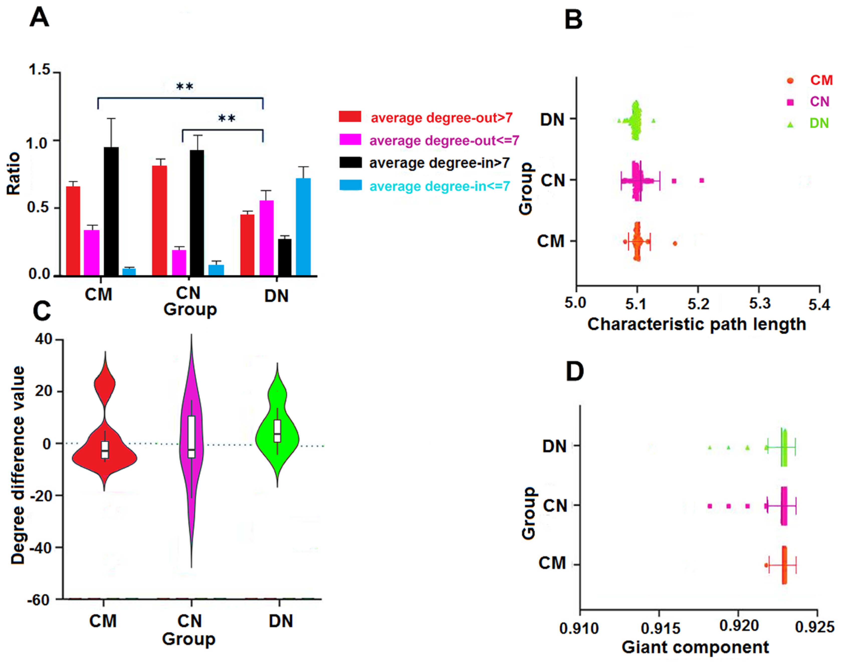
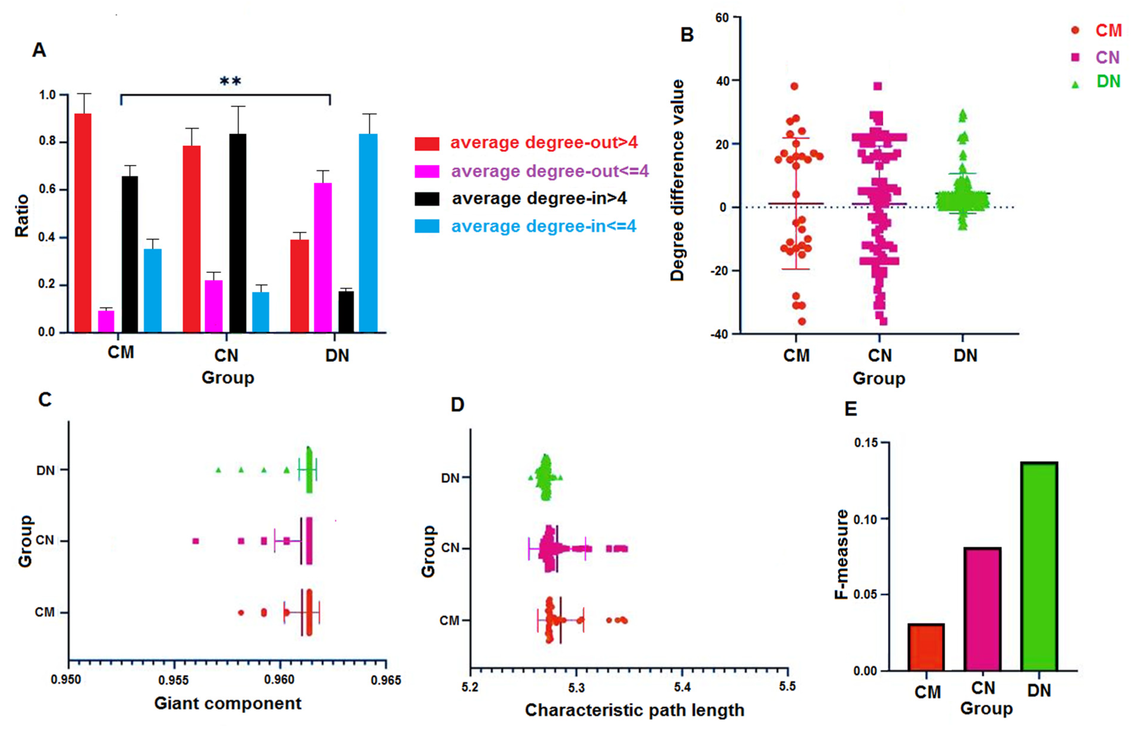
We selected to use the average degree of the network, 7, as the baseline and set up four standards: average degree-out > 7, average degree-out 7, average degree-in > 7, and average degree-in 7 (Figure 2A). This allowed us to demonstrate the degree (out and in) differences between nodes in the HNS. Comparative analysis revealed that the driver node’s degree-in was lower (29% average degree-in > 7), which distinguished it significantly from the module and core nodes (p < 0.05). Moreover, by comparing the difference of the value between the out-degree and in-degree of the three node sets, it is shown that the degree difference ≥ 0 is CN 45%, CM 39%, and DN 98%, respectively (Figure 2C).

3.4. Characteristic Path Lengths and Giant Components of HNSs

We evaluated the significance and robustness of the deleted genes for the network using characteristic path length and giant components after deleting single genes and random polygene combinations. The outcomes demonstrated that only a few nodes could disrupt the network after deleting the genes in the three HNSs (only deleting one node at a time) (Figure 2B,D). Notably, only one of these perturbed genes matched a set of biomarkers and drug targets for treating ovarian cancer, suggesting that these genes could be potential new targets for treating ovarian cancer. As seen in Figure 3, the three distinct node sets are more evident than the random nodes after each random polygene combination has been eliminated. The length (L) of the remaining network gradually increases after removing CM (from 4.812 to 5.881) and CN (from 4.722 to 6.541), which shows that the CM and the CN are crucial for information transmission between network nodes.
In contrast, there is no change in the remaining network length after removing the random DN combination. The changing trend of the GC is different from that of length; when the combination of random nodes is deleted in turn, the GC of the remaining network shows a gradual downward trend. One has a lower GC than the other nodes because it is the only network left without a CN. The synergy of the node sets between networks may be the reason why the remaining network’s GC rapidly declines after the removal of the CM set (seventh time) and the DN set (eighth time) in turn (CM from 0.819 to 0.812, CN from 0.810 to 0.798) (Figure 3).

3.5. Characteristic Path Lengths and Giant Components

We determined the Pearson correlation coefficient between length (L) and GC to determine whether the degree (in and out) of various node sets is a determining factor for L and GC. The findings demonstrate that the degree (in and out) of the driver nodes and L only have a marginally significant weak correlation (Table 1).

3.6. F-Measure and Perturbation Effect

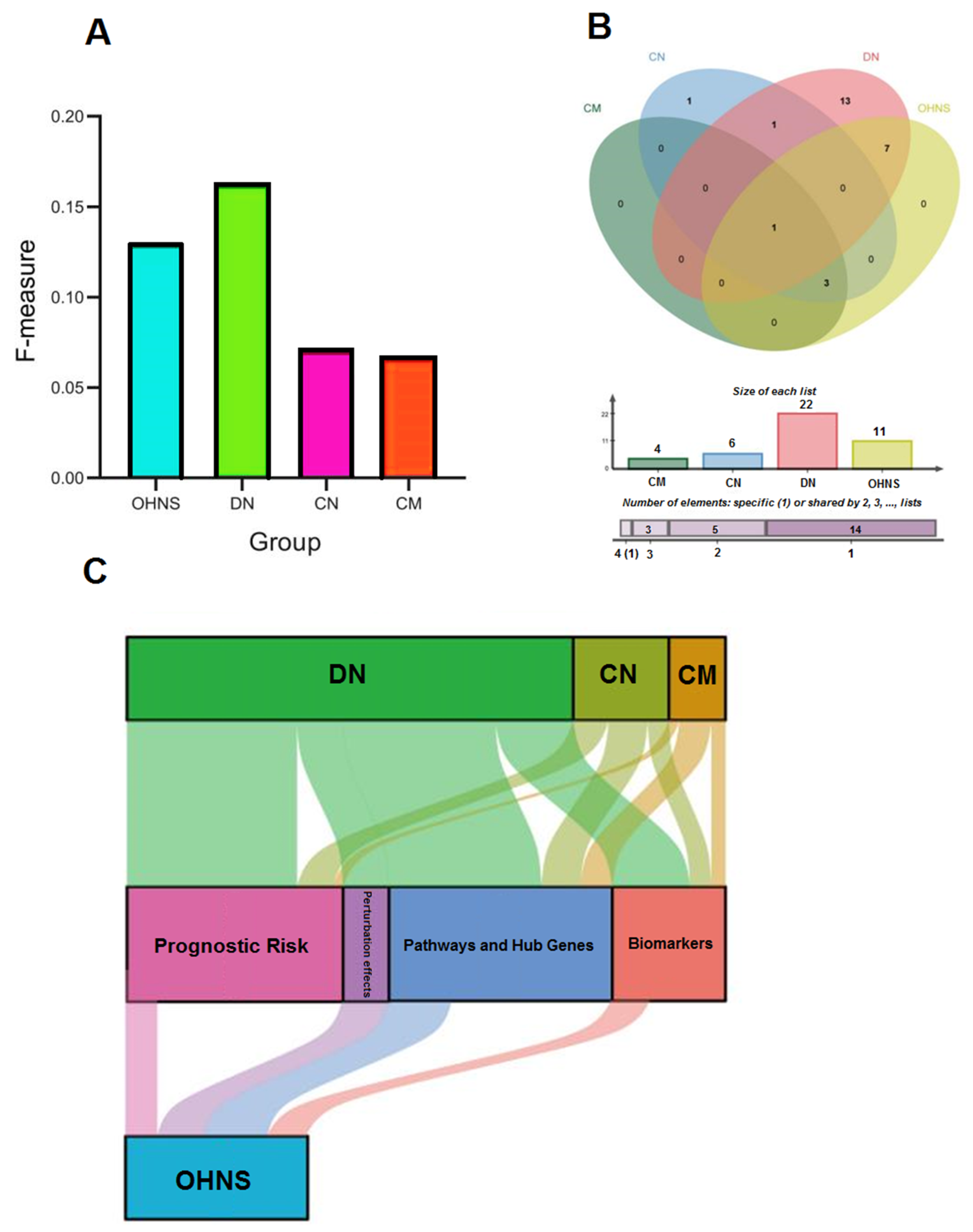
The target prediction capability of HNS was assessed using the F-measure in the list of drug targets and biomarker genes for ovarian cancer. We obtained 4, 6, and 22 markers and therapeutic targets when we mapped ovarian biomarkers and therapeutic targets from the CTD database to CM, CN, and DN. DN is more efficient than CM and CN at identifying drug targets and biomarkers (Figure 4A). It might also depend on the benefit of having a large enough number of DNs. The mapping outcomes of CN and CM overlap, which is consistent with the previous observation. It is important to note that the F-measure for OHNS is 0.134, almost identical to DN (Figure 4A). However, only one of the mapping outcomes from DN and CM overlaps (Figure 4B), meaning that their merging can boost the likelihood that drug targets and biomarkers will be discovered. We determined the Chronos dependency scores of CM, CN, and DN in 186 ovarian cancer cell lines using the genome-scale CRISPR-Cas9 knockout data. In 186 ovarian cancer cell lines, only 11 genes (MUC16, FOXA1, FBXL2, ARID1 A, COX15, COX17, SCO1, SCO2, NDUFA4L2, NDUFA, and PTEN) in OHNS had clear perturbation effects (Supplementary Table S9). It is interesting to note that OHNS also contains these 11 genes. A comparison of distributions of CM, CN, DN, and OHNS in biomarkers, pathway and hub genes, risk-prognostic genes, and perturbation effects is shown in Figure 4C.

3.7. Pathway Enrichment Analysis of HNSs

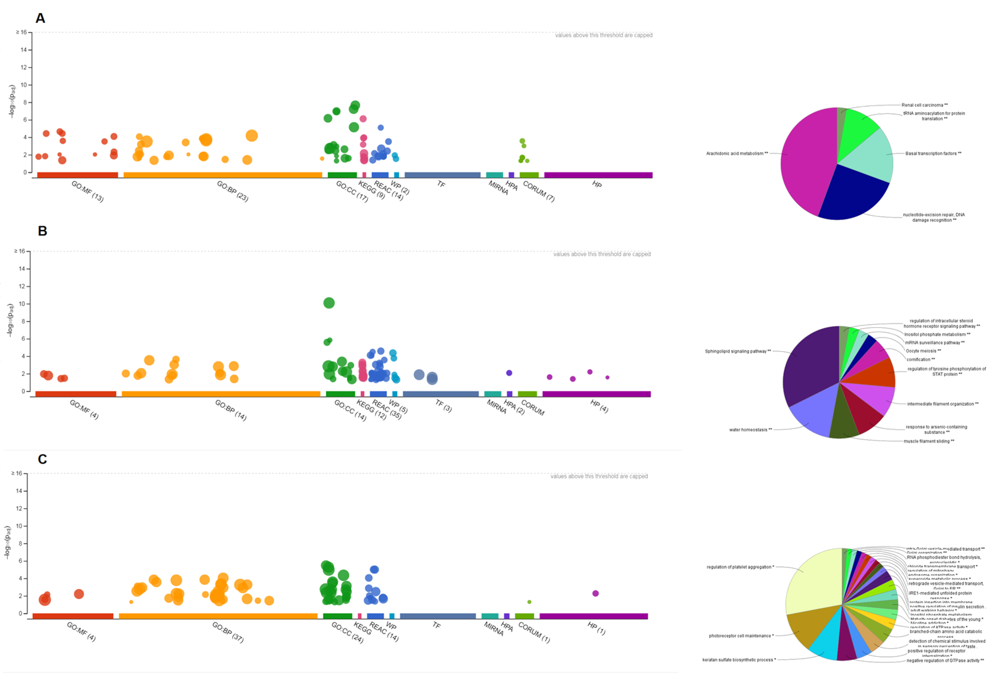
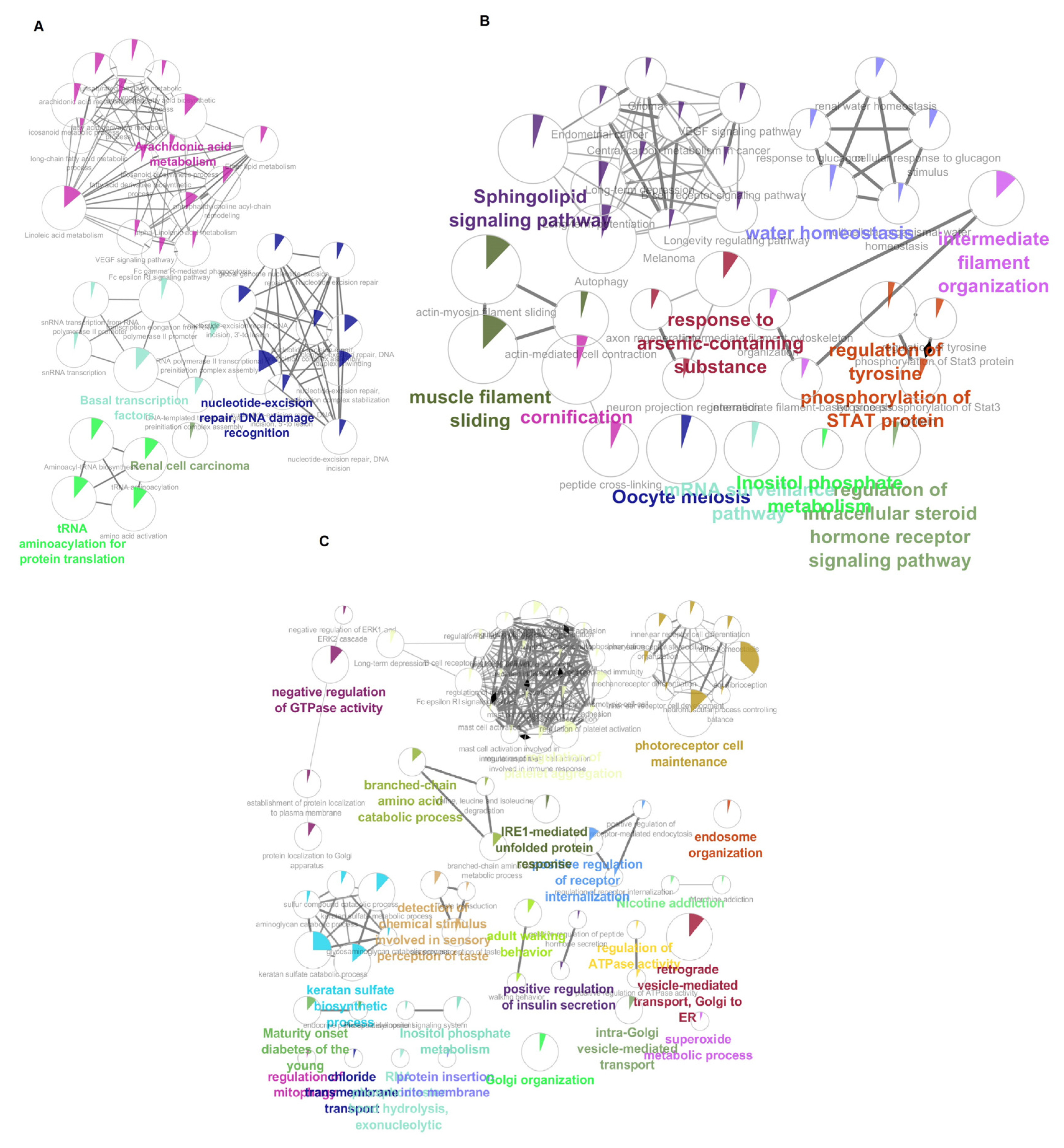
To examine the functions of HNS, online tools were used. In the findings, OHNS had 27 pathway overlaps and 10 functional overlaps (Supplementary Figure S1). Contrary to popular belief, CM and CN overlap, but they have distinct biological functions. This may be because of the many genes that do not exist in both. Using Cytoscape-ClueGo, we constructed a pathway network while visualizing the genes connecting different pathways. The results showed that 9, 33, and 43 pathways were present in CM, CN, and DN, respectively (Supplementary Figure S2). The pathway to which the CNs are enriched in the pathway enrichment, however, also contains the pathways of all the CMs (Supplementary Table S10) (Figure 5). After comparison with CN, CM, and DN, four unique pathways were discovered in OHNS: the endometrial cancer signaling, the PI3K/AKT pathway, the NER pathways, and the BMP pathway (Figure 6).

3.8. Survival Analysis of Perturbed Genes

The prognostic factors in the entire genome were observed, and the single-factor cox and log-rank tests were used to assess the prognostic significance based on the level of expression of a single gene. The findings revealed 2362 risk-prognosis-related genes in total, of which 3, 11, and 48 were discovered in CM, CN, and DN, respectively (Supplementary Table S11). Of the 48 DN risk prognostic genes, 11 were consistent with perturbed genes.

3.9. Tissue-Specific Enrichment and Correlation Analyses of Hub Genes

The scatter plot displayed the results of the correlation analysis of hub genes in multiple normal tissues. From Figure 7, the hub targets positively correlated with EOC were NDUFA, MUC16, MMP2, FOXA1, and ARID1A. The hub targets negatively correlated with EOC were SCO1, SCO2, COX15, COX17, FBXL2, PTEN, and NUFDA4L2.

3.10. Replication and Validation Analyses

The GEO data were processed using the same methodology, and 2825 differentially expressed genes in ovarian cancer were discovered (Supplementary Table S12). We constructed a set of 933 nodes and 4336 directed edges. Following network analysis, we discovered that the degree distribution of CM, CN, and DN in the GEO network, the degree difference, the F-measure, and the L and GC scores of deleting a single node almost precisely match the trend of TCGA (Figure 8).

3.11. The Deficiency of Proteins Expression and Biological Functions

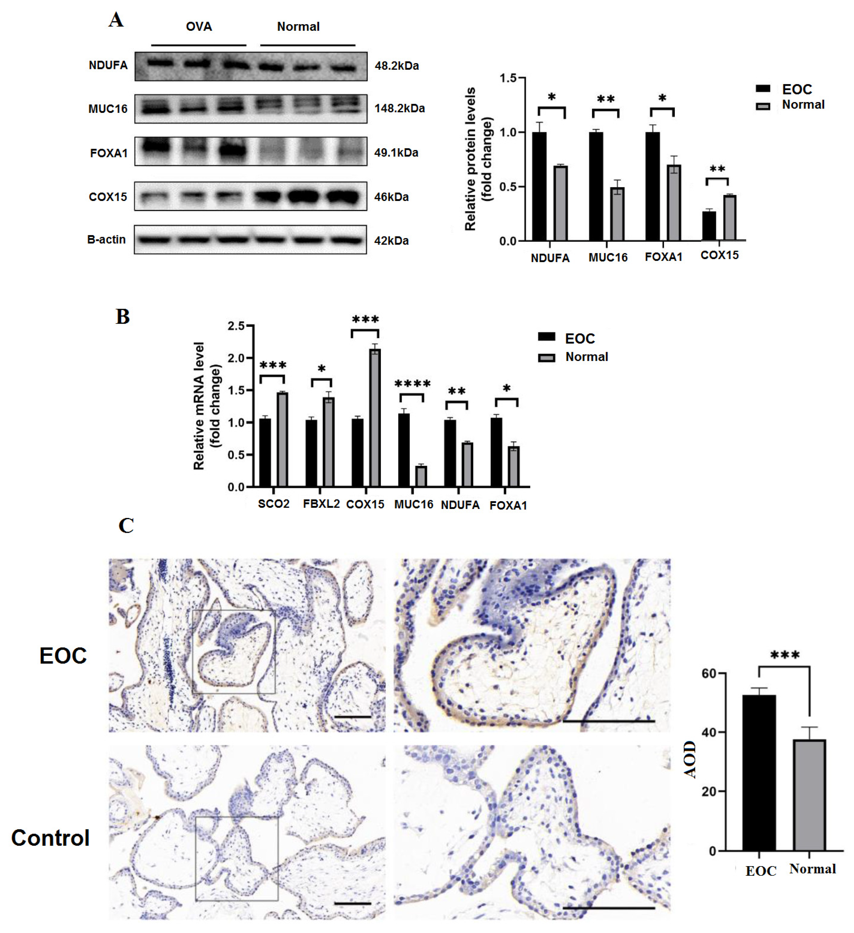
The expressions of COX15, MUC16, NDUFA, and FOXA1 protein in ovary tissues from three normal and three patients were evaluated by Western blot analysis (Figure 9A). The result coming from the qRT-PCR analysis also exhibited a similar outcome (Figure 9B). Immunohistochemistry revealed that the location of MUC16, NDUFA, and FOXA1 was in the nucleoplasm of ovary tissues, and the expressions were remarkably high in OVC samples than in the control group (Figure 9C).

4. Discussion

The network’s molecular targets were found using a variety of systems biology tools. The development of translational analysis as well as the design of the best network-based multi-target medications would benefit from further integration of various ‘-omics’ levels and databases. The current effort to use various network- and algorithm-based computational tools for clarifying the target network of herbal formulations and optimizing their molecular synergy looks to be advantageous, and it would also be a viable method for building network-based multi-component medications. The quality and quantity of the models will be improved by standardizing the drug design process, normalizing, and conducting research on herbal synergy with network-target sets, which will facilitate integration. The main concept of an effective target control approach is to disregard the nodes that are unnecessary to manage and concentrate on the nodes that are. For instance, in cancer, certain proteins are recognized as being necessary (for that malignancy) if they are necessary for the proliferation or survival of the tumor cells. Because of a mutation in the driver genes, some proteins become crucial in cancer. This indicates that these proteins are necessary for pathogenesis (i.e., the driver), and the tumor thereafter becomes dependent on the emergence of oncogenes [48]. In addition, in the post-genome era, selecting efficient “HNS” at the network level has gained much attention. It can assist us in learning more about the essential elements of the disease network. Since identifying HNS has good theoretical and application value, more studies about methods have been put forth. The methods can be viewed from two angles: one identifies the network’s fundamental structure, and the other considers the network’s controllability. It makes sense to assume that controlling the hubs will be crucial to controlling disease networks, given the crucial role nodes with a high degree (hubs) play in preserving the structural integrity of networks against failures and attacks [49], in spreading phenomena [50], and in synchronization [51]. Some hub nodes result in death or infertility, making them poor candidates for pharmacological therapy. Using network control theory to anticipate medication targets is a popular idea, but in reality, fully managing disease networks is difficult. The underlying significance and characteristics of the various forms of HNS are also not fully understood by researchers. They have only examined them from one angle, which has advantages and disadvantages. To overcome these obstacles, we compare different approaches from a variety of angles, such as core structure, control forces, and therapeutic value, and we emphasize the unique characteristics of each. The results show that DN has benefits in risk prognosis, pathway and hub genes, perturbation effects, and biomarkers. We come to the conclusion that DN will probably play a control role because of the cooperation of these four factors. The essential structure and control capabilities of the network were taken into account when building the OHNS. The F-measure indicates that the OHNS was more effective in comparison to CM and CN and was neck-and-neck with DN, which gives us the impression that combining various calculating techniques may raise the possibility of illness control. This might be a result of the OHNS, which controls the network and is built on the conservative evolution of core genes. On the other hand, 11 genes in OHNS were predicted to play a role in biomarkers, risk prognosis, and disruptions connected to ovarian cancer. After OHNS enrichment analysis, the same results were seen. The enriched pathways of CM and DN did not completely take up the enriched pathways of OHNS. These signaling pathways may be responsible for the structural centrality and control capacity of the ovarian cancer regulatory network. Our results are consistent with other studies that claim many MUC genes are expressed more frequently in ovarian cancers [52]. Since mucins are substantial extracellular proteins that can act as biomarkers (indeed, MUC16 is also referred to as CA125), overexpression of the MUC genes is clinically significant, and their overexpression is well established as a prognostic predictor [53]. When comparing normal cells to ovarian tumors, we discovered that FOXA1 was increased, whereas COX15, COX17, FBXL2, and cytochrome genes (SCO1 and SCO2) were downregulated. According to published research, FOXA1 is overexpressed in ovarian cancer, with aberrant expression linked to carcinogenesis and an aggressive character [54]. There have been reports of potential tumor suppressor functions of FBXL2 through the ubiquitin-mediated degradation of significant cell cycle regulators [55]. Low expression of SCO2, which is linked to a worse prognosis in populations of people with breast cancer, is one of the cytochrome oxidase C genes that also serve as a tumor suppressor [56].
Our analysis demonstrated dysregulated genes in the endometrial cancer signaling, the PI3K/AKT pathway, the NER pathways, and the BMP pathway in the normal cohort compared to the ovarian malignancies. It has been determined that PI3K/AKT mutations have a significant role in cell survival, proliferation, and angiogenesis in ovarian cancer; as a result, therapeutic targeting with PI3K/AKT inhibitors and mTOR inhibitors has been studied [57].
Although high levels of ATM protein and mRNA in ovarian carcinomas are associated with poor survival and platinum resistance, there is no information on the effect of the downregulation of ATM signaling in the normal cell [58]. Strong evidence points to the dysregulation of the mitochondrial, oxidative phosphorylation, and ubiquitination pathways as contributing factors to carcinogenesis [59]. In contrast to the normal cell where NDUFA4L2 expression is high, many NADH dehydrogenases (ubiquinone) 1 alpha (NDUFA) genes were downregulated in our cohort relative to ovarian malignancies, serving as a unique molecular target for treatment [60]. It has been noted that basal cell carcinogenesis suppresses NDUFA [61].
Numerous metabolic pathways (including retinoate biosynthesis, glycine, serotonin degradations, glucocorticoid receptor signaling, nicotine degradation, thyroid hormone metabolism, and the role of lipids), immune response pathways, and thyroid hormone metabolism were among the key upregulated pathways in our ovarian normal cohort compared to ovarian tumors (RIG1-like receptors in antiviral innate immunity, and the role of cytokines in mediating communication between immune cells). The strong body of literature demonstrating that reprogramming of biosynthesis and continuous cellular proliferation are hallmarks of tumorigenesis is consistent with the upregulation of metabolic pathways as the predominant phenotypic expression of differentially expressed genes in ovarian normal cohorts [62]. Metabolic reprogramming of glucose use is essential for the development and spread of tumors [63].
Unsurprisingly, the majority of the pathways that were downregulated in our data were those related to the DNA damage response, which promotes carcinogenesis by allowing uncontrolled progression through the cell cycle and metabolic reprogramming [64]. Based on the metabolic up- or downregulation of substances, such as serotonin and glycine, both of which are upregulated in our population, two subtypes of cell carcinoma have been identified [65]. We concentrated on ARID1A and PTEN because of their pre-existing connections to normal cells. Compared to cancer cells, transcription of the tumor suppressor gene PTEN was dramatically downregulated in normal cells, while transcription of the tumor suppressor gene ARID1A was significantly increased in cancer cells [66].
Additionally, Cheng et al. [67] proposed a network-based approach to demonstrate that clinical medication combinations can have superior efficacy according to the target distribution of two pharmaceuticals in the protein interaction network. In a clever graph research, Gates et al. [68] explained why a combination of drugs could in this model re-duce cancer proliferation to zero while a single drug could not, and they proposed that only combinations of interventions could entirely resolve the status of the proliferation variables. In order to obtain long-lasting clinical results, multi-target medications or pharmacological combinations are more likely to trigger a cascade of several pathways that will vigorously alter disease characteristics. A complete understanding of the meaning or characteristics of HNS calculated by different methods can prompt us to choose the appropriate method and help explore the space of gene combinations more effectively to identify synergistic gene interactions based on network topology. Therefore, it is reasonable to use more than one kind of method from multiple perspectives, not only considering the network’s core position but also the network’s control ability.
It should be noted that this study only used data from ovarian cancer. The universality of the research findings cannot be guaranteed, even if we select a thorough and reliable database. However, this work’s relevant hints at the OHNS method can provide strategies and ideas for other disease treatments and provide more insights into complex diseases from multiple perspectives.

5. Conclusions

We proposed the OHNS target combination based on the core structure and control ability of the ovarian cancer regulatory network and obtained four unique ovarian cancer-related pathways. At the same time, 11 genes were predicted, which may serve as potential candidates for new treatments and biomarkers for ovarian cancer. These genes assume the roles of risk prognosis, disease driver, and cell perturbation effect. Although further experimental studies are needed, our study shows that OHNS contains multiple disease biomarkers and therapeutic targets that can guide the therapeutic community to optimize appropriate strategies according to different cancer treatment targets, providing new avenues for disease intervention and drug discovery.

Supplementary Materials

The following supporting information can be downloaded at: https://www.mdpi.com/2079-7737/11/12/1851/s1, Table S1: List of ovarian cancer drug targets and biomarker genes downloaded from (Comparative Toxicology Database, http://ctdbase.org/ (accessed date 12 May 2022)); Table S2: Primers utilized in this study; Table S3: Differentially expressed genes from the TCGA; Table S4: The mapped regulatory network of ovarian cancer; Table S5: The core genes were found using the MCC algorithm; Table S6: Use HopcroftCKarp algorithm to get 214 drive nodes based on control centrality; Table S7: Use MCODE, ClusterOne and Connected Components to divide disease network modules and calculate entropy results; Table S8: In-degree, out-degree and difference between in-degree and out-degree of three HNS; Table S9: Chronos dependency scores of CM (core module), CN (core nodes), and DN (driver nodes) in 186 ovarian cancer cell lines; Table S10: Canonical Pathways of differentially transcribed genes in OVA; corrected p < 0.05; Table S11: 2362 risk prognostic genes and its distribution in CM, CN, DN; Table S12: 2825 differentially transcribed genes in normal cell versus ovarian cancer patients, corrected p < 0.05; |FC| ≥ 1.5; Figure S1: Gene Ontology of eight hub genes was constructed by integrating WikiPathways, Reactome, and KEGG; Figure S2: Analysis of CM, CN, and DN; (A) The GO biological functions of CM; (B) CN; and (C) DN by enrichment analysis.

Author Contributions

Conceptualization, A.A.E., D.A.M., A.I. and M.K.; methodology, M.M.J., D.A.M., A.I., A.B. and H.M.S.; software, M.M.J. and H.M.S.; validation, D.A.M., A.I., A.A.E., M.M.J., M.F.A. and S.S.; formal analysis, A.A.E., D.A.M., A.I., A.B., M.K., M.M.J. and H.M.S.; resources, M.F.A., M.M.J. and H.M.S.; writing—original draft preparation, A.A.E. and M.K.; writing—review and editing, J.W.K., D.A.M., A.I. and A.B.; visualization, S.S., M.F.A., M.M.J. and H.M.S.; funding acquisition, A.I. and M.F.A. All authors have read and agreed to the published version of the manuscript.

Funding

This research received no external funding.

Institutional Review Board Statement

Not applicable.

Informed Consent Statement

Informed consent was obtained from all subjects involved in the study.

Data Availability Statement

The datasets analyzed during the current study are available in the [TCGA] repository, [http://cancergenome.nih.gov/ (accessed on 12 May 2022)] and the BioProject and GEO at the National Centre for Biotechnology Information (NCBI). The corresponding data set accession number is GSE211669.

Acknowledgments

The authors would like to thank Professor Herman W. Barkema for helping with this work. We also thank the reviewers whose vital comments improved the manuscript.

Conflicts of Interest

The authors declare no conflict of interest.

References

  1. Dai, Y.F.; Zhao, X.M. A survey on the computational approaches to identify drug targets in the postgenomic era. BioMed Res. Int. 2015, 2015, 239654. [Google Scholar] [CrossRef] [PubMed]
  2. Lu, L.Y.; Chen, D.B.; Ren, X.L.; Zhang, Q.M.; Zhang, Y.C.; Zhou, T. Vital nodes identification in complex networks. Phys. Rep.-Rev. Sect. Phys. Lett. 2016, 650, 1–63. [Google Scholar]
  3. Kim, S.S.; Dai, C.; Hormozdiari, F.; van de Geijn, B.; Gazal, S.; Park, Y.; O’Connor, L.; Amariuta, T.; Loh, P.R.; Finucane, H.; et al. Genes with high network connectivity are enriched for disease heritability. Am. J. Hum. Genet. 2019, 104, 896–913. [Google Scholar] [CrossRef] [PubMed]
  4. Liao, H.; Mariani, M.S.; Medo, M.; Zhang, Y.C.; Zhou, M.Y. Ranking in evolving complex networks. Phys. Rep.-Rev. Sect. Phys. Lett. 2017, 689, 1–54. [Google Scholar] [CrossRef]
  5. Amberger, J.; Bocchini, C.; Scott, A.; Hamosh, A. OMIM.org: Leveraging knowledge across phenotype-gene relationships. Nucleic Acids Res. 2019, 47, 1038–1043. [Google Scholar] [CrossRef]
  6. Yu, G.C.; Wang, L.G.; Han, Y.Y.; He, Q.Y. clusterProfiler: An R Package for Comparing Biological Themes Among Gene Clusters. Omics-A J. Integr. Biol. 2012, 16, 284–287. [Google Scholar] [CrossRef]
  7. Jang, H.S.; Shah, N.M.; Du, A.Y.; Dailey, Z.Z.; Pehrsson, E.C.; Godoy, P.M.; Zhang, D.; Li, D.; Xing, X.; Kim, S.; et al. Transposable elements drive widespread expression of oncogenes in human cancers. Nat. Genet. 2019, 51, 611–617. [Google Scholar] [CrossRef]
  8. Luo, P.; Ding, Y.; Lei, X.; Wu, F.-X. deepDriver: Predicting Cancer Driver Genes Based on Somatic Mutations Using Deep Convolutional Neural Networks. Front. Genet. 2019, 10, 13. [Google Scholar] [CrossRef]
  9. Guo, W.-F.; Zhang, S.-W.; Liu, L.-L.; Liu, F.; Shi, Q.-Q.; Zhang, L.; Tang, Y.; Zeng, T.; Chen, L. Discovering personalized driver mutation profiles of single samples in cancer by network control strategy. Bioinformatics 2018, 34, 1893–1903. [Google Scholar] [CrossRef]
  10. Hu, Y.; Chen, C.H.; Ding, Y.Y.; Wen, X.; Wang, B.; Gao, L.; Tan, K. Optimal control nodes in disease-perturbed networks as targets for combination therapy. Nat. Commun. 2019, 10, 2180. [Google Scholar] [CrossRef]
  11. Zhang, Y.; Zhao, Z.; Yu, Y.; Liu, J.; Wang, P.; Li, B.; Zhang, X.; Chen, Y.; Wang, Z. Mining the Synergistic Core Allosteric Modules Variation and Sequencing Pharmacological Module Drivers in a Preclinical Model of Ischemia. Cpt-Pharmacomet. Syst. Pharmacol. 2018, 7, 269–280. [Google Scholar] [CrossRef] [PubMed]
  12. Liu, Y.Y.; Slotine, J.J.; Barabasi, A.L. Controllability of complex networks. Nature 2011, 473, 167–173. [Google Scholar] [CrossRef] [PubMed]
  13. Chin, C.H.; Chen, S.H.; Wu, H.H.; Ho, C.W.; Ko, M.T.; Lin, C.Y. cytoHubba: Identifying hub objects and sub-networks from complex interactome. BMC Syst. Biol. 2014, 8, S11. [Google Scholar] [CrossRef] [PubMed]
  14. Schwartz, J.M.; Otokuni, H.; Akutsu, T.; Nacher, J.C. Probabilistic controllability approach to metabolic fluxes in normal and cancer tissues. Nat. Commun. 2019, 10, 2725. [Google Scholar] [CrossRef] [PubMed]
  15. Jeong, H.; Mason, S.P.; Barabasi, A.L.; Oltvai, Z.N. Lethality and centrality in protein networks. Nature 2001, 411, 41–42. [Google Scholar] [CrossRef]
  16. Liu, X.; Pan, L. Detection of driver metabolites in the human liver metabolic network using structural controllability analysis. BMC Syst. Biol. 2014, 8, 51. [Google Scholar] [CrossRef]
  17. Grondin, C.J.; Davis, A.P.; Wiegers, J.A.; Wiegers, T.C.; Sciaky, D.; Johnson, R.J.; Mattingly, C.J. Predicting Molecular Mechanisms, Pathways, and Health Outcomes Induced by Juul E-Cigarette Aerosol Chemicals Using the Comparative Toxicogenomics Database. Curr. Res. Toxicol. 2021, 2, 272–281. [Google Scholar] [CrossRef]
  18. Stelzer, G.; Rosen, N.; Plaschkes, I.; Zimmerman, S.; Twik, M.; Fishilevich, S.; Stein, T.I.; Nudel, R.; Lieder, I.; Mazor, Y.; et al. The Genecards Suite: From Gene Data Mining to Disease Genome Sequence Analyses. Curr. Protoc. Bioinform. 2016, 54, 1–30. [Google Scholar] [CrossRef]
  19. Brown, G.R.; Hem, V.; Katz, K.S.; Ovetsky, M.; Wallin, C.; Ermolaeva, O.; Tolstoy, I.; Tatusova, T.; Pruitt, K.; Maglott, D.R.; et al. Gene: A Gene-Centered Information Resource at Ncbi. Nucleic Acids Res. 2015, 43, 36–42. [Google Scholar] [CrossRef]
  20. Watts, D.J.; Strogatz, S.H. Collective dynamics of ‘small-world’ networks. Nature 1998, 393, 440–442. [Google Scholar] [CrossRef]
  21. Wang, Y.; Zhang, S.; Li, F.; Zhou, Y.; Zhang, Y.; Wang, Z.; Zhang, R.; Zhu, J.; Ren, Y.; Tan, Y.; et al. Therapeutic Target Database 2020: Enriched Resource for Facilitating Research and Early Development of Targeted Therapeutics. Nucleic Acids Res. 2020, 48, D1031–D1041. [Google Scholar] [CrossRef]
  22. Kim, S.; Chen, J.; Cheng, T.; Gindulyte, A.; He, J.; He, S.; Li, Q.; Shoemaker, B.A.; Thiessen, P.A.; Yu, B.; et al. Pubchem in 2021: New Data Content and Improved Web Interfaces. Nucleic Acids Res. 2021, 49, 1388-d95. [Google Scholar] [CrossRef] [PubMed]
  23. Piñero, J.; Ramírez-Anguita, J.M.; Saüch-Pitarch, J.; Ronzano, F.; Centeno, E.; Sanz, F.; Furlong, L.I. The Disgenet Knowledge Platform for Disease Genomics: 2019 Update. Nucleic Acids Res. 2020, 48, 845-d55. [Google Scholar] [CrossRef] [PubMed]
  24. Leek, J.T.; Johnson, W.E.; Parker, H.S.; Jaffe, A.E.; Storey, J.D. The Sva Package for Removing Batch Effects and Other Unwanted Variation in High-Throughput Experiments. Bioinformatics 2012, 28, 882–883. [Google Scholar] [CrossRef] [PubMed]
  25. Blankenberg, D.; Gordon, A.; Von Kuster, G.; Coraor, N.; Taylor, J.; Nekrutenko, A.; Galaxy Team. Manipulation of FASTQ data with Galaxy. Bioinformatics 2010, 26, 1783–1785. [Google Scholar] [CrossRef] [PubMed]
  26. Bolger, A.M.; Lohse, M.; Usadel, B. Trimmomatic: A flexible trimmer for Illumina sequence data. Bioinformatics 2014, 30, 2114–2120. [Google Scholar] [CrossRef]
  27. Kim, D.; Langmead, B.; Salzberg, S.L. HISAT: A fast spliced aligner with low memory requirements. Nat. Methods 2015, 12, 357–360. [Google Scholar] [CrossRef]
  28. Love, M.I.; Huber, W.; Anders, S. Moderated estimation of fold change and dispersion for RNA-seq data with DESeq2. Genome Biol. 2014, 15, 550. [Google Scholar] [CrossRef]
  29. Bader, G.D.; Donaldson, I.; Wolting, C.; Ouellette, B.F.; Pawson, T.; Hogue, C.W. BIND—The biomolecular interaction network database. Nucleic Acids Res. 2001, 29, 242–245. [Google Scholar] [CrossRef]
  30. Xenarios, I.; Salwinski, L.; Duan, X.J.; Higney, P.; Kim, S.M.; Eisenberg, D. DIP, the Database of Interacting Proteins: A research tool for studying cellular networks of protein interactions. Nucleic Acids Res. 2002, 30, 303–305. [Google Scholar] [CrossRef]
  31. Chatr-Aryamontri, A.; Breitkreutz, B.J.; Heinicke, S.; Boucher, L.; Winter, A.; Stark, C.; Nixon, J.; Ramage, L.; Kolas, N.; O’Donnell, L.; et al. The BioGRID interaction database: 2013 update. Nucleic Acids Res. 2012, 41, 816–823. [Google Scholar] [CrossRef] [PubMed]
  32. Pagel, P.; Kovac, S.; Oesterheld, M.; Brauner, B.; Dunger-Kaltenbach, I.; Frishman, G.; Montrone, C.; Mark, P.; Stümpflen, V.; Mewes, H.W.; et al. The MIPS mammalian protein–protein interaction database. Bioinformatics 2005, 21, 832–834. [Google Scholar] [CrossRef] [PubMed]
  33. Szklarczyk, D.; Gable, A.L.; Lyon, D.; Junge, A.; Wyder, S.; Huerta-Cepas, J.; Simonovic, M.; Doncheva, N.T.; Morris, J.H.; Bork, P.; et al. STRING v11: Protein–protein association networks with increased coverage, supporting functional discovery in genome-wide experimental datasets. Nucleic Acids Res. 2019, 47, 607–613. [Google Scholar] [CrossRef]
  34. Shannon, P.; Markiel, A.; Ozier, O.; Baliga, N.S.; Wang, J.T.; Ramage, D.; Amin, N.; Schwikowski, B.; Ideker, T. Cytoscape: A software environment for integrated models of biomolecular interaction networks. Genome Res. 2003, 13, 2498–2504. [Google Scholar] [CrossRef] [PubMed]
  35. Mulligan, M.K.; Mozhui, K.; Prins, P.; Williams, R.W. Genenetwork: A Toolbox for Systems Genetics. Methods Mol. Biol. 2017, 1488, 75–120. [Google Scholar]
  36. Hopcroft, J.E.; Karp, R.M. A n^5/2 Algorithm for Maximum Matchings in Bipartite Graphs. Annu. Symp. Switch. Autom. Theory 1973, 2, 225–231. [Google Scholar] [CrossRef]
  37. Lu, C.; Hu, X.; Wang, G.; Leach, L.J.; Luo, Z.W. Why do essential proteins tend to be clustered in the yeast interactome network? Mol. BioSyst. 2010, 6, 871–877. [Google Scholar] [CrossRef]
  38. Repana, D.; Nulsen, J.; Dressler, L.; Bortolomeazzi, M.; Venkata, S.K.; Tourna, A.; Yakovleva, A.; Palmieri, T.; Ciccarelli, F.D. The Network of Cancer Genes (NCG): A comprehensive catalogue of known and candidate cancer genes from cancer sequencing screens. Genome Biol. 2019, 20, 1. [Google Scholar] [CrossRef]
  39. Lotia, S.; Montojo, J.; Dong, Y.; Bader, G.D.; Pico, A.R. Cytoscape app store. Bioinformatics 2013, 29, 1350–1351. [Google Scholar] [CrossRef]
  40. Przulj, N.; Wigle, D.A.; Jurisica, I. Functional topology in a network of protein interactions. Bioinformatics 2004, 20, 340–348. [Google Scholar] [CrossRef]
  41. Morris, J.H.; Apeltsin, L.; Newman, A.M.; Baumbach, J.; Wittkop, T.; Su, G.; Bader, G.D.; Ferrin, T.E. clusterMaker: A multi-algorithm clustering plugin for Cytoscape. BMC Bioinform. 2011, 12, 436. [Google Scholar] [CrossRef] [PubMed]
  42. Sherman, B.T.; Huang, D.W.; Tan, Q.; Guo, Y.; Bour, S.; Liu, D.; Stephens, R.; Baseler, M.W.; Lane, H.C.; Lempicki, R.A. DAVID Knowledgebase: A gene-centered database integrating heterogeneous gene annotation resources to facilitate high-throughput gene functional analysis. BMC Bioinform. 2007, 8, 426. [Google Scholar] [CrossRef] [PubMed]
  43. Raudvere, U.; Kolberg, L.; Kuzmin, I.; Arak, T.; Adler, P.; Peterson, H.; Vilo, J.G. Profiler: A web server for functional enrichment analysis and conversions of gene lists (2019 update). Nucleic Acids Res. 2019, 47, 191–198. [Google Scholar] [CrossRef] [PubMed]
  44. Bindea, G.; Mlecnik, B.; Hackl, H.; Charoentong, P.; Tosolini, M.; Kirilovsky, A.; Fridman, W.-H.; Pagès, F.; Trajanoski, Z.; Galon, J. ClueGO: A Cytoscape plug-in to decipher functionally grouped gene ontology and pathway annotation networks. Bioinformatics 2009, 25, 1091–1093. [Google Scholar] [CrossRef]
  45. Warde-Farley, D.; Donaldson, S.L.; Comes, O.; Zuberi, K.; Badrawi, R.; Chao, P.; Franz, M.; Grouios, C.; Kazi, F.; Lopes, C.T.; et al. The Genemania Prediction Server: Biological Network Integration for Gene Prioritization and Predicting Gene Function. Nucleic Acids Res. 2010, 38, 214–220. [Google Scholar] [CrossRef]
  46. GTEx Consortium. The Genotype-Tissue Expression (Gtex) Project. Nat. Genet. 2013, 45, 580–585. [Google Scholar] [CrossRef]
  47. Yang, S.Y.C.; Lheureux, S.; Karakasis, K.; Burnier, J.V.; Bruce, J.P.; Clouthier, D.L.; Danesh, A.; Quevedo, R.; Dowar, M.; Hanna, Y.; et al. Landscape of genomic alterations in high-grade serous ovarian cancer from exceptional long- and short-term survivors. Genome Med. 2018, 10, 81. [Google Scholar] [CrossRef]
  48. Albert, R.; Jeong, H.; Barabasi, A.L. Error and attack tolerance of complex networks. Nature 2000, 406, 378–382. [Google Scholar] [CrossRef]
  49. Nishikawa, T.; Motter, A.E.; Lai, Y.C.; Hoppensteadt, F.C. Heterogeneity in oscillator networks: Are smaller worlds easier to synchronize? Phys. Rev. Lett. 2003, 91, 014101. [Google Scholar] [CrossRef]
  50. Wang, W.; Slotine, J.J. On partial contraction analysis for coupled nonlinear oscillators. Biol. Cybern. 2005, 92, 38–53. [Google Scholar] [CrossRef]
  51. Giuntoli, R.L.; Rodriguez, G.C.; Whitaker, R.S.; Dodge, R.; Voynow, J.A. Mucin gene expression in ovarian cancers. Cancer Res. 1998, 58, 5546–5550. [Google Scholar] [PubMed]
  52. Singh, A.P.; Senapati, S.; Ponnusamy, M.P.; Jain, M.; Lele, S.M.; Davis, J.S.; Remmenga, S.; Batra, S.K. Clinical potential of mucins in diagnosis, prognosis, and therapy of ovarian cancer. Lancet Oncol. 2008, 9, 1076–1085. [Google Scholar] [CrossRef] [PubMed]
  53. Wang, K.; Guan, C.; Fang, C.; Jin, X.; Yu, J.; Zhang, Y.; Zheng, L. Clinical significance and prognostic value of Forkhead box A1 expression in human epithelial ovarian cancer. Oncol. Lett. 2018, 15, 4457–4462. [Google Scholar] [CrossRef] [PubMed]
  54. Heo, J.; Eki, R.; Abbas, T. Deregulation of F-box proteins and its consequence on cancer development, progression and metastasis. Semin. Cancer Biol. 2016, 36, 33–51. [Google Scholar] [CrossRef] [PubMed]
  55. Won, K.Y.; Lim, S.-J.; Kim, G.Y.; Kim, Y.W.; Han, S.-A.; Song, J.Y.; Lee, D.-K. Regulatory role of p53 in cancer metabolism via SCO2 and TIGAR in human breast cancer. Hum. Pathol. 2012, 43, 221–228. [Google Scholar] [CrossRef] [PubMed]
  56. Mabuchi, S.; Kuroda, H.; Takahashi, R.; Sasano, T. The PI3K/AKT/mTOR pathway as a therapeutic target in ovarian cancer. Gynecol. Oncol. 2015, 137, 173–179. [Google Scholar] [CrossRef]
  57. Abdel-Fatah, T.M.; Arora, A.; Moseley, P.; Coveney, C.; Perry, C.; Johnson, K.; Kent, C.; Ball, G.; Chan, S.; Madhusudan, S. ATM, ATR and DNA-PKcs expressions correlate to adverse clinical outcomes in epithelial ovarian cancers. BBA Clin. 2014, 2, 10–17. [Google Scholar] [CrossRef]
  58. Hager, K.M.; Gu, W. Understanding the non-canonical pathways involved in p53-mediated tumor suppression. Carcinogenesis 2014, 35, 740–746. [Google Scholar] [CrossRef]
  59. Minton, D.R.; Fu, L.; Mongan, N.P.; Shevchuk, M.M.; Nanus, D.M.; Gudas, L.J. Role of NADH Dehydrogenase (Ubiquinone) 1 Alpha Subcomplex 4-Like 2 in Clear Cell Renal Cell Carcinoma. Clin. Cancer Res. 2016, 22, 2791–2801. [Google Scholar] [CrossRef]
  60. Mamelak, A.J.; Kowalski, J.; Murphy, K.; Yadava, N.; Zahurak, M.; Kouba, D.J.; Howell, B.G.; Tzu, J.; Cummins, D.L.; Liegeois, N.J.; et al. Downregulation of NDUFA1 and other oxidative phosphorylation-related genes is a consistent feature of basal cell carcinoma. Exp. Dermatol. 2005, 14, 336–348. [Google Scholar] [CrossRef]
  61. Phan, L.M.; Yeung, S.C.J.; Lee, M.H. Cancer metabolic reprogramming: Importance, main features, and potentials for precise targeted anti-cancer therapies. Cancer Biopsy. Med. 2014, 11, 1–19. [Google Scholar]
  62. Hay, N. Reprogramming glucose metabolism in cancer: Can it be exploited for cancer therapy? Nat. Rev. Cancer 2016, 16, 635–649. [Google Scholar] [CrossRef] [PubMed]
  63. Turgeon, M.O.; Perry, N.J.S.; Poulgiannis, G. DNA damage, repair and cancer metabolism. Front. Oncol. 2018, 8, 5955. [Google Scholar] [CrossRef] [PubMed]
  64. Pandey, N.; Lanke, V.; Vinod, P.K. Network-based metabolic characterization of renal cell carcinoma. Sci. Rep. 2020, 10, 5955. [Google Scholar] [CrossRef]
  65. Fuh, K.C.; Java, J.J.; Chan, J.K.; Kapp, D.S.; Monk, B.J.; Burger, R.A.; Young, R.C.; Alberts, D.S.; McGuire, W.P.; Markman, M.; et al. Differences in presentation and survival of Asians compared to Caucasians with ovarian cancer: An NRG Oncology/GOG Ancillary study of 7914 patients. Gynecol. Oncol. 2019, 154, 420–425. [Google Scholar] [CrossRef]
  66. Cheng, F.; Kovacs, I.A.; Barabasi, A.L. Network-based prediction of drug combinations. Nat. Commun. 2019, 10, 1197. [Google Scholar] [CrossRef]
  67. Gates, A.J.; Correia, R.B.; Wang, X.; Rocha, L.M. The effective graph reveals redundancy, canalization, and control pathways in biochemical regulation and signaling. Proc. Natl. Acad. Sci. USA 2021, 118, e2022598118. [Google Scholar] [CrossRef]
  68. Wang, Y.; Yang, H.; Chen, L.; Jafari, M.; Tang, J. Network-based modeling of herb combinations in traditional Chinese medicine. Brief. Bioinform. 2021, 22, 106. [Google Scholar] [CrossRef]
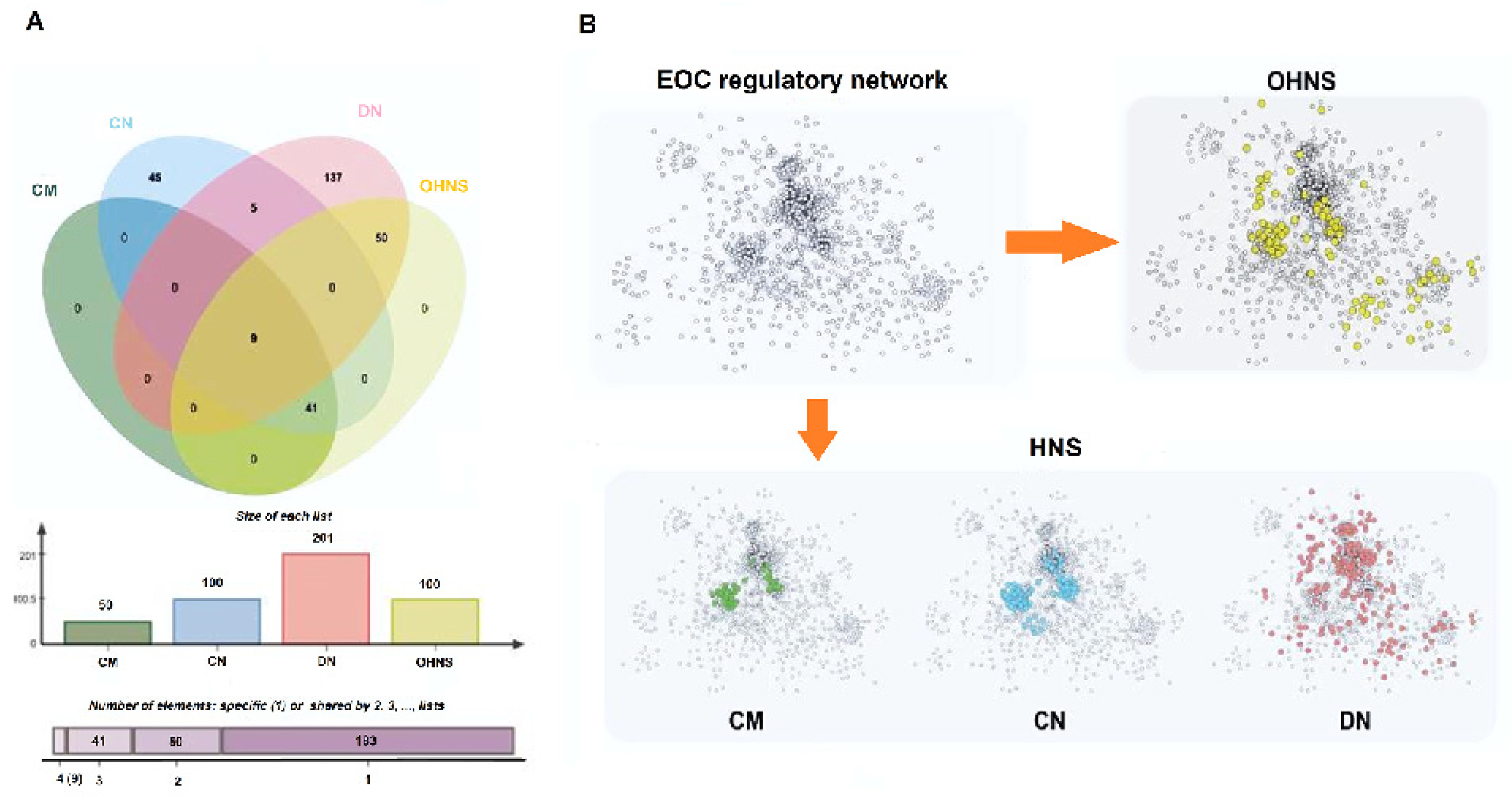
Figure 1. Distribution map of the HNS: (A) the Venn diagram represents the relationships among the HNS. Charts showing the list size, and the intersection size repartition are located underneath the diagram; (B) green represents the CM (core module), blue represents the CN (core nodes), pink represents the DN (driver nodes), and yellow represents the OHNS.
Biology 11 01851 g001
Figure 2. Network analysis of CM (core module), CN (core nodes), and DN (driver nodes): (A) the distribution probability of 4 levels of the HNS; (B) comparison of the changes in the characteristic path length after the nodes in CM, CN, and DN are deleted; (C) comparison of the degree difference values in CM, CN, and DN; (D) comparison of the changes in the giant component after the nodes in CM, CN, and DN are deleted. All data are represented as the mean ± SEM. ** p < 0.05.
Biology 11 01851 g002
Figure 3. Performance comparisons of CM (core module), CN (core nodes), DN (driver nodes), and the OHNS of the network: (A) the disturbance of CM, CN, DN, OHNS, and RN on the network; the smaller the giant component, the greater the disturbance of the network; (B) CM, CN, DN, OHNS, and RN are important in the network; the larger the characteristic path length, the more important.
Biology 11 01851 g003
Figure 4. Correlation and F-measure: (A) the F-measure of genes overlapping with CM (core module), CN (core nodes), DN (driver nodes), and the OHNS in the CTD list; (B) comparison of the overlapping genes with CM, CN, DN, and OHNS in the CTD list; (C) comparison of distributions of CM, CN, DN, and OHNS in biomarkers, pathway and hub genes, risk-prognostic genes, and perturbation effects. The width of the extended branch in the figure corresponds to the data flow size.
Biology 11 01851 g004
Figure 5. Enrichment analysis of the pathways related to CM (core module), CN (core nodes), DN (driver nodes); the visible output in GO terms is an interactive Manhattan plot that illustrates the enrichment analysis results: (A) this figure represents the result of a multiquery with CM; (B) this figure represents the result of a multiquery with CN; (C) this figure represents the result of a multiquery with DN. * and ** denoted that the biological pathways are significantly different at p < 0.05 and p < 0.01 levels, respectively.
Biology 11 01851 g005
Figure 6. Gene ontology was constructed by integrating WikiPathways, Reactome, and KEGG. Each node shows a pathway or term and the percentage of visible shared genes between pathways or terms and edges present relationships between pathways: (A) relationship between enriched pathways and genes in CM (core module); (B) relationship between enriched pathways and genes in CN (core nodes); (C) relationship between enriched pathways and genes in DN (driver nodes).
Biology 11 01851 g006
Figure 7. Spearman correlation analysis of hub genes in EOC and multiple normal tissues (AK). Rows represented “−Log10P”, and the vertical line in the middle of the graph indicates p = 0.05. Columns represent the correlation between hub genes and EOC, and “0” value axis indicates no correlation between hub genes and EOC. The upper part of the “0” value axis indicates a negative correlation, and the lower part indicates a positive correlation. Red shape represented positive correlation of the gene with different cancers. Green shape represented negative correlation of the gene with different cancers. Blue shape represented none correlation of the gene with different cancers.
Biology 11 01851 g007
Figure 8. GEO data validation results: (A) the distribution probability of 4 levels of the HNS; (B) compare the degree difference values in CM (core module), CN (core nodes), and DN (driver nodes); (C) comparison of the changes in the giant component after the nodes in CM, CN, and DN are deleted; (D) comparison of the changes in the characteristic path length after the nodes in CM, CN, and DN are deleted; (E) the F-measure of genes overlapping with CM, CN, and DN in the CTD list. All data are represented as the mean ± SEM. ** p < 0.05.
Biology 11 01851 g008
Figure 9. Dysregulated hub genes: (A) Western blotting showed alteration in the levels of MUC16, NDUFA, FOXA1, and COX15; (B) q-PCR assay was adopted to verify the changes of several hub genes; (C) IHC staining images of healthy samples and EOC samples and relative quantitative analysis in two groups; * p < 0.05, ** p < 0.01, *** p < 0.001, **** p < 0.0001.
Biology 11 01851 g009
Table 1. The correlation between the degree (out and in), the characteristic path length and the giant component in CM, CN, and DN.
CharacteristicCorrelationCM-OUTCM-INCN-OUTCN-INDN-OUTDN-IN
GCPearson correlation coefficient0.0450.1210.0650.1310.0240.084
Significance0.8010.3340.4990.1770.5620.271
LPearson correlation coefficient0.0350.2110.0410.1120.341 **0.503 **
Significance0.8210.0620.6940.0820.000.00
** Denoted by the different letters are significantly different at p < 0.01.
Publisher’s Note: MDPI stays neutral with regard to jurisdictional claims in published maps and institutional affiliations.