Article

Paramutation-like Epigenetic Conversion by piRNA at the Telomere of Drosophila virilis

1
Department of Ecology and Evolutionary Biology, University of Kansas, Lawrence, KS 66045, USA
2
Stowers Institute for Medical Research, Kansas City, MO 64110, USA
3
Divisions of Basic Sciences and Human Biology, Fred Hutchinson Cancer Research Center, Seattle, WA 98109, USA
4
Department of Molecular and Integrative Physiology, University of Kansas Medical Center, Kansas City, KS 66160, USA
5
Division of Genetic Medicine, Department of Pediatrics, University of Washington and Seattle Children’s Hospital, Seattle, WA 98195, USA
6
Department of Laboratory Medicine and Pathology, University of Washington, Seattle, WA 98195, USA
7
Department of Biological Sciences, Texas Tech University, Lubbock, TX 79409, USA
8
Engelhardt Institute of Molecular Biology, Russian Academy of Sciences, Moscow 119991, Russia
*
Author to whom correspondence should be addressed.
Biology 2022, 11(10), 1480; https://doi.org/10.3390/biology11101480
Submission received: 25 August 2022 / Revised: 27 September 2022 / Accepted: 4 October 2022 / Published: 9 October 2022
(This article belongs to the Special Issue Regulation of Mobile Genetic Elements at the Molecular Level)

Abstract

:

Simple Summary

Paramutation is an epigenetic phenomenon in which one allele triggers an epigenetic conversion of another allele. How paramutation occurs is poorly understood, but it frequently involves small RNAs that can silence an alternate allele in trans. In one well-known case in Drosophila, small RNAs known as piRNAs mediate paramutation. In this paper, we describe a novel system of epigenetic conversion in Drosophila virilis that demonstrates paramutation-like behavior. This occurs at the telomere, where retrotransposon arrays are known to be regulated by piRNAs. This paramutation-like behavior demonstrates that the unique properties of the Drosophila telomere may play a role in triggering epigenetic conversion in trans. This system promises to reveal new mechanisms that underlie paramutation-like behavior.

Abstract

First discovered in maize, paramutation is a phenomenon in which one allele can trigger an epigenetic conversion of an alternate allele. This conversion causes a genetically heterozygous individual to transmit alleles that are functionally the same, in apparent violation of Mendelian segregation. Studies over the past several decades have revealed a strong connection between mechanisms of genome defense against transposable elements by small RNA and the phenomenon of paramutation. For example, a system of paramutation in Drosophila melanogaster has been shown to be mediated by piRNAs, whose primary function is to silence transposable elements in the germline. In this paper, we characterize a second system of piRNA-mediated paramutation-like behavior at the telomere of Drosophila virilis. In Drosophila, telomeres are maintained by arrays of retrotransposons that are regulated by piRNAs. As a result, the telomere and sub-telomeric regions of the chromosome have unique regulatory and chromatin properties. Previous studies have shown that maternally deposited piRNAs derived from a sub-telomeric piRNA cluster can silence the sub-telomeric center divider gene of Drosophila virilis in trans. In this paper, we show that this silencing can also be maintained in the absence of the original silencing allele in a subsequent generation. The precise mechanism of this paramutation-like behavior may be explained by either the production of retrotransposon piRNAs that differ across strains or structural differences in the telomere. Altogether, these results show that the capacity for piRNAs to mediate paramutation in trans may depend on the local chromatin environment and proximity to the uniquely structured telomere regulated by piRNAs. This system promises to provide significant insights into the mechanisms of paramutation.

1. Introduction

Paramutation is a phenomenon in which one allele (termed paramutagenic) can impart an altered state of expression or function on another allele (termed paramutable) in the absence of alterations in the DNA sequence. One well characterized system of paramutation is found in maize where the B’ allele at the b1 locus has the capacity to silence the expression of purple pigmentation derived from the B-I allele [1,2]. After passing through B’/B-I heterozygotes, the original B-I allele becomes converted to a state designated B’*. This paramutated B’* allele can then in turn paramutate additional B-I alleles, converting them to a state that no longer produces purple pigment. This mode of communication and conversion between alleles is mediated by an RNA-dependent mechanism, tandem repeats of noncoding DNA, and the chromatin structure of these repeats [3,4,5]. In addition to maize, paramutation and paramutation-like phenomena are observed in other plants and a number of different animals, such as mice, flies, and nematodes [6]. In mice and nematodes, paramutation-like phenomena also appear mediated by the inheritance of RNA molecules [7,8,9] and, in flies, piwi-interacting RNAs (piRNAs) play a critical role in mediating this mode of epigenetic conversion [10]. Although RNA molecules, DNA methylation, and the state of the chromatin structure seem to be crucial players in the establishment and maintenance of paramutation, the question of how this trans effect is established still remains unclear. Understanding the role that each of these epigenetic modifications plays in systems of paramutation may help to elucidate the establishment of this transgenerational signal.
The primary function of piRNAs in Drosophila is to silence selfish transposable elements that proliferate within the genome. piRNAs are derived primarily from clusters of transposable elements, known as piRNA clusters, which have been modified at the chromatin level to become a source of transcripts designated for piRNA biogenesis. In the germline, dual-strand piRNA clusters become a source of sense and anti-sense piRNA, and anti-sense piRNAs can target TEs, through reverse complementarity, for either transcriptional, co-transcriptional, or post-transcriptional silencing [11,12,13].
A critical aspect of germline TE regulation in Drosophila depends on the maternal provisioning of piRNAs through the female germline. Syndromes of hybrid dysgenesis first revealed that maintenance of TE repression across generations required the maternal deposition of piRNA [14]. Subsequently, it has been shown that this maternal deposition is also required to trigger piRNA biogenesis at piRNA clusters, thus maintaining a continuous line of TE defense across generations [15,16,17]. Interestingly, this mode of epigenetic inheritance also plays a role in epigenetic gene silencing and paramutation through the off-target generation of genic piRNAs. In Drosophila melanogaster, a syndrome of paramutation has been identified that is triggered by an array of transgenes [10,18]. This transgene array producing piRNAs in the germline can also paramutate a related copy in trans and trigger piRNA biogenesis from a newly paramutated array. The newly paramutated array can in turn trigger piRNA biogenesis in trans to new non-paramutated transgene arrays and so on. This silencing mechanism has been observed to persist for up to 50 generations [10]
In the investigation of hybrid dysgenesis in Drosophila virilis, a related phenomenon has been described for a genic piRNA cluster residing near the telomere. In particular, one strain of D. virilis (the inducer strain, also known as strain 160) was noted to carry several sub-telomeric piRNA clusters that produced piRNAs from genes rather than transposons [19]. One of these sub-telomeric clusters, located on chromosome 2, contained the gene center divider (cdi), which had been silenced in the germline through conversion into a dual-strand piRNA cluster. Why the cdi gene has adopted piRNA cluster status is not known, though it should be noted that the strain carrying the cdi dual-strand cluster is also the strain that induces dysgenesis. This strain 160 is the only P-like strain in the D. virilis syndrome of dysgenesis and is noted by a large excess of TEs that are seemingly absent or in low abundance in other strains [19,20,21,22,23,24,25,26]. The polymorphic nature of this piRNA cluster (where the active piRNA cluster allele is designated cdiA and the inactive piRNA cluster allele is designated cdiI) was used to investigate the manner in which maternal piRNAs could trigger cluster piRNA biogenesis in the next generation [16]. This revealed that maternal deposition was critical for cdi cluster maintenance. Paternal transmission of the cdiA allele cluster led to weak production of cdi piRNAs in the germline, whereas maternal transmission maintained the robust expression of cdi piRNAs. Strikingly, the maternal transmission of the cdiA allele triggered piRNA production in trans from the paternally inherited non-cluster cdiI allele, though the production of piRNA in trans was weaker compared to that maintained in cis from the maternally transmitted allele. Nonetheless, the production of dual-strand piRNA biogenesis could also be maintained in the absence of the original cdiA allele in subsequent generations [27]. This is as expected from a system of paramutation.
In this paper, we further investigate this transgenerational epigenetic silencing of the sub-telomeric cdi piRNA cluster in Drosophila virilis. While previous studies of cdi showed the capacity for piRNA biogenesis in trans could be maintained independent of the cdiA piRNA allele, the production of functional polyadenylated cdi transcript has not been investigated. In addition, previous studies investigated neither the expression of cdi nor piRNA abundance in the first generation of progeny lacking the silencing allele. For this reason, it is not known how penetrant this silencing is in the first generation where the silencing allele has been segregated away. Nor is it even known whether the epigenetic conversion of the cdiI allele to a piRNA generating cluster is sufficient to lead to genic silencing. In this paper, we show that the silencing of the cdiI allele in trans can persist in females in the absence of the original cdiA allele piRNA cluster. Though we have yet to examine this persistence in subsequent generations and conversion of the paramutable cdiI allele into a paramutagenic state, this is consistent with bona fide paramutation that depends on the maternal deposition of piRNA. In contrast to the previously defined system of paramutation in Drosophila melanogaster [10], this does not depend on the presence of a transgene array. Moreover, an investigation of the telomere structure between the two strains with long reads reveals significant structural differences between the two alleles. This suggests that the sub-telomeric location itself may contribute to epigenetic conversion. Since piRNAs play a critical role in regulating retrotransposons in Drosophila, we propose that proximity to the telomere plays an important role in rendering a gene susceptible to epigenetic conversion by piRNA. This is supported by previous studies demonstrating that inducer strain 160, which carries the silent cdiA genic piRNA cluster, also carries a significantly greater abundance of piRNAs that target the telomeric TART retrotransposon [27].

2. Materials and Methods

2.1. Fly Stocks and Maintenance

Two Drosophila virilis strains of the dysgenic syndrome were used: Strain 160 (here indicated as cdiA strain with the active piRNA cluster) and Strain 9 (here indicated as cdiI with the inactive cluster) (Lozovskaya et al., 1990). All stocks and crosses were maintained on standard yeast food at 25 °C.

2.2. Cross Scheme

The general scheme was to cross cdiA strain females to cdiI males, collect F1 hybrid females, backcross them to cdiI males, and collect cdiI/cdiI backcross progeny. Backcross progeny were genotyped to identify cdiI/cdiI individuals for RT-qPCR based analysis of cdi expression in the ovary. Two experimental replicates were performed with nearly identical conditions, aside from an accidental incubator overheating event whereby Replicate A daughters were exposed to elevated heat for five to ten hours.

2.3. Replicate A

A total of 40 virgin cdiA strain females (0–3 days old) were selected, aged for one day, and each individually crossed to 2–3 cdiI strain males. Each of these was designated as a “grandmother replicate”. Vials were flipped two times, every three days, for a total of three broods from each grandmother. From these crosses, the ten most productive vials were selected, and from each of these vials, five to ten F1 virgin females were selected for backcrossing. Backcrosses with 2–3 cdiI strain males were performed with F1 virgin females collected 0–3 days after eclosion and aged for an additional 5 days. As before, vials were flipped twice, every three days. After laying, ovaries were removed and stored at −80 °C from the 11–14-day-old F1 females. This provided sets of sibling cohorts of five to ten F1 mothers from each grandmother replicate.
From each of these F1 females (five females per grandmother replicate), eight backcross progeny were selected and mated to cdiI strain males. The 11–14-day-old backcross progeny were then dissected. Ovaries were removed and stored at −80 °C and carcasses were retained for genotyping to identify cdiI/cdiI backcross genotypes. This scheme yielded five grandmother replicates, each of which yielded four F1 mothers. From each F1 mother, one cdiI/cdiI daughter was obtained. Due to a power outage and incubator overheating, these daughters experienced a period of elevated heat, estimated to have lasted between five and ten hours.

2.4. Replicate B

Crosses and collections were performed as in Replicate A with slight modifications. All females were mated at 0–3 days rather than being mated at 5–8 days. Brood vials were flipped twice every three to five days, instead of every three days. Ovaries from F1 mothers were collected 12–15 days post eclosion. Ovaries from backcross daughters were collected 12–16 days post eclosion. Finally, there was no period of elevated heat.

2.5. Daughter cdiI/cdiI Indel PCR Genotyping Assay and Sanger Sequencing Verification

Since it was essential to measure cdi expression in the daughters lacking the cdiA allele, only cdiI/cdiI daughters were selected. A 301 bp deletion was found in the cdiI strain only, ~73 kb away from the cdi locus. This was used to develop an indel PCR genotyping assay using daughter carcasses. The distinct amplicon sizes between alleles were easily distinguished from each other. Eight daughters were genotyped to identify cdiI homozygotes. Genotype verification was performed by sequencing identified individuals and confirming cdiI homozygosity with two SNPs inside the cdi locus.

2.6. Ovary RNA Extractions

Ovary RNA extractions were performed using the Zymo Research Direct-zolTM RNA miniprep kit (R2051). A total of 0.4 mL of the Zymo TRI ReagantTM was used to lyse the ovaries. All RNA extractions/purifications were carried out according to the Zymo Research Direct-zolTM RNA miniprep kit user manual. All ovary pairs were extracted individually and labeled according to their corresponding sample number. Upon extraction, all RNA samples were stored at −80 °C.

2.7. DNase Treatment

All samples were subjected to a DNase treatment (Thermo Fisher, Waltham, MA, USA, TURBO DNA-freeTM Kit (AM1907)) to ensure no DNA contamination was present in the RNA. A total of 10 μL of Turbo DNaseTM was mixed with 25 μL of 10× Turbo DNase BufferTM and added to samples, followed by incubation at 37 °C for 20 min, and DNase inactivation.

2.8. Reverse Transcription

All RNA samples were diluted to 100 ng/μL using RNase free water. The samples with an RNA concentration lower than 100 ng/μL were not diluted. RNA was converted to cDNA in half reactions using the Thermo Fisher SuperScript ® IV First-Strand cDNA synthesis reaction kit (11754050) with 1 μL of 50 μM Oligo d(T) (Thermo Fisher: N8080127). Oligo d(T) primer was used to select for functional polyadenylated mRNA transcripts. Reactions were carried out according to the ThermoFisher SuperScript® IV First-Strand cDNA synthesis reaction kit user manual (0.5 μL of Oligo d(T) primer, 0.5 μL of 10 mM dNTP mix, 1 μL of DNase free water, 2 μL of 5× SSIV Buffer, 0.5 μL of 100 nM DTT, 0.5 μL of RNaseOUTTM Recombinant RNase inhibitor, 0.5 μL of Superscript® IV Reverse Transcriptase enzyme (200 U/mL), and 2 μL of RNA template (200 ng)). If sample RNA concentration < 100 ng/μL, 5.5 μL of RNA template was added.

2.9. qPCR Analysis

The expression of cdi was measured using quantitative PCR. Primers were designed to amplify the mRNA transcripts of exon 5 in cdi. Rp49 was used as a control with primers designed to span an intron between exon 2 and 3. A melt curve analysis for Rp49 was conducted in order to identify any DNA contamination. No DNA contamination was identified. Primers were selected using the primer design guidelines provided in the PowerUPTM SYBRTM Green Master Mix User Guide along with Primer-BLAST. Primer sequences are as follows: cdi_qPCR_1_fwd: GTTTAGAGACTGTTGGTTCA; cdi_qPCR_1_rev: CAGCTTCGATGCGTGCGATA; Rp49_qPCR_1_fwd: TCCAGCATACAGGCCCAAGA; RP49_qPCR_1rev: ACGACGCACTCTGTTGTCAA.
The RT-qPCR analysis was conducted on an Applied Biosysems® QuantStudio® 3 Real-Time PCR System. The PowerUPTM SYBRTM Green Master Mix was used as the fluorescent dye. Three technical replicates were conducted on each set of experiments.

2.10. Statistical Analysis

Statistical analysis was performed in RStudio. In order to measure the cdi expression in the ovary samples, the critical threshold (Ct) value of the cdi samples was subtracted from the Ct value of the RP49 samples and taken to the power of 2, assuming 100% primer efficiency (i.e., 2(Rp49 CT-cdi CT)).

2.11. Nanopore Sequencing. TE Library and Telomere Annotation

A new D. virilis TE library was generated using RepeatModeler2 [28] on a PacBio assembly of cdiA strain 160 [29] and a nanopore assembly of cdiI strain 9 [22]. Additional nanopore reads were further obtained for the cdiA strain using the same approach as in [30]. In particular, DNA was extracted from a pool of whole adult females using the Qiagen Blood and Cell Culture DNA kit. Sequencing was performed using the Ligation Sequencing Kit (1D) on an Oxford Nanopore Minion and base-calling was performed with Albacore, retaining all reads. These two new assembly specific repeat libraries were further refined using RepeatAfterMe [31], permitting only repeats represented in 5 or more copies within a genome assembly. The consolidation of these two libraries, along with a previous one generated from the reference genome [27], was achieved using CD-hit [32]. The identification of single nanopore reads from the cdiA and cdiI strains was achieved by using cdi gene sequence in a BLAST query of all nanopore reads. All reads containing cdi and further distal sequence were collected and annotated with RepeatMasker using the final TE library. Visualization was performed using custom R scripts.

2.12. piRNA Sequences and Mapping

piRNA sequences were obtained from a previous study [27] and mapping was performed with BWA aln, randomly assigning multiple mappers [33]. Mapping was performed to whole genome assemblies from cdiA strain 160 [29] and cdiI strain 9 [22]. In the case of the cdiA strain, the artifictual telomeric TART array (with only a fragment remaining) was removed from the assembly image in Figures 2 and 3 based on being present in only one read among all individual PacBio and nanopore reads and that read most likely being a duplex read. For F1 progeny, mapping was performed on both assemblies combined. To normalize mapping against haploid and diploid assemblies, pure strain courts mapped to a haploid genome were divided by two.

2.13. Whole Mount In Situ RNA Hybridization

Ovary dissection, fixation, proteinase K treatment, re-fixation and hybridization steps were performed as described (PMID: 29813067). Matrices for probe preparation were prepared by PCR using cdi_F ATGTCGGAAACACTGCCACT and cdi_R GATAATACGACTCACTATAGGCAACTAACGATCCGATGC primers of genomic DNA of the cdiA strain. Labeling of RNA probes with DIG-11-UTP (Roche, Switzerland) was made by MAXIscript T7 kit (Ambion, Austin, TX, USA). Anti-DIG-AP antibodies (Roche, Basel, Switzerland) were used in 1:2000 dilution. Images obtained by binocular microscope Nikon Alphaphot-2 YS2 (Tokyo, Japan).

3. Results

3.1. Gene Silencing of cdi in the Ovary Is Maintained in the Absence of the Original Silencing cdiA Allele

In the system of D. virilis hybrid dysgenesis, one allele of cdi (designated cdiA) is an active sub-telomeric dual-strand genic piRNA cluster and an alternate allele (designated cdiI) is not. A previous study [27] showed that matrilineal backcross granddaughters of F1 female progeny carrying the cdiA allele with males carrying the cdiI allele could maintain piRNA biogenesis at the cdi locus in the absence of the original cdiA allele (Figure 1A). How this coincides with gene expression is not known. In this paper, we investigated the expression of the gene cdi in the absence of the original silent cdiA allele. This was achieved in two independent experiments (Figure 1B). In each experiment, five grandmothers homozygous (cdiA/cdiA) for the silent piRNA cluster cdi allele were individually crossed to males homozygous (cdiI/cdiI) for the non-silent allele. From each grandmother, four individual F1 female progeny were subsequently backcrossed to cdiI/cdiI males. After laying, ovaries from F1 females were collected for real-time qRT-PCR. Multiple backcross progeny were then selected per F1 female and ovaries were also collected. Backcross progeny carcasses were then genotyped using an indel PCR assay to identify cdiI/cdiI females and genotypes were confirmed using PCR and Sanger sequencing based on SNPs located within cdi that distinguish the two alleles. This genotyping allowed us to identify four mother–daughter pairs per each of the five grandmothers, each daughter having the cdiI/cdiI genotype. In total, this yielded 20 mother–daughter pairs per experiment, with cdiA/cdiI heterozygous mothers and each cdiI/cdiI daughter lacking the original piRNA cluster cdiA allele. In one experiment (Experiment A), due to equipment failure, these daughters experienced a period of elevated heat of approximately 30 degrees for between five to ten hours one day prior to collection. However, this had no apparent effect on results compared to Experiment B for which there was no period of elevated heat. The expression of cdi for all genotypes, as well as homozygous cdiA/cdiA and cdiI/cdiI controls subjected to the same regime, was evaluated using qRT-PCR on polyadenylated mRNA reverse transcribed with oligo-dT. Expression was measured relative to rp49 (Figure 1C).
To identify the determinants of cdi expression first we used a linear regression model (linear model: Daughter-relative cdi expression ~Plate + Control/Genotype). Control/Genotype included status as being pure strain cdiA/cdiA, pure strain cdiI/cdiI or cdiI/cdiI daughters (Supplemental Document S1). The model was tested on both experimental sets (Experiment A and B) together and separately. When both experimental sets were tested together, there was no significant effect by experiment (Experiment A vs. Experiment B, p = 0.2274), but the experiment was retained in the full model (linear model: Daughter-relative cdi expression ~ Experiment + Plate + Control/Genotype). In this model, daughters lacking the original cdiA piRNA cluster allele and homozygous for the cdiI/cdiI non-cluster allele showed significantly lower expression than pure strain cdiI/cdiI controls (p < 0.0001) (Figure 1C). This showed that the epigenetic conversion of cdi occurs in a manner similar to paramutation, though the analysis of further generations is required to determine if silencing could persist and if the newly silenced alleles themselves had the capacity to trigger epigenetic conversion and become paramutagenic. Despite this epigenetic conversion, the expression of cdi in cdiI/cdiI daughters was significantly (p < 0.0001) higher than in pure strain cdiA/cdiA females (Figure 1C). This indicates that, while epigenetic conversion is occurring, it is not fully penetrant.
Considering the experimental design, we were also able to evaluate several other predictors of cdi expression in cdiI/cdiI daughters. A model was used to determine if the cdi expression of the daughters was influenced by their corresponding grandmothers, or the grandmother ID. From every grandmother, we collected four progeny (designated mothers), and from each mother, we collected one cdiI/cdiI daughter. Thus, the mothers from each grandmother were sisters, and the subsequent daughters were cousins. We wanted to test if the variation of cdi expression among cousins could be explained by the grandmother from each cohort. In order to test this, a linear model (linear model: Daughter-relative cdi expression ~Experiment + Plate + grandmotherID) was used to test for significant differences across cousins that can be explained by the grandmother ID. Our findings initially showed significant differences in cdi expression between sets of cousins of different grandmothers (Supplemental Document S1). However, after performing a Tukey’s test to control for multiple comparisons, the adjusted p-values showed no significant differences between grandmother cohorts. Thus, the cdi expression variation across daughter cohorts is not apparently explained by a transgenerational grandmother effect (Supplemental Document S1).
Since this candidate system of paramutation is driven by a maternal effect, we wanted to test if the cdi expression in the ovaries of mothers predicted the cdi expression in their corresponding daughter ovaries, lacking the original silencing cdiA allele. The linear model used (linear model: Daughter-relative cdi expression ~Experiment + Plate + grandmotherID + mother relative cdi expression) showed no significant effect of mother cdi expression (p-value = 0.94). This finding suggests that the cdi expression of the daughters cannot be predicted by the cdi expression of the mothers. One possibility is that genetic variation among recombinant individuals may contribute to variation in cdi expression.
While cdiI/cdiI daughters lacked a copy of the cdiA allele, their mothers were heterozygous cdiA/cdiI. We sought to determine if this additional copy of the silent piRNA-cluster allele caused a lower expression of cdi in mothers. Using a paired t-test of mother–daughter pairs for relative cdi expression, we found a significantly higher level of expression in daughters compared to their heterozygous mothers (two-tailed p = 0.0367, avg Rp49 norm. expression Daughter: 0.0036, Mother: 0.0029). Thus, epigenetic conversion is not fully penetrant since it appears that daughters lacking the original cdiA allele have higher expression of cdi compared to their heterozygous mothers carrying the cdiA allele (Figure 1C).

3.2. Single-Read Nanopore Sequencing Analysis of the Subtelomeric Regions of cdiA and cdiA Strains

Many species of the Drosophila genus use telomeric retrotransposons to maintain chromosome ends [34,35,36] and the piRNA machinery regulates these telomeric retrotransposons [37,38,39,40,41]. In Drosophila, the region subterminal to the retrotransposon array is also composed of repetitive telomere-associated sequences (TAS). Recent genetic studies [42] indicate that telomere capping by HipHop plays a role in regulating retrotransposon expression near the telomeres, though apparently through HP1 rather than the piRNA pathway. To determine whether there are structural differences at the telomere that might explain the difference between the cdiI and cdiA alleles, we examined available assemblies and single long reads. A previous PacBio assembly of the strain carrying the cdiA allele [29] contains an array more than 27 kb in length composed of TART elements at the end of this chromosome. However, a manual inspection of individual PacBio reads carrying the cdi gene sequence showed that there was no single long read that contained such a TART array connected to cdi. Thus, this TART array appeared to be an artifact of the assembly. For this reason, we sought further clarification of the structure of the chromosome ends using nanopore sequencing. The number of reads per each sequencing run was similar (cdiI strain: 1.16 million reads; cdiA strain: 1.10 million reads) with similar length distributions (cdiI strain: 308 K reads > 10 kb; cdiA strain: 240 K reads > 10 kb). Reads were collected that contained significant BLAST hits to cdi as well as some additional distal sequence. These reads were then individually annotated with RepeatMasker with a custom D. virilis TE library (Supplemental Document S2). Even though there was a similar number of reads of similar length for each sequencing run, the results from the identified reads were quite different between the two strains (Note: There was a single read from the cdiA strain that was an apparent nanopore artifact that was removed. This read contained a ~40 kb array that was composed entirely of TART elements. Critically, they were arranged in a tandem inverted orientation with an essentially identical structure in the inverted direction. This is consistent with it being a duplex read and, since no other nanopore reads indicated this telomeric TART array, removal was warranted).
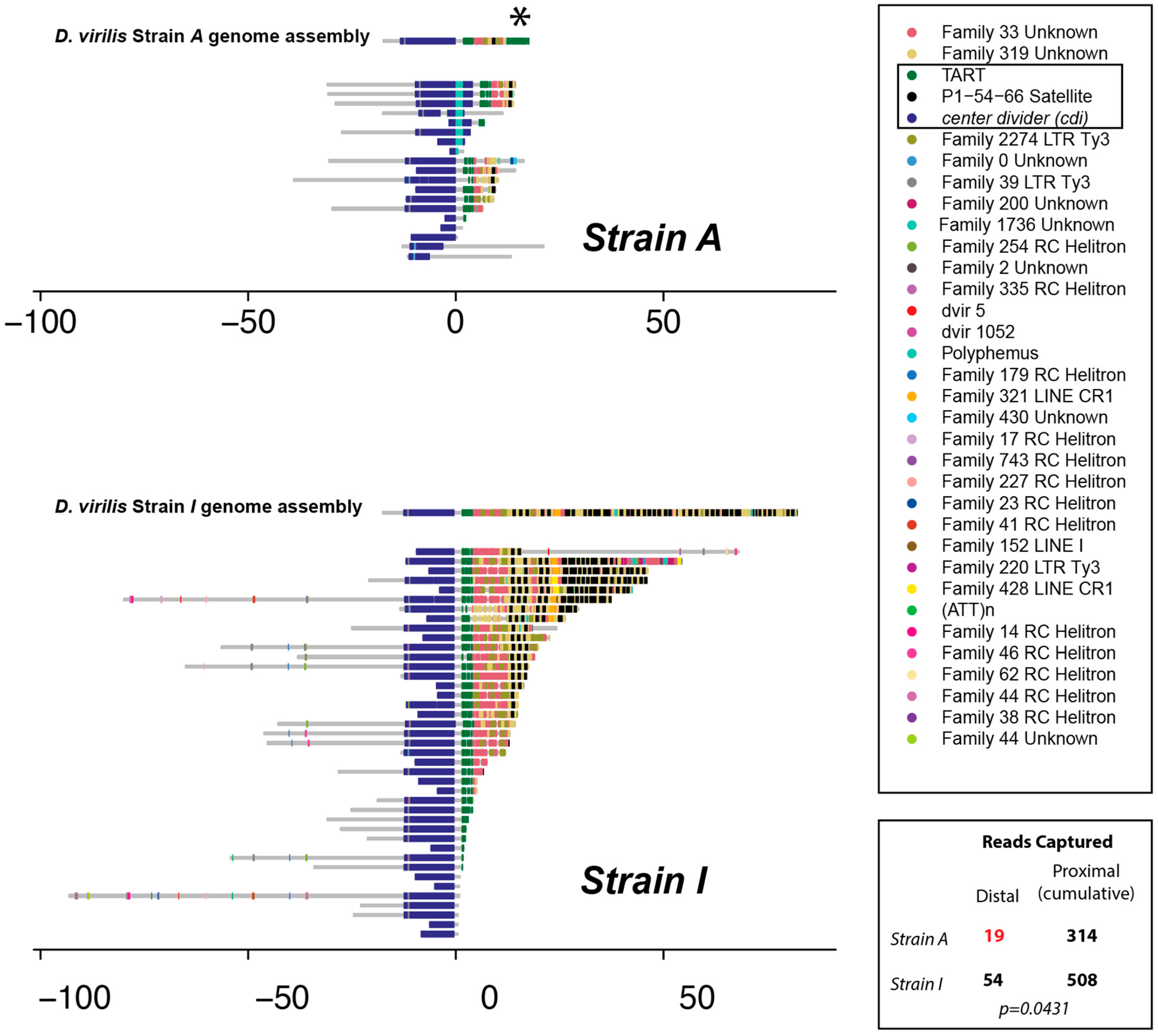
In particular, many fewer reads carrying cdi plus additional distal sequence were found from the cdiA strain (Figure 2), and no single read attached to cdi had a telomeric length greater than 20.7 kb outside of the removed read. In general, half as many reads are expected to cover the very terminal end of a chromosome compared to an internal region because an internal region will have sequence reads initiated from two flanking sides, whereas a terminal end will only be covered by reads initiated from one flanking side. We investigated this by counting the nanopore reads captured by BLAST using the distal most 500 bp non-repetitive fragment from the reference genome and comparing to the number of reads captured by nine additional 500 bp non-repetitive fragments approximately every 100 kb, extending proximal 1 Mb. There is a significant depletion of reads captured by BLAST from the distal end in the cdiA strain (Figure 2, two-tailed Fisher’s exact test, p = 0.0431). Thus, based on read coverage and read length, it appears that the chromosome carrying cdi in the cdiA strain is truncated just distally from cdi.
In addition, the inferred sequence consensus from the gathered reads at the terminus is different between the cdiA and the cdiI strains. Even though TART elements are known to be abundant at the telomeres, D. virilis differs from this standard arrangement. A previous study found D. virilis has satellite arrays at the very terminus rather than retrotransposons [43]. We found the same arrangement in nanopore reads from strain cdiI. In particular, the end of the cdiI chromosome is made of an assemblage of “54–66” satellite sequences, the same sequence identified previously [43]. These sequences appear not nearly as extensive in strain cdiA (Figure 2). Overall, nanopore sequencing indicates that the telomere end itself may be closer to cdiA, with fewer satellite sequences between cdi and the cap. Importantly, the analysis of strain-specific nanopore reads also revealed a TART fragment (green, Figure 2) just flanking cdi in both strains. We propose that this TART fragment just flanking cdi in both strains plays an important role in epigenetic silencing and conversion, due to differences in TART piRNA abundance between strains. While we lack formal evidence, one possibility is that the segregating Polyphemus or proximity of the cap to cdi capacitated the production of TART piRNAs and the conversion of cdi to a dual-strand cluster in the cdiA strain. This enhanced TART piRNA production may then mediate paramutation by targeting the TART fragment that is shared in both strains and is just flanking cdi.
Further investigation identified a segregating Polyphemus DNA transposon insertion inside cdi in the cdiA strain only (Figure 2, Upper Panel, Turquoise color), located within the sixth exon in the sense orientation. Though not in all reads from the cdiA strain, we sought to determine if this Polyphemus insertion might be important for the establishment of the genic cdi piRNA cluster and silencing of cdi. If critical, the insertion would have been present in heterozygous mothers since daughters show a signature of epigenetic conversion. Since the DNA of grandmothers and mothers was not collected, heterozygous first-generation backcross daughters were screened for the segregating Polyphemus insertion using PCR with primers flanking and within the insertion. This allowed the determination of the mother’s Polyphemus insertion status. In experiment A, 14 out of 19 heterozygous daughters screened had a Polyphemus insertion. One cohort (6_4) was excluded from this analysis due to the lack of a heterozygote daughter. In experiment B, 0 out of 17 heterozygote daughters screened had a Polyphemus insertion, and DNA from the remaining three cohorts (36_2, 36_5, and 39_5) was unavailable upon screening. Given that the insertion was missing from all heterozygous daughters tested in experiment B, it is unlikely that the Polyphemus insertion is responsible for the reduced expression of cdi, though the segregating insertion may have had a role in cluster establishment. We added inferred Polyphemus insertion status of the mother into the model and found no significant effect with respect to expression level of cdi (p = 0.891, Supplemental Document S1).

3.3. piRNA Mapping to Individual Parental Genome Assemblies of the Telomere

The previous investigation of the cdi piRNA cluster in D. virilis showed that maternal deposition of cdi piRNAs triggered cdi piRNA cluster activity in trans on the opposing allele within the ovary [16,27]. It was also shown that this piRNA cluster activity of cdi could be maintained in the absence of the original cdiA cluster allele in subsequent generations [27]. Given the differences in the telomeric structure, we mapped germline piRNA sequences [27] to the different assemblies obtained from the cdiI strain [21,22] and the cdiA strain [29] (Figure 3, excluding the apparent assembly artifact, with a residual distal TART fragment remaining and indicated by an asterisk). This was performed to compare how piRNAs differentially map to the structurally distinct chromosome ends and ensure that piRNA mappings based on a different reference assembly were accurate. We performed this using piRNAs sequenced from pure strains but also the reciprocal female progeny. In the case of reciprocal progeny, we combined two haploid assemblies for mapping. We normalized mapping density between parents (mapped to a haploid genome) and offspring (mapped to a diploid genome) by dividing mapping density by two for haploid mappings. As previously reported, significant differences in piRNA mapping can be seen directly within the cdi cluster, but, across the large satellite array, piRNA mapping does not reveal a difference. However, there is a significant difference in piRNA mapping density in the two TART fragments. One of these TART fragments (shown with an asterisk) is attributed to be an assembly artifact in the cdiA strain. However, another TART fragment that was found in nanopore reads in both strains just distal from cdi shows a distinct pattern of piRNA mapping between the two strains. In particular (as indicated with an arrow in Figure 3), there is abundant TART piRNA mapping to the strain with the cdiA allele, but these piRNAs are not abundant in the strain with the cdiI allele. This is consistent with the previous observation that TART piRNAs are vastly more abundant in inducer strain 160, which carries the cdiA allele [27]. By comparing strain specific assemblies, we confirmed that there is a TART piRNA target immediately next to cdi in both strains. We propose that excess TART piRNA, perhaps primed by a truncated satellite array, in the cdiA strain targets this shared TART element and contributes to the paramutation-like behavior at the telomere.

4. Discussion

In this paper, we extended prior studies of a genic piRNA cluster residing near a chromosome end in Drosophila virilis [16,19,27] and showed that this cluster has paramutagenic behavior with respect to gene expression. In particular, we showed that the females lacking the original silencing allele showed a low expression of cdi if their mothers carried the silent allele. Thus, with respect to cdi, such homozygous females lacking the silent piRNA cluster allele show reduced expression compared to genetically identical females whose mother lacked the silent allele. However, to prove that this is a complete system of paramutation, it is necessary to show that the converted allele also has the capacity to in turn paramutate another allele. In this paper, we only performed the analysis across two generations. In addition, to show that this is a system of paramutation as seen in plants, it would be important to show that paramutation can occur in both cross directions—where the silencing cluster allele is inherited both maternally and paternally. This is unlikely to be the case since the maternal provisioning of piRNA appears critical for the maintenance of the silent state.

4.1. Paramutation or a Maternal Effect?

Plants and animals seem to have different mechanisms of paramutation [6]. In plants, the capacity to trigger epigenetic conversion of an alternate allele is independent of parent-of-origin. However, this appears not to be the case in Drosophila. In another piRNA-mediated system of paramutation in Drosophila, paramutation is mediated by a maternal effect [10] and an allele only has the capacity for paramutation when present in the mother. A male that is heterozygous for the same epi-allele does not transmit a converted allele in the next generation. This reliance on the female germline for paramutation is explained by the fact that only the female germline provisions piRNAs. In this way, the phenomenon of paramutation is entangled in the concept of a maternal effect. Genetically, a maternal effect occurs when the genotype of the mother determines the trait of an offspring. However, while subsequent generations have not been evaluated for this system, the previously described system of paramutation in Drosophila does not rely strictly on a maternal effect. This is because, genetically, a paramutated allele can maintain its capacity to paramutate across more than one maternal generation. Since the original triggering allele can mediate its effect in the grandmother, the great grandmother, and so on, this is not strictly a maternal effect but a matrilineal effect. In some sense, one may also argue that this represents a form of transient imprinting since the female always transmits an inactive allele, though this mode of imprinting is lost once an allele is passed through the male germline for one generation.
When considering whether this mode of epigenetic conversion can be considered paramutation, one may also consider the timing of the establishment and maintenance of the epigenetic conversion in trans. Maternally provisioned piRNAs play a role in dual-strand cluster establishment [15,16,17] and this is the mechanism by which allelic conversion is maintained across generations. However, it is also clear that epigenetic conversion is occurring in the F1 heterozygous progeny of mothers that are homozygous. This is because studies have found, based on single nucleotide variants that distinguish the alleles [16,27], that piRNAs are derived from both alleles in the germline of F1 progeny. So, in some sense, the establishment of epigenetic conversion is initiated in the female germline and then maintained in the next generation through maternal piRNA deposition. It is unknown whether transient epigenetic conversion is also established in the heterozygous male germline, but it is conceivable that conversion is transiently established in the male germline but not maintained in the next generation due to paternal failure in piRNA transmission. This should be subject to future investigation.

4.2. What Drives Epigenetic Conversion of a Sub-Telomeric Genic piRNA Cluster?

While piRNAs are clearly important for the mode of epigenetic conversion of the sub-telomeric gene cdi, the properties of the locus and allele that enable this conversion are not clearly understood. In the study that originally identified the cdi piRNA cluster, several other genic piRNA clusters were identified near the telomeres of other chromosomes [19]. This suggests that some property of the telomeres in this strain might contribute to this phenomenon. In fact, we investigated a second cluster residing near another telomere, but the germline expression of both alleles was too low to fully evaluate. It is important to note that the sub-telomeric location of these genic piRNA clusters may be purely serendipitous. In this case, the property that confers this behavior may be independent of genomic position. Nonetheless, because telomeres often have unique genetic and epigenetic properties, we believe this association is unlikely to be a coincidence.
If in fact the sub-telomeric position is critical, what specific properties of the telomeres in this system mediate the conversion of cdi to a dual-strand cluster in the original cdiA strain and what property of the alternate telomere allele allows this conversion? Previous studies have shown [27] that there are both greater numbers of TART sequences as well as TART piRNAs in the germline of the cdiA strain. This may contribute, through some mechanism of spreading, to cdi cluster establishment in the cdiA strain. In this case, the maintenance of the silent state of cdi over multiple generations may depend on persisting elevated TART copy numbers and piRNA abundance. This could be tested in subsequent generations of backcrossing. If the capacity to maintain the silent state of cdi in recombinant progeny depended on global TART abundance or piRNA levels, then paramutation would not simply be the property of the original allele. Instead, it would rely on an epistatic interaction with variation dispersed through the genome. Though we cannot formally conclude it, we also showed that the telomeric repeat region appears shorter in the original cdiA strain. Thus, length and proximity to the capping proteins might be contributors. Under this model, only the genetic manipulation of the locus would allow one to distinguish whether simple physical distance or the repetitive sub-telomeric sequences themselves are critical.

Supplementary Materials

The following supporting information can be downloaded at: https://www.mdpi.com/article/10.3390/biology11101480/s1, Supplemental Document S1: Statistical Tables and Supplemental Document S2: TE Library.

Author Contributions

Conceptualization, A.P.D., S.Y.F., M.B.E. and J.P.B.; formal analysis, A.P.D., S.Y.F., M.B.E. and J.P.B.; visualization, A.P.D., M.D. and J.P.B.; investigation, A.P.D., S.Y.F., M.G.Z., M.B.E. and J.P.B.; data curation, S.C., D.A.R., C.M.S. and D.E.M.; writing—original draft preparation, A.P.D. and J.P.B.; writing—review and editing, A.P.D., S.C., S.Y.F., M.B.E. and J.P.B.; funding acquisition, S.Y.F., R.S.H., M.B.E. and J.P.B. All authors have read and agreed to the published version of the manuscript.

Funding

This project was supported by NSF MCB Awards 1413532 and 2025197 (PI: JPB), NIH-NIGMS/COBRE Award P20GM103638 (PI: Susan Lunte), NIH-NIGMS/KINBRE Award P20GM103418 (PI: Douglas Wright), the Stowers Institute for Medical Research and the University of Kansas. RNA in situ hybridization experiments were supported by Russian Science Foundation grant 22-74-10050.

Institutional Review Board Statement

Not applicable.

Informed Consent Statement

Not applicable.

Data Availability Statement

Nanopore sequence data are available in the SRA under BioProject Number PRJNA884378. Stowers Institute Original data underlying this manuscript can be accessed from the Stowers Original Data Repository at http://www.stowers.org/research/publications/libpb-1741.

Acknowledgments

Many thanks to members of the Blumenstiel lab for discussions. Thanks also to three anonymous reviewers for the comments and suggestions.

Conflicts of Interest

The authors declare no conflict of interest.

References

  1. Chandler, V.L.; Stam, M.; Sidorenko, L.V. 7 Long-distance Cis and Trans interactions mediate paramutation. Adv Genet. 2002, 46, 215–234. [Google Scholar] [CrossRef]
  2. Coe, E.H. A regular and continuing conversion-type phenomenon at the b locus in maize. Proc. Natl. Acad. Sci. USA 1959, 45, 828–832. [Google Scholar] [CrossRef] [Green Version]
  3. Belele, C.L.; Sidorenko, L.; Stam, M.; Bader, R.; Arteaga-Vazquez, M.A.; Chandler, V.L. Specific Tandem Repeats Are Sufficient for Paramutation-Induced Trans-Generational Silencing. PLoS Genet. 2013, 9, e1003773. [Google Scholar] [CrossRef] [PubMed] [Green Version]
  4. Alleman, M.; Sidorenko, L.; McGinnis, K.; Seshadri, V.; Dorweiler, J.E.; White, J.; Sikkink, K.; Chandler, V.L. An RNA-dependent RNA polymerase is required for paramutation in maize. Nature 2006, 442, 295–298. [Google Scholar] [CrossRef] [PubMed]
  5. Arteaga-Vazquez, M.; Sidorenko, L.; Rabanal, F.A.; Shrivistava, R.; Nobuta, K.; Green, P.J.; Meyers, B.C.; Chandler, V.L. RNA-mediated trans -communication can establish paramutation at the b1 locus in maize. Proc. Natl. Acad. Sci. USA 2010, 107, 12986–12991. [Google Scholar] [CrossRef] [PubMed] [Green Version]
  6. Hollick, J.B. Paramutation and related phenomena in diverse species. Nat. Rev. Genet. 2017, 18, 5–23. [Google Scholar] [CrossRef] [PubMed]
  7. Rassoulzadegan, M.; Grandjean, V.; Gounon, P.; Vincent, S.; Gillot, I.; Cuzin, F. RNA-mediated non-mendelian inheritance of an epigenetic change in the mouse. Nature 2006, 441, 469–474. [Google Scholar] [CrossRef] [PubMed] [Green Version]
  8. Minkina, O.; Hunter, C.P. Stable Heritable Germline Silencing Directs Somatic Silencing at an Endogenous Locus. Mol. Cell 2017, 65, 659–670.e5. [Google Scholar] [CrossRef] [Green Version]
  9. Sapetschnig, A.; Sarkies, P.; Lehrbach, N.J.; Miska, E.A. Tertiary siRNAs Mediate Paramutation in C. elegans. PLoS Genet. 2015, 11, e1005078. [Google Scholar] [CrossRef] [PubMed] [Green Version]
  10. de Vanssay, A.; Bouge, A.L.; Boivin, A.; Hermant, C.; Teysset, L.; Delmarre, V.; Antoniewski, C.; Ronsseray, S. Paramutation in Drosophila linked to emergence of a piRNA-producing locus. Nature 2012, 490, 112–115. [Google Scholar] [CrossRef] [PubMed]
  11. Sato, K.; Siomi, M.C. The piRNA pathway in Drosophila ovarian germ and somatic cells. Proc. Jpn. Acad. Ser. B 2020, 96, 32–42. [Google Scholar] [CrossRef] [Green Version]
  12. Czech, B.; Hannon, G.J. One Loop to Rule Them All: The Ping-Pong Cycle and piRNA-Guided Silencing. Trends Biochem. Sci. 2016, 41, 324–337. [Google Scholar] [CrossRef] [Green Version]
  13. Ozata, D.M.; Gainetdinov, I.; Zoch, A.; O’Carroll, D.; Zamore, P.D. PIWI-interacting RNAs: Small RNAs with big functions. Nat. Rev. Genet. 2019, 20, 89–108. [Google Scholar] [CrossRef] [Green Version]
  14. Brennecke, J.; Malone, C.D.; Aravin, A.A.; Sachidanandam, R.; Stark, A.; Hannon, G.J. An Epigenetic Role for Maternally Inherited piRNAs in Transposon Silencing. Science 2008, 322, 1387–1392. [Google Scholar] [CrossRef] [Green Version]
  15. Akkouche, A.; Mugat, B.; Barckmann, B.; Varela-Chavez, C.; Li, B.; Raffel, R.; Pélisson, A.; Chambeyron, S. Piwi Is Required during Drosophila Embryogenesis to License Dual-Strand piRNA Clusters for Transposon Repression in Adult Ovaries. Mol. Cell 2017, 66, 411–419.e4. [Google Scholar] [CrossRef]
  16. Le Thomas, A.; Marinov, G.K.; Aravin, A.A. A transgenerational process defines piRNA biogenesis in Drosophila virilis. Cell Rep. 2014, 8, 1617–1623. [Google Scholar] [CrossRef]
  17. Le Thomas, A.; Stuwe, E.; Li, S.; Du, J.; Marinov, G.; Rozhkov, N.; Chen, Y.-C.A.; Luo, Y.; Sachidanandam, R.; Toth, K.F.; et al. Transgenerationally inherited piRNAs trigger piRNA biogenesis by changing the chromatin of piRNA clusters and inducing precursor processing. Genes Dev. 2014, 28, 1667–1680. [Google Scholar] [CrossRef] [Green Version]
  18. Ronsseray, S.; Boivin, A.; Anxolabehere, D. P-element repression in Drosophila melanogaster by variegating clusters of P-lacZ-white transgenes. Genetics 2001, 159, 1631–1642. [Google Scholar] [CrossRef]
  19. Rozhkov, N.V.; Aravin, A.A.; Zelentsova, E.S.; Schostak, N.G.; Sachidanandam, R.; McCombie, W.R.; Hannon, G.J.; Evgen’Ev, M.B. Small RNA-based silencing strategies for transposons in the process of invading Drosophila species. RNA 2010, 16, 1634–1645. [Google Scholar] [CrossRef] [Green Version]
  20. Evgen’ev, M.B.; Zelentsova, H.; Shostak, N.; Kozitsina, M.; Barskyi, V.; Lankenau, D.H.; Corces, V.G. Penelope, a new family of transposable elements and its possible role in hybrid dysgenesis in Drosophila virilis. Proc. Natl. Acad. Sci. USA 1997, 94, 196–201. [Google Scholar] [CrossRef]
  21. Funikov, S.Y.; Kulikova, D.A.; Krasnov, G.S.; Rezvykh, A.P.; Chuvakova, L.N.; Shostak, N.G.; Zelentsova, E.S.; Blumenstiel, J.P.; Evgen’Ev, M.B. Spontaneous gain of susceptibility suggests a novel mechanism of resistance to hybrid dysgenesis in Drosophila virilis. PLoS Genet. 2018, 14, e1007400. [Google Scholar] [CrossRef]
  22. Rezvykh, A.; Funikov, S.; Protsenko, L.; Kulikova, D.; Zelentsova, E.; Chuvakova, L.; Blumenstiel, J.; Evgen’Ev, M. Evolutionary Dynamics of the Pericentromeric Heterochromatin in Drosophila virilis and Related Species. Genes 2021, 12, 175. [Google Scholar] [CrossRef]
  23. Lyozin, G.T.; Makarova, K.S.; Velikodvorskaja, V.V.; Zelentsova, H.S.; Khechumian, R.R.; Kidwell, M.G.; Koonin, E.V.; Evgen’ev, M.B. The structure and evolution of Penelope in the virilis species group of Drosophila: An ancient lineage of retroelements. J. Mol. Evol. 2001, 52, 445–456. [Google Scholar] [CrossRef]
  24. Petrov, D.A.; Schutzman, J.L.; Hartl, D.L.; Lozovskaya, E.R. Diverse transposable elements are mobilized in hybrid dysgenesis in Drosophila virilis. Proc. Natl. Acad. Sci. USA 1995, 92, 8050–8054. [Google Scholar] [CrossRef] [Green Version]
  25. Vieira, J.; Vieira, C.P.; Hartl, D.L.; Lozovskaya, E.R. Factors contributing to the hybrid dysgenesis syndrome in Drosophila virilis. Genet. Res. 1998, 71, 109–117. [Google Scholar] [CrossRef]
  26. Blumenstiel, J.P. Whole genome sequencing in Drosophila virilis identifies Polyphemus, a recently activated Tc1-like transposon with a possible role in hybrid dysgenesis. Mob. DNA 2014, 5, 6. [Google Scholar] [CrossRef] [Green Version]
  27. Erwin, A.A.; Galdos, M.A.; Wickersheim, M.L.; Harrison, C.C.; Marr, K.D.; Colicchio, J.M.; Blumenstiel, J.P. piRNAs Are Associated with Diverse Transgenerational Effects on Gene and Transposon Expression in a Hybrid Dysgenic Syndrome of D. virilis. PLoS Genet. 2015, 11, e1005332. [Google Scholar] [CrossRef] [Green Version]
  28. Flynn, J.M.; Hubley, R.; Goubert, C.; Rosen, J.; Clark, A.G.; Feschotte, C.; Smit, A.F. RepeatModeler2 for automated genomic discovery of transposable element families. Proc. Natl. Acad. Sci. USA 2020, 117, 9451–9457. [Google Scholar] [CrossRef]
  29. Hemmer, L.W.; Dias, G.B.; Smith, B.; Van Vaerenberghe, K.; Howard, A.; Bergman, C.; Blumenstiel, J.P. Hybrid dysgenesis in Drosophila virilis results in clusters of mitotic recombination and loss-of-heterozygosity but leaves meiotic recombination unaltered. Mob. DNA 2020, 11, 10. [Google Scholar] [CrossRef] [Green Version]
  30. Miller, D.E.; Staber, C.; Zeitlinger, J.; Hawley, R.S. Highly Contiguous Genome Assemblies of 15 Drosophila Species Generated Using Nanopore Sequencing. G3 Genes|Genomes|Genetics 2018, 8, 3131–3141. [Google Scholar] [CrossRef]
  31. Hubley, R. Dfam-consortium/RepeatAfterMe: RepeatAfterMe_V0.0.4. 2022. Available online: https://doi.org/10.5281/zenodo.7076442.
  32. Fu, L.; Niu, B.; Zhu, Z.; Wu, S.; Li, W. CD-HIT: Accelerated for clustering the next-generation sequencing data. Bioinformatics 2012, 28, 3150–3152. [Google Scholar] [CrossRef]
  33. Li, H.; Durbin, R. Fast and accurate short read alignment with Burrows—Wheeler transform. Bioinformatics 2009, 25, 1754–1760. [Google Scholar] [CrossRef] [Green Version]
  34. Biessmann, H.; Mason, J.M.; Ferry, K.; D’Hulst, M.; Valgeirsdottir, K.; Traverse, K.L.; Pardue, M.-L. Addition of telomere-associated HeT DNA sequences “heals” broken chromosome ends in Drosophila. Cell 1990, 61, 663–673. [Google Scholar] [CrossRef]
  35. Casacuberta, E.; Pardue, M.-L. Transposon telomeres are widely distributed in the Drosophila genus: TART elements in the virilis group. Proc. Natl. Acad. Sci. USA 2003, 100, 3363–3368. [Google Scholar] [CrossRef] [Green Version]
  36. Pardue, M.-L.; DeBaryshe, P. Retrotransposons Provide an Evolutionarily Robust Non-Telomerase Mechanism to Maintain Telomeres. Annu. Rev. Genet. 2003, 37, 485–511. [Google Scholar] [CrossRef]
  37. Khurana, J.S.; Xu, J.; Weng, Z.; Theurkauf, W.E. Distinct Functions for the Drosophila piRNA Pathway in Genome Maintenance and Telomere Protection. PLoS Genet. 2010, 6, e1001246. [Google Scholar] [CrossRef] [Green Version]
  38. Savitsky, M.; Kwon, D.; Georgiev, P.; Kalmykova, A.; Gvozdev, V. Telomere elongation is under the control of the RNAi-based mechanism in the Drosophila germline. Genes Dev. 2006, 20, 345–354. [Google Scholar] [CrossRef] [Green Version]
  39. Shpiz, S.; Kalmykova, A. Role of piRNAs in the Drosophila telomere homeostasis. Mob. Genet. Elements 2011, 1, 274–309. [Google Scholar] [CrossRef] [Green Version]
  40. Radion, E.; Morgunova, V.; Ryazansky, S.; Akulenko, N.; Lavrov, S.; Abramov, Y.; Komarov, P.A.; Glukhov, S.I.; Olovnikov, I.; Kalmykova, A. Key role of piRNAs in telomeric chromatin maintenance and telomere nuclear positioning in Drosophila germline. Epigenetics Chromatin 2018, 11, 40. [Google Scholar] [CrossRef] [Green Version]
  41. Shpiz, S.; Olovnikov, I.; Sergeeva, A.; Lavrov, S.; Abramov, Y.; Savitsky, M.; Kalmykova, A. Mechanism of the piRNA-mediated silencing of Drosophila telomeric retrotransposons. Nucleic Acids Res. 2011, 39, 8703–8711. [Google Scholar] [CrossRef]
  42. Cui, M.; Bai, Y.; Li, K.; Rong, Y.S. Taming active transposons at Drosophila telomeres: The interconnection between HipHop’s roles in capping and transcriptional silencing. PLoS Genet. 2021, 17, e1009925. [Google Scholar] [CrossRef] [PubMed]
  43. Biessmann, H.; Zurovcova, M.; Yao, J.G.; Lozovskaya, E.; Walter, M.F. A telomeric satellite in Drosophila virilis and its sibling species. Chromosoma 2000, 109, 372–380. [Google Scholar] [CrossRef]
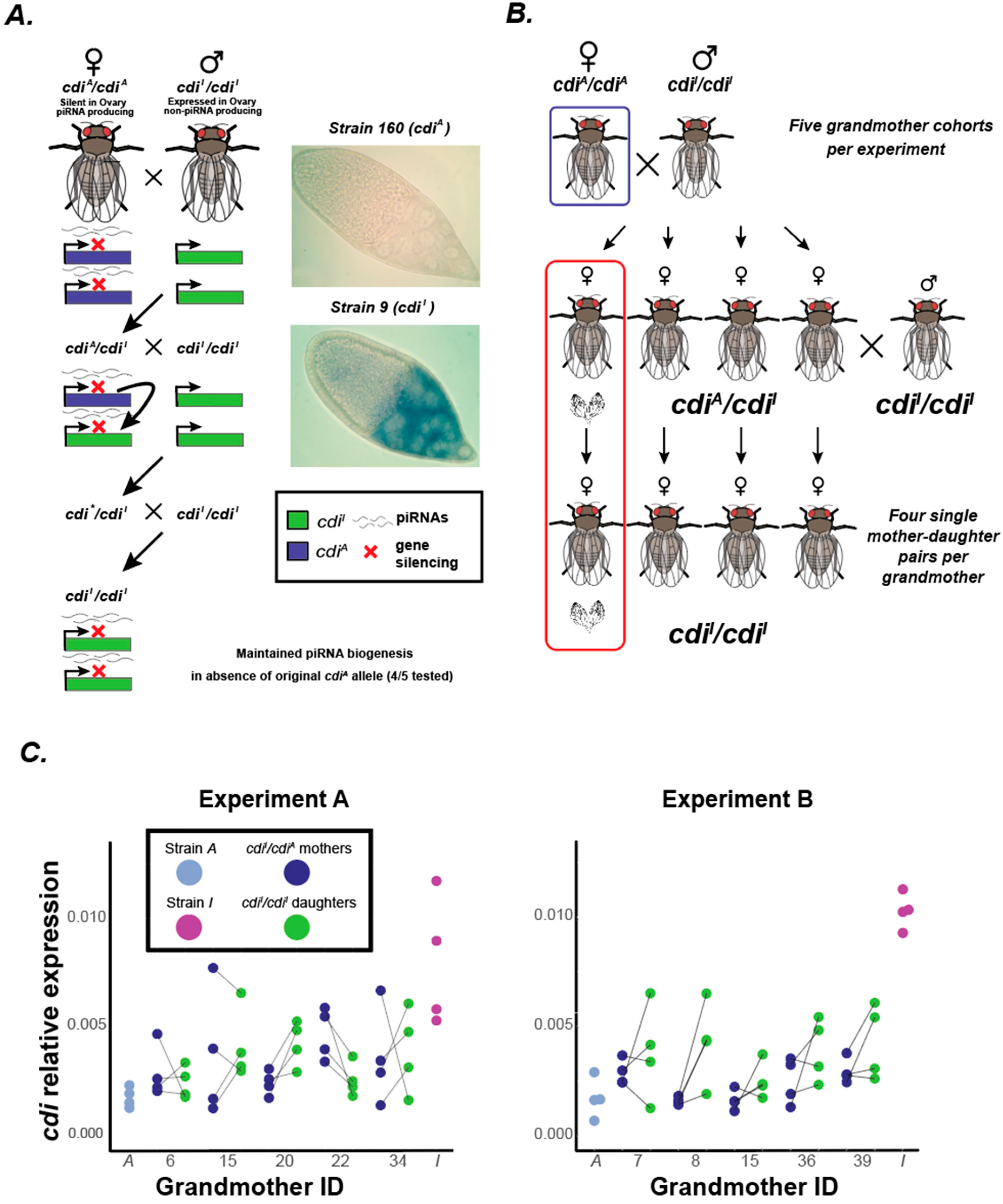
Figure 1. Experimental design and cdi expression. (A) The system of piRNA-mediated silencing of cdi by conversion to a dual-strand piRNA cluster. The cdiA allele is silenced and produces piRNAs from both strands. Results from previous studies indicate that a maternally inherited cdiA allele has the capacity to induce piRNA biogenesis from a non-silenced allele in trans. In addition, in subsequent generations, genic piRNA biogenesis can be maintained from cdi in the absence of the original triggering allele. In situ experiments show the expression of cdi in the egg chamber of strain 9 (which carries the expressed cdiI allele) and no expression in strain 160 (which carries the silent cdiA allele). (B) Experimental design for a single cohort derived from one of five grandmothers per experiment. (C) Low expression of cdi is maintained in the absence of the original triggering allele. Shown are relative expression values for cdi, normalized to Rp49. Each experiment presents the results of five cohorts, each daughter within a cohort being a cousin and uniquely derived from a single mother. Values presented are averages of three technical replicates per individual.
Biology 11 01480 g001
Figure 2. Repeat annotation of individual nanopore reads from Strain A and Strain I. The cdi gene sequence was used as a BLAST query for nanopore reads and reads carrying cdi along with further distal sequence were annotated with the new TE library and RepeatMasker. One read, a duplex read, was excluded from Strain A. Note the turquoise insertion of Polyphemus in some reads of Strain A. * indicates a fragment of TART that is likely an assembly artifact, but shown for completeness. Bottom right indicates results from nanopore read BLAST capture analysis. There is significant depletion of distal read coverage in the cdiA strain. This indicates a shorter telomere array.
Biology 11 01480 g002
Figure 3. piRNA mappings to unique assemblies. Denoted in the top section are assembly repeat regions of cdi and distal sequence. * indicates a fragment of the TART element that is a putative assembly artifact, but shown for completeness. piRNA mappings—from within a strain and also across to the other strain—are shown for pure parental strains in top panel, with a closeup on cdi in smaller panels, upper left of each section. The arrow indicates piRNA mapping to the TART fragment just immediately flanking cdi. In F1 progeny, the layout is the same as above for parental strains, but piRNAs were mapped to both assemblies at the same time. Despite a different structure of the distal repetitive sequences, there is not abundant piRNA mapping to this satellite array. This long satellite array may function as a buffer from the piRNA-mediated regulation of the telomere end.
Biology 11 01480 g003
Publisher’s Note: MDPI stays neutral with regard to jurisdictional claims in published maps and institutional affiliations.