Article

Systematic Studies of the Circadian Clock Genes Impact on Temperature Compensation and Cell Proliferation Using CRISPR Tools

1
Department of Health Sciences, Institutes of Physical Science and Information Technology, Anhui University, Hefei 230601, China
2
Moeden Experiment Technology Center, Institutes of Physical Science and Information Technology, Anhui University, Hefei 230601, China
*
Authors to whom correspondence should be addressed.
These authors contributed equally.
Biology 2021, 10(11), 1204; https://doi.org/10.3390/biology10111204
Submission received: 12 October 2021 / Revised: 12 November 2021 / Accepted: 15 November 2021 / Published: 18 November 2021
(This article belongs to the Section Cell Biology)

Abstract

:

Simple Summary

One of the major characteristics of the circadian clock is temperature compensation, and previous studies suggested a single clock gene may determine the temperature compensation. In this study, we report the first full collection of clock gene knockout cell lines using CRISPR/Cas9 tools. Our full collections indicate that the temperature compensation is a complex gene regulation system instead of being regulated by any single gene. Besides, we systematically compared the proliferation rates and circadian periods using our full collections, and we found that the cell growth rate is not dependent on the circadian period. Therefore, complex interaction between clock genes and their protein products may underlie the mechanism of temperature compensation, which needs further investigations.

Abstract

Mammalian circadian genes are capable of producing a self-sustained, autonomous oscillation whose period is around 24 h. One of the major characteristics of the circadian clock is temperature compensation. However, the mechanism underlying temperature compensation remains elusive. Previous studies indicate that a single clock gene may determine the temperature compensation in several model organisms. In order to understand the influence of each individual clock gene on the temperature compensation, twenty-three well-known mammalian clock genes plus Timeless and Myc genes were knocked out individually, using a powerful gene-editing tool, CRISPR/Cas9. First, Bmal1, Cry1, and Cry2 were knocked out as examples to verify that deleting genes by CRISPR is effective and precise. Cell lines targeting twenty-two genes were successfully edited in mouse fibroblast NIH3T3 cells, and off-target analysis indicated these genes were correctly knocked out. Through measuring the luciferase reporters, the circadian periods of each cell line were recorded under two different temperatures, 32.5 °C and 37 °C. The temperature compensation coefficient Q10 was subsequently calculated for each cell line. Estimations of the Q10 of these cell lines showed that none of the individual cell lines can adversely affect the temperature compensation. Cells with a longer period at lower temperature tend to have a shorter period at higher temperature, while cells with a shorter period at lower temperature tend to be longer at higher temperature. Thus, the temperature compensation is a fundamental property to keep cellular homeostasis. We further conclude that the temperature compensation is a complex gene regulation system instead of being regulated by any single gene. We also estimated the proliferation rates of these cell lines. After systematically comparing the proliferation rates and circadian periods, we found that the cell growth rate is not dependent on the circadian period.

1. Introduction

Circadian rhythms play important roles in maintaining homeostasis in mammals. The circadian clock system is a hierarchical system that incorporates external environmental cues and internal autonomous rhythms [1,2]. The circadian rhythms widely exist in almost every organ and cell in mammals. The suprachiasmatic nucleus (SCN) of hypothalamus, as the central pacemaker, regulates the biological rhythm of peripheral organs and synchronizes the peripheral clocks through complex signal cascade responses [3,4]. This timing system is very important for normal physiology and behavior. One of the major characteristics of the circadian clock is that it is temperature compensated [5]. In order for organisms to adapt to changes in the external environment to maintain normal physiological functions, changes in temperature within a certain range will not produce much change in the circadian rhythm of the organism, which is called temperature compensation [6]. Having a temperature-compensated circadian clock brings many benefits to animals, even to endothermic animals. However, the mechanism underlying temperature compensation remains elusive.
Q10 is used to represent the temperature dependence of a biochemical reaction process. The range of Q10 in circadian systems is around 0.8–1.2 [7], which indicates the system is temperature compensated. A number of mutation studies have investigated the molecular basis of temperature compensation in model organisms. Both Frq mutants in Neurospora and Per mutants in Drosophila have been shown to largely compromise temperature compensation [8,9], indicating that a single clock gene may directly determine the properties of temperature compensation. While in mammals, the Tau mutation in hamster (mutations in Csnk1e gene that encodes CK1ε) adversely affects the temperature compensation with Q10 value 1.487, compared with Q10 1.096 in wild types [10]. Furthermore, in vitro biochemical phosphorylation activity of CK1δ and CK1ε are temperature-insensitive [5,11,12], indicating temperature compensation may be a property derived from biochemical reactions by clock kinases CK1δ and CK1ε. For the simplest model organism cyanobacteria, the temperature compensation is determined by the ATPase activity of its key clock protein KaiC [13,14]. These studies indicate that the temperature compensation may be affected by a single clock gene and/or its protein products.
Almost every cell in our body has circadian oscillators [15]. The regulatory feedback loop of biological rhythm includes positive regulatory elements and negative regulatory elements. A set of core clock genes constitute a highly conserved cell-autonomous transcription-translation feedback loop (TTFL) [16], including core regulatory factors Bmal1, Clock (its homologous gene Npas2), Per1, Per2, Per3, Cry1, Cry2 [17] and auxiliary factors Rora, Rorb, Rorc, Dbp, Nfil3, Ciart, Csnk1d, Csnk1e, Dec1, Dec2, Beta-trcp, Fbxl3, Fbxl21, Timeless, Myc, etc [18]. The basic helix-loop-helix domains (bHLH) of BMAL1 and CLOCK combine with each other to bind DNA [19,20], and both proteins contain the PAS (PER-ARNT-SIM) domain, which interacts to form an asymmetric heterodimer [21]. BMAL1/CLOCK binds to the E-box enhancer and activates the expression of per (Per1, Per2, Per3) and Cry (Cry1, Cry2) genes [22,23,24]. When PER and CRY in the cytoplasm continue to accumulate to a certain amount [25,26,27], both proteins are phosphorylated by casein kinase family CK1ε (encoded by Csnk1e) and CK1δ (encoded by Csnk1d) [28,29,30]. Phosphorylated PER and CRY form heterodimers and enter the nucleus, disengaging BMAL1/CLOCK from the E-box to inhibit BMAL1/CLOCK [31]. On the other hand, phosphorylated PER and CRY are ubiquitinated through E3 ligase β-TRCP (encoded by Beta-trcp) and FBXL3/FBXL21 (encoded by Fbxl3/Fbxl21) [32,33,34] and degraded through the ubiquitin-mediated pathway. In the auxiliary circuits, REV-ERBs and RORs encoded by Nr1d1, Nr1d2, and Rora, Rorb, and Rorc are activated when the BMAL1/CLOCK heterodimer binds to the upstream region [35,36]. REV-ERBs negatively and RORs positively regulate the expression of the Bmal1 gene respectively [37]. In addition, DBP and NFIL3 act as activators and inhibitors, respectively, involved in regulating the transcription of D-box genes in gene promoters, such as Per3 [38,39]. These interrelated and mutually regulated loops mediated by cis-acting elements such as E-box, RORE and D-box regions form a complex clock network. Furthermore, NPAS2 can bind to BMAL1 to form heterodimer, which also regulates the expression of clock proteins PER and CRY [40]. DEC1 (encoded by Dec1) and DEC2 (encoded by Dec2), as members of the bHLH protein family, mainly form homodimers (or heterodimers) and bind to the E-box region of the target gene promoter, thereby inhibiting gene transcription [41]. Recently, it has been discovered that CHRONO translated by Ciart is functionally familiar with CRY protein [42], interacts with core proteins BMAL1 and CLOCK, and plays a transcriptional inhibitory role in the negative feedback loop of transcription and translation. Timeless is a gene found in Drosophila that can regulate biological rhythm [43]. In mouse experiments, knockout of Timeless can lead to embryonic death in mice. There are currently few studies on the mammalian Timeless gene in the field of chronobiology. The Myc gene, as a new member involved in the regulation of circadian rhythms that has been studied in recent years, can down-regulate the expression of the clock-controlled gene Bmal1 by activating the inhibition of REV-ERBs [44].
We wonder if there is a single gene in the mammalian circadian system that could determine the temperature compensation. We want to test this idea systematically. Thus, we take advantage of a powerful genetic tool, CRISPR, to knock out each individual gene that was identified as a component of the mammalian circadian clockwork. CRISPR (Clustered Regularly Interspaced Short Palindromic Repeats) is a microbial nuclease system that participates in the defense against phage and plasmid invasions [45,46]. Many types of CRISPR systems have been identified in a variety of bacteria and archaea hosts. The CRISPR/cas9 system is one of the most efficient gene-editing systems, and CRISPR/Cas9 is reconstructed in mammalian cells by optimizing the functions of endonuclease Cas9 and the essential RNA components in mammalian cells [46,47]. In addition, crRNA and tracrRNA can be fused together to produce a chimeric single guide RNA (sgRNA) [46], and Cas9 can be specifically directed to the PAM sequence through the 20-nt guide sequence in sgRNA. In view of its ease of implementation, multiplexing and other advantages, Cas9 has been used to edit eukaryotic cells and make specific mutations through NHEJ and HDR.
In this study, we successfully knocked out each clock gene in NIH3T3 fibroblast cells [48,49]. We performed multiple Lumicycle reporter experiments on cell lines with single gene editing at 32.5 °C and 37 °C to obtain data on the circadian period, and calculated Q10 values for each cell line. We found that except for Beta-trcp-D11, the Q10 values of other cell lines were within a stable and normal range between 0.8 and 1.2, which indicates a complex regulatory network of temperature compensation in mammalian cells. Since temperature compensation is a basic characteristic of the biological clock and cellular homeostasis, the removal of a single clock gene can hardly affect it. Previous studies had controversial conclusions that whether the circadian cycles and the cell cycles are strongly coupled. Yeom et al. reported that the circadian system does not regulate the cell cycle rhythm in rat-1 fibroblasts [50], while Bieler et al. showed that circadian cycles and cell cycles are mutually coupled [51]. Thus, in order to understand the correlation between cell proliferation and the circadian period, we further investigated whether loss of individual clock gene would affect the cell proliferation in NIH3T3 cells. We found that the absence of clock genes such as Clock and Cry significantly prolongs cell proliferation. However, the cell growth rate is not dependent on the circadian period.

2. Materials and Methods

2.1. Cell Culture

NIH3T3-B1-B10 is an NIH3T3 cell line constructed in our laboratory with fusion expression of Bmal1 promoter and the firefly luciferase. NIH3T3-B1-B10 cells were maintained in a complete medium (Dulbecco’s Modified Eagle Medium supplemented with 10% fetal bovine serum, and 100 U/mL penicillin-streptomycin, HyClone, Logan, UT, USA) at 37 °C (5% CO2). The NIH3T3 cell line was provided by Dr. Xu at Anhui University.

2.2. Single Guide RNA Design

After selecting the gene to be knocked out, the exon sequence of the corresponding gene was found on the NCBI website, and the sgRNA design website of Zhang Feng’s laboratory (http://crispr.mit.edu (accessed on 10 October 2018)) was used to design the sgRNA. Then, the sgRNA sequence with the highest score and the lowest off-target rate was chosen (Table S1). These sgRNAs were not chosen due to any specific domain of each gene. The sgRNA sequence and complementary strand were designed with BbsI restriction site sticky ends and sent to company for synthesis (General Biol, Anhui, China).

2.3. Construction of Vectors and Cell Lines Screening

The single-stranded sgRNA sequences designed for 25 candidate genes were annealed, phosphorylated and ligated into the pX459 vector (pSpCas9{BB}-2A-Puro was a gift from Feng Zhang, Addgene plasmid No. 48139) which was digested by BbsI enzyme. The constructed plasmid of each individual gene was transfected to NIH3T3-B1-B10 cells according to the instructions of Lipo6000 (#C0529, Beyotime, Shanghai, China). Twenty-four hours after transfection, cells were screened with 2 μg/mL puromycin. Next, monoclonal cell lines were screened by the limiting dilution method.

2.4. Identification of Knock-Out Cell Lines

After the selected monoclonal cell line growing to cell populations, we used the genome extraction kit (Tiangen, Beijing, China) to extract their genomes. We also designed the primers (Table S2) to locate ~800 bp upstream and downstream of the target editing site to amplify the target fragment. According to the instructions of T7E1 endonuclease kit (#E001, Viewsolid, Beijing, China), the amplified target fragments were analyzed for gene editing results. Besides, the amplified target fragments were ligated into the pLB vector with subsequent sequencing.

2.5. Off-Target Analysis

In the sgRNA design process, the off-target site sequence and score will be given for each sgRNA (http://crispr.mit.edu (accessed on 10 October 2018)). We chose the most likely off-target site, and designed primers (Tables S3 and S4) for PCR amplification. After each single cell clone was screened for interested gene, these primers were used to amplify the possible off-target site and sent for subsequent sequencing.

2.6. Bioluminescence Recording

We seeded 2 × 10⁵ cells in a 35 mm cell culture dish (#9102, Costar, Washington, DC, USA) with 2 mL medium. Replicated experiment groups and control groups were set up together. On the next day, we added 100 nM dexamethasone to synchronize the cells. Two hours later, cells were replaced with recording media without phenol red indicator, with 100 μM D-Luciferin. These dishes were covered with round cover glasses with a diameter of 40 mm. Bioluminescence data were analyzed with the LumiCycle analysis program (Actimetrics, IL, USA) to obtain circadian parameters such as period and amplitude.

2.7. Immuno-Blotting Assays

Mouse antibodies against Myc-tag (#M047-3, MBL, Japan), rabbit antibody against GFP-tag (D110008, Sangon, Shanghai, China) and Rabbit antibody against HistonH3A (#D151007, Sangon, Shanghai, China) were subjected to Western blot according to the manufacturer’s protocol. For WB, HEK-293T cells transfected with the desired plasmids by using Lipo8000 Transfection Reagent (Beyotime, Beijing, China). Cells were harvested in RIPA buffer (2 M Tris pH 8.0, 5 M NaCl, 0.5 mM EDTA, 1% NP-40, 10% glycerinum). After quantification by using BCA Protein Assay Kit (P0012, Beyotime, Beijing, China), lysates were added to the 5 X SDS loading dye, boiled for 5 min, and run on a 12.5% SDS-PAGE gel followed by Western blot analysis. Immunoreactive bands were detected by Typhoon laser scanners (FLA-9500, GE, Boston, MA, USA).

2.8. Cell Proliferation Assay

The cell suspension was diluted to keep a cell concentration of 2000 cells/mL and was seeded to a 96-well plate with 200 μL per well. A zero-setting well (medium only without cells) and a control well were set up at the beginning. For each cell line, 5 groups of duplicate control groups were set, and 6 plates of the same cells were plated simultaneously. After the cells adhered to the bottom, we added 10 μL of 5 mg/mL MTT reagent to all the experimental and control wells, including the zero-setting well, and then wrapped the 96-well plate with tin foil and put it back into the cell culture incubator for four hours. Next, we aspirated the medium carefully to avoid touching the crystals at the bottom of the well. 150 μL of DMSO was added to each well and the plate was shaken at high speed for 30 min in a shaker to ensure that the blue crystals were fully dissolved. The absorbance of each well was measured at a wavelength of 490 nm by a multifunctional microplate reader.
24 h later, the absorbance value of the second plate of cells was measured by the same method. The third plate of cells was measured 36 h later. Notably, the medium in the remaining three untreated culture dishes was replaced with fresh media. One plate of cells was selected for measurement every other day until the six plates of cells were completely measured. According to the collected six sets of data, the cell doubling curve was drawn with time, days as the abscissa and OD value as the ordinate. The SPSS software was used to fit the two-division data to calculate the cell proliferation rate.

3. Results

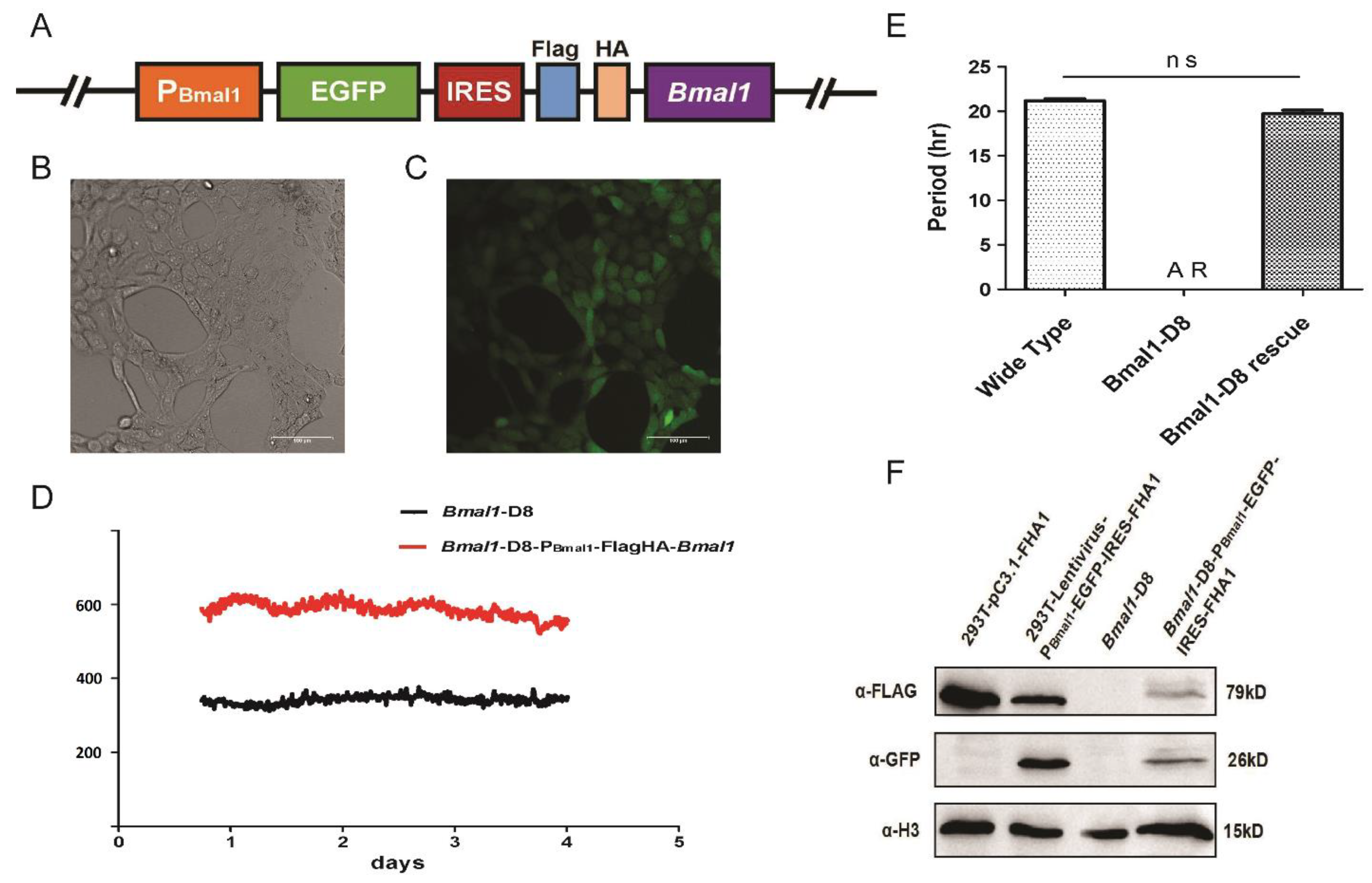
3.1. Cellular Circadian Gene Bmal1 Deletion by the CRISPR System Resulted in Loss of Circadian Rhythms

To understand the influence of each individual clock gene on the cellular physiology, we used CRISPR/Cas9 system to edit 23 clock genes plus Myc and Timless in NIH3T3-B1-B10 cells. This cell line can stably express luciferase, which is driven by a Bmal1 promoter, so as to observe the circadian rhythm. Taking Bmal1 as an example, the procedure of gene editing using CRISPR tools is presented (Figure 1). The single guide RNA sequence was designed and constructed to the pX459 vector as described in the Material & Methods (Figure 1A). A stable Bmal1 deletion cell line, Bmal1-D8, was established after normal procedures. Sequencing analysis identified that both alleles of Bmal1 were mutated by the CRISPR technique (Figure 1B). One allele had a deletion of 125 bp leading to a premature termination, while the other allele had a deletion of 2 bp (Figure 1B and Figure S1). Next, we asked if the deletion of Bmal1 will eliminate the circadian rhythms. We did the LumiCycle reporter assay to monitor the circadian rhythms, and the results showed that the knockout of Bmal1 resulted in a loss of the circadian rhythm (Figure 1C,D).
In order to prove that the phenotype is indeed caused by the deletion of Bmal1, we carried out complementation experiments. First, a lentiviral complement vector was constructed, which contains a Bmal1 encoding box under the CMV promoter. We used 5xMyc as a tag fused to the N-terminus of Bmal1, in order to detect the complemented protein (Figure 1E). By western blotting studies, we confirmed that this lentiviral construct can successfully express exogenous BMAL1 in various cell lines including Bmal-D8 cells (Figure 1F). However, expression of exogenous Bmal1 under the CMV promoter failed to rescue the circadian rhythm in Bmal-D8 cells (Figure 1G).

3.2. The Expression of Exogenous Bmal1 under a Bmal1 Promoter Can Rescue the Rhythm in Bmal-D8 Cells

CMV is a strong constitutive promoter, which may explain why the circadian rhythm of Bmal1-D8 has not been restored. Next, we constructed another lentiviral vector (Figure 2A). This new lentiviral construct contains the Bmal1 promoter which drives the expressions of EGFP and Flag/HA tagged BMAL1 proteins simultaneously. In this construct, a ribosome binding site IRES was introduced between EGFP and the interested gene to ensure EGFP and the interested protein, BMAL1, can express at the same time (Figure 2B,C). It was found that the circadian rhythm was restored after expressing the exogenous BMAL1 under Bmal1 promoter (Figure 2D), but the robustness of the rhythm did not return to the level of wild type, possibly because these are mixed cells. The Bmal1-D8 is successfully rescued, as the FLAG tag on the expressed BMAL1 can be detected using western blotting (Figure 2F). Moreover, the circadian period of Bmal1-D8 with complemented FH-BMAL1 recovered to the same extent as that of the wild-type cells (Figure 2E).

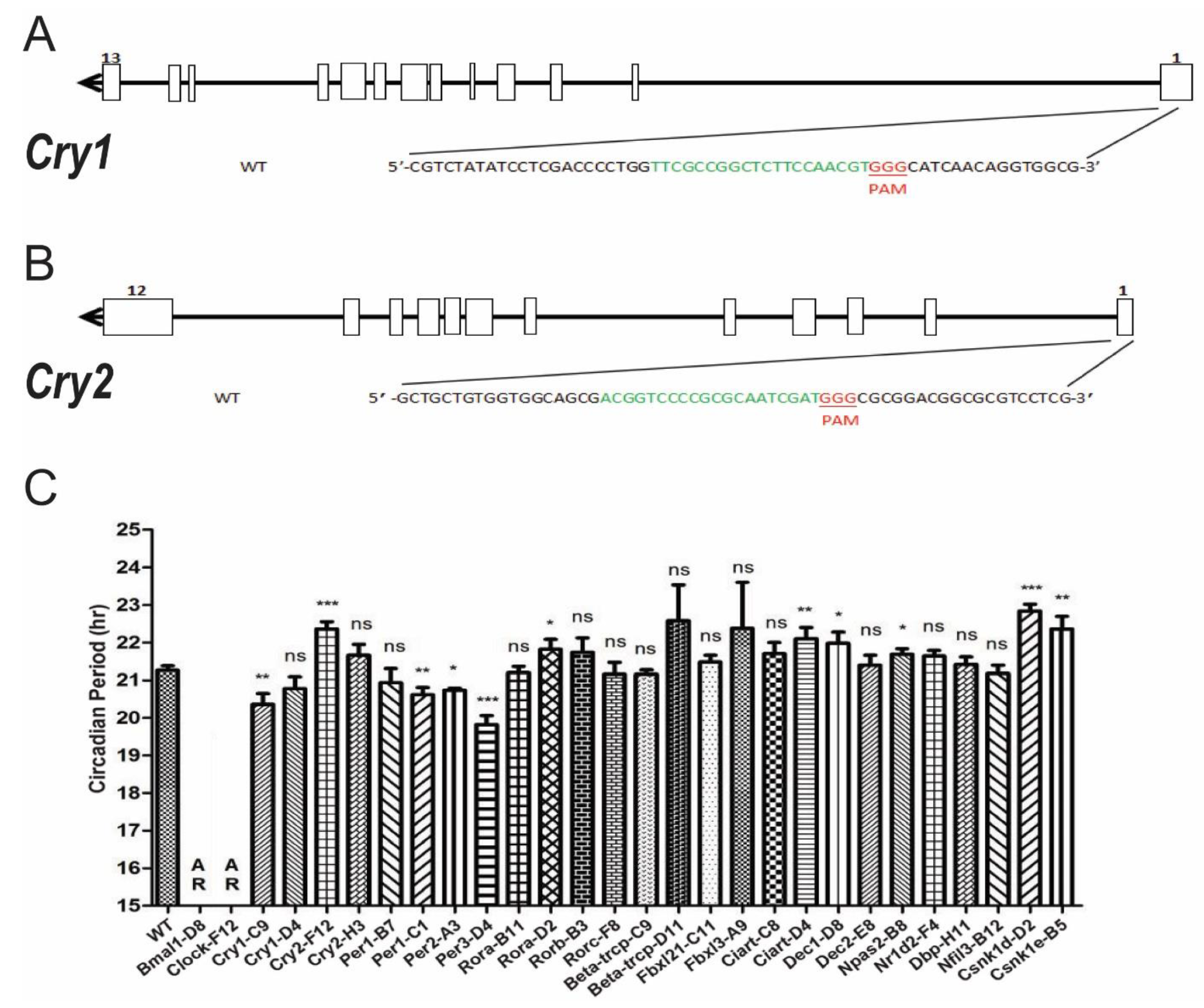
3.3. Systematic Knocking out Clock Genes Using CRISPR/Cas9

Taking Bmal1 as an example, the loss of circadian rhythm in Bmal1-D8 cells showed that CRISPR/Cas9 is an effective tool to knock out clock genes. We would like to further knock out circadian genes that will show period changes. Based on previous knowledge, loss of Cry1 or Cry2 genes will shorten or lengthen the circadian periods, respectively [52]. Thus, we choose Cry1 and Cry2 genes to further verify the effects of knocking out genes by the CRISPR/Cas9 tools. As illustrated in Figure 3A,B, sgRNAs targeting the exon 1 of either Cry1 or Cry2 genes were chosen and constructed into pX459 vectors to knock out corresponding genes. After screening for successful knock-out cell lines, we obtained Cry1-C9 and Cry2-F12 respectively. The period estimations using Lumicycle reporter assays showed that Cry1-C9 cells have a significantly shortened period by 0.48 h while Cry2-F12 cells have a significantly lengthened period by 1.1 h (Figure 3C, Table 1). These circadian period phenotypes are comparable with those of gene knock-out animals [52]. Therefore, CRISPR/Cas9 tools are reliable to knock out each individual gene.
Next, we designed sgRNAs targeting known clock genes and transfected each construct to NIH3T3-B1-B10 cells. Except for Nr1d1, Myc, and Timeless genes, we obtained knock-out cells of other genes mentioned in the Introduction (Table 1 and Supplemental materials). The comparison of circadian period changes between knockout mice and knockout cell lines demonstrates that most of the period phenotype changes are consistent for the same gene, indicating that the circadian system at cellular level is the same as the animal level. There was no available information about the circadian phenotypes of Nr1d2 or Nfil3 knockout animals, and we obtained the knockout cell lines of these two genes. There are a few conflicting results between knockout animals and knockout cell lines, for example, Csnk1d−/+ showed a shortened period, while Csnk1d-D2 showed a lengthened period (Figure 3C, Table 1). However, the animal study of Csnk1d was a heterozygous animal. In general, knocking out an individual circadian gene did not exhibit a dramatic effect on circadian period.

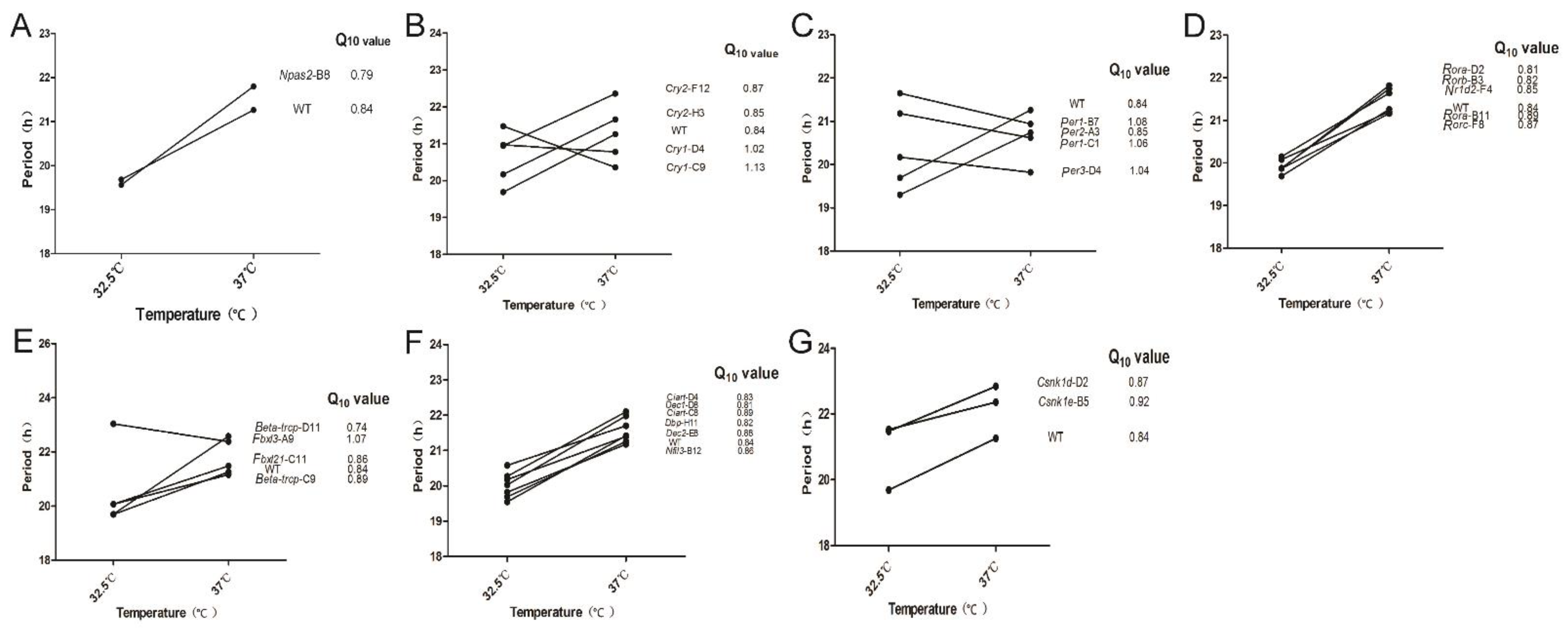
3.4. Temperature Compensation Evaluated in Cells Which Have Systematically Knocked out Clock Genes

According to preliminary explorations of the circadian oscillation of NIH3T3-B1-B10 at different temperatures (data not shown), we selected 32.5 °C and 37 °C in order to record robust circadian oscillation of cells under physiological range. The circadian period at different temperatures was measured by the Lumicycle luminometer and the effect of gene editing on temperature compensation was analyzed by calculating the numerical value of Q10 based on the following equation. τ1 and τ2 represent the circadian period at temperature T1 and T2, respectively (T1 = 32.5 °C, T2 = 37.0 °C).
Q 10 = ( τ 1 τ 2 ) 10 T 2 T 1
By calculating the Q10, we found that knocking out each single clock gene had little effect on temperature compensation. For all the cell lines we obtained, the Q10 value ranges from 0.8 to 1.2, except of Beta-trcp-D11 cell line (Figure 4). We observed that there are two categories in which the circadian period changes with the increase of temperature. One category includes Cry1-C9, Cry1-D4, Per1-B7, Per1-C1, Per3-D4, and Fbxl3-A9, whose periods become shorter with the increase of temperature, while the circadian periods of the other category become longer (Figure 4). We wondered if different types of the clock genes show different trends in terms of temperature compensation. Thus, we plot the Q10 values based on the types of these clock genes: core positive regulator (Figure 4A), core negative regulators (Figure 4B,C), Bmal1 gene regulators (Figure 4D), E3 ligases (Figure 4E), auxiliary regulators (Figure 4F), and casein kinase genes (Figure 4G). However, the Q10 is relatively consistent in all types. Thus, we conclude that the existence and maintenance of temperature compensation is regulated by complex gene networks rather than single clock genes. In addition, the Q10 value of Beta-trcp-D11 is less than 0.8. We preliminarily concluded that Beta-trcp may be more related to the maintenance of temperature compensation since it is involved in the stability of PER proteins. Though a dramatic change in Q10 was observed in Tau (CK1ε-R178C) mutant animals [10], Csnk1e-B5 cells showed little effect on Q10. Thus, not like KaiC in cyanobacteria, key kinases in the mammalian clock system do not determine the characteristics of temperature compensation.

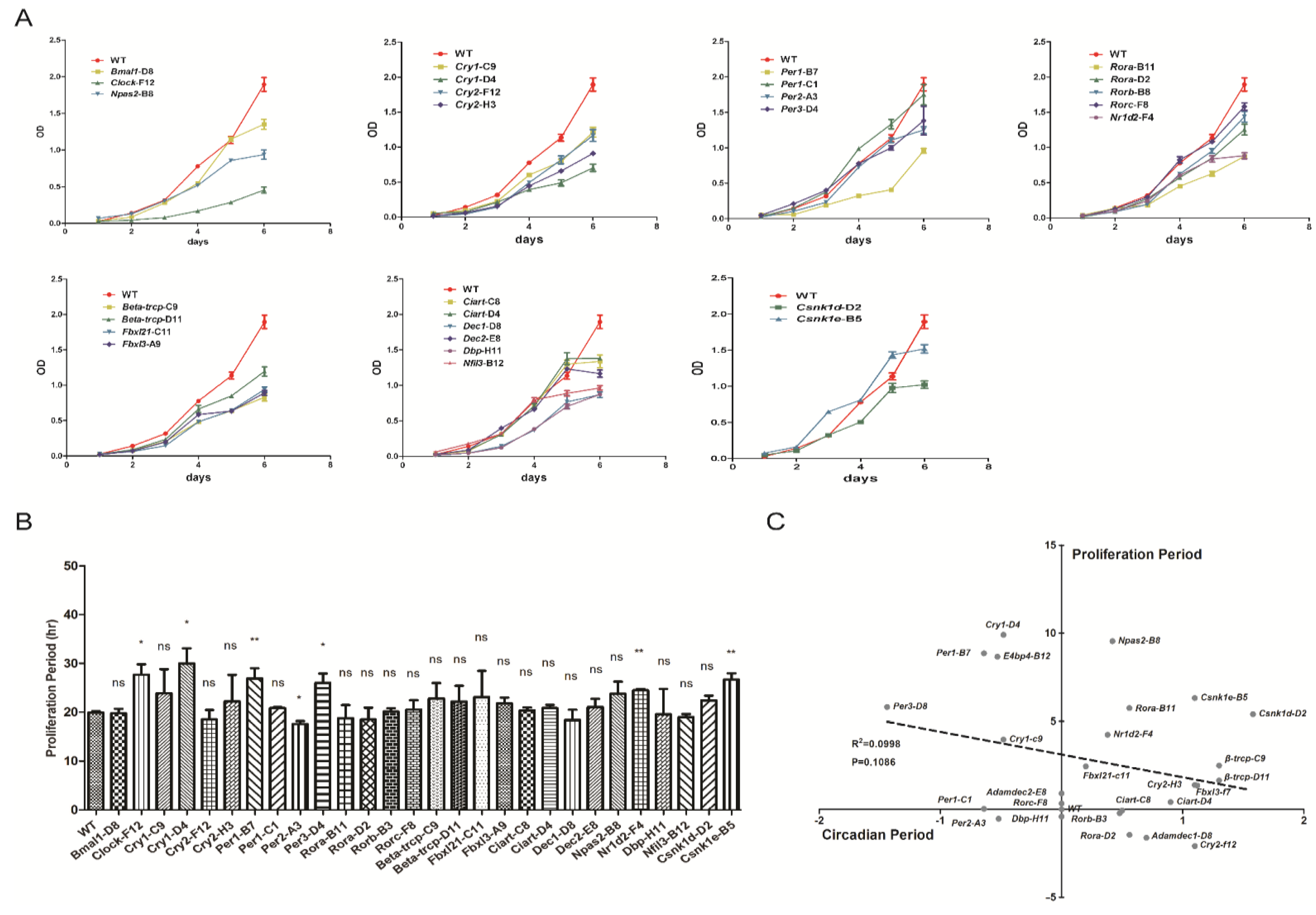
3.5. Proliferation Rates Evaluated in Cells Which Have Systematically Knocked out Clock Genes

Since we have observed different circadian period changes of these knocked-out cell lines, we planned to investigate whether there is a correlation between circadian period change and the cell proliferation rate change. We used MTT colorimetric method to determine the cell growth curve. According to the six groups of data collected, the cell doubling curve was plotted based on the recorded OD values (Figure 5A). We inoculated the same number of cells on the first day, and all the cell lines were maintained in logarithmic growth phases within six days. Again, we plot the growth curve of these cell lines based on different roles these genes play in the circadian clock networks. Next, we used SPSS software to calculate the doubling time of cell lines with two-division mode Y = a × 2bt (Figure 5B). We found that the doubling time of most gene-edited cell lines was not different from that of the wild type. Nevertheless, the doubling time of Clock-F12, Cry1-D4, Per1-B7, Per3-D4, Nr1d2-F4, and Csnk1e-B5 were significantly prolonged, while that of Per2-A3 was significantly shortened. However, the mechanisms underlying the changed proliferation rates need to be further studied. Taken together, by systematically comparing the circadian period and proliferation rates of these cell lines, it showed that no strong correlation can be found between the circadian period and the cell proliferation (R2 = 0.0998, p value = 0.1086, Figure 5C).

4. Discussion

In this study, we reported a systematic investigation on the temperature compensation and cell growth rate over currently known mammalian circadian genes in a fibroblast cell line. We applied a powerful genetic manipulation tool, CRISPR/Cas9, to generate a series of mutant cell lines that have individual circadian gene being knocked out (Table 1 and Supplemental materials). Most of these cell lines showed robust circadian oscillations, except the Bmal1-D8 and Clock-F12 cell lines. Bmal1 gene was taken as an example to verify the gene deletion by CRISPR/Cas9. Deletion of Bmal1 resulted in loss of the circadian rhythm, while complementation of exogenous Bmal1 under the Bmal1 promoter can rescue the rhythm. Most cell mutants recapitulate the circadian phenotypes of the period changes, which were collected using knockout animal studies. For example, Cry1-C9 cells showed a half-hour shortened period, and Cry2-F12 cells showed one hour longer period (Table 1, and supplemental materials). Therefore, our mutant cells represent the first full collection of knockouts of clock genes. According to our sequencing results of the second-high possibility of potential off-target gene locus (Table S3), there were no off-target mutations in this study. However, we cannot exclude the possible off-target mutations in the less potential loci.
Our data demonstrated that the period length of circadian rhythms is temperature compensated in each mutant cell line, except the arrhythmic ones, Bmal1-D8 and Clock-F12. Although previous results indicate that CK1δ/ε might be the important factor that determines the temperature compensation in the mammalian system [11,12], our data show that CK1δ and CK1ε depletion cells are still temperature compensated, with Q10 value close to that of the wild type cells (Figure 4G). This indicates that temperature compensation is not determined by a single factor. A single factor might be the key in other model organisms, such as frq in neurospora [9] and KaiC in cyanobacteria [12,13]. However, we observed a few mutant cells which showed a different trend in terms of period changes at different temperatures, 32.5 °C and 37 °C. For instance, Cry1-C9 showed a longer period at lower temperature, while WT and most other mutant cells showed a shorter period at lower temperature (Figure 4). We sort all known circadian genes into several categories, positive transcription factors, negative regulators, Bmal1 gene regulators, E3 ligases leading to degradations, auxiliary regulators and kinases. Then we compared the period change in these different categories. The period-got-increased trend appeared differently in these categories, indicating that this phenomenon might be gene-specific, but not dependent on the properties of a specific category. Next, we considered the effect of period change on the trend of temperature compensation. In the mutant cells that have shorter period, Cry1-C9, Per1-C1and Per3-D4 (Figure 3C), the trends are opposite to those of other cell lines (Figure 4). This is a very interesting point, which means that cells with slower clock at lower temperature (32.5 °C) can run faster at higher temperature (37 °C). In most cases, for example, wild-type cells, cells’ clock runs faster at lower temperature and runs slower at higher temperature, resulting in Q10 value less than one (Figure 4). These data indicate that the temperature compensation is a fundamental property of the circadian clock which is maintained in a homeostatic way under physiological temperature ranges.
Thus, we conclude that the temperature compensation is not determined by properties of each gene product, nor the properties of any limb in the circadian system. We speculate that the temperature compensation might be achieved by a network of multiple genes, which need further studies. Not like the circadian protein KaiC, which shows temperature compensated kinase and ATPase activities in pure biochemical reactions, none of the known mammalian clock gene products possess temperature compensation. Casein kinase 1ε has been implied to play important role in temperature compensation [10], but knock-out of CK1ε showed similar Q10 to that of WT cells (Figure 4G). Other studies have tried to double knock out CK1δ and CK1ε but failed to get target cells, which indicate at least one allele of CK1δ/ε is essential for cell growth [67].
We also investigated the relation between circadian period and cell growth rate. Compared to the changes of the circadian period of these mutant cells, the changes of the growth rate are larger in some of the cells (Figure 5B). Our results demonstrate that the cell growth rate is not dependent on the circadian period. Previous studies indicate that the circadian cycle gates the cell cycle, but we showed that the growth rate of most mutant cells is independent of the lengthened period nor shortened period. It may be possible that the circadian cycle gates the cell cycle, but the non-circadian functions of these genes may play roles at different phases of cell cycles. In order to fully understand the relation between circadian rhythms and cell cycle, further study is necessary.

5. Conclusions

In conclusion, we knocked out the known circadian genes in mouse fibroblasts and found that their biological rhythms are not destroyed except Bmal1-D8 and Clock-F12 cell lines. We concluded that the temperature compensation is a complex gene regulation system instead of being regulated by any single gene. The temperatures we used in this study are limited to 32.5 °C and 37 °C. Although the bioluminescence rhythm at 25 °C was not consistent in our hands, more temperatures are required to test in the near future. Furthermore, we found that the cell growth rate is not dependent on the circadian period by analyzing and comparing the proliferation rates and circadian periods of knockout cell lines.

Supplementary Materials

The following are available online at https://www.mdpi.com/article/10.3390/biology10111204/s1, Figure S1. The sgRNA targeting the Bmal1 gene and the sequences of both alleles in the Bmal1 deletion cell strain. Figure S2. Predicted translation products of both alleles in the Bmal1 deletion cell strain. Figure S3. The sgRNA targeting the Clock gene and the sequences of both alleles in the Clock deletion cell strain. Figure S4. Predicted translation products of both alleles in the Clock deletion cell strain. Figure S5. The sgRNA targeting the Cry1 gene and the sequences of both alleles in the Cry1 deletion cell strains. Figure S6. Predicted translation products of both alleles in the Cry1 deletion cell strain (Cry1-C9). Figure S7. Predicted translation products of both alleles in the Cry1 deletion cell strain (Cry1-D4). Figure S8. The sgRNA targeting the Cry2 gene and the sequences of both alleles in the Cry2 deletion cell strain. Figure S9. Predicted translation products of both alleles in the Cry2 deletion cell strain (Cry2-F12). Figure S10. Predicted translation products of both alleles in the Cry2 deletion cell strain (Cry2-H3). Figure S11. The sgRNA targeting the Per1 gene and the sequences of both alleles in the Per1 deletion cell strain. Figure S12. Predicted translation products of both alleles in the Per1 deletion cell strain (Per1-C1). Figure S13. Predicted translation products of both alleles in the Per1 deletion cell strain (Per1-B7). Figure S14. The sgRNA targeting the Per2 gene and the sequences of both alleles in the Per2 deletion cell strain. Figure S15. The sgRNA targeting the Per3 gene and the sequences of both alleles in the Per3 deletion cell strain. Figure S16. Predicted translation products of both alleles in the Per3 deletion cell strain. Figure S17. The sgRNA targeting the Rora gene and the sequences of both alleles in the Rora deletion cell strain. Figure S18. Predicted translation products of both alleles in the Rora deletion cell strain (Rora-B11). Figure S19. Predicted translation products of both alleles in the Rora deletion cell strain (Rora-D2). Figure S20. The sgRNA targeting the Rorb gene and the sequences of both alleles in the Rorb deletion cell strain. Figure S21. The sgRNA targeting the Rorc gene and the sequences of both alleles in the Rorc deletion cell strain. Figure S22. Predicted translation products of both alleles in the Rorc deletion cell strain. Figure S23. The sgRNA targeting the Beta-trcp gene and the sequences of both alleles in the Beta-trcp deletion cell strain. Figure S24. Predicted translation products of both alleles in the Beta-trcp deletion cell (C9). Figure S25. Predicted translation products of both alleles in the Beta-trcp deletion cell (D11). Figure S26. The sgRNA targeting the Fbxl21 gene and the sequences of both alleles in the Fbxl21 deletion cell strain. Figure S27. Predicted translation products of both alleles in the Fbxl21 deletion cell. Figure S28. The sgRNA targeting the Fbxl3 gene and the sequences of both alleles in the Fbxl3 deletion cell strain. Figure S29. Predicted translation products of both alleles in the Fbxl3 deletion cell. Figure S30. The sgRNA targeting the Ciart gene and the sequences of both alleles in the Ciart deletion cell strain. Figure S31. Predicted translation products of both alleles in the Ciart deletion cell (C8). Figure S32. Predicted translation products of both alleles in the Ciart deletion cell (D4). Figure S33. The sgRNA targeting the Dec1 gene and the sequences of both alleles in the Dec1 deletion cell strain. Figure S34. Predicted translation products of both alleles in the Dec1 deletion cell. Figure S35. The sgRNA targeting the Dec2 gene and the sequences of both alleles in the Dec2 deletion cell strain. Figure S36. Predicted translation products of both alleles in the Dec2 deletion cell. Figure S37. The sgRNA targeting the Npas2 gene and the sequences of both alleles in the Npas2 deletion cell strain. Figure S38. Predicted translation products of both alleles in the Npas2 deletion cell. Figure S39. The sgRNA targeting the Nr1d2 gene and the sequences of both alleles in the Nr1d2 deletion cell strain. Figure S40. Predicted translation products of both alleles in the Nr1d2 deletion cell. Figure S41. The sgRNA targeting the Dbp gene and the sequences of both alleles in the Dbp deletion cell strain. Figure S42. The sgRNA targeting the Nfil3 gene and the sequences of both alleles in the Nfil3 deletion cell strain. Figure S43. Predicted translation products of both alleles in the Nfil3 deletion cell. Figure S44. The sgRNA targeting the Csnk1d gene and the sequences of both alleles in the Csnk1d deletion cell strain (Knocking-out of CK1d was reported in Guo et al., JBR, 2019).Figure S45. The sgRNA targeting the Csnk1e gene and the sequences of both alleles in the Csnk1e deletion cell strain (Knocking-out of CK1e was reported in Guo et al., JBR, 2019). Figure S46. The bioluminescence of the wild-type, Bmal1-D8, Clock-F12, and Npas2-B8 cells. Figure S47. The bioluminescence of the Cry1 and Cry2 knock-out cells. Figure S48. The bioluminescence of the Per1, Per2, and Per3 knock-out cells. Figure S49. The bioluminescence of the Rora-B11, Rora-D2, Rorb-B3, Rorc-F8, Nr1d2-F4 cells. Figure S50. The bioluminescence of the Beta-trcp-C9, Beta-trcp-D11, Fbxl21-C11, Fbxl3-A9 cells. Figure S51. The bioluminescence of the Ciart-C8, Ciart-D4, Dec1-D8, Dec2-E8, Dbp-H11, Nfil3-B12 cells. Figure S52. The bioluminescence of the casein kinase genes, Csnk1d-D2, Csnk1e-B5 cells. Table S1. The sequence information of sgRNAs used in this study. Table S2. The sequence information of primers to amplify targeted sites. Table S3. The most possible off-target sites for each individual gene in this study. Table S4. Primers to amplify the most possible off-target site for individual genes.

Author Contributions

Y.Z. and X.Q. conceived and designed research; Y.W. (Yue Wu), T.T., Y.W. (Yin Wu) and Y.Y. performed research; all authors were involved in analyzing data; Y.Z. and X.Q. wrote and edited the manuscript. All authors have read and agreed to the published version of the manuscript.

Funding

This work was financially supported by the grants from the National Natural Science Foundation of China (32071158), Anhui Provincial Natural Science Foundation (1608085MH212 and 2008085MC68), and the Research Start Grant from Anhui University (Y040418157).

Institutional Review Board Statement

Not applicable.

Informed Consent Statement

Not applicable.

Data Availability Statement

Not applicable.

Acknowledgments

We are grateful to the staff, Xi Zhu and Minrui Xu for providing technical support with using the facility of Institute of Physical Science and Information Technology at Anhui University.

Conflicts of Interest

The authors declare that they have no conflict of interest.

References

  1. Albrecht, U. Timing to perfection: The biology of central and peripheral circadian clocks. Neuron 2012, 74, 246–260. [Google Scholar] [CrossRef] [PubMed] [Green Version]
  2. Allada, R.; Chung, B.Y. Circadian organization of behavior and physiology in Drosophila. Annu. Rev. Physiol. 2010, 72, 605–624. [Google Scholar] [CrossRef] [PubMed] [Green Version]
  3. Balsalobre, A.; Brown, S.A.; Marcacci, L.; Tronche, F.; Kellendonk, C.; Reichardt, H.M.; Schütz, G.; Schibler, U. Resetting of circadian time in peripheral tissues by glucocorticoid signaling. Science 2000, 289, 2344–2347. [Google Scholar] [CrossRef] [Green Version]
  4. McNamara, P.; Seo, S.B.; Rudic, R.D.; Sehgal, A.; Chakravarti, D.; FitzGerald, G.A. Regulation of CLOCK and MOP4 by nuclear hormone receptors in the vasculature: A humoral mechanism to reset a peripheral clock. Cell 2001, 105, 877–889. [Google Scholar] [CrossRef] [Green Version]
  5. Qin, X.; Mori, T.; Zhang, Y.; Johnson, C.H. PER2 Differentially Regulates Clock Phosphorylation versus Transcription by Reciprocal Switching of CK1ε Activity. J. Biol. Rhythm. 2015, 30, 206–216. [Google Scholar] [CrossRef] [PubMed] [Green Version]
  6. Anderson, R.W.; Laval-Martin, D.L.; Edmunds, L.N., Jr. Cell cycle oscillators. Temperature compensation of the circadian rhythm of cell division in Euglena. Exp. Cell Res. 1985, 157, 144–158. [Google Scholar] [CrossRef] [PubMed]
  7. Ruby, N.F.; Heller, H.C. Temperature sensitivity of the suprachiasmatic nucleus of ground squirrels and rats in vitro. J. Biol. Rhythm. 1996, 11, 126–136. [Google Scholar] [CrossRef] [Green Version]
  8. Lakin-Thomas, P.L. Choline depletion, frq mutations, and temperature compensation of the circadian rhythm in Neurospora crassa. J. Biol. Rhythm. 1998, 13, 268–277. [Google Scholar] [CrossRef] [PubMed]
  9. Huang, Z.J.; Curtin, K.D.; Rosbash, M. PER protein interactions and temperature compensation of a circadian clock in Drosophila. Science 1995, 267, 1169–1172. [Google Scholar] [CrossRef] [Green Version]
  10. Tosini, G.; Menaker, M. The tau mutation affects temperature compensation of hamster retinal circadian oscillators. Neuroreport 1998, 9, 1001–1005. [Google Scholar] [CrossRef]
  11. Isojima, Y.; Nakajima, M.; Ukai, H.; Fujishima, H.; Yamada, R.G.; Masumoto, K.H.; Kiuchi, R.; Ishida, M.; Ukai-Tadenuma, M.; Minami, Y.; et al. CKIepsilon/delta-dependent phosphorylation is a temperature-insensitive, period-determining process in the mammalian circadian clock. Proc. Natl. Acad. Sci. USA 2009, 106, 15744–15749. [Google Scholar] [CrossRef] [PubMed] [Green Version]
  12. Shinohara, Y.; Koyama, Y.M.; Ukai-Tadenuma, M.; Hirokawa, T.; Kikuchi, M.; Yamada, R.G.; Ukai, H.; Fujishima, H.; Umehara, T.; Tainaka, K.; et al. Temperature-Sensitive Substrate and Product Binding Underlie Temperature-Compensated Phosphorylation in the Clock. Mol. Cell 2017, 67, 783–798.e20. [Google Scholar] [CrossRef] [PubMed] [Green Version]
  13. Terauchi, K.; Kitayama, Y.; Nishiwaki, T.; Miwa, K.; Murayama, Y.; Oyama, T.; Kondo, T. ATPase activity of KaiC determines the basic timing for circadian clock of cyanobacteria. Proc. Natl. Acad. Sci. USA 2007, 104, 16377–16381. [Google Scholar] [CrossRef] [PubMed] [Green Version]
  14. Ito-Miwa, K.; Furuike, Y.; Akiyama, S.; Kondo, T. Tuning the circadian period of cyanobacteria up to 6.6 days by the single amino acid substitutions in KaiC. Proc. Natl. Acad. Sci. USA 2020, 117, 20926–20931. [Google Scholar] [CrossRef] [PubMed]
  15. Akiyama, M.; Moriya, T.; Shibata, S. Physiological, pharmacological and molecular aspects of mammalian biological clocks. Nihon Yakurigaku Zasshi. Folia Pharmacol. Jpn. 1998, 112, 243–250. [Google Scholar] [CrossRef] [Green Version]
  16. Kondratov, R.V.; Shamanna, R.K.; Kondratova, A.A.; Gorbacheva, V.Y.; Antoch, M.P. Dual role of the CLOCK/BMAL1 circadian complex in transcriptional regulation. FASEB J. Off. Publ. Fed. Am. Soc. Exp. Biol. 2006, 20, 530–532. [Google Scholar] [CrossRef] [Green Version]
  17. Edery, I. Circadian rhythms in a nutshell. Physiol. Genom. 2000, 3, 59–74. [Google Scholar] [CrossRef]
  18. Takahashi, J.S. Transcriptional architecture of the mammalian circadian clock. Nat. Rev. Genet. 2017, 18, 164–179. [Google Scholar] [CrossRef] [Green Version]
  19. Buhr, E.D.; Takahashi, J.S. Molecular components of the Mammalian circadian clock. Handb. Exp. Pharmacol. 2013, 217, 3–27. [Google Scholar] [CrossRef] [Green Version]
  20. Gustafson, C.L.; Partch, C.L. Emerging models for the molecular basis of mammalian circadian timing. Biochemistry 2015, 54, 134–149. [Google Scholar] [CrossRef]
  21. Huang, N.; Chelliah, Y.; Shan, Y.; Taylor, C.A.; Yoo, S.H.; Partch, C.; Green, C.B.; Zhang, H.; Takahashi, J.S. Crystal structure of the heterodimeric CLOCK:BMAL1 transcriptional activator complex. Science 2012, 337, 189–194. [Google Scholar] [CrossRef] [PubMed] [Green Version]
  22. Brown, S.A.; Ripperger, J.; Kadener, S.; Fleury-Olela, F.; Vilbois, F.; Rosbash, M.; Schibler, U. PERIOD1-associated proteins modulate the negative limb of the mammalian circadian oscillator. Science 2005, 308, 693–696. [Google Scholar] [CrossRef] [PubMed] [Green Version]
  23. Duong, H.A.; Robles, M.S.; Knutti, D.; Weitz, C.J. A molecular mechanism for circadian clock negative feedback. Science 2011, 332, 1436–1439. [Google Scholar] [CrossRef] [PubMed] [Green Version]
  24. Lee, C.; Etchegaray, J.P.; Cagampang, F.R.; Loudon, A.S.; Reppert, S.M. Posttranslational mechanisms regulate the mammalian circadian clock. Cell 2001, 107, 855–867. [Google Scholar] [CrossRef]
  25. Ye, R.; Selby, C.P.; Ozturk, N.; Annayev, Y.; Sancar, A. Biochemical analysis of the canonical model for the mammalian circadian clock. J. Biol. Chem. 2011, 286, 25891–25902. [Google Scholar] [CrossRef] [PubMed] [Green Version]
  26. Koike, N.; Yoo, S.H.; Huang, H.C.; Kumar, V.; Lee, C.; Kim, T.K.; Takahashi, J.S. Transcriptional architecture and chromatin landscape of the core circadian clock in mammals. Science 2012, 338, 349–354. [Google Scholar] [CrossRef] [Green Version]
  27. Stratmann, M.; Stadler, F.; Tamanini, F.; van der Horst, G.T.; Ripperger, J.A. Flexible phase adjustment of circadian albumin D site-binding protein (DBP) gene expression by CRYPTOCHROME1. Genes Dev. 2010, 24, 1317–1328. [Google Scholar] [CrossRef] [Green Version]
  28. Eide, E.J.; Vielhaber, E.L.; Hinz, W.A.; Virshup, D.M. The circadian regulatory proteins BMAL1 and cryptochromes are substrates of casein kinase Iepsilon. J. Biol. Chem. 2002, 277, 17248–17254. [Google Scholar] [CrossRef] [Green Version]
  29. Keesler, G.A.; Camacho, F.; Guo, Y.; Virshup, D.; Mondadori, C.; Yao, Z. Phosphorylation and destabilization of human period I clock protein by human casein kinase I epsilon. Neuroreport 2000, 11, 951–955. [Google Scholar] [CrossRef]
  30. Lowrey, P.L.; Shimomura, K.; Antoch, M.P.; Yamazaki, S.; Zemenides, P.D.; Ralph, M.R.; Menaker, M.; Takahashi, J.S. Positional syntenic cloning and functional characterization of the mammalian circadian mutation tau. Science 2000, 288, 483–492. [Google Scholar] [CrossRef] [Green Version]
  31. Partch, C.L.; Green, C.B.; Takahashi, J.S. Molecular architecture of the mammalian circadian clock. Trends Cell Biol. 2014, 24, 90–99. [Google Scholar] [CrossRef] [PubMed] [Green Version]
  32. Siepka, S.M.; Yoo, S.H.; Park, J.; Song, W.; Kumar, V.; Hu, Y.; Lee, C.; Takahashi, J.S. Circadian mutant Overtime reveals F-box protein FBXL3 regulation of cryptochrome and period gene expression. Cell 2007, 129, 1011–1023. [Google Scholar] [CrossRef] [Green Version]
  33. Hirano, A.; Yumimoto, K.; Tsunematsu, R.; Matsumoto, M.; Oyama, M.; Kozuka-Hata, H.; Nakagawa, T.; Lanjakornsiripan, D.; Nakayama, K.I.; Fukada, Y. FBXL21 regulates oscillation of the circadian clock through ubiquitination and stabilization of cryptochromes. Cell 2013, 152, 1106–1118. [Google Scholar] [CrossRef] [PubMed] [Green Version]
  34. Shirogane, T.; Jin, J.; Ang, X.L.; Harper, J.W. SCFbeta-TRCP controls clock-dependent transcription via casein kinase 1-dependent degradation of the mammalian period-1 (Per1) protein. J. Biol. Chem. 2005, 280, 26863–26872. [Google Scholar] [CrossRef] [PubMed] [Green Version]
  35. Sato, T.K.; Panda, S.; Miraglia, L.J.; Reyes, T.M.; Rudic, R.D.; McNamara, P.; Naik, K.A.; FitzGerald, G.A.; Kay, S.A.; Hogenesch, J.B. A functional genomics strategy reveals Rora as a component of the mammalian circadian clock. Neuron 2004, 43, 527–537. [Google Scholar] [CrossRef] [PubMed] [Green Version]
  36. Preitner, N.; Damiola, F.; Lopez-Molina, L.; Zakany, J.; Duboule, D.; Albrecht, U.; Schibler, U. The orphan nuclear receptor REV-ERBalpha controls circadian transcription within the positive limb of the mammalian circadian oscillator. Cell 2002, 110, 251–260. [Google Scholar] [CrossRef]
  37. Reppert, S.M.; Weaver, D.R. Coordination of circadian timing in mammals. Nature 2002, 418, 935–941. [Google Scholar] [CrossRef]
  38. Yamaguchi, S.; Mitsui, S.; Yan, L.; Yagita, K.; Miyake, S.; Okamura, H. Role of DBP in the circadian oscillatory mechanism. Mol. Cell. Biol. 2000, 20, 4773–4781. [Google Scholar] [CrossRef] [Green Version]
  39. Oishi, K.; Miyazaki, K.; Kadota, K.; Kikuno, R.; Nagase, T.; Atsumi, G.; Ohkura, N.; Azama, T.; Mesaki, M.; Yukimasa, S.; et al. Genome-wide expression analysis of mouse liver reveals CLOCK-regulated circadian output genes. J. Biol. Chem. 2003, 278, 41519–41527. [Google Scholar] [CrossRef] [Green Version]
  40. Kume, K.; Zylka, M.J.; Sriram, S.; Shearman, L.P.; Weaver, D.R.; Jin, X.; Maywood, E.S.; Hastings, M.H.; Reppert, S.M. mCRY1 and mCRY2 are essential components of the negative limb of the circadian clock feedback loop. Cell 1999, 98, 193–205. [Google Scholar] [CrossRef] [Green Version]
  41. Fukada, Y.; Okano, T. Circadian clock system in the pineal gland. Mol. Neurobiol. 2002, 25, 19–30. [Google Scholar] [CrossRef]
  42. Annayev, Y.; Adar, S.; Chiou, Y.Y.; Lieb, J.D.; Sancar, A.; Ye, R. Gene model 129 (Gm129) encodes a novel transcriptional repressor that modulates circadian gene expression. J. Biol. Chem. 2014, 289, 5013–5024. [Google Scholar] [CrossRef] [PubMed] [Green Version]
  43. Price, J.L.; Blau, J.; Rothenfluh, A.; Abodeely, M.; Kloss, B.; Young, M.W. Double-time is a novel Drosophila clock gene that regulates PERIOD protein accumulation. Cell 1998, 94, 83–95. [Google Scholar] [CrossRef] [Green Version]
  44. Altman, B.J.; Hsieh, A.L.; Sengupta, A.; Krishnanaiah, S.Y.; Stine, Z.E.; Walton, Z.E.; Gouw, A.M.; Venkataraman, A.; Li, B.; Goraksha-Hicks, P.; et al. MYC Disrupts the Circadian Clock and Metabolism in Cancer Cells. Cell Metab. 2015, 22, 1009–1019. [Google Scholar] [CrossRef] [PubMed] [Green Version]
  45. Hsu, P.D.; Zhang, F. Dissecting neural function using targeted genome engineering technologies. ACS Chem. Neurosci. 2012, 3, 603–610. [Google Scholar] [CrossRef] [PubMed] [Green Version]
  46. Sternberg, S.H.; Haurwitz, R.E.; Doudna, J.A. Mechanism of substrate selection by a highly specific CRISPR endoribonuclease. RNA 2012, 18, 661–672. [Google Scholar] [CrossRef] [PubMed] [Green Version]
  47. Wang, H.; Yang, H.; Shivalila, C.S.; Dawlaty, M.M.; Cheng, A.W.; Zhang, F.; Jaenisch, R. One-step generation of mice carrying mutations in multiple genes by CRISPR/Cas-mediated genome engineering. Cell 2013, 153, 910–918. [Google Scholar] [CrossRef] [Green Version]
  48. Traynard, P.; Feillet, C.; Soliman, S.; Delaunay, F.; Fages, F. Model-based investigation of the circadian clock and cell cycle coupling in mouse embryonic fibroblasts: Prediction of RevErb-α up-regulation during mitosis. Bio. Syst. 2016, 149, 59–69. [Google Scholar] [CrossRef] [PubMed] [Green Version]
  49. Gao, L.; Chen, H.; Li, C.; Xiao, Y.; Yang, D.; Zhang, M.; Zhou, D.; Liu, W.; Wang, A.; Jin, Y. ER stress activation impairs the expression of circadian clock and clock-controlled genes in NIH3T3 cells via an ATF4-dependent mechanism. Cell. Signal. 2019, 57, 89–101. [Google Scholar] [CrossRef]
  50. Yeom, M.; Pendergast, J.S.; Ohmiya, Y.; Yamazaki, S. Circadian-independent cell mitosis in immortalized fibroblasts. Proc. Natl. Acad. Sci. USA 2010, 107, 9665–9670. [Google Scholar] [CrossRef] [Green Version]
  51. Bieler, J.; Cannavo, R.; Gustafson, K.; Gobet, C.; Gatfield, D.; Naef, F. Robust synchronization of coupled circadian and cell cycle oscillators in single mammalian cells. Mol. Syst. Biol. 2014, 10, 739. [Google Scholar] [CrossRef] [PubMed]
  52. van der Horst, G.T.; Muijtjens, M.; Kobayashi, K.; Takano, R.; Kanno, S.; Takao, M.; De Wit, J.; Verkerk, A.; Eker, A.P.; Van Leenen, D.; et al. Mammalian Cry1 and Cry2 are essential for maintenance of circadian rhythms. Nature 1999, 398, 627–630. [Google Scholar] [CrossRef]
  53. Kitanishi, K.; Igarashi, J.; Hayasaka, K.; Hikage, N.; Saiful, I.; Yamauchi, S.; Uchida, T.; Ishimori, K.; Shimizu, T. Heme-binding characteristics of the isolated PAS-A domain of mouse Per2, a transcriptional regulatory factor associated with circadian rhythms. Biochemistry 2008, 47, 6157–6168. [Google Scholar] [CrossRef] [PubMed]
  54. Cermakian, N.; Monaco, L.; Pando, M.P.; Dierich, A.; Sassone-Corsi, P. Altered behavioral rhythms and clock gene expression in mice with a targeted mutation in the Period1 gene. EMBO J. 2001, 20, 3967–3974. [Google Scholar] [CrossRef] [PubMed] [Green Version]
  55. Zheng, B.; Larkin, D.W.; Albrecht, U.; Sun, Z.S.; Sage, M.; Eichele, G.; Lee, C.C.; Bradley, A. The mPer2 gene encodes a functional component of the mammalian circadian clock. Nature 1999, 400, 169–173. [Google Scholar] [CrossRef] [PubMed]
  56. Shearman, L.P.; Jin, X.; Lee, C.; Reppert, S.M.; Weaver, D.R. Targeted disruption of the mPer3 gene: Subtle effects on circadian clock function. Mol. Cell. Biol. 2000, 20, 6269–6275. [Google Scholar] [CrossRef] [PubMed]
  57. Yang, X.O.; Pappu, B.P.; Nurieva, R.; Akimzhanov, A.; Kang, H.S.; Chung, Y.; Ma, L.; Shah, B.; Panopoulos, A.D.; Schluns, K.S.; et al. T helper 17 lineage differentiation is programmed by orphan nuclear receptors ROR alpha and ROR gamma. Immunity 2008, 28, 29–39. [Google Scholar] [CrossRef] [Green Version]
  58. Liu, A.C.; Tran, H.G.; Zhang, E.E.; Priest, A.A.; Welsh, D.K.; Kay, S.A. Redundant function of REV-ERBalpha and beta and non-essential role for Bmal1 cycling in transcriptional regulation of intracellular circadian rhythms. PLoS Genet. 2008, 4, e1000023. [Google Scholar] [CrossRef]
  59. Pourcet, B.; Zecchin, M.; Ferri, L.; Beauchamp, J.; Sitaula, S.; Billon, C.; Delhaye, S.; Vanhoutte, J.; Mayeuf-Louchart, A.; Thorel, Q.; et al. Nuclear Receptor Subfamily 1 Group D Member 1 Regulates Circadian Activity of NLRP3 Inflammasome to Reduce the Severity of Fulminant Hepatitis in Mice. Gastroenterology 2018, 154, 1449–1464.e20. [Google Scholar] [CrossRef]
  60. Pittendrigh, C.S. On temperature independence in the clock system controlling emergence time in Drosophila. Proc. Natl. Acad. Sci. USA 1954, 40, 1018–1029. [Google Scholar] [CrossRef] [Green Version]
  61. Goriki, A.; Hatanaka, F.; Myung, J.; Kim, J.K.; Yoritaka, T.; Tanoue, S.; Abe, T.; Kiyonari, H.; Fujimoto, K.; Kato, Y.; et al. A novel protein, CHRONO, functions as a core component of the mammalian circadian clock. PLoS Biol. 2014, 12, e1001839. [Google Scholar] [CrossRef] [PubMed]
  62. Honma, S.; Kawamoto, T.; Takagi, Y.; Fujimoto, K.; Sato, F.; Noshiro, M.; Kato, Y.; Honma, K. Dec1 and Dec2 are regulators of the mammalian molecular clock. Nature 2002, 419, 841–844. [Google Scholar] [CrossRef] [PubMed]
  63. Ohsaki, K.; Oishi, K.; Kozono, Y.; Nakayama, K.; Nakayama, K.I.; Ishida, N. The role of {beta}-TrCP1 and {beta}-TrCP2 in circadian rhythm generation by mediating degradation of clock protein PER2. J. Biochem. 2008, 144, 609–618. [Google Scholar] [CrossRef]
  64. Barnes, J.W.; Tischkau, S.A.; Barnes, J.A.; Mitchell, J.W.; Burgoon, P.W.; Hickok, J.R.; Gillette, M.U. Requirement of mammalian Timeless for circadian rhythmicity. Science 2003, 302, 439–442. [Google Scholar] [CrossRef] [PubMed] [Green Version]
  65. Eng, G.W.L.; Virshup, D.M. Site-specific phosphorylation of casein kinase 1 δ (CK1δ) regulates its activity towards the circadian regulator PER2. PLoS ONE 2017, 12, e0177834. [Google Scholar] [CrossRef] [Green Version]
  66. Meng, Q.J.; Logunova, L.; Maywood, E.S.; Gallego, M.; Lebiecki, J.; Brown, T.M.; Sládek, M.; Semikhodskii, A.S.; Glossop, N.R.J.; Piggins, H.D.; et al. Setting clock speed in mammals: The CK1 epsilon tau mutation in mice accelerates circadian pacemakers by selectively destabilizing PERIOD proteins. Neuron 2008, 58, 78–88. [Google Scholar] [CrossRef] [Green Version]
  67. Tsuchiya, Y.; Umemura, Y.; Minami, Y.; Koike, N.; Hosokawa, T.; Hara, M.; Ito, H.; Inokawa, H.; Yagita, K. Effect of Multiple Clock Gene Ablations on the Circadian Period Length and Temperature Compensation in Mammalian Cells. J. Biol. Rhythm. 2016, 31, 48–56. [Google Scholar] [CrossRef]
Figure 1. NIH3T3 cells with Bmal1−/− genotypes are arrhythmic. (A) The schematic diagram of sgRNA design information and knockout vector construction of the Bmal1 gene is shown. On the top, the schematic of Bmal1 was drawn. In the enlarged region at the 14th exon, the bases marked in green are the sequence of the picked sgRNA. Then, the sgRNA sequence was cloned into the px459 vector. (B) A monoclonal cell line, Bmal1-D8, showed that both alleles of the Bmal1 gene were edited. According to the analysis on the predicted mRNA, neither edited alleles of Bmal1 is capable of producing functional Bmal1 proteins. (C) The luminescence recording of the WT NIH3T3 cells that bear with a luciferase reporter under the Bmal1 promoter. (D) The bioluminescence rhythm of the Bmal1-D8 cells, which is arrhythmic. (E) Illustration of the main components of the constructed lentiviral vector to complement the Bmal1 gene. The Bmal1 coding sequence tagged with five constitutive myc tags is under the CMV promoter. (F) Myc epitope tags as a label to detect whether the cells are successfully transfected with exogenous Bmal1. In several cell lines, 293T, NIH3T3, and Bmal1-D8 cells, lentiviral constructs can successfully express Bmal1 proteins. Histone3 is shown as a loading control. Histone3 is shown as a control. (G) However, both Bmal1-D8 and Bmal1-D8 cells supplemented with lentivirus were arrhythmic. Representative luminescent trace or image was shown.
Biology 10 01204 g001
Figure 2. The lentivirus of exogenous Bmal1 driven by the Bmal1 promoter rescues the circadian phenotype of Bmal1-D8. (A) Illustration of the main components of the new constructed lentiviral complement vector, in which the Flag-HA tagged Bmal1 is driven by the native Bmal1 promoter. (B) Bmal1-D8 cells were transfected with the new lentiviral vector and the bright filed image is shown. (C) The fluorescent image of Bmal1-D8 cells transfected with the new lentiviral vector indicates that GFP is successfully expressed. (D) The bioluminescence rhythm results of Bmal1-D8 and lentivirus infected Bmal1-D8-PBmal1-FlagHA-Bmal1 cells. (E) The calculated circadian period of wide type, Bmal1-D8, Bmal1-D8 rescue cells. Bmal1-D8 rescue cells showed a period closed to that of the wild-type cells. (F) Western blot images show that the GFP and Flag-HA tagged Bmal1 can simultaneously express in both HEK293T and Bmal1-D8 cells. Histone3 are shown here as loading controls. ns, none significant. Representative luminescent trace or image was shown.
Biology 10 01204 g002
Figure 3. Circadian period phenotypes of cell lines with individual clock gene being knocked out. (A) The schematic diagram of sgRNA designed to knock out the Cry1 gene in NIH3T3 cells. (B) The schematic diagram of sgRNA designed to knock out the Cry2 gene in NIH3T3 cells. (C) The statistical results of the circadian period of the monoclonal cell lines for the clock genes in this study. Similar to the animal studies, Cry1 deletion cell lines showed shortened period, while Cry2 deletion cell lines showed lengthened period. The period changes were summarized in Table 1. Results are expressed as mean ± SD, n ≥ 3 for each group. ns, no significant, * p < 0.05, ** p < 0.01, *** p < 0.001.
Biology 10 01204 g003
Figure 4. The circadian period changes of monoclonal cell lines at different temperatures. Clock genes in this study were classified into different types. (A) The circadian period changes of wild-type and Npas2-B8 cells at different temperatures. Since Bmal1 deletion and Clock deletion cells were arrhythmic, Npas2 is shown here to represent the core positive regulator group. (B) The circadian period changes of Cry1-D4, Cry1-C9, Cry2-F12, Cry2-H3 cells at different temperatures. (C) The circadian period changes of Per1-B7, Per1-C1, Per2-A3, Per3-D4 cells. Cry and Per genes are core negative regulators. (D) The circadian period changes of Rora-B11, Rora-D2, Rorb-B3, Rorc-F8, Nr1d2-F4 cells, which represent regulators for the Bmal1 gene. (E) The circadian period changes of Beta-trcp-C9, Beta-trcp-D11, Fbxl21-C11, Fbxl3-A9 cells at different temperatures. These genes represent E3 ligases. (F) The circadian period changes of Ciart-C8, Ciart-D4, Dec1-D8, Dec2-E8, Dbp-H11, Nfil3-B12 cells, which represent auxiliary regulators. (G) The circadian period changes of the casein kinase genes, Csnk1d-D2, Csnk1e-B5 cells at different temperatures. On the right is their Q10 value. Periods of each cell line were calculated based on multiple experiments.
Biology 10 01204 g004
Figure 5. The proliferation periods of clock gene mutant cells are not correlated with their circadian periods. (A) Cell proliferation curves of wild-type and clock gene deletion monoclonal cell lines for six consecutive days. (B) The proliferation period of each cell line was calculated and plotted. (C) The relationship between the circadian period changes and the proliferation period of the monoclonal cell lines. Linear relationship was applied to fit the data in panel C. Results are expressed as mean ± SD. n ≥ 3 in each group. ns, no significant, * p < 0.05, ** p < 0.01.
Biology 10 01204 g005
Table 1. Comparison of rhythm cycle changes between knockout mice and cell lines.
GenesAllelesMouse Mutant PhenotypesCellular Mutation Phenotypes
Bmal1Bmal1−/−Rhythm disorder [39]Rhythm disorder
ClockClock∆19/∆19Rhythm disorder [39]Rhythm disorder
Npas2Npas2−/−Shorten 0.2 h [53]Lengthen 0.42 h
Cry1Cry1−/−Shorten 0.5 h [52]Shorten 0.48 h
Cry2Cry2−/−Lengthen 1 h [52]Lengthen 1.1 h
Per1Per1−/−Shorten 0.5 h [54]Shorten 0.64 h
Per2Per2brdmlShorten 1.5 h [55]Shorten 0.52 h
Per3Per3−/−Shorten 0–0.5 h [56]Shorten 1.44 h
RoraStaggererShorten 0.5 h [35]Lengthen 0.56 h
RorbRorb−/−Lengthen 0.5 h [57]Lengthen 0.48 h
RorcRorc−/−WT [58]WT
Nr1d1Nr1d1−/−Shorten 0.5 h [59]No cell lines
Nr1d2Nr1d2−/−N/ALengthen 0.38 h
DbpDbp−/−Shorten 0.5 h [60]WT
Nfil3Nfil3−/−Nfile3N/AWT
CiartCiart−/−Lengthen [61]Lengthen 0.5–0.9 h
Dec1Dec1−/−Lengthen 0–0.15 h [62]Lengthen 0.7 h
Dec2Dec2−/−WT [62]WT
Beta-trcpBeta-trcp−/−WT [63]WT/Lengthen 1.3 h
Fbxl21Fbxl21−/−WT [33]Lengthen 0.2 h
Fbxl3Fbxl3−/−Lengthen 2–3 h [32]Lengthen 1.12 h
MycMyc−/−Lengthen [44]No cell lines
TimelessTimeless−/−Embryo death [64]No cell lines
Csnk1dCsnk1d−/+Shorten 0.5 h [65]Lengthen 1.58 h
Csnk1eCsnk1e−/−Lengthen 0.3 h [66]Lengthen 1.1 h
N/A: Not available.
Publisher’s Note: MDPI stays neutral with regard to jurisdictional claims in published maps and institutional affiliations.

