Simple Summary
Recently, there has been an emergence of a hypervirulent pathotype of Aeromonas hydrophila, vAh strain. The strain was responsible for the acute mass mortalities among catfish in the USA. One of the unique abilities of the vAh strain is to utilize myo-inositol as a sole carbon source and this ability has been linked to contribute to its virulence. Therefore, the present study was carried out to screen and assess the virulence of myo-inositol-utilizing strains among Aeromonas spp. in Malaysia. Out of the 124 Aeromonas isolates screened, only A. dhakensis strain 1P11S3 was able to utilize myo-inositol as a sole carbon source. The only myo-inositol-utilizing strain was compared with five non-myo-inositol-utilizing Aeromonas spp. in an experimental challenge test using red hybrid tilapia (Oreochromis mossambicus × O. niloticus). Our findings demonstrated that the most virulent strains were A. dhakensis strain 4PS2 and A. hydrophila strain 8TK3, followed by A. dhakensis strain 1P11S3 (the only myo-inositol-utilizing strain), A. veronii strain 6TS5, A. caviae strain 7X11 and the least virulent strain was A. jandaei strain 7KL3 under current disease model. Therefore, more data are needed to assess the influence of myo-inositol utilizing ability on the pathogenesis of Aeromonas spp.
Abstract
The genus Aeromonas has been recognised as an important pathogenic species in aquaculture that causes motile Aeromonas septicaemia (MAS) or less severe, chronic infections. This study compares the pathogenicity of the different Aeromonas spp. that were previously isolated from freshwater fish with signs of MAS. A total of 124 isolates of Aeromonas spp. were initially screened for the ability to grow on M9 agar with myo-inositol as a sole carbon source, which is a discriminatory phenotype for the hypervirulent A. hydrophila (vAh) pathotype. Subsequently, LD50 of six selected Aeromonas spp. were determined by intraperitoneal injection of bacterial suspension containing 103, 105, and 107 CFU/mL of the respective Aeromonas sp. to red hybrid tilapias. The kidneys, livers and spleens of infected moribund fish were examined for histopathological changes. The screening revealed that only A. dhakensis 1P11S3 was able to grow using myo-inositol as a sole carbon source, and no vAh strains were identified. The LD50–240h of A. dhakensis 1P11S3 was 107 CFU/mL, while the non-myo-inositol utilizing A. dhakensis 4PS2 and A. hydrophila 8TK3 was lower at 105 CFU/mL. Similarly, tilapia challenged with the myo-inositol A. dhakensis 1P11S3 showed significantly (p < 0.05) less severe signs, gross and histopathological lesions, and a lower mortality rate than the non-myo-inositol A. dhakensis 4PS2 and A. hydrophila 8TK3. These findings suggested that myo-inositol utilizing A. dhakensis 1P11S3 was not a hypervirulent Aeromonas sp. under current experimental disease challenge conditions, and that diverse Aeromonas spp. are of concern in aquaculture farmed freshwater fish. Therefore, future study is warranted on genomic level to further elucidate the influence of myo-inositol utilizing ability on the pathogenesis of Aeromonas spp., since this ability correlates with hypervirulence in A. hydrophila strains.
1. Introduction
Aquaculture is one of the most important sectors that provides valuable sources of protein, aside from generating income for certain countries. FAO [] reported a new height in global aquaculture production in 2018, with 114.5 million tonnes in live weight, generating USD 263.6 billion of farmgate sale value. Malaysia has been relying on the aquaculture sector as an important source of revenue, and FAO [] also states that fish is an essential food in Southeast Asian nations, such as Malaysia, Indonesia, Myanmar, Thailand, Vietnam, Philippines, and Cambodia. In 2018, it was reported that the red hybrid tilapia (Oreochrommis niloticus × O. mossambicus) and black tilapia (Oreochromis spp.) accounted for 30.7% of total freshwater production in Malaysia, wherein red hybrid tilapia alone constituted about 97% of total tilapia production []. Tilapia farming is preferred because of their resilience in many environmental conditions, disease resistance, marketability, and easier-to-produce market-sized fish using a varied range of feed, ranging from natural organisms to humanmade pellets [,].
However, disease outbreak is one of the main problems which resulted in economic, environmental and social impacts on aquaculture. Fish farms are vulnerable to losses due to outbreaks of bacterial infections such as motile Aeromonas septicemia (MAS) []. This disease is caused by members of the genus Aeromonas, such as Aeromonas hydrophila, A. veronii, A. dhakensis, A. jandaei, A. sobria and A. caviae [,,,,,]. The disease was described as having two forms, the acute hemorrhagic septicemia and the chronic ulcerative syndrome [,,]. The genus Aeromonas plays an important role in diseases of aquaculture, with Aeromonas spp. showing a ubiquitous distribution in aquatic habitats including freshwater, seawater, estuaries and even in chlorinated water [,]. The ability of Aeromonas spp. to thrive in a wide range of temperatures, from 4 °C to 42 °C, and tolerate up to 5.5 g/L NaCl and in a range of pH from 5 to 10 contributes to their widespread distribution [,]. Aeromonas spp. are generally opportunistic pathogens that are normal residents of the fish gut microbiota [].
Recently, hypervirulent vAh strains were identified in diseased carp species in China and from diseased channel catfish from the USA, which were found to cause acute persistent MAS []. It was reported as a primary pathogen [,] among grass carp (Ctenopharyngodon idella) and channel catfish (Ictalurus punctatus) suffering from mass mortalities without concurrent infection [,,]. Most of the affected fish were of market size [,], while another study reported the strain in juvenile fish [].
The vAh strains have an apparently unique ability among A. hydrophila strains to utilize myo-inositol as a sole carbon source and Hossain et al. [] further reported that myo-inositol utilizing gene cluster was part of epidemic associated region in vAh strains. Therefore, this characteristic has been used to screen vAh isolates among A. hydrophila [,,]. Nevertheless, the only known vAh strains are among A. hydrophila, while A. dhakensis, A. hydrophila, A. jandaei, A. veronii, A. sobria and A. caviae were believed to be unable to utilize inositol [,,,]. However, with more recent data, there is a possibility that horizontal gene transfer may have happened between vAh and other notorious virulent Aeromonas spp., such as A. dhakensis. Therefore, this study screens for the myo-inositol utilizing ability and evaluate its influence on the pathogenicity of non- and myo-inositol utilizing Aeromonas spp. in red hybrid tilapia based on the results of LD50 and histopathological changes.
2. Materials and Methods
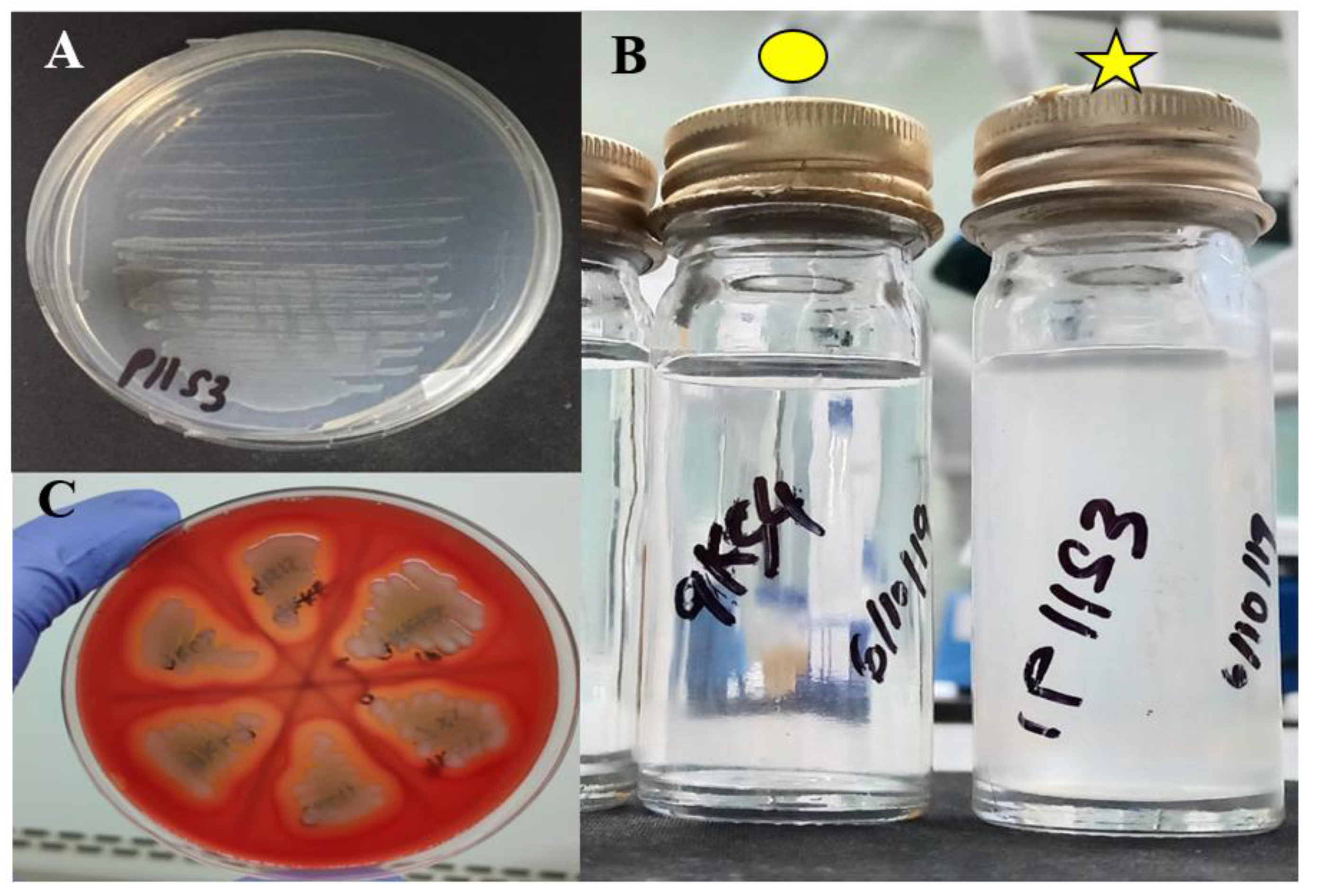
2.1. Collection of Aeromonas spp.
A total of 124 Aeromonas isolates that were previously obtained from fish with chronic ulcerative infection were used. The isolates were identified in previous experiment [] as A. dhakensis (n = 53), A. hydrophila (n = 25), A. veronii (n = 27), A. caviae (n = 10), and A. jandaei (n = 9) using primers that identified the gyrB gene []. The isolates were streaked onto tryptic soy agar (TSA) (Oxoid, Hampshire, UK) and further sub-cultured onto Aeromonas selective agar (ASA) (Oxoid, Hampshire, UK) and incubated at 30 °C overnight.
2.2. Screening for Myo-Inositol Utilizing Strains and Blood Agar Characteristics
All isolates were screened for the ability to utilize myo-inositol using both agar and broth methods. The M9 agar (Difco, Sparks, MD, USA) with myo-inositol was prepared as previously described []. A total of 30 mL of sterile 10% myo-inositol and 2 mL of 1.0 M MgSO4 were added into the agar after it was cooled down to 55 °C. Observation for myo-inositol utilizing strains was completed after 24 h of incubation at 30 °C for both agar and broth methods. The bacteria that grew beyond 48 h were discarded. The isolates were also screened using TSA with 7% horse blood to check for the type of haemolysis.
2.3. Acclimatization of Red Hybrid Tilapia
A total of 380 juvenile red hybrid tilapias (Oreochromis mossambicus × O. niloticus) with mean ± standard deviation (SD) weight of 70.00 ± 6.81 g were obtained from a local commercial fish farm. The fish were acclimatized in a 500 L fibre-glass tank for 14 days with constant aeration. Approximately 30% of the water was changed while water quality parameters were monitored daily. The mean ± SD of water parameters, including pH (6.71 ± 0.14), temperature (25.51 °C ± 0.04 °C), salinity (0.25 ± 0.01 ppt) and total dissolved solid (342.5 ± 5.7 mg/L) were determined using a YSI meter (Yellow Springs Instrument, Inc., Yellow Springs, OH, USA). Throughout the experiment, the fish were fed two times a day ad libitum with commercial feed containing 32% crude protein. All procedures involving fish in this study were approved by the Institutional Animal Care and Use Committee, Universiti Putra Malaysia (AUP No: UPM/IACUC/AUP-R076/2019).
2.4. Preparation of Different Concentrations of Aeromonas spp.
Six isolates of Aeromonas spp. were selected for the pathogenicity study based on the number of virulence genes present in each isolate []. The selected isolates were the myo-inositol utilizing A. dhakensis 1P11S3 and the non-myo-inositol utilizing A. dhakensis 4PS2, A. hydrophila 8TK3, A. veronii 6TS5, A. caviae 7X11, and A. jandaei 7KL3. The five non-myo-inositol utilizing Aeromonas spp. had highest number of virulence genes within each species (Table A1) []. Briefly, five colonies on ASA were randomly picked and inoculated into 1 L of tryptic soy broth (TSB; Oxoid), and incubated for 18 h at 30 °C. Serial dilutions were performed up to tenfold and 1 mL of TSB from each dilution was plated on TSA in triplicate, and further incubated at 30 °C overnight. The colony forming units per millilitre (CFU/mL) were determined using standard plate count.
2.5. Pathogenicity Study
For pathogenicity test, there were six Aeromonas spp. and a control group involved. Therefore, a total of 380 juvenile red hybrid tilapias were randomly distributed into glass tanks of 20 L (18 challenge tanks + 1 control tank). The fish were divided into six groups (6 groups × 3 bacterial dilutions × 20 fish/tank) of bacterial isolates and 1 group of control (n = 20). The fish were anaesthetised using MS222 (Western Chemical Inc., Ferndale, WA, USA) at a concentration of 150 µg/L [], before being inoculated intraperitoneally with 0.1 mL of the suspension containing respective Aeromonas sp. at 103 CFU/mL, 105 CFU/mL and 107 CFU/mL. Control fish were similarly inoculated with sterile TSB. All fish were observed for clinical signs and mortalities for a period of 10 days. The LD50–240h was calculated using Probit analysis with SPSS 25 [].
2.6. Histopathological Assessment
All moribund fish (groups with dilution of 107 CFU/mL) were immediately sampled and prior to histopathological evaluation, the fish were checked for lesions and haemorrhages. The kidney, liver and spleen samples were collected (n = 5 samples/group) and fixed in 10% neutral-buffered formalin for at least 24 h before routinely processed using paraffin-embedded technique, sectioned at 4 µm, stained with haematoxylin-eosin (H&E) and examined under a light microscope. The severity of the gross and histopathological changes in each of the internal organs were described and scored as follows; none: 0% affected (0), mild: <30% affected (1), moderate: 30–60% affected (2) and severe: >60% affected (3) [].
2.7. Statistical Analysis
The mean of histopathological scorings in selected internal organs of the fish were compared using Kruskal–Wallis H test (SPSS, IBM Corporation, NY, USA). Before carrying out the Kruskal–Wallis H test, normality of the variance was evaluated using Shapiro–Wilk test, respectively. The results were considered significant at p < 0.05.
3. Results
3.1. Characterization of Aeromonas spp.
Out of the 124 Aeromonas isolates tested in this study, one isolate, the A. dhakensis 1P11S3 was found to utilize myo-inositol. The growth on M9 agar was observed as early as 12 h post-incubation and could clearly be seen after 24 h, while the M9 broth became turbid after 24 h of incubation with A. dhakensis 1P11S3 (Figure A1). All isolates (n = 124) produced complete haemolysis (β-haemolysis) on 7% blood agar (n = 124).
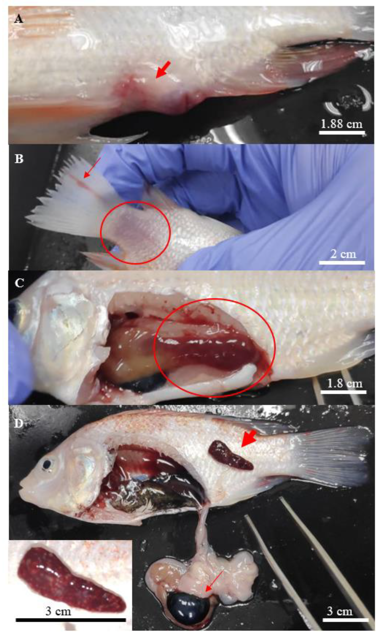
3.2. Clinicopathological Changes of Infected Fish
In general, all Aeromonas-infected fish showed various degrees of typical gross lesions and clinical signs of aeromoniasis. These symptoms included irregular breathing and lethargy, reduced feed consumption, displayed erratic movement with loss of balance, isolating and swimming to the surface of the water. The gross lesions included swollen and haemorrhagic site of injection (Figure 1A), scale loss that exposed the underlying skin at the caudal fin (Figure 1B) and necrosis of the fins (Figure 1B).
Figure 1.
Clinical signs found on the hybrid red tilapias infected with Aeromonas. (A) focal haemorrhage on the site of injection, (B) scale loss (circle) at the caudal peduncle with rotting caudal fin (thin arrow), (C) liver with deep haemorrhage (circle), and (D) haemorrhage with multiple white spots observed on the enlarged spleen (thick arrow), enlarged gallbladder (thin arrow), with a close-up picture of the affected spleen at the bottom left.
Post-mortem examinations revealed hepatomegaly (Figure 1C) in a majority of the infected fish, while multifocal splenic infarction (Figure 1D) was observed in fish infected with A. hydrophila 8TK3. Some infected fish had a haemorrhagic spleen and swollen gall bladder (Figure 1D). Overall, fish infected by A. hydrophila 8TK3 and A. dhakensis 4PS2 showed more severe gross lesions than other isolates. All control fish exhibited neither clinical sign nor gross lesion.
3.3. Lethal Dose of Aeromonas spp. in Red Hybrid Tilapia
The rates of mortality following infection by different isolates of Aeromonas were summarized in Table 1. Mortalities were observed as early as 24 h post-challenge in all infected groups, except for A. jandaei 7KL3 and lasted for 216 h. By 240 h, the cumulative mortality of fish challenged with 103 CFU/mL of bacteria ranged between 0% and 20%, between 20% and 60% with 105 CFU/mL and between 35% and 95% with 107 CFU/mL. The two highest cumulative mortalities (95% and 90%) involved non-myo-inositol utilizing A. dhakensis 4PS2 (107 CFU/mL) and A. hydrophila 8TK3 (107 CFU/mL), which was higher than the myo-inositol utilizing A. dhakensis 1P11S3 (107 CFU/mL) with 55% cumulative mortality.
Table 1.
The number of mortalities of red hybrid tilapias infected with six strains of Aeromonas spp.
The LD50–240h for all Aeromonas spp. were presented in Table 1. The lowest LD50–240h of 105 CFU/mL was for A. hydrophila 8TK3 and A. dhakensis 4PS2, lower than the 107 CFU/mL of A. dhakensis 1P11S3, A. veronii 6TS5 and A. caviae 7X11, while the highest LD50–240h of 1011 CFU/mL was calculated for A. jandaei 7KL3.
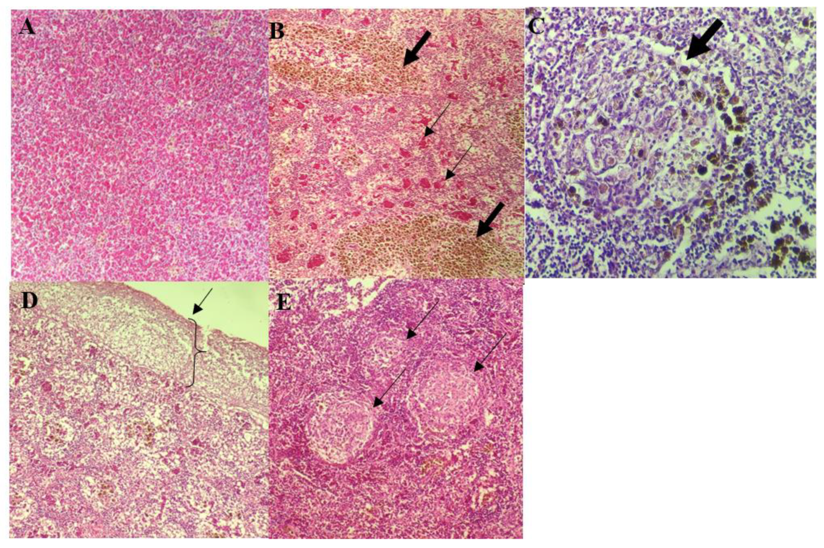
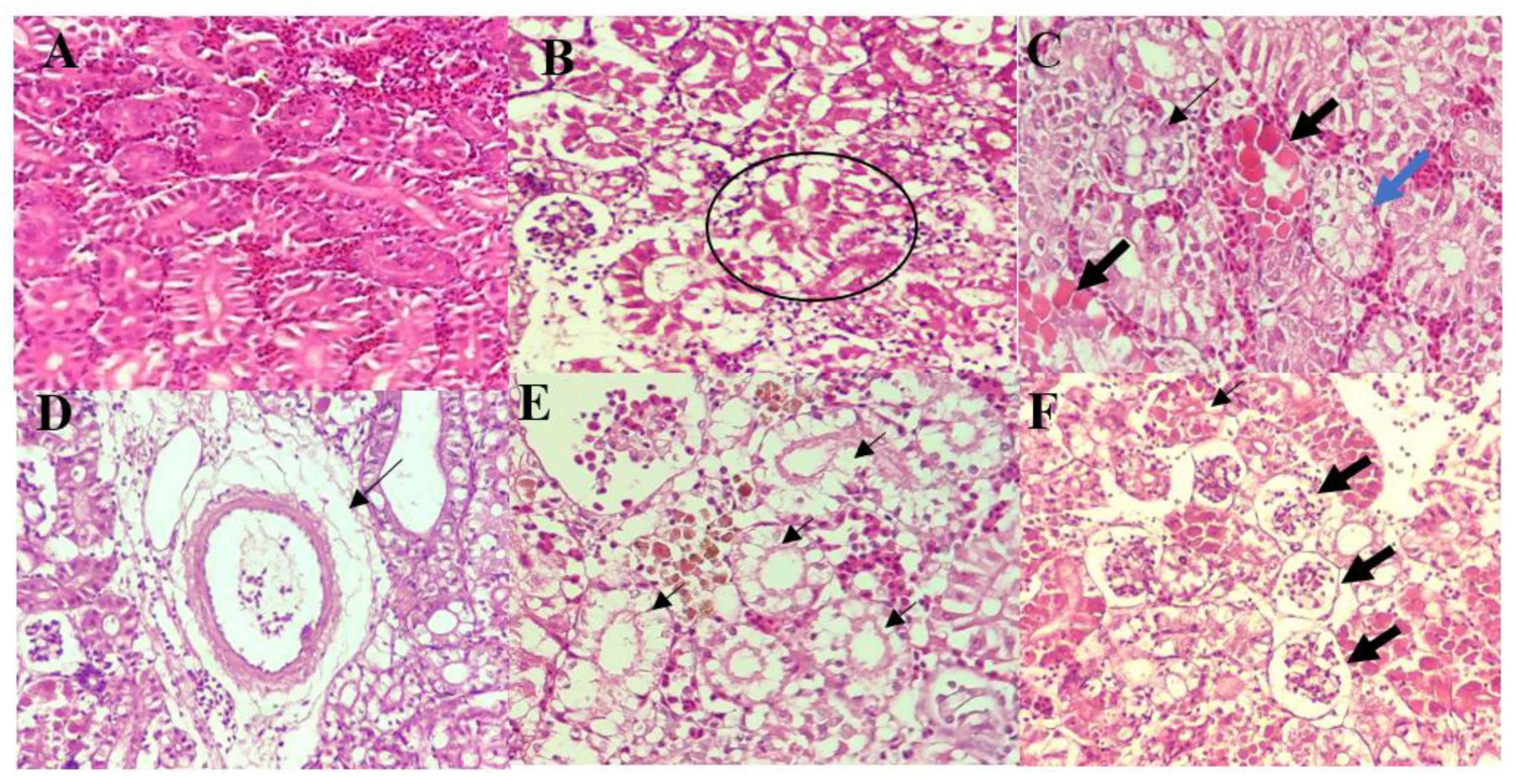
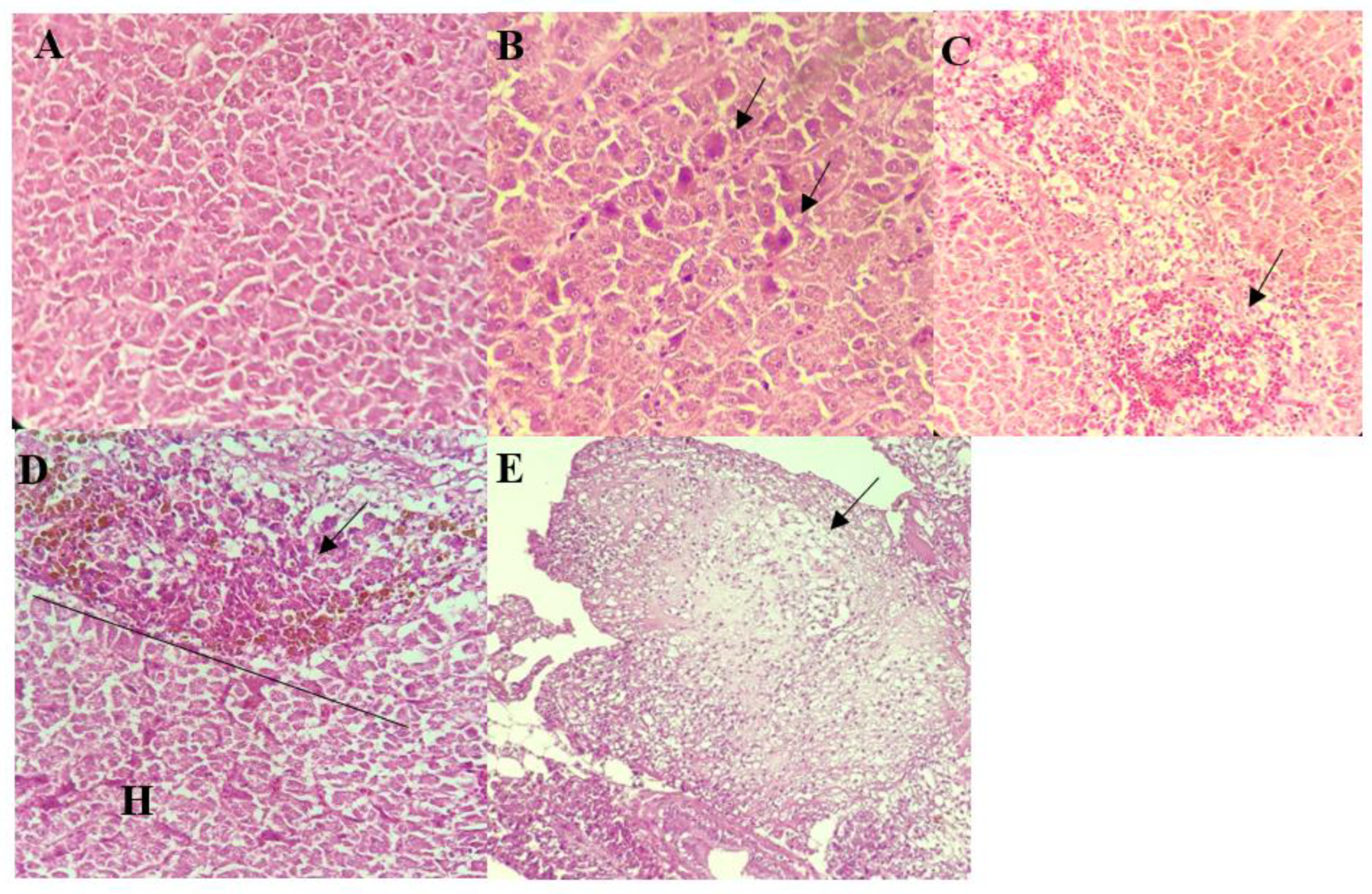
3.4. Histopathological Analysis
The livers of all infected fish appeared congested with necrosis of hepatocytes. These were frequently accompanied by moderate to severe hydropic degeneration of the hepatocytes (Figure 2B,C). Individual necrosis of the hepatocytes was most severe in fish infected by A. hydrophila 8TK3 (p < 0.05), followed by A. dhakensis 4PS2, A. dhakensis 1P11S3, A. veronii 6TS5, A. caviae 7X11, and A. jandaei 7KL3. Infiltration of inflammatory cells, haemorrhages and hemosiderosis were also observed in the liver of all groups (Figure 2D). In addition, liver of fish infected with A. hydrophila 8TK3 showed hepatic infarction (Figure 2E).
Figure 2.
Histological findings of the liver of control and infected red hybrid tilapias by Aeromonas spp. (A) Liver of control fish showed normal liver architecture and hepatocytes, 400×. (B) Individual hepatocyte necrosis exhibiting intense cytoplasmic eosinophilia (arrows), 400×. (C) Extensive hydropic degeneration and necrosis of cells surrounding a central vein (arrow), 400×. (D) Infiltration of inflammatory cells. Note that some inflammatory cells are showing brownish cytoplasmic pigmentation suggestive of hemosiderosis (arrow) ((H): healthy cells), 400×. (E) Hepatic infarction showing complete loss of liver architecture (arrow), 400×.
Normal splenic architecture was observed in control group with presence of numerous erythrocytes in the red pulp and mildly scattered MMC (Figure 3A). On the other hand, the spleens of infected fish appeared congested involving multiple blood vessels with numerous MMC (Figure 3B), and infiltration of inflammatory cells associated with multifocal areas of splenic necrosis (Figure 3C). Different Aeromonas spp. were observed to produce different severity of lesions, ranging from mild to severe. Spleens of fish infected by A. hydrophila 8TK3 showed complete loss of splenic architecture (Figure 3D). Fish infected with A. jandaei 7KL3 showed white pulp hypoplasia, affecting small multifocal areas (Figure 3E). In general, fish infected with A. hydrophila 8TK3 showed most severe splenic lesion (p < 0.05), followed by A. dhakensis 4PS2, A. dhakensis 1P11S3, A. veronii 6TS5, A. caviae 7X11 and A. jandaei 7KL3.
Figure 3.
Histological findings of the spleen of control and infected red hybrid tilapias by Aeromonas spp. (A) Normal spleen of control fish showing normal splenic architecture, 200×. (B) Presence of abundant MMC (thick arrow) and multiple areas of splenic vascular congestion (thin arrow), 400×. (C) Severe, focal area of necrosis (arrow), with the presence of MMC, 400×. (D) Severe necrosis observed mainly at the splenic surface. 200×. (E) Multifocal areas of splenic white pulp hypoplasia (arrow), 200×.
The kidneys of control fish showed normal architecture with intact, healthy tubular epithelia (Figure 4A). However, all groups of infected fish showed varying degree of tubular epithelial and glomerular degeneration and necrosis (Figure 4B–D). Necrosis of tubular epithelial cells was seen as either desquamation from the basement membrane or karyolysis of the nucleus (Figure 4C). The latter was only observed in fish infected by A. hyrophila 8TK3, A. dhakensis 4PS2, and A. caviae 7X11. Fish infected by A. jandaei 7KL3 showed perivascular oedema, accompanied by necrotic tubular epithelium and infiltration of inflammatory cells (Figure 4D). These lesions were not seen in fish infected by other species of Aeromonas. Additionally, the tubular epithelial changes were more severe among these groups (A. dhakensis 4PS2 and A. hydrophila 8TK3), where complete lysis of tubular epithelial cells was frequently observed (Figure 4E). All fish infected with Aeromonas spp. showed desquamation (Figure 4F) and atrophied glomerulus. Cumulatively, histopathological changes in the kidney were most severe in fish infected with A. dhakensis 4PS2 and A. hydrophila 8TK3 (p < 0.05), followed by A. jandaei 7KL3, A. dhakensis 1P11S3, A. veronii 6TS5, and A. caviae 7X11.
Figure 4.
Histological findings of the kidney of control and infected red hybrid tilapias by Aeromonas spp. (A) Kidney of control fish showing normal and intact renal epithelial cells, 400×. (B) Hydropic degeneration (circle) of tubular epithelium, 400×. (C) Karyolysis and severe cytoplasmic eosinophilia of the tubular epithelial cells (thick arrow), with detachment of tubular epithelium from the basement membrane (thin arrow), and severe vacuolation (blue arrow), 400×. (D) Dilution of tissue surrounding a blood vessel suggestive of perivascular oedema, 400×. (E) Severe lysis of tubular epithelium (thin arrow), 400×. (F) Necrosis of glomerulus (thick arrow) and karyolysis of tubular epithelial cells (thin arrow), 400×.
The result of all histopathological scoring of liver, spleen and kidney was presented in Table 2. In summary, the most severe histopathological changes were observed in liver (A. hydrophila 8TK3), spleen (A. hydrophila 8TK3) and kidney (A. hydrophila 8TK3 and A. dhakensis 4PS2) of the infected fish. The myo-inositol utilizing A. dhakensis 1P11S3 caused mild to moderate histopathological changes in liver, spleen and kidney of the red hybrid tilapia.
Table 2.
Summary of histopathological changes scoring in liver, spleen and kidney of red hybrid tilapias following experimental infection by Aeromonas spp.
4. Discussion
Recent reports on hypervirulent vAh strains have raised concern, particularly on the possibility of the spread of this hypervirulent pathotype in different countries and farmed fish species. This study identified only one strain, A. dhakensis 1P11S3, has the ability to use myo-inositol as a sole carbon source, which to our knowledge represents the first report of A. dhakensis that could utilize myo-inositol. None of the A. hydrophila isolates could utilize myo-inositol, suggesting that the vAh pathotype is not present or at least not common among farmed freshwater fish in Malaysia.
The subsequent challenge study produced typical signs and gross lesions of Aeromonas infection as reported by Soto-Rodriguez et al. [] and Saharia et al. [] in Nile tilapia (O. niloticus) and Indian major carp (Labeo rohita). There was increasing rate of mortality with increasing bacterial concentration, since more toxins would be released that could damage the internal organs of the fish []. Infected fish died as early as 24 h and as late as 216 h, highlighting the ability of Aeromonas spp. to cause acute-to-chronic infection in fish. The histopathological changes in the internal organs of red hybrid tilapia were also typical following Aeromonas spp. infection. In liver, changes were manifested as necrosis of liver parenchyma [,,]. Splenic necrosis was also observed, similarly reported by previous studies [,]. Necrosis of kidney was characterized by swollen, desquamated and vacuolated tubular epithelial cells with some cells having complete necrosis, while atrophied glomerulus were also reported in previous studies [,,]. Other histopathological changes such as the presence of polymorphonuclear cells in the inter tubular area, along with proliferated cell of glomerulus, were not observed in this study [,]. Alternatively, the presence of haemocyte aggregation and congestion in all selected internal organs indicated that liver, spleen and kidney are the target organ for Aeromonas systemic infection [,,]. In addition, histopathological changes in the liver, spleen, and kidney were highly suggestive of acute to subacute cellular injury. It is worth mentioning that no abnormalities were observed following infection by the myo-inositol utilizing A. dhakensis 1P11S3, indicating that inositol catabolism per se did not contribute to A. dhakensis pathogenesis in tilapia with current disease model. Furthermore, all isolates used in this study were obtained from diseased fish, without any relation with epidemic outbreak []. On the other hand, myo-inositol utilizing A. hydrophila vAh, that were isolated during epidemic outbreak among catfish, have been reported to cause severe clinical MAS symptoms with rapid onset of mortality []. Zhang et al. [] reported 90% mortality among catfish challenged with vAh strains; however, the disease model included clipping of adipose fin and immersion in a high dose of 107 CFU/mL vAh, processes that were not conducted in the present study. Therefore, modification of the challenge model should be conducted in future study.
Based on the LD50–240h, the most virulent Aeromonas strains were A. dhakensis 4PS2 and A. hydrophila 8TK3 (105 CFU/mL), while the least virulent was A. jandaei 7KL3 (1011 CFU/mL). Previous reports revealed the LD50 of A. dhakensis was between 102 and 105 CFU/mL when challenged in zebrafish (Danio rerio), rainbow trout (Oncorhynchus mykiss) and pacu (Piaractus mesopotamicus) [,,]. Similarly, few studies reported LD50 < 106 CFU/mL for A. hydrophila in zebrafish, rainbow trout and tilapia [,,].
When the LD50 of each isolate was compared with the presence of virulence genes [], A. dhakensis 4PS2 and A. hydrophila 8TK3 were found to have all of the virulence genes that were tested. In addition, both strains also possessed the aer gene, which might play a significant role in the virulence of Aeromonas. On the other hand, A. dhakensis 1P11S3 (myo-inositol utilizing Aeromonas) and A. caviae 7X11 shared the same number of virulence genes (only 62.5% of the tested virulence genes), whereas A. veronii 6TS5 had fewer virulence genes (50% of the tested genes), while sharing the same LD50–240h. In addition, those three strains also harboured the fla and ela genes, while A. jandaei harboured the fewest virulence genes (37.5% of the tested genes) and was found to lack the ahp, fla, alt, lip and aer genes. While there are many factors that can contribute to pathogenesis beyond the presence or absence of genes (e.g., gene expression), it was noteworthy that the presence of the aer gene was the most highly correlated with Aeromonas strain virulence. In addition, Zhang et al. [] reported that aerolysin as one of the most potent toxins produced by A. hydrophila, where acute mortality of catfish was observed as early as 3 h following exposure to either proaerolysin (inactive form) or aerolysin (activated form).
Following the challenge test, A. dhakensis 1P11S3 (myo-inositol utilizing) appeared to lack significant virulence under current tested conditions; however, it is possible that the ability to utilize myo-inositol may contribute to A. dhakensis persistence within aquaculture pond ecosystems through the use of myo-inositol, which is present in plant-based feeds that contain phytate []. Therefore, to arrive at the conclusion that myo-inositol utilizing ability did not translate to increased virulence in Aeromonas spp., more research and verification is required. We postulated that there may be different genomic representation of myo-inositol utilizing vAh with our A. dhakensis 1P11S3, since both are of different Aeromonas spp. In vAh strains, myo-inositol metabolism was found to be isolated, instead of dispersed like Corynebacterium glutamicum and Caulobacter crescentus [,]; the cluster was found in genomic island 2, an epidemic-associated region. The genomic island 2 was also determined to encode other putative virulence factors such as gene bpIA (probable oxidoreductase) and hitC (iron (III) ABC transporter) []. In addition, our study elucidated the influence of eight virulence genes on the virulence of Aeromonas sp. based on the result of the challenge test. However, the possibility of other virulence factors that might have even greater significance in myo-inositol utilizing Aeromonas sp. should not be neglected. Rasmussen-Ivey et al. [] reported that more virulent vAh isolates lacked several components of type VI secretion system (T6SS). Meanwhile, T6SS in A. dhakensis was very crucial for its virulence [], but in A. hydrophila, it was determined to have a least pronounced effect []. Nevertheless, horizontal gene transfer may have occurred between vAh strain and A. dhakensis 1P11S3, which to the author’s knowledge, no study has reported this ability among A. dhakensis. Therefore, future studies should be conducted immediately on the whole-genome sequencing of A. dhakensis 1P11S3, which can potentially reveal the in-depth state of the virulence of the isolate, alongside the genotypic properties of myo-inositol catabolism cluster of the isolate. The study also indicates that there are diverse, pathogenic Aeromonas spp. that contribute to disease prevalence within farmed freshwater fish in Malaysia.
5. Conclusions
In conclusion, this study reports for the first time that A. dhakensis could utilize myo-inositol as a sole carbon source. A subsequent pathogenicity study revealed that the non-myo-inositol utilizing A. hydrophila and A. dhakensis was the most pathogenic among the tested isolates. While no vAh strains were identified in this study, other virulent Aeromonas spp. isolates that were highly virulent in farmed fish were identified.
Author Contributions
M.A.-S.: Conceptualization, Validation, Data curation, Formal analysis, Investigation, Writing—original draft, Writing—review and editing. M.Y.I.-S.: Conceptualization, Validation, Data curation, Writing—review and editing, Funding acquisition, Supervision. M.Z.-S.: Data curation, Writing—review and editing. M.T.Y.: Writing—review and editing, S.A.: Data curation, Writing—review and editing. M.R.L.: Writing—review and editing, Data curation. M.S.M.: Data collection, Writing—review and editing. A.M.: Data collection, Writing—review and editing. M.H.N.M.-S.: Data collection, Writing—review and editing. J.Y.L.: Data collection, Writing—review and editing. Y.K.C.: Data collection, Writing—review and editing. Z.A.-D.: Data collection, Writing—review and editing. A.A.: Data collection, Writing—review and editing. B.L.: Data collection, Formal analysis, Writing—review and editing. M.N.A.A.: Supervision, Conceptualization, Data curation, Writing—review and editing. All authors have read and agreed to the published version of the manuscript.
Funding
This research was supported by using the grants of Long Term Research Grant Scheme (LRGS), code: LRGS/1/2019/UPM/01/1/2; Higher Institution Centre of Excellence (HiCoE), vot no.: 6369100 provided by the Ministry of Higher Education Malaysia.
Institutional Review Board Statement
All procedures involving fish in this study were approved by the Institutional Animal Care and Use Committee, Universiti Putra Malaysia (AUP No: UPM/IACUC/AUP-R076/2019).
Informed Consent Statement
Not applicable.
Data Availability Statement
Not applicable.
Conflicts of Interest
The authors declare no conflict of interest.
Appendix A
Table A1.
Summary of virulence genes identified in each selected Aeromonas isolates that were conducted in the previous study (Azzam-Sayuti et al., 2021).
Table A1.
Summary of virulence genes identified in each selected Aeromonas isolates that were conducted in the previous study (Azzam-Sayuti et al., 2021).
| Species | Strain | Virulence Genes | NCBI Accession Number | |||||||
|---|---|---|---|---|---|---|---|---|---|---|
| ahp | fla | act | ela | hly | alt | lip | aer | |||
| A. dhakensis | 1P11S3 * | + | + | + | + | + | MT371976 | |||
| A. dhakensis | 4PS2 | + | + | + | + | + | + | + | + | MT372009 |
| A. hydrophila | 8TK3 | + | + | + | + | + | + | + | + | MT372063 |
| A. veronii | 6TS5 | + | + | + | + | MT372033 | ||||
| A. caviae | 7X11 | + | + | + | + | + | MT372047 | |||
| A. jandaei | 7KL3 | + | + | + | MT372043 | |||||
* Indicate isolate that can utilize myo-inositol as a sole carbon source. Abbreviations: ahp, serine protease; fla, flagella; act, cytotoxic heat-labile enterotoxin; ela, elastase; hly, haemolysin; alt, cytotonic heat-labile; lip, lipase; aer, aerolysin.
Figure A1.
The growth characteristics of Aeromonas isolates. (A) positive growth of myo-inositol utilizing A. dhakensis 1P11S3 after 24 h of incubation, (B) turbidity observed on M9 with myo-inositol broth (star) as compared to clear appearance of the broth that indicates negative growth (round), and (C) complete haemolysis (b-haemolysis) surrounding A. dhakensis colonies on 7% blood agar.
References
- FAO. The State of World Fisheries and Aquaculture 2020. Sustainability in Action; FAO: Rome, Italy, 2020; Available online: http://www.fao.org/3/ca9229en/ca9229en.pdf (accessed on 26 June 2021).
- FAO. The State of World Fisheries and Aquaculture; FAO: Rome, Italy, 2010; p. 197. Available online: http://www.fao.org/documents/card/en/c/f9f071ce-7bee-5107-a343-097685d0d683/ (accessed on 26 June 2021).
- Mohamad, S.N.; Noordin, W.N.M.; Ismail, N.F.; Hamzah, A. Red hybrid tilapia (Oreochromis spp.) broodstock development programme in Malaysia: Status, challenges and prospects for future development. Asian Fish. Sci. 2021, 34, 73–81. [Google Scholar]
- Huicab-Pech, Z.; Landeros-Sánchez, C.; Castañeda-Chávez, M.; Lango-Reynoso, F.; López-Collado, C.; Platas Rosado, D. Current state of bacteria pathogenicity and their relationship with host and environment in tilapia Oreochromis niloticus. J. Aquac. Res. Dev. 2016, 7, 428. [Google Scholar]
- Wang, M.; Lu, M. Tilapia polyculture: A global review. Aquac. Res. 2016, 47, 2363–2374. [Google Scholar] [CrossRef]
- Monir, M.S.; Yusoff, S.M.; Mohamad, A.; Ina-Salwany, M.Y. Vaccination of tilapia against motile Aeromonas septicemia: A review. J. Aquat. Anim. Health 2020, 32, 65–76. [Google Scholar] [CrossRef]
- Singh, V.; Rathore, G.; Kapoor, D.; Mishra, B.N.; Lakra, W.S. Detection of aerolysin gene in Aeromonas hydrophila isolated from fish and pond water. Indian J. Microbiol. 2008, 48, 453–458. [Google Scholar] [CrossRef] [PubMed] [Green Version]
- Kozińska, A.; Pkala, A. Characteristics of disease spectrum in relation to species, serogroups, and adhesion ability of motile aeromonads in fish. Sci. World J. 2012, 2012, 949358. [Google Scholar] [CrossRef] [Green Version]
- Jagoda, S.S.d.S.; Wijewardana, T.G.; Arulkanthan, A.; Igarashi, Y.; Tan, E.; Kinoshita, S.; Watabe, S.; Asakawa, S. Characterization and antimicrobial susceptibility of motile aeromonads isolated from freshwater ornamental fish showing signs of septicaemia. Dis. Aquat. Organ. 2014, 109, 127–137. [Google Scholar] [CrossRef] [Green Version]
- Dong, H.T.; Techatanakitarnan, C.; Jindakittikul, P.; Thaiprayoon, A.; Taengphu, S.; Charoensapsri, W.; Khunrae, P.; Rattanarojpong, T.; Senapin, S. Aeromonas jandaei and Aeromonas veronii caused disease and mortality in Nile tilapia, Oreochromis niloticus (L.). J. Fish Dis. 2017, 40, 1395–1403. [Google Scholar] [CrossRef] [PubMed]
- Amal, M.N.A.; Koh, C.B.; Nurliyana, M.; Suhaiba, M.; Nor-Amalina, Z.; Santha, S.; Diyana-Nadhirah, K.P.; Yusof, M.T.; Ina-Salwany, M.Y.; Zamri-Saad, M. A case of natural co-infection of Tilapia Lake Virus and Aeromonas veronii in a Malaysian red hybrid tilapia (Oreochromis niloticus × O. mossambicus) farm experiencing high mortality. Aquaculture 2018, 485, 12–16. [Google Scholar] [CrossRef]
- Basri, L.; Nor, R.M.; Salleh, A.; Yasin, I.S.M.; Saad, M.Z.; Rahaman, N.Y.A.; Barkham, T.; Amal, M.N.A. Co-Infections of Tilapia Lake Virus, Aeromonas hydrophila and Streptococcus agalactiae in farmed red hybrid tilapia. Animals 2020, 10, 2141. [Google Scholar] [CrossRef] [PubMed]
- Stratev, D.; Odeyemi, O.A. Antimicrobial resistance of Aeromonas hydrophila isolated from different food sources: A mini-review. J. Infect. Public Health 2016, 9, 535–544. [Google Scholar] [CrossRef] [PubMed] [Green Version]
- Emeish, W.F.; Mohamed, H.M.; Elkamel, A.A. Aeromonas infections in African sharptooth catfish. J. Aquac. Res. Dev. 2018, 9, 548. [Google Scholar] [CrossRef]
- Saharia, P.; Pokhrel, H.; Kalita, B.; Hussain, I.A. Histopathological changes in Indian major carp, Labeo rohita (Hamilton), experimentally infected with Aeromonas hydrophila associated with hemorrhagic septicemia of Central Brahmaputra valley of Assam, India. J. Entomol. Zool. Stud. 2018, 6, 6–11. [Google Scholar]
- AlYahya, S.A.; Ameen, F.; Al-Niaeem, K.S.; Al-Sa’adi, B.A.; Hadi, S.; Mostafa, A.A. Histopathological studies of experimental Aeromonas hydrophila infection in blue tilapia, Oreochromis aureus. Saudi J. Biol. Sci. 2018, 25, 182–185. [Google Scholar] [CrossRef]
- Musthafa, M.S.; Asgari, S.M.; Elumalai, P.; Hoseinifar, S.H.; Doan, H.V. Protective efficacy of Shilajit enriched diet on growth performance and immune resistance against Aeromonas hydrophila in Oreochromis mossambicus. Fish Shellfish Immunol. 2018, 82, 147–152. [Google Scholar] [CrossRef] [PubMed]
- Hoel, S.; Vadstein, O.; Jakobsen, A.N. Species distribution and prevalence of putative virulence factors in mesophilic Aeromonas spp. isolated from fresh retail sushi. Front. Microbiol. 2017, 8, 931. [Google Scholar] [CrossRef] [PubMed]
- Soto-Rodriguez, S.A.; Lozano-Olvera, R.; Garcia-Gasca, M.T.; Abad-Rosales, S.; Gomez-Gil, B.; Ayalla-Arellano, J. Virulence of the fish pathogen Aeromonas dhakensis: Genes involved, characterization and histopathology of experimentally infected hybrid tilapia. Dis. Aquat. Organ. 2018, 129, 107–116. [Google Scholar] [CrossRef] [Green Version]
- Popovic, N.T.; Teskeredzic, E.; Strunjak-Perovic, I.; Coz-Rakovac, R. Aeromonas hydrophila isolated from wild freshwater fish in Croatia. Vet. Res. Commun. 2000, 24, 371–377. [Google Scholar] [CrossRef]
- Rasmussen-Ivey, C.R.; Hossain, M.J.; Odom, S.E.; Terhune, J.S.; Hemstreet, W.G.; Shoemaker, C.A.; Zhang, D.; Xu, D.-H.; Griffin, M.J.; Liu, Y.-J.; et al. Classification of a hypervirulent Aeromonas hydrophila pathotype responsible for epidemic outbreaks in warm-water fishes. Front. Microbiol. 2016, 7, 1615. [Google Scholar] [CrossRef] [Green Version]
- Awan, F.; Dong, Y.; Liu, J.; Wang, N.; Mushtaq, M.H.; Lu, C.; Liu, Y. Comparative genome analysis provides deep insights into Aeromonas hydrophila taxonomy and virulence-related factors. BMC Genom. 2018, 19, 712. [Google Scholar] [CrossRef] [Green Version]
- Bebak, J.; Wagner, B.; Burnes, B.; Hanson, T. Farm size, seining practices, and salt use: Risk factors for Aeromonas hydrophila outbreaks in farm-raised catfish, Alabama, USA. Prev. Vet. Med. 2015, 118, 161–168. [Google Scholar] [CrossRef] [PubMed]
- Hossain, M.J.; Sun, D.; McGarey, D.J.; Wrenn, S.; Alexander, L.M.; Martino, M.E.; Xing, Y.; Terhune, J.S.; Liles, M.R. An Asian origin of virulent Aeromonas hydrophila responsible for disease epidemics in United States-farmed catfish. Am. Soc. Microbiol. 2014, 5, 3. [Google Scholar] [CrossRef] [Green Version]
- Peatman, E.; Mohammed, H.; Kirby, A.; Shoemaker, C.A.; Yildirim-Aksoy, M.; Beck, B.H. Mechanisms of pathogen virulence and host susceptibility in virulent Aeromonas hydrophila infections of channel catfish (Ictalurus punctatus). Aquaculture 2018, 482, 1–8. [Google Scholar] [CrossRef]
- Hossain, M.J.; Waldbieser, G.C.; Sun, D.; Capps, N.K.; Hemstreet, W.B.; Carlisle, K.; Griffin, M.J.; Khoo, L.; Goodwin, A.E.; Sonstegard, T.S.; et al. Implication of lateral genetic transfer in the emergence of Aeromonas hydrophila isolates of epidemic outbreaks in channel catfish. PLoS ONE 2013, 8, e80943. [Google Scholar] [CrossRef] [PubMed] [Green Version]
- Baumgartner, W.A.; Ford, L.; Hanson, L. Lesions caused by virulent Aeromonas hydrophila in farmed catfish (Ictalurus punctatus and I. punctatus × I. furcatus) in Mississippi. J. Vet. Diagn. Investig. 2017, 29, 747–751. [Google Scholar] [CrossRef] [Green Version]
- Pang, M.; Jiang, J.; Xie, X.; Wu, Y.; Dong, Y.; Kwok, A.H.Y.; Zhang, W.; Yao, H.; Lu, C.; Leung, F.C.; et al. Novel insights into the pathogenicity of epidemic Aeromonas hydrophila ST251 clones from comparative genomics. Sci. Rep. 2015, 5, 9833. [Google Scholar] [CrossRef] [Green Version]
- Janda, J.M.; Abbott, S.L. The genus Aeromonas: Taxonomy, pathogenicity, and infection. Clin. Microbiol. Rev. 2010, 23, 35–73. [Google Scholar] [CrossRef] [Green Version]
- Pu, W.; Guo, G.; Yang, N.; Li, Q.; Yin, F.; Wang, P.; Zheng, J.; Zheng, J. Three species of Aeromonas (A. dhakensis, A. hydrophila and A. jandaei) isolated from freshwater crocodiles (Crocodylus siamensis) with pneumonia and septicemia. Lett. Appl. Microbiol. 2019, 68, 212–218. [Google Scholar] [CrossRef]
- Gao, X.; Tong, S.; Zhang, S.M.; Chen, Q.; Jiang, Z.; Jiang, Q.; Wei, W.; Zhu, J.; Zhang, X. Aeromonas veronii associated with red gill disease and its induced immune response in Macrobrachium nipponense. Aquac. Res. 2020, 51, 5163–5174. [Google Scholar] [CrossRef]
- Azzam-Sayuti, M.; Ina-Salwany, M.Y.; Zamri-Saad, M.; Yusof, M.T.; Annas, S.; Najihah, M.Y.; Liles, M.R.; Monir, M.S.; Zaidi, Z.; Amal, M.N.A. The prevalence, putative virulence genes, and antibiotic resistance profiles of Aeromonas spp. isolated from cultivated freshwater fish in peninsular Malaysia. Aquaculture 2021, 540, 736719. [Google Scholar] [CrossRef]
- Yáñez, M.A.; Catalán, V.; Apráiz, D.; Figueras, M.J.; Martínez-Murcia, A.J. Phylogenetic analysis of members of the genus Aeromonas based on gyrB gene sequences. Int. J. Syst. Evol. Microbiol. 2003, 53, 875–883. [Google Scholar] [CrossRef] [Green Version]
- Hanson, L.A.; Liles, M.R.; Hossain, M.J.; Griffin, M.J.; William, G. Motile Aeromonas septicemia. In Fish Health Section Blue Book, 2nd ed.; Bethesda, M.D., Ed.; American Fisheries Society—Fish Health Section: Bethesda, Maryland, 2014; Available online: https://units.fisheries.org/fhs/wp-content/uploads/sites/30/2017/08/1.2.9-Motile-Aeromonas-Septicemia-2014.pdf (accessed on 2 May 2021).
- Raj, N.S.; Swaminathan, T.R.; Dharmaratnam, A.; Raja, S.A.; Ramraj, D.; Lal, K.K. Aeromonas veronii caused bilateral exophthalmia and mass mortality in cultured Nile tilapia, Oreochromis niloticus (L.) in India. Aquaculture 2019, 512, 734278. [Google Scholar] [CrossRef]
- Priyatha, C.V.; Chitra, K.C. Acute toxicity of triclosan on the native freshwater fish, Anabas testudineus (Bloch, 1792): Behavioural alterations and histopathological lesions. Int. J. Life Sci. 2018, 6, 166–172. [Google Scholar]
- Abdullah, S.; Omar, N.; Yusoff, S.M.; Obukwho, E.B.; Nwunuji, T.P.; Hanan, L.; Samad, J. Clinicopathological features and immunohistochemical detection of antigens in acute experimental Streptococcus agalactiae infection in red tilapia (Oreochromis spp.). SpringerPlus 2013, 2, 286. [Google Scholar] [CrossRef] [Green Version]
- Soto-Rodriguez, S.A.; Cabanillas-Ramos, J.; Alcaraz, U.; Gomez-Gil, B.; Romalde, J.L. Identification and virulence of Aeromonas dhakensis, Pseudomonas mosselii and Microbacterium paraoxydans isolated from Nile tilapia, Oreochromis niloticus, cultivated in Mexico. J. Appl. Microbiol. 2013, 115, 654–662. [Google Scholar] [CrossRef]
- Chen, N.; Jiang, J.; Gao, X.; Li, X.; Zhang, Y.; Liu, X.; Yang, H.; Bing, X.; Zjamg, X. Histopathological analysis and the immune related gene expression profiles of mandarin fish (Siniperca chuatsi) infected with Aeromonas hydrophila. Fish Shellfish Immunol. 2018, 83, 410–415. [Google Scholar] [CrossRef] [PubMed]
- Abdelhamed, H.; Ibrahim, I.; Baumgartner, W.; Lawrence, M.L.; Karsi, A. Characterization of histopathological and ultrastructural changes in channel catfish experimentally infected with virulent Aeromonas hydrophila. Front. Microbiol. 2017, 8, 1519. [Google Scholar] [CrossRef]
- Stratev, D.; Stoev, S.; Vashin, I.; Daskalov, H. Some varieties of pathological changes in experimental infection of carps (Cyprinus carpio) with Aeromonas hydrophila. J. Aquac. Eng. Fish. Res. 2015, 1, 191–192. [Google Scholar] [CrossRef]
- Zhang, D.; Xu, D.; Shoemaker, C. Experimental induction of motile Aeromonas septicemia in channel catfish (Ictalurus punctatus) by waterborne challenge with virulent Aeromonas hydrophila. Aquac. Rep. 2016, 3, 18–23. [Google Scholar] [CrossRef] [Green Version]
- Orozova, P.; Barker, M.; Austin, D.A.; Austin, B. Identification and pathogenicity to rainbow trout, Oncorhynchus mykiss (Walbaum), of some aeromonads. J. Fish Dis. 2009, 32, 865–871. [Google Scholar] [CrossRef]
- Carriero, M.M.; Maia, A.A.M.; Sousa, R.L.M.; Henrique-Silva, F. Characterization of a new strain of Aeromonas dhakensis isolated from diseased pacu fish (Piaractus mesopotamicus) in Brazil. J. Fish Dis. 2016, 39, 1285–1295. [Google Scholar] [CrossRef]
- Hu, M.; Wang, N.; Pan, Z.H.; Lu, C.P.; Liu, Y.J. Identity and virulence properties of Aeromonas isolates from diseased fish, healthy controls and water environment in China. Lett. Appl. Microbiol. 2012, 55, 224–233. [Google Scholar] [CrossRef] [PubMed]
- Shahi, N.; Mallik, S.K.; Sahoo, M.; Das, P. Biological characteristics and pathogenicity of a virulent Aeromonas hydrophila associated with ulcerative syndrome in farmed rainbow trout, Oncorhynchus mykiss (Walbaum), in India. Isr. J. Aquac. 2013, 65, 926–936. [Google Scholar]
- Pauzi, N.A.; Mohamad, N.; Azzam-Sayuti, M.; Yasin, I.S.M.; Saad, M.Z.; Nasruddin, N.S.; Azmai, M.N.A. Antibiotic susceptibility and pathogenicity of Aeromonas hydrophila isolated from red hybrid tilapia (Oreochromis niloticus × Oreochromis mossambicus) in Malaysia. Vet. World 2020, 13, 2166–2171. [Google Scholar] [CrossRef]
- Zhang, D.; Pridgeon, J.W.; Klesius, P.H. Expression and activity of recombinant proaerolysin derived from Aeromonas hydrophila cultured from diseased channel catfish. Vet. Microbiol. 2013, 165, 478–482. [Google Scholar] [CrossRef] [PubMed]
- Kumar, V.; Sinha, A.K.; Makkar, H.P.S.; Boeck, G.D.; Becker, K. Phytate and phytase in fish nutrition. J. Anim. Physiol. Anim. Nutr. 2012, 96, 335–364. [Google Scholar] [CrossRef]
- Krings, E.; Krumbach, K.; Bathe, B.; Keller, R.; Wendisch, V.; Sahm, H.; Eggeling, L. Characterization of myo-inositol utilization by Corynebacterium glutamicum: The stimulon, identification of transporters, and influence on L-Lysine formation. J. Bacteriol. 2006, 188, 8054–8061. [Google Scholar] [CrossRef] [Green Version]
- Boutte, C.C.; Srinivasan, B.S.; Flannick, J.A.; Novak, A.F.; Martesans, A.T.; Batzoglou, S.; Viollier, P.H.; Crosson, S. Genetic and computational identification of a conserved bacterial metabolic module. PLoS Genet. 2008, 4, e1000310. [Google Scholar] [CrossRef] [Green Version]
- Chen, P.L.; Lamy, B.; Ko, W.C. Aeromonas dhakensis, an increasingly recognized human pathogen. Front. Microbiol. 2016, 7, 0793. [Google Scholar] [CrossRef]
- Rasmussen-Ivey, C.R.; Figueras, M.J.; McGarey, D.; Liles, M.R. Virulence factors of Aeromonas hydrophila: In the wake of reclassification. Front. Microbiol. 2016, 7, 01337. [Google Scholar] [CrossRef]
Publisher’s Note: MDPI stays neutral with regard to jurisdictional claims in published maps and institutional affiliations. |
© 2021 by the authors. Licensee MDPI, Basel, Switzerland. This article is an open access article distributed under the terms and conditions of the Creative Commons Attribution (CC BY) license (https://creativecommons.org/licenses/by/4.0/).