Article

Krüppel-homologue 1 Mediates Hormonally Regulated Dominance Rank in a Social Bee

1
Department of Ecology, Evolution and Behavior, The Hebrew University of Jerusalem, Jerusalem 9190401, Israel
2
Department of Ecology and Evolutionary Biology, University of Michigan, Ann Arbor, MI 48109, USA
*
Authors to whom correspondence should be addressed.
Biology 2021, 10(11), 1188; https://doi.org/10.3390/biology10111188
Submission received: 15 September 2021 / Revised: 9 November 2021 / Accepted: 10 November 2021 / Published: 15 November 2021
(This article belongs to the Section Behavioural Biology)

Abstract

:

Simple Summary

In diverse animal species, individuals establish dominance hierarchies by means of agonistic interactions. Dominance rank is functionally significant because it affects access to vital resources such as mates, food, and shelter, but little is known on the underlying genetic and molecular mechanisms, specifically in insects, and among females. We tested the hypothesis that Krüppel homologue 1, a key endocrine signaling gene, influences dominance among bumble bee female workers. We developed and validated a new nanoparticle-based protocol to down-regulate gene expression in bumble bees. Using this protocol, we show that Krüppel homologue 1 mediates endocrine influences, not only on fertility and wax production, but also on aggression and dominance rank. These findings, which establish the first causal link between a gene and a dominance rank in a social insect, are important for determining whether there are general molecular principles governing dominance ranks across gender and animal species.

Abstract

Dominance hierarchies are ubiquitous in invertebrates and vertebrates, but little is known on how genes influence dominance rank. Our gaps in knowledge are specifically significant concerning female hierarchies, particularly in insects. To start filling these gaps, we studied the social bumble bee Bombus terrestris, in which social hierarchies among females are common and functionally significant. Dominance rank in this bee is influenced by multiple factors, including juvenile hormone (JH) that is a major gonadotropin in this species. We tested the hypothesis that the JH responsive transcription factor Krüppel homologue 1 (Kr-h1) mediates hormonal influences on dominance behavior. We first developed and validated a perfluorocarbon nanoparticles-based RNA interference protocol for knocking down Kr-h1 expression. We then used this procedure to show that Kr-h1 mediates the influence of JH, not only on oogenesis and wax production, but also on aggression and dominance rank. To the best of our knowledge, this is the first study causally linking a gene to dominance rank in social insects, and one of only a few such studies on insects or on female hierarchies. These findings are important for determining whether there are general molecular principles governing dominance rank across gender and taxa.

1. Introduction

Social hierarchies are ubiquitous in invertebrates and vertebrates. Behavioral dominance is the basic principle organizing social hierarchies and is typically established by the outcome of multiple contests between pairs of conspecifics which are resolved with a clear dominance rank. Dominance rank is functionally significant given that high-ranked individuals typically gain priority access to resources, shelters, and mating opportunities [1,2,3,4]. Low dominance ranks, on the other hand, are commonly associated with stress and compromised physical conditions in many animals, including humans, even in systems with no apparent rank-related asymmetries with access to resources [5,6,7,8].
Although it is well established that dominance rank has fitness consequences, the possibility that social dominance can be genetically inherited has been a subject of recurrent debate [9,10]. The difficulties in linking dominance to specific genes stem from evidence that social rank is influenced by multiple factors that may interact in complex manners. There is evidence that a dominance rank can be passed on through social (e.g., matriline or network of allies) rather than genetic mechanisms [9]. An additional difficulty is that dominance rank is inherently context-dependent and may be expressed in only limited situations or with specific partners. The same individual may be dominant over one partner and submissive with another [11]. The outcome of a dominance hierarchy contest is commonly influenced by the individual intrinsic Recourse Holding Potential (RHP), which relates to attributes such as body size, mass, physical strength, resting metabolism, and fighting ability [12]. In addition, prior experience (e.g., winner, loser, or bystander effects; chronological age; fighting experience), and the subjective evaluation of a resource, including motivation, may also be crucial, adding additional levels of complexity [13,14]. Thus, it is not obvious how such group-level, context-dependent traits can be genetically inherited [10]. In spite of these difficulties, artificial selection in laboratory studies with various species including rodents [15,16] and insects [17], have shown that dominance rank can be selected, lending credence to the notion that social rank is influenced by genes. However, little is known of the identity of specific genes underlying the expression of dominance behavior and the mechanisms by which they influence dominance rank [9].
Genes related to dominance rank have been studied mostly in mammals and fishes. In some of these model systems, there has been significant progress in describing and understanding the neural circuits that control social status, and in linking specific genes to these pathways [18,19]. These studies have repeatedly associated dominance rank with certain key pathways, and particularly serotonergic and dopaminergic systems [9], and genes related to oxytocin and vasopressin signaling [20,21]. Some studies have established causal relationships between a specific gene product and social dominance (for example, [22,23]). However, the interpretation of the results of gene manipulation or mutation studies can be challenging given that these monoaminergic and peptidergic systems influence many neuronal processes and may affect motivation, mood, or agonistic behavior, regardless of social rank [24]. For example, mice with knockout serotonin transporter (SLC6A4) show reduced social dominance, but also display increased anxiety and reduced locomotion, which can decrease their motivation to be involved in an aggressive contest [25,26]. An additional complexity is that the link between aggression and dominance is not always simple, such that initial aggression may not be a good predictor of later dominance [27,28,29].
Relative to mammals and other vertebrates, little is known on the molecular and neuronal underpinnings governing dominance in insects. Given that social hierarchy behavior in mammals and other vertebrates depends on a collection of intricate cognitive traits [30,31,32,33], it is not clear to what extent the neuronal and molecular processes underlying social dominance are similar in vertebrates and invertebrates. Perhaps the best studied dominance-related gene in invertebrates is fruitless, which regulates courtship behavior and also contributes to gender-specific differences in dominance-related aggression of the fruit fly Drosophila melanogaster [34,35]. Dominance interactions are also a hallmark of social insects such as Polistes paper wasps, bumble bees, and queenless ants, which live in relatively small and simple societies (commonly dubbed “primitively eusocial”, [36]). As in other species, dominance is functionally significant because top-ranked females are more likely to reproduce [37]. Despite the long tradition of research on dominance in social insects [36,38], little is still known about the genes and molecular processes influencing dominance in this important group of insects. In contrast to studies with solitary insects such as Drosophila, in which dominance has been studied mostly in males in the context of access to receptive females [39], in social insects, dominance hierarchies are typically formed between females. This includes interactions between queens and between groups of unmated workers laying unfertilized haploid eggs that develop into males [37]. Given evidence in mammals that the regulation of dominance differs between males and females [40,41], it is also not clear to what extent implicating particular genes in social dominance in male flies is relevant to understanding dominance in social insects. Nevertheless, dominance behavior or dominance rank were correlated with certain gene expression patterns in social hierarchies between queens in highly social species (e.g., paper wasps [42,43] fire ants [44]), as well as in subsocial and facultatively social bees [45,46], and also between social insect workers [47,48]. The expression of some of the same genes (e.g., vg, beadex) and molecular processes (e.g., the insulin signaling pathway) were found in studies on multiple species and social systems, which is consistent with a conserved role in the regulation of dominance rank [43,45,46,48].
A common theme in the regulation of dominance rank in vertebrate and insects is the crucial role of the endocrine system. Studies with many species of vertebrates have established that social dominance is tightly regulated by various endocrine signals, and specifically the stress and sex steroid hormones [19,40,49,50]. In social insects, the best studied endocrine signal in the context of dominance rank is juvenile hormone (JH), a pleotropic hormone known to regulate diverse developmental, physiological, and behavioral processes in insects [51,52,53,54,55,56,57]. JH functions as a major gonadotropic hormone regulating fertility and reproductive physiology in most species studied so far [51,58,59]. However, there seems to be significant interspecies variability in adult insects, with evidence for species in which JH does not function as a gonadotropin [60,61,62,63]. For example, whereas in solitary bees, JH seems to retain its gonadotropic functions, in the highly eusocial honey bee it influences age-related division of labor, but not reproduction [64,65].
In Bombus terrestris, the best studied bumble bee, JH functions as a major gonadotropin coordinating multiple tissues and processes related to reproduction [47,66,67,68,69,70]. This includes oogenesis, vitellogenesis, exocrine gland activity, wax deposition, egg-laying, metabolic rate, and the expression of hundreds of brain and fat body transcripts [47,70,71]. Given the close association between dominance and reproduction in bumble bees, it has been long suspected that JH influences dominance, as well as other behaviors related to reproduction [66,67,72,73]. This hypothesis has been recently supported in JH manipulation experiments in which JH titers were reduced by surgical or chemical corpora allata (CA) nullification, and subsequently elevated by replacement therapy with JH-III, the natural JH of bumble bees [69,74]. These manipulation studies show that the relationships between JH, dominance, and aggression, are not simple. An individual with reduced JH levels is less likely to acquire a high dominance rank in small groups of orphan (“queenless”) workers, and this effect is fully reverted by topical treatment with JH-III. However, a clear dominance hierarchy was also established in groups in which all individuals are similarly manipulated to have either high or low JH levels. In these groups, body size and previous experience were good predictors of dominance rank. Thus, JH appears to be one of several factors influencing dominance behavior in bumble bees [69].
Given the established role of reproductive hormones in the regulation of dominance in vertebrates (see above) and the evidence that JH influences dominance in bumble bees and other social insects, we hypothesized that JH signaling pathway genes may influence dominance-related behaviors. To start testing this hypothesis, we focused on the C2H2 zinc-finger transcription factor Krupple-homolog 1 (Kr-h1), which is tightly regulated by JH in many tissues and is considered a pivotal JH signaling gene in insects [75,76,77,78,79,80,81]. The expression of Kr-h1 is high in the JH target tissues such as the brain, ovaries, antennae, fat bodies and ovaries, that are associated with reproduction and behavior [80,81,82,83]. In insects from diverse orders (e.g., the honey bee Apis mellifera [84], the fruit fly, Drosophila melanogaster [85], Oriental fruit fly, Bactrocera dorsalis [77], Migratory locust, Locusta migratoria [86] and Red flour beetle, Tribolium castaneum [87]), Kr-h1 functions as an early JH inducible gene acting downstream of the JH receptor [55,88,89,90] to regulate the expression of multiple pathways [89,91,92,93]. Studies in which Kr-h1 levels were knocked down using RNA interference (RNAi) approaches lend additional support to the premise that Kr-h1 mediates the influence of JH on oogenesis and fertility (e.g., Locusta migratoria [86]; Bactrocera dorsalis [77]; White-back planthopper, Sogatella furcifera [94]; Brown planthopper Nilaparvata lugens [95,96]; Asiatic rice borer, Chilo suppressalis [76]; Cotton Bollworm, Helicoverpa armigera [97]; and Red flour beetle Tribolium castaneum [98]). These studies show that Kr-h1 is a key gene mediating the influence of JH on physiology and behavior, making it an excellent candidate gene for studies on the molecular basis of dominance behavior.
In bumble bee workers, brain Kr-h1 transcript abundance is positively correlated with ovarian activity [83]. JH manipulation studies further show that Kr-h1 expression in the brain and fat bodies is upregulated by JH [47,70,83]. Studies with bumble bee queens are, however, less consistent. Amsalem et al. (2015) reported a higher fat body Kr-h1 levels in nest foundresses (which typically have high JH titers) compared to pre-diapause mated queens (with typically low JH levels) [99]. Jedlicka et al. (2016) reported higher levels in the fat body and other tissues (crop, hypopharyngeal glands, ventriculus, rectum) in diapausing (with typically low JH titers) compared to virgin gynes (low JH titers) and egg-laying queens (high JH titers) [100].
In order to test the hypothesis that Kr-h1 mediates the influence of JH signaling on dominance, we developed and validated a new RNAi procedure for down-regulating Kr-h1 RNA levels. We chose a gene knockdown approach rather than mutagenesis because JH signaling plays important roles during larval development and metamorphosis. We load the double strand RNA (dsRNA) onto nanoparticles allowing us to introduce relatively small amounts of RNA interfering with dsRNA and thus minimizing RNA toxicity and nonspecific effects. Using this improved protocol, we show that Kr-h1 not only mediates the influence of JH on fertility and wax production, but also on dominance and aggression.

2. Materials and Methods

2.1. Bees

We purchased incipient B. terrestris colonies from Polyam Pollination Services, Kibbutz, Yad-Mordechai, Israel, or Bio-Bee Biological Systems Ltd., Kibbutz Sde-Eliyahu, Israel. The colonies arrived at approximately 2–4 days post first worker emergence, and contained a queen, 5–10 workers, and brood at various stages of development. We housed each colony in a wooden nesting box (21 × 21 × 12 cm) with top and front walls made of transparent Plexiglass panels, enabling a clear view of the entire wax comb. All the nest boxes were placed in an environmental chamber (29 ± 1 °C; 55% ± 10% RH, continuously monitored with Hobo UX100-003 data loggers) at the Bee Research Facility at the Edmond J. Safra Campus of the Hebrew University of Jerusalem, Givat Ram, Jerusalem, Israel. We provisioned the colonies with ad libitum commercial sugar syrup and fresh honey bee-collected pollen (obtained from Polyam Pollination Services, Kibbutz, Yad-Mordechai, Israel). For most of the experiments we used newly emerged (callow) workers. Callow bees (up to about 12 h of age) do not yet show yellow pigmentation and were identified based on their white coloration. For all the experiments detailed below, we used groups of four orphan (queenless) workers. Additional details are provided in the sections describing the specific experiments.

2.2. Behavioral Observations

We performed behavioral observations as detailed in our previous study [69]. The days in which we performed observations differed between the experiments as reported for each experiment in the sections below. Observation sessions lasted 20–30 min and the measurements are presented as events per 30 min. We recorded threatening displays that include “buzzing”, and “pumping” [69,101,102,103]. Buzzing displays are characterized by fast, short wing vibrations of a worker facing another worker bee; “pumping” is characterized by abdominal contraction/extension movements (“pumping”) performed by a bee standing and facing a nestmate bee. We also recorded overt aggression, which typically follows threatening displays, and includes darting and attacks directed towards another bee. To estimate the dominance rank of individual bees within groups, we calculated for each bee a Dominance Index (DI) as in previous studies [69,101,103,104]. Briefly, the DI is the sum proportion of encounters between each pair of bees in which the focal bee did not retreat out of the total encounters (1- (Retreats/Total encounters)). Thus, it includes both encounters with and without threatening displays, but in which it was clear that the bees assessed each other. The worker with the highest DI value was termed “alpha” (α), followed by “beta” (β), “gamma” (γ) and “delta” (δ) in descending DI order. The observer was blind to the treatment to which the bees were subjected. Bees were individually tagged with numbered plastic disks (Opalith tags, Graze, factory beekeeping equipment, Germany) or painted with xylene free silver color paint (Pilot-PL01735), using a distinct pattern for each bee in a group to allow individual identification. In order to minimize the observer effects and other destructions, we observed the bees under dim red light and minimized vibrations that could potentially startle the bees.

2.3. Assessing Ovarian Activity and Wax Deposition

At the end of each experiment, we stored the focal bees at −20 °C. To assess ovarian state, we fixed the bee on a wax-filled dissecting plate under a stereomicroscope (Nikon SMZ645) and immersed it in honey bee saline (Huang et al., 1991). We cut three incisions through the lateral and ventral abdominal cuticle using fine scissors and immersed the internal organs in saline. We gently removed the ovaries into a drop of saline on a microscope slide and used an ocular ruler to measure the length of the four largest terminal oocytes in the ovaries (B. terrestris females typically have 8 ovarioles). We used the average terminal oocyte length at seven days of age as an index for ovarian activity. At this age, queenless workers typically show the entire range of terminal oocyte size, including bees with mature eggs, but still do not show signs of reabsorption [101,105]. To estimate the amount of wax deposited in a cage, we collected all the wax in the cage (honeypots, egg cups, etc.) and removed nectar, pollen, and eggs deposited in the cups, as needed. We scraped all the wax attached to the cardboard bottom while trying to avoid any pollen remains and bee poops. We absorbed the whole wax collected with paper towel, removing any liquid or moisture from it. The total amount of wax collected from a cage was weighed using an electronic balance.

2.4. Reverse-Transcription Real-Time Quantitative PCR (qPCR)

We separated the frozen head from the rest of the body and lightly freeze-dried the heads for 150 min (−50 °C, Heto Drywinner, Thermo scientific, Waltham, MA, USA). The frozen dried brains were stored in −70 °C until further analyses. In order to extract RNA from the abdominal fat body, we immersed the whole abdomen in RNA save solution (Biological Industries, SKU-01-891-1B). We made two lateral ventral longitudinal incisions and an additional connecting frontal cut, allowing us to remove the cuticle and expose the inner abdominal organs. We then carefully removed the digestive system, Malpighian tubes, ovaries, and any other tissue besides the fat body connected to the abdominal cuticle. The fat body tissue was separated from the cuticle and was stored in RNA Save solution for molecular analyses. We extracted total RNA using the Bioline Isolate II RNA extraction Kit according to the manufacturer’s instructions and assessed RNA quantity and quality using Nanodrop and agarose gel electrophoresis (0.8%). Complementary DNA (cDNA) was reverse-transcribed using the same protocol described in Section 2.5. We used the cDNA as a template in a qPCR reaction on Applied Biosystems StepOne Real-Time PCR sequence analyzer using the fast SYBR green master mix (Applied Biosystems, Cat# 4385612). We included three technical replicates for each biological sample. We chose Elongation factor 1α (Ef-1α) as the housekeeping loading control gene, given that its expression was unaffected by JH manipulation in previous studies [47,106]. We performed all the statistical tests on ΔCt values, which are normally distributed. For the graphical display, we used the relative quantification (RQ) expression representation calculated from ΔΔCt values and represented relative to the control group (which received an average level = 1.0).

2.5. Synthesis and Delivery of dsRNA

We used the siRNA sequence generator web-tool (Gene script, https://www.genscript.com/tools/sirna-target-finder, accessed on 21 May 2016) to predict the possible siRNAs products of our Kr-h1 and pGEM-T (control) dsRNA sequences, helping us to design effective and specific dsRNA constructs. These predicted siRNAs were further screened for possible off-target effects. To generate a template for synthesizing Kr-h1 dsRNA constructs, we extracted total RNA from the fat bodies of individual bumble bees (Section 2.4) and reverse-transcribed it (300 ng/reaction) following the manufacturer’s protocol (Cat. Bio-27036, Bioline meridian biosciences, USA). The synthesized cDNA was then used as a template in polymerase chain reactions (PCR), with a first pair of primers designed to amplify a 668 bp long fragment (Table S1, row 1). This DNA product was purified and cleaned using DNA clean and concentrator-5 kit (Cat. D4003, Zymo Research, Murphy Ave. Irvine, CA, USA) and used it as a template for a PCR with T7 promotor sequence linked to nested primers designed to amplify a 440 bp long Kr-h1 fragment (Table S1, row 2).
For the negative control gene, we used a 443 bp long fragment of pGEM-T easy plasmid (Promega, Cat. A1360, Woods Hollow Road, Madison, WI, USA) for which there is no similar sequence in the B. terrestris genome (NCBI; confirmed using Blast searches). We amplified a pGEM-T easy plasmid sequence using costume designed nested primers linked to T7 sequences (Table S1, row 3). These PCR reactions generated double-strand DNA produce that were separated on agarose gels (0.8%). We excised bands in appropriate sizes, which we cleaned using the Zymoclean gel elution kit (Cat. D4001, Zymo Research, Murphy Ave. Irvine, CA, USA). We generated dsRNA with 1 µg gel-purified PCR products as a template and the RNA Maxx-Transcription Kit (Cat. 200339, Agilent Technologies, Santa Clara, USA) following the manufacturer’s guidelines. We precipitated the dsRNA product and purified it using Lithium Chloride (LiCl) and Ethanol. In brief, 4 M LiCl (0.1 volume) and pre-chilled 100% Ethanol (2.5 volumes) were added to a reaction mixture to precipitate dsRNA for 2 h at −20 °C. We centrifuged the samples at 13,000 g for 15 min at 40 C and washed the pellet with 70% ethanol (100 µL/sample, 1:1 ratio). We resuspended the dsRNA pellet in DNase/RNase free water and assessed the RNA quality (on 0.8% agarose electrophoresis) and quantity (using Nanodrop). The suspended dsRNA was aliquoted and stored at −70 °C until usage. Supplementary Table S1 summarizes the primers used in all our PCR and RT-PCR reactions.

2.5.1. Perfluorocarbon Nanoparticles

We developed a protocol for loading dsRNA onto perfluorocarbon nanoparticles (PFCnp, [107]) in order to reduce the amounts of injected dsRNA and improve knockdown efficacy. We used a stock solution (20 pmol) comprising 40% (v/v) perfluorooctyl bromide, 2% (w/v) safflower oil, 2% (w/v) of surfactant co-mixture, 1.7% (w/v) glycerin, and rest DNase/RNase free water, courtesy of the lab of Samuel A Wickline (University of South Florida, Health Heart Institute, Fowler Avenue Tampa, FL, USA), which we stored at 4 °C until further use. The freshly prepared PFCnp was measured to gain an average diameter size range of 150–250 nm and a surface charge of +0 ± 20 mV [107,108,109].

2.5.2. Assessing PFC Nanoparticles Toxicity

We serially diluted the PFCnp in ultrapure water while shaking well on rotor shaker and injected (chilled) callow worker bees (8 bees/treatment group) with decreasing volumes of each concentration (Supplementary Figure S1A). The solution was mixed steadily during the whole process of injection in order to keep a consistent distribution of particles. The injected amount was adjusted to the bee body size, as detailed in Table 1. Following injection, we placed the bees for 1–2 min on ice and then transferred them into groups of four of the same treatment into small wooden cages (not controlling for body size distribution across cages). Dead bees were removed each day and the survival rate was determined based on the number of bees alive on Day 7 (Supplementary Figure S1A). We measured the length of the four largest terminal oocytes for all bees alive on Day 7 (Section 2.3). Given that ovarian activity and survival were lower for bees injected with high PFCnp amounts, we chose not to use concentrations above 0.1 pmol in later in-vivo experiments (Supplementary Figure S1A, Kruskal-Wallis; χ2 = 45.95, df = 7; p < 0.0001).

2.5.3. Determining the Efficiency of dsRNA Loading onto the PFCnp

We loaded different concentrations (50–500 µg/mL) dsRNA onto PFCnp at various amounts (0.05–3 pmol), keeping the reaction mixture constant at 50 µL (Supplementary Figure S1B). We slowly (flow rate of ~0.3 mL/minutes) loaded the dsRNA while vortexing the PFCnp (Fine vortex, Korea). We then incubated the dsRNA-PFCnp solution at room temperature for 30 min while rotating it on a shaker (Polymax 1040, Heidolph Instruments) at 200 rpm. We next centrifuged the solution at 10800 g for 10 min at 40 C and collected the supernatant. We used a nanodrop (OD 230/260; ND-1000, Nanodrop Technologies, Inc) reader to estimate the amount of unbound dsRNA in the supernatant. To calculate loading efficiency (LE), we used the following formula: LE% = [1 − (unloaded dsRNA/total dsRNA)] × 100% [110]. These measurements suggest that PFCnp efficiently binds to dsRNA in a dose-dependent manner (Supplementary Figure S1B, Two-way ANOVA, F (PFC concentration) = 578.0, df = 3, p < 0.0001; F (dsRNA amount) = 290.2, df = 4, p < 0.0001; F (PFC concentration X dsRNA amount) = 24.84, df = 12, p < 0.0001).

2.5.4. Determining the Time of PFCnp Injection

We performed two experiments with newly emerged bees for selecting the time and age of injection. In the first experiment, we loaded different dsRNA amounts (50, 100 and 200 µg/mL) onto 0.1 pmol PFCnp and injected the solution to bees at different pair of day combinations (days 2 and 4, 3 and 5, or 4 and 5; days correspond to the age of the bees; Supplementary Figure S2A) and assessed ovarian activity at day 7 of the experiment. For each 2-day combination, we injected bees (n = 8) with 1, 2 or 3 µg Kr-h1 dsRNA. As a control for RNA toxicity, we injected bees with a similar amount of a plasmid dsRNA. We found that both the amount of Kr-h1 dsRNA loaded onto the nanoparticles and the days of injection affected the ovarian activity (Supplementary Figure S2A). Interestingly, in all three pair-day combinations tested, ovarian activity inhibition diminished with the increase in the amount of loaded dsRNA. We presume that this effect stems from the influence of the RNA on the surface charge of the nanoparticles. Unloaded PFCnps are positively charged, which should facilitate intake into cells; when loaded with dsRNA, the complex becomes more negative, which in high amounts may obstruct intake into cells [111,112,113]. Ovarian activity was lower (i.e., Kr-h1 knockdown was more effective) for bees injected on days 2 and 4 or 3 and 5 compared to those injected on days 4 and 6 (Supplementary Figure S2A). In contrast, to the clear effect of Kr-h1 dsRNA, bees injected with pG dsRNA had an ovarian activity similar to control bees, irrespective of the days of injection or dsRNA concentration.
In the second set of experiments, we injected bees with PFCnp loaded with 1 µg dsRNA at different days up to the seventh day post-emergence. The results of this experiment similarly suggest that injections of Kr-h1 dsRNA on both days 2 and 4, or 3 and 5 produce a better sequence-specific inhibition of ovarian activity compared to the two treatments in which the first injection was on Day 4 (Supplementary Figure S2B).

2.6. dsRNA Mediated Kr-h1 Knock-Down

2.6.1. RNA Interference (RNAi) with Naked dsRNA

We first injected 1-day-old callow bees with 5–20 µg naked dsRNA (suspended in water) and used qPCR to measure Kr-h1 transcript abundance 24–72 h after injection. Given that a single injection failed to produce significant down-regulation with any of the tested doses (data not shown), we switched to a double injection in subsequent experiments (as in Niu et al., 2017, 2016). We randomly assigned freshly collected callow bees to groups of four. On days 2 and 4 after emergence, we injected each bee with 5 µg dsRNA suspended in 5 µL of DNAse/RNAse free ultrapure water (SKU: 01-866-1B, Biological Industries, Beit-Haemek, Israel). We measured Kr-h1 transcript abundance 3 or 6 h after the second injection on day 4 of the experiment (Supplementary Figure S3B). In a set of complementary experiments, we similarly injected callow bees, and kept them alive for seven days (see experimental outline in Supplementary Figure S3A). We performed daily behavioral observations on days 3 to 5 (see Section 2.2 for details). At the end of the experiment, we measured the amount of wax deposited in each cage and assessed ovarian activity, as described in Section 2.3.

2.6.2. Down-Regulating Kr-h1 Expression Using dsRNA Loaded on PFCnp

Based on the results of the preliminary experiments (Section 2.5.4), we selected a protocol in which 0.1 µg dsRNA is loaded onto one microliter of PFCnp (0.1 pmol) suspended in water. We injected 10–20 µL PFCnp-dsRNA solution (adjusted to body size as described in Table 1) into the abdomen of focal bees on both day 3 and day 5 post-emergence (Table 1, Figure 1A). As a control for RNA toxicity, we similarly treated bees with PFCnp loaded with similar amounts of pGEMT dsRNA. We collected callow bees (<24 h after emergence from the pupa) from various source (“donor”) colonies (n = 5–6 colonies) and cold anesthetized them in 50 mL air-ventilated tubes immersed in ice (~2 °C) until they were immobile for 10–15 min. We measured the length of the front wing marginal cell as an index for body size [114], and classified the bees as “small”, “medium”, or “large” according to the ranges shown in Table 1. We injected different volumes of dsRNA-Kr-h1 loaded on PFCnp (“ds-Kr”), overall delivering 1–2 µg Kr-h1 dsRNA (according to body size; Table 1). As a control for the effects of dsRNA injection (i.e., “RNA toxicity”), we similarly injected bees with the pGEMT plasmid dsRNA (“ds-pG”). An additional control group was used to account for the effect of PFCnp injection. These bees were similarly treated and injected with similar amounts of PFCnp that was not loaded with dsRNA (“PFC”). Handling control bees (“Control”) were handled and chilled on ice similarly in parallel to bees of the other three groups, but not injected with either dsRNA or PFCnp. Injected bees were left anesthetized on ice (~2 °C) for 2–3 min post-treatment, and were carefully inspected. Bees with visible leakage of the injection solution were discarded and were not used in the experiments. To assess the influence of treatment on gene expression, we flesh-froze bees from all treatment groups in liquid nitrogen, at three, or nine hours after the second injection on Day 5. The bees were dissected, and tissues of interest were stored in an ultra-low freezer (−75–80 °C) until molecular analyses (Section 2.4).

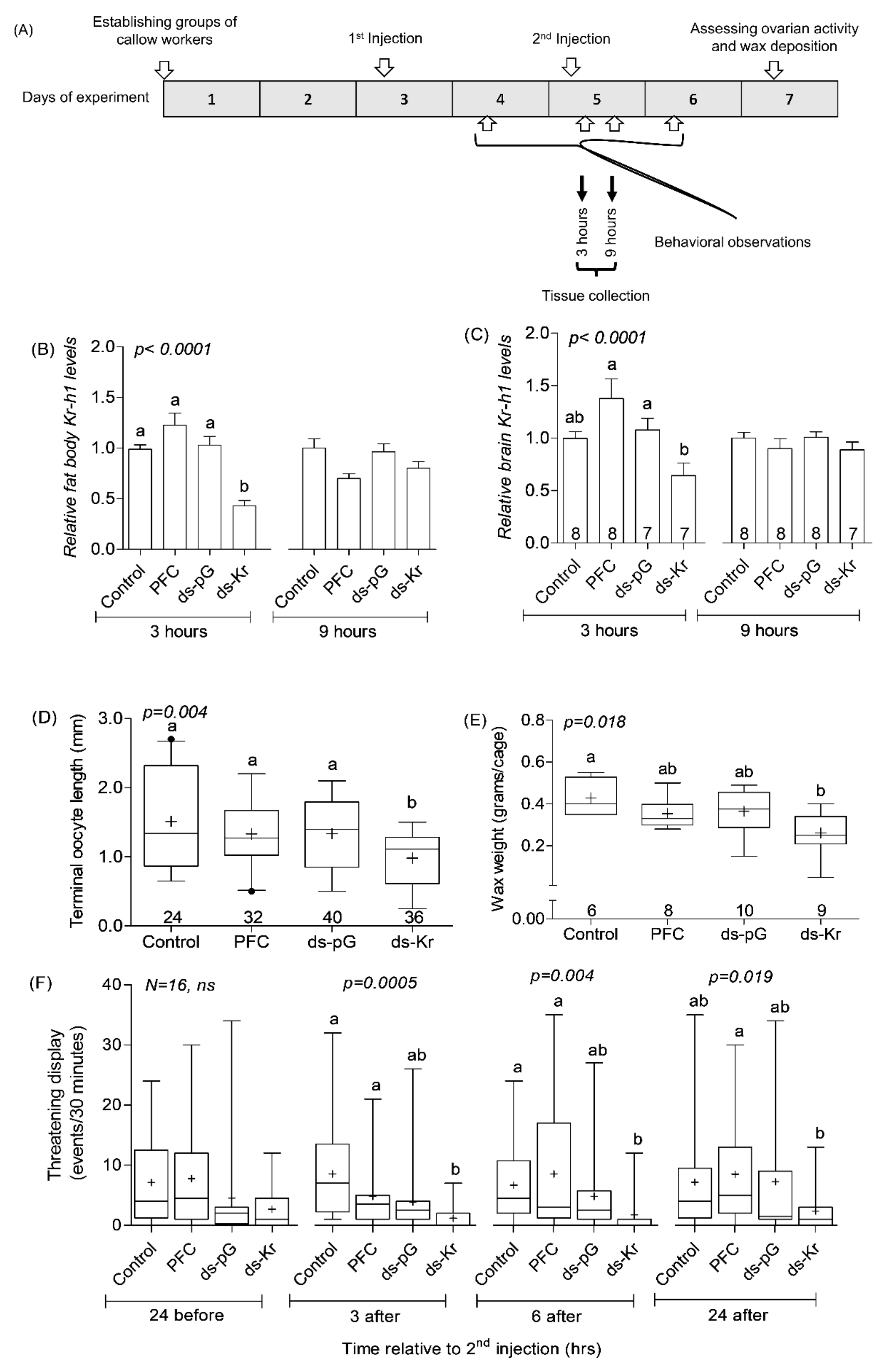
2.7. Experiment 1. The Influence of Kr-h1 on JH Regulated Physiology and Behavior in Groups of Similarly Treated Queenless Workers

We repeated this experiment with bees either injected with naked dsRNA or with dsRNA loaded on PFCnp. Naked dsRNA (5 µg/5 µL/bee) was injected on days 2 and 4 after group formation (see experiment outline in Supplementary Figure S3A). Fat bodies and ovaries for qPCR analyses were sampled at 3, and 6 h after the second injection on Day 4. We performed behavioral observations (Section 2.2.) on Day 3 (24 h after the first injection), Day 4 (3 and 6 h after the second injection), and Day 5 (24 h after the second injection). We assessed the amount of wax deposited and ovarian activity on Day seven, as described in Section 2.3.
In a second set of experiments, we injected dsRNA loaded on PFCnp following the procedure described in Section 2.6.2. The outline of this experiment (Figure 1A) is similar to that with the naked RNA, but with the following modifications: First, following the experiments shown in Supplementary Figure S3A, we decided to inject the bees on days 3 and 5, rather than on days 2 and 4 of the experiment. Second, we reduced the dsRNA amount (up to 5-fold, compared to naked dsRNA doses in Supplementary Figure S3B). Third, we collected the brain and fat body tissues for qPCR analyses at 3, and 9 h following the second injection on Day 5.

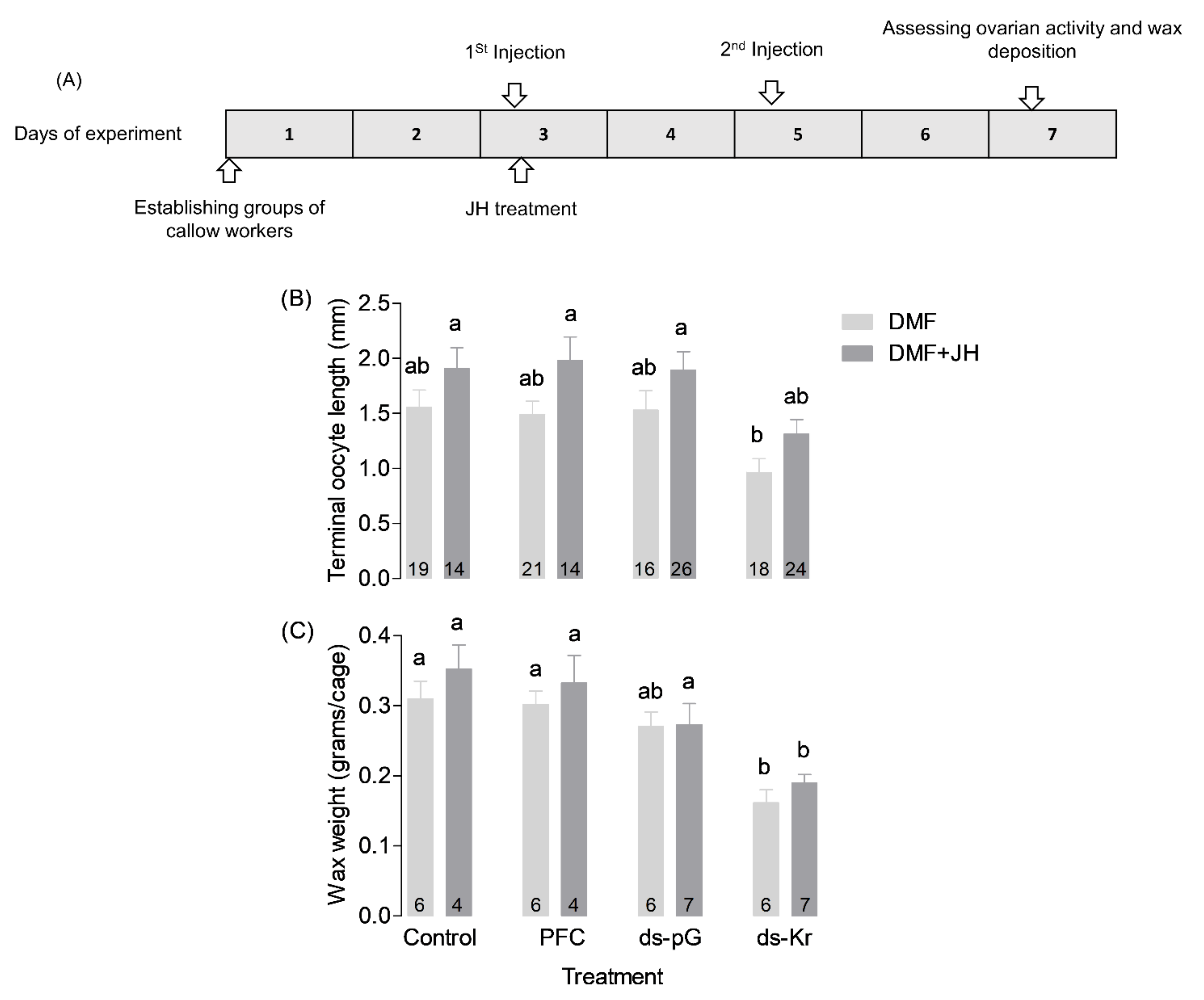
2.8. Experiment 2. The Influence of JH-III on Bees Treated with Kr-h1 dsRNA

The experimental outline (Figure 2A) was overall similar to that in Exp. 1, but following the 1st injection on day 3 of the experiment, half of the bees of all treatment groups were treated with 50 µg/bee JH-III dissolved in Dimethylformamide (DMF), and the other half with only the vehicle (DMF, 3.5 µL/bee). Previous studies showed that treatment with this JH dose stimulates oogenesis, wax deposition, and aggression [69]. The JH (in DMF) or DMF solution alone were topically applied to the thorax. Control bees were similarly handled and chilled on ice, but were not treated with either JH or DMF. All the bees were left on ice 10 min after treatments for maximizing JH absorption. The second dsRNA injection was similar to that described for the first experiment and was done on Day 5.

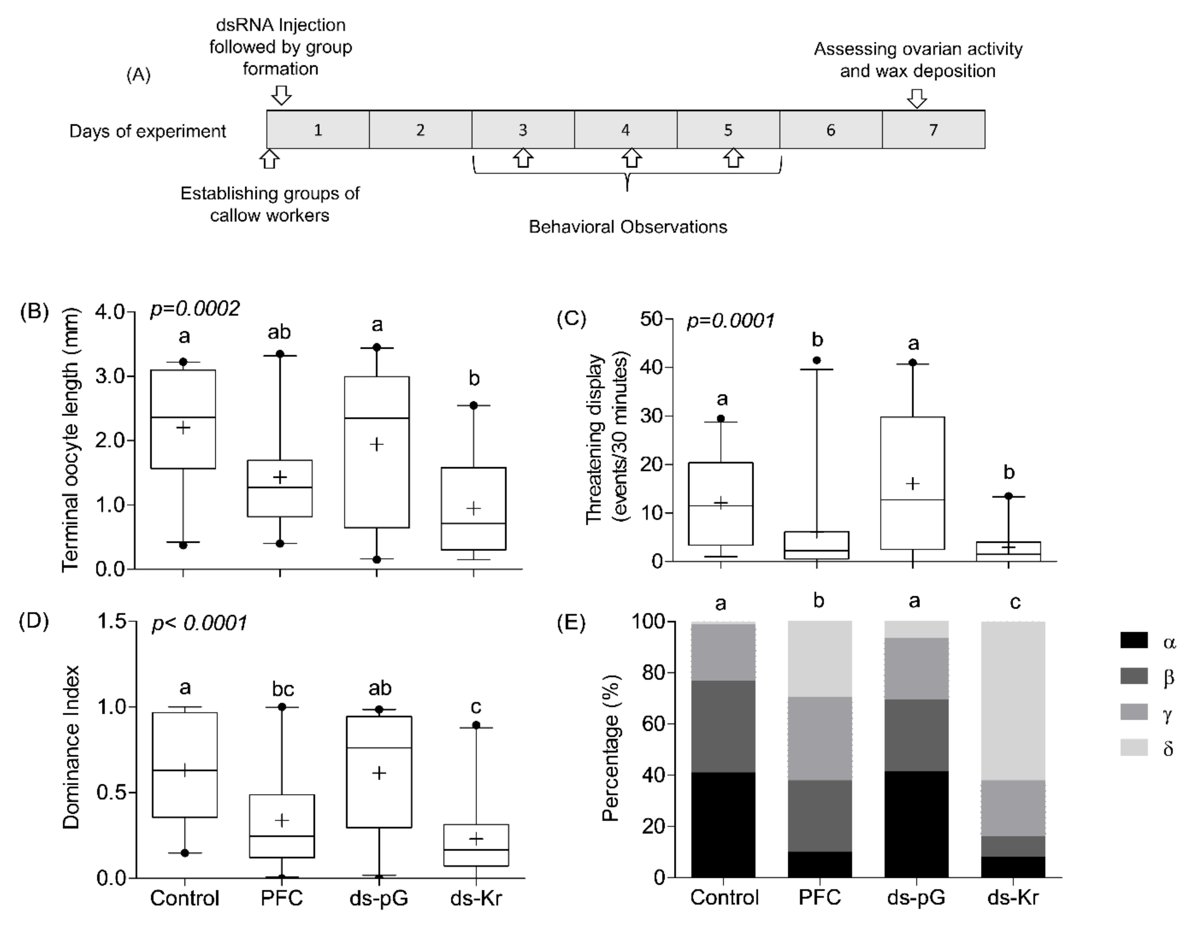
2.9. Experiment 3. The Influence of Kr-h1 on JH Regulated Physiology and Behavior in Groups of Queenless Workers, Each Subjected to a Different Treatment

The experimental outline was similar to that in Exp. 1, but with the following modifications (Figure 3A). First, the bees were treated only once on Day 1, rather than twice, as in Experiment 1. This was done in order to create the groups of bees with normal and reduced Kr-h1 levels at an early age, assuming the differences at early stages of group formation will affect the establishment of the dominance hierarchy. Secondly, the bees were injected two hours after being collected from the mother colony, rather than Day 3 in the first injection of Exp. 1. Thirdly, following the treatment on Day 1, we assigned the bees to mixed groups. Each one of these groups included one bee subjected to each of the four treatments (Control, PFC, ds-pG, ds-Kr). We aimed to use bees of a similar body size within each cage. Behavioral observations, wax collection, and oocyte length measurements were performed as described for Exp. 1 (Figure 3A and Section 2.2 and Section 2.3).

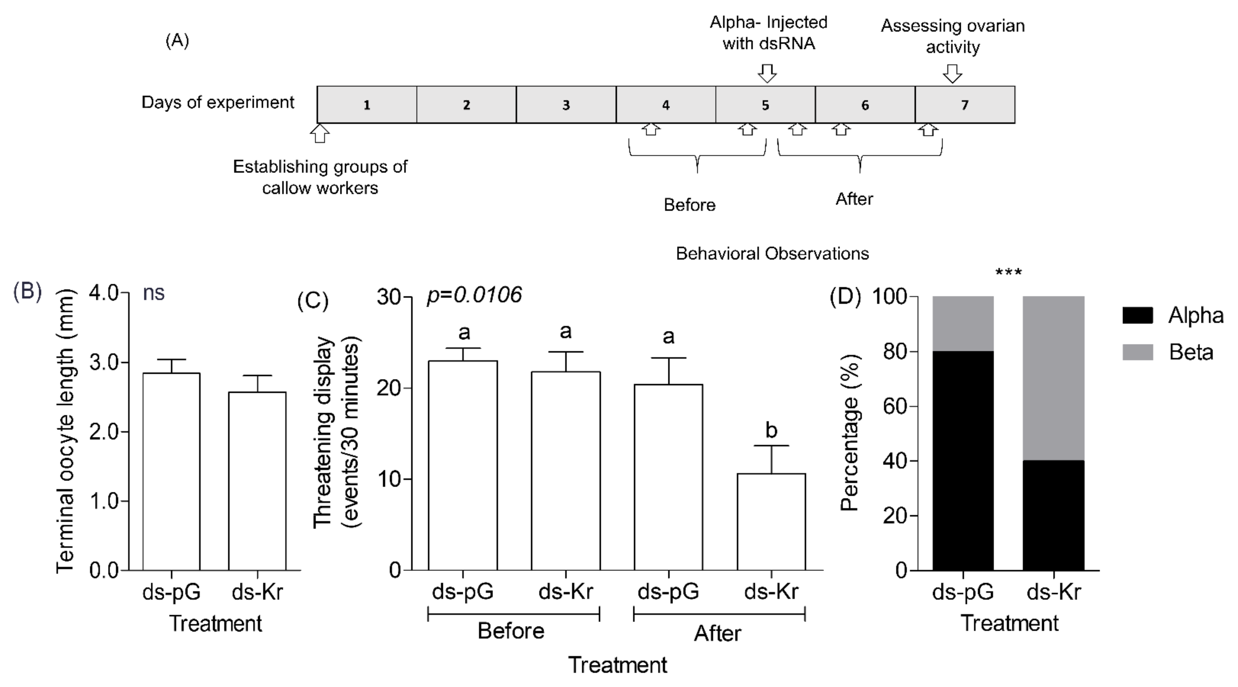
2.10. Experiment 4. The Influence of Kr-h1 on Aggression and Dominance in Groups of Queenless Workers That Have Already Established Dominance Hierarchy

The outline of this experiment is summarized in Figure 4A. We collected newly emerged bees from donor colonies, paint-marked them, and assigned them randomly to groups of four orphan workers. We performed a first set of pre-treatment behavioral observations (Section 2.2) on days 4 and 5 of the experiment and determined the dominance rank for individuals in each queenless group. On Day 5, we injected the α-ranked individual in each group with either ds-pG or ds-Kr (1–2 µg/bee, corrected for body size) as described in Section 2.6.2. To assess the influence on behavior, we performed behavioral observations during the following days (days 5, 6, and 7 of the experiment; Figure 4A). We assessed ovarian activity and wax deposition on Day 7, as in the other experiments.

2.11. Statistical Analyses

We used a one-sample Kolmogorov–Smirnov test to determine whether each set of data are normally distributed. Data that fitted a normal distribution were analyzed using one-way ANOVA followed by Tukey’s post-hoc test. Data that did not fit a normal distribution were analyzed using the non-parametric Kruskal–Wallis-H test followed by Dunn’s post-hoc tests. We used Chi-square tests to compare percentage data (e.g., Figure 3E and Supplementary Figure S4). Additional information on statistics that were relevant to only a certain experiment are reported in the corresponding methods section or figure legend.

3. Results

3.1. Experiment 1. The Influence of Kr-h1 on JH Regulated Physiology and Behavior in Groups of Similarly Treated Queenless Workers

We performed this experiment with naked dsRNA, and then after validating the nanoparticles protocol, repeated it with dsRNA loaded on PFCnp. The results using the two methods were similar overall, but the protocol with nanoparticles allowed us to inject lower amounts of dsRNA. Bees injected with naked Kr-h1 dsRNA, but not with naked control dsRNA (ds-pG), showed a significant reduction of about 50% in fat body and ovarian Kr-h1 mRNA levels compared to control bees when tested 3 h after the second injection (fat body, Supplementary Figure S3B, One-way ANOVA, F = 6.07, df = 2, p = 0.004; ovaries, Figure S3C, One-way ANOVA, F = 4.34, df = 2, p = 0.018). This influence of Kr-h1 dsRNA injection was transient and there was no similar reduction observed for bees analyzed six hours after the second injection (Fat body, Supplementary Figure S3B, F = 1.05, df = 2, p = 0.356; ovaries, Figure S3C, F = 1.25, df = 2, p = 0.295). Bees injected with Kr-h1, but not with pG, dsRNA, deposited less wax in their cages (Supplementary Figure S3D; One-way ANOVA F = 9.982, df = 2, p = 0.0004), and had less active ovaries (Supplementary Figure S3E; Kruskal–Wallis test, χ2 = 35.75, df = 2, p < 0.0001) compared to control bees. Injection of naked Kr-h1 dsRNA also affected behavior. Compared to handling control bees and those injected with pG dsRNA, the bees injected with naked Kr-h1 dsRNA showed fewer threatening displays when observed at three, but not at six hours, after the second injection (Supplementary Figure S3F; Kruskal–Wallis test, χ2(threatening display, 3 after) = 9.93, df = 2, p = 0.007; χ2(threatening display, 6 after) = 0.34, df = 2, p = 0.843, respectively). The number of threatening displays were not affected by treatment in the two other observation sessions (Supplementary Figure S3F, χ2(threatening display, 24 before) = 0.71, df = 2, p = 0.701; χ2(threatening display, 24 after) = 0.75, df = 2, p = 0.756).
The experiment in which we treated bees with dsRNA loaded onto PFCnp produced similar transient Kr-h1 down-regulation in the fat body that was seen at 3, but not at 9 h after dsRNA injection on Day 5 (Figure 1B; Kruskal–Wallis, χ2(Fat bodies,3 h) = 27.90, df = 3, p < 0.0001; χ2(fat bodies,9 h) = 7.70, df = 3, p = 0.052, respectively). In this experiment, we also measured Kr-h1 levels in the brain and found a similar transient downregulation (Figure 1C; Kruskal–Wallis, χ2(Brain,3 h) = 25.71, df = 3, p < 0.0001; χ2(Brain,9 h) = 5.28, df = 3, p = 0.15, respectively). In a set of complementary experiments, we tested the influence of Kr-h1 knockdown on JH regulated behavior and physiology. We found that bees injected with PFCnp loaded with Kr-h1 dsRNA showed significantly reduced ovarian activity compared to bees from all three control groups (Figure 1D, Kruskal–Wallis χ2 = 13.17, df = 3, p = 0.004). An overall similar pattern was seen for wax deposition, but the differences compared to the PFCnp and ds-pG treatment groups were not statistically significant in the post-hoc analyses (Figure 1E, One-way ANOVA, F = 3.94, df = 3, p = 0.018). Kr-h1 down-regulation also influenced the number of threatening displays recorded during the observations performed 3, 6 and 24 h after the second injection. Bees injected with Kr-h1 dsRNA performed fewer threatening displays (Figure 1F; Kruskal–Wallis test; χ2 = 17.69, df = 3, p = 0.0005; χ2 = 12.97, df = 3, p = 0.005; χ2 = 9.9, df = 3, p = 0.019; for bees observed 3, 6, or 24 h. post the 2nd injection, respectively). Post-hoc analyses for the observations performed at 3 and 6 h after the second injection revealed a statistically significant reduction in threatening display in bees treated with Kr-h1 dsRNA compared to bees in the control and PFC treatment groups, and a similar but statistically non-significant trend compared to the ds-pG treated bees. The number of threatening displays was significantly reduced in the Kr-h1 treated bees compared to the PFC, but not the Control or ds-pG treatments, at the observations performed 24 h after the second injection. Nevertheless, the overall trend was similar at the three post 2nd injection observations. Taken together, the experiments with both naked and PFCnp loaded dsRNA suggest that Kr-h1 mediates the influence of JH on ovarian activity, wax deposition, and agonistic behaviors.

3.2. Experiment 2. The Influence of JH on Bees Treated with Kr-h1 dsRNA

In order to test whether JH can act independently of Kr-h1, we measured wax deposition and ovarian activity for bees treated with JH right after the first Kr-h1 dsRNA injection. Controlled bees from each one of the RNAi treatments groups (i.e., Control, PFC, ds-pG, ds-Kr) were similarly treated, but were topically treated with only the vehicle (DMF). Given that there were two trials, we first performed a 3-way ANOVA using the square root transformed data (that fit normal distribution; Kolmogorov–Smirnov test, p > 0.05) with RNAi treatment, JH treatment, and trial as factors (Supplementary Table S2). Given that these analyses showed no significant effect for the trial, on either ovarian activity (p = 0.36), or wax deposition (p = 0.11), we reanalyzed the data using 2-way ANOVA followed by Holm–Sidak post-hoc tests (Figure 2). These analyses revealed a significant effect of RNAi treatment for both ovarian activity (Figure 2B; Two-way ANOVA, F = 6.987, df = 3, p < 0.001) and wax deposition (Figure 2C; F = 16.536, df = 3, p < 0.001). JH treatment had an overall significant effect on ovarian activity (Figure 2B; F = 11.238, df = 1, p = 0.001), but not on wax deposition (Figure 2C; F = 2.178, df = 1, p = 0.148). The interaction between the RNAi and JH treatments was not statistically significant for either ovarian activity (Figure 2B; Two-way ANOVA, F = 0.08, df = 3, p = 0.97), or wax deposition (Figure 2C; Two-way ANOVA, F = 0.235, df = 3, p = 0.87). Bees treated with JH-III had more active ovaries compared with bees treated with only the DMF vehicle. However, JH treatment could not fully recover the inhibitory effect of Kr-h1 downregulation on either ovarian activity or wax production (Figure 2B,C). Taken together, this experiment shows that a JH treatment that is sufficient to induce ovarian activity cannot fully recover the effect of down-regulating Kr-h1 expression levels. These findings are consistent with the premise that Kr-h1 mediates the influence of JH on ovarian activity and wax deposition. The trend of decrease in ovarian activity and wax deposition in bees treated with both Kr-h1 dsRNA and JH can be explained by the partial and transient effect of Kr-h1 down-regulation (Figure 1 and Supplementary Figure S3).

3.3. Experiment 3. The Influence of Kr-h1 on JH Regulated Physiology and Behavior in Groups of Queenless Workers, Each Subjected to a Different Treatment

Given that dominance rank is the product of interactions between individuals in a group, studies in which all the individuals are subjected to the same treatment (such as Exp. 1) may not reveal the full effect of Kr-h1 on dominance. To fill this gap, we performed an additional experiment in which each bee in a queenless group was subjected to a different treatment. The outline of this experiment is summarized in Figure 3A. Consistent with Exp. 1, treatment had a significant influence on ovarian activity (Figure 3B; Kruskal–Wallis χ2 = 19.20, df = 3, p = 0.0002). Ovarian activity was the lowest for bees treated with Kr-h1 dsRNA, but the post-hoc comparison was statistically significant only relative to the Control and ds-pG treated bees. In a pooled analysis of the three observations performed during days 3–5, we found that bees injected with Kr-h1 dsRNA showed fewer threatening displays compared to bees of the Control and ds-pG treatment groups, but not compared to the PFCnp treatment (Figure 3C; Kruskal–Wallis test, χ2 = 21.00, df = 3, p = 0.0001). Bees treated with Kr-h1 dsRNA also had low dominance index (Figure 3D, Kruskal–Wallis χ2 = 23.48, df = 3, p < 0.0001) and had significantly lower dominance rank (Figure 3E; contingency table analysis using 4X4 chi-square test, χ2 = 152.64, p <0.00001, followed by pair-wise Chi-square tests, and corrected for multiple comparisons). The differences in dominance index were statistically significant compared to the Control and ds-pG treated bees, but not compared to the nanoparticles control treatment (PFCnp; Figure 3D). The results of the behavioral analyses (threatening displays, dominance index, and dominance rank) were similar when we analyzed each observation session separately (days 3, 4, and 5; Supplementary Figure S4).

3.4. Experiment 4. The Influence of Kr-h1 on Aggression and Dominance in Groups of Queenless Workers That Have Already Established a Dominance Hierarchy

Given that Experiments 1 and 3 established that Kr-h1 influences the establishment of dominance hierarchies, we next asked whether it is also needed for maintaining a high dominance rank in established dominance hierarchies. To meet this goal, we knockdown Kr-h1 expression in the most dominant individuals (α rank) in groups that have already established stable dominance hierarchies (see experimental outline in Figure 4A). We found that α-rank bees injected with dsKr-h1 on Day 5 showed an ovarian activity similar to that of control α-rank bees injected with ds-pG when analyzed two days later (Figure 4B, Two-tailed, t-test, df = 8, p = 0.412). The frequency of threatening displays performed by the α-rank bee decreased significantly after ds-Kr dsRNA but not ds-pG dsRNA injection (Figure 4C shows the average for 30 min of the two observations before injection compared to the average of the three observations performed after dsRNA injection: Two-tailed, t-test, df = 8, p = 0.018, p = 0.445, respectively). In a complementary Two-way ANOVA with observation time (before or after dsRNA injection) and dsRNA treatment (Kr-h1 or pG), we found that the level of threatening displays is significantly lower for the bees injected with Kr-h1 dsRNA compared to the three other treatment groups (Figure 4C; Two-way ANOVA, F = 4.90, (Treatment), df = 1, p = 0.041; F = 7.72(Observation), df = 1, p = 0.013; F = 2.99 (Treatment x observations), df = 1, p = 0.103, Bonferroni post-hoc test). Consistent with the effects on the frequency of threatening displays, Kr-h1 knockdown also affected dominance rank: α rank bees injected with Kr-h1 dsRNA were more likely to lose their top dominance rank compared to α rank bees injected with pG-dsRNA (Figure 4D, Chi-square with Yates’ correction = 31.69, df = 1, p < 0.0001). Nevertheless, it is notable that in all the groups they did not have a rank lower than Betta.

4. Discussion

High social dominance rank has fitness advantages and a genetic bases, but little is known about the specific genes and molecular processes influencing dominance rank [9,115]. We know particularly little about the genes influencing dominance in females and in invertebrates. Bridging these gaps in knowledge is crucial for articulating the molecular organization principles governing dominance behavior and defining their generality across sex and phylogeny. To address his challenge, we developed and validated a new nanoparticle-based dsRNA-mediated RNAi protocol and used this procedure to down-regulate the expression of Kr-h1 in newly emerged adult bumble bee workers. The development of this novel protocol is an important contribution because it allows knocking down gene expression in a bumble bee using relatively low amounts of dsRNA, and reduced RNA toxicity and nonspecific effects. Using this approach, we showed that Kr-h1 mediates not only the effects of JH on fertility and wax deposition, but also on agonistic behavior and dominance rank. By showing that bees with reduced Kr-h1 transcript abundance show less dominance behavior and have a lower dominance rank compared to bees similarly injected with control dsRNA, we establish for the first time a causal link between a specific gene and dominance rank in a social insect in which dominance hierarchies are established between females. These findings provide a molecular link between JH and dominance and sets the stage for research on the molecular and neuronal bases of dominance in social insects and other female hierarchies.
RNAi technology in insects has faced challenges related to the stability, delivery, and knock-down effectivity of dsRNA/siRNA [116,117]. dsRNA that is not protected in the insect hemolymph is typically degraded and therefore injected dsRNA can be cleared from the hemolymph before effectively knocking down the targeted gene [118,119]. Indeed, our unpublished in-vitro degradation assay for dsRNA in bumble bee hemolymph showed that most of the tested dsRNA is degraded within about six hours. Nanoparticles offer a system to improve delivery, uptake, and efficiency (reduction in the amount of dsRNA injected). Nanoparticles may also help in protecting the introduced dsRNA from rapid degradation [120,121]. It is important to note that the B. terrestris genome lacks a Sid 2 gene, which is an important component of systemic RNAi signaling. There is evidence that the Sid-1/2 genes facilitate dsRNA uptake into cells and therefore improve the effectivity of RNAi [122,123]. Thus, the lack of Sid2 means that the development of RNAi technologies in this species is expected to be even more challenging [124]. Our validated PFCnp RNAi protocol enables us to significantly reduce (~5 fold) the amount of dsRNA injected, which is important for decreasing dsRNA toxicity and nonspecific effects. However, it should be noted that some of our experiments suggest that unloaded PFCnp may have some biological effects, perhaps due to the positive charge of their surface.
In vertebrates in which the physiological and molecular mechanisms affecting dominance behavior are better understood, particularly in males, it is established that the sex steroid hormones that coordinate processes related to reproduction also influence dominance [125,126]. JH seems to overall have similar coordinating functions in females of simple (“primitive”) insect societies [65,69,127,128,129,130,131]. The bumble bee B. terrestris provides some of the best support for this notion. Endocrine manipulation experiments show that JH coordinates processes associated with reproduction in many tissues. These include ovary activation, wax deposition, regulation of biosynthetic pathways in exocrine glands, and gene expression in the brain and fat body [47,70]. Similar to vertebrates’ sex steroids, JH also influences behaviors such as aggression, dominance, and circadian rhythms [69,74]. The new findings presented above show that worker bees with down-regulated Kr-h1 mRNA levels are overall similar to bees with naturally or artificially reduced JH titers. Bees with knocked down Kr-h1 levels have smaller ovaries (Figure 1D, Figure 2B, Figure 3A, Figure S2 and Figure S3E), deposited less wax in their cages (Figure 1E, Figure 2C and Figure S3C), and showed fewer threatening displays, compared to bees similarly injected with control dsRNA. These findings are consistent with evidence that Kr-h1 is a JH responsive gene in B. terrestris [47,83]. Our gene knock-down experiments establish Kr-h1 as a pivotal signaling gene that is expressed in multiple tissues and acts downstream of JH in bumble bees, consistent with its key roles in JH signaling pathways in other insect species [82,86,89,94,95,96,97,132,133,134,135,136]. These findings also set the stage for a deeper study on JH signaling pathways and the genes with which Kr-h1 interacts in bumble bees, as well as other hymenopteran insects.
The effects of Kr-h1 knock-down on diverse processes such as oogenesis, wax deposition, and behavior are consistent with the premise that Kr-h1 mediates the influence of JH by modulating gene expression in diverse tissues including the fat body, wax glands and brain. Our qPCR measurements are consistent with this premise by showing that bees injected with Kr-h1 dsRNA show reduced Kr-h1 RNA abundance in the brain, fat body, and ovaries (Figure 1B,C, and Supplementary Figure S3B,C). The effects of Kr-h1 dsRNA injection were substantial, albeit our qPCR mRNA measurements suggest that Kr-h1 down-regulation was partial and transient: Kr-h1 transcript abundance was significantly reduced when measured three hours post injection, but not a few hours later (Figure 1B,C and Figure S3B,C). This transient RNA knockdown is consistent with evidence suggesting that modulation of JH amounts in newly emerged adult bees is sufficient to produce lasting effects, including on ovarian activity measured about a week post JH treatment [68,69,70,74,83,137]. Similar long-lasting effects of single early JH manipulations have been also reported for other insects [138,139,140,141,142,143]. Given that JH is not stable in the hemolymph over time [74,144,145,146,147,148], the lasting effects of a single acute JH treatment early in life is consistent with organizational effects, or with models in which JH levels at early age regulate cascades of processes that after the initial activation do not require continuous high JH levels. Our current finding suggests that many of these effects are mediated by Kr-h1.
As in other species, the establishment of dominance hierarchies among bumble bee workers is achieved through repeated agonistic interactions characterized by frequent threatening displays [69,72,101,102,104]. Our findings lend credence to the hypothesis that Kr-h1 mediates the influence of JH on dominance in bumble bees, providing the first causal link between a specific gene and dominance in social insects. Kr-h1 mRNA levels are higher in the brain of dominant compared to low-ranked individuals [83] and our results show that our RNAi protocol significantly reduced Kr-h1 in the brain (Figure 1). These results are consistent with the premise that Kr-h1 mediates JH effects on dominance by modulating gene expression in the brain tissue. However, it is also possible that Kr-h1-mediated JH effects in other tissues (such as the ovaries) lead to physiological changes that influence dominance. For example, JH may affect metabolism that can affect fighting ability and RHP. Moreover, JH activation of the ovaries may lead to increase production of ecdysteroids [103] or other endocrine signals that may act on the brain. Worker bees with down-regulated Kr-h1 mRNA levels showed fewer threatening displays towards other individuals in all the experiments in which we performed behavioral observations. Notably, in most hierarchies in the mix treatment experiment, the dsRNA injected bees had the lowest (δ) or next to lowest (γ) dominance rank, which was significantly lower than for bees injected with control dsRNA (Figure 3D). Top-ranked α bees in already established hierarchies of older bees for which we knocked down Kr-h1 levels showed reduced threatening displays compared to the period before dsRNA injection and compared to similar α bees injected with control dsRNA and were more likely to lose their top dominance rank (Figure 4). These later results may suggest that Kr-h1 is involved not only in the establishment but also in the maintenance of dominance hierarchies, but additional research is needed for testing this hypothesis.
In spite of the overall agreement, some of our results suggest that Kr-h1 knockdown did not always mirror the effect of reducing JH levels. Dominant bees in already established dominance hierarchies that were treated with Precocene-I to reduce circulating JH levels, showed significantly lower ovarian activity, but not a lesser amount of threatening displays compared to similar bees treated with only the vehicle [69]. In contrast, here we show that similar bees treated with Kr-h1 dsRNA performed fewer threatening displays and were more likely to lose their top dominance rank but had similarly active ovaries compared to bees treated with control dsRNA. Although we cannot exclude the possibility that these differences stem from technical or biological (e.g., genetic, or developmental) variability between the two experiments, it is worth noting that recent studies with other insects show that JH reduction and Kr-h1 knockdown do not necessarily have identical effects. JH acts on additional transcription factors other than Kr-h1 and has various epigenetic effects that may regulate the activity of Kr-h1 or other downstream processes [91,133,149,150,151,152,153]. In the future, it will be important to explore whether additional factors other than JH affect Kr-h1 activity and its effects on dominance. Likewise, it is important to test whether JH influences dominance by acting on molecular processes other than those regulated by Kr-h1.

5. Conclusions

Although it is well established that dominance rank has significant fitness consequences across the animal kingdom, it has been proven difficult to study in molecular terms. Given its complexity and strong context dependency (including the traits of individuals with whom the focal subject interacts with), a comprehensive understanding of dominance behavior requires studying diverse social systems. Our study provides the first causal link between a specific gene and dominance rank in female hierarchies of insects. Although additional studies are needed for determining the generality of our findings, they may suggest a common theme across species and gender by which key genes in endocrine signaling pathways controlling reproduction also affect dominance. The implication of Kr-h1 in the regulation of dominance in bees sets the stage for studies on the molecular, neurobiological, and anatomical underpinnings of social dominance in insects and other invertebrates as well as in female social hierarchies. This provides an important step towards articulating general models for the genetic and molecular regulation of dominance hierarchies. Our new nanoparticle-based RNAi protocol allows us to knockdown gene expression with relatively low dsRNA amounts and reduced RNA toxicity and nonspecific effects. Abdominal injection of our dsRNA nanoparticle complex enables Kr-h1 mRNA knockdown not only in abdominal tissues such as the fat body and ovaries, but also in the brain.

Supplementary Materials

The following are available online at https://www.mdpi.com/article/10.3390/biology10111188/s1, Figure S1: title: The influence of PFCnp concentration on ovarian activity and dsRNA binding efficiency, Figure S2: title: The influence of the days of dsRNA injection on ovarian activity, Figure S3: title: The influence of naked Kr-h1 dsRNA injection on JH-regulated physiology and behavior, Figure S4: title: The influence dsRNA mediated Kr-h1 knock-down on dominance and agonistic behavior—separate analyses for each observation day, Table S1: title: List of primers used in the study, Table S1: title: Summary of a three-way ANOVA analysis for Exp. 2 (Figure 2B,C).

Author Contributions

Conceptualization, A.P. and G.B.; methodology, A.P.; validation, A.P.; formal analysis, A.P.; resources, G.B.; writing—original draft preparation, A.P.; writing—review and editing, G.B.; visualization, A.P.; supervision, G.B.; project administration, G.B.; funding acquisition, G.B. All authors have read and agreed to the published version of the manuscript.

Funding

This research was funded by grants from the US-Israel Binational Agricultural Research and Development Fund (BARD, grant No. IS-4418-11, and IS-5077-18), ISF-NSFC joint research program (grant No. 3391/20), the United States-Israel Binational Science Foundation (BSF grant No. 2017188).

Institutional Review Board Statement

Not applicable.

Informed Consent Statement

Not applicable.

Data Availability Statement

Not applicable.

Acknowledgments

This research was supported by grants from the US-Israel Binational Agricultural Research and Development Fund (BARD, grant No. IS-4418-11, and IS-5077-18, to G.B.), ISF-NSFC joint research program (grant No. 3391/20 to G.B.), the United States-Israel Binational Science Foundation (BSF grant No. 2017188 to G.B.), and The Planning and Budgeting Committee (PBC) Fellowship Program for Outstanding Chinese and Indian Post-Doctoral Fellows (to A.P).

Conflicts of Interest

The authors declare no conflict of interest.

References

  1. Fero, K.; Simon, J.L.; Jourdie, V.; Moore, P.A. Consequences of social dominance on crayfish resource use. Behaviour 2007, 144, 61–82. [Google Scholar] [CrossRef]
  2. Georgiev, A.V.; Klimczuk, A.C.E.; Traficonte, D.M.; Maestripieri, D. When violence pays: A cost-benefit analysis of aggressive behavior in animals and humans. Evol. Psychol. 2013, 11, 678–699. [Google Scholar] [CrossRef] [PubMed] [Green Version]
  3. Van Kleef, G.A.; Cheng, J.T. Power, status, and hierarchy: Current trends and future challenges. Curr. Opin. Psychol. 2020, 33, iv–xiii. [Google Scholar] [CrossRef] [PubMed]
  4. Cowlishaw, G.; Dunbar, R.I.M. Dominance rank and mating success in male primates. Anim. Behav. 1991, 41, 1045–1056. [Google Scholar] [CrossRef]
  5. Gesquiere, L.R.; Learn, N.H.; Simao, M.C.M.; Onyango, P.O.; Alberts, S.C.; Altmann, J. Life at the top: Rank and stress in wild male baboons. Science 2011, 333, 357–360. [Google Scholar] [CrossRef] [Green Version]
  6. Archie, E.A.; Altmann, J.; Alberts, S.C. Social status predicts wound healing in wild baboons. Proc. Natl. Acad. Sci. USA 2012, 109, 9017–9022. [Google Scholar] [CrossRef] [Green Version]
  7. Snyder-Mackler, N.; Sanz, J.; Kohn, J.N.; Voyles, T.; Pique-Regi, R.; Wilson, M.E.; Barreiro, L.B.; Tung, J. Social status alters chromatin accessibility and the gene regulatory response to glucocorticoid stimulation in rhesus macaques. Proc. Natl. Acad. Sci. USA 2019, 116, 1219–1228. [Google Scholar] [CrossRef] [PubMed] [Green Version]
  8. Bartolomucci, A. Social stress, immune functions and disease in rodents. Front. Neuroendocrinol. 2007, 28, 28–49. [Google Scholar] [CrossRef]
  9. Van der Kooij, M.A.; Sandi, C. The genetics of social hierarchies. Curr. Opin. Behav. Sci. 2015, 2, 52–57. [Google Scholar] [CrossRef]
  10. Barrette, C. Dominance cannot be inherited. Trends Ecol. Evol. 1987, 2, 251. [Google Scholar] [CrossRef]
  11. Fonberg, E. Dominance and aggression. Int. J. Neurosci. 1988, 41, 201–213. [Google Scholar] [CrossRef]
  12. Biro, P.A.; Stamps, J.A. Do consistent individual differences in metabolic rate promote consistent individual differences in behavior? Trends Ecol. Evol. 2010, 25, 653–659. [Google Scholar] [CrossRef] [PubMed]
  13. Oliveira, R.F.; McGregor, P.K.; Latruffe, C. Know thine enemy: Fighting fish gather information from observing conspecific interactions. Proc. R. Soc. B Biol. Sci. 1998, 265, 1045–1049. [Google Scholar] [CrossRef] [Green Version]
  14. Chase, I.D.; Bartolomeo, C.; Dugatkin, L.A. Aggressive interactions and inter-contest interval: How long do winners keep winning? Anim. Behav. 1994, 48, 393–400. [Google Scholar] [CrossRef] [Green Version]
  15. Feder, Y.; Nesher, E.; Ogran, A.; Kreinin, A.; Malatynska, E.; Yadid, G.; Pinhasov, A. Selective breeding for dominant and submissive behavior in Sabra mice. J. Affect. Disord. 2010, 126, 214–222. [Google Scholar] [CrossRef]
  16. Masur, J.; Benedito, M.A.C. Genetic selection of winner and loser rats in a competitive situation. Nature 1974, 249, 284. [Google Scholar] [CrossRef] [PubMed]
  17. Moore, A.J.; Haynes, K.F.; Preziosi, R.F.; Moore, P.J. The evolution of interacting phenotypes: Genetics and evolution of social dominance. Am. Nat. 2002, 160, S186–S197. [Google Scholar] [CrossRef]
  18. Wang, F.; Kessels, H.W.; Hu, H. The mouse that roared: Neural mechanisms of social hierarchy. Trends Neurosci. 2014, 37, 674–682. [Google Scholar] [CrossRef]
  19. Maruska, K.P.; Zhang, A.; Neboori, A.; Fernald, R.D. Social opportunity causes rapid transcriptional changes in the social behaviour network of the brain in an African cichlid fish. J. Neuroendocrinol. 2013, 25, 145–157. [Google Scholar] [CrossRef] [Green Version]
  20. Lema, S.C.; Sanders, K.E.; Walti, K.A. Arginine Vasotocin, Isotocin and nonapeptide receptor gene expression link to social status and aggression in sex-dependent patterns. J. Neuroendocrinol. 2015, 27, 142–157. [Google Scholar] [CrossRef]
  21. Timmer, M.; Cordero, M.I.; Sevelinges, Y.; Sandi, C. Evidence for a role of oxytocin receptors in the long-term establishment of dominance hierarchies. Neuropsychopharmacology 2011, 36, 2349–2356. [Google Scholar] [CrossRef] [PubMed]
  22. Lazzari, V.M.; Becker, R.O.; de Azevedo, M.S.; Morris, M.; Rigatto, K.; Almeida, S.; Lucion, A.B.; Giovenardi, M. Oxytocin modulates social interaction but is not essential for sexual behavior in male mice. Behav. Brain Res. 2013, 244, 130–136. [Google Scholar] [CrossRef]
  23. Caldwell, H.K.; Dike, O.E.; Stevenson, E.L.; Storck, K.; Young, W.S. Social dominance in male vasopressin 1b receptor knockout mice. Horm. Behav. 2010, 58, 257–263. [Google Scholar] [CrossRef] [PubMed] [Green Version]
  24. Chiao, J.Y. Neural basis of social status hierarchy across species. Curr. Opin. Neurobiol. 2010, 20, 803–809. [Google Scholar] [CrossRef]
  25. Holmes, A.; Murphy, D.L.; Crawley, J.N. Abnormal behavioral phenotypes of serotonin transporter knockout mice: Parallels with human anxiety and depression. Biol. Psychiatry 2003, 54, 953–959. [Google Scholar] [CrossRef] [PubMed]
  26. Kalueff, A.V.; Fox, M.A.; Gallagher, P.S.; Murphy, D.L. Hypolocomotion, anxiety and serotonin syndrome-like behavior contribute to the complex phenotype of serotonin transporter knockout mice. Genes Brain Behav. 2007, 6, 389–400. [Google Scholar] [CrossRef]
  27. Williamson, C.M.; Lee, W.; Curley, J.P. Temporal dynamics of social hierarchy formation and maintenance in male mice. Anim. Behav. 2016, 115, 259–272. [Google Scholar] [CrossRef]
  28. So, N.; Franks, B.; Lim, S.; Curley, J.P. A social network approach reveals associations between mouse social dominance and brain gene expression. PLoS ONE 2015, 10, e0134509. [Google Scholar] [CrossRef] [Green Version]
  29. Francis, R.C. On the relationship between aggression and social dominance. Ethology 1988, 78, 223–237. [Google Scholar] [CrossRef]
  30. Fernald, R.D. Cognitive skills needed for social hierarchies. Cold Spring Harb. Symp. Quant. Biol. 2014, 79, 229–236. [Google Scholar] [CrossRef] [Green Version]
  31. Langley, E.J.G.; Van Horik, J.O.; Whiteside, M.A.; Madden, J.R. Group social rank is associated with performance on a spatial learning task. R. Soc. Open Sci. 2018, 5, 171475. [Google Scholar] [CrossRef] [Green Version]
  32. Boogert, N.J.; Reader, S.M.; Laland, K.N. The relation between social rank, neophobia and individual learning in starlings. Anim. Behav. 2006, 72, 1229–1239. [Google Scholar] [CrossRef]
  33. Massen, J.J.M.; Pašukonis, A.; Schmidt, J.; Bugnyar, T. Ravens notice dominance reversals among conspecifics within and outside their social group. Nat. Commun. 2014, 5, 3679. [Google Scholar] [CrossRef] [PubMed] [Green Version]
  34. Vrontou, E.; Nilsen, S.P.; Demir, E.; Kravitz, E.A.; Dickson, B.J. Fruitless regulates aggression and dominance in drosophila. Nat. Neurosci. 2006, 9, 1469–1471. [Google Scholar] [CrossRef] [PubMed]
  35. Chan, Y.B.; Kravitz, E.A. Specific subgroups of FruM neurons control sexually dimorphic patterns of aggression in Drosophila melanogaster. Proc. Natl. Acad. Sci. USA 2007, 104, 19577–19582. [Google Scholar] [CrossRef] [Green Version]
  36. Pardi, L. Dominance order in Polistes wasps. Physiol. Zool. 1948, 21, 1–13. [Google Scholar] [CrossRef]
  37. Wilson, E.O. The Insect Societies; Harvard University Press: Cambridge, MA, USA, 1971; Volume 1, ISBN 0674454952. [Google Scholar]
  38. Free, J.B. The behaviour of egg-laying workers of bumblebee colonies. Br. J. Anim. Behav. 1955, 3, 147–153. [Google Scholar] [CrossRef]
  39. Zwarts, L.; Versteven, M.; Callaerts, P. Genetics and neurobiology of aggression in drosophila. Fly 2012, 6, 35–48. [Google Scholar] [CrossRef] [Green Version]
  40. Williamson, C.M.; Lee, W.; DeCasien, A.R.; Lanham, A.; Romeo, R.D.; Curley, J.P. Social hierarchy position in female mice is associated with plasma corticosterone levels and hypothalamic gene expression. Sci. Rep. 2019, 9, 7324. [Google Scholar] [CrossRef] [Green Version]
  41. Grieb, Z.A.; Ross, A.P.; McCann, K.E.; Lee, S.; Welch, M.; Gomez, M.G.; Norvelle, A.; Michopoulos, V.; Huhman, K.L.; Albers, H.E. Sex-dependent effects of social status on the regulation of arginine-vasopressin (AVP) V1a, oxytocin (OT), and serotonin (5-HT) 1A receptor binding and aggression in Syrian hamsters (Mesocricetus auratus). Horm. Behav. 2021, 127, 104878. [Google Scholar] [CrossRef]
  42. Toth, A.L.; Tooker, J.F.; Radhakrishnan, S.; Minard, R.; Henshaw, M.T.; Grozinger, C.M. Shared genes related to aggression, rather than chemical communication, are associated with reproductive dominance in paper wasps (Polistes metricus). BMC Genomics 2014, 15, 75. [Google Scholar] [CrossRef] [PubMed] [Green Version]
  43. Manfredini, F.; Brown, M.J.F.; Toth, A.L. Candidate genes for cooperation and aggression in the social wasp Polistes dominula. J. Comp. Physiol. A 2018, 204, 449–463. [Google Scholar] [CrossRef] [PubMed] [Green Version]
  44. Manfredini, F.; Riba-Grognuz, O.; Wurm, Y.; Keller, L.; Shoemaker, D.W.; Grozinger, C.M. Sociogenomics of cooperation and conflict during colony founding in the fire ant Solenopsis invicta. PLoS Genet. 2013, 9, 1003633. [Google Scholar] [CrossRef]
  45. Steffen, M.A.; Rehan, S.M. Genetic signatures of dominance hierarchies reveal conserved cis-regulatory and brain gene expression underlying aggression in a facultatively social bee. Genes Brain Behav. 2020, 19, e12597. [Google Scholar] [CrossRef]
  46. Withee, J.R.; Rehan, S.M. Social aggression, experience, and brain gene expression in a subsocial bee. Integr. Comp. Biol. 2017, 57, 640–648. [Google Scholar] [CrossRef] [Green Version]
  47. Shpigler, H.Y.; Herb, B.; Drnevich, J.; Band, M.; Robinson, G.E.; Bloch, G. Juvenile hormone regulates brain-reproduction tradeoff in bumble bees but not in honey bees. Horm. Behav. 2020, 126, 104844. [Google Scholar] [CrossRef] [PubMed]
  48. Okada, Y.; Watanabe, Y.; Tin, M.M.Y.; Tsuji, K.; Mikheyev, A.S. Social dominance alters nutrition-related gene expression immediately: Transcriptomic evidence from a monomorphic queenless ant. Mol. Ecol. 2017, 26, 2922–2938. [Google Scholar] [CrossRef]
  49. Wingfield, J.C.; Hegner, R.E.; Dufty, A.M.; Ball, G.F. The “Challenge Hypothesis”: Theoretical implications for patterns of testosterone secretion, mating systems, and breeding strategies. Am. Nat. 1990, 136, 829–846. [Google Scholar] [CrossRef]
  50. Terburg, D.; Van Honk, J. Approach-avoidance versus dominance-submissiveness: A multilevel neural framework on how testosterone promotes social status. Emot. Rev. 2013, 5, 296–302. [Google Scholar] [CrossRef]
  51. Adams, M.E. Juvenile hormones. In Encyclopedia of Insects; Academic Press: Cambridge, MA, USA, 2009; ISBN 9780123741448. [Google Scholar] [CrossRef]
  52. Goodman, W.G.; Cusson, M. The juvenile hormones. In Insect Endocrinology; Gilbert, L.I., Ed.; Academic Press: Cambridge, MA, USA, 2012; pp. 310–365. ISBN 9780123847492. [Google Scholar] [CrossRef]
  53. Jindra, M.; Palli, S.R.; Riddiford, L.M. The juvenile hormone signaling pathway in insect development. Annu. Rev. Entomol. 2013, 58, 181–204. [Google Scholar] [CrossRef]
  54. Jindra, M.; Bellés, X.; Shinoda, T. Molecular basis of juvenile hormone signaling. Curr. Opin. Insect Sci. 2015, 11, 39–46. [Google Scholar] [CrossRef]
  55. Pandey, A.; Bloch, G. Juvenile hormone and ecdysteroids as major regulators of brain and behavior in bees. Curr. Opin. Insect Sci. 2015, 12, 26–37. [Google Scholar] [CrossRef]
  56. Riddiford, L.M. Juvenile hormone action: A 2007 perspective. J. Insect Physiol. 2008, 54, 895–901. [Google Scholar] [CrossRef]
  57. Truman, J.W.; Riddiford, L.M. Endocrine insights into the evolution of metamorphosis in insects. Annu. Rev. Entomol. 2002, 47, 467–500. [Google Scholar] [CrossRef] [Green Version]
  58. De Loof, A.; Baggerman, G.; Breuer, M.; Claeys, U.; Cerstiaens, A.; Clynen, E.; Janssen, T.; Schoofs, L.; Broeck, J. Vanden Gonadotropins in insects: An overview. Arch. Insect Biochem. Physiol. 2001, 47, 129–138. [Google Scholar] [CrossRef] [PubMed]
  59. Riddiford, L.M. How does juvenile hormone control insect metamorphosis and reproduction? Gen. Comp. Endocrinol. 2012, 179, 477–484. [Google Scholar] [CrossRef] [PubMed]
  60. Delisle, J.; Cusson, M. Juvenile hormone biosynthesis, oocyte growth and vitellogenin accumulation in Choristoneura fumiferana and C. rosaceana: A comparative study. J. Insect Physiol. 1999, 45, 515–523. [Google Scholar] [CrossRef]
  61. Dong, S.-Z.; Ye, G.-Y.; Guo, J.Y.; Hu, C. Roles of ecdysteroid and juvenile hormone in vitellogenesis in an endoparasitic wasp, Pteromalus puparum (Hymenoptera: Pteromalidae). Gen. Comp. Endocrinol. 2009, 160, 102–108. [Google Scholar] [CrossRef] [PubMed]
  62. Klowden, M.J. Endocrine aspects of mosquito reproduction. Arch. Insect Biochem. Physiol. 1997, 35, 491–512. [Google Scholar] [CrossRef]
  63. Panaitof, S.C.; Scott, M.P. Effect of juvenile hormone on vitellogenin gene expression in the fat body of burying beetles, Nicrophorus orbicollis. Arch. Insect Biochem. Physiol. 2006, 63, 82–91. [Google Scholar] [CrossRef]
  64. Bloch, G.; Wheeler, D.E.; Robinson, G.E. Endocrine influences on the organization of insect societies. In Hormones, Brain and Behavior; Pfaff, D.W., Arnold, A.P., Rubin, R.T., Eds.; Academic Press: Cambridge, MA, USA, 2002; pp. 195–235. ISBN 9780125321044. [Google Scholar] [CrossRef]
  65. Robinson, G.E.; Vargo, E.L. Juvenile hormone in adult eusocial hymenoptera: Gonadotropin and behavioral pacemaker. Arch. Insect Biochem. Physiol. 1997, 35, 559–583. [Google Scholar] [CrossRef]
  66. Bloch, G.; Borst, D.W.; Huang, Z.-Y.; Robinson, G.E.; Cnaani, J.; Hefetz, A. Juvenile hormone titers, juvenile hormone biosynthesis, ovarian development and social environment in Bombus terrestris. J. Insect Physiol. 2000, 46, 47–57. [Google Scholar] [CrossRef]
  67. Röseler, P.F. Juvenile hormone control of oogenesis in bumblebee workers, Bombus terrestris. J. Insect Physiol. 1977, 23, 985–992. [Google Scholar] [CrossRef]
  68. Amsalem, E.; Teal, P.; Grozinger, C.M.; Hefetz, A. Precocene-I inhibits juvenile hormone biosynthesis, ovarian activation, aggression and alters sterility signal production in bumble bee (Bombus terrestris) workers. J. Exp. Biol. 2014, 217, 3178–3185. [Google Scholar] [CrossRef] [Green Version]
  69. Pandey, A.; Motro, U.; Bloch, G. Juvenile hormone interacts with multiple factors to modulate aggression and dominance in groups of orphan bumble bee (Bombus terrestris) workers. Horm. Behav. 2020, 117, 104602. [Google Scholar] [CrossRef] [PubMed]
  70. Shpigler, H.; Amsalem, E.; Huang, Z.Y.; Cohen, M.; Siegel, A.J.; Hefetz, A.; Bloch, G. Gonadotropic and physiological functions of juvenile hormone in bumblebee (Bombus terrestris) workers. PLoS ONE 2014, 9, e100650. [Google Scholar] [CrossRef] [PubMed] [Green Version]
  71. Shpigler, H.Y.; Magory Cohen, T.; Ben-Shimol, E.; Ben-Betzalel, R.; Levin, E. Juvenile hormone functions as a metabolic rate accelerator in bumble bees (Bombus terrestris). Horm. Behav. 2021, 136, 105073. [Google Scholar] [CrossRef]
  72. Van Doorn, A. Factors influencing dominance behaviour in queenless bumblebee workers (Bombus terrestris). Physiol. Entomol. 1989, 14, 211–221. [Google Scholar] [CrossRef]
  73. Larrere, M.; Couillaud, F. Role of juvenile hormone biosynthesis in dominance status and reproduction of the bumblebee, Bombus terrestris. Behav. Ecol. Sociobiol. 1993, 33, 335–338. [Google Scholar] [CrossRef]
  74. Pandey, A.; Motro, U.; Bloch, G. Juvenile hormone affects the development and strength of circadian rhythms in young bumble bee (Bombus terrestris) workers. Neurobiol. Sleep Circadian Rhythms 2020, 9, 100056. [Google Scholar] [CrossRef]
  75. Zhang, J.; Liu, X.; Liu, Y.; An, Y.; Fang, H.; Michaud, J.P.; Zhang, H.; Li, Y.; Zhang, Q.; Li, Z. Molecular characterization of primary juvenile hormone responders Methoprene-Tolerant (Met) and Krüppel Homolog 1 (Kr-h1) in Grapholita molesta (Lepidoptera: Tortricidae) with clarification of their roles in metamorphosis and reproduction. J. Econ. Entomol. 2019, 112, 2369–2380. [Google Scholar] [CrossRef] [PubMed]
  76. Tang, Y.; He, H.; Qu, X.; Cai, Y.; Ding, W.; Qiu, L.; Li, Y. RNA interference-mediated knockdown of the transcription factor Krüppel homologue 1 suppresses vitellogenesis in Chilo suppressalis. Insect Mol. Biol. 2020, 29, 183–192. [Google Scholar] [CrossRef] [PubMed]
  77. Yue, Y.; Yang, R.-L.; Wang, W.-P.; Zhou, Q.-H.; Chen, E.-H.; Yuan, G.-R.; Wang, J.-J.; Dou, W. Involvement of Met and Kr-h1 in JH-mediated reproduction of female Bactrocera dorsalis (Hendel). Front. Physiol. 2018, 9, 482. [Google Scholar] [CrossRef] [PubMed]
  78. Li, K.L.; Yuan, S.Y.; Nanda, S.; Wang, W.X.; Lai, F.X.; Fu, Q.; Wan, P.J. The roles of E93 and Kr-h1 in metamorphosis of Nilaparvata lugens. Front. Physiol. 2018, 9, 1677. [Google Scholar] [CrossRef] [PubMed] [Green Version]
  79. Holtof, M.; Van Lommel, J.; Gijbels, M.; Dekempeneer, E.; Nicolai, B.; Broeck, J.V.; Marchal, E. Crucial role of juvenile hormone receptor components Methoprene-Tolerant and Taiman in sexual maturation of adult male desert locusts. Biomolecules 2021, 11, 244. [Google Scholar] [CrossRef]
  80. Zhu, Z.; Tong, C.; Qiu, B.; Yang, H.; Xu, J.; Zheng, S.; Song, Q.; Feng, Q.; Deng, H. 20E-mediated regulation of BmKr-h1 by BmKRP promotes oocyte maturation. BMC Biol. 2021, 19, 39. [Google Scholar] [CrossRef]
  81. Guo, W.; Song, J.; Yang, P.; Chen, X.; Chen, D.; Ren, D.; Kang, L.; Wang, X. Juvenile hormone suppresses aggregation behavior through influencing antennal gene expression in locusts. PLoS Genet. 2020, 16, e1008762. [Google Scholar] [CrossRef]
  82. Fussnecker, B.; Grozinger, C. Dissecting the role of Kr-h1 brain gene expression in foraging behavior in honey bees (Apis mellifera). Insect Mol. Biol. 2008, 17, 515–522. [Google Scholar] [CrossRef]
  83. Shpigler, H.; Patch, H.M.; Cohen, M.; Fan, Y.; Grozinger, C.M.; Bloch, G. The transcription factor Krüppel homolog 1 is linked to hormone mediated social organization in bees. BMC Evol. Biol. 2010, 10, 120. [Google Scholar] [CrossRef] [Green Version]
  84. Grozinger, C.M.; Sharabash, N.M.; Whitfield, C.W.; Robinson, G.E. Pheromone-mediated gene expression in the honey bee brain. Proc. Natl. Acad. Sci. USA 2003, 100 (Suppl. 2), 14519–14525. [Google Scholar] [CrossRef] [Green Version]
  85. Minakuchi, C.; Zhou, X.; Riddiford, L.M. Krüppel homolog 1 (Kr-h1) mediates juvenile hormone action during metamorphosis of Drosophila melanogaster. Mech. Dev. 2008, 125, 91–105. [Google Scholar] [CrossRef] [PubMed] [Green Version]
  86. Song, J.; Wu, Z.; Wang, Z.; Deng, S.; Zhou, S. Krüppel-homolog 1 mediates juvenile hormone action to promote vitellogenesis and oocyte maturation in the migratory locust. Insect Biochem. Mol. Biol. 2014, 52, 94–101. [Google Scholar] [CrossRef] [PubMed]
  87. Konopova, B.; Jindra, M. Juvenile hormone resistance gene Methoprene-tolerant controls entry into metamorphosis in the beetle Tribolium castaneum. Proc. Natl. Acad. Sci. USA 2007, 104, 10488–10493. [Google Scholar] [CrossRef] [PubMed] [Green Version]
  88. Kim, K.; Albishi, N.M.; Palli, S.R. Identification of juvenile hormone-induced posttranslational modifications of Methoprene tolerant and Krüppel homolog 1 in the yellow fever mosquito, Aedes aegypti. J. Proteom. 2021, 242, 104257. [Google Scholar] [CrossRef] [PubMed]
  89. Guo, S.; Wu, Q.-W.; Tian, Z.; Zhu, L.; King-Jones, K.; Zhu, F.; Wang, X.; Liu, W. Krüppel homolog 1 regulates photoperiodic reproductive plasticity in the cabbage beetle Colaphellus bowringi. Insect Biochem. Mol. Biol. 2021, 134, 103582. [Google Scholar] [CrossRef] [PubMed]
  90. Kayukawa, T.; Minakuchi, C.; Namiki, T.; Togawa, T.; Yoshiyama, M.; Kamimura, M.; Mita, K.; Imanishi, S.; Kiuchi, M.; Ishikawa, Y.; et al. Transcriptional regulation of juvenile hormone-mediated induction of Krüppel homolog 1, a repressor of insect metamorphosis. Proc. Natl. Acad. Sci. USA 2012, 109, 11729–11734. [Google Scholar] [CrossRef] [Green Version]
  91. Zhang, T.; Song, W.; Li, Z.; Qian, W.; Wei, L.; Yang, Y.; Wang, W.; Zhou, X.; Meng, M.; Peng, J.; et al. Krüppel homolog 1 represses insect ecdysone biosynthesis by directly inhibiting the transcription of steroidogenic enzymes. Proc. Natl. Acad. Sci. USA 2018, 115, 3960–3965. [Google Scholar] [CrossRef] [Green Version]
  92. Zhu, Z.-D.; Hu, Q.-H.; Tong, C.-M.; Yang, H.-G.; Zheng, S.-C.; Feng, Q.-L.; Deng, H.-M. Transcriptomic analysis reveals the regulation network of BmKrüppel homolog 1 in the oocyte development of Bombyx mori. Insect Sci. 2021, 28, 47–62. [Google Scholar] [CrossRef]
  93. Ling, L.; Raikhel, A. Cross-talk of insulin-like peptides, juvenile hormone, and 20-hydroxyecdysone in regulation of metabolism in the mosquito Aedes aegypti. Proc. Natl. Acad. Sci. USA 2021, 118, e2023470118. [Google Scholar] [CrossRef]
  94. Hu, K.; Tian, P.; Yang, L.; Tang, Y.; Qiu, L.; He, H.; Ding, W.; Li, Y. Molecular characterization of the Krüppel-homolog 1 and its role in ovarian development in Sogatella furcifera (Hemiptera: Delphacidae). Mol. Biol. Rep. 2020, 47, 1099–1106. [Google Scholar] [CrossRef]
  95. Jiang, J.; Xu, Y.; Lin, X. Role of Broad-Complex (Br) and Krüppel homolog 1 (Kr-h1) in the ovary development of Nilaparvata lugens. Front. Physiol. 2017, 8, 1013. [Google Scholar] [CrossRef]
  96. Lin, X.; Yao, Y.; Wang, B. Methoprene-tolerant (Met) and Krüpple-homologue 1 (Kr-h1) are required for ovariole development and egg maturation in the brown plant hopper. Sci. Rep. 2015, 5, 18064. [Google Scholar] [CrossRef] [Green Version]
  97. Zhang, W.; Ma, L.; Liu, C.; Chen, L.; Xiao, H.-J.; Liang, G.-M. Dissecting the role of Krüppel homolog 1 in the metamorphosis and female reproduction of the cotton bollworm, Helicoverpa armigera. Insect Mol. Biol. 2018, 27, 492–504. [Google Scholar] [CrossRef]
  98. Minakuchi, C.; Namiki, T.; Shinoda, T. Krüppel homolog 1, an early juvenile hormone-response gene downstream of Methoprene-tolerant, mediates its anti-metamorphic action in the red flour beetle Tribolium castaneum. Dev. Biol. 2009, 325, 341–350. [Google Scholar] [CrossRef] [Green Version]
  99. Amsalem, E.; Galbraith, D.A.; Cnaani, J.; Teal, P.E.A.; Grozinger, C.M. Conservation and modification of genetic and physiological toolkits underpinning diapause in bumble bee queens. Mol. Ecol. 2015, 24, 5596–5615. [Google Scholar] [CrossRef] [PubMed]
  100. Jedlička, P.; Ernst, U.R.; Votavová, A.; Hanus, R.; Valterová, I. Gene expression dynamics in major endocrine regulatory pathways along the transition from solitary to social life in a bumblebee, Bombus terrestris. Front. Physiol. 2016, 7, 574. [Google Scholar] [CrossRef] [PubMed]
  101. Bloch, G.; Borst, D.W.; Huang, Z.-Y.; Robinson, G.E.; Hefetz, A. Effects of social conditions on juvenile hormone mediated reproductive development in Bombus terrestris workers. Physiol. Entomol. 1996, 21, 257–267. [Google Scholar] [CrossRef]
  102. Duchateau, M.J. Agonistic behaviours in colonies of the bumblebee Bombus terrestris. J. Ethol. 1989, 7, 141–151. [Google Scholar] [CrossRef]
  103. Geva, S.; Hartfelder, K.; Bloch, G. Reproductive division of labor, dominance, and ecdysteroid levels in hemolymph and ovary of the bumble bee Bombus terrestris. J. Insect Physiol. 2005, 51, 811–823. [Google Scholar] [CrossRef] [PubMed]
  104. Van Doorn, A.; Heringa, J. The ontogeny of a dominance hierarchy in colonies of the bumblebee Bombus terrestris (Hymenoptera, Apidae). Insectes Sociaux 1986, 33, 3–25. [Google Scholar] [CrossRef]
  105. Duchateau, M.J.; Velthuis, H.H.W. Ovarian development and egg laying in workers of Bombus terrestris. Entomol. Exp. Appl. 1989, 51, 199–213. [Google Scholar] [CrossRef]
  106. Hagai, T.; Cohen, M.; Bloch, G. Genes encoding putative Takeout/juvenile hormone binding proteins in the honeybee (Apis mellifera) and modulation by age and juvenile hormone of the takeout-like gene GB19811. Insect Biochem. Mol. Biol. 2007, 37, 689–701. [Google Scholar] [CrossRef] [PubMed]
  107. Morawski, A.M.; Winter, P.M.; Yu, X.; Fuhrhop, R.W.; Scott, M.J.; Hockett, F.; Robertson, J.D.; Gaffney, P.J.; Lanza, G.M.; Wickline, S.A. Quantitative “magnetic resonance immunohistochemistry” with ligand-targeted 19F nanoparticles. Magn. Reson. Med. 2004, 52, 1255–1262. [Google Scholar] [CrossRef]
  108. Chen, J.; Pan, H.; Lanza, G.M.; Wickline, S.A. Perfluorocarbon nanoparticles for physiological and molecular imaging and therapy. Adv. Chronic Kidney Dis. 2013, 20, 466–478. [Google Scholar] [CrossRef] [PubMed] [Green Version]
  109. Hughes, M.; Caruthers, S.; Tran, T.; Marsh, J.; Wallace, K.; Cyrus, T.; Partlow, K.; Scott, M.; Lijowski, M.; Neubauer, A.; et al. Perfluorocarbon nanoparticles for molecular imaging and targeted therapeutics. Proc. IEEE 2008, 96, 397–415. [Google Scholar] [CrossRef]
  110. Shen, S.; Wu, Y.; Liu, Y.; Wu, D. High drug-loading nanomedicines: Progress, current status, and prospects. Int. J. Nanomed. 2017, 12, 4085–4109. [Google Scholar] [CrossRef] [PubMed] [Green Version]
  111. Behzadi, S.; Serpooshan, V.; Tao, W.; Hamaly, M.A.; Alkawareek, M.Y.; Dreaden, E.C.; Brown, D.; Alkilany, A.M.; Farokhzad, O.C.; Mahmoudi, M. Cellular uptake of nanoparticles: Journey inside the cell. Chem. Soc. Rev. 2017, 46, 4218–4244. [Google Scholar] [CrossRef] [PubMed]
  112. Foroozandeh, P.; Aziz, A.A. Insight into cellular uptake and intracellular trafficking of nanoparticles. Nanoscale Res. Lett. 2018, 13, 339. [Google Scholar] [CrossRef]
  113. Asati, A.; Santra, S.; Kaittanis, C.; Perez, J.M. Surface-charge-dependent cell localization and cytotoxicity of cerium oxide nanoparticles. Am. Chem. Soc. Nano 2010, 4, 5321–5331. [Google Scholar] [CrossRef] [Green Version]
  114. Yerushalmi, S.; Bodenhaimer, S.; Bloch, G. Developmentally determined attenuation in circadian rhythms links chronobiology to social organization in bees. J. Exp. Biol. 2006, 209, 1044–1051. [Google Scholar] [CrossRef] [Green Version]
  115. Jandt, J.M.; Tibbetts, E.A.; Toth, A.L. Polistes paper wasps: A model genus for the study of social dominance hierarchies. Insectes Sociaux 2013, 61, 11–27. [Google Scholar] [CrossRef]
  116. Zhu, K.Y.; Palli, S.R. Mechanisms, applications, and challenges of insect RNA interference. Annu. Rev. Entomol. 2020, 65, 293–311. [Google Scholar] [CrossRef] [PubMed] [Green Version]
  117. Christiaens, O.; Whyard, S.; Vélez, A.M.; Smagghe, G. Double-stranded RNA technology to control insect pests: Current status and challenges. Front. Plant Sci. 2020, 11, 451. [Google Scholar] [CrossRef] [PubMed]
  118. Wang, K.; Peng, Y.; Pu, J.; Fu, W.; Wang, J.; Han, Z. Variation in RNAi efficacy among insect species is attributable to dsRNA degradation in vivo. Insect Biochem. Mol. Biol. 2016, 77, 1–9. [Google Scholar] [CrossRef] [PubMed]
  119. Cooper, A.M.W.; Yu, Z.; Biondi, M.; Song, H.; Silver, K.; Zhang, J.; Zhu, K.Y. Stability of double-stranded RNA in gut contents and hemolymph of Ostrinia nubilalis larvae. Pestic. Biochem. Physiol. 2020, 169, 104672. [Google Scholar] [CrossRef]
  120. Wang, K.; Peng, Y.; Chen, J.; Peng, Y.; Wang, X.; Shen, Z.; Han, Z. Comparison of efficacy of RNAi mediated by various nanoparticles in the rice striped stem borer (Chilo suppressalis). Pestic. Biochem. Physiol. 2020, 165, 104467. [Google Scholar] [CrossRef]
  121. Nel, A.E.; Mädler, L.; Velegol, D.; Xia, T.; Hoek, E.M.V.; Somasundaran, P.; Klaessig, F.; Castranova, V.; Thompson, M. Understanding biophysicochemical interactions at the nano-bio interface. Nat. Mater. 2009, 8, 543–557. [Google Scholar] [CrossRef]
  122. Braukmann, F.; Jordan, D.; Miska, E.A. A genetic pathway encoding double-stranded RNA transporters and interactors regulates growth and plasticity in Caenorhabditis elegans. bioRxiv 2019, 694414. [Google Scholar] [CrossRef]
  123. Lichtenberg, S.S.; Tsyusko, O.V.; Palli, S.R.; Unrine, J.M. Uptake and bioactivity of Chitosan/double-stranded RNA polyplex nanoparticles in Caenorhabditis elegans. Environ. Sci. Technol. 2019, 53, 3832–3840. [Google Scholar] [CrossRef]
  124. Sadd, B.M.; Barribeau, S.M.; Bloch, G.; de Graaf, D.C.; Dearden, P.; Elsik, C.G.; Gadau, J.; Grimmelikhuijzen, C.J.P.; Hasselmann, M.; Lozier, J.D.; et al. The genomes of two key bumblebee species with primitive eusocial organization. Genome Biol. 2015, 16, 76. [Google Scholar] [CrossRef] [Green Version]
  125. Dekkers, T.J.; van Rentergem, J.A.A.; Meijer, B.; Popma, A.; Wagemaker, E.; Huizenga, H.M. A meta-analytical evaluation of the dual-hormone hypothesis: Does cortisol moderate the relationship between testosterone and status, dominance, risk taking, aggression, and psychopathy? Neurosci. Biobehav. Rev. 2019, 96, 250–271. [Google Scholar] [CrossRef] [PubMed]
  126. Anestis, S.F. Testosterone in juvenile and adolescent male chimpanzees (Pan troglodytes): Effects of dominance rank, aggression, and behavioral style. Am. J. Phys. Anthropol. 2006, 130, 536–545. [Google Scholar] [CrossRef] [PubMed]
  127. Barth, R.H.; Lester, L.J.; Sroka, P.; Kessler, T.; Hearn, R. Juvenile hormone promotes dominance behavior and ovarian development in social wasps (Polistes annularis). Experientia 1975, 31, 691–692. [Google Scholar] [CrossRef] [PubMed]
  128. Bloch, G.; Wheeler, D.E.; Robinson, G.E. Endocrine influences on the organization of insect societies. In Hormones, Brain and Behavior Online; Pfaff, D.W., Arnold, A.P., Rubin, R.T., Eds.; Academic Press: San Diego, CA, USA, 2009; pp. 1027–1070. ISBN 9780080887838. [Google Scholar] [CrossRef]
  129. Bortolotti, L.; Bogo, G.; Pošćić, F.; Duchateau, M.J. Effect of a topical treatment with juvenile hormone analogues on dominance, ovarian development and corpora allata size in Bombus terrestris workers. Bull. Insectology 2020, 73, 303–311. [Google Scholar]
  130. Tibbetts, E.A.; Huang, Z.Y. The challenge hypothesis in an insect: Juvenile hormone increases during reproductive conflict following queen loss in Polistes Wasps. Am. Nat. 2010, 176, 123–130. [Google Scholar] [CrossRef] [PubMed] [Green Version]
  131. Tibbetts, E.A.; Izzo, A.S. Endocrine mediated phenotypic plasticity: Condition-dependent effects of juvenile hormone on dominance and fertility of wasp queens. Horm. Behav. 2009, 56, 527–531. [Google Scholar] [CrossRef]
  132. Moda, L.M.; Vieira, J.; Guimarães Freire, A.C.; Bonatti, V.; Bomtorin, A.D.; Barchuk, A.R.; Simões, Z.L.P. Nutritionally driven differential gene expression leads to heterochronic brain development in honeybee castes. PLoS ONE 2013, 8, e64815. [Google Scholar] [CrossRef] [Green Version]
  133. Roy, A.; Palli, S.R. Epigenetic modifications acetylation and deacetylation play important roles in juvenile hormone action. BMC Genomics 2018, 19, 934. [Google Scholar] [CrossRef]
  134. Gassias, E.; Maria, A.; Couzi, P.; Demondion, E.; Durand, N.; Bozzolan, F.; Aguilar, P.; Debernard, S. Involvement of Methoprene-tolerant and Krüppel homolog 1 in juvenile hormone-signaling regulating the maturation of male accessory glands in the moth Agrotis ipsilon. Insect Biochem. Mol. Biol. 2021, 132, 103566. [Google Scholar] [CrossRef]
  135. Li, X.; Chen, T.; Jiang, H.; Huang, J.; Huang, M.; Xu, R.; Xie, Q.; Zhu, H.; Su, S. Effects of methyl farnesoate on Krüppel homolog 1 (Kr-h1) during vitellogenesis in the Chinese mitten crab (Eriocheir sinensis). Anim. Reprod. Sci. 2021, 224, 106653. [Google Scholar] [CrossRef]
  136. Duportets, L.; Bozzolan, F.; Abrieux, A.; Maria, A.; Gadenne, C.; Debernard, S. The transcription factor Krüppel homolog 1 is linked to the juvenile hormone-dependent maturation of sexual behavior in the male moth, Agrotis ipsilon. Gen. Comp. Endocrinol. 2012, 176, 158–166. [Google Scholar] [CrossRef] [PubMed]
  137. Shpigler, H.Y.; Siegel, A.J.; Huang, Z.Y.; Bloch, G. No effect of juvenile hormone on task performance in a bumblebee (Bombus terrestris) supports an evolutionary link between endocrine signaling and social complexity. Horm. Behav. 2016, 85, 67–75. [Google Scholar] [CrossRef] [PubMed]
  138. Argue, K.J.; Yun, A.J.; Neckameyer, W.S. Early manipulation of juvenile hormone has sexually dimorphic effects on mature adult behavior in Drosophila melanogaster. Horm. Behav. 2013, 64, 589–597. [Google Scholar] [CrossRef] [Green Version]
  139. Lee, S.S.; Ding, Y.; Karapetians, N.; Rivera-Perez, C.; Noriega, F.G.; Adams, M.E. Hormonal signaling cascade during an early-adult critical period required for courtship memory retention in drosophila. Curr. Biol. 2017, 27, 2798–2809.e3. [Google Scholar] [CrossRef] [Green Version]
  140. Mathiron, A.G.E.; Earley, R.L.; Goubault, M. Juvenile hormone manipulation affects female reproductive status and aggressiveness in a non-social parasitoid wasp. Gen. Comp. Endocrinol. 2019, 274, 80–86. [Google Scholar] [CrossRef]
  141. Montagna, T.S.; Raizer, J.; Antonialli-Junior, W.F. Effect of larval topical application of juvenile hormone on caste determination in the independent-founding eusocial wasp Mischocyttarus consimilis (Hymenoptera: Vespidae). Open J. Anim. Sci. 2015, 5, 174–184. [Google Scholar] [CrossRef] [Green Version]
  142. Norman, V.C.; Hughes, W.O.H.H. Behavioural effects of juvenile hormone and their influence on division of labour in leaf-cutting ant societies. J. Exp. Biol. 2016, 219, 8–11. [Google Scholar] [CrossRef] [PubMed] [Green Version]
  143. Sasagawa, H.; Sasaki, M.; Okada, I. Hormonal control of the division of labor in adult honeybees (Apis mellifera L.). I. Effect of methoprene on Corpora allata and hypopharyngeal gland, and its α-Glucosidase activity. Appl. Entomol. Zool. 1989, 24, 66–77. [Google Scholar] [CrossRef] [Green Version]
  144. Whitmore, D.; Whitmore, E.; Gilbert, L.I. Juvenile hormone induction of esterases: A mechanism for the regulation of juvenile hormone titer. Proc. Natl. Acad. Sci. USA 1972, 69, 1592–1595. [Google Scholar] [CrossRef] [Green Version]
  145. Borovsky, D.; Carlson, D.A.; Ujváry, I. In Vivo and In Vitro biosynthesis and metabolism of methyl farnesoate, juvenile hormone III, and juvenile hormone III acid in the mosquito Aedes aegypti. J. Med. Entomol. 1992, 29, 619–629. [Google Scholar] [CrossRef]
  146. Ajami, A.M.; Riddiford, L.M. Comparative metabolism of the Cecropia juvenile hormone. J. Insect Physiol. 1973, 19, 635–645. [Google Scholar] [CrossRef]
  147. Kramer, S.J.; Wieten, M.; de Kort, C.A.D. Metabolism of juvenile hormone in the colorado potato beetle, Leptinotarsa decemlineata. Insect Biochem. 1977, 7, 231–236. [Google Scholar] [CrossRef]
  148. Lanzrein, B. The activity and stability of injected juvenile hormones (JH I, JH II, and JH III) in last-instar larvae and adult females of the cockroach Nauphoeta cinerea. Gen. Comp. Endocrinol. 1979, 39, 69–78. [Google Scholar] [CrossRef]
  149. George, S.; Gaddelapati, S.C.; Palli, S.R. Histone deacetylase 1 suppresses Krüppel homolog 1 gene expression and influences juvenile hormone action in Tribolium castaneum. Proc. Natl. Acad. Sci. USA 2019, 116, 17759–17764. [Google Scholar] [CrossRef] [PubMed] [Green Version]
  150. Li, K.; Jia, Q.-Q.; Li, S. Juvenile hormone signaling—A mini review. Insect Sci. 2019, 26, 600–606. [Google Scholar] [CrossRef]
  151. Mao, Y.; Li, Y.; Gao, H.; Lin, X. Krüppel homologue 1 interacts directly with Hairy and regulates ecdysis in the brown planthopper. Insect Mol. Biol. 2020, 29, 293–300. [Google Scholar] [CrossRef] [PubMed]
  152. Saha, T.T.; Roy, S.; Pei, G.; Dou, W.; Zou, Z.; Raikhel, A.S. Synergistic action of the transcription factors Krüppel homolog 1 and Hairy in juvenile hormone/Methoprene-tolerant-mediated gene-repression in the mosquito Aedes aegypti. PLoS Genet. 2019, 15, e1008443. [Google Scholar] [CrossRef] [PubMed]
  153. Tsang, S.S.K.; Law, S.T.S.; Li, C.; Qu, Z.; Bendena, W.G.; Tobe, S.S.; Hui, J.H.L. Diversity of insect sesquiterpenoid regulation. Front. Genet. 2020, 11, 1027. [Google Scholar] [CrossRef]
Figure 1. The influence of nanoparticles bounded Kr-h1 dsRNA on gene expression, physiology, and behavior. (A) Experiment outline. (B) Fat body Kr-h1, mRNA levels. (C) Brain Kr-h1, mRNA levels. Kr-h1 mRNA levels were measured for bees sampled at 3 or 9 h after the second injection. (D) Ovarian activity. (E) Wax deposited in the cage. (F) Threatening displays. Control—untreated bees, PFC—bees injected with nanoparticles not loaded with dsRNA, ds-pG—bees injected with pG dsRNA, ds-Kr—bees injected with Kr-h1 dsRNA. In (B,C,F), a separate statistical analysis was performed for each time point. The sample size reported within bras in panel (B) are also applicable for panel (C). The box plot presents the median (−) and mean (+) values, with the frame spanning over the first to the third quartile; the whiskers depict the 5th/95th percentile; outliers are depicted with black dots. Treatments marked with different small letters were statistically different in a Kruskal-Wallis test followed by Dunne’s post-hoc analysis (D,F) or ANOVA followed by Tukey’s post-hoc test (E). Sample size depicts the number of bees in panels (BD,F), and the number of cages (i.e., groups) in panel (E).
Biology 10 01188 g001
Figure 2. Treatment with JH-III could not fully recover the effect of Kr-h1 down-regulation. (A) Experimental outline. (B) Ovarian activity at seven days of age. (C) Total amount of wax deposited. Statistical analysis for the length of terminal oocyte was performed using square root transformed data, which fits normal distribution. Treatments marked with different small letters are statistically different in Two-way ANOVA followed by Holm–Sidak post-hoc tests. Numbers within bars show sample size, which is the number of bees in (B), and the number of cages in (C). Further details are shown in Figure 1.
Biology 10 01188 g002
Figure 3. The influence of Kr-h1 down-regulation on dominance and aggression in mixed treatment groups. (A) Experimental outline. (B) Ovarian activity at seven days of age. (C) Threatening displays. (D) Dominance index. (E) Proportion of dominance rank. The data shown in panel (CE) are the average of the observations of days 3, 4, and 5; an overall similar pattern was obtained when each day was analyzed separately (Supplementary Figure S4). Treatments marked with different small letters in panels (BD) are statistically different in a Kruskal–Wallis H test followed by Dunn’s post-hoc test, and in Chi square tests in panel (E) (see text for details). Sample size is n = 22 for all analyses. Further details are shown in Figure 2.
Biology 10 01188 g003
Figure 4. The influence Kr-h1 knock-down on behavior and ovarian activity of dominant queenless bees in established dominance hierarchies. (A) Experimental outline. (B) Ovarian activity at seven days of age of dominant (alpha) bees injected with Kr-h1 dsRNA (ds-kr) or control dsRNA (ds-pG). (C) Threatening displays of dominant bees injected with Kr-h1 or control dsRNA before or after injection; the values before injection are the average of the observations in days 4 and 5, and the values after injection are the average of the observations in days 5 (after injection), 6, and 7. Treatments marked with different small letters are statistically different in two-way ANOVA analysis followed by Bonferroni post-hoc analysis. (D) Dominance rank of alpha bees after injection with either Kr-h1 or pG dsRNA. The vertical bars (in B,C) depict mean ± SE. n = 5 bees per treatment group. ***—p < 0.0001, in Chi-square with Yates’ correction.
Biology 10 01188 g004
Table 1. Injection volumes and RNA amounts injected into bees of different body sizes.
Body Size
Category
Marginal Wing Cell Length (mm)Injection Volume (µL/Bee)dsRNA Amount
(µg/Bee)
Small2.6–2.8101.0
Medium2.9–3.0151.5
Large3.1–3.3202.0
Publisher’s Note: MDPI stays neutral with regard to jurisdictional claims in published maps and institutional affiliations.

