Article

Development of a Carotenoid-Rich Microalgae Colorant by Microencapsulation

1
iBET, Instituto de Biologia Experimental e Tecnológica, Apartado 12, 2781-901 Oeiras, Portugal
2
ITQB, Instituto de Tecnologia Química e Biológica António Xavier, Universidade Nova de Lisboa, Av. da República, 2780-157 Oeiras, Portugal
3
LAQV/Requimte, Faculdade de Ciências e Tecnologia, Universidade Nova de Lisboa, Campus de Caparica, 2829-516 Caparica, Portugal
4
BioEcoUVa, Research Institute on Bioeconomy—High Pressure Processes Group, Department of Chemical Engineering and Environmental Technology, University of Valladolid, Prado de la Magdalena s/n, 47011 Valladolid, Spain
5
MicroLab, Instituto Superior Técnico, Universidade de Lisboa, Av. Rovisco Pais 1, 1049-001 Lisboa, Portugal
6
iMed.ULisboa, Research Institute for Medicines, Faculty of Pharmacy, Universidade de Lisboa, Av. Professor Gama Pinto, 1649-003 Lisboa, Portugal
*
Author to whom correspondence should be addressed.
Colorants 2024, 3(1), 39-52; https://doi.org/10.3390/colorants3010003
Submission received: 28 November 2023 / Revised: 20 December 2023 / Accepted: 28 December 2023 / Published: 12 January 2024

Abstract

:
Dunaliella salina has been recognized as an excellent biomass source of carotenoid, which can be used as a natural orange coloring agent for food products. The most eco-friendly approach for extracting carotenoid is through supercritical carbon dioxide extraction, as it yields highly concentrated extracts while preventing pigment thermal degradation. However, there are limitations when a lipophilic extract is considered a food ingredient, in particular very difficult handling and low solubility in water-based products. The aim of this study was to develop a hydrosoluble form of a natural carotenoid-rich extract recovered from algae biomass within a biorefinery concept to be incorporated in aqueous-based food products. A two-step process was developed, starting with the emulsification of the supercritical extract into a mixture of maltodextrin and gum arabic, using soy lecithin as an emulsifier. The emulsification was followed by a spray-drying step. The impact of process variables on the encapsulation yield, efficiency, emulsion properties, and particle characteristics was studied. The resulting particles exhibited an intense orange color and good water dispersibility, facilitating uniform yellow coloring when incorporated into an aqueous-based product. Overall, spray-drying emulsions containing carotenoids derived from Dunaliella salina prove to be a promising strategy for the global market demand for natural colorants.

1. Introduction

The marine environment is widely recognized as a valuable and underexploited source of bioactive compounds [1]. Particularly, microalgae, which are photosynthetic eukaryotic microorganisms, have attracted significant attention due to their potential as an industrially feasible feedstock for bioactive compounds [2]. Microalgae produce a wide spectrum of nutrients and biomolecules, including carbohydrates, proteins, enzymes, fibers, vitamins, minerals, and pigments. Importantly, many of these pigments are essential and cannot be synthesized by humans or animals, necessitating their acquisition through dietary sources [3]. The mass cultivation of microalgae offers several distinct advantages in terms of sustainability. Cultivating microalgae is a straightforward and cost-effective process that can utilize wastewater as a growth medium and fix carbon dioxide from the atmosphere. Also, they exhibit higher biomass productivity when compared to traditional terrestrial plants and do not compete for agricultural land with conventional crops [3,4,5,6,7].
Carotenoids represent a category of naturally occurring pigmented compounds synthesized by plants and microorganisms [8]. Microalgae, in particular, emerge as highly productive producers of these lipid-soluble pigments [4]. Among various microalgae species, Dunaliella salina holds the distinction of accumulating the highest levels of β-carotene, reaching up to 14% of its dry weight. Additionally, this microalgae can synthesize other valuable carotenoids such as zeaxanthin, lutein, phytofluene, and phytoene [5,9,10]. These pigments have found diverse applications across various market segments as natural coloring agents and bioactive ingredients in the food, nutraceuticals, pharmaceuticals, and cosmetic industries [11]. Consequently, carotenoids have reached a strong and increasing demand in the market [9]. Indeed, the global carotenoid market, which was valued at 1.44 billion USD in 2019, is expected to grow to USD 1.84 billion in 2027, with a compound annual growth rate of 3.4% [12]. Given the remarkable carotenoid content of Dunaliella salina and the market demand, this microalgae has emerged as a promising and sustainable feedstock for the commercial production of carotenoids [9].
Carotenoids are stored and protected within the cell wall of Dunaliella salina. While the FDA has designated Dunaliella salina generally recognized as safe, incorporating the whole cells into food, nutraceuticals, or cosmetic products may not be the most effective approach. These pigments are enclosed within a non-rigid cell wall structure, potentially restricting their release and bioavailability [13]. In this context, carotenoid extraction plays a crucial role in pigment production. The most suitable sustainable solvent for carotenoid extraction is supercritical carbon dioxide, which provides highly concentrated extracts and avoids thermal degradation of carotenoids [14]. The elevated pressures drive supercritical fluid into the microalgae cells. Since the diffusion rate of the supercritical fluid resembles that of a gas, mass transfer is significantly accelerated. Consequently, the extraction time can be considerably shorter compared to traditional solvent extraction methods [15]. However, there are limitations when considering a lipophilic extract as a food ingredient due to its poor physiochemical properties, notably its challenging handling and low water solubility [16].
Encapsulation techniques can help overcome the above-mentioned issues. Encapsulation entraps a target ingredient in a wall material to isolate the ingredient from the environment [17,18,19]. Among various encapsulating methods, spray-drying emulsions represent a widely employed dehydration technique in the food industry due to their cost-effectiveness and ability to produce high-quality powders that offer excellent protection, stabilization solubility, and controlled release of lipophilic bioactive compounds [17,20]. To obtain these emulsions, lipophilic bioactive compounds are homogenized, forming droplets dispersed within the aqueous phase. The process of spray drying emulsions allows the encapsulation of the carotenoid-rich extract in a solid carrier, yielding a dispersible powder [21].
Several materials are accessible for commercial use as wall materials in spray-dryer applications. These materials include both natural and modified polysaccharides, gums, lipids, proteins, and synthetic polymers. A suitable wall material for spray drying should exhibit functional characteristics such as emulsifying, geling, film-forming, high solubility, and low cost [22,23,24]. Maltodextrin offers good oxidative stability and high water solubility, but tends to exhibit poor oil retention, emulsifying efficiency, and emulsion stability [16,22,25]. Conversely, gum arabic demonstrates a strong emulsifying capacity [16,26]. Mixing both materials has been shown to offer advantages, as the partial replacement of gum arabic with maltodextrin results in wall materials with enhanced solubility [16,27]. Also, soy lecithin is a natural emulsifier that stabilizes oil droplets during the incorporation of a lipophilic extract into oil-in-water emulsions, producing microcapsules with improved stability and water solubility [28].
Spray drying has been employed for drying microalgae suspensions, including Dunaliella salina biomass [29]. However, there is limited information available in the literature regarding the encapsulation of Dunaliella salina carotenoids through spray drying. The authors, instead of using carotenoid-concentrated extract, encapsulated β-carotene-rich cells of Dunaliella salina within a polymer matrix [13,30,31]. In addition, no studies have explored the application of encapsulated carotenoids powders derived from Dunaliella salina for the purpose of coloring and/or enhancing the color of food products.
Within this context, the objective of this work was to explore the integration of encapsulation processes by (1) studying the emulsions containing carotenoid-rich extract from Dunaliella salina and (2) drying the emulsions using a spray dryer to produce particles that enhance the dispersibility of carotenoids in water-based products. This study marked the first attempt to assess the feasibility of applying a highly concentrated extract recovered from Dunaliella salina biomass. The research involved analyzing the yield of collected particles and encapsulation efficiency to optimize the encapsulation conditions. As a proof of concept, the incorporation of the developed water-soluble carotenoid forms was evaluated in an aqueous-based product. This approach represents a significant step forward in the challenging task of producing new natural carotenoid-rich colorants with improved dispersibility in water-based products, addressing the needs of nutraceutical and food industries.

2. Materials and Methods

2.1. Raw Material

Dunaliella salina extract used as core material in the encapsulation experiments was provided by NATECO2 (Wolnzach, Germany) within a European project framework, D-Factory, whose main goal was the development of a sustainable algae biorefinery. The microalga Dunaliella salina from Monzón Biotech S.L. (Monzón, Spain) presented a total carotenoid content of 8 wt.%. The carotenoid-rich extract, obtained by ultra-high-pressure extraction with supercritical carbon dioxide (up to 1000 bar) of freeze-dried biomass, presented a total carotenoid content of 28 wt.%.

2.2. Chemicals

The reagents employed in the spray-drying emulsion methodology included the following: nitrogen (99.99% purity, Air Liquid, Lisbon, Portugal), gum arabic (Sigma-Aldrich, St. Louis, MI, USA), maltodextrin DE 13.0–17.0 (Sigma-Aldrich, St. Louis, MI, USA), soy lecithin (Solgar, Inc., Leonia, NJ, USA), and olive oil (Gallo, Abrantes, Portugal).

2.3. Carotenoid Encapsulation by Spray-Drying Emulsion

2.3.1. Preparation of Oil-in-Water Emulsions

A solution of maltodextrin and gum arabic (50 wt.%) was prepared by dissolving both in distilled water at a concentration of 20 g/100 mL. Soy lecithin, used as an emulsifier, was added at a ratio of 20 mg/g lipophilic extract in all samples. The soy lecithin was combined with the extract, which was previously diluted in olive oil (50 wt.%). Subsequently, the oil phase was added to the polymer aqueous phase, with varying concentrations relative to the total solids of the emulsion (3–37 wt.%). These concentrations were determined based on prior research [16] and preliminary experimental results. The oil-in-water (o/w) emulsions were prepared by subjecting the mixture to homogenization using an ultra-turrax blender (IKA-Werke GmbH & Co. KG, Staufen, Germany) for a duration of 5 min at a stirring speed ranging from 6500 to 21,500 rpm. Then, the resulting emulsions were promptly subjected to the spray-drying process.

2.3.2. Spray-Drying Process Optimization

O/w emulsions were dried using a laboratory-scale spray dryer, the Büchi Mini Spray Dryer B-290 (Büchi Labortechnik, Flawil, Switzerland), which was equipped with a nozzle featuring a 0.7 mm tip and a nozzle cap with a diameter of 1.4 mm. The emulsions were fed into the system via a peristaltic pump. The processing conditions were as follows: the inlet air temperature ranged from 110 to 220 °C, with an aspirator flow rate of 36 m3/h, a compressed nitrogen flow rate of 439 L/h, and a feed flow rate of 4.5 mL/min. The collected particles were stored at 4 °C and protected from light until further use. The encapsulation yield was determined by calculating the ratio of the weight of the resulting powder collected in the vessel after spray drying to the weight of all solids present in the emulsion (including both wall and core material), expressed as a percentage. Encapsulation efficiency was assessed as the ratio of carotenoids extracted from the particles to the total carotenoid content in the initial feed solution, also expressed as a percentage.

2.4. Oil-in-Water Emulsion Characterization

2.4.1. Emulsion Droplet Size

The emulsion droplet size distribution of o/w emulsions was assessed using dynamic light scattering (DLS) with a Zetasizer Nano Zs instrument (Malvern Instruments, Worcestershire, UK). All measurements were conducted at room temperature. The instrument provides data on the mean diameter of the particle size distribution (Dv50) and the polydispersity index (PDI). PDI values below 0.4 indicate a narrow particle-size distribution.

2.4.2. Emulsion Stability

The o/w emulsions were transferred into a glass tube with an internal diameter of 15 mm and a height of 50 mm, filling it to a height of 20 mm. The tube was then tightly sealed with a plastic cap and stored at room temperature. Emulsion stability was assessed by observing phase separation at two time points: 15 min and 1 day after emulsion preparation. The degree of creaming index was quantified using a creaming index (CI), which represents the percentage of the cream layer (HC) formed at the surface of the emulsion relative to the total volume of the emulsion in the tube (HE) [32]:
CI, vol.% = HC⁄HE × 100

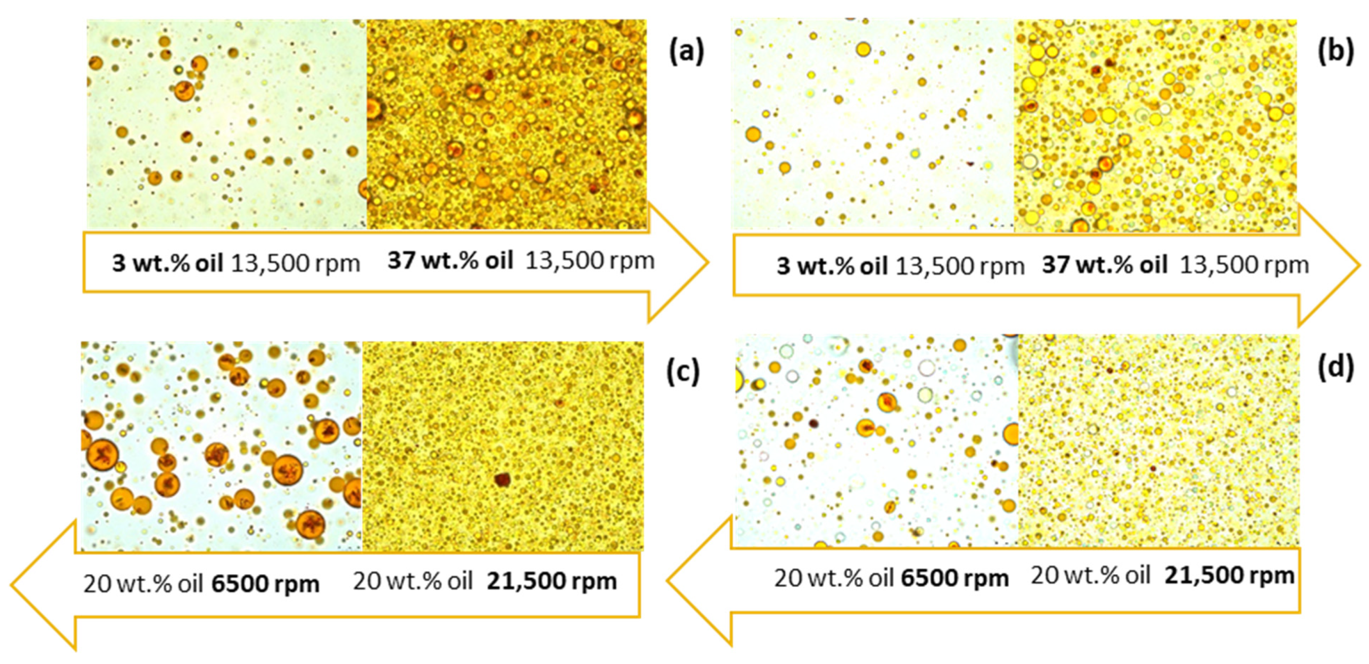
2.4.3. Emulsion Morphology

O/w emulsions were examined under a microscope to assess their respective microstructures. Right after their production, the emulsions were gently agitated using a magnetic stirrer to maintain uniformity. A droplet was then collected and placed onto a microscope slide, which was covered with a cover slip. An optical microscope, the Leica DM6 B (Leica Microsystems CMS GmbH, Wetzlar, Germany), was employed to analyze the microstructure of both the freshly prepared and reconstituted emulsions using a ×100 oil immersion objective. Digital image files were captured using Leica LAS X 2018.1.0 software.

2.4.4. Emulsion Reconstitution

Dried particles were weighted (n = 3) and mixed with distilled water to obtain a reconstituted emulsion with the same dry matter content as before the drying process. After 1 h of continuous rotation at 500 rpm using an orbital mixer (Orbit 300, Labnet International, Inc., Edison, NJ, USA), samples were taken for the assessment of droplet size distribution, stability, and morphological characteristics, following the same procedure as described for fresh emulsions.

2.5. Spray-Dried Particle Characterization

2.5.1. Total Carotenoid Content (TCC)

The determination of TCC in the particles was carried out using high-performance liquid chromatography (HPLC). For carotenoid analysis, an Alliance e2695 HPLC model (Waters, Milford, MA, USA), equipped with a degasser, an autosampler, and a PDA detector, was employed. Separation of carotenoids was achieved using a YMC 30 Carotenoid Column (Classical Analytical HPLC Column S-5 μm, 250 × 4.6 mm, YMC) coupled with a Guard Cartridges YMC C30 pre-column (5 μm, 10 × 4.0 mm, YMC). The mobile phase consisted of methanol and MTBE (80:20) at 25 °C. The detection of carotenoids was performed at a wavelength of 450 nm, and the flow rate was set to 1 mL/min.
Calibration curves for lutein and zeaxanthin were found to be linear in the concentration range of 0.5–100 mg/L, while for all-trans β carotene, 9-cis β carotene, and α-carotene, linearity was observed in the range of 0.5–200 mg/L, using MTBE as the solvent. Prior to analysis, all the samples were prepared by dissolving them in MTBE, followed by centrifugation and filtration through a 0.45 µm HLPC filter. The total duration of the assay was 45 min, with retention times for lutein, zeaxanthin, α-carotene, all-trans beta-carotene, and 9-cis-β-carotene recorded at 6.7, 7.9, 20.5, 26, and 30 min, respectively. A representative chromatogram is included in the Supplementary Materials file (Figure S1).

2.5.2. Particle Size Distribution

The particle size of the samples was assessed using laser diffraction, employing a Malvern Mastersizer 2000. For measurements in a dry state, a Scirocco 2000 dry disperser (Malvern Instruments, Malvern, UK) was utilized. Particle size measurements are presented as volume distribution and defined as Dv50, which is derived as the average of three separated measurements. Additionally, the span value is reported, calculated as the ratio of Dv50 to (Dv90–Dv10). Span values close to 1 indicate a narrow particle size distribution.

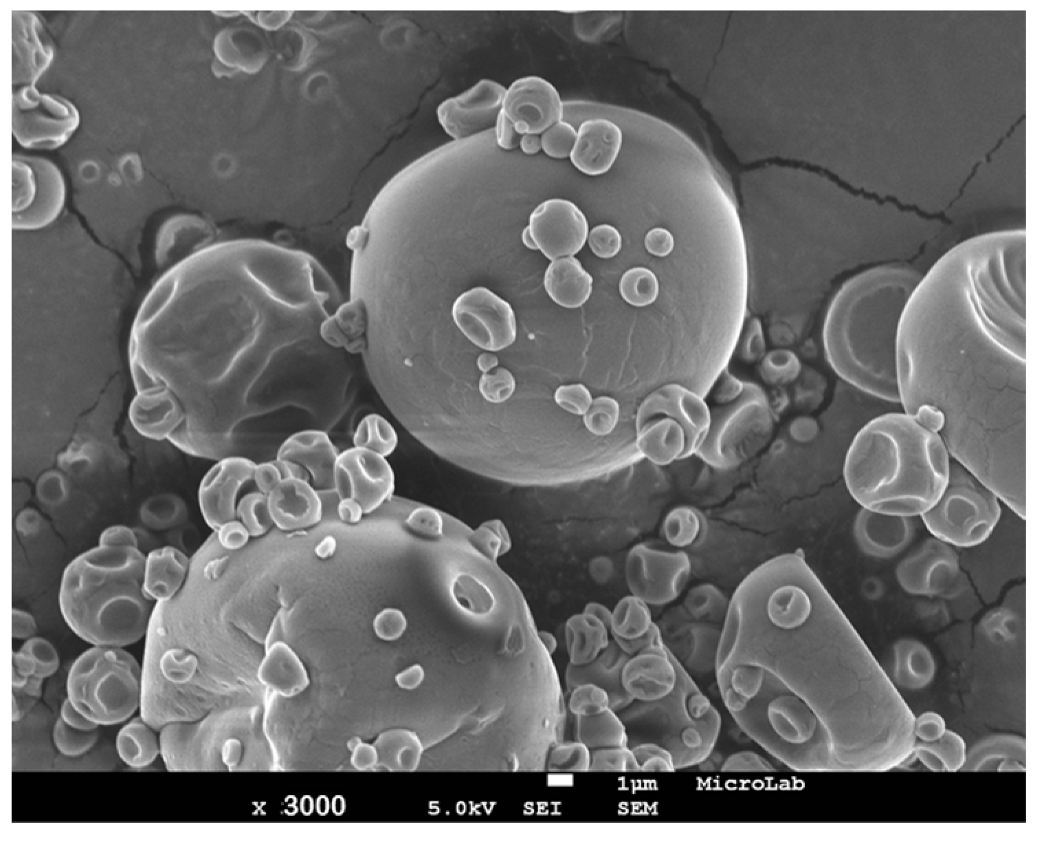
2.5.3. Particle Morphology

To examine the shape and structure of the particles, scanning electron microscopy (FEG-SEM) (Jeol, SM-5310 model, Tokyo, Japan) was employed, operating at a voltage of 20 to 25 kV. Prior to imaging, the samples were coated with a thin layer of gold, approximately 300 Å thick, in an argon atmosphere.

2.5.4. Particle Color Properties

The color of particles was evaluated using the CIELab method with a Minolta Colorimeter CR-200 (Minolta, Osaka, Japan). This method utilizes three distinct attributes or characteristics of visual perception: tonality, luminosity, and chromatism.
The CIELab color space system is founded on a continuous Cartesian representation of three orthogonal axes: L*, a*, and b*. The L* coordinate L* signifies brightness, with L* = 0 representing black and L* = 100 representing colorless. The a* axis represents the green-to-red color component, where a* > 0 corresponds to red and a* < 0 corresponds to green. The b* axis signifies the blue-to-yellow color component, where b* > 0 represents yellow and b* < 0 represents blue. The chroma (color purity) is denoted as C*, while the hue angle, represented by h°, indicates the sample’s color (0° or 360° = red, 90° = yellow, 180° = green, and 270° = blue). The values of C* and h° were determined using the following expressions:
C* = (a* + (b*)2)1/2
h° = arctan(b*/a*)
Subsequently, these values were converted into RGB color values (representing red, green, and blue) using the OpenRGB 0.9 software (Logicol, Trieste, Italy).

2.6. Incorporation of Dunaliella Salina scCO2 Extract and Spray-Dried Particles in an Aqueous-Based Product

The jelly solution was prepared using commercial jelly powder (Royal, Portugal). To prepare the solution, the jelly powder was dissolved in boiling water at a ratio of 1:50 (%, w/v). The Dunaliella salina scCO2 extract and the produced colorant powder were then introduced into the mixture at a solid concentration of 0.001 wt.%. The resulting preparations were poured into plastic cups, sealed with screw caps, and stored in a refrigerator. Visual assessments were conducted to evaluate the uniformity of color distribution within the mixtures.

2.7. Experimental Design and Statistical Analysis

Response surface methodology was employed to assess the influence of process conditions on the encapsulation process of Dunaliella salina scCO2 extract. The data related to encapsulation yield and efficiency, obtained through experiments conducted under specific conditions set by a central composite circumscribed design, were analyzed using the Modde® v.12 software (Umetrics, Umeå, Sweden). The statistical significance of the design model and the effects of various factors were determined by evaluating the p-values. Any factor or model adjustment was considered statistically significant when the resulting p-value was less than the predefined α = 0.05. The assessment was carried out using ANOVA and a t-test.

3. Results and Discussion

Aiming at developing a hydrosoluble form of scCO2 extract from Dunaliella salina, a two-step encapsulation process was developed: (1) emulsifying step: o/w emulsion of the scCO2 extract (lipophilic character) in an aqueous solution containing the wall material (maltodextrin and gum arabic); (2) spray-drying step: drying step to develop hydrosoluble forms of carotenoid-rich extract derived from Dunaliella salina.

3.1. Encapsulation Process Optimization

When applying spray drying to dry emulsions, it is crucial to carefully consider and manage various experimental factors for each specific system being studied (Figure S2). To achieve the desired high encapsulation yield and efficiency while accounting for the expected process factors and complex interactions, a systematic and rational approach was necessary.
Utilizing the knowledge gained from prior research, along with preliminary experimental findings and awareness of instrumental constraints, certain factors were held constant. These include the composition of the emulsion aqueous phase, the composition of the emulsion oil phase, the spray dryer aspirator flow rate, the compressed nitrogen flow rate, and the feed flow rate. By maintaining these factors at fixed values, the complexity of the space to be explored was reduced, simplifying the research process. The remaining parameters and their ranges, such as the oil phase concentration in the emulsion (3–37 wt.%), the stirring speed (6500–21,500 rpm), and the inlet air temperature (110–220 °C), were chosen based on available information for other biomass [16,22,23,26,33,34].
An appropriate balance between the number of necessary experimental trials and the knowledge acquired was determined. Considering the three factors and the defined objectives, a central composite, circumscribed design was chosen. This design encompasses a full factorial design and star points. Additionally, central replicate points were integrated to evaluate experimental variability, result uncertainty, and the goodness of fit of the mathematical model employed. Using response surface methodology, the optimum conditions for encapsulating the Dunaliella salina scCO2 extract were determined.
The experimental conditions and outcomes for each encapsulation trial are detailed in Table 1. The highest encapsulation yield was 69.0 ± 0.9 wt.% (Table 1—experiment N11). This recovery was obtained at a 3 wt.% oil phase concentration in the emulsion, 13,500 rpm, and 165 °C of inlet air temperature. This finding was consistent with previous reports. According to Durmaz et al., an encapsulation yield of 50.8–70.2% was obtained for spray-drying Dunaliella salina biomass using maltodextrin [13]. Bustos-Garza et al. reported a 49.4–50.9% encapsulation yield for the spray-drying of astaxanthin derived from Haematococcus pluvialis in a mixture of gum arabic and maltodextrin [35].
The encapsulation experiment with the highest encapsulation efficiency was obtained at 30 wt.% oil phase concentration in the emulsion, 17,500 rpm, and 130 °C of inlet air temperature, which was 80.0 ± 13.3 wt.% (Table 1—experiment N4). The obtained values were on par with the encapsulation efficiency of 39.3–74.2% previously reported by Foo et al. for spray-dried fucoxanthin from Chaetoceros calcitrans in gum arabic and maltodextrin [36].
The oil phase concentration in the emulsion has a significant effect (p < 0.05) on both evaluated responses: encapsulation yield and efficiency. In addition, the encapsulation efficiency was significantly affected (p < 0.05) by some interactions between factors, namely oil concentration in the emulsion and stirring speed (the model fit is provided in the Supplementary Materials file—Figure S2, Tables S1 and S2). Contour plots representing the encapsulation yield and efficiency as a function of the oil phase concentration in the emulsion, stirring speed during the emulsification step, and inlet air temperature of spray drying are presented in Figure 1.
Figure 1a illustrates a significant impact (p < 0.05) of oil phase concentration in the emulsion on the encapsulation yield. A decreasing amount of oil phase resulted in an improvement in encapsulation yield. This effect was more pronounced at higher inlet air temperatures (p = 0.05) and higher stirring speeds (p = 0.05). Insufficiently high inlet air temperatures can lead to incomplete water vaporization, resulting in inadequately dried powder that adheres to the drying chamber wall [26]. The process of water evaporation is more effective at higher inlet air temperatures due to the improved efficiency of heat and mass transfer between the chamber wall and the moving fluid, ultimately resulting in higher yields [26].
From Figure 1b, when a high oil phase concentration in the emulsion was employed, an increase in the stirring speed led to a rise in encapsulation yield. This observation can possibly be attributed to the smaller emulsion droplet size achieved, which promotes the retention of carotenoids during the spray-drying process. This, in turn, makes it more challenging for the oil to diffuse to the drying surface of the particles [37,38]. Interestingly, higher inlet air temperatures were found to decrease the encapsulation efficiency (Figure 1b). This phenomenon may be associated with the carotenoids losses attributed to oxidation and/or degradation induced by elevated temperatures [37].
The above results show that low oil phase concentration in the emulsion, medium stirring speed, and medium inlet air temperature were the best encapsulation conditions within the studied range to obtain the highest yield. The greatest encapsulation efficiency was attained using a high concentration of the oil phase and a high stirring speed at a lower inlet air temperature. Consequently, processing factors were demonstrated to have different effects on each response variable evaluated. The improvement in the process of carotenoid encapsulation might be not only in the encapsulation yield but also in the encapsulation efficiency. In this way, the goal was to optimize the encapsulation of carotenoids, maximizing the encapsulation yield and efficiency. The operational parameters for emulsion spray drying were determined through a multiple linear regression approach employing an automated optimizer (Modde® v.12). The software generated twelve sets of optimal encapsulation conditions. Among these, the parameter-setting combination with the lowest probability of failure was identified as follows: 30 wt.% oil phase concentration in the emulsion, 17,500 rpm stirring speed, and an inlet air temperature of 200 °C (same conditions as experiment N8).

3.2. Oil-in-Water Emulsion Characterization

In order to ensure the effective retention of carotenoid-rich extract during the spray-drying process, assessing emulsion properties such as droplet size, morphology, and stability is a critical step [37]. The mean diameters of the particle size distribution and creaming index for both freshly prepared and reconstituted emulsions are presented in Table 1.
The mean droplet diameters of the fresh emulsions ranged from 4.3 to 16.9 μm with a PDI < 0.4, indicating that emulsions are monodisperse. Emulsions prepared with a higher concentration of the oil phase and a lower stirring speed (experiments N3, N7, N12, and N13) exhibited a greater mean diameter in the droplet size distribution (Dv50 ≥ 9.4 μm). These emulsions contained droplets with diameters exceeding the limit of the equipment (10 μm); therefore, it was not possible to determine their distribution of different sizes in droplets (PDI). Reducing the oil phase concentration in the emulsion and increasing the stirring speed resulted in a reduction in droplet size and narrowed the droplet distribution. The incorporation of carotenoids, which are lipophilic compounds, into aqueous-based products necessitates the employment of emulsifiers. Emulsifiers, acting as surface-active agents, facilitate the rapid absorption of oil droplets at the o/w interface during the homogenization process, thus preventing the dispersed-phase droplets in emulsions from clustering together [21,35,39]. Therefore, the reduction in droplet size could be explained by increasing the concentration of gum arabic, which is known to have good emulsifying properties. Also, the reduction in droplet size as stirring speed increases can be attributed to the heightened local dissipation of energy within the emulsion-breaking zone, thus avoiding droplet coalescence [40]. Lima and co-workers found higher values (from 40.9 to 52.3 μm) when producing carotenoid-enriched emulsions with gum arabic and a similar homogenizing system (ultra-turrax blender) [21].
Microscopic examination was carried out on both freshly prepared and reconstituted emulsions to assess their respective microstructures. Figure 2a displays the microscopic images of fresh emulsions prepared with different oil phase concentrations at the same stirring speed. It is visually evident that an increase in the oil phase concentration in the emulsion resulted in larger droplets, even at the same stirring speed. In Figure 2b, microscopic images of fresh emulsions prepared with different stirring speeds but with the same oil phase concentration are presented. It is noticeable that higher stirring speeds correspond to smaller mean diameters of emulsion droplets. These visual observations align with the results obtained for emulsion droplet size.
Following the preparation of o/w emulsions, they underwent spray drying to directly obtain particles, which were subsequently re-suspended in water. The particles exhibited excellent solubility in water due to the solubility properties conferred by maltodextrin. Figure 2c,d shows the microscopic images of reconstituted emulsions prepared with different oil phase concentrations and stirring speeds. The effects of both oil phase concentration and stirring speed on the droplet morphology of the reconstituted emulsions mirrored the observations made for the freshly prepared emulsions. Notably, the processing of emulsions through spray drying led to a reduction in droplet sizes, although a non-negligible loss of droplets was observed. The visual separation of an emulsion can be regarded as an indication of physical instability. When emulsions undergo instability, the oil phase tends to rise to the top while the denser aqueous phase settles at the bottom [41]. In this evaluation, the physical stability of the emulsions was assessed by analyzing the extent of the cream layer. The larger the creaming index, the greater the agglomeration of the emulsion droplets, which results in a less stable emulsion. According to the results presented in Table 1, all emulsions were stable 15 min after preparation, presenting an CI equal to 0 vol.%. This time was considered sufficient to ensure emulsion stability throughout the spray dryer experiment. After one day of preparation, the emulsions were physically unstable, with CI varying between 2.5 and 15 vol.%. Since the presented strategy (spray drying of emulsions) is a continuous process, the physical stability of the emulsion during the entire experiment was ensured (<15 min).

3.3. Spray-Dried Particle Characterization

Yellow-to-orange, dry, easily dispersible powders were successfully produced by spray-drying emulsions. The total carotenoid content, color properties, and particle size of the obtained particles are given in Table 1. Assessing these properties is of significance as they dictate the potential applications of these powders in the food industry [13].
The highest total carotenoid content was 2.9 ± 0.5 wt.% (Table 1—experiment N12). This concentration was obtained at 37 wt.% oil phase concentration in the emulsion, 13,500 rpm, and 165 °C of inlet air temperature. As expected, the total carotenoid content was highly affected by the oil phase concentration in the emulsion. This effect was more pronounced when applying a higher stirring speed during the emulsion preparation. The particles produced in this study exhibited a notably higher carotenoid content compared to the findings of Leach and co-workers [30]. The authors studied the encapsulation of Dunaliella salina biomass through spray drying, employing maltodextrin and gum arabic as the encapsulation matrix, resulting in a total carotenoid content ranging from 0.16 to 0.79 wt.%. The increased carotenoids observed in the current study can primarily be attributed to the utilization of a highly concentrated extract of Dunaliella salina rather than the entire biomass cell. Additionally, the addition of soy lecithin as an emulsifier may have effectively prevented the migration of the oil phase, which contains carotenoids, to the surface of particles.
Studies have indicated that the average particle size generated by a spray dryer for different biological materials can reach up to 50 μm [42]. Particle size is influenced not only by the parameters of the spray-drying process but also by the emulsion properties, including solid content, size, and stability [37]. In Table 1, the particle sizes of the different powders obtained via spray-drying emulsions at various inlet air temperatures are depicted. The mean particle sizes for the encapsulated carotenoid samples in this study ranged between 7.6 and 39.4 μm. Additionally, polydisperse particles were obtained with span values above 1. A decrease in particle size was observed with an increase in the concentration of wall material and stirring speed during the emulsification step. It is noteworthy that larger particle sizes have been reported when applying the same encapsulation process to whole biomass instead of using a highly concentrated carotenoid extract [13]. In their study, the encapsulation of Dunaliella salina cell suspension with maltodextrin by spray drying resulted in a mean diameter ranging from 80.1 to 143.7 μm.
The carotenoid-rich particles were subjected to SEM analysis, and no non-encapsulated extract was detected in the product. Figure 3 displays an SEM microphotograph of particles obtained under optimal conditions (Table 1—experiment N8). All samples exhibited a similar morphology characterized by a spherical shape with extensive dented, smooth surfaces and an absence of fractures. The surface irregularities observed on the particles are influenced by factors such as the composition of the feed solution, droplet size, and temperature during the spray-drying process [35]. The spherical shape arises from the atomization of the feed solution, with the generated droplets solidifying as they cool [36]. The development of dents can be attributed to the rapid water evaporation and consequent particle shrinkage during the drying process [35]. These surface features can be advantageous for enhancing particle dispersibility and rehydration during emulsion reconstitution [21]. The absence of fractures in the particles may be attributed to the film-forming properties imparted by gum arabic, which enhance the retention and protection of the carotenoid-rich extract [22]. In contrast, Orset et al. applied a similar technique to dry a suspension of Dunaliella salina cells, resulting in damaged cell structures with porous surfaces. Consequently, carotenoids were more exposed to environmental risk factors such as oxygen and light. However, the morphology of the particles obtained in the present study aligns with previous research on carotenoid encapsulation from various sources using the spray-drying technique [21,22,35,36].
Since Dunaliella salina scCO2 extract has been suggested as a natural colorant due to its vibrant orange-to-yellow appearance, CIELab parameters were employed to assess its color characteristics. In the supplementary information file, these CIELab parameters, including L* (reflecting lightness), a* (representing redness-greenness), b* (indicating yellowness-blueness), C* (chroma), and h° (hue), are provided. These colorimetric parameters were subsequently converted to RGB, as presented in Table 1. The data obtained fall within the first quadrant of the CIELAB color chart. All particles exhibit distinctive colors associated with carotenoids, confirming that the pigment is encapsulated within the carriers. The lightness values ranged from L* = 52.3 to L* = 78.4. Notably, the L* values for spray dryer samples increased with an increase in maltodextrin and gum arabic concentrations in the feed solution. A higher concentration of the encapsulating agent resulted in a whitening effect on the powder. All the samples display positive a* values, consistent with their orange color, which ranged from a* = 5.0 to a* = 18.3. The a* value exhibited a direct correlation with the carotenoid content, decreasing as the amount of encapsulation agents (aqueous phase) increased, leading to a decrease in the amount of extract (oil phase). Positive values of the b* parameter were observed, ranging from b* = 35.4 to b* = 59.2, indicating that the resulting particles had a yellowish color. The b* value was inversely proportional to the carotenoid content. Other studies have also reported a strong relationship between the total carotenoid content and the color parameters a* and b* [21,35,36]. Minor variations were detected in the C* and hue angle h° parameters.

3.4. Incorporation of Dunaliella Salina scCO2 Extract and Particles in an Aqueous-Based Food Product

The Dunaliella salina scCO2 extract and the resulting colorant powder were introduced into the jelly mixture at a concentration of 0.001 wt.%. Pictures were captured after refrigeration, and these images are displayed in Figure 4. The images clearly illustrate the effective blending of the orange color into the product following the incorporation of water-soluble particles, in contrast to the scCO2 extract, which led to color segregation within the jelly.

4. Conclusions

The study reported herein highlights the potential of emulsification followed by spray drying for encapsulation as an effective method for encapsulating natural pigments derived from Dunaliella salina. The optimum conditions for carotenoid encapsulation include a 30 wt.% emulsion oil phase concentration, an emulsification stirring speed of 17,500 rpm, and a spray drying inlet air temperature of 200 °C. Under these conditions, an encapsulation yield of 39 wt.% and an efficiency of 74 wt.% were obtained. Furthermore, the resulting particles exhibit desirable characteristics for utilization as food ingredients, as they present a vibrant orange hue and excellent water dispersibility. This feature enables uniform yellow color integration when incorporated into aqueous-based products. Notably, this study marks the first exploration of utilizing a concentrated extract derived from Dunaliella salina biomass rather than the entire cell. This approach enables the attainment of a higher carotenoid concentration in the final formulation. The results confirm that encapsulation of carotenoid pigments using natural polysaccharides through emulsion-based spray-drying is a promising strategy for developing water-soluble forms of liposoluble pigments, thereby enhancing their dispersibility in aqueous-based products. These results are useful for broadening the utilization of healthy natural colorants derived from algal biomass in the fields of foods, pharmaceuticals, and cosmetics.

Supplementary Materials

The following supporting information can be downloaded at: https://www.mdpi.com/article/10.3390/colorants3010003/s1, Figure S1: HPLC carotenoid profile at 450 nm of the particle produced with an emulsion oil concentration of 37%, 13,500 rpm of emulsification stirring seed, and 165 °C of spray drying inlet air temperature. (1) lutein; (2) zeaxanthin; (3) α-carotene; (4) all-trans β-carotene; and (5) 9-cis-β-carotene; Figure S2. Ishikawa diagram displaying the variables impacting the dispersion attributes of the reconstituted microcapsules. The black-shaded variables were included in the multifactorial optimization; Table S1. Linear and interaction effects and respective significance levels (p) of the tested variables (factors: emulsion oil concentration with respect to total solids (A); emulsification stirring speed (B); and spray drying inlet air temperature (C)) and interactions on encapsulation yield and efficiency; Table S2. ANOVA analysis for encapsulation yield and efficiency obtained using different spray-drying emulsion conditions.

Author Contributions

Conceptualization, A.N.N., L.F.G., C.B. and A.A.M.; methodology, A.N.N., J.M., S.R.-R., I.D.N., L.F.G. and C.B.; validation, A.N.N., J.M., S.R.-R., I.D.N. and L.F.G.; formal analysis, A.N.N., J.M., S.R.-R., I.D.N. and L.F.G.; investigation, A.N.N. and J.M.; resources, C.B. and A.A.M.; writing—original draft preparation, A.N.N.; writing—review and editing, A.N.N., J.M. S.R.-R., L.F.G., C.B. and A.A.M.; visualization, A.N.N. and L.F.G.; supervision, C.B. and A.A.M.; funding acquisition, C.B. and A.A.M. All authors have read and agreed to the published version of the manuscript.

Funding

C.B. (post-doctoral grant SFRH/BPD/79533/2011) acknowledges the grant support by the Portuguese Fundação para a Ciência e a Tecnologia, Portugal. A.A.M. is thankful for the IF Starting Grant—GRAPHYT (IF/00723/2014). This work was supported by the Associate Laboratory for Green Chemistry LAQV [UID/QUI/50006/2013, POCI-01-0145-FEDER–007265]; the European KBBE P7 project “D- Factory” [613870]; the Portuguese Fundação para a Ciência e Tecnologia (PEst-OE/EQB/LA0004/2011); and the iNOVA4Health Research Unit (LISBOA-01-0145-FEDER-007344), which is cofunded by Fundação para a Ciência e Tecnologia/Ministério da Ciência e do Ensino Superior through national funds and by FEDER under the PT2020 Partnership Agreement.

Institutional Review Board Statement

Not applicable.

Informed Consent Statement

Not applicable.

Data Availability Statement

The authors declare that the data supporting the findings of this work are available within the paper and its Supplementary Information file. Should any raw data files be needed in another format, they are available from the corresponding author upon reasonable request.

Acknowledgments

The authors would like to acknowledge NATECO2 (Wolnzach, Germany) for providing the supercritical carbon dioxide extract from Dunaliella salina and Monzón Biotech S.L. (Monzón, Spain) for providing the Dunaliella salina for the supercritical carbon dioxide extract. The authors are grateful to Mário Santos for the use of the Minolta Colorimeter CR-200 at INIAV (Instituto Nacional de Investigação Agrária e Veterinária).

Conflicts of Interest

The authors declare no conflicts of interest.

References

  1. Herrero, M.; del Pilar Sánchez-Camargo, A.; Cifuentes, A.; Ibáñez, E. Plants, seaweeds, microalgae and food by-products as natural sources of functional ingredients obtained using pressurized liquid extraction and supercritical fluid extraction. TrAC Trends Anal. Chem. 2015, 71, 26–38. [Google Scholar] [CrossRef]
  2. Hosseini, S.R.P.; Tavakoli, O.; Sarrafzadeh, M.H. Experimental optimization of SC-CO2 extraction of carotenoids from Dunaliella salina. J. Supercrit. Fluids 2017, 121, 89–95. [Google Scholar] [CrossRef]
  3. Sovová, H.; Stateva, R.P. New developments in the modelling of carotenoids extraction from microalgae with supercritical CO2. J. Supercrit. Fluids 2019, 148, 93–103. [Google Scholar] [CrossRef]
  4. Di Lena, G.; Casini, I.; Lucarini, M.; Lombardi-Boccia, G. Carotenoid profiling of five microalgae species from large-scale production. Food Res. Int. 2019, 120, 810–818. [Google Scholar] [CrossRef]
  5. Srinivasan, R.; Chaitanyakumar, A.; Mageswari, A.; Gomathi, A.; Pavan Kumar, J.G.S.; Jayasindu, M.; Bharath, G.; Shravan, J.S.; Gothandam, K.M. Oral administration of lyophilized Dunaliella salina, a carotenoid-rich marine alga, reduces tumor progression in mammary cancer induced rats. Food Funct. 2017, 8, 4517–4527. [Google Scholar] [CrossRef] [PubMed]
  6. Molino, A.; Mehariya, S.; Di Sanzo, G.; Larocca, V.; Martino, M.; Leone, G.P.; Marino, T.; Chianese, S.; Balducchi, R.; Musmarra, D. Recent developments in supercritical fluid extraction of bioactive compounds from microalgae: Role of key parameters, technological achievements and challenges. J. CO2 Util. 2020, 36, 196–209. [Google Scholar] [CrossRef]
  7. Mouahid, A.; Crampon, C.; Toudji, S.A.A.; Badens, E. Effects of high water content and drying pre-treatment on supercritical CO2 extraction from Dunaliella salina microalgae: Experiments and modelling. J. Supercrit. Fluids 2016, 116, 271–280. [Google Scholar] [CrossRef]
  8. Adadi, P.; Barakova, N.V.; Krivoshapkina, E.F. Selected Methods of Extracting Carotenoids, Characterization, and Health Concerns: A Review. J. Agric. Food Chem. 2018, 66, 5925–5947. [Google Scholar] [CrossRef]
  9. Rammuni, M.N.; Ariyadasa, T.U.; Nimarshana, P.H.V.; Attalage, R.A. Comparative assessment on the extraction of carotenoids from microalgal sources: Astaxanthin from H. pluvialis and β-carotene from D. salina. Food Chem. 2019, 277, 128–134. [Google Scholar] [CrossRef]
  10. Pourkarimi, S.; Hallajisani, A.; Nouralishahi, A.; Alizadehdakhel, A.; Golzary, A. Factors affecting production of beta-carotene from Dunaliella salina microalgae. Biocatal. Agric. Biotechnol. 2020, 29, 101771. [Google Scholar] [CrossRef]
  11. López, G.D.; Álvarez-Rivera, G.; Carazzone, C.; Ibáñez, E.; Leidy, C.; Cifuentes, A. Bacterial Carotenoids: Extraction, Characterization, and Applications. Crit. Rev. Anal. Chem. 2023, 53, 1239–1262. [Google Scholar] [CrossRef] [PubMed]
  12. Saini, R.K.; Prasad, P.; Lokesh, V.; Shang, X.; Shin, J. Carotenoids: Dietary Sources, Extraction, Encapsulation, Bioavailability, and Health Benefits—A Review of Recent Advancements. Antioxidants 2022, 11, 795. [Google Scholar] [CrossRef] [PubMed]
  13. Konar, N.; Durmaz, Y.; Gurbuz, B.; Genc Polat, D.; Mert, B. Spray-Drying Optimization for Dunaliella salina and Porphyridium cruentum Biomass. Drying Technology 2023, 41, 2371–2384. [Google Scholar] [CrossRef]
  14. Saini, R.K.; Keum, Y.-S. Carotenoid extraction methods: A review of recent developments. Food Chem. 2018, 240, 90–103. [Google Scholar] [CrossRef] [PubMed]
  15. Gong, M.; Bassi, A. Carotenoids from microalgae: A review of recent developments. Biotechnol. Adv. 2016, 34, 1396–1412. [Google Scholar] [CrossRef]
  16. Montero, P.; Calvo, M.M.; Gómez-Guillén, M.C.; Gómez-Estaca, J. Microcapsules containing astaxanthin from shrimp waste as potential food coloring and functional ingredient: Characterization, stability, and bioaccessibility. LWT Food Sci. Technol. 2016, 70, 229–236. [Google Scholar] [CrossRef]
  17. Lim, A.S.L.; Roos, Y.H. Spray drying of high hydrophilic solids emulsions with layered interface and trehalose-maltodextrin as glass formers for carotenoids stabilization. J. Food Eng. 2016, 171, 174–184. [Google Scholar] [CrossRef]
  18. Loksuwan, J. Characteristics of microencapsulated β-carotene formed by spray drying with modified tapioca starch, native tapioca starch and maltodextrin. Food Hydrocoll. 2007, 21, 928–935. [Google Scholar] [CrossRef]
  19. Rocha, G.A.; Fávaro-Trindade, C.S.; Grosso, C.R.F. Microencapsulation of lycopene by spray drying: Characterization, stability and application of microcapsules. Food Bioprod. Process. 2012, 90, 37–42. [Google Scholar] [CrossRef]
  20. Kha, T.C.; Nguyen, M.H.; Roach, P.D.; Stathopoulos, C.E. Microencapsulation of Gac oil: Optimisation of spray drying conditions using response surface methodology. Powder Technol. 2014, 264, 298–309. [Google Scholar] [CrossRef]
  21. Lima, P.M.; Dacanal, G.C.; Pinho, L.S.; Pérez-Córdoba, L.J.; Thomazini, M.; Moraes, I.C.F.; Favaro-Trindade, C.S. Production of a rich-carotenoid colorant from pumpkin peels using oil-in-water emulsion followed by spray drying. Food Res. Int. 2021, 148, 110627. [Google Scholar] [CrossRef] [PubMed]
  22. Guadarrama-Lezama, A.Y.; Dorantes-Alvarez, L.; Jaramillo-Flores, M.E.; Pérez-Alonso, C.; Niranjan, K.; Gutiérrez-López, G.F.; Alamilla-Beltrán, L. Preparation and characterization of non-aqueous extracts from chilli (Capsicum annuum L.) and their microencapsulates obtained by spray-drying. J. Food Eng. 2012, 112, 29–37. [Google Scholar] [CrossRef]
  23. Rascón, M.P.; Beristain, C.I.; García, H.S.; Salgado, M.A. Carotenoid retention and storage stability of spray-dried encapsulated paprika oleoresin using gum Arabic and Soy protein isolate as wall materials. LWT Food Sci. Technol. 2011, 44, 549–557. [Google Scholar] [CrossRef]
  24. Liang, R.; Huang, Q.; Ma, J.; Shoemaker, C.F.; Zhong, F. Effect of relative humidity on the store stability of spray-dried beta-carotene nanoemulsions. Food Hydrocoll. 2013, 33, 225–233. [Google Scholar] [CrossRef]
  25. Coronel-Aguilera, C.P.; San Martín-González, M.F. Encapsulation of spray dried b-carotene emulsion by fluidized bed coating technology. LWT Food Sci. Technol. 2015, 62, 187–193. [Google Scholar] [CrossRef]
  26. Tonon, R.V.; Grosso, C.R.F.; Hubinger, M.D. Influence of emulsion composition and inlet air temperature on the microencapsulation of flaxseed oil by spray drying. Food Res. Int. 2011, 44, 282–289. [Google Scholar] [CrossRef]
  27. Janiszewska-turak, E.; Dellarosa, N.; Tylewicz, U.; Laghi, L.; Romani, S.; Dalla, M.; Witrowa-rajchert, D. The influence of carrier material on some physical and structural properties of carrot juice microcapsules. Food Chem. 2017, 236, 134–141. [Google Scholar] [CrossRef]
  28. Wu, D.; Lu, J.; Zhong, S.; Schwarz, P.; Chen, B.; Rao, J. Effect of chitosan coatings on physical stability, antifungal and mycotoxin inhibitory activities of lecithin stabilized cinnamon oil-in-water emulsions. LWT 2019, 106, 98–104. [Google Scholar] [CrossRef]
  29. Orset, S.; Leach, G.C.; Morais, R.; Young, A.J. Spray-drying of the microalga Dunaliella salina: Effects on beta-carotene content and isomer composition. J. Agric. Food Chem. 1999, 47, 4782–4790. [Google Scholar] [CrossRef]
  30. Leach, G.; Oliveira, G.; Morais, R. Spray-drying of Dunaliella salina to produce a β-carotene rich powder. J. Ind. Microbiol. Biotechnol. 1998, 20, 82–85. [Google Scholar] [CrossRef]
  31. Morowvat, M.H.; Ghasemi, Y. Spray-Drying Microencapsulation of β-Carotene Contents in Powdered Dunaliella salina Biomass. Int. J. Pharm. Clin. Res. 2016, 8, 1533–1536. [Google Scholar]
  32. McClements, D.J. Food Emulsions: Principles, Practice and Techniques, 3rd ed.; CRC Press: Boca Raton, FL, USA, 1999. [Google Scholar]
  33. Donhowe, E.G.; Flores, F.P.; Kerr, W.L.; Wicker, L.; Kong, F. Characterization and invitro bioavailability of β-carotene: Effects of microencapsulation method and food matrix. LWT Food Sci. Technol. 2014, 57, 42–48. [Google Scholar] [CrossRef]
  34. Frascareli, E.C.; Silva, V.M.; Tonon, R.V.; Hubinger, M.D. Effect of process conditions on the microencapsulation of coffee oil by spray drying. Food Bioprod. Process. 2012, 90, 413–424. [Google Scholar] [CrossRef]
  35. Bustos-Garza, C.; Yáñez-Fernández, J.; Barragán-Huerta, B.E. Thermal and pH stability of spray-dried encapsulated astaxanthin oleoresin from Haematococcus pluvialis using several encapsulation wall materials. Food Res. Int. 2013, 54, 641–649. [Google Scholar] [CrossRef]
  36. Foo, S.C.; Khong, N.M.H.; Yusoff, F. Physicochemical, microstructure and antioxidant properties of microalgae- derived fucoxanthin rich microcapsules. Algal Res. 2020, 51, 102061. [Google Scholar] [CrossRef]
  37. Bustamante, A.; Masson, L.; Velasco, J.; Del Valle, J.M.; Robert, P. Microencapsulation of H. pluvialis oleoresins with different fatty acid composition: Kinetic stability of astaxanthin and alpha-tocopherol. Food Chem. 2016, 190, 1013–1021. [Google Scholar]
  38. Gharsallaoui, A.; Roudaut, G.; Chambin, O.; Voilley, A.; Saurel, R. Applications of spray-drying in microencapsulation of food ingredients: An overview. Food Res. Int. 2007, 40, 1107–1121. [Google Scholar] [CrossRef]
  39. McClements, D.J. Food Emulsions: Principles, Practice and Techniques, 2nd ed.; CRC Press: Boca Raton, FL, USA, 2005. [Google Scholar]
  40. Chen, G.; Tao, D. An experimental study of stability of oil–water emulsion. Fuel Process. Technol. 2005, 86, 499–508. [Google Scholar] [CrossRef]
  41. Chung, C.; Mcclements, D.J.; Chung, C.; Mcclements, D.J. Structure–function relationships in food emulsions: Improving food quality and sensory perception. Food Struct. 2014, 1, 106–126. [Google Scholar] [CrossRef]
  42. Konar, N.; Durmaz, Y.; Genc Polat, D.; Mert, B. Optimization of spray drying for Chlorella vulgaris by using RSM methodology and maltodextrin. J. Food Process. Preserv. 2022, 46, e16594. [Google Scholar] [CrossRef]
Figure 1. Contour plots of the effects of different emulsion oil phase concentrations, emulsification stirring speed, and spray drying inlet air temperature on (a) encapsulation yield and (b) encapsulation efficiency.
Colorants 03 00003 g001
Figure 2. Effect of oil phase concentration on the droplet morphology of the fresh (a) and reconstituted emulsions (b). Effect of stirring speed on the droplet morphology of the fresh (c) and reconstituted emulsions (d). Optical microscopy through a ×100 oil immersion objective.
Colorants 03 00003 g002
Figure 3. SEM microphotograph of the particles produced with an oil phase concentration of 30 wt.% and 17,500 rpm stirring speed during emulsification and at 200 °C of inlet air temperature. SEM images were captured with ×3000 magnification.
Colorants 03 00003 g003
Figure 4. Incorporation of (a) scCO2 extract and (b) particles in a water-based food product—jelly.
Colorants 03 00003 g004
Table 1. Summary of experimental results for the encapsulation of scCO2 extract from Dunaliella salina.
Encapsulation ProcessEmulsion CharacterizationParticle Characterization
FreshReconstituted
Exp.Oil (wt.%)Stirring (rpm)Inlet T 1 (°C)Outlet T 2 (°C)EY 3 (wt.%)EE 4 (wt.%)Dv50 5 (µm)PDI 6CI 7 1 Day (vol.%)Dv50 (µm)PDICI 1 Day (vol.%)Dv50 (µm)SpanTCC 8 (wt.%)RGB Color
N11095001307539315.330.0610.04.90.262.50121.80.31Colorants 03 00003 i001
N21017,500130855744.320.262.54.80.110.0582.00.04
N33095001307631589.41ND10.08.9ND10.00161.81.75
N43017,5001307625805.220.265.01.40.345.00147.12.40
N51095002009854325.330.0610.05.10.135.00101.90.32
N61017,5002001055844.320.222.54.70.130.0592.00.04
N730950020010528729.41ND10.010.5ND10.003911.22.16
N83017,50020011039745.220.265.08.20.425.002116.72.22
N92013,5001107542525.050.0710.06.60.395.00111.91.04
N102013,50022011548275.050.0710.05.00.115.00132.00.54
N11313,5001659269134.370.215.04.60.122.5091.90.04
N123713,50016598167910.00ND15.07.00.347.502617.62.92
N1320650016590403716.87ND15.09.4ND7.50141.90.74
N142021,5001658546374.560.185.02.00.391.2592.20.74
N152013,5001658842345.050.0710.02.30.455.00101.70.67
N162013,5001657642475.050.0710.05.00.205.0091.80.93
 
N17
 
2013,5001658843445.050.0710.02.10.455.00101.70.88
1 inlet air temperature; 2 outlet air temperature; 3 encapsulation yield; 4 encapsulation efficiency; 5 mean diameter of the particle size distribution; 6 polydispersity index; 7 creaming index; 8 total carotenoid content. ND—not determined.
Disclaimer/Publisher’s Note: The statements, opinions and data contained in all publications are solely those of the individual author(s) and contributor(s) and not of MDPI and/or the editor(s). MDPI and/or the editor(s) disclaim responsibility for any injury to people or property resulting from any ideas, methods, instructions or products referred to in the content.

