Article

Preparation and Characterization of Chitosan-Coated Poly(l-Lactic Acid) Fibers and Their Braided Rope

Faculty of Chemistry, Materials and Bioengineering, Kansai University, Suita, Osaka 564-8680, Japan
*
Author to whom correspondence should be addressed.
Fibers 2015, 3(4), 380-393; https://doi.org/10.3390/fib3040380
Submission received: 31 August 2015 / Accepted: 24 September 2015 / Published: 1 October 2015

Abstract

:
Novel chitosan (CS)-coated poly(l-lactic acid) (PLA) fibers (CS–PLA) were prepared by reaction of an alkali and CS under heat treatment without a chemical binder. These treatments induced hydrolysis on the PLA surface, formation of ionic bonds between the carboxyl groups of the PLA surface and the amino groups of CS, and dehydration between the carboxyls and amines. The prepared fibers were characterized by scanning electron microscopy and mechanical strength tests. The presence of CS on the fiber surface was observed by the visual test of CS–PLA with amido black 10B and confirmed by the amine ratio obtained by X-ray photoelectron spectroscopy. The coating thickness of CS on the surface of the PLA fibers was approximately 28 nm, as determined from calculations based on the results of Kjeldahl nitrogen analysis and elemental analysis. The degradation properties of CS–PLA were also investigated. These properties were apparently enhanced by hydrophilicity resulting from the CS-coating treatment. Furthermore, braided ropes prepared using CS–PLA became tight with increasing number of core ropes. Results indicate that the objective tensile strength and flexibility of the braided rope could be controlled by adjusting the number of core fibers.

1. Introduction

Poly(l-lactic acid) (PLA) is a common and important polymer in aliphatic polyester because it is derived from renewable resources, nontoxic in the natural environment, biodegradable, and biocompatible; in addition, it exhibits good physical properties compared with those of commercial polymers [1,2,3,4,5,6,7]. PLA is extensively studied in biomaterial applications such as tissue cultures, implants, resorbable materials, and drug delivery systems [8,9,10,11]. For example, PLA is sometimes used in specific biomaterial applications such as bone fixation materials and absorbable surgical sutures because lactic acid, the hydrolysate of PLA, is metabolized by lactase dehydrogenase. Moreover, PLA has the advantage of being able to be formed into shapes such as plates, pins, screws, wires, and porous 3D scaffolds because of its excellent mechanical strength. However, PLA also suffers some shortcomings such as low hydrolyzability and poor adhesion and growth of cells because the PLA surface is hydrophobic. Recently, some PLA surface modification techniques have been reported, including alkali treatment, enzymatic treatment, plasma techniques, coating with a hydrophilic polymer, and crosslinking with proteins, alginate, and chitosan (CS) [12,13,14,15,16,17,18,19]. In particular, alkali treatment resulted in remarkable decreases of the contact angles on the PLA film and increases in bone cell functions and chondrocyte attachment and growth [20,21,22].
CS, which is derived from a polysaccharide (chitin), has been also used as a biomedical material because of its wound healing effect, hemostasis, antibacterial activity, biocompatibility, and biodegradability [23,24,25,26]. CS can be dissolved in dilute organic acids such as acetic acid. CS is a positively charged polymer that exhibits high charge density in solution. This charge density allows CS to form ionic complexes with a wide variety of anionic polymers [27]. Complexation with CS facilitates cellular transfection by interaction with cell membranes. Therefore, the hydrophobicity of PLA might be improved drastically by coating CS onto the PLA surface because CS contains numerous hydrophilic components such as hydroxyl and amino groups in its molecular structure. Moreover, as CS exhibits antibacterial properties, its application to surgical sutures, which implies a combination of moderate hydrophilicity and functionality, would be expected.
In this study, a novel and versatile PLA fiber coated with CS (CS–PLA) without a chemical binder was prepared by alkali treatment of the PLA, immersion of the PLA into a CS solution, and subsequent heat treatment. The obtained fibers were characterized by scanning electron microscopy (SEM), X-ray photoelectron spectroscopy (XPS), and analysis of their degradation properties. Moreover, to demonstrate the general applicability of CS–PLA as a biomaterial, a braided rope was constructed using the obtained fibers.

2. Experimental Section

2.1. Materials and Methods

CS (degree of deacetylation: 84.7%; viscosity: 40 mPa∙s) was supplied by Koyo Chemical Co., Ltd (Sakaiminato, Japan). A piece of the original poly(l-lactic acid) (PLA) fiber (560T96) was composed of 56 filaments (mean diameter: 24 µm). Other chemicals were used without further purification.
The PLA fibers (1.0 g) were immersed in 30% NaOH aq. solution at 60 °C for 10 or 300 s (alkali treatment). Then, some of the fibers were washed with distilled water until they were neutralized (first wash process). These fibers were then immersed in 0.5% CS solution dissolved in 0.5% acetic acid for 1 min at room temperature (CS treatment) and thoroughly washed with distilled water until they were pH neutral (second wash process). The fibers were then heat-treated in an oven at 100 °C for 1 h (heat treatment).
In a second approach, PLA fibers (1.0 g) treated with 30% NaOH aq. solution at 60 °C for 60 s (alkali treatment) was washed with 1% NaOH aq. solution for 1 min (first wash process). These fibers were then immersed in 0.5% CS solution dissolved in 0.5% acetic acid for 1 min at room temperature (CS treatment). Next, the fibers were washed with 1% NaOH aq. solution for 1 min and with distilled water until they were pH neutral (second wash process). The fibers were then heat-treated in an oven at 100 °C for 1 h (heat treatment).
The braided rope was prepared using a Braider STL (Kokubun, Ltd, Hamamatsu, Japan). After each fiber was spooled into a cassette, 16 cassettes were placed into the braider; the fibers were then braided into a single rope. First, as-received PLA fibers were braided as a core rope. One or two core ropes were positioned at the center of the braider, and two types of braided ropes with single- and double-core ropes were prepared using CS–PLA (P-3) as a cover fiber. The braided rope without a core rope, consisting only of P-3, was also prepared.

2.2. Qualitative and Quantitative Determination of CS

CS–PLA was immersed in 1.0% amido black 10B solution and dried at room temperature to verify the presence of CS on the surface. The developed color of the fiber was visually observed.
The nitrogen content derived from CS was quantitatively determined using Kjeldahl nitrogen analysis. A 0.5-g sample of CS–PLA was placed in a flask with 10 mL concentrated sulfuric acid and 0.05 g copper sulfate; the mixture was then heated using a heating apparatus. After 10 min, the mixture was cooled and a small amount of hydrogen peroxide was added until the dark-colored mixture became colorless. The mixture was heated again for approximately 1 h to completely degrade the organic compounds. The decomposed mixture was diluted with distilled water and placed on a Kjeldahl apparatus. Ammonia gas, which was liberated by the addition of NaOH aq. solution, was collected in 0.05 M HCl solution by steam distillation. The obtained HCl solution was titrated with 0.01 M NaOH aq. solution to a phenolphthalein endpoint, and the amount of CS on the surface of the fibers was calculated from this result. The coating amount of CS was also calculated from the results of elemental analysis.

2.3. Surface Characterization

The surface morphology of the PLA fibers was studied by SEM (JSM-6700 microscope, JEOL, Tokyo, Japan). The chemical composition of various treated fiber surfaces was determined by XPS. XPS spectra were collected using an ESCA-3400 electron spectrometer (Kratos Analytical Ltd, Manchester, UK) equipped with a monochromatized Mg–Kα X-ray source. The spectrometer was operated at 10 kV and 20 mA. The spectrum for each sample was collected at the binding energy for C1s (300.5–276.85 eV) and N1s (410.05–389.85 eV) using a constant dwell time. The C1s peak has three peak separations according to the chemical components, i.e., the hydrocarbon main chain (C–C) at 285.0 eV, ether (C–O) at 286.5 eV, and ester (COO) at 289.2 eV. Moreover, the intensity of the amine (C–NH) peak was observed as the peak maximum of the N1s binding energy at 400 eV. Therefore, the ester (COO/C) and the amine (N/C) ratios were calculated on the basis of each peak intensity obtained after peak separation of the C1s or N1s range according to Equations (1) and (2).
ester  ( COO / C )  ratio =  COO peak intensity at  289.2  eV   C - C peak intensity at  285.0  eV 
amine  ( N / C )  ratio =  C - NH peak intensity at  400  eV   C - C peak intensity at  285.0  eV 

2.4. Degradation In Vitro

The PLA fibers were maintained at 37 °C for six months in phosphate buffered saline (PBS, pH 7.2). At the end of this period, the samples were rinsed three times with distilled water and dried at 40 °C. The degradation rate was calculated from each weight according to Equation (3):
Degradation rate %  ( w / w ) =   W 0 W 1     W 0 × 100
where W0 and W1 are the weights of the initial and degraded fibers, respectively.

2.5. Tensile Strength

The fiber tensile strength was measured according to the JIS (Japanese Industrial Standard) 1013-8.5 methods using an STA-1150 universal testing machine (Orientec Co., Ltd, Tokyo, Japan). The tensile strength of the braided ropes was measured using an Autograph AG-TS (Shimadzu, Kyoto, Japan). The initial sample length was 10 mm, and the stretching rate was 10.0 mm/min. The force at the breaking point was measured as tensile stress, which was converted into tensile strength.

3. Results and Discussion

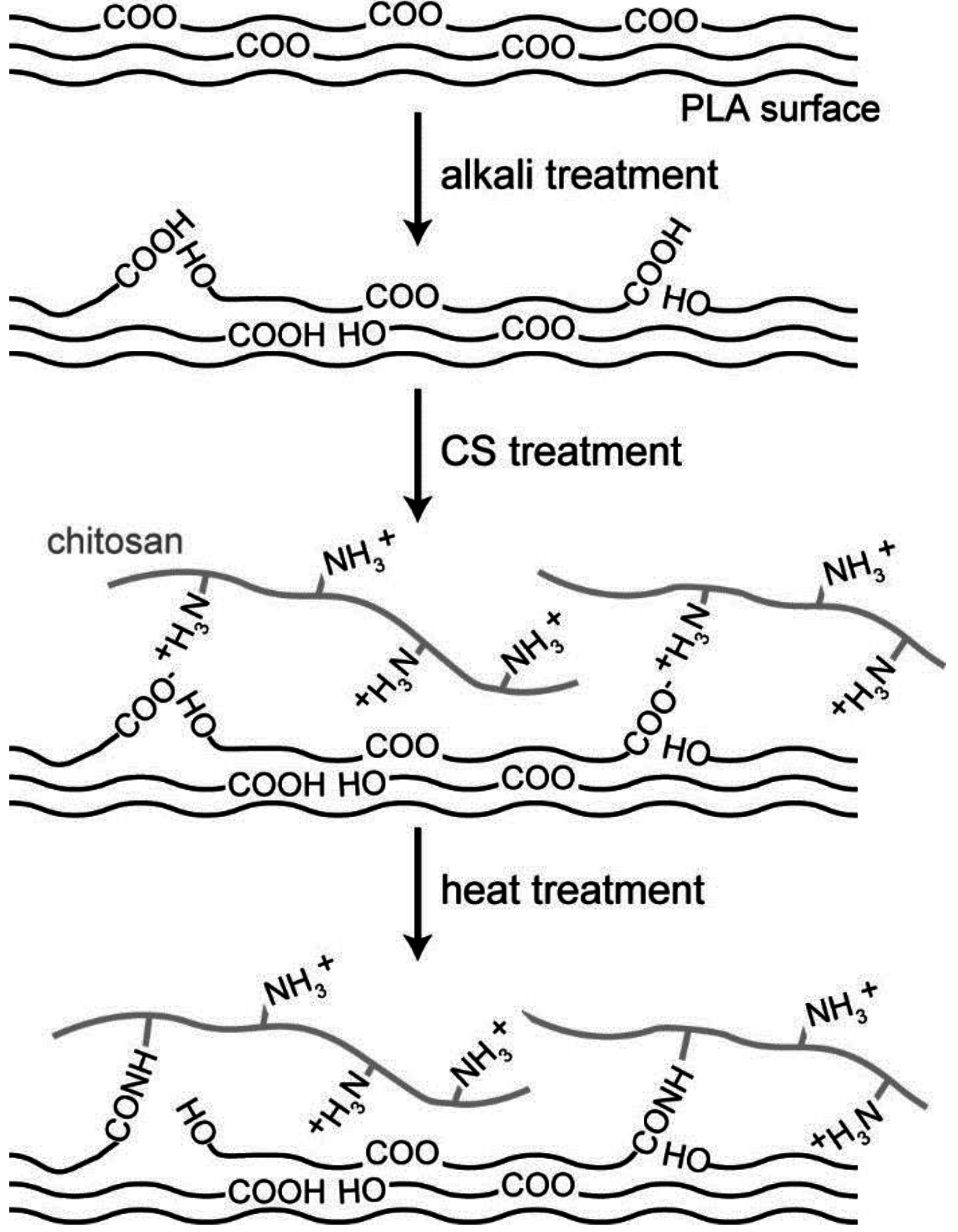
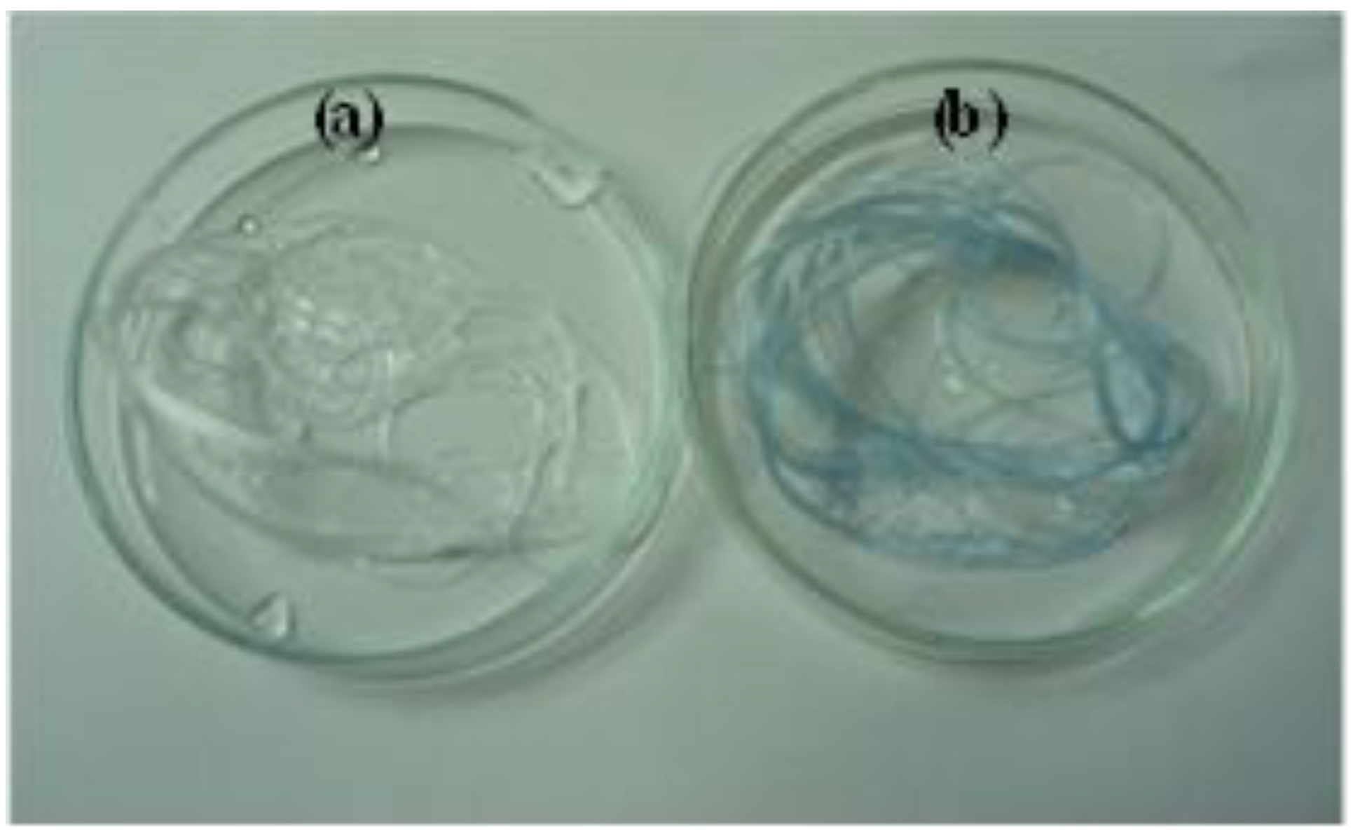
3.1. Preparation of CS–PLA

Figure 1 shows a schematic of the fabrication process. The PLA fiber was treated with 30% NaOH aq. solution and 0.5% CS solution to generate ionic interactions between the carboxyl groups of the PLA and the amino groups of CS. Data related to the preparation of the treated fibers are shown in Table 1. As a control, fiber samples P-1 and P-2 were not treated with CS solution. Fiber samples P-3, P-4, and P-5 were coated with CS on their surface. P-5 differs from P-3 and P-4 with respect to the wash processes used in their preparation. P-3, P-4, and P-5 were not strongly degraded through these processes; however, P-5 exhibited a slightly rigid texture. Through heat treatment, CS–PLA was prepared by partial dehydration between the carboxyl and amino groups.
Figure 1. The schematic illustration of the process in this study.
Fibers 03 00380 g001
Table 1. Preparation data of PLA fibers.
Treatment ProcessesP-1P-2P-3P-4P-5
alkali treatment (sec) a103001030060
first wash process bWWWWS
CS treatment (min) c111
second wash process bWWS & W
heat treatment (h) d11111
Notes: a Alkali treatment was used 30% NaOH aq. solution at 60 °C; b W; washed with distilled water, S; washed with 1% NaOH aq. solution; c 0.5% CS solution at room temperature was used for CS treatment; d dried at 100 °C.

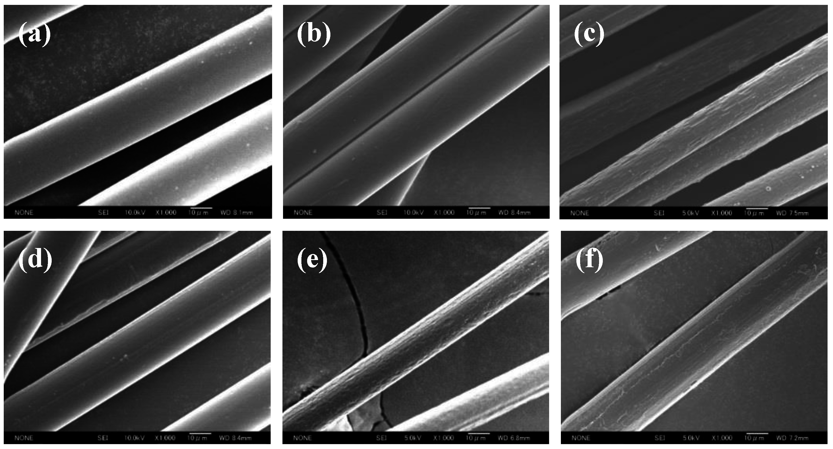
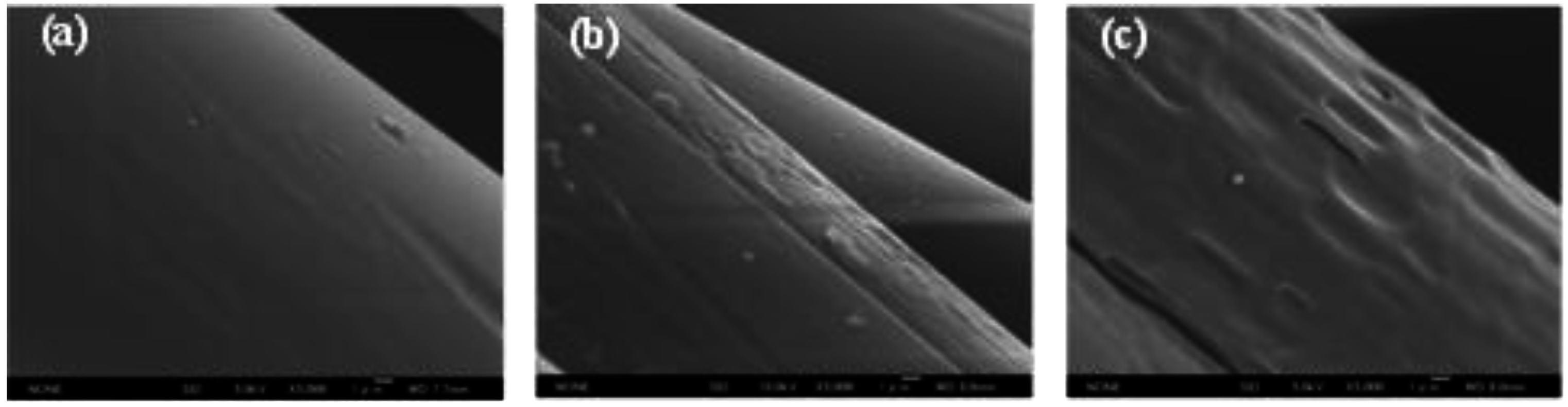
3.2. Surface Morphology

SEM images of the PLA fibers (P-1 and P-2) treated with 30% NaOH aq. solution for predetermined times and those of the CS–PLA fibers (P-3, P-4, and P-5) are shown in Figure 2. The PLA fibers exhibited a rough surface, and their mean diameters were small depending on the degree of alkali treatment. In particular, several cracks were observed on the P-2 surface, whereas few cracks were observed on the P-1 surface. This difference indicates that the degree of alkali treatment strongly affects the ester hydrolysis on the fiber surface. The surface morphology of P-3 was smooth, whereas that of P-4 was rough and some cracks were observed. The surface of each CS–PLA sample was slightly smoother than the surface of the PLA fibers with no CS coating. In particular, the surface morphology of P-5 was smoother and its mean diameter was slightly greater relative to P-3. These observations indicate that CS was additionally coated onto the PLA surface though a coagulation process. The SEM images show that P-5 was coated with CS to a greater extent compared with P-3 and P-4, although it was extensively washed before heat treatment.
Figure 2. SEM images of PLA fibers: (a) original PLA fiber; (b) P-1; and (c) P-2; and CS-PLA (d) P-3; (e) P-4 and (f) P-5.
Fibers 03 00380 g002

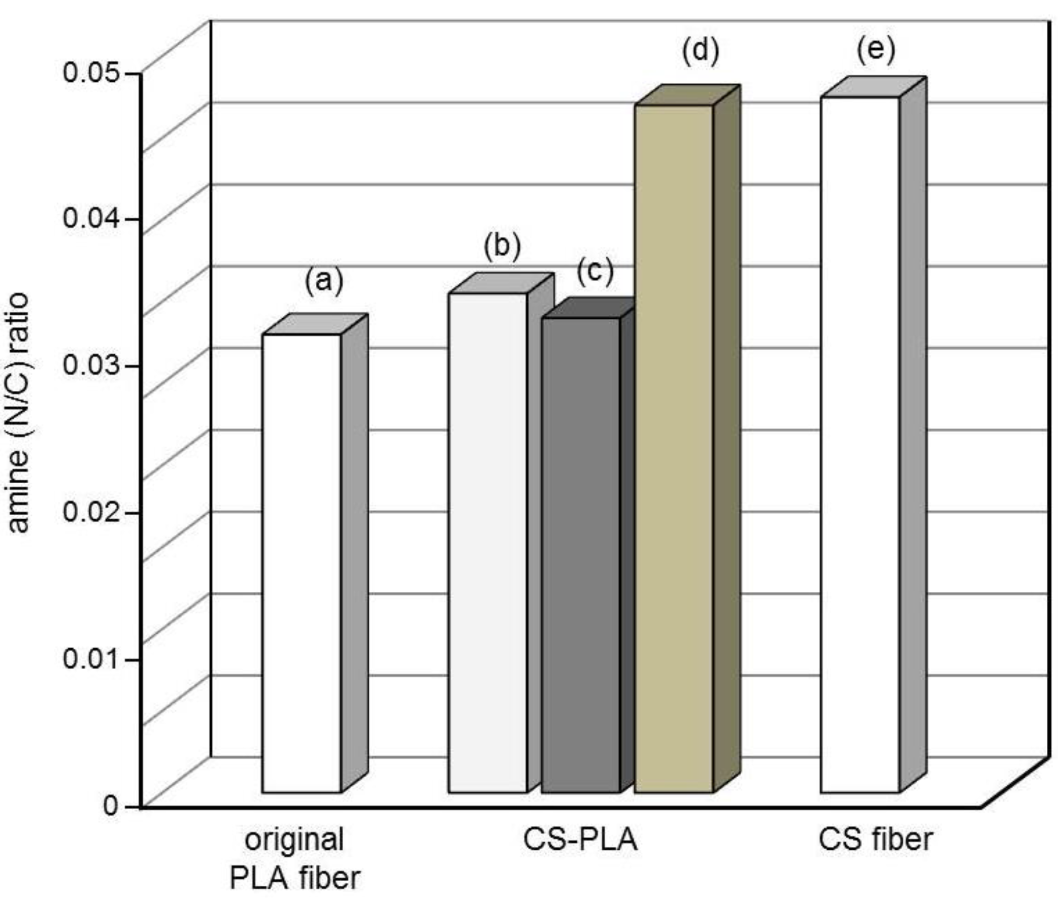
3.3. Qualitative and Quantitative Determination of CS

From the result of the surface morphology, the qualitative and quantitative analyses of CS existing on the fiber were performed using P-3 because the surface of P-2 was rough and had some cracks compared with that of P-1, resulting from the difference of the degree of alkali treatment. The results of visual tests with amido black 10B are shown in Figure 3.
Figure 3. PLA fibers stained with amido black 10B: (a) original PLA fiber and (b) CS–PLA (P-3).
Fibers 03 00380 g003
Since amido black 10B contains two anionic sulfonate groups, it strongly adsorbs CS, which contains cationic amino groups. The images in Figure 3 show the obvious difference between the original PLA and P-3 fibers. P-3 exhibits a clear blue coloration, whereas the original PLA fiber was not stained by amido black 10B. These results suggest that CS exists on the surface of P-3. Additionally, when the PLA fiber without alkali treatment was treated with CS solution, the obtained fiber was not stained by amido black 10B. Thus, it was suggested that the partial hydrolysis of ester linkages by alkali treatment is necessary to coat CS on the fibers’ surface.
The CS coating on each PLA fiber was quantitatively analyzed by Kjeldahl nitrogen analysis and elemental analysis; the results are shown in Table 2. These results indicate that a small amount of CS was coated onto the fibers’ surface. Only slight differences were observed between the results obtained by Kjeldahl nitrogen analysis and elemental analysis. From these results, by calculating the mean diameter of the PLA fibers, which is approximately 24 µm, the coating thickness of CS on the PLA surface was calculated to be approximately 28 nm.
Table 2. Quantitative data of CS content in CS–PLA (P-3).
Measurement methodCS (g)/CS–PLA (g) a
Kjeldahl nitrogen analysis4.69 × 10−3
elemental analysis4.62 × 10−3
Notes: a CS content was calculated from nitrogen content, based on unit molecular weight from deacetylation degree of CS.

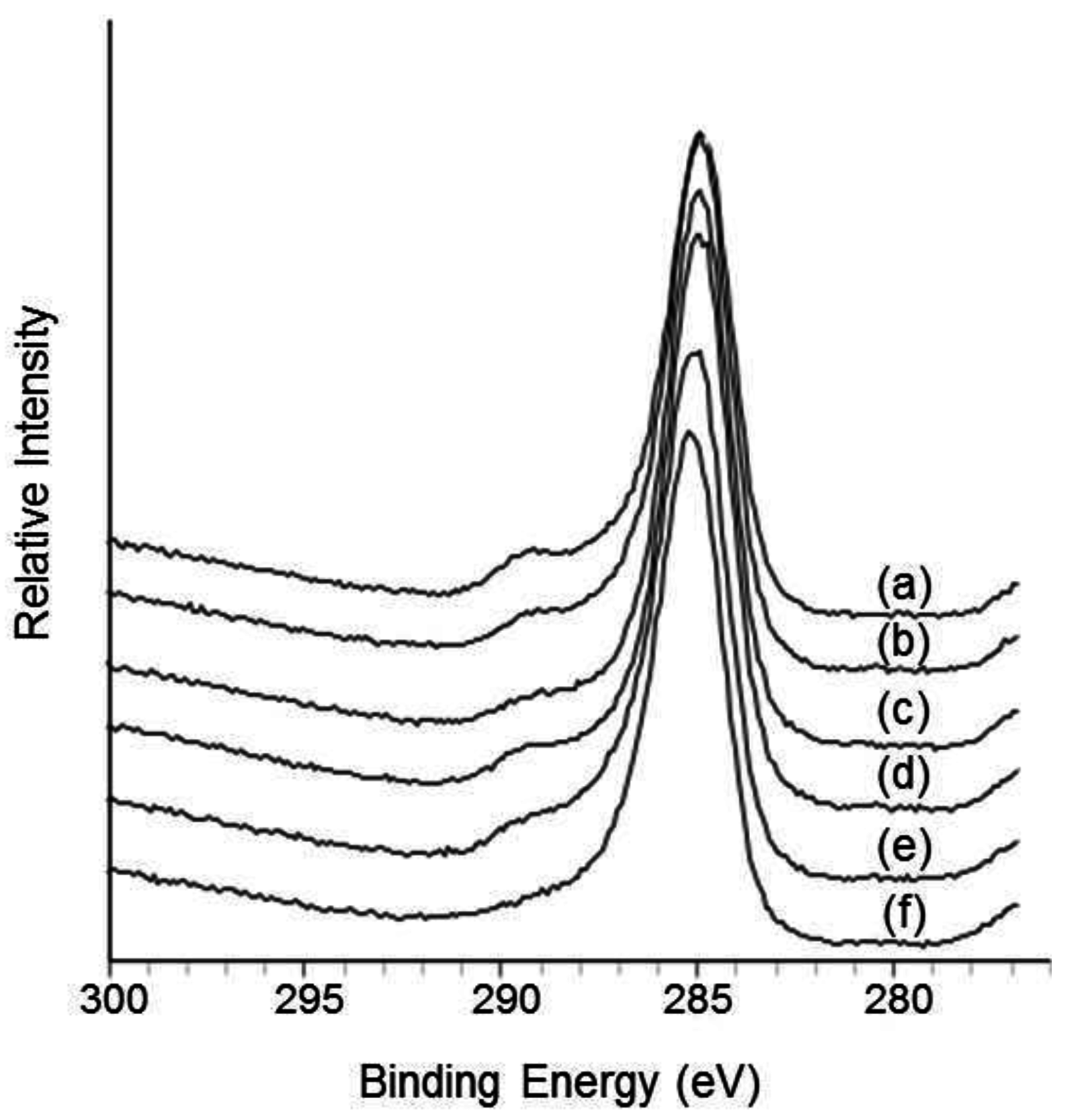
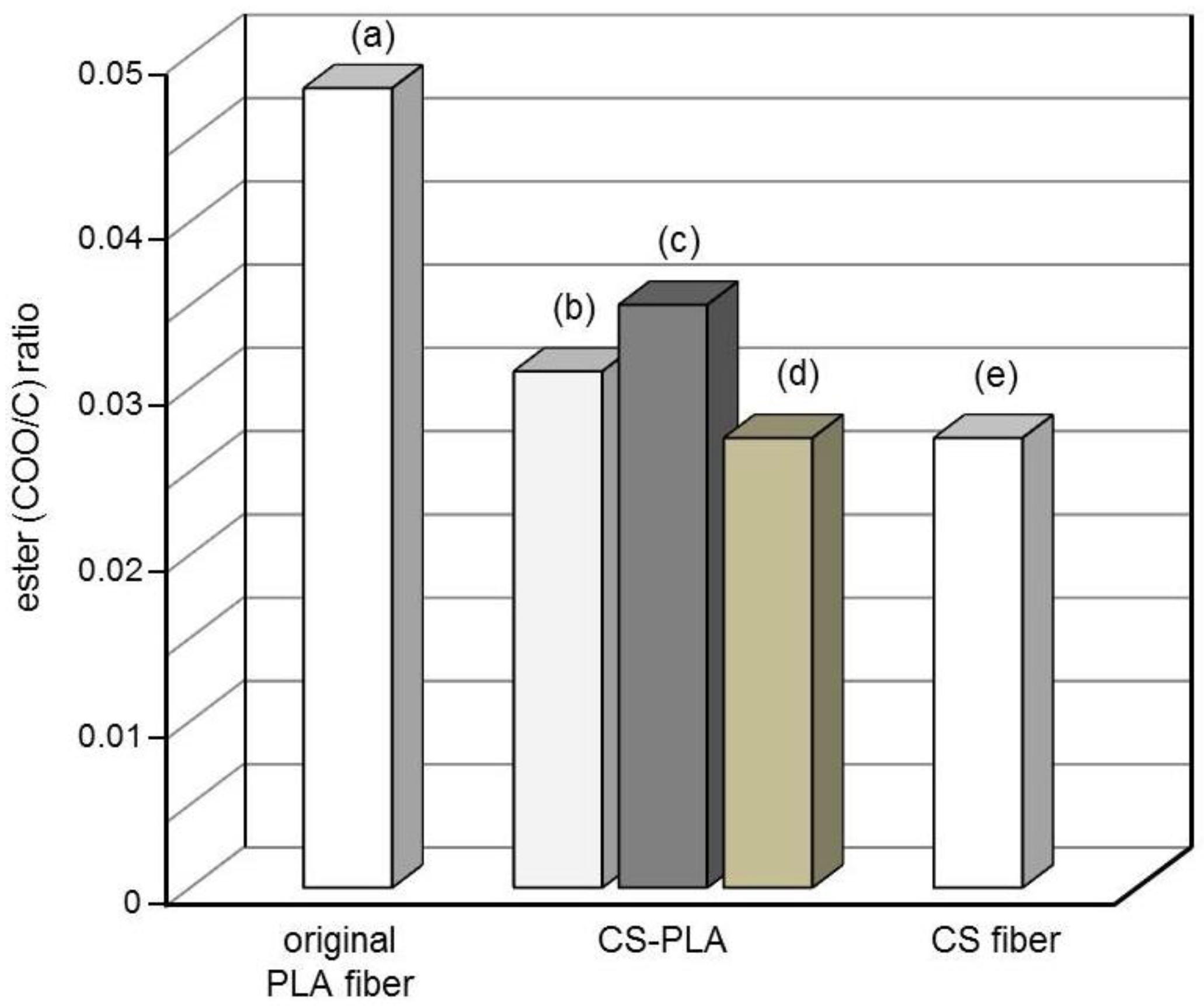
3.4. Surface Characterization

The XPS C1s scan spectra on the surface of P-3, P-4, and P-5 are shown in Figure 4. In this study, the original PLA fiber and CS fibers prepared by previously reported methods were used as reference samples [28]. The C1s peak for fibers can be deconvoluted into three peak components mainly associated with C–C, C–O, and COO species. The ratio of these peaks was attributed to the chemical structure on the fibers’ surface. In particular, the proportion of the COO peak clearly changed when the PLA surface was subjected to alkali or CS treatment. Figure 5 shows the ester (COO/C) ratio calculated from XPS results. The COO/C ratios of P-3, P-4, and P-5 approached that of the CS fibers. These results indicate that the PLA surface was clearly coated by CS.
Figure 4. XPS C1s spectra of PLA fibers and CS fiber: (a) original PLA fiber; (b) P-2; (c) P-3; (d) P-4; (e) P-5, and (f) CS fiber.
Fibers 03 00380 g004
Figure 5. Ester (COO/C) ratio of fibers from XPS analysis: (a) original PLA fiber; (b) P-3; (c) P-4; (d) P-5 and (e) CS fiber.
Fibers 03 00380 g005
The XPS N1s spectra of the fibers’ surface were also analyzed. In the case of P-3, P-4, and P-5, the proportion of the C–NH peak increased compared with that of the original PLA fiber. Since CS contains amide groups in its molecular structure, small amounts of CS bonded to the fibers’ surface can be easily detected. Figure 6 shows variation of the N/C ratios determined by XPS. The N/C ratios of P-3, P-4, and P-5 clearly increased compared with that of the original PLA fiber. P-5 especially exhibited a very high N/C ratio among the CS–PLA samples because it was coated with a greater amount of CS, as observed by SEM.
Figure 6. Amine (N/C) ratio of fibers from XPS analysis: (a) original PLA fiber; (b) P-3; (c) P-4; (d) P-5, and (e) CS fiber.
Fibers 03 00380 g006

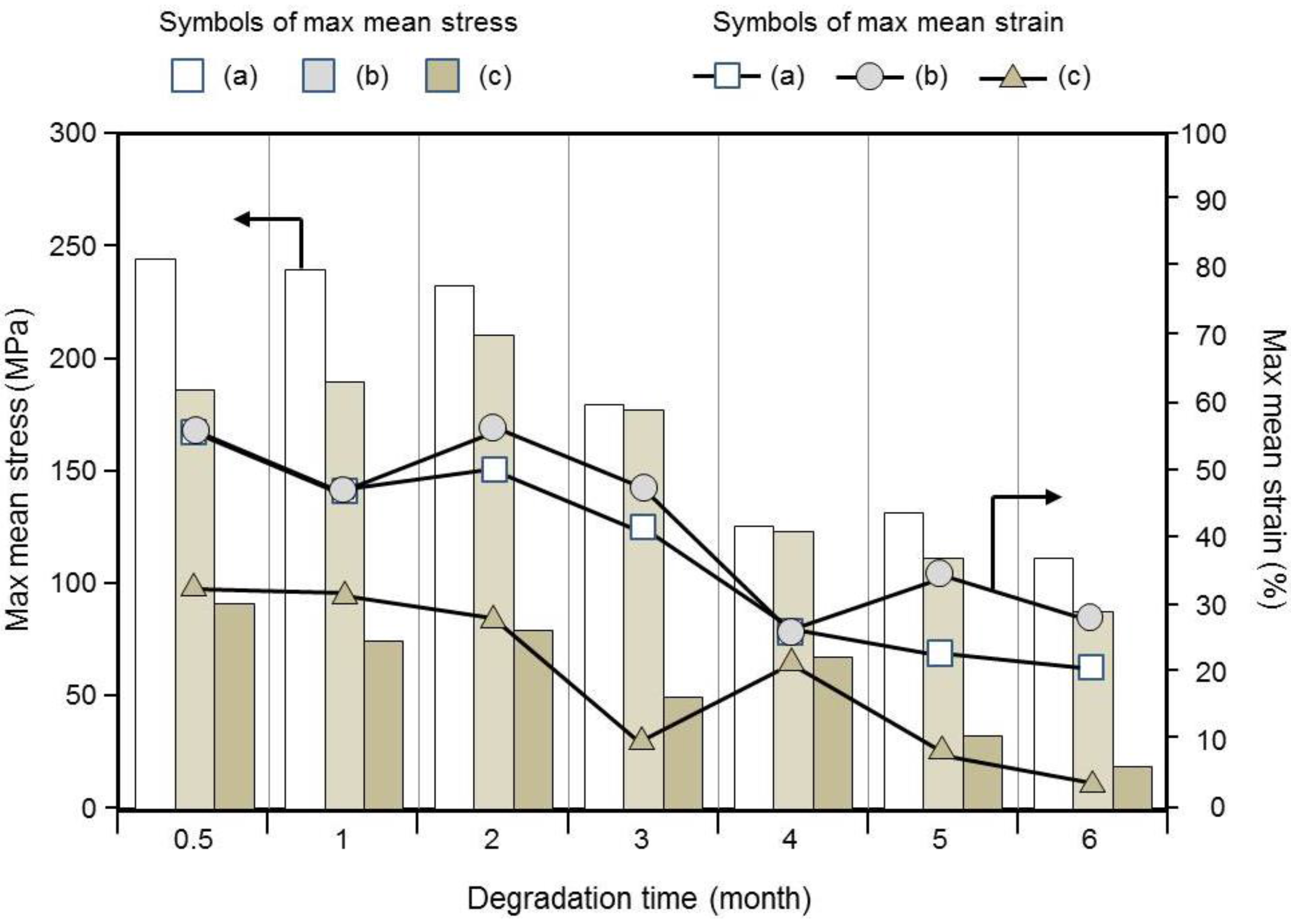
3.5. Degradation Properties

Figure 7 shows the degradation rate calculated from the weight loss of each fiber sample in PBS. The degradation rate of P-3 was slightly higher than that of the original PLA fiber. P-3 exhibited 0.46% (w/w) degradation, whereas P-4 exhibited 0.90% (w/w) degradation. Hence, the degradation rate of P-4 was substantially greater than those of the original PLA and P-3 fibers. This clear difference indicates that the degradation ability depends on the degree of both alkali and CS treatments as they enhanced the surface hydrophilicity. This result on the fibers’ surface is remarkably similar to that on the surface of PLA films by alkali treatment [21]. Moreover, the degradation rates of these three samples were low because of the absence of decay-accelerating species such as enzymes, bacteria, and H3O+ [29]. Notably, however, other authors have reported that the mass of PLA remained almost unchanged for up to 12 months in PBS [30,31].
Figure 7. The degradation rate % (w/w) calculated from the weight loss of the fibers in PBS (1M, pH 7.2) at 37 °C: (a) original PLA fiber; (b) P-3 and (c) P-4.
Fibers 03 00380 g007
The SEM images of CS–PLA hydrolyzed for six months are shown in Figure 8. The original PLA fiber exhibited relatively fewer cracks on its surface. In contrast, the surfaces of the CS–PLA fibers were rough and showed some cracks. In particular, P-4 exhibited a highly hydrolyzed morphology that demonstrated numerous cracks and high roughness over the entire surface of the fibers. These SEM images suggest that P-4 exhibited the highest degradation rate among the investigated PLA fibers.
Figure 8. SEM images of hydrolyzed PLA fibers: (a) original PLA fiber; (b) P-3 and (c) P-4. The fibers were immersed in PBS (1M, pH 7.2) at 37 °C for 6 months.
Fibers 03 00380 g008
Figure 9 shows the max mean stress and strain of the degraded fibers. The initial max mean stress of the original PLA fiber and P-3 and P-4 fibers were 245, 198, and 95 MPa, respectively. The tensile strength of all the fiber samples tended to decrease over time. These results suggest that the presence of cracks on the fibers’ surface tended to diminish the tensile strength. After six months, the max mean stress of the original PLA fiber and P-3 and P-4 fibers were 111, 85, and 18 MPa, respectively. The max mean stress and strain of P-3 were slightly lower than those of the original PLA fiber. This suggests that both alkali and CS treatments affected the degradation ability of the PLA fibers. Moreover, the max mean stress and strain of P-4 were clearly much lower than those of the other fiber samples from 0 to 6 months later. After six months, the stress of the original PLA and P-4 fibers decreased by 45.3% and 18.6%, respectively, compared with those at month zero. CS–PLA fibers exhibited greater degradation according to the results of the degradation rate measurements, surface morphology observations, and tensile strength tests. These results indicate that the hydrophilicity of the fiber surface was remarkably enhanced depending on the degree of alkali and CS treatments. Moreover, control of these treatments will enable future adjustment of the degradation behavior of the PLA fibers.
Figure 9. Max mean stress (MPa) and strain (%) of degraded PLA fibers: (a) original PLA fiber; (b) P-3 and (c) P-4.
Fibers 03 00380 g009

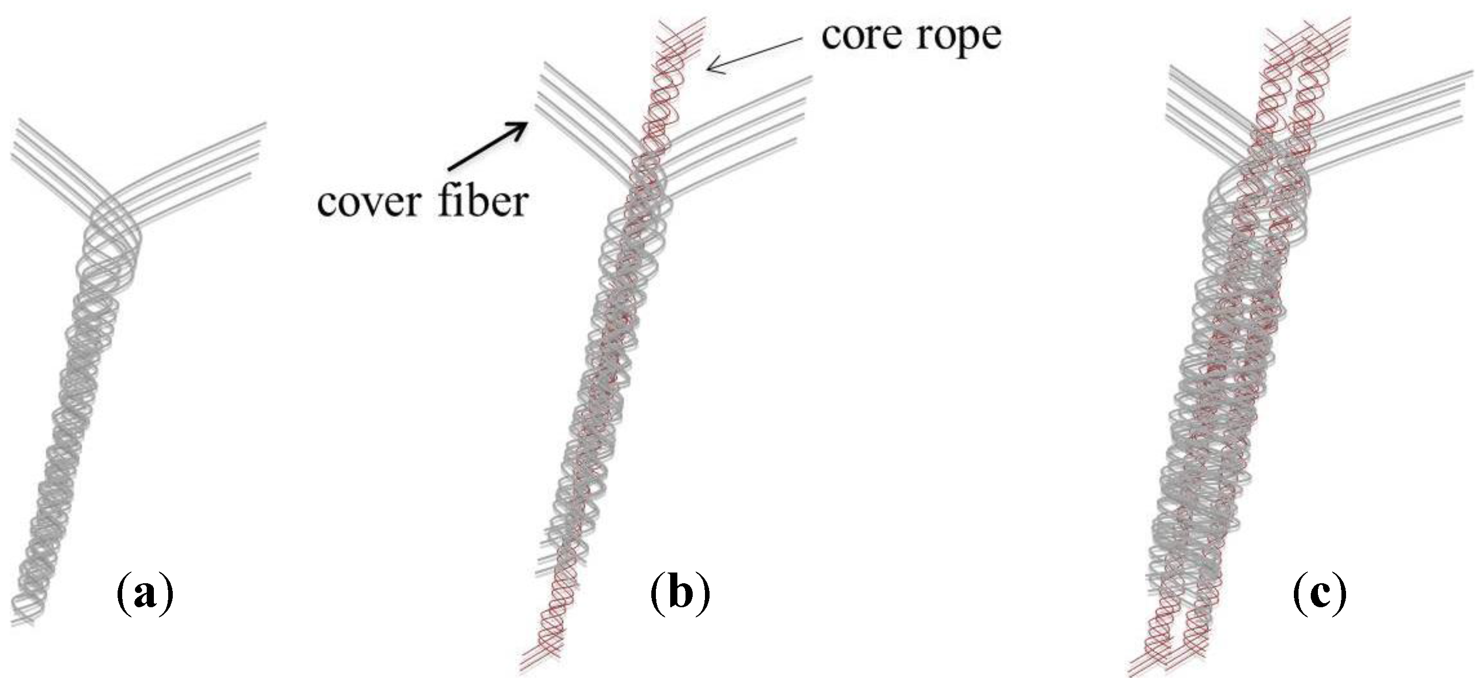
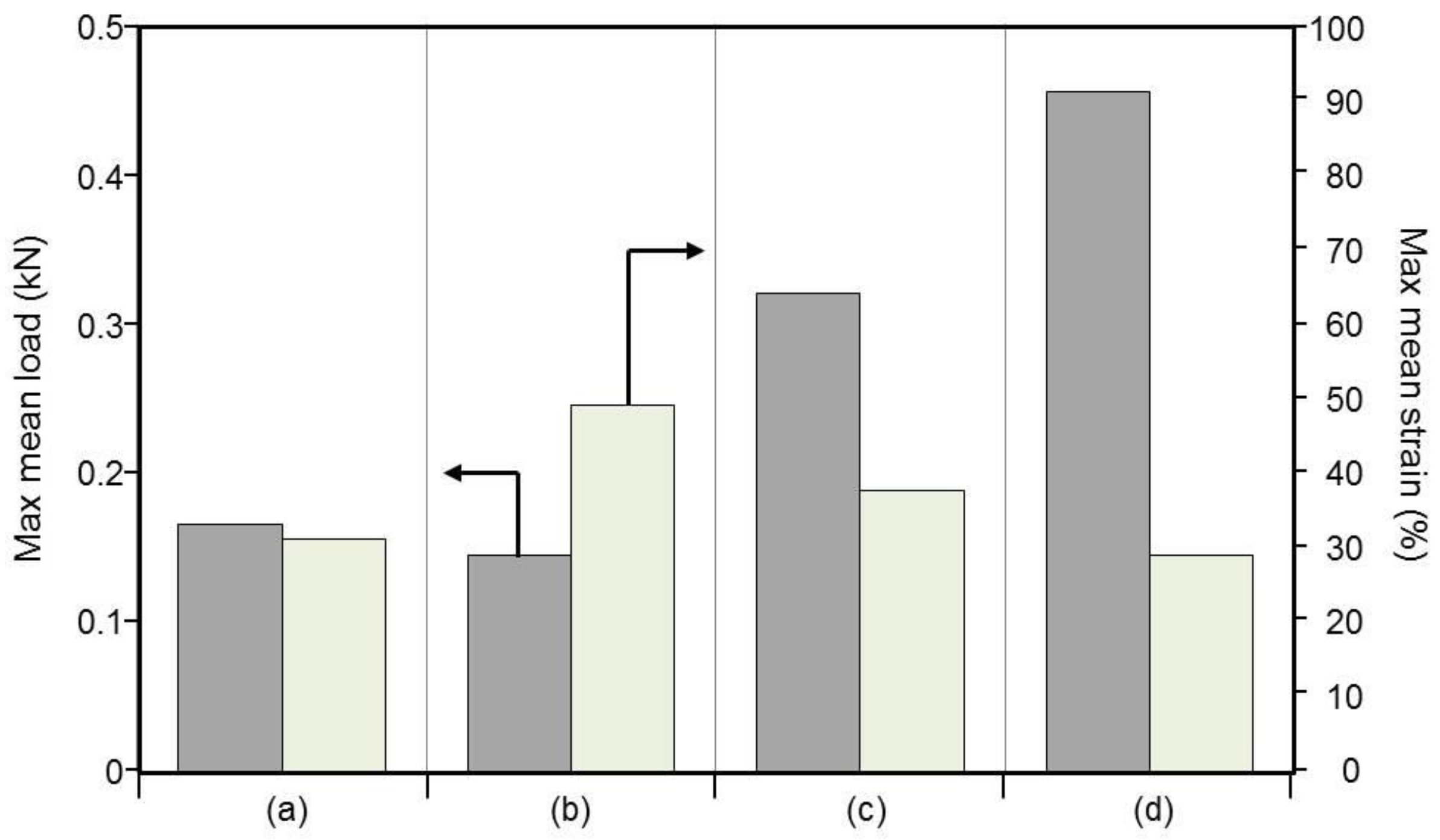
3.6. Mechanical Properties of Braided Ropes

Figure 10 shows an illustration of the braided ropes. In this study, the core rope prepared from the original PLA fiber and the braided ropes with non-, single-, or double-core ropes were prepared using a braider. The tensile strength of these braided ropes was measured using an Autograph AG-TS apparatus; the results are shown in Figure 11. The stress and strain of the braided rope prepared from only the original PLA fiber (a) was 0.17 kN and 32%, whereas those of the braided rope prepared from only P-3 (b) were 0.14 kN and 48%; Meanwhile, the stresses of single- (c) and double-core braided ropes (d) were 0.32 and 0.46 kN, respectively; these values are respectively about two-fold and three-fold greater than the stress value of the non-core braided rope (b). In contrast, the strain of non- (b), single- (c), and double-core (d) braided ropes gradually decreased with increasing number of core ropes. Hence, these results suggest that the objective tensile strength and flexibility of the braided rope can be controlled by adjusting the number of core ropes and shape of the braided rope. These braided ropes will be useful for biomaterial applications that require substantial strength, such as artificial tendons. The test for cell attachment and growth in vivo and in vitro using these braided ropes will be the focus of future investigations.
Figure 10. The illustration of the braided ropes. Original PLA fibers were braided as a core rope. P-3 as a cover fiber was braided around the core rope. (a) Non-; (b) single-; and (c) double-core braided ropes were prepared.
Fibers 03 00380 g010
Figure 11. Max mean load (kN) and strain (%) of braided ropes prepared from PLA fiber: (a) core fiber prepared from original PLA fiber; (b) P-3 braided rope without core fiber; (c) P-3 braided rope with single-core fiber, and (d) P-3 braided rope with double-core fiber.
Fibers 03 00380 g011

4. Conclusions

Alkali treatment introduced active species through partial hydrolysis of ester groups on the surface of the PLA fibers. Subsequent CS treatment generated an ionic bond between the amino group of CS and the carboxyl group of PLA. CS–PLA was finally prepared by the formation of amide bonds through heat treatment. SEM images of the fibers revealed that the morphology of the PLA surface became rough with increasing degrees of alkali and CS treatments. The presence of CS on the fibers’ surface after the alkali and CS treatments was clearly verified by the amido black 10B absorption tests and XPS. We also deduced that the coating thickness of CS on the fibers’ surface was approximately 28 nm from the quantitative analysis results of the nitrogen content. Moreover, analysis of the degradation rate, surface morphology, and tensile strength of the samples revealed that CS–PLA exhibited a higher degradation rate than the unmodified PLA. This greater degradation rate resulted from the enhancement of the hydrophilicity as a consequence of the alkali and CS treatments. The braided ropes with non-, single-, and double-core ropes were also prepared and became tight with increasing number of core ropes. They indicated that the objective tensile strength and flexibility of the braided rope could be controlled by adjusting the number of core ropes and the shape of the braided rope. Therefore, we expect that the braided rope prepared from CS–PLA could be utilized in biomaterial applications such as artificial tendons.

Acknowledgments

This work was supported by the Kansai University Grant-in-Aid of the Promotion and Upgrading of Education and Research, 2015, “Generation of bio-interactive polymers leading future-oriented medicalsˮ.

Author Contributions

Tetsuya Furuike and Hiroshi Tamura designed experiments, and Hideaki Nagahama and Thitirat Chaochai performed the experiments. Tetsuya Furuike wrote the manuscript.

Conflicts of Interest

The authors declare no conflict of interest.

References

  1. Ikada, Y.; Tsuji, H. Biodegradable polyesters for medical and ecological applications. Macromol. Rapid Commun. 2000, 21, 117–132. [Google Scholar] [CrossRef]
  2. Tsuji, H.; Ikada, Y. Blends of aliphatic polyesters. II. Hydrolysis of solution-cast blends from poly(l-lactide) and poly(ε-caprolactone) in phosphate-buffered solution. J. Appl. Polym. Sci. 1998, 67, 405–415. [Google Scholar] [CrossRef]
  3. Saravanan1, M.; Domb, A.J. A contemporary review on—Polymer stereocomplexes and its biomedical application. Eur. J. Nanomed. 2013, 5, 81–96. [Google Scholar] [CrossRef]
  4. Razak, S.I.A.; Sharif, N.F.A.; Rahman, W.A.W.A. Biodegradable polymers and their bone applications: A review. Int. J. Basic Appl. Sci. 2012, 12, 31–49. [Google Scholar]
  5. Wachsen, O.; Platkowski, K.; Reichert, K.-H. Thermal degradation of poly-l-lactide-studies on kinetics, modelling and melt stabilisation. Polym. Degrad. Stab. 1997, 57, 87–94. [Google Scholar] [CrossRef]
  6. Aoyagi, Y.; Yamashita, K.; Doi, Y. Thermal degradation of poly[(R)-3-hydroxybutyrate], poly[ε-caprolactone], and poly[(S)-lactide]. Polym. Degrad. Stab. 2002, 76, 53–59. [Google Scholar] [CrossRef]
  7. Lunt, J. Large-scale production, properties and commercial applications of polylactic acid polymers. Polym. Degrad. Stab. 1998, 59, 145–152. [Google Scholar] [CrossRef]
  8. Jain, R.A. The manufacturing techniques of various drug loaded biodegradable poly(lactide-co-glycolide) (PLGA) devices. Biomaterials 2000, 21, 2475–2490. [Google Scholar] [CrossRef]
  9. Mikos, A.G.; Lyman, M.D.; Freed, L.E.; Langer, R. Wetting of poly(l-lactic acid) and poly(dl-lactic-co-glycolic acid) foams for tissue culture. Biomaterials 1994, 15, 55–58. [Google Scholar] [CrossRef]
  10. Mi, F.-L.; Syu, S.-S.; Lin, Y.-M.; Wu, Y.-B.; Peng, C.-K.; Tsai, Y.-H. Chitin/PLGA blend microspheres as a biodegradable drug delivery system: A new delivery system for protein. Biomaterials 2003, 24, 5023–5036. [Google Scholar] [CrossRef]
  11. Nagahama, K.; Mori, Y.; Ohya, Y.; Ouchi, T. Biodegradable nanogel formation of polylactide-grafted dextran copolymer in dilute aqueous solution and enhancement of its stability by stereocomplexation. Biomacromolecules 2007, 8, 2135–2141. [Google Scholar] [CrossRef] [PubMed]
  12. Nanda, R.; Sasmal, A.; Nayak, P.L. Preparation and characterization of chitosan–polylactide composites blended with Cloisite 30B for control release of the anticancer drug paclitaxel. Carbohydr. Polym. 2011, 83, 988–994. [Google Scholar] [CrossRef]
  13. Preechawong, D.; Peesan, M.; Supaphol, P.; Rujiravanit, R. Preparation and characterization of starch/poly(l-lactic acid) hybrid foams. Carbohydr. Polym. 2005, 59, 329–337. [Google Scholar] [CrossRef]
  14. Chen, C.; Dong, L.; Cheung, M.K. Preparation and characterization of biodegradable poly(l-lactide)/chitosan blends. Eur. Polym. J. 2005, 41, 958–966. [Google Scholar] [CrossRef]
  15. Peesan, M.; Supahol, P.; Rujiravanit, R. Preparation and characterization of hexanoly chitosan/polylactide blend films. Carbohydr. Polym. 2005, 60, 343–350. [Google Scholar] [CrossRef]
  16. Cai, K.; Yao, K.; Lin, S.; Yang, Z.; Li, X.; Xie, H.; Qing, T.; Gao, L. Poly(d,l-lactic acid) surfaces modified by silk fibroin: Effects on the culture of osteoblast in vitro. Baiomaterials 2002, 23, 1153–1160. [Google Scholar] [CrossRef]
  17. Chu, C.F.L.; Lu, A.; Liszkowski, M.; Sipehia, R. Enhanced growth of animal and human endothelial cells on biodegradable polymers. Biochim. Biophys. Acta 1999, 1472, 479–485. [Google Scholar] [CrossRef]
  18. Kito, H.; Matsuda, T. Biocompatible coatings for luminal and outer surfaces of small-caliber artificial grafts. J. Biomed. Mater. Res. 1996, 30, 321–330. [Google Scholar] [CrossRef]
  19. Zhu, A.; Zhang, M.; Wu, J.; Shen, J. Covalent immobilization of chitosan/heparin complex with a photosensitive hetero-bifunctional crosslinking reagent on PLA surface. Biomaterials 2002, 23, 4657–4665. [Google Scholar] [CrossRef]
  20. Ding, Z.; Chen, J.; Gao, S.; Chang, J.; Zhang, J.; Kang, E.T. Immobilization of chitosan onto poly-l-lactic acid film surface by plasma graft polymerization to control the morphology of fibroblast and liver cells. Biomaterials 2004, 25, 1059–1067. [Google Scholar] [CrossRef]
  21. Tsuji, H.; Ishida, T. Poly(l-lactide). X. Enhanced surface hydrophilicity and chain-scission mechanisms of poly(l-lactide) film in enzymatic, alkali, and phosphate-buffered solutions. J. Appl. Polym. Sci. 2003, 87, 1628–1633. [Google Scholar] [CrossRef]
  22. Park, G.E.; Pattison, M.A.; Park, K.; Webster, T.J. Accelerated chondrocyte functions on NaOH-treated PLGA scaffolds. Biomaterials 2005, 26, 3075–3082. [Google Scholar] [CrossRef] [PubMed]
  23. Tokura, S.; Ueno, K.; Miyazaki, S.; Nishi, N. Molecular weight dependent antimicrobial activity by chitosan. Macromol. Symp. 1997, 120, 1–9. [Google Scholar] [CrossRef]
  24. Okamoto, Y.; Minami, S.; Matsuhashi, A.; Sashiwa, H.; Saimoto, H.; Shigemasa, Y.; Tanigawa, T.; Tanaka, Y.; Tokura, S. Application of polymeric n-acetyl-d-glucosamine (chitin) to veterinary practice. J. Vet. Med. Sci. 1993, 55, 743–747. [Google Scholar] [CrossRef] [PubMed]
  25. Dodane, V.; Vilivalam, V.D. Pharmaceutical applications of chitosan. Pharm. Sci. Technol. Today 1998, 1, 246–253. [Google Scholar] [CrossRef]
  26. Dash, M.; Chiellini, F.; Ottenbrite, R.M.; Chiellini, E. Chitosan-A versatile semi-synthetic polymer in biomedical applications. Prog. Polym. Sci. 2011, 36, 981–1014. [Google Scholar] [CrossRef]
  27. Suh, J.-K.F.; Matthew, H.W.T. Application of chitosan-based polysaccharide biomaterials in cartilage tissue engineering: A review. Biomaterials 2000, 21, 2589–2598. [Google Scholar] [PubMed]
  28. Tamura, H.; Tsuruta, Y.; Itoyama, I.; Worakitkanchanakul, W.; Rujiravanit, R.; Tokura, S. Preparation of chitosan filament applying new coagulation system. Carbohydr. Polym. 2004, 56, 205–211. [Google Scholar] [CrossRef]
  29. Cam, D.; Hyon, S.-H.; Ikada, Y. Degradation of high molecular weight poly(l-lactide) in alkaline medium. Biomaterials 1995, 16, 833–843. [Google Scholar] [CrossRef]
  30. Li, S.; Garreum, H.; Vert, M. Structure-property relationships in the case of the degradation of massive poly(α-hydroxy acids) in aqueous media. J. Mater. Sci. Mater. Med. 1990, 1, 198–206. [Google Scholar] [CrossRef]
  31. Tsuji, H.; Ikada, Y. Properties and morphology of poly (l-lactide) 4. Effects of structural parameters on long-term hydrolysis of poly (l-lactide) in phosphate-buffered solution. Polym. Degrad. Stab. 2000, 67, 179–189. [Google Scholar] [CrossRef]

