Article

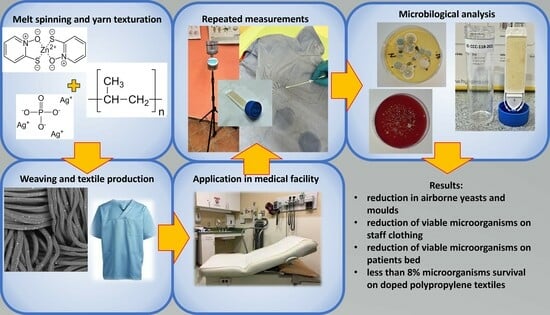
Field Study of Activity of Antimicrobial Polypropylene Textiles

Chemosvit Fibrochem, s.r.o., Štúrova 101/2, 059 21 Svit, Slovakia
*
Author to whom correspondence should be addressed.
Fibers 2023, 11(11), 97; https://doi.org/10.3390/fib11110097
Submission received: 27 June 2023 / Revised: 12 October 2023 / Accepted: 30 October 2023 / Published: 10 November 2023

Abstract

:
In this work, an in situ study is presented of the impact of textile materials used in healthcare facilities on microbial colonization of textile surfaces. The available literature describes antimicrobial active textiles and their effectiveness in laboratory conditions. However, the quantification of the impact on the microbiome of healthcare facilities has not been investigated so far. Polypropylene yarns doped with silver phosphate glass and zinc pyrithione were prepared and used for the production of bed sheets and clothing for healthcare personnel. Subsequently, measurements of airborne particles and viable microorganisms on given textiles were conducted in a private surgery clinic for 3 weeks, comparing the counts of viable microorganisms before and after replacing staff clothing and bedding on examination and the surgical bed with said polypropylene cloth. A significant reduction in airborne particles and viable microorganisms was expected based on previous studies on the use of polypropylene textiles in operating rooms. In this study, a significant reduction in viable airborne fungi and viable microorganisms on monitored textiles was observed by multiple methods. However, the effect on airborne microorganisms seems insignificant in areas with frequent patient traffic. The textile described here represents a new additional way of protecting patients and medical personnel from healthcare-associated infections while using a modification of proven production procedures and commercially usable materials without legislative restrictions.

Graphical Abstract

1. Introduction

In healthcare settings, great attention is paid to cleanliness, and compliance with a range of routines, such as hand hygiene, is required to mitigate healthcare-associated infections (HAIs) [1,2]. The frequency of HAIs can be illustrated, for example, by the most recent summary report of the National Health Care Safety Network (USA), which states there were 331,897 cases of HAIs reported in 2015–2017, with surgical site infections accounting for 43% of cases being most prevalent [3]. The most recent surveillance report of the European Centre for Disease Prevention and Control states an average prevalence of HAIs in Europe at 5.9% of hospitalized patients, where prevalence values for individual countries ranged from 2.9% to 10.0% of hospitalized patients, with respiratory tract infections as the most frequently reported (21.4%) and cause and surgical site infections being the third most common cause (18.4%) [4]. A similar point prevalence survey conducted in Slovakia, where this study was conducted, states that 4% of hospitalized patients contracted an HAI in 2016–2017, with urinary tract infections (25.8%), pneumonia, and other lower respiratory tract infections (20%) being the most common and surgical site infections being the fourth most common (12.5%) [5]. As a measure to prevent the spread of pathogens in medical facilities, surfaces with antimicrobial properties, such as copper possessing an oligomeric effect, are utilized [6]. While most surfaces, such as floors, tables, and handles, are easily sanitized by wiping with appropriate disinfectants, the situation is different for textile surfaces. Textiles present porous surfaces prone to retaining moisture and possessing increased specific surface area to host microorganisms. In fact, in environments with controlled air quality, sources of airborne particles carrying microorganisms may be traced to used textiles [7,8]. Studies examining the survival times of individual pathogens show that the base material of textile is also an important factor; in fact, the survival of bacteria at room temperature was the longest, up to 206 days, on polyester [6]. In fact, textile surfaces such as curtains and upholstery have been found to be contaminated over time in healthcare settings and host pathogens for up to 90 days [9]. To mitigate these risks, textiles utilizing new biocidal treatments are developed by many researchers, utilizing, for example, metallic nanoparticles, plasma treatments [10], new organic biocidal substances [11], and their combinations with inorganic nanoparticles [12]. However, new biocides have to comply with relevant regulations [13]; thus, their application in the near future is limited to prototypes.
In fact, the fiber-forming polymer itself may hinder the growth of bacteria and fungi; even the structure of the textile (woven, knitted, and spunbond) can affect its suitability for use in healthcare settings [7]. Additionally, active ingredients relying on metallic ions, such as zinc [14] and silver [15], have been demonstrated to be active toward antibiotic-resistant bacteria. In light of the above facts, one may suggest that polypropylene fabric [7] containing antimicrobial substances utilizing an oligodynamic effect may bring a substantial reduction in both airborne particles and present living microorganisms. The possible difference between laboratory settings and real-life working environments points to a necessity to conduct a study of the aforementioned hypothesis. During the investigation of standards for textiles in medical settings and textiles actually used in healthcare, the authors found a private surgery clinic in Levoča district, Slovakia, willing to participate in the study proposed above, albeit under the condition of anonymity due to public perception of any possible findings. As active ingredients, silver phosphate glass (SPG) and zinc pyrithione (ZP) were chosen as sources of silver and zinc ions since they are intended for use in thermoplastics and possess valid registrations required for use in the EU market. As for quantification of living organisms on textile surfaces, determination of present adenosine triphosphate was suggested: All living organisms produce and utilize adenosine triphosphate (ATP), and standardized methods that use the detection and quantification of present ATP are used to confirm and quantify the sum of all living microorganisms on the given surface [16].

2. Materials and Methods

2.1. Materials

Isotactic polypropylene homopolymer with melt flow index of 25 g/10 min. in form of pellets was supplied by Slovnaft a.s., Bratislava, Slovakia. Low-density polyethylene (LDPE) homopolymer wax in a powder form used as dispersing agent was produced by Honeywell, Morris Plains, NJ, USA. Pigments used to dope-dye polypropylene yarns, titanium dioxide, carbon black, PV-Echt-Rosa, copper phthalocyanine, and phthalocyanine green G, were purchased from NYLUS SK, Poprad, Slovakia. Active antimicrobial ingredients silver phosphate glass and zinc pyrithione were supplied by SANITIZED AG, Burgdorf, Switzerland. Both silver phosphate glass and zinc pyrithione were supplied in the form of fine ground powders with a nominal median particle size of 2 µm declared by the supplier.
Sets of ATP sampling tests UltraSnap, including SystemSURE luminometer, were purchased from Hygiena LLC, Camarillo, CA, USA. Envirocheck® Contact DC Disinfection Control contact plates, tryptic soy agar contact plates, blood agar plates, glucose, tryptone, yeast extract, and agar for agar plates were supplied by Merck KGaA, Darmstadt, Germany.

2.2. Methods

2.2.1. Textile Samples Preparation

Polypropylene (PP) yarns were produced in three steps: compounding, melt spinning, and false-twist texturation. A masterbatch containing 5% by weight of zinc pyrithione (ZP) was produced and prepared by thoroughly mixing 500 g of ZP and 250 g of fine powdered polyethylene wax in Mixaco vertical container mixer (MIXACO Maschinenbau Dr. Herfeld GmbH & Co. KG, Neuenrade, Germany) and subsequent addition of 9.25 kg of polypropylene. The resulting mixture was compounded on ZSK-25 twin screw compounder (Coperion GmbH, Stuttgart, Germany). In a similar manner, a masterbatch containing 6% silver phosphate glass (SPG) was prepared by compounding 600 g of SPG, 300 g of polyethylene wax, and 9.1 kg of PP. Mixtures of pigments were compounded the same way to provide masterbatches containing 5% of pigments for corresponding resulting color shade in the form of pellets for dope dyeing for final yarn colors light-blue, gray-blue, and red. Six mixtures, each containing pigment masterbatch, masterbatch of chosen antimicrobial additive, and polypropylene, were prepared in a fashion to obtain one yarn containing 0.10% of ZP and one yarn containing 0.15% of SPG for each color produced; the recipes for each mixture are listed in Table 1.
Each of the given mixtures was melt spun using an industrial melt spinning line with a single screw extruder (Research Institute for Man-Made Fibres, a.s., Svit, Slovakia) with D = 45 mm and L/D = 44, at 230 °C with melt throughput 151 g∙min−1 through two shafts, each with one gear metering pump supplying four spinnerets with 50 orifices and orifice diameter 0.35 mm heated at 245 °C, supplying each spinneret with 18.9 g∙min−1 of polymer melt. The as-spun fiber was oriented between two godets: the pull-off speed (1st godet) was set to 1480 m∙min−1, and the take-up speed was 1450 m/min. The speeds of second godets were adjusted to 1450 ± 20 m∙min−1 individually for each mixture to ensure tension of pre-oriented fiber during winding to be 5 ± 0.5 cN. As-spun pre-oriented multifilament fibers were false-twist textured using Barmag AFK-6 draw texturizing machine (OC Oerlikon, Pfäffikon, Switzerland) at texturation speed of 400 m∙min−1, D/Y = 1.55 and draw ratio of 1.545, and heater temperature 170 °C, connecting two ends by intermingling in air jet at supply air pressure of 150 kPa resulting in yarns with linear density 167 ± 5 decitex.
A simple plain weave fabric was woven using pairs of PP yarns with the same color: PP fibers containing SPG were used for the warp beam with 26 ends per cm of warp width, and PP fibers containing ZP were used for the weft, resulting in weft density of 22.1 threads/cm and the resulting fabrics having a grammage of 95 ± 5% g∙m−2.

2.2.2. Replacement of Staff Clothing and Bedding in a Small Surgery Department and Frequency of Measurements

Of the aforementioned fabrics, the textiles dyed purple were used for production of four pairs of women’s tunics; two pairs of men’s tunics were made from the light blue fabric, while beige fabrics were used as bed sheets. The samples were then used in the private surgery clinic by staff and on two beds, replacing the currently worn cotton-polyester blend clothing and bedding. Men’s tunics were to be worn by the attending physician, and women’s tunics were to be worn by nurses. The light-blue fabrics were used as bed sheets on the examining bed in room no. 1 and the surgical bed in room no. 2. During examination and surgery, another disposable polymer sheet covered the bed sheet on both the examining and surgical beds. Two rooms of the clinic in question were examined: Room no. 1 is the doctor’s office with an area of 32 m2 and a high frequency of visits, approximately 35 patients each day with 1 physician and 1 nurse present. These persons were present in the room for 32.5 h per week. In this room, an ambient germicidal lamp is in operation outside the business hours of the clinic. The doctor’s office no. 2 has an area of 9 m2 and is used for minor surgery and preparation of tools with only one patient, physician, and nurse present if used. The average weekly time of occupation of room no. 2 is 22.5 h, with a daily surface (floor and tables) disinfection and cleaning routine.
Of the below-mentioned samplings, for each method, the first sampling, numbered “0”, was taken before replacing the usual staff clothing and bedding with presented polypropylene textiles with antimicrobial agents. Starting the next day, the staff clothing and beddings were replaced with the above-mentioned textiles; the measurements of air quality and textile viable microbe counts numbered sampling 1 were made one week after the replacement, and subsequent samplings numbered 2 and 3 were made with an interval of one week. Finally, after measurement 3 was taken, the 4 samples, nurse’s tunic, physician’s tunic, bedding from examination bed, and bedding from surgery bed, were each separately closed in a sealable plastic bag and left under ambient temperature for 24 h. Subsequently, new samplings with swabs for ATP and contact plates, as described below in the text, were taken, cultivated, and evaluated. This measurement is listed as measurement 4 and shows the rate of survival of microbes from measurement no. 3 without any additional bioburden.

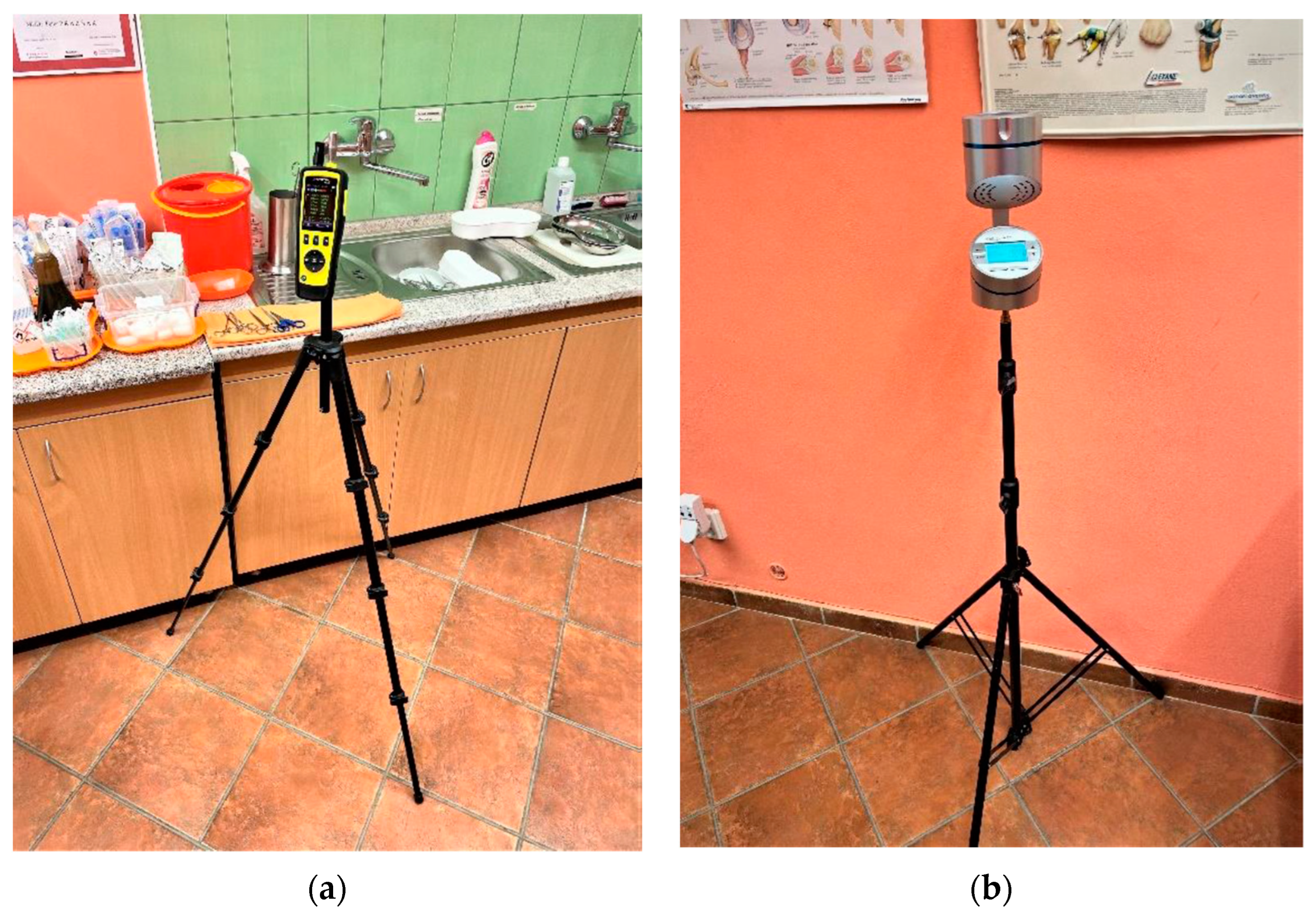
2.2.3. Evaluation of Impact on Airborne Particles, Airborne Microorganisms, and Viable Microorganisms on Textile Samples

Concentration of airborne particles was measured using TROTEC PC220 discrete particle counter (Trotec GmbH, Heinsberg, Germany); in every measurement, five separate particle count measurements in short succession were taken between 9:30 and 10:30 during clinic operation with the discrete particle counter in the corner of a given room. For each size fraction of airborne particles, the counts were then averaged.
Counts of viable microorganisms in ambient air in observed rooms were determined using Aeroscop MAS-100 NT (Merck KGaA, Darmstadt, Germany) with perforated air inlet of 300 0.6 mm-wide orifices, performing three samplings with 600 L of air taken during 15 min of air in each sampling: one sampling used agar plates for counting of yeasts and molds; another used blood agar plates, and the last used glucose tryptone yeast extract agar plates (GTY plate) for counting of airborne bacteria. After each sampling, the agar plates were transported in cooling box (3–6 °C) to certified laboratory for cultivation and analysis (Public Health Authority of the Slovak Republic, Poprad, Slovakia). Plates from each sampling were cultivated 24 h at corresponding temperatures (25 °C for agar plates, 30 °C for GTY plates, and 37 °C for blood agar plates) and evaluated by a certified microbiological laboratory according to ISO 21527 for counts of yeasts and molds in the agar plates and bacteria on the blood agar and GTY plates.
A quantification of total surviving microorganisms on studied surfaces was conducted using UltraSnap Surface ATP test swab with subsequent luminometric quantification of present ATP. In each measurement, an area of 10 × 10 cm was provided according to provided user instructions. Used swab is then immersed in provided agent containing luciferase enzyme for 10 s and immediately evaluated by provided luminometer. The results are expressed in relative light units (LRU) and thus provide a means of monitoring relative change in the quantity of living organisms on the monitored surface over time.
Measurement of counts of viable microorganisms on textiles was conducted by two separate methods: Envirocheck Contact DC Disinfection Control and 55 mm tryptic soy agar contact plates.
Envirocheck Contact DC Disinfection Control are flexible contact paddles with two sides containing CASO tryptic soy agar with different neutralizers and inhibitors, one for determination of yeasts and bacteria, the other side for counting molds. After swabbing a surface with an Envirocheck paddle, the paddle was transported together with other samples in a cooling box to certified laboratory. On the same day, a 48 h cultivation of said paddle at 37 °C was initiated. After incubation, the density of microorganisms on the measured paddle of 9.4 cm2 was determined by comparison with the model density chart supplied within the Envirocheck sets in limits of 103 to 107 for bacteria and yeasts and 102–105 for molds. In a similar manner, the soy contact plates were open at the site of measurement, and the agar surface was pressed with steady pressure for a few seconds on the investigated textile surface, immediately closed and stored, and transported in the cooling box with other samples and brought to microbiological laboratory for cultivation and evaluation. Soy agar contact plates were cultivated for 48 h at 37 °C.
As a control for compliance with the necessary procedures and an indicator of sample contamination, in each measurement using agar plates, one additional unused agar plate of each type was transported to the site and from the site in the same cooling box. Respective discovery of microbial growth in this unused agar plate would indicate contamination, and such results would be ignored. All analyses of microbial cultivation were contracted to and conducted by accredited laboratory of the Public Health Authority of the Slovak Republic in Poprad, Slovakia.

3. Results

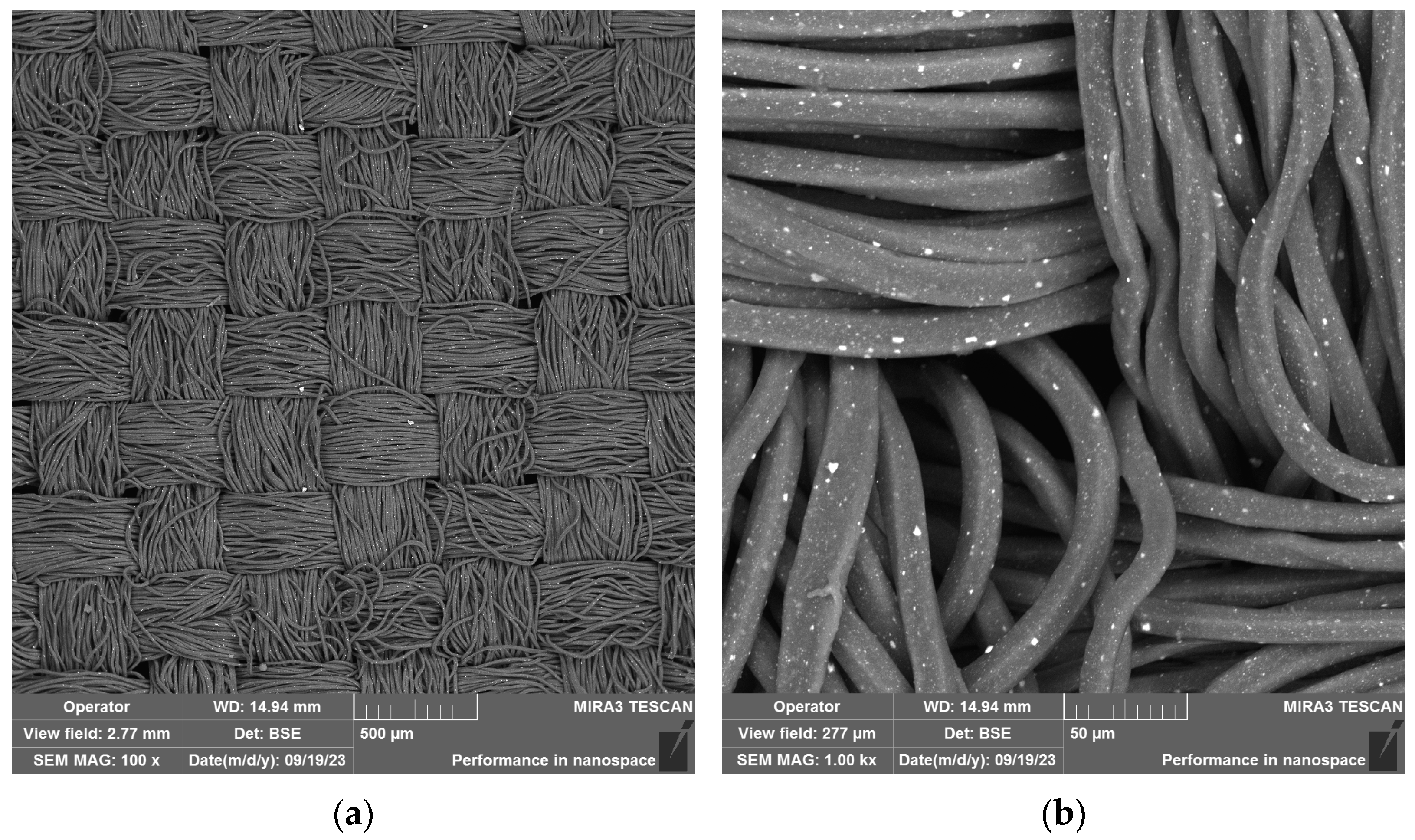
3.1. Preparation of Yarns and Vowen Textile

Polypropylene yarns containing ZP a SPG were prepared according to the process described in Section 2.2.1 above. The tensile properties of the produced yarns are summarized in Table 2, and scanning electron microscope (SEM) images of the produced fabric are presented in Figure 1.

3.2. Evaluation of Airborne Particles and Airborne Microorganisms

On four occasions, with a weekly interval of measurements, an electronic discrete particle counter (DPI) was set up for a period of approximately 45 min, conducting five discrete measurements as described above, placement of the DPI devices shown in Figure 2.
The observed values of found airborne particles per m3 of air in room no.1 are listed in Table 3, values observed in room no. 2 are listed in Table 4, and all listed values are averages of five independent readings on a given day and location. Sampling numbered 0 is the measurement of the initial conditions before the replacement of bed sheets and staff apparel with ZP and SPG doper polypropylene fabric.
As described in Section 2.2.3 above, simultaneously with airborne discreet particle counting, three samplings of airborne microorganisms were conducted. Each sampling had a different plate to distinguish different types of microorganisms, with 600 L of ambient air passing through the aeroscope for each plate. The results for all four sampling dates expressed in colony-forming units per cubic meter of ambient air (CFU/m3) are presented in Table 5 and Table 6.
The selected standardized method displays a range sufficient to reflect the effect of developed fabric on the numbers of yeasts and molds, whatever the impact on counts is.

3.3. Quantification and Identification of Microorganisms Present on Textile Surfaces

Simultaneously with aeroscopic sampling, swabs of clothing and bedding, as described in Section 2.2.3 and depicted in Figure 3, were conducted; all sampling sessions are numbered in accordance with the measurement description in Section 2.2.2. All results of ATP swabs are listed in Table 7.
As illustrated in Table 7, there is observable variability in the data, but a reduction in living microorganisms is evident in all cases. For unambiguous quantification, averages of samplings 1 to 3 for a given surface can be compared to sampling 0; such comparisons are summarized in Table 8.
Further quantification of viable microorganisms is provided by the evaluation of Envirocheck contact plates, which allow estimation of the order of magnitude of numbers of CFU per the area of the given sampler, which is 9.4 cm2. Also, more resolution of the number of molds vs. the number of bacteria can be made. A summarization of results from the cultivation of Envirocheck contact plates at 37 °C during 48 h is presented in Table 9 in logarithmic form, where a value of “0” means no yeasts and bacteria or molds were found to grow in the given sample.
The most detailed description of viable microbes found on monitored textiles is provided by cultivation and analysis of soy contact plates at 30 °C. The summarization of numbers and identified organisms across individual measurements are presented in Table 10, Table 11 and Table 12 below.

4. Discussion

In this study, a combination of two approaches of mitigation of transfer of pathogens described in the literature was chosen: substitution of usually used textiles woven using continuous filament polypropylene yarn-based fabric as described in [7] and doping of the polymer used with antimicrobial properties, such as described in [2,12,13,17,18] and others. Additionally, the impact of such textiles was verified and quantified in real-life conditions in a medical facility with frequent movement of staff and patients. The results listed in Section 3 demonstrate the viability of the presented method for the production of textiles for use in healthcare facilities. At the concentrations of ZP and SPG used in this study, no effect on coloring was observed, and the reduction in bacteria and fungi surviving on these textiles is apparent. However, no significant impact on airborne particles and airborne microorganisms, as described in [7], was observed. The reason may be attributed to the difference in other factors between this study and the study presented in [7] since the room investigated in the former study was an operating room, where all staff used surgical masks and proper surgical gowns at all times, and filtered air was continuously supplied to the room, while in our study, the patients came in their own clothes for short surgical procedures and, besides the use of germicidal lamps, no means of air purification were used.
Regarding the choice of antimicrobial agents and means of their implementation, one of the authors’ previous experiences with antifouling modification of apparel and introduction of said goods to the markets of the European Union was of key influence: use of nanoparticles and antibacterial substances not recognized by current legislation [13] were avoided. Concerns for environmental and health risks associated with the use of nanoparticles were expressed and summarized in a recent literature review [19], and metal particles, especially copper, have an impact on textile color [18]. The use of ZP is also limited in the EU; however, this restriction only applies to use in cosmetic preparations [20]. By following this approach, a textile was developed that does not expect significant obstacles for further testing in similar environments and for subsequent market launch.

5. Conclusions

Application of presented polypropylene textiles doped with silver phosphate glass and zinc pyrithione in real-life conditions in a doctor’s office exhibited a reduction in the count of bacteria and fungi surviving on their surfaces and was proven to retain their antimicrobial properties over a monitored period. The counting of discrete airborne particles showed an increase in the number of particles smaller than 0.5 µm by almost approximately 50% in the examination room, while a decrease in the number of particles of 10 µm of approximately 1/3 is indicated in the data for room no. 1. Relative differences are even smaller for room no. 2, which had much fewer attending patients per day. The impact on observed dust particles in ambient air in the observed rooms of the clinic is thus rather ambiguous, and the authors conclude that the expected reduction in airborne particles was not observed in this study. Regarding viable airborne microorganisms, a clear reduction in yeasts and molds was shown in room no. 2, the surgery room with a lower frequency of patient visits. However, this effect was not observed in room no. 1, the doctor’s office, which had patient traffic multiple times higher. Most promising are the results of total viable counts on studied textile surfaces: all used methods confirmed the reduction in viable microorganisms by at least 44% (doctor’s tunic) relative to measurement before the replacement of commonly used cotton and polyester-based textiles with presented antimicrobial-doped polypropylene. The relative reduction in viable microbes for both the examination table and surgical table was at least 80% in all cases. Visible differences between the observed rooms can be attributed to the number of passing patients and, thus, a higher bioburden in room no. 1. This reduction was most pronounced in the final observation after 24 h of isolation, which indicates that less than 8% of the original microorganisms survived on presented textiles after 24 h without an additional bioburden.

Author Contributions

Conceptualization, A.B.; investigation, M.K. and B.B.; writing—original draft preparation, T.Z. and B.B.; writing—review and editing, M.K.; supervision, A.B.; project administration, T.Z.; funding acquisition, A.B. All authors have read and agreed to the published version of the manuscript.

Funding

This publication was created thanks to support under the Operational Programme Integrated Infrastructure for the project Centre for the Development of Textile Intelligence and Antimicrobial Technologies, ITMS code 313011AVF5, co-financed by the European Regional Development Fund.

Data Availability Statement

The data presented in this study are available on request from the corresponding author.

Conflicts of Interest

The authors declare no conflict of interest. The funders had no role in the design of the study; in the collection, analyses, or interpretation of data; in the writing of the manuscript; or in the decision to publish the results.

References

  1. Cummings, K.L.; Anderson, D.J.; Kaye, K.S. Hand hygiene noncompliance and the cost of hospital-acquired methicillin-resistant Staphylococcus aureus infection. Infect. Control Hosp. Epidemiol. 2010, 31, 357–364. [Google Scholar] [CrossRef]
  2. Stewart, S.; Robertson, C.; Pan, J.; Kennedy, S.; Dancer, S.; Haahr, L.; Reilly, J. Epidemiology of healthcare-associated infection reported from a hospital-wide incidence study: Considerations for infection prevention and control planning. J. Hosp. Infect. 2021, 114, 10–22. [Google Scholar] [CrossRef]
  3. Weiner-Lastinger, L.M.; Abner, S.; Edwards, J.R.; Kallen, A.J.; Karlsson, M.; Magill, S.S.; Dudeck, M.A. Antimicrobial-resistant pathogens associated with adult healthcare-associated infections: Summary of data reported to the National Healthcare Safety Network, 2015–2017. Infect. Control. Hosp. Epidemiol. 2020, 41, 1–18. [Google Scholar] [CrossRef] [PubMed]
  4. Suetens, C.; Kärki, T.; Plachouras, D. ECDC surveillance report. In Point Prevalence Survey of Healthcare Associated Infections and Antimicrobial Use in European Acute Care Hospitals 2016–2017; ECDC: Stockholm, Sweden, 2023; pp. 9–13. ISBN 978-92-9498-636-8. [Google Scholar]
  5. Štefkovičová, M.; Litvová, S.; Mikas, J.; Kopilec Garabášová, M.; Jamrichová, M.; Prostináková, Z. Second Point Prevalence Survey of Healthcare-Associated Infections in The Slovak Republic as a Part of The European Survey. Zdr. Listy 2019, 7, 12–18. [Google Scholar]
  6. Kampf, G. How long can nosocomial pathogens survive on textiles? A systematic review. GMS Hyg. Infect. Control. 2020, 15, 1–15. [Google Scholar] [CrossRef]
  7. Ljungqvist, B.; Reinmüller, B. Some observations on protective efficacy of surgical clothing systems with additional clothing components concerning airborne bacteria-carrying particles measured during ongoing surgery. Eur. J. Parenter. Pharm. Sci. 2021, 26, 1–14. [Google Scholar] [CrossRef]
  8. Ullmann, C.; Ljungqvist, B.; Reinmüller, B. Some aspects of protective efficacy of surgical clothing systems concerning airborne microorganisms based on results from measurements in a dispersal chamber and during surgical procedures. Eur. J. Parenter. Pharm. Sci. 2017, 22, 51–58. [Google Scholar]
  9. Mitchell, A.; Spencer, M.; Edmiston, C., Jr. Role of healthcare apparel and other healthcare textiles in the transmission of pathogens: A review of the literature. J. Hosp. Infect. 2015, 90, 285–292. [Google Scholar] [CrossRef] [PubMed]
  10. Botelho, C.M.; Fernandes, M.M.; Souza, J.M.; Dias, N.; Sousa, A.M.; Teixeira, J.A.; Fangueiro, R.; Zille, A. New Textile for Personal Protective Equipment—Plasma Chitosan/Silver Nanoparticles Nylon Fabric. Fibers 2021, 9, 5:1–5:13. [Google Scholar] [CrossRef]
  11. Gomes, L.; Monteiro, P.; Cotas, J.; Gonçalves, A.M.; Fernandes, C.; Gonçalves, T.; Pereira, L. Seaweeds’ pigments and phenolic compounds with antimicrobial potential. Biomol. Concepts 2022, 13, 89–102. [Google Scholar] [CrossRef] [PubMed]
  12. Staneva, D.; Atanasova, D.; Angelova, D.; Grozdanov, P.; Nikolova, I.; Grabchev, I. Antimicrobial Properties of Chitosan-Modified Cotton Fabric Treated with Aldehydes and Zinc Oxide Particles. Materials 2023, 16, 5090. [Google Scholar] [CrossRef]
  13. Official Journal of the European Union. Regulation (Ec) No 1272/2008 of the European Parliament and of the Council: Regulation on Classification, Labelling and Packaging of Substances and Mixtures, Amending and Repealing Directives 67/548/EEC and 1999/45/EC, and Amending Regulation (EC) No 1907/2006; Official Journal of the European Union: Brussels, Belgium, 2008. [Google Scholar]
  14. Brayner, R.; Ferrari-Iliou, R.; Brivois, N.; Djediat, S.; Benedetti, M.F.; Fiévet, F. Toxicological impact studies based on Escherichia coli bacteria in ultrafine ZnO nanoparticles colloidal medium. Nano Lett. 2006, 6, 866–870. [Google Scholar] [CrossRef]
  15. Rajski, L.; Juda, M.; Los, A.; Witun, E.; Malm, A. Medical textiles with silver/nanosilver and their potential application for the prevention and control of healthcare-associated infections–mini-review. Curr. Issues Pharm. Med. Sci. 2019, 32, 104–107. [Google Scholar] [CrossRef]
  16. van Arkel, A.; Willemsen, I.; Kluytmans, J. The correlation between ATP measurement and microbial contamination of inanimate surfaces. Antimicrob. Resist. Infect. Control. 2021, 10, 116. [Google Scholar] [CrossRef]
  17. Yang, S.-Y.; Lee, M.-J. Poly (Methyl Methacrylate)-Containing Silver-Phosphate Glass Exhibits Potent Antimicrobial Activity without Deteriorating the Mechanical and Biological Properties of Dental Prostheses. Polymers 2023, 15, 297. [Google Scholar] [CrossRef] [PubMed]
  18. Novi, V.T.; Gonzalez, A.; Brockgreitens, J.; Abbas, A. Highly efficient and durable antimicrobial nanocomposite textiles. Sci. Rep. 2022, 12, 17332. [Google Scholar] [CrossRef] [PubMed]
  19. Kumah, E.A.; Fopa, R.D.; Harati, S.; Boadu, P.; Zohoori, F.V.; Pak, T. Human and environmental impacts of nanoparticles: A scoping review of the current literature. BMC Public Health 2023, 23, 1059. [Google Scholar] [CrossRef] [PubMed]
  20. Official Journal of the European Union. Commission Regulation (EU) 2021/1902 of 29 October 2021 Amending Annexes II, III and V to Regulation (EC) No 1223/2009 of the European Parliament and of the Council as Regards the Use in Cosmetic Products of Certain Substances Classified as Carcinogenic, Mutagenic or Toxic for Reproduction (Text with EEA Relevance); Official Journal of the European Union: Brussels, Belgium, 2021. [Google Scholar]
Figure 1. SEM images of resulting woven textile: (a) 100× magnification showing the structure of the textile; (b) 1000× magnification showing the surface of produced yarns: the seemingly quadrangular cross section of filaments is a result of texturation process and particles of pigments, and additives protruding from surface of filaments are visible.
Fibers 11 00097 g001
Figure 2. Placement of the DPIs in investigated rooms. (a) Placement of DPI in room no. 1—doctor’s office with area 32 m2; (b) placement of Aeroscop MAS-100 NT for airborne microorganism quantification in room no. 1.
Fibers 11 00097 g002
Figure 3. Determination and quantification of present ATP on physician’s tunic before replacement with PP fabric tunic. (a) Application of UltraSnap swab; the wet stains are marks left by the soy agar contact plates; (b) output of subsequent luminometric measurement.
Fibers 11 00097 g003
Table 1. Recipes for individual doped polymer mixtures; quantities in grams.
Yarn ColorAdditiveAdditive MasterbatchPigment MasterbatchPolypropylene
PurpleSPG25015608190
ZP20015608240
Light blueSPG25012008550
ZP20012008600
BeigeSPG25015608190
ZP20015608240
Table 2. Tensile properties of SPG- and ZP-doped yarns.
Yarn ColorAdditiveRelative Tenacity (cN/dtex)Strain at Break (%)
PurpleSPG2.6182.1
ZP2.6081.6
Light blueSPG2.7935.8
ZP2.7134.2
BeigeSPG2.8739.8
ZP2.6339.3
Table 3. Distribution of dust particles according to size in individual samplings in room no. 1 and average values for each measurement.
Particle Size
No.0.3 µm0.5 µm1.0 µm2.5 µm5.0 µm10 µmConditions
072,61322,6886119129624712921.9 °C/47.9%
1109,18929,49154259711687622.1 °C/49.3%
2137,56036,561622610801658322.3 °C/48.1%
3106,45429,580592311161939622.1 °C/47.7%
Table 4. Distribution of dust particles according to size in individual measurements in room no. 2 and average values for each measurement.
Particle Size
No.0.3 µm0.5 µm1.0 µm2.5 µm5.0 µm10 µmConditions
090,91127,366605310551508321.9 °C/47.9%
1117,65332,873627311241718522.1 °C/49.3%
2130,96335,359645011331578422.3 °C/48.1%
356,69216,9543766661934922.1 °C/47.7%
Table 5. Analysis of cultivated aeroscope plates from room no. 1, results for each sampling expressed in CFU/m3.
No. of
Measurement
Yeasts and MoldsCultivated on Blood MediumCultivated on GTK Medium
052>535>535
124>535>535
255>535>535
355>535>535
Table 6. Analysis of cultivated aeroscope plates from room no. 2, results for each sampling expressed in CFU/m3.
No. of
Measurement
Yeasts and MoldsCultivated on Blood MediumCultivated on GTK Medium
057>535>535
123>535>535
237>535>535
332>535>535
Table 7. Quantification of present ATP on examined textiles in RLU.
No.*Bedding in Room No. 1Bedding in Room No. 2Tunic,
Nurse
Tunic, PhysicianConditions **
0400106677221.9 °C/47.9%
1541194022.1 °C/49.3%
2163211422.3 °C/48.1%
381357922.1 °C/47.7%
* Number of sampling as stated in Section 2.2.2; ** conditions as ambient temperature in °C/relative humidity.
Table 8. Comparison of present ATP on examined textiles in RLU.
ResultBedding in Room No. 1Bedding in Room No. 2Tunic, NurseTunic, Physician
04001066772
Average 1–32663221
Reduction−94%−94%−52%−71%
Table 9. Decimal logarithms of CFUs as indicated by Envirocheck contact plates after incubation.
Bedding in Room No. 1Bedding in Room No. 2Tunic—NurseTunic—Physician
No. of SamplingYeasts and BacteriaMoldsYeasts and BacteriaMoldsYeasts and BacteriaMoldsYeasts and BacteriaMolds
033444343
132333243
232303232
330323230
430323032
Table 10. Quantification of present microbes in CFU/contact plate and identified organisms found on bedding in room no. 1.
No. of
Measurement
CFU per
Contact Plate
Identified OrganismsConditions **
060aerobic sporulants, saprophytic staphylococci, micrococci, and two CFU micromycetes21.9 °C/47.9%
147aerobic sporulants, saprophytic staphylococci, micrococci, and bacillus cereus22.1 °C/49.3%
222aerobic sporulants, saprophytic staphylococci, and micrococci22.3 °C/48.1%
32aerobic sporulants22.1 °C/47.7%
43 *saprophytic staphylococci21.8 °C/47.8%
* Present, number of CFU estimated; ** conditions as ambient temperature in °C/relative humidity.
Table 11. Quantification of present microbes in CFU/contact plate and identified organisms found on bedding in room no. 2.
No. of
Measurement
CFU per
Contact Plate
Identified OrganismsConditions **
096aerobic sporulants, saprophytic staphylococci, and micrococci21.9 °C/47.9%
13 *saprophytic staphylococci22.1 °C/49.3%
25 *micrococci22.3 °C/48.1%
31 *micrococci22.1 °C/47.7%
414aerobic sporulants, saprophytic staphylococci, and micrococci21.8 °C/47.8%
* Present, number of CFU estimated; ** conditions as ambient temperature in °C/relative humidity
Table 12. Quantification of present microbes in CFU/contact plate and identified organisms found on nurse’s tunic.
No. of
Measurement
CFU per Contact PlateIdentified OrganismsConditions ***
0100 **aerobic sporulants, saprophytic staphylococci, micrococci, and one CFU micromycetes21.9 °C/47.9%
1100 **aerobic sporulants, saprophytic staphylococci, micrococci, Streptococcus sp., and two CFU micromycetes22.1 °C/49.3%
2100 **aerobic sporulants, saprophytic staphylococci, and micrococci22.3 °C/48.1%
3100 **aerobic sporulants, saprophytic staphylococci, and micrococci22.1 °C/47.7%
430aerobic sporulants, saprophytic staphylococci, micrococci, Acinetobacter sp., and Pantoea sp.21.8 °C/47.8%
** high number of CFU per plate, probably >100; and *** conditions as ambient temperature in °C/relative humidity.
Disclaimer/Publisher’s Note: The statements, opinions and data contained in all publications are solely those of the individual author(s) and contributor(s) and not of MDPI and/or the editor(s). MDPI and/or the editor(s) disclaim responsibility for any injury to people or property resulting from any ideas, methods, instructions or products referred to in the content.

