Article

Biofouling of FeNP-Coated SWRO Membranes with Bacteria Isolated after Pre-Treatment in the Sea of Cortez

by
Maria Magdalena Armendáriz-Ontiveros
1,
Gustavo A. Fimbres Weihs
2,*,
Sergio de los Santos Villalobos
2,* and
Sergio G. Salinas-Rodriguez
3
1
Instituto Tecnológico de Sonora. 5 de Febrero 818 Sur, Cd. Obregón, Sonora, C.P. 85000, Mexico
2
CONACYT–Instituto Tecnológico de Sonora, 5 de Febrero 818 Sur, Cd. Obregón, Sonora, C.P. 85000, Mexico
3
IHE Delft Institute for Water Education, Environmental Engineering and Water Technology Department, Westvest 7, 2611 AX Delft, The Netherlands
*
Authors to whom correspondence should be addressed.
Coatings 2019, 9(7), 462; https://doi.org/10.3390/coatings9070462
Submission received: 14 June 2019 / Revised: 11 July 2019 / Accepted: 19 July 2019 / Published: 23 July 2019
(This article belongs to the Special Issue Novel Marine Antifouling Coatings)

Abstract

:
Commercial seawater reverse osmosis (SWRO) membranes were coated with iron nanoparticles (FeNPs) and biofouled with a bacterium strain isolated from the Sea of Cortez, Mexico. This strain was selected and characterized, as it was the only cultivable strain in pretreated seawater. Molecular identification of the strain showed that it belongs to Bacillus halotolerans MCC1. This strain was Gram positive with spore production, and was susceptible to Fe+2 toxicity with a minimum inhibitory concentration of 1.8 g L−1. Its biofouling potential on both uncoated and FeNP coated reverse osmosis (RO) membranes was measured via biofilm layer thickness, total cell count, optical density and organic matter. The FeNP-coated RO membrane presented a significant reduction in biofilm cake layer thickness (>90%), total cells (>67%), optical density (>42%) and organic matter (>92%) with respect to an uncoated commercial membrane. Thus, Bacillus halotolerans MCC1 shows great potential to biofoul RO membranes as it can pass through ultrafiltration membranes due to its spore producing ability; nonetheless, FeNP-coated membranes represent a potential alternative to mitigate RO membrane biofouling.

1. Introduction

Halotolerant bacteria are of great biotechnological importance for industry, as they are easy to grow under limited nutritional requirements. Moreover, their tolerance to high salt concentrations minimizes laboratory contamination risks [1]. Several researchers investigating halotolerant bacteria have focused on their capacity to produce antibiotics and enzymes [2], while others have used them to design microbial inoculants to increase the salinity tolerance of crops [3]. For example, in India, a halotolerant bacillus has been used for the biosynthesis of enzymes (L-Glutaminase) in bioethanol production [4]. In Algerian wetland ecosystems, a study found that halotolerant strains belonging to the genera Haloferax, Halococcus and Haloarcula showed a high production of molecules with important biotechnological applications, such as in the coastal agriculture, pharmaceutical and environmental fields [5]. In Spain, the dominant halotolerant genera isolated from the Bras del Port salt basins included Salinivibrio, Pseudomonas, Alteromonas, Alcaligenes, Acinetobacter and Flavobacerium. These halotolerant microorganisms have exhibited potential for several applications in the food, pharmaceutical, medical and environmental fields [6].
In the seawater reverse osmosis (RO) desalination industry, halotolerant bacteria have gained great interest because they are involved in membrane biofouling, one of the biggest problems faced by membrane-based desalination [7]. Biofouling is an operational problem in SWRO that can reduce production efficiency, leading to severe economic impacts [8]. Biofouling involves microorganism attachment and growth on the membrane surface, and is very difficult to treat due to the ability of microorganisms to multiply even if most are eliminated in pretreatment [9]. As biomass accumulates in the feed channel of membrane modules, this leads to technical problems (increases in the working pressure or reduction of permeate flux, as well as the need of more frequent chemical cleaning, among others), decreasing the process productivity, reducing the life span of membranes and increasing process costs [10,11]. To reduce the impact of biofouling problems, some research efforts have focused on coating the membrane surface with various nanoparticles (NPs) that are known to have antimicrobial properties, such as Ag, CuO, ZnO and TiO2 [12,13,14,15]. In addition, NPs have been used as antifouling coatings in different marine industrial environments in order to prevent biofouling [16]. However, these NPs are expensive and difficult to synthetize; therefore, their use as a membrane coating increases desalination costs. Iron nanoparticles (FeNPs) are a promising coating agent, as they cost less than other NPs, are easy to synthesize and have a biocide effect by promoting Fenton or Fenton-like reactions, producing oxidative stress by generating reactive oxygen species (ROS), disrupting lipids, proteins and DNA, and eventually causing bacterial death [17,18,19].
Interestingly, most biofouling studies of NP coatings have been carried out with strains that are not native from seawater, such as Escherichia coli and Pseudomonas aeruginosa [20,21]. Those studies are not very useful for the desalination industry, since those strains are not ones that commonly cause RO membrane biofouling. Hence, the studying and understanding of seawater halotolerant bacteria is paramount for the RO desalination industry due to their negative effect on membrane durability and productivity. Moreover, the shoreline of the Sea of Cortez is home to 32 RO desalination plants with capacities ranging from 100 to 17,280 m3 d−1 [22] and one under construction [23]. Further, the number of RO desalination plants in that region has been increasing in recent years (up by 68% in three years) [22] due to frequent water scarcity. For these reasons, the aim of this paper is to study the effect of FeNP coating of RO membranes on the extent of biofouling caused by bacteria native to the Sea of Cortez. This is achieved by isolating and characterizing the only bacterium strain found after a typical pretreatment of seawater sampled from the Sea of Cortez, Mexico (Bacillus halotolerans MCC1), and using it as a model biofoulant.

2. Materials and Methods
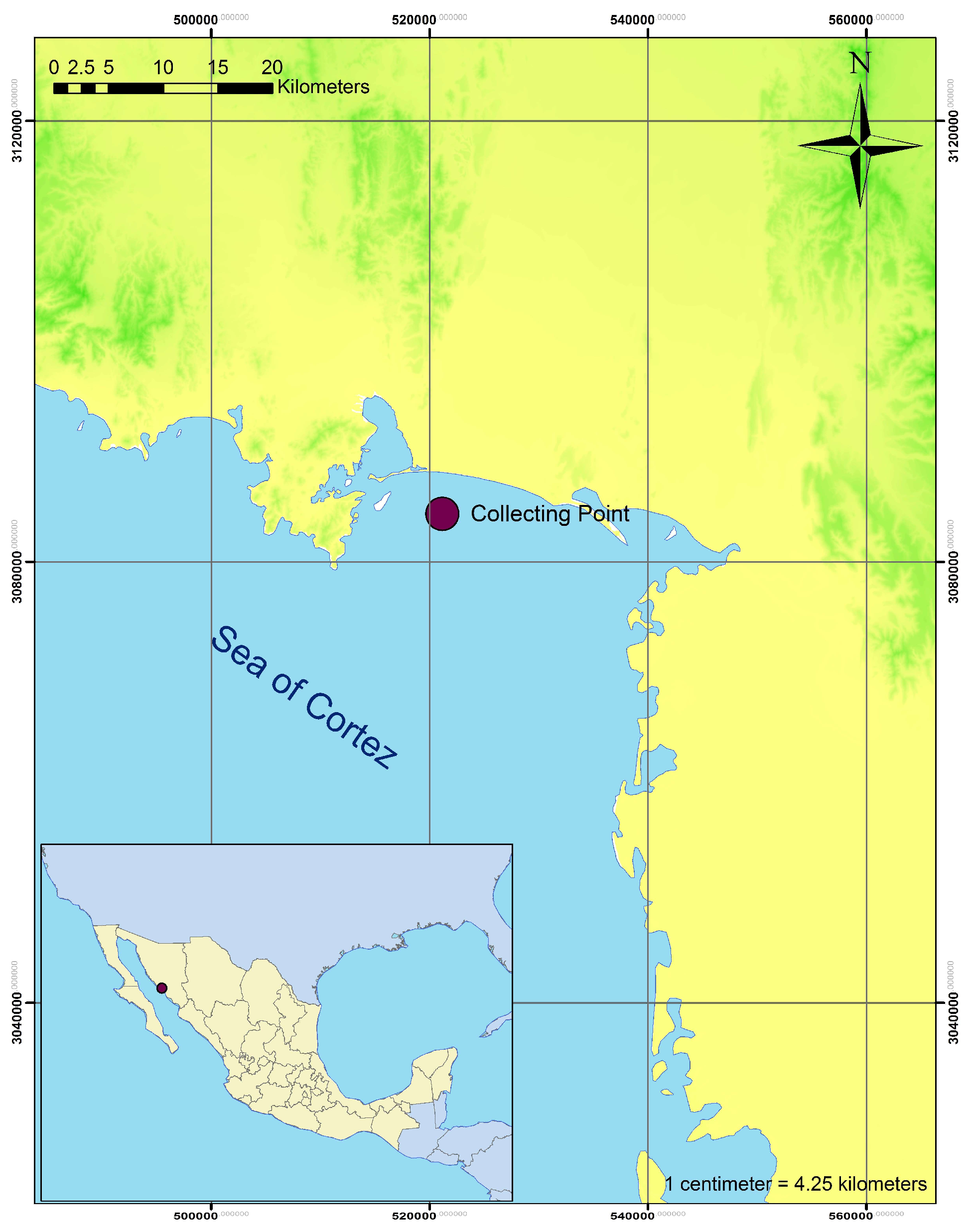
2.1. Sampling

Seawater samples were obtained from the Sea of Cortez, at Cochorit beach near Empalme, Sonora, Mexico at a depth of 4 m and at a distance of 3.76 km from the coast (27° 53’ 1.28′ N, 110° 47’ 5.56′ W) (Figure 1). The samples were collected following the procedure detailed in the field manual for water quality sampling [24]. Five samples were taken throughout the year (spring, summer, autumn and two in winter) and at different times of the day. The physicochemical parameters measured at the time of sampling are shown in Table 1. Three hour after sampling, the collected water was pretreated by 5 μm cartridge filtration and 0.1 µm ultrafiltration (PURIKOR, PKUF-M) with a constant working pressure of 0.2 MPa and a permeate flow rate of 3.7 L min−1, as a representative analogue of the pretreatment that takes place in typical large-scale desalination plants [25].

2.2. Bacterial Isolation and Molecular Identification

Petri dishes containing nutrient agar (NA) supplemented with sterilized pretreated seawater (34.99 g L−1 of salinity) were used as a culture medium. The pretreated seawater was previously sterilized in an autoclave (Felisa FE-399), for 15 min and 0.103 MPa at 121 °C. Pretreated seawater was used to provide nutritional conditions for bacterial growth, similar to those observed in their natural environment (Table 1).
To determine the cultivable bacterial population and diversity of the unsterilized pretreated seawater, the serial dilution (1:10) method was used, preparing dilutions up to 10−3 [26]. Finally, 1 mL of each dilution was dispersed, onto the NA prepared in the Petri dishes, and incubated for 24 h at 28 °C [27]. The bacterial isolation was done by triplicate.
Macro and microscopic bacterial characterization, spore production and Gram staining were carried out using the Wirtz–Conklin method [28] and the Hycel stain kit (Hycel, Cat 541), respectively. All obtained isolates were preserved in the Native Endophytes and Soil Microorganisms Collection (COLMENA) [29].
Molecular identification was carried out for the isolated bacterium strain that grew in the previously mentioned Petri dishes with culture medium, which was isolated from the pretreated seawater. Genomic DNA was extracted using the methodology of Reader and Broda [30]. Bacteria molecular identification was carried out amplifying the 16S ribosomal gene (16S rRNA) [31]. The PCR protocol was carried out according to the procedure described by Villa-Rodriguez et al [32]. The DNA sequences were processed using the FinchTV 1.4.0 software package (Geospiza Inc., Denver, CO, USA). The phylogenetic tree was created by Neighbor Joining methodology [31] using MEGA 7.0.

2.3. Minimum Inhibitory Fe+2 Concentration

The minimum inhibitory concentration of Fe+2 was determined by the inoculation of 1 × 10⁴ colony forming units (CFU) onto Petri dishes containing NA supplemented with sterile pretreated seawater. Further, the culture medium was supplemented with Fe+2, at concentrations of 1.0, 1.2, 1.4, 1.6, 1.8 and 2 g L−1. The inoculated Petri dishes, using three independent replicates, were incubated at 28 °C for six days. The bacterial population in each Petri dish was determined by CFU count every 24 h.

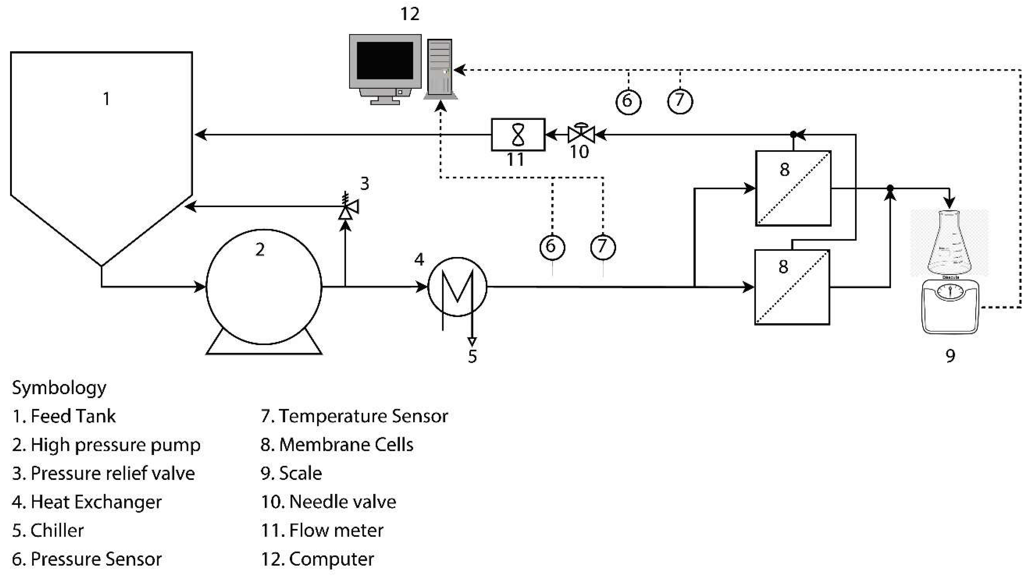
2.4. Accelerated Biofouling Test on RO Membranes

Accelerated biofouling on two commercial RO membranes (Dow Filmtec SW30HR) was carried out using a high concentration of the studied bacterial strain and providing it with sufficient nutrients in the feed water. One of these membranes was coated with the minimum inhibitory concentration of FeNPs (FeNP membrane) via immersion, ensuring the NP dispersion by sonication for 1 h, following the methodology of Armendariz et al. [18]. The membrane was immersed into fresh DI water for 10 min to activate the carboxyl groups and then the membrane was dipped into the aqueous suspension of FeNPs for 24 h, followed by rinsing with an ultrasonic bath for 5 min. The other membrane was not coated (uncoated membrane) and used as blank for reference and comparison. The FeNPs were synthetized and characterized [18,19] according to the method of Baltazar et al. [33] and Arancibia-Miranda et al. [34]. The chemical structures and morphology of the FeNPs were characterized by X-ray Diffraction (XRD) using a Shimadzu XRD-6000 diffractometer (Kyoto City, Japan) and Scanning Electron Microscopy (SEM) using a Nova NanoSEM-200 (FEI Company, Hillsboro, OR, USA).
The bacterial growth on the RO membrane surface took place in CF042 crossflow membrane cells (Sterlitech Corp., Kent, WA, USA) (Figure 2). The feed water comprised sterile pretreated seawater and a high amount (109 CFU mL−1) of the studied strain. In addition, a sterile nutrient broth was added daily to the feed water, to guarantee optimal bacterial growth. The operating pressure was 6.38 MPa during the test, which ran for 90 h at an average temperature of 30 °C, while the pH remained in a range of 6–8. A low crossflow velocity of 0.15 m s−1 was used in order to minimize shear stress and encourage a high biofouling rate. After the accelerated biofouling test, biofouled membranes were analyzed by scraping 1 cm2 of biofilm. Then, the optical density of the scraped biofilm was determined by spectrophotometry, the biofilm cake layer was measured using inverted microscopy, and a Neubauer chamber using an optical microscope in 100× was used to carry out the total cell count in the biofilm. The amount of organic matter was determined by the catalytic combustion method [10].

2.5. Statistical Analysis

An analysis of variance of simple classification based on a linear model of fixed effects was performed for the CFUs found over time, and the statistical differences between the mean values were determined by the Tukey method for p ≤ 0.001. For the biofilm cake layer, optical density (OD), total cell count (TCC) and organic matter (OM), the theoretical t-student probability distribution for quantitative continuous variables for significance levels of p ≤ 0.001 was used. A simple linear regression analysis based on a linear model of fixed effects was performed. The degree of goodness of fit between the Fe+2 concentration and the final CFU count was determined. From these equations, the optimal inhibitory concentration of bacterial growth was determined using an ordinary least squares method. The software STATISTIC version 8.5 (StatSoft, Tulsa, OK, USA) was used for the statistical analyses.

3. Results and Discussion

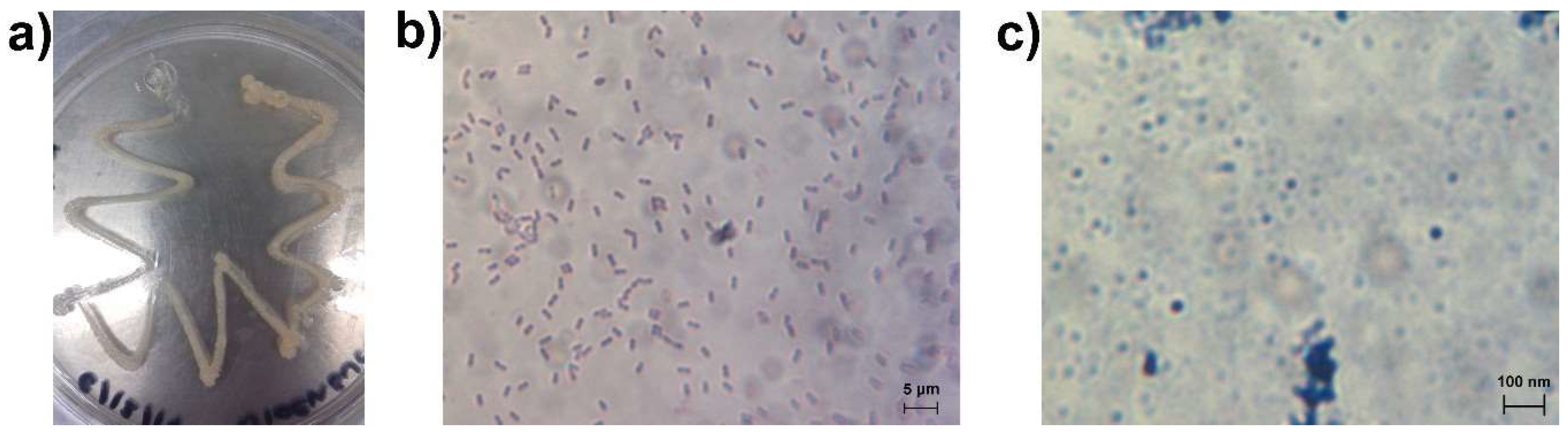
3.1. Bacteria Growth, Isolation and Molecular Identification

After 24 h of incubation, only one bacterial isolate (MCC1) grew for each repetition in direct pretreated seawater. No growth of CFUs was observed for the 10−3, 10−2, 10−1 dilutions. This bacterial strain showed medium size colonies (5.0 ± 1.0 mm), convex, whitish such as wax, rounded edges, fusiform and circular shape (Figure 3a). In addition, the Gram stain showed a Gram positive cell with a size of 2.8 ± 0.2 μm (Figure 3b), and spore size of 18.0 ± 0.0 nm (Figure 3c). As the ultrafiltration membrane nominal pore size of 0.1 μm is smaller than the typical sizes of bacteria, this indicates that only spores could pass through the filter and grow on the Petri dishes, thus originating the CFU found [35]. The ultrafiltration membrane did not show integrity problems during the lapse of the testing.
Molecular identification showed that this particular strain belongs to Bacillus halotolerans MCC1. This result confirms the macro and microscopic traits observed for the strain. Its Gram staining (Figure 3b) showing a Gram positive Bacillus strain, agrees with reports that most sea water Bacillus are Gram positive [36]. Moreover, this strain belongs to the specie B. halotolerans, which agrees with its source of isolation (ultrafiltered seawater). This finding indicates that the strain MCC1 is able to tolerate high electrical conductivity (48,100 ± 350 µS cm−1) and is not retained by the ultrafiltration process, due to its ability to produce spores that are smaller than the UF membrane pore size (as seen in Figure 3c). Spore production is an adaptive strategy to compensate the osmotic stress generated by high salt concentrations in the environment [37], and is an ability that not all Bacillus possess [38,39]. These bacterial traits could be highly associated with the role of the strain MCC1 on biofouling RO membranes.

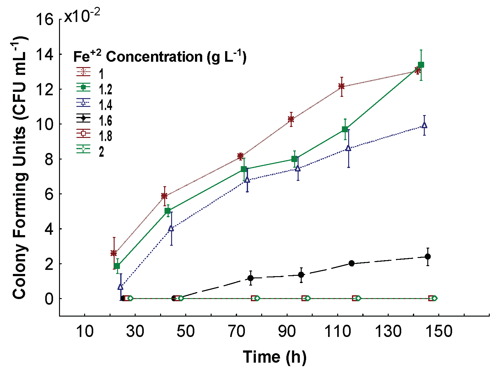
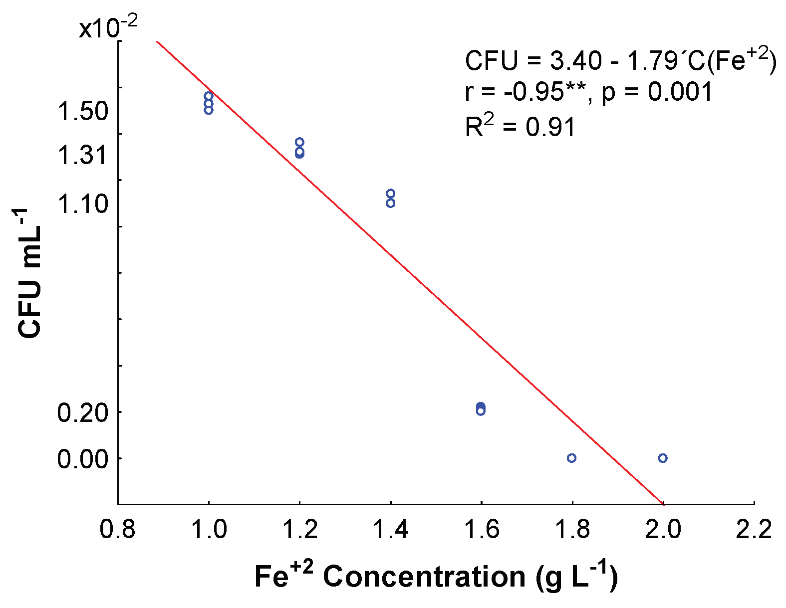
3.2. Minimum Inhibitory Fe+2 Concentration

Figure 4 shows the rate of bacterial growth in NA supplemented with various concentrations of Fe+2. The data shows that the biocidal effect of Fe+2 was greater from 24 to 48 h, at any concentration used. After 72 h, large bacterial populations were observed for the range of 1.0 to 1.6 g L−1 Fe+2. It is clear that Bacillus halotolerans MCC1 can easily grow in Fe+2 concentrations of 1 to 1.4 g L−1, as Fe is an essential element for life [40]. However, from 1.6 g L−1 the strain MCC1 is not able to maintain its optimal growth rate within the first 48 h. From that moment, the bacterium shows adaptive strategies (Fe+2 resistance) to be able to survive, because at 72 h of incubation it is possible to observe CFUs. This could be attributed to MCC1 having adaptive abilities under extreme conditions, also evidenced by the sporulation capacity reported in this work (Figure 3c) [37]. Finally, the biocidal effect of Fe+2 at higher concentrations (1.8 to 2 g L−1) is greater, since there was negligible bacterial growth.
A significant decrease in the final bacterial population for the strain MCC1 was observed with increasing Fe+2 concentration. There was a negative correlation with a highly significant (95% confidence) dependence between these variables (r = 0.95, p < 0.001). The degree of goodness of fit was high (R2 = 0.91) for a linear regression model (Figure 5). The greatest decrease in CFUs for MCC1 was observed when the Fe+2 concentration was increased from 1.4 to 1.6 g L−1, presenting 50% of the total inhibition by Fe+2. At Fe+2 concentrations of 1.8 g L−1 and higher, no bacterial growth was observed. Despite the previously mentioned capability of Bacillus halotolerans MCC1 to adapt to extreme conditions, there is a clear adaptability limit after which it cannot survive. Therefore, the minimum inhibitory Fe+2 concentration was 1.8 g L−1, as the data suggests that this bacterium cannot grow at higher concentrations. At concentrations above that level, the accumulation of Fe+2 can stop cell growth or cause death due to DNA damage [40].

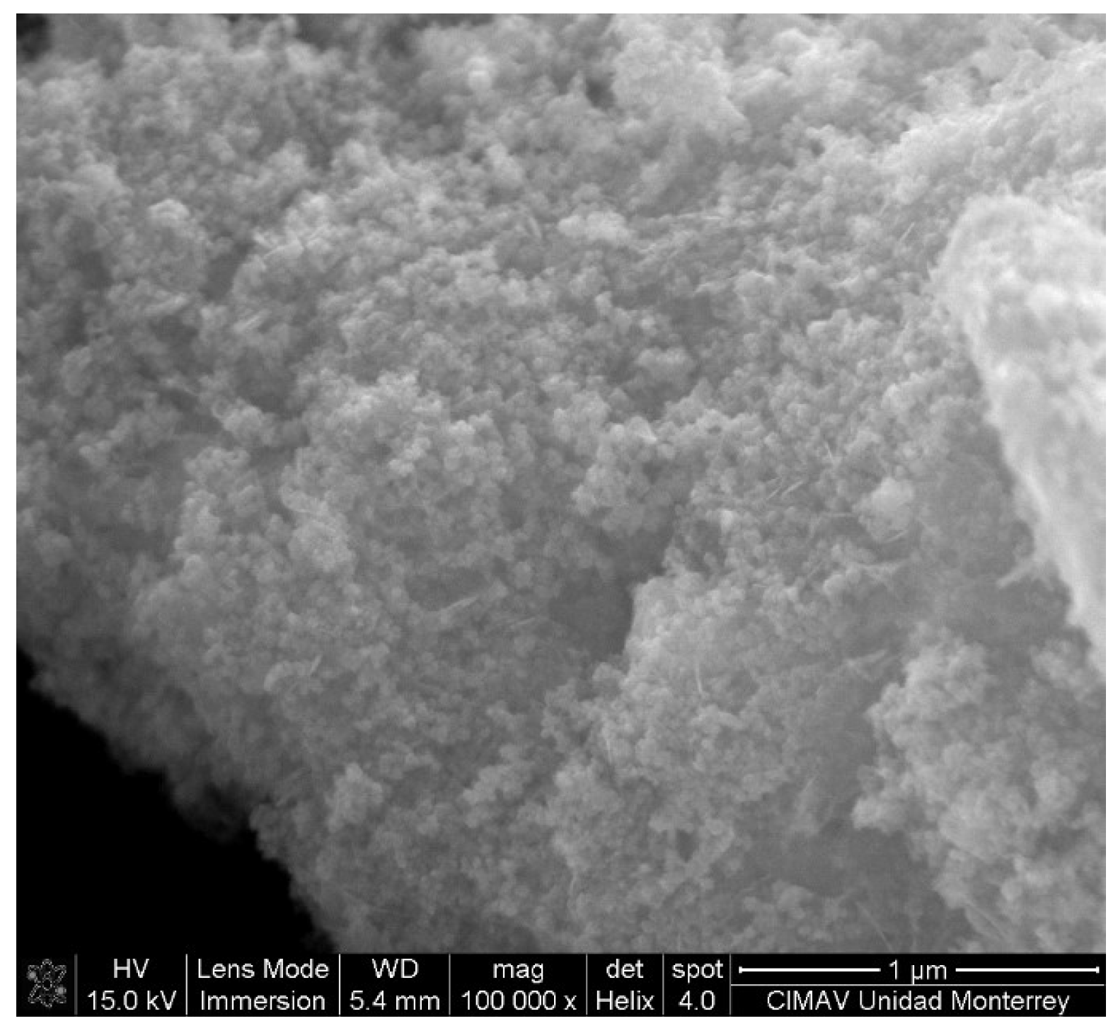
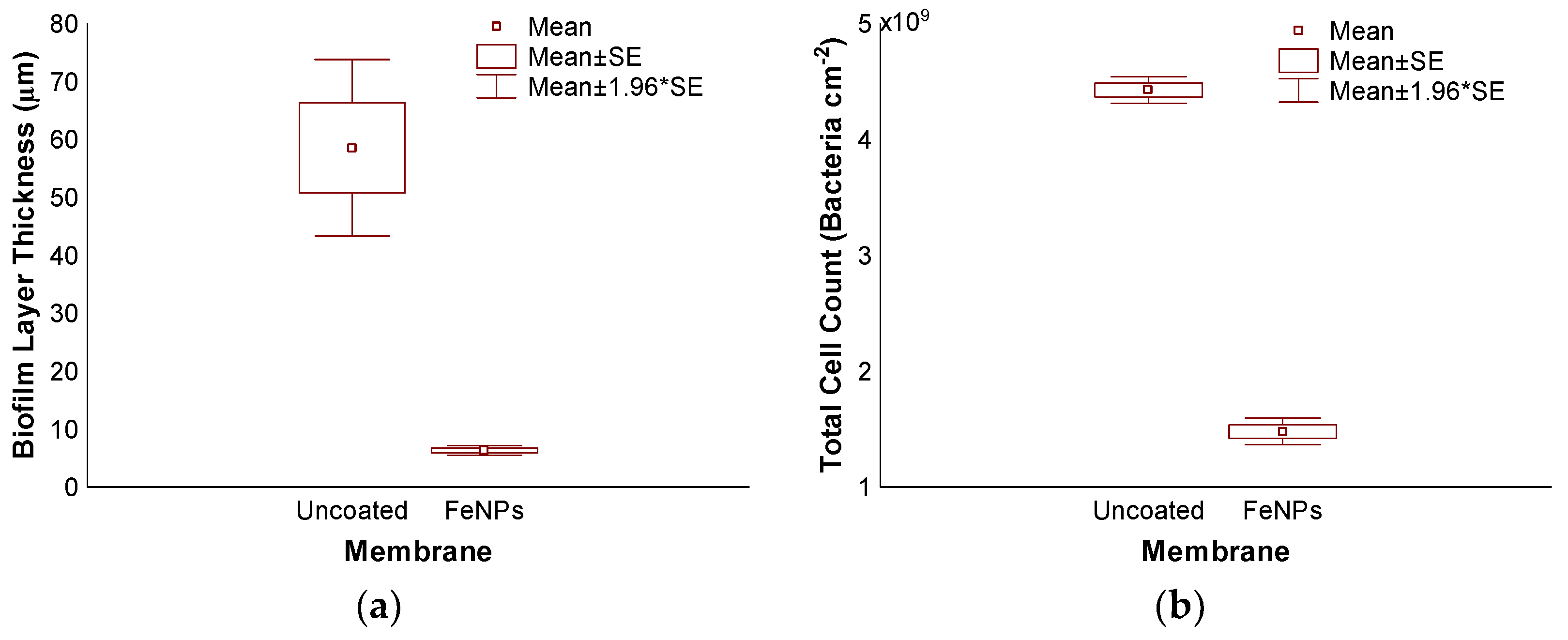
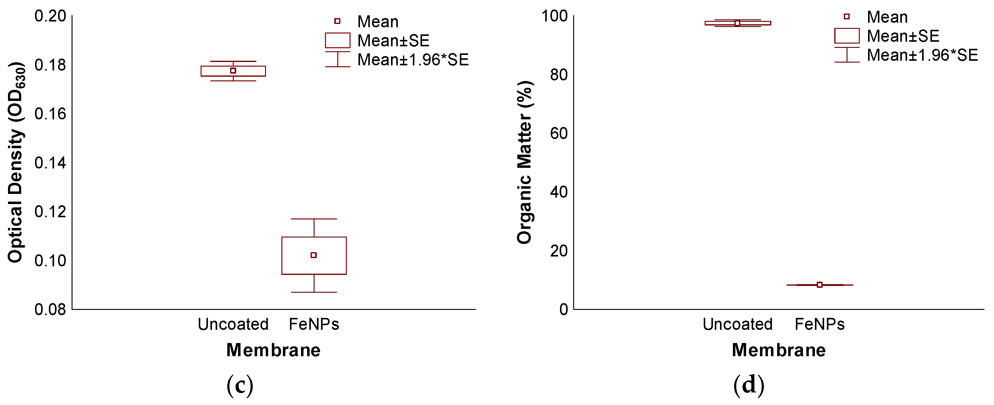
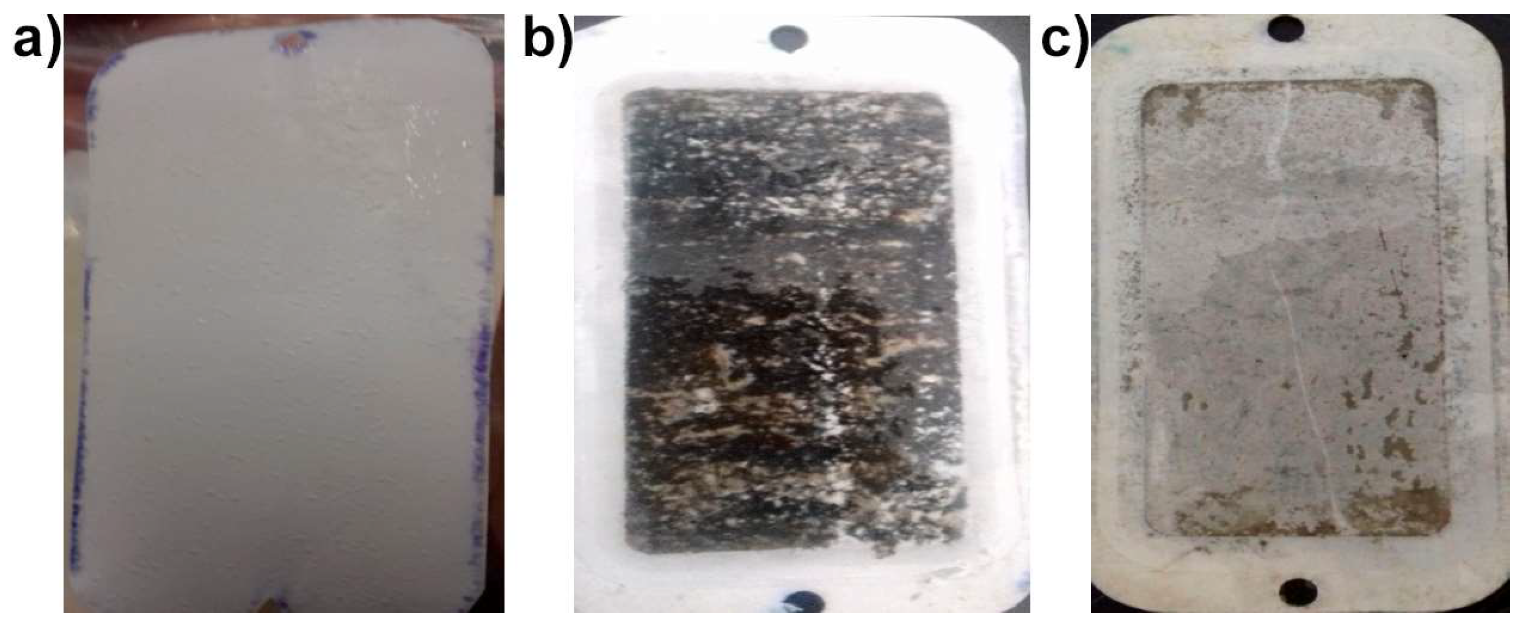
3.3. Accelerated Biofouling Test on RO Membranes

The SEM image (Figure 6) depicts the FeNPs, showing a mean particle size around 14.6 ± 1.2 nm. Figure 7 shows the XRD peaks for the FeNPs. The crystalline structure of the FeNPs was confirmed by XRD analysis (2θ = 44.5°, 65.0° and 82.4°) [41]. The accelerated biofouling test showed that the bacteria can exhibit growth under conditions of very high salinity, in the range of 3%–8% salt concentration by weight (see Figure 8). This result suggests that the bacteria indeed exhibits sporulation, as endospore production allows the bacteria to survive in extreme environmental conditions [42,43]. In addition, the ability of Bacillus halotolerans MCC1 to biofoul RO membranes was also demonstrated in this test. A biofilm cake layer was formed on the membrane surface during the experiment, resulting in a decrease in permeate flux. This strongly suggests that this bacterium strain can be useful as a model biofoulant, especially for desalination plants in the Sea of Cortez. Further, as can be seen in Figure 8c, the FeNP coated membrane showed significantly less biofouling compared to the uncoated membrane (Figure 8b), evidencing the antimicrobial effect of FeNPs already demonstrated in the medical field [44].
The FeNP coating reduced the biofilm thickness by 90% (Figure 9a), and the bacterial density on the membrane showed a highly significant reduction of 67% compared to the uncoated membrane, which can be attributed to the FeNPs (see Figure 9b). The optical density also showed a highly significant reduction related to the FeNP coating, as it was 42% lower than for the uncoated membrane (Figure 9c). In addition, the organic matter content in the biofilm was lower (about 92%) for the FeNP-coated membrane compared to the uncoated membrane (Figure 9d). All these results can be related to the Bacillus halotolerans MCC1 being unable to produce iron-chelating metabolites (siderophores), which limits its ability to tolerate and grow under high Fe concentration [45]. Therefore, the direct exposure of this strain to the FeNPs located on the membrane affects its reproduction and growth rates. This can also be associated with the oxidative stress that has been shown to be caused by the generation of ROS by FeNPs, leading to damage of cellular DNA, lipids and proteins, causing bacterial death [46,47]. The existence of this latter mechanism also suggests that the Fenton reaction facilitated by the FeNPs can cause the death of different bacteria, not only of Bacillus halotolerans MCC1 [19,46], since the FeNPs in the presence of dissolved oxygen and water form hydrogen peroxide and Fe+2 ions. The hydrogen peroxide and Fe+2 then, in turn, initiate the Fenton reaction cycle, which produces several ROSs with strong biocidal effects. However, in this reaction the FeNPs also are oxidized to Fe+2, which eventually limits the FeNPs’ effectiveness as a biocide due to the leaching of Fe+2 ions [48]. Nevertheless, the FeNPs can still hinder biofilm growth and decrease biofouling problems in desalinations plants.

4. Conclusions

Bacillus halotolerans MCC1 is capable of biofouling RO membranes in desalination plants where it is present in the feed seawater. Its sporulation ability helps it pass through UF membranes and causes problems for RO desalination membranes. This bacterium strain is therefore proposed as a model biofoulant for desalination plants in the Sea of Cortez.
Despite its resilience and adaptive capabilities, Bacillus halotolerans MCC1 is susceptible to Fe+2 ions (1.8 g L−1) and to FeNPs. Although it is well known that FeNPs have a biocidal effect, continued studies of RO membrane biofouling with other types of seawater bacteria strains is recommended, in order to adequately design and plan biofouling reduction strategies in seawater desalination plants. In addition, it is advisable to carry out FeNP leaching tests to assess how long the NPs adhered to the RO membrane will last, as well as the stability of the FeNP coating.

Author Contributions

Conceptualization, M.M.A.-O., G.A.F.W., S.d.l.S.V. and S.G.S.-R.; formal analysis, M.M.A.-O.; Resources, G.A.F.W. and S.d.l.S.V.; data curation, M.M.A.-O.; writing—original draft preparation, M.M.A.-O.; writing—review and editing, G.A.F.W., S.d.l.S.V. and S.G.S.-R.; supervision, G.A.F.W., S.d.l.S.V. and S.G.S.-R.; project administration, G.A.F.W.; funding acquisition, G.A.F.W.

Funding

This research was funded by CONACYT, grant number PDCPN 2015-1221 and Cátedras Project 2338 and by the Instituto Tecnológico de Sonora through a PROFAPI fund (Project 2019-0054).

Acknowledgments

MM Armendariz-Ontiveros would like to thank CONACYT for scholarship funding.

Conflicts of Interest

The authors declare no conflict of interest.

References

  1. Ramírez, N.; Sandoval, A.; Serrano, J. Las bacterias halófilas y sus aplicaciones biotecnológicas. Rev. Soc. Venez. Microbiol. 2004, 24, 12–23. [Google Scholar]
  2. Ayala Borda, P.V.; Vargas Calle, V.A.; Solis Valdivia, J.L. Producción de bacteriocinas por bacterias halófilas y halotelerantes de la laguna Chairkota, Potosí-Bolivia. Rev. Investig. Inform. Salud. 2017, 12, 42–51. [Google Scholar]
  3. Etesami, H.; Beattie, G. Mining halophytes for plant growth-promoting halotolerant bacteria to enhance the salinity tolerance of non-halophytic crops. Front. Microbiol. 2018, 9, 148. [Google Scholar] [CrossRef]
  4. Indira, D.; Das, B.; Balasubramanian, P.; Jayabalan, R. Sea Water as a reaction medium for bioethanol production. In Microbial Biotechnology; Patra, J.K., Das, G., Shin, H.-S., Eds.; Springer: Singapore, Singapore, 2018; pp. 171–192. [Google Scholar]
  5. Menasria, T.; Aguilera, M.; Hocine, H.; Benammar, L.; Ayachi, A.; Bachir, A.S.; Dekak, A.; Monteoliva-Sánchez, M. Diversity and bioprospecting of extremely halophilic archaea isolated from Algerian arid and semi-arid wetland ecosystems for halophilic-active hydrolytic enzymes. Microbiol. Res. 2018, 207, 289–298. [Google Scholar] [CrossRef]
  6. Poli, A.; Finore, I.; Romano, I.; Gioiello, A.; Lama, L.; Nicolaus, B. Microbial diversity in extreme marine habitats and their biomolecules. Microorganisms 2017, 5, 25. [Google Scholar] [CrossRef]
  7. Lee, J.; Kim, I.S. Microbial community in seawater reverse osmosis and rapid diagnosis of membrane biofouling. Desalination 2011, 273, 118–126. [Google Scholar] [CrossRef]
  8. Sun, Y.; Ji, Y.; Lang, Y.; Wang, L.; Liu, B.; Zhang, Z. A comparative study on the impact of the carbon nanotubes-modified polydimethylsiloxane nanocomposites on the colonization dynamics of the pioneer biofilm communities. Int. Biodeterior. Biodegrad. 2018, 129, 195–201. [Google Scholar] [CrossRef]
  9. Veza, J.M.; Ortiz, M.; Sadhwani, J.J.; Gonzalez, J.E.; Santana, F.J. Measurement of biofouling in seawater: some practical tests. Desalination 2008, 220, 326–334. [Google Scholar] [CrossRef]
  10. Drioli, E.; Criscuoli, A.; Macedonio, F. Membrane-Based Desalination: An Integrated Approach (MEDINA); Iwa Publishing: London, UK, 2011. [Google Scholar]
  11. Powell, C.L.; Hilal, N.; Wright, C.J. Atomic force microscopy study of the biofouling and mechanical properties of virgin and industrially fouled reverse osmosis membranes. Desalination 2017, 404, 313–321. [Google Scholar] [CrossRef]
  12. García, A.; Quintero, Y.; Vicencio, N.; Rodríguez, B.; Ozturk, D.; Mosquera, E.; Corrales, T.P.; Volkmann, U.G. Influence of TiO2 nanostructures on anti-adhesion and photoinduced bactericidal properties of thin film composite membranes. RSC Adv. 2016, 6, 82941–82948. [Google Scholar] [CrossRef]
  13. Pérez-Sicairos, S.; Miranda-Ibarra, S.A.; Lin-Ho, S.W.; Álvarez-Sánchez, J.; Pérez-Reyes, J.C.; Corrales-López, K.A.; Morales-Cuevas, J.B. Membranas de nanofiltración, preparadas viá polimerización en interfase, dopadas con nanopartículas de ZnO: Efecto en su desempeño. Rev. Mex. Ing. Quim. 2016, 15, 961–975. [Google Scholar]
  14. Lee, S.Y.; Kim, H.J.; Patel, R.; Im, S.J.; Kim, J.H.; Min, B.R. Silver nanoparticles immobilized on thin film composite polyamide membrane: characterization, nanofiltration, antifouling properties. Polym. Adv. Technol. 2007, 18, 562–568. [Google Scholar] [CrossRef]
  15. García, A.; Rodríguez, B.; Oztürk, D.; Rosales, M.; Diaz, D.I.; Mautner, A. Incorporation of CuO nanoparticles into thin-film composite reverse osmosis membranes (TFC-RO) for antibiofouling properties. Polym. Bull. 2017, 75, 2053–2069. [Google Scholar] [CrossRef]
  16. Lang, Y.; Sun, Y.; Yu, M.; Ji, Y.; Wang, L.; Zhang, Z. Differential colonization dynamics of marine biofilm-forming eukaryotic microbes on different protective coating materials. Polymers 2019, 11, 161. [Google Scholar] [CrossRef] [PubMed]
  17. Zhou, L.; Zhuang, W.; Wang, X.; Yu, K.; Yang, S.; Xia, S. Potential effects of loading nano zero valent iron discharged on membrane fouling in an anoxic/oxic membrane bioreactor. Water Res. 2017, 111, 140–146. [Google Scholar] [CrossRef] [PubMed]
  18. Armendáriz-Ontiveros, M.M.; García, A.G.; De Los Santos Villalobos, S.; Weihs, G.F. Biofouling performance of RO membranes coated with Iron NPs on graphene oxide. Desalination 2019, 451, 45–58. [Google Scholar] [CrossRef]
  19. Armendariz Ontiveros, M.; Quintero, Y.; Llanquilef, A.; Morel, M.; Argentel Martínez, L.; García García, A.; Garcia, A. Anti-biofouling and desalination properties of thin film composite reverse osmosis membranes modified with copper and iron nanoparticles. Materials 2019, 12, 2081. [Google Scholar] [CrossRef] [PubMed]
  20. Ayache, C.; Manes, C.; Pidou, M.; Croue, J.P.; Gernjak, W. Microbial community analysis of fouled reverse osmosis membranes used in water recycling. Water Res. 2013, 47, 3291–3299. [Google Scholar] [CrossRef]
  21. Belila, A.; El-Chakhtoura, J.; Otaibi, N.; Muyzer, G.; Gonzalez-Gil, G.; Saikaly, P.; Van Loosdrecht, M.; Vrouwenvelder, J.; Saikaly, P.; Vrouwenvelder, H. Bacterial community structure and variation in a full-scale seawater desalination plant for drinking water production. Water Res. 2016, 94, 62–72. [Google Scholar] [CrossRef] [Green Version]
  22. GWI. Desalting Plant Inventory. In DesalData; Global Water Intelligence: Oxford, UK, 2015. [Google Scholar]
  23. CONAGUA. Taller del Proyecto de la Planta Desaladora para las Ciudades de Guaymas y Empalme, Estado de Sonora; CONAGUA: Mexico City, Mexico, 2017; pp. 1–71. [Google Scholar]
  24. Center, A.W.R.R. Field Manual for Water Quality Sampling; Arizona Water Resources research Center: Tucson, AZ, USA, 1995; pp. 6–37. [Google Scholar]
  25. Al-Ghamdi, M.; Ghaffour, N. Development of a Novel UF Membrane Cleaning Method Using Carbon Dioxide; IDA World Congress: Sao Paulo, Brazil, 2017. [Google Scholar]
  26. Ben-David, A.; Davidson, C.E. Estimation method for serial dilution experiments. J. Microbiol. Methods 2014, 107, 214–221. [Google Scholar] [CrossRef] [Green Version]
  27. La Cono, V.; Smedile, F.; La Spada, G.; Arcadi, E.; Genovese, M.; Ruggeri, G.; Genovese, L.; Giuliano, L.; Yakimov, M.M. Shifts in the meso- and bathypelagic archaea communities composition during recovery and short-term handling of decompressed deep-sea samples. Environ. Microbiol. Rep. 2015, 7, 450–459. [Google Scholar] [CrossRef] [PubMed]
  28. Schaeffer, A.B.; Fulton, M.D. A Simplified method of staining endospores. Science 1933, 77, 194. [Google Scholar] [CrossRef] [PubMed]
  29. Villalobos, S.; Parra Cota, F.I.; Herrera Sepúlveda, A.; Valenzuela Aragón, B.; Estrada Mora, J.C. Colmena: colección de microorganismos edáficos y endófitos nativos, para contribuir a la seguridad alimentaria nacional. Rev. Mex. Cienc. Agric. 2018, 9, 191–202. [Google Scholar]
  30. Raeder, U.; Broda, P. Rapid preparation of DNA from filamentous fungi. Lett. Appl. Microbiol. 1985, 1, 17–20. [Google Scholar] [CrossRef]
  31. Weisburg, W.G.; Barns, S.M.; A Pelletier, D.; Lane, D.J. 16S ribosomal DNA amplification for phylogenetic study. J. Bacteriol. 1991, 173, 697–703. [Google Scholar] [CrossRef] [PubMed] [Green Version]
  32. Villa-Rodríguez, E.; Parra-Cota, F.; Castro-Longoria, E.; López-Cervantes, J.; Santos-Villalobos, S.D.L. Bacillus subtilis TE3: A promising biological control agent against bipolaris sorokiniana, the causal agent of spot blotch in wheat (Triticum turgidum L. subsp. durum). Boil. Control. 2019, 132, 135–143. [Google Scholar] [CrossRef]
  33. Baltazar, S.E.; García, A.; Romero, A.H.; Rubio, M.A.; Arancibia-Miranda, N.; Altbir, D. Surface rearrangement of nanoscale zerovalent iron: the role of pH and its implications in the kinetics of arsenate sorption. Environ. Technol. 2014, 35, 2365–2372. [Google Scholar] [CrossRef] [PubMed]
  34. Arancibia-Miranda, N.; Baltazar, S.E.; García, A.; Muñoz-Lira, D.; Sepúlveda, P.; Rubio, M.A.; Altbir, D. Nanoscale zero valent supported by Zeolite and Montmorillonite: Template effect of the removal of lead ion from an aqueous solution. J. Hazard. Mater. 2016, 301, 371–380. [Google Scholar] [CrossRef]
  35. Carrera, M.; Zandomeni, R.; FitzGibbon, J.; Sagripanti, J.-L. Difference between the spore sizes of Bacillus anthracis and other Bacillus species. J. Appl. Microbiol. 2007, 102, 303–312. [Google Scholar] [CrossRef]
  36. Guo, W.; Cui, P.; Chen, X. Complete genome of Bacillus sp. Pc3 isolated from the Antarctic seawater with antimicrobial activity. Mar. Genom. 2015, 20, 1–2. [Google Scholar] [CrossRef]
  37. Angert, E.R. Alternatives to binary fission in bacteria. Nat. Rev. Genet. 2005, 3, 214–224. [Google Scholar] [CrossRef] [PubMed]
  38. Nogi, Y.; Soda, K.; Oikawa, T. Flavobacterium frigidimaris sp. nov., isolated from Antarctic seawater. Syst. Appl. Microbiol. 2005, 28, 310–315. [Google Scholar] [CrossRef] [PubMed]
  39. Siefert, J.L.; Larios-Sanz, M.; Nakamura, L.K.; Slepecky, R.A.; Paul, J.H.; Moore, E.R.B.; Fox, G.E.; Jurtshuk, J.P. Phylogeny of Marine Bacillus Isolates from the Gulf of Mexico. Curr. Microbiol. 2000, 41, 84–88. [Google Scholar] [CrossRef] [PubMed]
  40. Dixon, S.J.; Stockwell, B.R. The role of iron and reactive oxygen species in cell death. Nat. Methods 2013, 10, 9–17. [Google Scholar] [CrossRef] [PubMed]
  41. Arancibia-Miranda, N.; Baltazar, S.E.; García, A.; Romero, A.H.; Rubio, M.A.; Altbir, D. Lead removal by nano-scale zero valent iron: Surface analysis and pH effect. Mater. Res. Bull. 2014, 59, 341–348. [Google Scholar] [CrossRef]
  42. Samad, N.S.A.; Amid, A.; Jimat, D.N.; Shukor, A.N.A. Isolation and identification of halophilic bacteria producing halotolerant protease. Sci. Herit. J. 2017, 1, 7–9. [Google Scholar] [CrossRef]
  43. Yadav, A.N.; Verma, P.; Kumar, M.; Pal, K.K.; Dey, R.; Gupta, A.; Padaria, J.C.; Gujar, G.T.; Kumar, S.; Suman, A.; et al. Diversity and phylogenetic profiling of niche-specific Bacilli from extreme environments of India. Ann. Microbiol. 2015, 65, 611–629. [Google Scholar] [CrossRef]
  44. Dinali, R.; Ebrahiminezhad, A.; Manley-Harris, M.; Ghasemi, Y.; Berenjian, A. Iron oxide nanoparticles in modern microbiology and biotechnology. Crit. Rev. Microbiol. 2017, 7, 1–15. [Google Scholar] [CrossRef]
  45. Kharangate-Lad, A.; Bhosle, S. Effect of zerovalent iron (Zvi) nanoparticles on siderophores produced by halophilic and halotolerant adhered bacteria from the mangrove ecosystem. BAOJ Chem. 2017, 3, 24. [Google Scholar]
  46. Palza, H. Antimicrobial polymers with metal nanoparticles. Int. J. Mol. Sci. 2015, 16, 2099–2116. [Google Scholar] [CrossRef]
  47. Prabhu, S.; Poulose, E.K. Silver nanoparticles: mechanism of antimicrobial action, synthesis, medical applications, and toxicity effects. Int. Nano Lett. 2012, 2, 32. [Google Scholar] [CrossRef] [Green Version]
  48. Lu, H.J.; Wang, J.K.; Ferguson, S.; Wang, T.; Bao, Y.; Hao, H.X. Mechanism, synthesis and modification of nano zerovalent iron in water treatment. Nanoscale 2016, 8, 9962–9975. [Google Scholar] [CrossRef] [PubMed]
Figure 1. Map showing the location of the water collection point.
Coatings 09 00462 g001
Figure 2. Representative diagram of the experimental setup for accelerated biofouling of RO membranes.
Coatings 09 00462 g002
Figure 3. Macro and microscopic traits of the B. halotolerans MCC1 strain. (a) Bacterial colonies on nutritive agar; (b) bacterial Gram stain; (c) bacterial sporulation.
Coatings 09 00462 g003
Figure 4. Effect of Fe+2 concentration on the growth rate of B. halotolerans MCC1.
Coatings 09 00462 g004
Figure 5. Final bacterial population of the strain MCC1 at different Fe+2 concentrations. C (Fe+2): concentration of Fe+2. R: correlation coefficient. p: probability. R2: coefficient of determination without adjustment. **: highly significant.
Coatings 09 00462 g005
Figure 6. SEM image of FeNPs.
Coatings 09 00462 g006
Figure 7. XRD of the FeNPs showing the peaks associated with the Fe (red arrows) and iron oxides (black arrows) present on the FeNPs.
Coatings 09 00462 g007
Figure 8. Photographs of the RO membranes before and after the accelerated biofouling tests: (a) membrane before the test, (b) bacterial growth on uncoated membrane, and (c) bacterial growth on FeNP-coated membrane.
Coatings 09 00462 g008
Figure 9. Effect of FeNP coating on biofilm parameters obtained after biofouling of RO membranes by Bacillus halotolerans MCC1: (a) thickness of biofilm layer formed onto the membranes; (b) bacterial count obtained from scraped biofilm; (c) optical density of scraped biofilm; (d) organic matter in scraped biofilm.
Coatings 09 00462 g009aCoatings 09 00462 g009b
Table 1. Physicochemical parameters of seawater samples.
Temperature (°C)Electrical Conductivity (µS cm−1)Total Dissolved Solids (mg L−1)Salt Fraction (%)Dissolved Oxygen (mg L−1)pH
24.1 ± 0.348,100 ± 35034.99 ± 0.13.56 ± 0.08.42 ± 0.18.1 ± 0.2

