Article

The Antibacterial Properties and Safety of a Nanoparticle-Coated Parquet Floor

College of Materials Science and Engineering, Nanjing Forestry University, Nanjing 210037, China
*
Author to whom correspondence should be addressed.
Coatings 2019, 9(6), 403; https://doi.org/10.3390/coatings9060403
Submission received: 27 April 2019 / Revised: 16 June 2019 / Accepted: 17 June 2019 / Published: 21 June 2019
(This article belongs to the Special Issue Recent Developments in Antibacterial and/or Antifouling Surfaces)

Abstract

:
Floor antibacterial technology prevents the human body from cross-infection with bacterial diseases. The most commonly used approach to endow daily-used floors with antibacterial properties is to apply a thin film of antibacterial agents on the parquet floor surface. In the present study, five commercial antibacterial nanoparticles were first dispersed in melamine resin solution, and then applied on a floor. Afterwards, the antibacterial properties of the nanoparticle-coated floor were investigated, in which Escherichia coli was used as the target bacteria. The impact of the nanoparticle dispersing agents on the ultimate antibacterial properties of the floor were also investigated. The results showed that silver nanoparticle-loaded hydroxyl zirconium sodium phosphate (Ag-HZDP) was most suitable as the antibacterial agent of a melamine coating for parquet flooring. With the help of sodium hexametaphosphate, the antibacterial agent was able to disperse well in the melamine resin solution and was also able to disperse well on the floor surface. When the loading amount of Ag-HZDP was 1 wt % or higher, the prepared antibacterial floor was able kill almost all the bacteria cultivated on its surface. Moreover, the prepared antibacterial floor had a lower toxicity compared with a pristine cedar substrate. The present study provides an effective way to provide daily-used parquet floors with excellent antibacterial properties.

Graphical Abstract

1. Introduction

Nowadays, people are more inclined to design their home with natural style when decorating their houses. Thus, solid wood floors like oak, bamboo, and white ash are popular to most people when they choose their ground decoration materials [1,2,3]. Traditionally used solid wood floors use high-quality woods as the raw materials, which have the merits of low toxicity, high comfort, and delectable appearance [4,5,6]. However, pure wood floors consume abundant forest resources. Since most high-quality trees, such as black alder and oak, have relatively long growth cycles, the production of pure wood flooring may lead to excessive deforestation [7].
Comparatively, parquet flooring, which is also called a tongue-and-groove floor, is composed of wood laminate surface ply, wood stripe center ply, and veneer ground ply, or veneer surface ply and plywood substrate [8]. A parquet floor uses large amounts of fast-growing woods as its raw materials, resulting in a reduction of the high-quality wood consumption [9,10]. Moreover, the tongue-and-groove structure, along with the multilayer structure of parquet flooring, can avoid anisotropy of the wood’s inner structure, leading to longtime use stability [11,12]. Besides, a parquet floor still keeps the merits of natural grain and a comfortable feeling, making it a promising substitute for pure wood floors [13].
It is generally known that wood is mainly composed of carbohydrates with a multi-void structure [14,15]. Many studies have shown that wood species have antimicrobial capability [16,17,18]. However, when the wood is used as a ground decoration material, its surface antimicrobial capability is more likely to be inhibited due to surface coating before practical service and surface contamination after long-time use [19,20]. Meanwhile, as ground decoration materials, which are mainly used in public and home flooring, the spread of bacteria on the floor surface is inevitable, bringing about serious threats to human health [21]. Thus, it is necessary to conduct antibacterial treatment for the floor. The most commonly-used approach to endow polymers with antibacterial properties is to incorporate antibacterial ingredients into the polymers [22,23,24]. For wood decoration products, it is easy apply antibacterial agents, especially antibacterial nanoparticles, on the surface of a parquet floor [25,26]. During the production process of a parquet floor, it is usual to use melamine-impregnated paper to veneer the floor surface [27]. Based on this fact, it is feasible to fabricate a parquet floor with antibacterial coatings through dispersing antibacterial nanoparticles in the melamine resin solution first, and veneering the antibacterial agent-loaded, melamine-impregnated paper on the parquet floor afterwards [28]. Since the dispersed state of the nanoparticles affects the final antibacterial properties remarkably, it is also of significance to choose a suitable dispersing agent for the antibacterial nanoparticles in melamine solution [29]. To the best of our knowledge, although the concept of an antibacterial floor has been already proposed, the detailed study of antibacterial nanoparticle-coated flooring is still lacking.
Thus, in the present study, five commercial antibacterial nanoparticles are introduced, dispersed in melamine resin solution, and applied on a parquet floor surface. A multilayer cedar parquet floor was chosen as the floor sample, since cedar is an abundant planted fast-growing wood in Asia, Europe, and America [30]. The antibacterial properties of the nanoparticle-coated floor were investigated, in which Escherichia coli was used as the target bacteria. The impact of the dispersing agents on the dispersive state of the nanoparticles in the melamine solution [31] and the ultimate antibacterial properties of the floor were investigated in detail. Toxicity tests for the prepared antibacterial floor were also conducted.

2. Materials and Methods

2.1. Materials

A nine-layer cedar parquet floor substrate was used, in which the face veneer was Chinese fir, named Cunninghamia Lanceolata, with a density of 0.4 g/cm3, and the undercoated wood-based panels were plywood with 8-layer poplar veneers with backside anti-deformation grooves (500 × 60 × 25 mm3, water content: 10–12 wt %), which were supplied by Zhejiang Fudeli Wood Co., Ltd. (Shaoxing, China). The ultraviolet curing acrylic resin solution (white color, solid content >95%, viscosity: 1000–1200 mPa s) and the melamine resin solution (transparent appearance, solid content: 52.3 wt %, viscosity: 45 mPa s, pH value: 9) were supplied by Jiangsu Daya Wood Co., Ltd. (Danyang, China). Five commercial nanoparticle antibacterial agents, including silver nanoparticle-loaded hydroxyl zirconium sodium phosphate (Ag-HZDP), silver nanoparticle-loaded zeolite (Ag-Z), far infrared ceramic nanoparticles (FICN), Titanium oxide nanoparticles (Nano-TiO2), and zinc oxide nanoparticles (Nano-ZnO), were all purchased from Nanjing Lisheng Chemical Company (Nanjing, China). Five dispersing agents, including polyacrylamide (PAA), sodium hexametaphosphate (Na6P6O18), sodium carboxymethylcellulose (Na-CMC), sodium dodecylbenzene sulfonate (SDBS), and sodium polyacrylate (PAAS), were also purchased from Nanjing Lisheng Chemical Company. Escherichia coli (ATCC 11229) was purchased from China General Microbiological Culture Collection Center (Beijing, China).

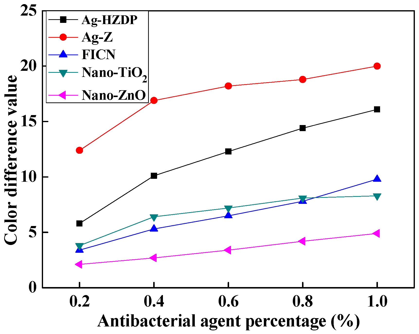
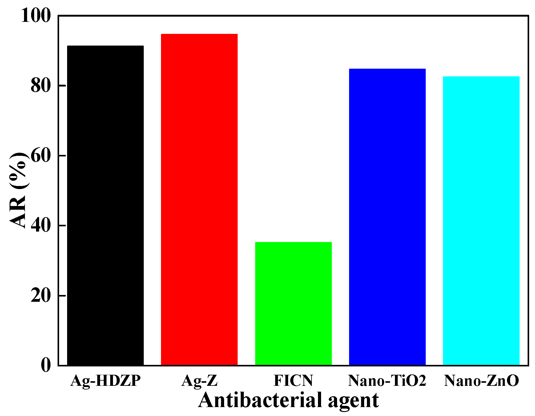
2.2. Selection of Antibacterial Agent

In order to find the effect of the antibacterial agent on the properties of the melamine resin coating, five antibacterial agents were first added into the melamine resin solution, in which the weight content of the antibacterial agent in the melamine resin solution was 1 wt %. The prepared antibacterial agent and melamine resin solutions were then applied onto the surface of the cedar floor substrate. The optimized antibacterial agent was selected through investigating the color difference of the coated floor substrate after 7 days until the color of the floor did not change. The floor surface was first divided into three approximately even areas, named as the top, middle, and bottom parts, based on their position. The color difference of the floor was calculated using a WSC-S color difference meter (Shanghai Precision Technology Co., Ltd., Shanghai, China), based on ISO 105-J03:2009 [32], in which 3 spots were randomly selected from the pre-divided areas of the coated floor surface.

2.3. Selection of Dispersing Agent

Fifteen mL of melamine resin solution, 1.5 g of selected antibacterial agent, and 0.15 g of dispersing agents were added into a 20 mL glass tube. The mixture was mechanically shaken for ~2 h. Afterwards, the glass tube was put into a test tube rack and left for 24 h. The optimized dispersing agent was selected through investigating the stability of the antibacterial agents’ dispersion in water, and calculating the subsidence value (H) of each antibacterial agent’s dispersion. Specifically, H was calculated as:
H = H0Hsub
where H0 is the initial height of the antibacterial agent dispersion (mm), and Hsub is the final height of the antibacterial agent dispersion after 24 h of subsidence. The calculation of H was schematically illustrated in Scheme 1.

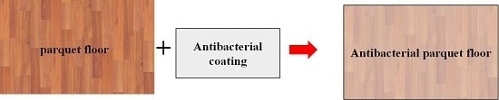
2.4. Preparation of Antibacterial Cedar Parquet Floor

In 100 mL melamine solution, a certain amount of the selected dispersing agent and the antibacterial agent, in which the mass ratio between dispersing agent and antibacterial agent was 1/10, were put into the melamine resin solution. After 2 h of mechanical shaking, the antibacterial melamine resin solutions with antibacterial agent mass contents of 0.2, 0.4, 0.6, 0.8, and 1.0 wt % were obtained. At the same time, the cedar floor was brushed with an ultraviolet curing coat. The obtained antibacterial melamine resin solutions were then applied onto the surface of the coated cedar floor using a brush. The coated floor was then aged and dried under UV light at 110 °C for ~24 h, and the antibacterial cedar parquet floor was finally obtained. The thickness of the antibacterial coating was ~1 mm, which was measured by a micrometer. For comparison, the pure melamine-coated cedar parquet floor without the introduction of the antibacterial agent was also prepared.

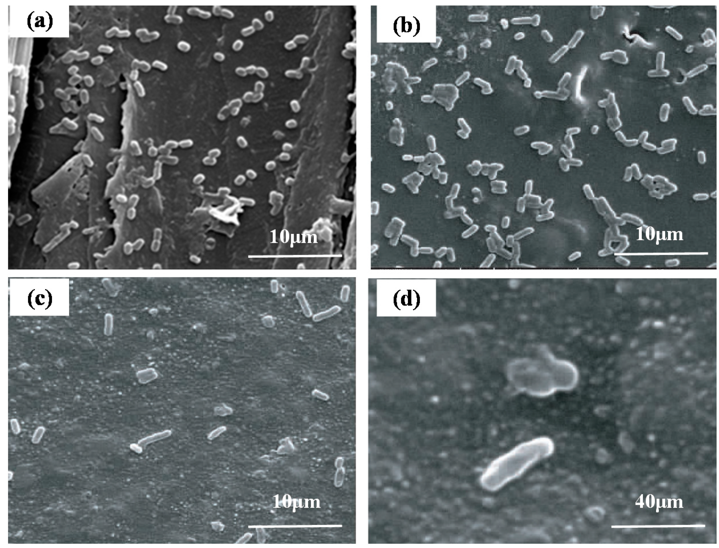
2.5. Antibacterial Properties Analysis of the Cedar Parquet Floor

The antibacterial properties of the prepared cedar parquet floor were analyzed through cultivating the Escherichia coli on the floor surface and investigating the number variation after 24 h of cultivation. The analysis was mainly based on China enterprise standard of QB/T2591-2003 [33] (test for antimicrobial activity and efficacy). The detailed steps were as follows. The cedar parquet floor sample (50 mm × 50 mm) was first washed with 70% ethanol to kill the bacteria on its surface, and then fully dried naturally. At the same time, the Escherichia coli was cultivated in nutrient agar at 36 ± 1 °C for 16 h, and was transferred into nutrient broth and cultivated at 36 ± 1 °C for 16 h. This cultivation broth was diluted 500 times with 1/500 nutrient broth (pH = 7.0 ± 0.2). The Escherichia coli was then transferred and dispersed on a slope medium surface evenly, and cultured at 36 °C for 24 h. The bacteria was then moved to an agar culture medium and cultivated at 36 °C for 24 h. The transferring steps were repeated for ~10 days. Afterwards, a 0.4 mL solution of cultivated bacteria (~5.0 × 10⁵ cfu/mL) was added onto the surface of the floor sample, and the floor surface was covered by a PE film (50 mm × 50 mm). The floor sample was then incubated at a relative humidity of over 90% (37 °C) for 24 h. The floor sample was then completely washed with 30 mL of a 1% NaCl solution containing Tween 80 with a pH value of 7.0 ± 0.2. The remained active bacteria was then counted. The final antibacterial ratio (AR) was calculated using the following equation:
AR = (BC)/B × 100%
where AR is antibacterial ratio, B is the average remaining bacterial numbers of the pure melamine resin solution-coated cedar parquet floor, and C is the average remaining bacterial numbers of the antibacterial melamine resin solution-coated cedar parquet floor.

2.6. Free Formaldehyde Content (FFC) and Total Volatile Organic Compounds (TVOC) Tests of the Cedar Parquet Floor

FFC value (CFF) was determined in accordance with Chinese National Standards (GB 18580-2001) [34] as follows. The cedar parquet floor sample (100 cm × 100 cm) was first put into a testing chamber with an air exchange rate of 1.0 ± 0.05 h−1 and a loading rate of 1.0 ± 0.02 m2/m3. The ambient temperature, relative humidity, and air velocity on the sample surface were set at 23 ± 0.5 °C, 50% ± 3%, and 0.1–0.3 m/s, respectively. The parquet floor sample was then put in the chamber for 5 days to release the formaldehyde. An appropriate amount of the air in the chamber was withdrawn at predetermined intervals and injected into an absorption bottle with distilled water. The content of the formaldehyde (Cf) in the air was quantified based on the absorbance value of 412 nm recorded by the ultraviolet spectrophotometer. The formaldehyde release equilibrium was reached when the Cf bias between two samples was lower than 5%. The CFF was then calculated as:
CFF = Cf/Vair
where Cf is formaldehyde content in the sampling air (mg), Vair is the volume of the sampling air (m3).
The TVOC value (CTVOC) was determined in accordance with (HJ571-2010) [35], and calculated using the following equation:
CTVOC = xCVOC/Vair
where x is the air exchange rate (m3/m2 h), CVOC is the amount of the VOC in the sampling air (mg), and Vair is the volume of the sampling air (m3). Specifically, CVOC was quantified by gas chromatograph equipment GC436 produced by Bruker Co., Ltd. (Billerica, MA, USA).

2.7. Cytotoxicity Tests of the Cedar Parquet Floor

The cytotoxicity effect of the cedar parquet floor was analyzed using the methyl thiazolyl tetrazolium (MTT) assay. Briefly, the floor sample was put into a 96-well plate with RPMI-1640 medium containing 10% fetal bovine serum (FBS), and the cells were allowed to grow in an incubator for 72 h at 37 °C. The cell culture fluid was then filtrated and kept at 4 °C as the extract liquid. Subsequently, liver cells were seeded and grown in 104 cells/well (200 μL/well) in a 96-well plate for 24 h in a RPMI-1640 medium containing 10% FBS. After that, the media was removed and replaced with the prepared extract liquid for exposure. Then, the liquid was removed and the cells were incubated with 20 μL of MTT (5 mg/mL in PBS) in fresh medium for 4 h at 37 °C. After the medium liquid was removed, the residual was dissolved in DMSO (150 μL/well). The respective absorbance values (Abs.) were read at 490, 570, and 630 nm using a microplate reader (Sunrise RC TS TC, Grödig, Austria) to determine the relative growth rate (RGR) value of the cells. The results were given as the mean of three independent experiments. The cytotoxicity grade was evaluated based on ISO 10993-5:1999 [36]. For comparison, the pristine cedar substrate was also assessed using the cytotoxicity test.

3. Results and Discussion

In order to analyze the effect of the antibacterial agent on the properties of the melamine resin solution coating, the selection of the optimized antibacterial agent was conducted first. Figure 1 shows the color difference of the melamine resin solution-coated floor with or without the introduction of the antibacterial agents. One could observe that compared with the floor coated with pure melamine resin solution, all the floor samples coated with melamine resin solution loaded with antibacterial agents showed a color difference. With the increase of the antibacterial agent loading amount, the color difference also increased. It could be seen that when the loading amount of the antibacterial agents reached 1 wt %, the color difference value of the floor coated with Ag-Z was as high as 20%. At the same time, the values of all the other four samples were lower than 15%, indicating Ag-HZDP, FICN, Nano-TiO2, and Nano-ZnO did not affect the properties of the melamine resin solution much.
The rough AR of the coated floor was then investigated, with the results shown in Figure 2. In each melamine resin solution, the loading amount of the antibacterial agent was 1 wt %. It could be clearly seen from Figure 2 that compared with other antibacterial agents, antibacterial agents incorporated with silver nanoparticles, Ag-Z and Ag-HZDP, had the highest antibacterial efficiency; the ARs of these two agents could reach 90% or higher. It is generally known that silver ions can generate bacteria function disorder and ingredient destruction through contact reactions [37]. Even minute amounts of silver ions can firmly attach to the bacteria cytomembrane and coagulate the protein, resulting in destruction of synthetase activity [38]. Moreover, the silver ions can also destroy the transmission system and respiratory system of the bacteria, which results in excellent antibacterial properties for silver nanoparticle loaded antibacterial agents [39]. On the contrary, the semiconductor antibacterial agents, such as titanium oxide and zinc oxide nanoparticles, have a totally different antibacterial mechanism. Specifically, under the irradiation of incident light, especially ultra-violet light, the electrons (e) of the semiconductors delocalized from the conduction band (CB) to the valence band (VB). The holes (h) in the VB react with the H2O molecules or O2 molecules to generate highly oxidative radicals [40]. The radicals kill the bacteria through redox reactions. Since the antibacterial properties of these semiconductors require continuous light irradiation, and the electron/hole recombination rate is high, the antibacterial ability of these semiconductors is relatively low compared with silver nanoparticle loaded antibacterial agents [41]. Combined with the results from Figure 1, Ag-HZDP was selected as the optimized antibacterial agent and used for the following test.
Since the dispersing status of the nanoparticles in the solution significantly affects the final antibacterial properties of the cedar parquet floor, the optimized dispersing agent was then selected. Table 1 shows the dispersion of Ag-HZDP in melamine resin solution using five alternative dispersing agents. It could be clearly seen from Table 1 that the Ag-HZDP was not well dispersed in melamine solution without the introduction of the dispersing agents, for which the subsidence value was ~2.5. When the dispersing agents were introduced into the system, the Ag-HZDP dispersed diversely in the melamine resin solution using different dispersing agents. One can clearly observe from Table 1 that the subsidence value of Ag-HZDP in both Na6P6O18 and SDBS decreases, indicating a better dispersing stability of the Ag-HZDP in the melamine solution. Comparatively, the other three dispersing agents could not enhance the dispersing stability of the Ag-HZDP, which meant these three ones were not suitable as dispersing agents for the Ag-HZDP in melamine solutions. Combined with the final appearance and solution status shown in Table 1, it can be seen that with the assistance of the Na6P6O18, Ag-HZDP was uniformly dispersed in the melamine resin solution without changing its color. Thus, the Na6P6O18 was selected as the optimized dispersing agent and used for the following test.
Based on the above mentioned results, Ag-HZDP and Na6P6O18 were used as the optimized antibacterial agent and dispersing agent, respectively, to prepare the final cedar parquet floor. Figure 3 shows the AR of the Ag-HZDP-melamine solution-coated floor with and without the Na6P6O18. It can be seen that without the assistance of the Na6P6O18, the AR of the floor reached ~90% only when the Ag-HZDP loading amount was higher than 1 wt %. Comparatively, for Na6P6O18 assisted Ag-HZDP, the floor had an AR of higher than 90% when the loading amount of the Ag-HZDP was only 0.6 wt %. The results indicated that with the help of Na6P6O18, the Ag-HZDP was better dispersed, both in the melamine resin solution and on the surface of the cedar parquet floor, resulting in better antibacterial effects.
Figure 4 shows the SEM images of the cedar parquet floor surface after cultivation of the Escherichia coli. It could be directly revealed from Figure 4a that the melamine-coated cedar parquet floor without antibacterial agent did not have any antibacterial ability. After 24 h of cultivation, numerous Escherichia coli colonies still grew on the floor surface. It could be also seen from Figure 4b that pure melamine resin did not have antibacterial ability either, resulting in abundant growth of the Escherichia coli on the surface of pure melamine resin-coated cedar parquet floor. Since the flooring is mainly used for public and home flooring, and the flooring cannot be used practically without coatings, the antibacterial treatment to the coatings is inevitable. After the antibacterial agent, Ag-HZDP, was added into the melamine resin solution, the antibacterial ability of the cedar parquet floor was dramatically enhanced. After 24 h of cultivation, nearly no Escherichia coli colonies still grew on the floor surface, due to the presence of the Ag-HZDP. From the magnified SEM image shown in Figure 4d, it can be seen that the morphology of some Escherichia coli had been changed. This indicated that the Ag-HZDP has promising antibacterial properties and is capable of being added into a melamine resin solution to enhance the antibacterial ability of cedar parquet flooring when combined with Na6P6O18. Since one of the most common uses for floor products is in home flooring, where the floor is in contact with human skin continually, the toxicity of the floor is extremely significant. Thus, it is necessary to conduct conventional toxicity tests for a prepared antibacterial floor. The FFC and TVOC tests were conducted first. The CFF and CTVOC values of the floor were 0.084 mg/m3 and 0.386 mg/m2·h, respectively, which were both lower than Chinese national standards and European Union standards [42,43].
The cytotoxicity of the prepared antibacterial floor was then analyzed using MTT method. Table 2 shows the RGR values of the floor. On the basis of the RGR values, it can be seen that the toxicity grade of the pristine cedar substrate was 3 according to United States Pharmacopeia (USP). When the antibacterial coating was applied on the cedar floor surface, all the RGR values of the prepared antibacterial floor were obviously enhanced compared with the value of the pristine cedar substrate. Moreover, the cytotoxicity grade of the prepared antibacterial floor was similar to that of the cedar floor with antibacterial-agent-free melamine coating. The results indicated the prepared antibacterial floor had a lower toxicity grade and was promising for daily use as a flooring product.

4. Conclusions

In summary, a novel antibacterial cedar parquet floor was prepared in the present study. The optimized antibacterial agent and the dispersing agent were selected, and the antibacterial properties and the toxicity tests were conducted in detail. The results showed that Ag-HZDP and Na6P6O18 were suitable as antibacterial and dispersing agents in a melamine coating for flooring. With the Ag-HZDP-loaded melamine resin solution applied onto the floor surface, the prepared antibacterial floor could inhibit and kill almost all the Escherichia coli bacteria cultivated on its surface. Moreover, the prepared antibacterial floor had a lower toxicity compared with pristine cedar substrate. The present study provides an effective way to fabricate daily-used parquet flooring with excellent antibacterial properties and lower toxicity.

Author Contributions

Formal Analysis, C.J.; Methodology, Y.Z.; Conceptualization and Data Curation, J.C.; Supervision and Writing—Original Draft Preparation, L.G.

Funding

This research was funded by Natural Science Foundation of Jiangsu Province, China (No. BK20160938), the Youth Science and Technology Innovation Fund (No. CX2016019), and the Major Project of Natural Science Foundation of the Jiangsu Higher Education Institutions of China (No. 18KJA220002).

Acknowledgments

Advanced analysis and testing center of Nanjing Forestry University is acknowledged.

Conflicts of Interest

The authors declare no conflict of interest.

References

  1. Yamulki, S.; Morison, J.I.L. Annual greenhouse gas fluxes from a temperate deciduous oak forest floor. Forestry 2017, 90, 541–552. [Google Scholar] [CrossRef]
  2. Zhang, H.Y.; He, Q.; Lu, X.N.; Pizzi, A.; Mei, C.T.; Zhan, X.X. Energy release rate measurement of welded bamboo joints. J. Renew. Mater. 2018, 6, 450–456. [Google Scholar] [CrossRef]
  3. MacFarlane, D.W.; Meyer, S.P. Characteristics and distribution of potential ash tree hosts for emerald ash borer. For. Ecol. Manag. 2005, 213, 15–24. [Google Scholar] [CrossRef]
  4. Zhang, H.Y.; Pizzi, A.; Zhou, X.H.; Lu, X.N.; Wang, Z.Q. The study of linear vibrational welding of moso bamboo. J. Adhes. Sci. Technol. 2018, 32, 1–10. [Google Scholar] [CrossRef]
  5. De Figueiredo Latorraca, J.V.; Teixeira, D.E.; Batista, D.C. Overlay of Eucalyptus urophylla cement-bonded particleboard for application as flooring panels. For. Prod. J. 2009, 59, 65–69. [Google Scholar]
  6. Salca, E.-A. Black alder (Alnus glutinosa L.)—A resource for value-added products in furniture industry under European screening. Curr. For. Rep. 2019, 5, 41–54. [Google Scholar] [CrossRef]
  7. Huang, W.; Wilkes, A.; Sun, X.; Terheggen, A. Who is importing forest products from Africa to China? An analysis of implications for initiatives to enhance legality and sustainability. Environ. Dev. Sustain. 2013, 15, 339–354. [Google Scholar] [CrossRef]
  8. Hesser, F.; Wohner, B.; Meints, T.; Stern, T.; Windsperger, A. Integration of LCA in R&D by applying the concept of payback period: Case study of a modified multilayer wood parquet. Int. J. Life Cycle Assess. 2017, 22, 307–316. [Google Scholar]
  9. Berti, S.; Burato, P.; Dionisi-Vici, P.; Allegretti, O. Orange wood for parquet and engineered flooring use. BioResources 2017, 13, 586–596. [Google Scholar] [CrossRef]
  10. Blanchet, P.; Beauregard, R.; Erb, A.; Lefebvre, M. Comparative study of four adhesives used as binder in engineered wood parquet flooring. For. Prod. J. 2003, 53, 89. [Google Scholar]
  11. Heudorf, U.; Angerer, J. Internal exposure to PAHs of children and adults living in homes with parquet flooring containing high levels of PAHs in the parquet glue. Int. Arch. Occup. Environ. Health. 2001, 74, 91–101. [Google Scholar] [CrossRef] [PubMed]
  12. Saelzer, E.; Maack, J.; Moeck, T. Special impact sound insulation cases, Part 1: Laminate and parquet flooring, dry and raised floor constructions and terrace flooring. Bauphysik 2012, 34, 223–228. [Google Scholar]
  13. Derler, S.; Kausch, F.; Huber, R. Systematic patterns and random fluctuations in time series of coefficients of friction measured on floor surfaces. Saf. Sci. 2005, 43, 751–770. [Google Scholar] [CrossRef]
  14. Zigon, J.; Pizzi, A.; Zhang, H.; Sega, B.; Cop, M.; Sernek, M. The influence of heat and chemical treatments of beech wood on the shear strength of welded and UF bonded specimens. Eur. J. Wood Wood Prod. 2015, 73, 685–687. [Google Scholar] [CrossRef]
  15. Bruez, E.; Haidar, R.; Alou, M.T.; Vallance, J.; Bertsch, C.; Mazet, F.; Fermaud, M.; Deschamps, A.; Guerin-Dubrana, L.; Compant, S.; et al. Bacteria in a wood fungal disease: Characterization of bacterial communities in wood tissues of esca-foliar symptomatic and asymptomatic grapevines. Front. Microbiol. 2015, 6, 1137. [Google Scholar] [CrossRef] [PubMed]
  16. Milling, A.; Kehr, R.; Wulf, A.; Smalla, K. Survival of bacteria on wood and plastic particles: Dependence on wood species and environmental conditions. Holzforschung 2005, 59, 72–81. [Google Scholar] [CrossRef]
  17. Joycharat, N.; Thammavong, S.; Limsuwan, S.; Homlaead, S.; Voravuthikunchai, S.P.; Yingyongnarongkul, B.-E.; Dej-Adisai, S.; Subhadhirasakul, S. Antibacterial substances from Albizia myriophylla wood against cariogenic Streptococcus mutans. Arch. Pharmacal Res. 2013, 36, 723–730. [Google Scholar] [CrossRef]
  18. Siedenbiedel, F.; Tiller, J.C. Antimicrobial Polymers in Solution and on Surfaces: Overview and functional principles. Polymers 2012, 4, 46–71. [Google Scholar] [CrossRef]
  19. Kandelbauer, A.; Widsten, P. Antibacterial melamine resin surfaces for wood-based furniture and flooring. Prog. Org. Coat. 2009, 65, 305–313. [Google Scholar] [CrossRef]
  20. Gysels, K.; Delalieux, F.; Deutsch, F.; Van Grieken, R.; Camuffo, D.; Bernardi, A.; Sturaro, G.; Busse, H.-J.; Wieser, M. Indoor environment and conservation in the Royal Museum of Fine Arts, Antwerp, Belgium. J. Cult. Heritage 2004, 5, 221–230. [Google Scholar] [CrossRef]
  21. Abrishami, S.H.; Tall, B.D.; Bruursema, T.J.; Epstein, P.S.; Shah, D.B. Bacterial adherence and viability on cutting board surfaces. J. Food Saf. 1994, 14, 153–172. [Google Scholar] [CrossRef]
  22. Alvarez-Paino, M.; Munoz-Bonilla, A.; Fernandez-Garcia, M. Antimicrobial polymers in the nano-world. Nanomaterials 2017, 7, 48. [Google Scholar] [CrossRef]
  23. Timofeeva, L.; Kleshcheva, N. Antimicrobial polymers: Mechanism of action, factors of activity, and applications. Appl. Microbiol. Biotechnol. 2011, 89, 475–492. [Google Scholar] [CrossRef] [PubMed]
  24. Munoz-Bonilla, A.; Fernandez-Garcia, M. Polymeric materials with antimicrobial activity. Prog. Polym. Sci. 2012, 37, 281–339. [Google Scholar] [CrossRef]
  25. Kim, S.; Kim, H.-J. Anti-bacterial performance of colloidal silver-treated laminate wood flooring. Int. Biodeterior. Biodegrad. 2006, 57, 155–162. [Google Scholar] [CrossRef]
  26. Munoz-Bonilla, A.; Fernandez-Garcia, M. The roadmap of antimicrobial polymeric materials in macromolecular nanotechnology. Eur. Polym. J. 2015, 65, 46–62. [Google Scholar] [CrossRef] [Green Version]
  27. Seo, J.; Jeon, J.; Lee, J.-H.; Kim, S. Thermal performance analysis according to wood flooring structure for energy conservation in radiant floor heating systems. Energy Build. 2011, 43, 2039–2042. [Google Scholar] [CrossRef]
  28. Braun, M.; Sun, Y. Antimicrobial polymers containing melamine derivatives. I. Preparation and characterization of chloromelamine-based cellulose. J. Polym. Sci. Part A 2004, 42, 3818–3827. [Google Scholar] [CrossRef]
  29. Kuplennik, N.; Tchoudakov, R.; Zelas, Z.B.-B.; Sadovski, A.; Fishman, A.; Narkis, M. Antimicrobial packaging based on linear low-density polyethylene compounded with potassium sorbate. LWT-Food Sci. Technol. 2015, 62, 278–286. [Google Scholar] [CrossRef]
  30. Chakra, C.; Raob, K.; Rajendar, V. Nanocomposites of ZnO and TiO2 have enhanced antimicrobial and antibacterial properties than their disjoint counterparts. Dig. J. Nanomater. Biostruct. 2017, 12, 185–193. [Google Scholar]
  31. Youssef, A.; Abdel-Aziz, M.; El-Sayed, E.; Abdel-Aziz, M.; El-Hakim, A.A.; Kamel, S.; Turky, G. Morphological, electrical & antibacterial properties of trilayered Cs/PAA/PPy bionanocomposites hydrogel based on Fe3O4-NPs. Carbohydr. Polym. 2018, 196, 483–493. [Google Scholar] [PubMed]
  32. ISO 105-J03:2009 Textiles—Tests for Colour Fastness—Part J03: Calculation of Colour Differences; International Organization for Standardization: Geneva, Switzerland, 2009.
  33. QB/T 2591-2003 Antimicrobial Plastics—Test for Antimicrobial Activity and Efficacy; National Development and Reform Commission: Beijing, China, 2003.
  34. GB 18580-2001 The Indoor Decorating and Refurbishing Material-Limit of Formaldehyde Emission of Wood-Based Panels and Finishing Products; Standardisation Administration of China: Beijing, China, 2001.
  35. HJ571-2010 Technical Requirement for Environmental Labeling Products—Wood Based Panels and Finishing Products; Ministry of Environmental Protection of China: Beijing, China, 2010.
  36. ISO 10993-5:1999 Biological Evaluation of Medical Devices. Part 5. Tests for Cytotoxicity: In Vitro Methods; International Organization for Standardization: Geneva, Switzerland, 1999.
  37. Le Ouay, B.; Stellacci, F. Antibacterial activity of silver nanoparticles: A surface science insight. Nano Today 2015, 10, 339–354. [Google Scholar] [CrossRef] [Green Version]
  38. Ferraris, M.; Perero, S.; Ferraris, S.; Miola, M.; Vernè, E.; Skoglund, S.; Blomberg, E.; Wallinder, I.O. Antibacterial silver nanocluster/silica composite coatings on stainless steel. Appl. Surf. Sci. 2017, 396, 1546–1555. [Google Scholar] [CrossRef]
  39. Liu, C.; Liu, J.; Ning, X.; Chen, S.; Liu, Z.; Jiang, S.; Miao, D. The effect of polydopamine on an Ag-coated polypropylene nonwoven fabric. Polymers 2019, 11, 627. [Google Scholar] [CrossRef]
  40. Geng, A.; Meng, L.; Han, J.; Zhong, Q.; Li, M.; Han, S.; Mei, C.; Xu, L.; Tan, L.; Gan, L. Highly efficient visible-light photocatalyst based on cellulose derived carbon nanofiber/BiOBr composites. Cellulose 2018, 25, 4133–4144. [Google Scholar] [CrossRef]
  41. Qi, K.; Cheng, B.; Yu, J.; Ho, W. Review on the improvement of the photocatalytic and antibacterial activities of ZnO. J. Alloy. Compd. 2017, 727, 792–820. [Google Scholar] [CrossRef]
  42. Roumeli, E.; Papadopoulou, E.; Pavlidou, E.; Vourlias, G.; Bikiaris, D.; Paraskevopoulos, K.; Chrissafis, K. Synthesis, characterization and thermal analysis of urea–formaldehyde/nanoSiO2 resins. Thermochim. Acta 2012, 527, 33–39. [Google Scholar] [CrossRef]
  43. Jin, Y.; Cheng, X.; Zheng, Z. Preparation and characterization of phenol–formaldehyde adhesives modified with enzymatic hydrolysis lignin. Bioresour. Technol. 2010, 101, 2046–2048. [Google Scholar] [CrossRef] [PubMed]
Scheme 1. Illustration of subsidence value of the antibacterial nanoparticles in melamine resin solution.
Coatings 09 00403 sch001
Figure 1. Effect of different quantities of antibacterial agents on the color of the coatings.
Coatings 09 00403 g001
Figure 2. Antibacterial ratio of different antibacterial agents loaded with melamine resin coatings.
Coatings 09 00403 g002
Figure 3. Antibacterial ratio of Ag-HZDP-melamine resin solution-coated cedar parquet floor, with and without Na6P6O18.
Coatings 09 00403 g003
Figure 4. SEM image of cedar parquet floor (a) without coating, (b) with pure melamine resin solution, (c) with Ag-HZDP-loaded melamine resin solution, and (d) magnified cedar parquet floor with Ag-HZDP-loaded melamine resin solution.
Coatings 09 00403 g004
Table 1. The subsidence condition and final appearance of Ag-HZDP in melamine resin solution using five dispersing agents.
Dispersing AgentSubsidence Value/ConditionAppearance
Nil2.5 mmNormal color
PAAPrecipitates appearGrey in color
Na6P6O181 mmUniformly dispersed
Na-CMCNa-CMC insolubleNanoparticle/Na-CMC separated
SDBS1.25 mmBubble appears, slight color change in solution
PAASSolution thickenedViscosity increased, slight color change in solution
Table 2. Cytotoxic effects of the prepared antibacterial floor.
Sample490 nm570 nm630 nm
Abs.RGR/%GradeAbs.RGR/%GradeAbs.RGR/%Grade
Pristine cedar substrate without melamine coating0.109032.0940.528725.8240.464333.273
Cedar floor with melamine coating (no antibacterial agent)0.222168.4520.992160.4320.461933.103
Prepared antibacterial floor0.205263.2520.894354.4520.461832.893

