Article

Evaluation of Mesenchymal Stem Cells and Osteoblasts’ Adhesion and Proliferation in the Presence of HA-AL Biomaterials

by
Oana-Elena Nicolaescu
1,
Adina Turcu-Stiolica
1,
Renata-Maria Varut
1,*,
Andreea-Gabriela Mocanu
1,*,
Ionela Belu
1,
Livia Elena Sima
2 and
Johny Neamtu
1
1
Pharmacy Department I, University of Medicine and Pharmacy, 2-4 Petru Rares Str., 200349 Craiova, Romania
2
Institute of Biochemistry, Romanian Academy, 296 Splaiul Independentei, 060031 Bucharest, Romania
*
Authors to whom correspondence should be addressed.
Coatings 2019, 9(12), 782; https://doi.org/10.3390/coatings9120782
Submission received: 10 October 2019 / Revised: 7 November 2019 / Accepted: 19 November 2019 / Published: 22 November 2019

Abstract

:
There is an increased interest in developing biocomposite implants with high biocompatibility in order to be used as grafts or prostheses in orthopedic surgery. The purpose of the study was to determine the biocompatibility of titanium implants coated with synthesized hydroxyapatite-alendronate composites. The implants were obtained using Matrix Assisted Pulsed Laser Evaporation technique (MAPLE). The hydroxyapatite-alendronate composites were synthesized using the wet precipitation method. Immunofluorescence microscopy showed that composites support mesenchymal stem cells (MSCs) adhesion. Bone cells as well as human MSCs adhere to hydroxyapatite (HA)-based thin films obtained by matrix assisted laser deposition onto titanium. Alendronate doping into the films increased the number of cell-biomaterial focal points as compared to HA only. Thus, the synthesis of hydroxyapatite-alendronate composite (HA-AL) may be considered a viable solution for including the bisphosphonate on the surface of metallic prosthetic components used in orthopedics.

1. Introduction

Among the various types of ceramic materials (calcium phosphate ceramics, alumina ceramics, zirconium ceramic, ceramic composite materials, metal ceramic composite materials, plastic ceramic composite materials), calcium phosphate bioceramics are closest to the natural bone. The CaO-P2O5 binary system consists of several binary compounds: C4P (Ca4P2O9)—tetracalcium phosphate, C3P (Ca3(PO4)2)—tricalcium phosphate, C2P (Ca2HPO4)—dicalcium phosphate, CP (CaH2PO4)—monocalcium phosphate, Ca8H2(PO4)6·5H2O—octacalcium phosphate, with or without adjacent polymorphic varieties. Hydroxyapatite lies between C4P and C3P. This compound is extremely interesting as a bioceramic material because its structure can be easily modified depending on temperature and vapor pressure of water. Among the calcium phosphate based bioceramics, crystalline hydroxyapatite is the most stable phase when put in contact with body fluids. Amorphous HA and tricalcium phosphate are less stable and they are resorbed by the body faster. Another important characteristic of HA is bioactivity. After it is implanted, it promotes the adherence of the implant to the tissue that surrounds it by forming a functional connective structure [1,2,3].
Bisphosphonates are a group of anti-resorptive drugs that suppress osteoclast activity and increase osteoblast proliferation. They display an increased affinity for hydroxyapatite crystals and are retained for an extended time in the bone. Bisphosphonates are used for the treatment of a variety of bone diseases such as osteoporosis, Paget’s disease, myeloproliferative disease and bone cancer. The main disadvantage of bisphosphonates is the low bioavailability they exhibit through oral or intravenous administration [4].
Local release of bisphosphonates from various biocomposites such as hydroxyapatite-alendronate is a solution frequently studied in the recent years in an attempt to avoid prosthesis-induced problems and side effects that may occur due to bisphosphonates long-term treatment [5].
Bone disorders are difficult to treat because the blood flow reaching the bone is reduced. Therefore, local drug concentration is also reduced. An incompatibility between metal implants and bone structure leads to an inflammatory reaction. This is a defense mechanism of the body as it considers the prosthesis components as non-self. Considering that the viability of the implant depends on the processes that occur at the bone-implant interface, the physicochemical optimization of the surface of implants used in orthopedic surgery is fundamental in achieving fast and consistent bone integration [5]. There is a great interest among physicists, biologists and physicians to develop biomimetic surfaces of calcium phosphates and proteins that would improve cell adhesion and thus reduce bone integration time [6]. Antiresorptive therapies with bisphosphonates that inhibit osteoclasts can be implemented in order to avoid periprosthetic bone loss [4]. Although the main mechanism of action of bisphosphonates is to inhibit bone resorption, there are studies that also prove having a positive effect on osteoblasts. A number of studies show a growth in the differentiation of osteoblasts from mesenchymal stem cells and a positive influence over their proliferation and maturation. Moreover, these studies show that bisphosphonates can prevent osteoblast apoptosis [7]. The local release of the bisphosphonates is preferred in order to avoid loss of bone mass and other side effects that may appear due to long-term treatment. Therefore, a large dose of the drug can be administered directly to the area of interest in order to reduce bone loss and have a positive effect on bone integration time [8]. Furthermore, it also has a positive effect on accelerating the secondary fixation of the prosthesis. Additionally, a good osseointegration is obtained for the osteoporotic bone [9]. Implants that deliver bisphosphonates locally were tested in preclinical studies on several laboratory animals (rats, rabbits, dogs). The results obtained are promising in terms of implant viability [10].
There are a number of methods used in research for hydroxyapatite synthesis such as sol-gel method [11], hydrothermal technique [12], multiple emulsion technique [13], biomimetic deposition technique [14], electrodeposition technique [15]. Compared to these methods, the wet precipitation technique has several advantages such as low reaction temperature, ease in synthesis and cost effectiveness. Moreover, it yields harmless reaction byproducts [16].
The purpose of the study was to determine the biocompatibility of hydroxyapatite-alendronate (HA-AL) coated titanium implants. The HA-AL composites were synthesized by wet precipitation method. Then, they were deposited on titanium implants, by Matrix Assisted Pulsed Laser Evaporation Technique (MAPLE) in order to obtain thin films.
MAPLE was successfully used for the deposition of several compounds such as organic polymers, bovine serum albumin, biomolecules and urease [17].
The experimental design of the biocompatibility studies consisted of several well-defined steps, namely the primary evaluation of biomaterials in interaction with a model bone cell line—SaOs2. In vitro biocompatibility tests were performed using mesenchymal stem cells (MSCs) or SaOs2 cells seeded on the proposed biomaterials.

2. Materials and Methods

All reagents used were of analytical grade and they were purchased either from Sigma Aldrich (calcium nitrate, diammonium phosphate; Saint Louis, MO, USA) or Merck, Darmstadt, Germany (ammonia, sodium alendronate trihydrate).

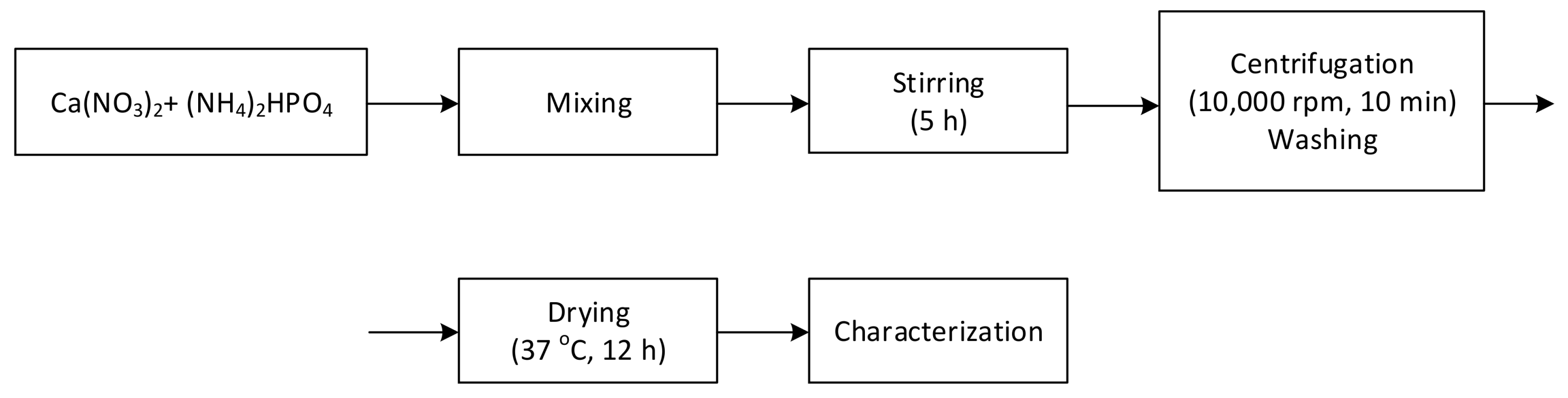
2.1. Hydroxyapatite Synthesis by Wet Precipitation Method

Two solutions of calcium nitrate of concentration 1.08 M (12.75 g Ca(NO3)2·4H2O in 50 mL solution) and ammonium hydrogen phosphate 0.65 M (4.292 g (NH4)2HPO4 in 50 mL solution) were obtained initially. The pH of these two solutions was adjusted to 10 using NH4OH [18].
The calcium nitrate solution was heated initially to 90 °C. Then ammonium hydrogen phosphate was added under constant stirring (600 rpm). The addition rate of the second reagent was 0.1 mL/min (Table 1). It was added using a peristaltic pump.
The product was maintained in the reaction environment for 5 h at a constant temperature under constant stirring. Afterwards, it was centrifuged at 10,000 rpm for 10 min (Eppendorf 5804 centrifuge, Hamburg, Germany) and washed repeatedly with distilled water until no traces of ammonia remained. The powder was heated overnight at 37 °C and then triturated.

2.2. Hydroxyapatite-Alendronate Synthesis

The compounds containing bisphosphonates were obtained by dripping alendronate solution to the reaction environment immediately after the ammonium hydrogen phosphate was added.
The outline of the operations performed in the synthesis of the compound is shown in Figure 1.
Several concentrations of alendronate solutions (5, 10 and 20 mM) were used for the syntheses. The compounds obtained from these syntheses were abbreviated in accordance with the alendronate concentrations used HA-AL 5 mM, HA-AL 10 mM and HA-AL 20 mM.

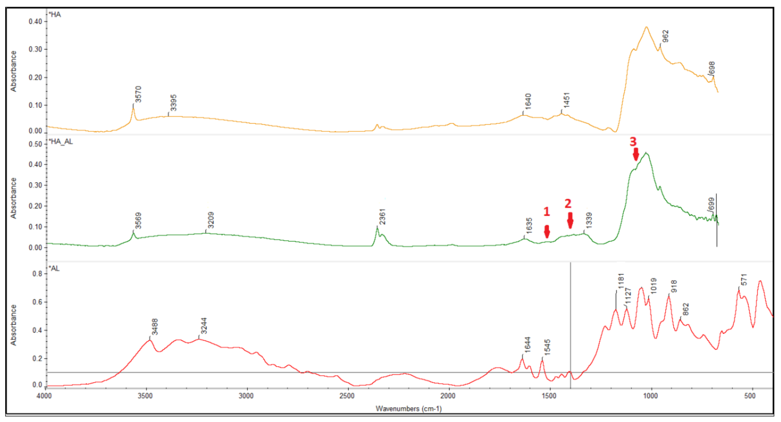
2.3. Physico-Chemical Characterization of Hydroxyapatite and HA-AL Composite

The synthesized hydroxyapatite and HA-AL composite were characterized by spectrophotometry. These analyses were carried out using an Avatar Nicolet spectrophotometer (iN 10, Thermo Scientific, Waltham, MA, USA) in KBr pellets within the range 4000–400 cm−1.
Moreover, the powders were characterized by X-ray diffraction in a previous study [19].

2.4. MAPLE Deposition

MAPLE deposition technique was used to transfer the synthesized HA-AL composites on titanium implants. The experiments were performed at the “Laser-Surface-Plasma Interactions” Laboratory from Lasers Department, National Institute for Laser Plasma and Radiation Physics Bucharest Romania.
The deposition experiments were performed using a 205 COMPexPro laser (Coherent, Santa Clara, CA, USA) with the following characteristics: wavelength 248 nm, pulse length 25 ns, fluence 0.7 J/cm2 and target-substrate distance 5 cm. Water was the solvent chosen for target preparation. Suspensions of synthesized HA-AL 5 mM, synthesized HA-AL 20 mM and synthesized HA were first obtained. The suspensions were poured in a copper device with a diameter of 3 cm, and then, they were frozen in liquid nitrogen. The target was introduced in the reaction chamber and then irradiated.
Grade 4 titanium implants were washed in an acetone, alcohol and water mixture in an ultrasound bath and then they were introduced in the reaction chamber.

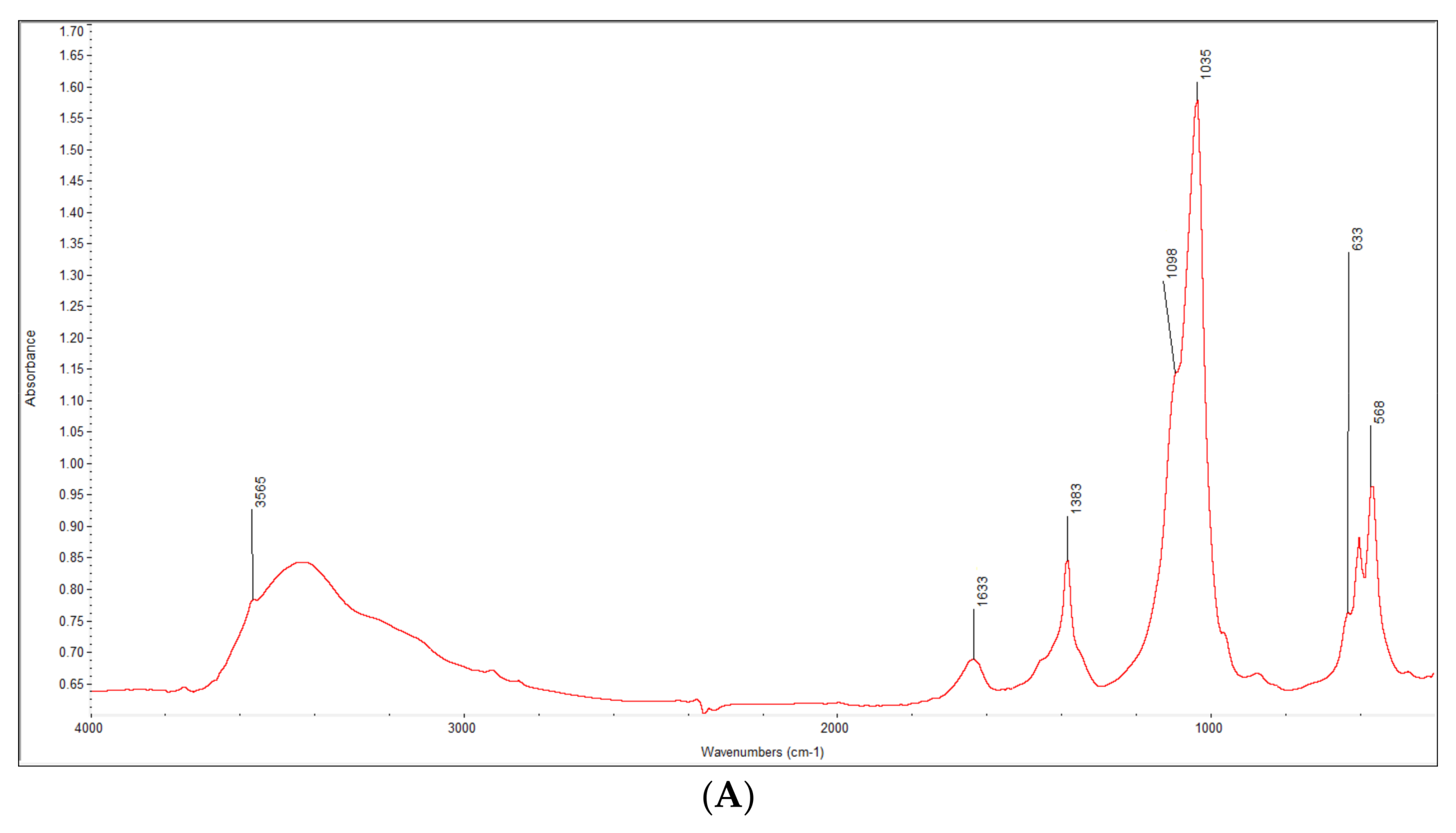
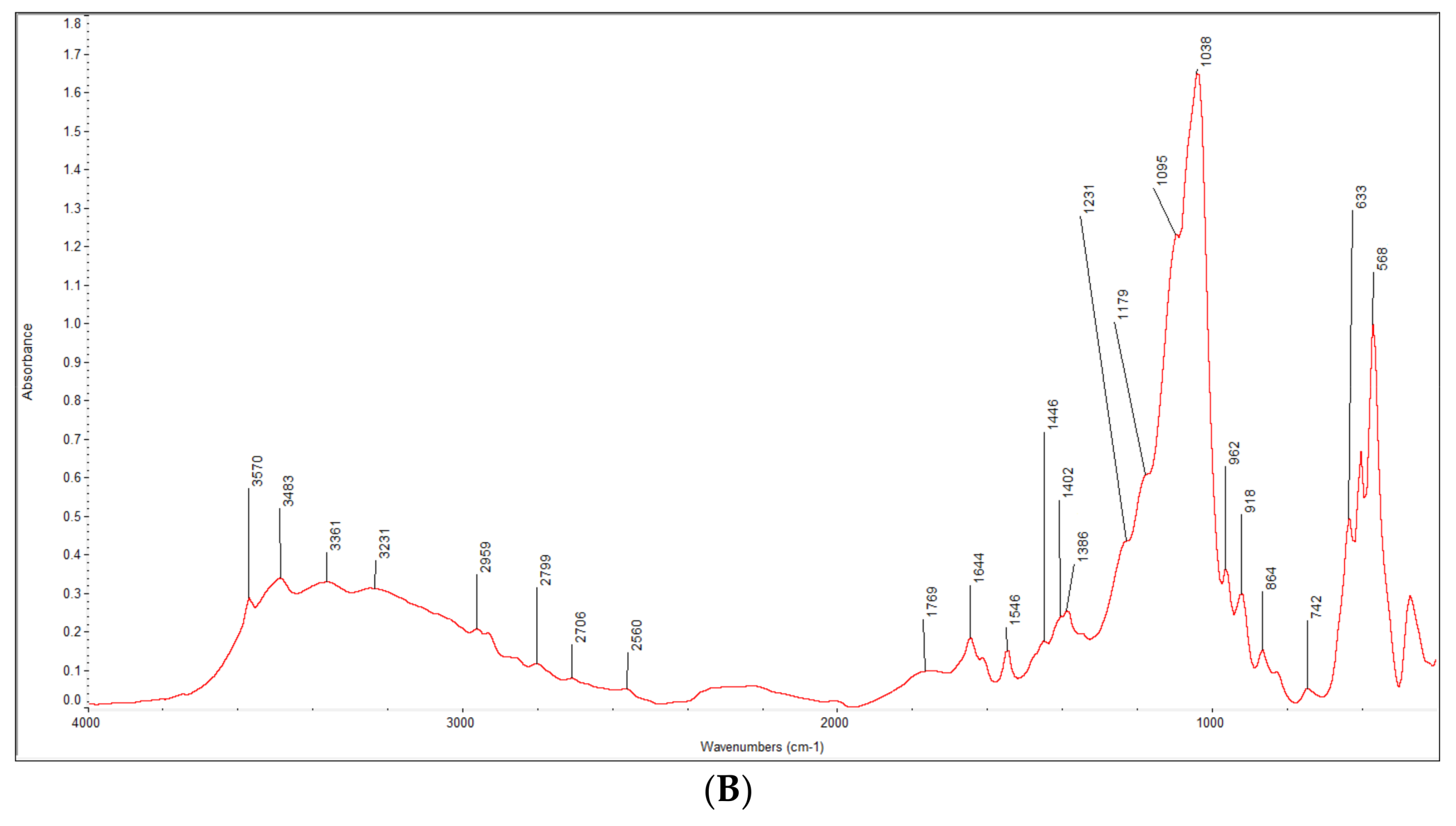
2.5. Spectral Characterization

MAPLE film spectrum was assessed with a Nicolet iN10 (Thermo Scientific, Waltham, MA, USA) configured for reflection setup. An accumulation of 128 spectral samples was used to minimize noise and increase signal. Aperture was set to 200 μm × 200 μm.

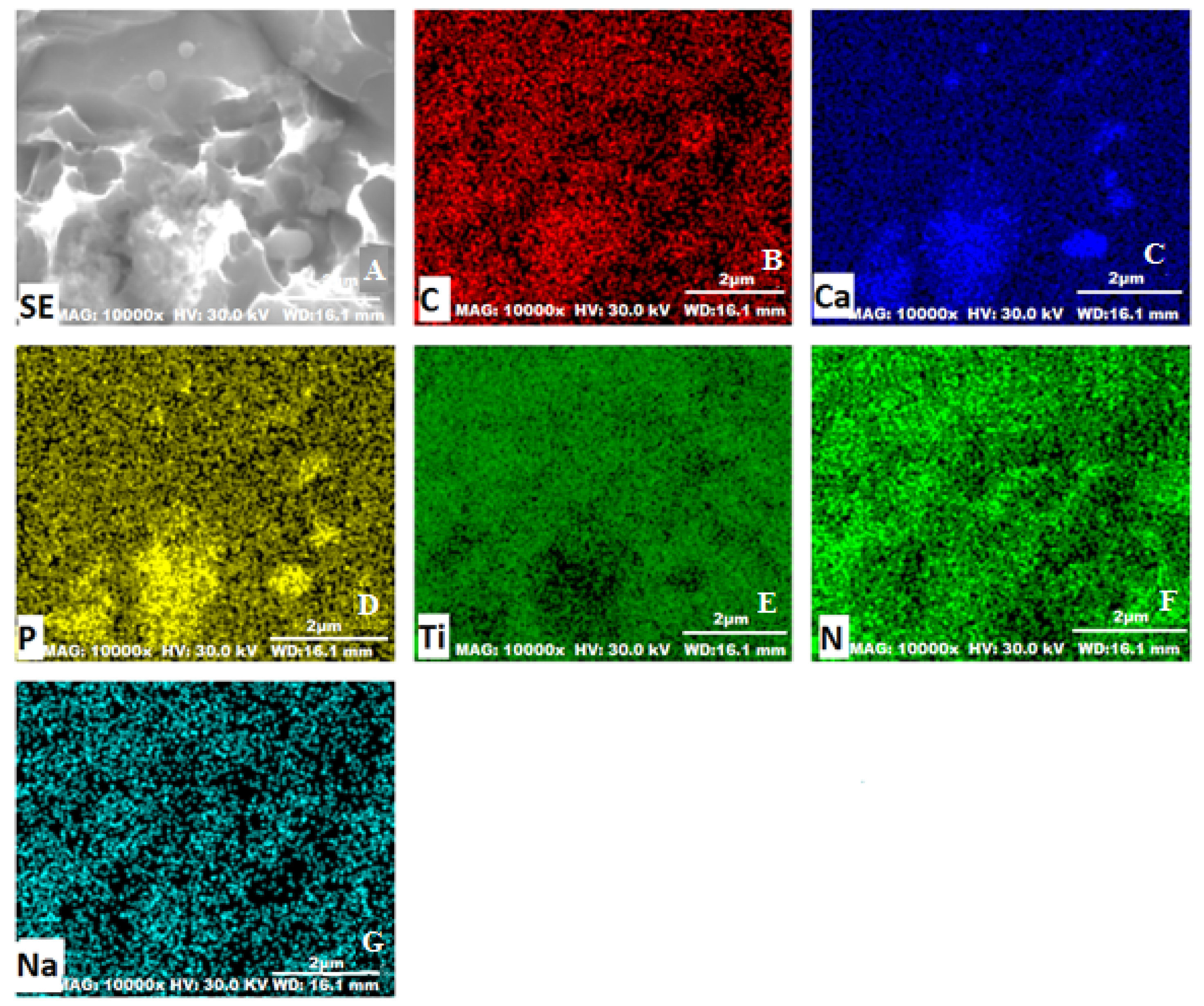
2.6. Scanning Electron Microscopy

Scanning electron microscopy (SEM) with energy dispersive X-ray (EDX) analytical system was used to evaluate the surface structure of the MAPLE deposited films. These analyses were carried out using a Hitachi 8230 microscope (Tokyo, Japan).

2.7. Biocompatibility Assays of HA-AL Biomaterials

2.7.1. Isolation and Characterization of Mesenchymal Stem Cells

Mesenchymal stem cells (primary cells) were obtained from bone marrow samples donated by patients who underwent surgery (implant) at Orthopedic Surgery Clinic of the Emergency Hospital in Craiova, Romania. The harvest was performed during the orthopedic surgery procedure for total femur arthroplasty. All subjects included in the study have given their informed, written consent in order to perform the research and publish the study’s results. The research was conducted with the consent of the Hospital’s Ethics Committee (No. 68/11.07.2016).
Bone marrow samples were the source for MSC. These were isolated by ficoll density gradient centrifugation [20] using a method adapted from Meinel et al. 2005 [21]. The cells were then grown in Dulbecco’s Modified Eagle Medium (DMEM) supplemented with 10% Fetal Calf Serum (FCS) (Biochrom AG, Cambridge, UK), L-Gln and Pen/Strep (Gibco, Invitrogen, Carlsbad, CA, USA).

2.7.2. In Vitro Cell Culture on Biomaterials

Bone Cell Culture and Seeding Cells

The experiments were performed on human mesenchymal stem cells (MSC) and SaOs2 cells. Both MSC and SaOs2 were seeded at a density of 5000 cells/cm2 in multi-wells plates (24 wells) (Santa Cruz, Heidelberg, Germany) and cultivated for the specified period of time.

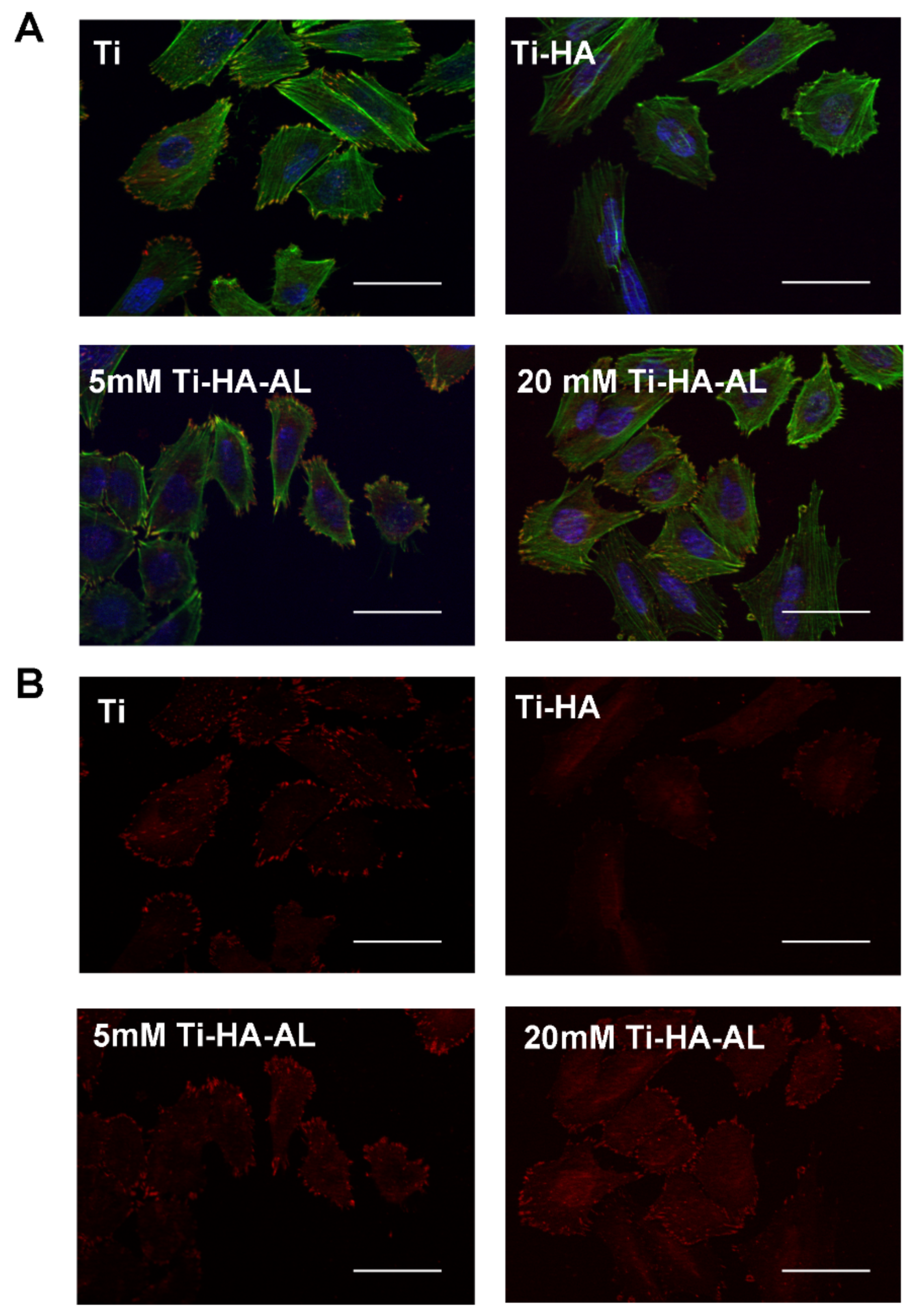
Fluorescence Microscopy

MSC grown under standard conditions or in direct contact with biomaterials (HA-AL coated titanium, HA-AL 5 mM coated titanium, HA-AL 20 mM coated titanium) were analyzed for adhesion and proliferation by immunofluorescence. The standards chosen were borosilicate glass (CS), uncoated titanium (Ti), non-doped HA coated titanium (HA) and alendronate solutions having different concentrations (alendronate 10−9 M, alendronate 10−8 M).
To evaluate cell adhesion, the samples were fixed with 4% paraformaldehyde (PFA) for 10 min and permeabilized with 0.2% Triton-X-100 for 3 min at room temperature. The cells were sequentially incubated for 30 min each with primary antibodies against vinculin and Alexa Fluor 594 conjugated secondary antibodies. PBS washes were performed after each stage. Alexa Fluor 488 conjugated phalloidin was added together with the secondary antibody to label actin filaments. Finally, the specimens were mounted in Prolong Gold Antifade reagent (Invitrogen, Carlsbad, CA, USA) and visualized using a Zeiss Axio Imager fluorescence microscopy (Carl Zeiss Microscopy, Jena, Germany).

2.7.3. MTS Assay

Cell proliferation was determined using CellTiter 96® Aqueous One Solution Cell Proliferation Assay (Promega, Madison, WI, USA) kit and read at 450 nm. After being cultured, the materials together with the adherent cells were transferred to new plates and incubated with 300 µL fresh medium containing MTS reagent. After approximately 90 min, the supernatant was pipetted in duplicate into a 96 well plate for spectroscopic detection using a Mithras instrument (Berthold Technologies, Bad Wildbad, Germany).

3. Results

3.1. Characterization of the Synthesized HA-AL Composite

HA-AL composite spectrum was compared to 1:1 (w/w) alendronate-hydroxyapatite mixture spectrum (Figure 2). The 1:1 alendronate-hydroxyapatite mixture spectrum shows the 1644 cm−1 peak that corresponds to the N–H scissoring vibration. For the synthesized compound, the peak has shifted at 1634 cm−1 when compared to the 1:1 mixture. Both spectra showed the presence of PO43− characteristic peaks of hydroxyapatite at 568 and 633 cm−1. Moreover, the characteristic stretching vibration of the hydroxyl group of the hydroxyapatite molecule is observed at 3565 cm−1 in the HA-AL spectrum and at 3570 cm−1, respectively. A more extensive spectrophotometric analysis of the composites was conducted in a previous study [19].
The MAPLE film spectrum is dominated by the absorption bands of HA, but some changes of the broad absorption band at 1500 are easily observed. The red points (1 and 2) indicate the broadening given by the convolution of peaks around 1500 cm−1 from AL absorption spectra. Moreover, the highest peak around 1000 cm−1 is deformed by the convolution of peaks around 1019 cm−1 from AL spectra (Figure 3). No bands characteristic to new compounds, that might appear after MAPLE deposition, can be observed in the spectrum. This suggests that both substances, HA and AL, remain as they are after coating.
The distribution of calcium and phosphorus is specific to hydroxyapatite. Moreover, the distribution of carbon, sodium and nitrogen specific to alendronate suggests the successful and homogenous deposition of the HA-AL composite on titanium (Figure 4).

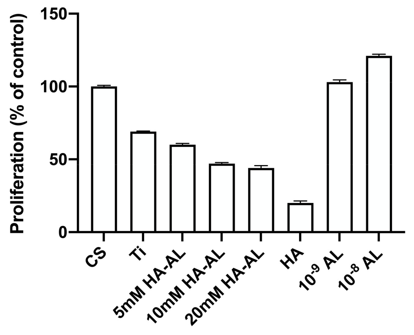
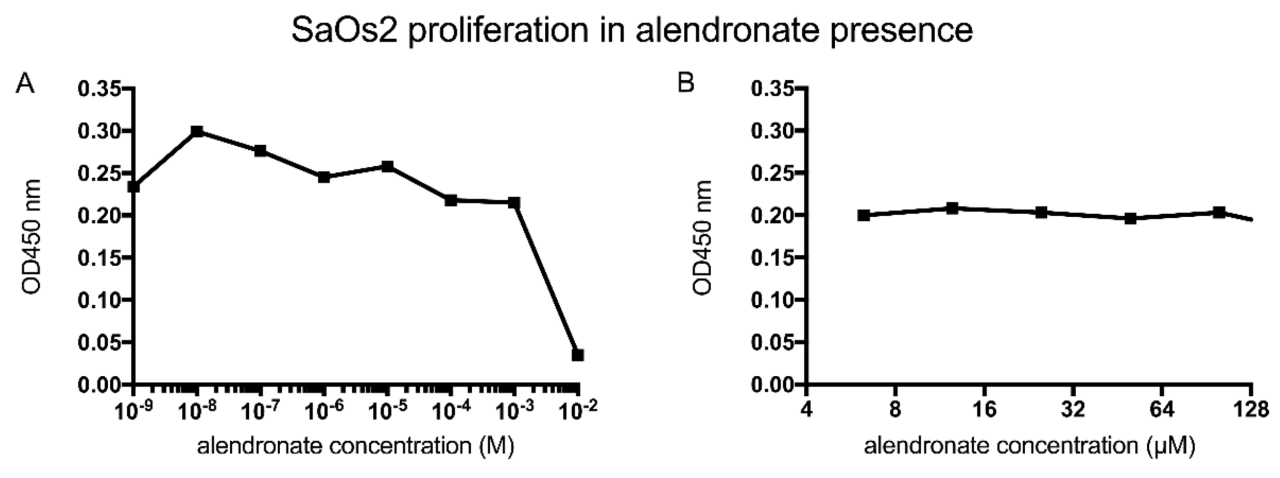
3.2. Testing the Biocompatibility of HA-AL Biomaterials

The synthesized HA powders were evaluated against the commercially available material to establish their effect on the proliferation of SaOs2 cells, an accepted model bone cell line. Thus, the powders were diluted with culture medium and incubated for 72 h. Then, the conditioned medium was harvested and applied onto pre-seeded SaOs2 cells upon dilution with fresh media. Subsequently, the number of viable cells in each condition medium was assessed indirectly by MTS assay.
The results showed a decrease of SaOs2 cells’ proliferation in the medium containing synthesized HA compared to commercial HA, for all dilutions tested (Figure 5).
Hydroxyapatite had a positive effect on proliferation at low concentrations—up to 10% conditioned medium mixed with 90% fresh medium-regardless of the powder’s origin. The synthesized HA had an inhibitory effect, when the concentration of the extract was increased (starting at 25% conditioned medium) while commercial HA did not negatively affect bone cell proliferation.
Alendronate potentiates proliferation at a concentration of 10−8 M (0.0065 µg alendronate in 200 µL—the volume of the well) and at concentrations higher than 10−4 M (6.5 µg alendronate in 200 µL—the volume of the well) it has a toxic effect on bone cells (Figure 6A). Within this range, we further tested a 2-fold dilution of alendronate and determined that for concentrations between 7 and 120 µM there is no negative impact on SaOs2 cell proliferation (Figure 6B). These results are consistent with the release profiles of alendronate, presented in a previous study [22].
A progressive decrease of cell proliferation was observed 72 h upon growing in contact with AL doped hydroxyapatite containing increased concentrations of the drug (Table 2). However, we observed no negative impact of alendronate on cell adhesion as shown by the morphology of actin filaments and the shape and distribution of focal contacts (Figure 7).
Next, the experiments aimed at characterizing the proliferation and adhesion of MSCs upon growing onto HA-AL films deposited by MAPLE. For the proliferation assay, primary cells were cultured for 8 days in contact with the biomaterials, as they have a slower expansion dynamic compared to SaOs2 cells. Results of the MTS assay showed that cells proliferate better on 5 mM HA-AL then on the 20 mM HA-AL and similarly to uncoated Ti (Figure 8). Cells grown with minute concentrations of alendronate (10−9 to 10−8 M) added to the culture medium show similar proliferation to those grown on cover slips (CS).
MSCs adhere and spread onto the HA-AL films as shown by the immunofluorescence microscopy performed after 72 h (Figure 9).

4. Discussion

4.1. Synthesis and Characterization of the Synthesized Hydroxyapatite and HA-AL

Wet precipitation was the method chosen for hydroxyapatite synthesis. This method has several advantages such as a high degree of crystallinity depending on the synthesis conditions [19]. We also had very good results when using the wet precipitation method for the synthesis of hydroxyapatite-ciprofloxacin composites [23].
This method allows the variation of several parameters that may influence morphology, structure and size of the obtained hydroxyapatite crystals. These parameters are synthesis temperature, pH, stirring rate and addition rate of the reactants. In a previous study we determined that the best experimental conditions for the HA-AL 20 mM synthesis were temperature: 90 °C, addition rate: 0.1 mL·min−1, stirring rate: 600 rpm. A composite with a high incorporation efficiency of the drug was subsequently obtained. Furthermore, the hydroxyapatite appeared as a single crystalline phase [24].
The spectral characterization by FTIR analysis confirmed the presence of alendronate for all synthesized HA-AL composites. Furthermore, a well-structured absorption band appears between 3000–3600 cm−1 for all composites, containing a medium intense band at 3565 cm−1, characteristic for the hydroxyl group. Previous studies suggest that there are certain interactions between alendronate and hydroxyapatite that might involve the OH group [19,25].
The structural characterization by X-ray diffraction (XRD) analysis conducted in a previous study confirmed that this method determined the synthesis of a hexagonal-crystallized single-phase hydroxyapatite [19].
Both SEM-EDX and FTIR analyses confirm that the Matrix Assisted Pulsed Laser Evaporation has been successfully employed to deposit thin films of alendronate doped HA on Ti substrates (Figure 4).

4.2. Human Bone Cell Proliferation in the Presence of HA, Alendronate and HA-AL

It was necessary to validate each individual component in order to study the proliferative potential of bone cells in the presence of implant materials. The first test conducted was on synthesized hydroxyapatite (HA) compared to commercial HA (SIGMA).
Synthesized hydroxyapatite, when used at low concentrations, exhibited an effect on cell proliferation similar to commercial hydroxyapatite. Since the aim of the study was to use HA powder in order to obtain nanoscale thin films by matrix assisted pulsed laser evaporation, we considered that the synthesized HA could be used without negative effects caused by high concentrations.
The second test determined the values at which alendronate can be used in implantology without having any side effects on cell proliferation in the injured area.
SaOs2 cells were exposed to serial dilutions of alendronate in growth medium. Their proliferation was quantified 72 h after exposure. The first observation was that alendronate potentiates proliferation at concentrations between 10−9–10−7 M with a high at 10−8 M. At higher alendronate concentrations, proliferation decreases drastically. In the μM range in which the compound is included in the implant coatings, serial dilutions of the compound did not affect the degree of cell proliferation (Figure 6B).
In the next experiment we evaluated the titanium biomaterials coated by 5 mM hydroxyapatite–alendronate composite (HA-AL) and 20 mM HA-AL, respectively. Cell proliferation 72 h after seeding on HA-AL films (Table 2) was lower compared to non-doped HA or uncoated titanium (Ti) and also slightly lower compared with the borosilicate glass control plate (CS = cover slip). This effect may appear due to the concentration of incorporated alendronate but also due to physical characteristics of the coating films.
We performed an immunofluorescence experiment at 72 h after cultivating in order to analyze the effect of the HA-AL composite coatings on cell adhesion. The fluorescence microscopy images show that SaOs2 cells adhere to all four types of tested biomaterials (Figure 7A). Synthesized HA-AL biomaterials show an increase in the number of positive vinculin focal points as compared to HA only coatings (Figure 7B), suggesting a better cell adhesion to the substrate. This is consistent with the results obtained by Boanini et al. that show osteoblasts well attached and spread on the seventh day of culture onto zoledronate-hydroxyapatite coatings [26].

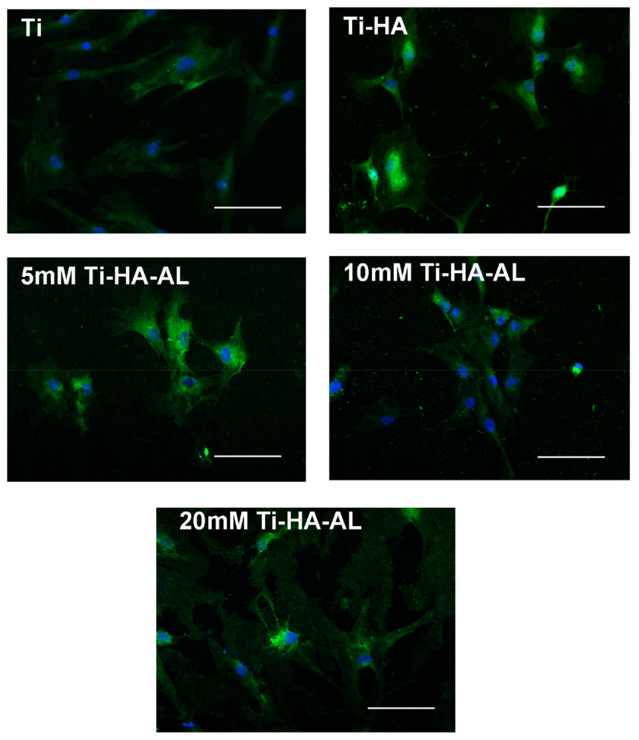
4.3. Human Mesenchymal Stem Cells’ Proliferation on HA-AL Films

During post-trauma bone regeneration, most of the cells recruited to repair the lesion come from the bone marrow—these are the mesenchymal stem cells. They differentiate into osteoblasts and repair the bone by intramembranous ossification under the influence of the microenvironment and due to the inflammatory response at the injured tissue [27].
Human MSCs have a low in vitro proliferation rate. Thus, we chose a longer culture period in order to highlight any differences between the studied materials.
An increase in proliferation due to treatment with 10−8 M alendronate was observed (Figure 8). This is consistent with the previous experiment on SaOs2 cells (Figure 6). Results similar to those obtained on titanium were generated by the samples originating from 5 mM alendronate solutions (Figure 8).
MSC adhesion on biomaterial surface was evaluated by immunofluorescence by labeling actin filaments in order to assay the cell organization on the surface of the implant material at 72 h after attachment (Figure 9). It is known that MSC are morphologically similar to fibroblasts—they are large and elongated cells with sizes that can reach 200 microns [28]. After assessing the images, a decrease of cellular area for both HA and HA-AL samples compared to the uncovered titanium (Figure 9) can be observed. On titanium, the cells have a characteristic elongated appearance. This is probably due to differences in physical features between the surface of the titanium and the coatings.
The best spreading is obtained on 5 mM HA-AL films (Figure 9). This is consistent with the optimal viability obtained at 8 days (Figure 8). The 20 mM HA-AL films show autofluorescence, which makes the morphological analysis more difficult, but there is a clear reshaping of the cells that adopt a stellar form. An improved osteoblast cell adhesion for bisphosphonates containing samples as compared to reference samples (HA) was also obtained by Boanini et al. [26].
All these are evidence of behavioral changes in response to signals received by cells through surface receptors from the materials they have adhered to.

5. Conclusions

The wet precipitation method was selected for the synthesis of hydroxyapatite due to its advantages such as obtaining a compound with a high crystallization rate.
Part of the study was dedicated to the evaluation of the biocompatibility of implant materials coated with alendronate doped hydroxyapatite (HA-AL). Bone cells adhesion to the biomaterials was evaluated using immunofluorescence to label actin filaments. Both osteosarcoma cells and human MSCs adhere to all MAPLE deposited HA thin films. The alendronate included in the films increased the number of cell-biomaterial focal points as compared to HA alone.
Therefore, the HA-AL composite can be considered as a viable solution when including a bisphosphonate in the coating materials of prosthetic metal components.

Author Contributions

All authors have equally contributed to the work reported.

Funding

This research was funded by the grant number 153/26.03.2019.

Conflicts of Interest

The authors declare no conflict of interest. The funders had no role in the design of the study; in the collection, analyses, or interpretation of data; in the writing of the manuscript; or in the decision to publish the results.

References

  1. Jaffe, W.L.; Scott, D.F. Total hip arthroplasty with hydroxyapatite-coated prosthesis. J. Bone Jt. Surg. 1996, 78, A1818–A1934. [Google Scholar] [CrossRef] [PubMed]
  2. Negrilă, C.C.; Predoi, M.V.; Iconaru, S.L.; Predoi, D.L. Development of zinc-doped hydroxyapatite by sol-gel method for medical applications. Molecules 2018, 23, 2986. [Google Scholar] [CrossRef] [PubMed]
  3. Cacciotti, I.; Bianco, A. High thermally stable Mg-substituted tricalcium phosphate via precipitation. Ceram. Int. 2011, 37, 127–137. [Google Scholar] [CrossRef]
  4. Suratwala, S.J.; Cho, S.K.; Van Raalte, J.J.; Park, S.H.; Seo, S.W.; Chang, S.S.; Gardner, T.R.; Lee, F.Y. Enhancement of periprosthetic bone quality with topical hydroxyapatite-bisphosphonate composite. J. Bone Jt. Surg. Am. 2008, 90, 2189–2196. [Google Scholar] [CrossRef] [PubMed]
  5. Puleo, D.A. Encyclopedia of Biomaterials and Biomedical Engineering; Marcel Dekker Inc.: New York, NY, USA, 2004; pp. 190–198. [Google Scholar]
  6. Wilson, C.J.; Clegg, R.E.; Leavesley, D.I.; Pearcy, M.J. Mediation of biomaterial-cell interactions by adsorbed proteins: A review. Tissue Eng. 2005, 11, 1–18. [Google Scholar] [CrossRef]
  7. Knoch, F.; Eckhardt, C.; Alabre, C.I.; Schneider, E.; Rubash, H.E.; Shanbhag, A.S. Anabolic effects of bisphosphonates on peri-implant bone stock. Biomaterials 2007, 28, 3549–3559. [Google Scholar] [CrossRef]
  8. Matthew, T.; Drake, M.D.; Bart, L.; Clarke, M.D.; Sundeep Khosla, M.D. Bisphosphonates: Mechanism of action and role in clinical practice. Mayo Clin. Proc. 2008, 83, 1032–1045. [Google Scholar]
  9. Cavalli, L.; Brandi, M.L. Periprosthetic bone loss: Diagnostic and therapeutic approaches. F1000Research 2013, 2, 266. [Google Scholar] [CrossRef]
  10. Vignoletti, F.; Abrahamsson, I. Quality of reporting of experimental research in implant dentistry. Critical aspects in design, outcome assessment and model validation. J. Clin. Periodontol. 2012, 39, 6–27. [Google Scholar] [CrossRef]
  11. Chai, C.S.; Ben-Nissan, B. Bioactive nanocrystalline sol-gel hydroxyapatite coatings. J. Mater. Sci. 1999, 10, 465–469. [Google Scholar]
  12. Manafi, S.A.; Joughehdoust, S. Synthesis of hydroxyapatite nanostructure by hydrothermal condition for biomedical application. Iran. J. Pharm. Sci. 2009, 5, 89–94. [Google Scholar]
  13. Nayak, A.K. Hydroxyapatite synthesis methodologies: An overview. Int. J. ChemTech Res. 2010, 2, 903–907. [Google Scholar]
  14. Thamaraiselvi, T.V.; Prabakaran, K.; Rajeswari, S. Synthesis of hydroxyapatite that mimics bone mineralogy. Trends Biomater. Artif. Organs 2006, 19, 81–83. [Google Scholar]
  15. Shikhanzadeh, M. Direct formation of nanophase hydroxyapatite on cathodically polarized electrodes. J. Mater. Sci. Mater. Med. 1998, 9, 67–72. [Google Scholar] [CrossRef] [PubMed]
  16. Rodríguez-Lugo, V.; Karthik, T.V.K.; Mendoza-Anaya, D.; Rubio-Rosas, E.; Villaseñor Cerón, L.S.; Reyes-Valderrama, M.I.; Salinas-Rodríguez, E. Wet chemical synthesis of nanocrystalline hydroxyapatite flakes: Effect of pH and sintering temperature on structural and morphological properties. R. Soc. Open Sci. 2018, 5, 180962. [Google Scholar] [CrossRef] [PubMed]
  17. Bubb, D.M.; Wu, P.K.; Horwitz, J.S.; Callahan, J.H.; Galicia, M.; Vertes, A. The effect of the matrix on film properties in matrix-assisted pulsed laser evaporation. J. Appl. Phys. 2002, 91, 2055–2058. [Google Scholar] [CrossRef]
  18. Boanini, E.; Gazzano, M.; Rubini, K.; Bigi, A. Composite nanocrystals provide new insight on alendronate interaction with hydroxyapatite structure. Adv. Mater. 2007, 19, 2499–2502. [Google Scholar] [CrossRef]
  19. Neamtu, J.; Bubulica, M.V.; Rotaru, A.; Ducu, C.; Balosache, O.E.; Manda, V.C.; Turcu-Stiolica, A.; Nicolicescu, C.; Melinte, R.; Popescu, M.; et al. Hydroxyapatite-alendronate composite systems for biocompatible materials. J. Therm. Anal. Calorim. 2017, 127, 1567–1582. [Google Scholar] [CrossRef]
  20. Sima, L.E.; Stan, G.E.; Morosanu, C.O.; Melinescu, A.; Ianculescu, A.; Melinte, R.; Neamtu, J.; Petrescu, S.M. Differentiation of mesenchymal stem cells onto highly adherent radio frequency-sputtered carbonated hydroxylapatite thin films. J. Biomed. Mater. Res. A 2010, 95, 1203–1214. [Google Scholar] [CrossRef]
  21. Meinel, L.; Fajardo, R.; Hofmann, S.; Langer, R.; Chen, J.; Snyder, B. Silk implants for the healing of critical size bone defects. Bone 2005, 37, 688–698. [Google Scholar] [CrossRef]
  22. Rotaru, L.T.; Varut, R.M.; Nicolaescu, O.; Bubulica, M.; Belu, I. In vitro release studies of alendronate from HA-AL composite deposited on titanium metal substrate. J. Sci. Arts 2019, 19, 443–448. [Google Scholar]
  23. Ciocilteu, M.V.; Mocanu, A.G.; Mocanu, A.; Ducu, C.; Nicolaescu, O.E.; Manda, V.C.; Turcu-Stiolica, A.; Nicolicescu, C.; Melinte, R.; Balasoiu, M.; et al. Hydroxyapatite-ciprofloxacin delivery system: Synthesis, characterisation and antibacterial activity. Acta Pharm. 2018, 68, 129–144. [Google Scholar] [CrossRef] [PubMed]
  24. Turcu-Stiolica, A.; Bubulica, M.V.; Nicolaescu, O.E. A design of experiment approach to the synthesis of alendronate-incorporated hydroxyapatite. Rev. Chim. Buchar. 2018, 69, 1944–1948. [Google Scholar]
  25. Capra, P.; Dorati, R.; Colonna, C.; Bruni, G.; Pavanetto, F.; Genta, I.; Conti, B. A preliminary study on the morphological and release properties of hydroxyapatite-alendronate composite materials. J. Microencapsul. 2011, 28, 395–405. [Google Scholar] [CrossRef] [PubMed]
  26. Boanini, E.; Torricelli, P.; Gazzano, M.; Fini, M.; Bigi, A. The effect of zoledronate-hydroxyapatite nanocomposites on osteoclasts and osteoblast-like cells in vitro. Biomaterials 2012, 33, 722–730. [Google Scholar] [CrossRef]
  27. Colnot, C. Skeletal cell fate decisions within periosteum and bone marrow during bone regeneration. J. Bone Miner. Res. 2009, 24, 274–282. [Google Scholar] [CrossRef]
  28. Haasters, F.; Prall, W.C.; Anz, D.; Bourquin, C.; Pautke, C.; Endres, S.; Mutschler, W.; Docheva, D.; Schieker, M. Morphological and immunocytochemical characteristics indicate the yield of early progenitors and represent a quality control for human mesenchymal stem cell culturing. J. Anat. 2009, 214, 759–767. [Google Scholar] [CrossRef]
Figure 1. Synthesis of the hydroxyapatite-alendronate compound.
Coatings 09 00782 g001
Figure 2. Fourier-transform infrared spectroscopy (FT-IR, Thermo Scientific, Waltham, MA, USA) spectrum of HA-AL 5 mM compound (A) and FTIR spectrum of 1:1 alendronate-hydroxyapatite mixture (B).
Coatings 09 00782 g002aCoatings 09 00782 g002b
Figure 3. FTIR Reflection Spectra of MAPLE deposited HA-AL compound (green) versus pure HA (orange) and pure AL (red).
Coatings 09 00782 g003
Figure 4. SEM-EDX ionic distribution of the HA-AL films on titanium: (A) Secondary electron (SE) image; SEM images and X-ray mapping of elemental (B) Carbon (C); (C) Calcium (Ca); (D) Phosphor (P); (E) Titanium (Ti); (F) Nitrogen (N); (G) Sodium (Na).
Coatings 09 00782 g004
Figure 5. Comparative evaluation of human bone cell proliferation in the presence of synthesized HA and commercial HA. Data are presented as mean values ± SD.
Coatings 09 00782 g005
Figure 6. Identification of the range of alendronate concentrations that do not interfere with bone cell proliferation. (A) SaOs2 cells’ proliferation in the presence of a 10-fold dilution series of alendronate. (B) SaOs2 cells’ proliferation in the presence of a 2-fold dilution series extracted from the previous test so as to not decrease the expansion capacity of the cells.
Coatings 09 00782 g006
Figure 7. Immunofluorescence images of SaOs2 cells adhesion to biomaterials: actin (green) was labeled with Alexa Fluor 488 conjugated phalloidin, and vinculin was firstly labeled with primary antibodies and then with secondary antibodies conjugated with Alexa Fluor 594 (red); the nuclei are labeled with DAPI (blue) for cell identification. (A)—merged 3-channel images showing the distribution of cytoskeleton and focal adhesions; (B)—single channel images showing vinculin staining of focal adhesions. Scale bar = 50 μm.
Coatings 09 00782 g007
Figure 8. Evaluation of bone progenitor proliferation on implant grade titanium coated with MAPLE deposited HA-AL films.
Coatings 09 00782 g008
Figure 9. Immunofluorescence images of MSCs adhesion on HA-AL biomaterials: actin (green) was labeled with Alexa Fluor 488 conjugated phalloidin; the nuclei are labeled with DAPI (blue) for cell identification. Scale bar = 100 μm.
Coatings 09 00782 g009
Table 1. Experimental parameters for the HA-AL 20 mM synthesis and the quantity of alendronate contained in the HA-AL composite.
SampleTemperature (oC)Flow (mL/min)Stirring (rpm)pHMaturation (Days)AL (mg) in 5 mg HA-AL (HPLC)Conc. AL (w/w) (%)Incorporation Efficiency (%)Size (nm)
1900.16001000.01510.3029.258565397
Table 2. SaOs2 cells’ proliferation on the surface of HA-AL biomaterials upon 72 h culture.
SamplesHA5 mM HA-AL10 mM HA-ALTiCs
OD 450 nm1.812 ± 0.0041.530 ± 0.0081.421 ± 0.0081.825 ± 0.0031.593 ± 0.006

