Review

Functionalized Textile Based Therapy for the Treatment of Atopic Dermatitis

Institute of Textiles and Clothing, The Hong Kong Polytechnic University, Hung Hom, Kowloon, Hong Kong, China
*
Author to whom correspondence should be addressed.
Coatings 2017, 7(6), 82; https://doi.org/10.3390/coatings7060082
Submission received: 8 May 2017 / Revised: 9 June 2017 / Accepted: 12 June 2017 / Published: 17 June 2017
(This article belongs to the Special Issue Fabric Coatings)

Abstract

:
Atopic dermatitis (AD) is a common chronic inflammatory skin condition characterized by intense puritus and skin dryness. The pathogenesis for AD has not been fully understood to date. Complementary therapies are very popular as effective treatment for AD among clinical practitioners. This study presents a comprehensive review of published works associated with textiles-based complementary therapies for AD treatment such as wet-wrap dressing, functionalized textiles, and the application of hydrogel techniques in the textile industry to provide a better understanding of the development and design of new textiles-based transdermal therapies.

1. Introduction

Atopic dermatitis (AD), also termed eczema, is a common chronic inflammatory skin condition characterized by intense puritus and a waxing and waning course [1,2]. The term ‘dermatitis’ was derived from the Greek ‘derma’, which means skin, and ‘itis’ means inflammation, whereas eczema came from the Greek ‘ekzema’ with the meaning of ‘to boil over’ and refers to acute manifestation of this disease. However, dermatitis and eczema are often used synonymously [3].
Children are the most vulnerable group to suffer from AD. The typical onset of this condition generally occurs in children under five years of age, but the majority of patients have a spontaneous remission before adolescence [4]. The prevalence of this condition has increased steadily worldwide over the past few decades, with an estimated morbidity of 15% to 30% of affected adolescents, especially in industrialized countries [2,5]. AD imposes a great influence on the quality of life of children and their parents and causes significant social, economic, and health care costs [6].
The clinical manifestation of AD varies widely in individuals and generally presents intense pruritus, dryness, lichenification and excoriation, macular erythema, raised bumps, secondary infection, etc. [7,8]. However, the distribution of eczematous symptoms changes with age [7]. Patients in infancy commonly experience lesion on the cheeks, forehead, and scalp. Children may suffer severe eruptions on the elbow and knee flexures, the sides of the neck, the wrists, and the ankles [8]. In adolescence, lichenified plaques usually emerge on the flexures, head, and neck. Each stage is characterized by intense pruritus that continues throughout the day and worsens at night, thereby leading to sleep loss and impairing the patient’s quality of life [2].
To date, there is no consensus on the pathogenesis for AD, but generally it is agreed that this disorder results from a complex interaction between genetically mediated epidermal barrier dysfunction and immunologic disturbance [5,9]. Strictly speaking, this is true for extrinsic AD (IgE-high AD) with considerable evidence involving barrier conditions and immunodeficiency, whereas the causation for intrinsic AD (IgE-normal AD) remains elusive [10]. The epidermal barrier dysfunction typically results from genetic mutations in the filaggrin, which is an epidermal protein that acts as a waterproof mortar between keratinocytes in the outermost layer of the skin [1]. This facilitates the invasion of environmental allergens and induces allergic responses to external antigens, resulting in elevated serum IgE. This provides an explanation for extrinsic AD characterized by the barrier dysfunction of the skin and chronic inflammation. Conversely, intrinsic AD, which is non-allergic, exhibits normal serum IgE values and the normal skin barrier function [10]. Moreover, another difference between extrinsic and intrinsic AD resides in the different degrees of sensitization to antigens. Extrinsic AD patients are predisposed to being sensitized to protein antigens, while nonprotein antigens such as haptens and metals are typically seen in intrinsic AD patients. However, Bieber [2] suggested that the absence of IgE-mediated sensitization for intrinsic AD patients may be a transient factor and proposed a unified hypothesis. This hypothesis holds that the development of AD has three phases, and nonatopic intrinsic AD is the initial phase with a high prevalence among infants during which no sensitization emerges. Subsequently, a transition from nonatopic dermatitis to true AD occurs, during which the majority of AD patients suffer from IgE-mediated sensitization to food and/or environmental allergens. Meanwhile, scratching causes cellular damage and thus induces IgE autoantibodies, thereby exacerbating the inflammation [11].
A recent study has reported a new mechanism that links cutaneous inflammation and epidermal barrier dysfunction and that potentially unifies the conflicting outside-in and inside-out hypothesis of AD, i.e., inhabitation of phospholipase A2 (PLA2) [12]. The house dust mite-derived PLA2 was found to generate neolipid antigens in human skin, which contributes to inflammation. The inflammation is controlled and eliminated through inhibition of PLA2 by the skin barrier protein filaggrin. Thus PLA2 links skin inflammation and barrier dysfunction and provides a therapeutic approach to treat AD.

2. Textiles-Based Complementary Therapy

The treatment of AD is still a clinical challenge, and so far it is still not completely curable. Basically, the treatment regime focuses on relieving the symptoms and preventing acute exacerbations as well as improving cosmetic appearance to enhance quality of life [13]. It is important to address the skin barrier defect with the avoidance of specific and nonspecific trigger factors and to maintain skin hydration with regular use of emollients [7]. Further treatment depends on the severity of AD, including a combination of multiple therapeutic agents in a step-wise fashion. Topical or systemic corticosteroids are still considered the mainstay therapy for the treatment of moderate-to-severe AD. They are normally used as anti-inflammatory medications to treat AD in any stage of inflammation and to reduce itching [14]. However, long-term use and the overdosage of corticosteroids may cause a series of adverse effects, including atrophy, hypopigmentation, striae distensae, and skin infections [15]. Therefore, the use of corticosteroids is not highly recommended, especially for younger children. A wide variety of complementary therapies such as traditional Chinese medicine [16], wet wrap dressing [17], and a wide variety of functional textiles [18] have thus been advocated and developed. However, the present study only focuses on textiles-based therapies for AD treatment and makes an elaborate review of the development and application of textile-based therapy.

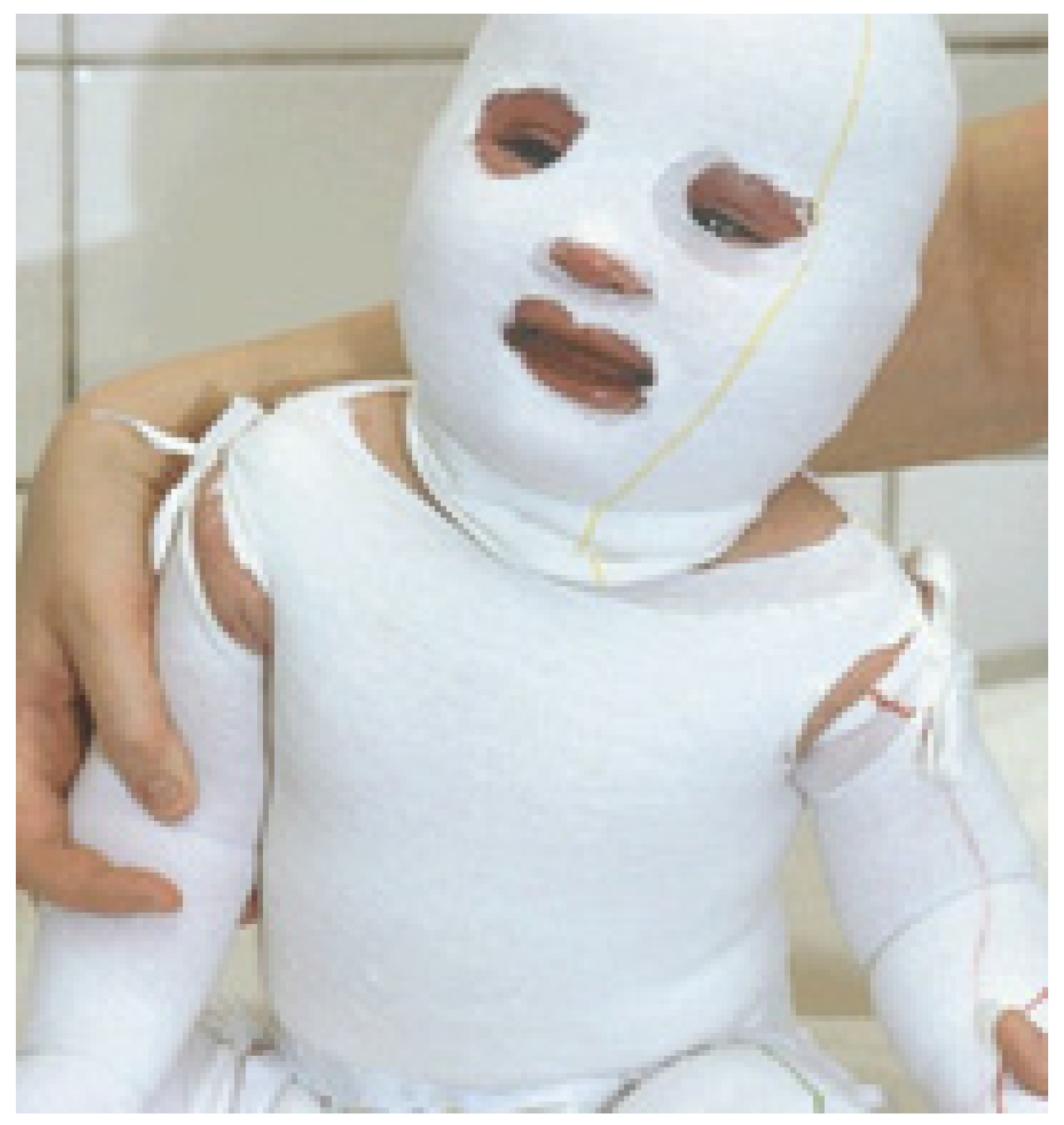
2.1. Wet-Wrap Dressing

A wet-wrap dressing is a double layer of tubular bandage or gauze consisting of a first moist inner layer and second dry outer layer and involves the application of a topical medication (Figure 1) [14,19]. The use of dampened bandages to reduce pruritus and inflammation is an ancient medical remedy. Their use was described in an ancient surgical textbook by Liston in 1846 [20], but their use for AD treatment was first reported in 1987 by Nicol and his colleagues [21]. Subsequently, wet-wrap dressings attracted widespread attention among clinicians and practitioners [22].
In 1991, Goodyear and co-workers reported that wet-wrap dressings are an efficacious therapy to treat AD [23]. Since then, numerous researchers have reported successful outcomes of the treatment of AD using wet-wrap dressing [22,24,25]. Researchers from Mayo Clinic reported that 239 patients out of 266 hospitalizations saw a significant improvement in pruritus and the severity of AD after using wet-wrap therapy in conjunction with topical corticosteroid or emollient cream [17]. A study conducted by Janmohamed et al. demonstrated that wet wrap therapy with diluted corticosteroids showed obvious therapeutic effect on severe AD [26]. Currently, wet-wrap dressing is primarily considered a second-line treatment recommended for severe or refractory AD, and no precise instructions exist. The effectiveness differs and depends on the condition, the topical drugs used, the time of occlusion, and the duration of treatment [14].
It is generally assumed that wet-wrap dressing is associated with a cooling effect [17]. The cooling effect of a wet-wrap dressing may decrease skin temperature and cause vasoconstriction, thereby relieving the inflammation and pruritus. Sleep disturbance is thus controlled with the pruritus alleviation. Wet-wrap dressing not only helps to rehydrate the skin and enhances the absorption of the topical medication, but can also provide a physical barrier against scratching and hasten the restoration of an impaired epidermal function [4].
To conclude, wet-wrap dressing shows several advantages for the treatment of AD, including increasing skin hydration, promoting penetration of medication, impeding scratching, and improving sleep disturbance. The disadvantages, however, should not be ignored. The most dangerous adverse effect is the systemic absorption of topical steroid, with a transient increase in cortisol levels. Wet-wrap dressing may also cause skin maceration, secondary infections, and folliculitis [27,28]. Additionally, wet-wrap dressing is a short-term therapy; the dressings easily get dried out and must be continuously dampened. This often increases the burden of caregivers and families [19].

2.2. Healthcare Textiles

Apart from antimicrobial textiles as aforementioned, some novel coated fabrics for the treatment of AD patients have also been reported in recent years [29,30]. These fabrics were functionalized with healthcare merits by coating cotton fabrics with various finishing agents such as anion-generating agents and borage oil [30,31].
Anion-generating textiles are designed to develop high value-added textiles and have been extensively studied in the past few decades [32,33,34]. This concept was developed based on the fact that negative ions can exert beneficial effects on human health [35]. Researchers were thus inspired to design and develop a type of healthcare textile that can generate negative ions [33,36]. Currently, the development of anion textiles has made great progress, and related products are commercially available [37]. Tourmaline, which can generate negative ions and electromagnetic radiation in the far-infrared region by mechanical forces such as rubbing and vibrating, is the most commonly used anion-generating agent for the development of functionalized fibers [38,39]. Kim and his colleagues first performed a clinical trial to employ anion textile, produced by polyester filaments impregnated with nano-sized fine-crusted tourmaline powder, for the treatment of AD [29] (Figure 2). They found that such textiles helped to significantly improve the transepidermal water loss, skin erythema, and hydration of AD patients and might provide a therapeutic option for them. It should be noted that, however, the textile material, polyester, may exacerbate the severity of AD and thus it has yet to be further investigated.
Borage oil used for AD treatment was proposed on a hypothesis that patients with AD generally suffer from metabolic aberration of linoleic acid to gamma-linolenic acid (GLA) caused by inactivation of the delta-6 desaturase enzyme [40,41]. Borage oil has been extensively studied as a complementary therapy in AD treatment due to its abundant content of GLA [42,43,44]. Kanehara and co-workers developed a borage oil-coated fabric and examined its clinical efficacy in AD treatment [30]. This coated fabric was produced by chemical bonding formed between borage oil and cotton fibers. The borage oil could be gradually released from the cotton fibers and absorbed by the skin. Clinical trials demonstrated that the symptoms of erythema and itch were alleviated, and the transepidermal water loss (TEWL) was also controlled. Nevertheless, Foster et al. suggested that borage oil, whether administered orally or topically, did not have a significant therapeutic effect but may be useful in some individual patients with less severe AD [45]. This conclusion was drawn on the basis of objective analysis on typical clinical cases and is thus credible and acceptable. Also, this viewpoint was consistent with the analysis of Bamford et al. [44].

2.3. Medical Textiles

Medical textiles are the products and constructions used for medical and biological applications such as first aid, clinical, and hygienic purposes. Textiles act as a good carrier for microorganisms, e.g., mould and fungi, which may cause deterioration of fibers, strength loss, discoloration, and even potential health risks, e.g., skin diseases [46,47]. With the growing public awareness, antimicrobial properties have attracted increasing attention among practitioners and have been successfully imparted to textiles to improve the resilience against microorganisms [47,48,49]. The detrimental effects caused by colonization of microorganisms can be controlled by durable antimicrobial finishing using antimicrobial agents [50], including metals and metal salts [47,51], quaternary ammonium salts [52], chitosan [53], triclosan [46], and natural dyestuffs [54,55]. Currently, antimicrobials have been playing an increasingly important role in addressing textile hygiene in clinical and sensitive environments.

2.3.1. Antibacterial Agents for AD Treatment

The development of antibacterial textiles has received extensive attention, and applications in AD treatment have also been reported [18,56]. This proposal was developed on the basis of the fact that a majority of AD patients suffer from recurrent bacterial infections, primarily caused by the colonization of Staphylococcus aureus (S. aureus) [10]. Moreover, the colonization of S. aureus is generally correlated with the severity of AD [57]. The reason may consist in the immunomodulatory toxins with superantigens properties secreted by S. aureus, which can stimulate theactivation of T cells and macrophages and thus induce skin inflammation and exacerbate AD [58]. Therefore, antimicrobial textiles may be a desirable alternative therapy to treat AD by killing S. aureus colonization. Clinically, some metal nanoparticles and organic antibacterial agents have been reported to develop antibacterial textiles for AD treatment primarily due to low toxicity and good compliance (Table 1).

Metal Nanoparticles

Metal nanostructured materials, including titania nanoparticles, silver nanoparticles, zinc oxide nanoparticles, and copper nanoparticles, have attracted tremendous attention among researchers due to their good antimicrobial activities for the development of antimicrobial textiles (Table 1) [47,68]. Among these, since ancient times silver has been the most commonly used antimicrobial agent to fight against infections and prevent spoilage. Silver shows low toxicity to humans, is effective against a broad spectrum of microorganisms, shows high efficiency, and has an ‘oligodynamic’ nature [46,69]. The forms of application of silver are diverse, including various oxidation states (Ag+, Ag2+, and Ag3+) and nanoparticles which act as sources of silver atoms [48,70]. It has been proposed that the antibacterial mechanism seemingly results from the release of silver ions, which pass through the cell membrane of bacteria, causing structural changes in the cell walls by binding to negatively charged components in proteins and nucleic acids leading to proteins inactivation, metabolism disturbance, and growth inhibition, up to the cell’s death [46,47].
The applications of metal nanoparticles generally fall into two categories: (1) inorganic nanoparticles and their nanocomposites; and (2) inorganic nanoparticles loaded organic carriers. The strategies for the fabrication of antimicrobial textiles include conventional exhaust, padding, spraying, pad-dry-cure process, and sol-gel techniques [47,68,71]. Additionally, surface modification using various methods provides a promising approach to developing antimicrobial textiles. In order to improve the durability against domestic laundering and dry cleaning, polymeric binders such as acrylic resin and polysiloxane, are commonly used for the fixation of metal nanoparticles onto the fabrics [47].

Organic Antibacterial Agents

Two representative organic antibacterial agents for the development of antibacterial textiles in AD treatment are citric acid and citrus grandis osbeck extract. The application of citric acid for the development of antimicrobial textiles was inspired by the concept of the ‘acid mantle’, which is formed when sweat and sebum combine and act as a barrier to bacteria, viruses, and other skin contaminants [65]. This concept has led to the development of acidic emollients for skin care and disposable materials with infection control functions such as disposable gowns and masks [65,72]. The antibacterial property of citric acid coated textiles has been fully elucidated [73,74]. Jaeger and co-workers first investigated the application of citric acid-coated textiles on AD patients and reported their research results in 2014 [65]. In their study, a citric acid-coated textile with a pH value of 5.5–6.5 was fabricated by esterification reaction, leading to the formation of a very thin citric acid layer on the surface of cellulose fibers. Clinical trials showed that this kind of textile was well tolerated and had therapeutic effects on AD. It is thus promising as a novel therapeutic strategy for AD. Further studies are required to provide more evidence for clinical practice.
Additionally, the selection of suitable fabric materials is also of high importance to AD subjects because the fabric materials may be a cause of triggering or worsening the lesions. Natural fibers such as cotton, silk, and viscose are preferential owing to their hygienic properties [56]. Lyocell also provides a well-established and promising choice for the development of antimicrobial textiles [75].
Yi et al. first introduced a dyeing method to develop the antibacterial textiles for AD treatment [66]. The dyeability and antibacterial activities of citrus grandis osbeck extract against Staphylococcus aureus were fully investigated. The optimal dye condition was determined. They further studied the clinical efficacy of cotton underclothes dyed with the citrus grandis osbeck extract in AD treatment by in vivo methodology [67]. It was observed that the dyed fabrics could alleviate the severity of AD and decrease the SCORAD (Scoring Atopic Dermatitis) index value and thus could be utilized for AD treatment as a complementary therapy.

2.3.2. Herbal Medicine Coated Textile Therapy for AD Treatment

Traditional Chinese medicine (TCM) is one of the oldest healing systems and has been applied for thousands of years for a wide variety of clinical treatments of different types of diseases and symptoms in China [76,77]. In terms of AD treatment, an increasing number of studies have been reported to be associated with TCM owing to its minimal adverse effects and ease of application [78,79,80]. Numerous herbal medicines (Table 2) have been shown to be efficacious and beneficial in controlling the severity of AD and improving the quality of life of patients despite the dispute over the efficacy and standardization of herbal medicines. Table 2 presents several typical prescriptions with traditional herbal medicines used in Asian countries.
In clinical practice, Hui and coworkers recently developed a textile therapy functionalized with TCM for AD treatment using microencapsulation technology [90,91]. The fabrication strategy was an emulsion chemical crosslinking method (Figure 3A), by which the herbal medicines (PentaHerbs formula and cortex moutan) were successfully loaded into chitosan and sodium alginate composite microcapsules. The resultant microcapsules were fully evaluated in terms of surface morphology, particle size and distribution, in vitro drug release behavior, and cytotoxicity testing. A pad-dry-cure approach was then employed to coat the microcapsules on the surface of cotton fabrics (Figure 3B). The drug loaded textiles were proven to show excellent controlled drug release properties, with a release duration of seven days [91]. However, there was a great loss of major active ingredients of the herbal medicine in the fabrication of the microcapsule, resulting in both low drug loading and encapsulation efficiency. Clinical trials demonstrated that the medical textile therapy failed to treat AD patients [90,91].
Considering the skin dryness of AD patients, a dual-functional medical textile therapy, i.e., both moisture and drug supply, was developed by Wang and coworkers using hydrogel technology (Figure 4) [92]. Poloxamer-based hydrogel was fabricated by a ‘cold method’, through which the loss of herbal medicine was avoided, and sufficient moisture was incorporated into the hydrogel. The physiological property of poloxamers is closely associated with the molecular weight, the ratio of poly(ethylene oxide)/poly(propylene oxide) (PEO/PPO) moieties, and the concentration. The amphiphilic property stems from the hydrophilic ethylene oxide moiety and the hydrophobic propylene oxide moiety [93]. Consequently, the poloxamer solution can exhibit a reversible phase transition from sol to gel (low temperature sol gel boundary) and from gel to sol (high temperature boundary) as the temperature monotonically increases (Figure 4B) [94]. The characteristic temperature responsive property makes poloxamer-based hydrogel an attractive alternative in the development of transdermal therapies [95]. Moreover, the herbal medicine (cortex moutan extract) loaded in the hydrogel showed excellent controlled release and percutaneous diffusional behavior. Clinical trials indicated that herbal medicine loaded poloxamer-based hydrogel can moisturize skin and relieve the symptoms of AD, thus providing a promising alternative for the treatment of AD. In spite of this, further studies are still required to optimize the drug loading hydrogel fabrication and coating process for promoting medical textile therapy production in a large scale and standardization approach.
The gelling process for poloxamer-based hydrogel typically consists of two steps, i.e., a micellization process (formation of spherical micelles) and a gelation process (stacking of spherical micelles) (Figure 5) [96,97,98]. The micellization process of PEO-PPO-PEO copolymers is endothermic and driven by a decrease in the polarity of ethylene oxide (EO) and propylene oxide (PO) segments as the temperature increases and by the entropy gain in water when unimers aggregate to form micelles (hydrophobic effect) [98,99].

3. Fixation Strategies of Antibacterial Agents on Fabrics

Textiles are an excellent carrier of pharmaceuticals through contact with the skin and have found applications for medical, hygienic, and health purposes [100]. The incorporation of antibacterial agents or clinical drugs into textiles is a critical step to fabricate such functionalized textiles. A wide variety of fixation approaches have been proposed for the coating of different antibiotic drugs onto the fabrics. Basically, the fixation approaches can be divided into three categories: (1) inclusion inside the fiber; (2) coating outside the fabrics by textile finishing techniques such as exhaust and pad-dry-cure; and (3) application of encapsulation techniques such as micro- or nano-capsule technology, cyclodextrin, and hydrogel technology.

3.1. Inclusive Method

The inclusive method refers to natural, organic, or inorganic additives incorporated into fibers prior to extrusion, which allows the production of different functional fibers for a wide variety of purposes [38,101]. This strategy is mainly applicable for the synthesis polymeric fibers. Electrospinning is the most commonly used method to blend active agents into the fibers. Two typical agents that have been blended into fibers for AD treatment are tourmaline and silver. Tijing et al. developed tourmaline-decorated polyurethane composite nanofibers using an electrospinning technique, which showed excellent superhydrophilic and antibacterial properties [38]. An important step to achieve well-dispersed tourmaline nanoparticles in polyurethane nanofibers was separate fabrication of tourmaline and polyurethane solutions. The tourmaline containing composite fiber developed by Lin et al. using melt spinning can generate anion and present outstanding permeability and mechanical properties [37,101]. Clinical trials conducted by Kim and coworkers showed that tourmaline anion textile may significantly relieve the symptoms and produce positive results in the treatment of AD patients [29].
Silver loaded seaweed-based (alginic acid) cellulosic fiber has been made commercially available by Smartfiber AG (SeaCell® Active, Rudolstadt, Germany) [102]. Amine oxide and water are required to separate the regenerated (alginic acid) cellulose directly, and the fiber is then produced by adding finely ground seaweed into the spinning solution during the spinning process (Figure 6). The cellulose solvent amine oxide is N-methylmorpholine-N-oxide (NMMO) monohydrate (NMMO·H2O, 13.3% water w/w) [103,104]. The dissolution and regeneration processes are resulted from the hydrogen-bonding characteristics of the hygroscopic NMMO. In these processes, NMMO and water can be almost completely recycled, with a recovery ratio of more than 99% [104]. Therefore, it is an eco-friendly and pollution-free strategy to fabricate functional cellulose fibers.
The homogeneous mixing of organic and inorganic additives in the fibers make it possible to produce various functional materials on this common cellulose base [105,106]. Due to the excellent capability of absorbing the minerals of seaweed, inorganic antibacterial agents such as nano-silver and nano-zinc particles can be absorbed in the core of the fully formed cellulosic fiber during the activation of the fibers [102,107]. This process is totally different from the commonly used method of mixing the additives in the spinning solution. It is generally agreed that the metals are bound via the carboxyl and hydroxyl groups of the cellulose as well as of the seaweed [108,109]. Fluhr and coworkers clinically demonstrated that this sliver-loaded seaweed fiber is safe and applicable in AD treatment according to the positive experimental results [75]. However, extensive follow-up studies are still required to confirm the clinical efficacy for the treatment of AD.
Smartfiber AG has also introduced a seaweed-based cellulosic regenerated fiber containing zinc (Smartcel Sensitive™, Rudolstadt, Germany) in recent years (Figure 7) [106,110]. Zinc is blended in the fiber in the form of high quality zinc oxide, which is a natural and medically approved white zinc with revitalizing effects and hygienic properties [111]. This zinc loaded seaweed-based fiber gives relief to people with sensitive skin and allows faster skin regeneration and complication-free wound healing. A clinical trial conducted by Wiegand and his colleagues demonstrated that this functionalized textile can significantly reduce AD severity and pruritus and improve the subjective sleep quality of AD patients [110]. Moreover, their study also concluded that this zinc loaded textile was well tolerated by AD patients and had good biocompatibility. Hence, this functional textile may be a promising alternative as a complementary therapy for AD treatment.

3.2. Textile Finishing Approaches

The inclusive method can provide good durability and a considerably slow release of active agents. Nonetheless, this approach is both time-consuming and sophisticated. On the contrary, conventional textile finishing methods such as pad-dry-cure process (Figure 8) are the most commonly used strategies for antibacterial agents coating on textiles [51,112]. However, a challenge for the textile finishing consists in the relatively poor durability. A wide variety of methods have been employed to improve the durability of the textile finishing, e.g., treating the fiber with resin, condensates, or crosslinking agents, chemical modification of the fiber by covalent bond formation, the use of graft polymerization or copolymerization, and nanotechnology [18,47].

3.3. Encapsulation Techniques

3.3.1. Micro/Nano-Capsules
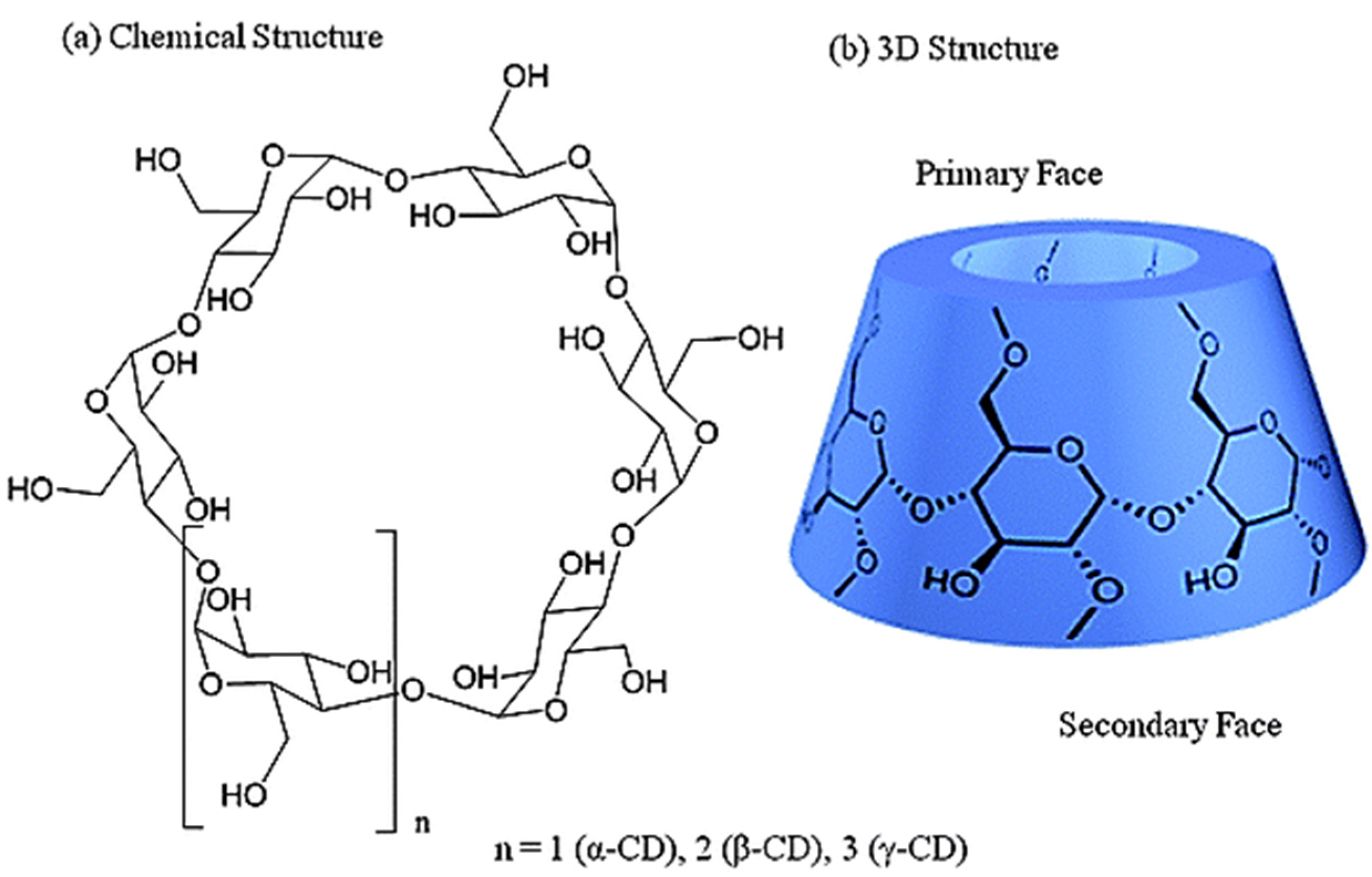
The microencapsulation process refers to the encapsulation of a substance into the membrane of polymers on the microscopic scale [113,114]. With the concept of microencapsulation, it should be possible to encapsulate a wide variety of substances in the core of microcapsules using different fabrication methods [115]. Accordingly, the fabrics can be designed and produced for tailored functions when coated by the desired microcapsules [116,117]. In this aspect, numerous examples have been described with regard to conferring textiles with various functions using microencapsulation technology, including durable fragrances, skin softeners, insect repellents, antimicrobials, phase change materials, and special medical applications [118,119,120,121]. In terms of AD treatment, Hui et al. reported a textile based therapy functionalized with herbal medicine loaded microcapsules [90,91,122]. The herbal medicine was encapsulated within polymer capsules and then coated on the fabrics. However, the clinical efficacy of this microcapsule coated textile based therapy has not been reported.

3.3.2. Cyclodextrin

Cyclodextrins (CDs) are non-reducing oligosaccharides in a cyclic form produced through the enzymatic degradation of starch [68,123]. Three cyclodextrins, i.e., α-, β-, and γ-CD, can be obtained by the separation method using different precipitation agents such as n-octanol and toluene, among which β-CD is the most common and commercially available type due to the ease of synthesis and the reasonable price [68,124]. These cyclic oligosaccharides are arranged in a truncated-cone shape with a hydrophilic outer surface and an internal hydrophobic hollow cavity (Figure 9) [123]. The nature of the hydrophilic exterior and hydrophobic interior of the cavity imparts CDs with the remarkable ability to entrap a large variety of hydrophobic active ingredients, which explains why β-CD has been widely applied in textile field [68,125,126].
CDs can be grafted on the fabrics via two approaches, i.e., the physical method by immsersing the fabric in CDs solution and the chemical method through the formation of chemical bonds, which each present different finishing effects [124]. CDs provide an admirable carrier to develop antibacterial and medical textiles. The commonly used antibacterial agents, including triclosan, ciprofloxacin, and even silver, have been successfully coated on the fabric through the formation of inclusion complexes with CDs [68,124,127]. However, as far as AD treatment is concerned, there are still no publications involving CDs functionalized textiles. In terms of treating allergic diseases, Radu et al. reported an enlightening study that prepared antiallergic pajamas grafted with β-CD and with antiallergic active principles absorbed in the cavity [128]. However, the wearing comfort and softness of the fabric were greatly affected by β-CD.

3.3.3. Hydrogel Technology

Application of Hydrogel in Functional Textiles

Hydrogel refers to a class of three-dimensional cross-linked polymeric networks with the ability to absorb large quantities of water or biological fluids within the bulk structure while maintaining the dimensional stability [129]. The amount of water absorbed may vary from around 10% to thousands of times the weight of the dry polymeric network, which may also be influenced by the capillary effect and osmotic pressure [130,131]. The water absorbing capacity makes hydrogels suitable for widespread applications in a variety of fields, including tissue engineering [132], the biomedical and pharmaceutical industry [133], and smart textiles [134]. Additionally, hydrogels also constitute an excellent alternative drug carrier.
Textiles functionalized with hydrogel have been extensively exploited over the past few decades [135,136]. Hydrogel can endow textiles, including cotton, polyester, silk, and hemp, with novel functionalities such as aesthetic appeal, healthcare, and smart wetting properties [135]. The hydrogels applied to textiles are generally accomplished using coating technology such as graft polymerization, the sol gel technique, and finishing [137].
Poly (N-isopropyl acrylamide) (PNIPAAm) is a thermo-responsive polymer which shows hydrophilic-hydrophobic transition in aqueous solution at around 32–34 °C and has been extensively studied for textile use (Figure 10) [134,135,138]. The textiles can be functionalized with excellent moisture management properties. Jiang and co-workers developed a functionalized cotton fabric coated with PNIPAAm via atom transfer radical polymerization [139]. This fabric exhibited an excellent switchability from superhydrophilicity to superhydrophobicity with changing temperature. Yang and colleagues further studied PNIPAAm-coated cotton fabric, which showed an extraordinary capability for collecting water from humid air [134]. The application of PNIPAAm combined with chitosan can make textiles with thermal/pH dual-responsive properties [140]. This dual-responsive function can also be achieved by incorporating pH-dependent monomer acrylic acid (AAc) into NIPAAm [141]. Likewise, thermal/light dual-responsive textiles can also be developed by copolymerizing a light-responsive azobenzene moiety into NIPAAm [142]. More recently, Schiphorst et al. reported a novel thermal/light dual-responsive cotton fabric functionalized with a surface-grafted spiropyran-NIPAAm hydrogel. This dual-responsive fabric was shown to be capable of dimensional changes upon irradiation with visible light or a temperature stimulus and may find application in breathable textiles and agricultural purposes with moisture collection capacity [136].
Smart hydrogel can also be employed to develop functional textiles with healthcare functions such as moisturizing, whitening, wound healing, and even anti-ageing effects on human skin [135]. Liu et al. offered poly (N-isopropyl acrylamide)/polyurethane (PNIPAAm/PU) grafted nonwoven fabric with antibacterial function against S. aureus and E. coli via chitosan modification [143]. In this context, Ribeiro and coworkers investigated the applicability of chitosan hydrogel dressing in the re-establishment of skin lesions [144]. Recently, Wang et al. developed a skin moistening functional textile by coating thermos-responsive poly (ethylene glycol)-poly(-caprolactone)-poly(ethylene glycol) (PEG-PCL-PEG) hydrogel [145]. The drug loaded hydrogel makes the functional textiles attractive in the development of skin care and wound treatment products. Actually, drugs or nutrients loaded in hydrogels are administered through the transdermal delivery mode.
Currently, numerous studies have been reported involving the application of hydrogels as a drug carrier for a transdermal drug delivery system [146,147]. For example, a transdermal therapy, desonide hydrogel, consisting of methylparaben and Carbopol 981 was approved for AD treatment by the Food and Drug Administration (FDA) in 2006 [148,149]. This hydrogel therapy was shown to be effective in improving the quality life of AD patients [149]. Recently, a microemulsion based hydrogel loaded with benzocaine was constructed for dermal purposes [150]. Nayak and colleagues reported carboxymethyl cellulose/gelatin copolymer hydrogel containing lidocaine as a transdermal delivery [151]. The percutaneous behavior could greatly be enhanced with a microneedle and an ultrasound. In this regard, poloxamer 407, which is a thermosensitive amphiphilic block copolymer, has found extensive applications as a drug carrier matrix in the development of transdermal delivery systems [152].

Hydrogel Based Transdermal Therapy

Transdermal therapy, i.e., transdermal drug delivery systems, refers to the self-contained discrete dosage forms from which drugs can penetrate across the skin and into the systemic circulation [153,154]. Transdermal therapy encompasses a wide range of non-invasive or minimally invasive technologies for the delivery of drugs across the skin without needles and thus constitutes an attractive alternative to oral administration and hypodermic injection [153,155]. Since it was first approved for clinical use by the FDA in 1979, transdermal therapy has seen rapid progress due to its favorable advantages, including high patient compliance, enhanced therapeutic efficiency by avoiding the peaks and troughs, and avoidance of the hepatic first-pass and gastrointestinal metabolism [154,155,156]. Currently, transdermal drug systems have greatly progressed from the first-generation system in which only the small, lipophilic, and low-dose drugs could be delivered to the third-generation enhancement strategies by which the delivery of macromolecules such as insulin and the influenza vaccine is achievable [155].
Two major components of conventional transdermal formulations are the targeted drugs and drug reservoirs, which usually are plasters, creams, and ointments [154,157]. Currently, conventional transdermal formulations are being gradually replaced by a wide range of novel hydrogel formulations such as semisolid and film-based systems in order to minimize skin irritation, promote dosage flexibility, enhance patient acceptability, and improve ease of use [147]. The forms of hydrogel applied for transdermal formulations include semisolid matrix, patch, nanogel, emulgel, and combined use with vesicular systems such as liposomes and transfersomes [158,159,160,161,162].
Hydrogels are polymeric networks with three-dimensional configuration that can absorb large quantities of water or biological fluids [146]. They are composed of homopolymers or copolymers and are insoluble in water due to the presence of chemical or physical crosslinks. The relatively high moisture content facilitates control of the transepidermal water loss and enhances skin hydration and elasticity, thereby providing a better feel when applied to the skin [147,163]. Therefore, hydrogel offers an ideal alternative for the development of novel transdermal formulations, especially for the treatment of skin-related disorders [131,164]. In this respect, the well-known polymeric materials for hydrogel formulations include polysaccharides (starch, cellulose and its derivatives, chitosan, alginate, hyaluronate), proteins (collagens, gelatins, caseins, albumins), and synthetic polymers (polyvinyl alcohol, polyvinylpyrrolidone, polyethylene glycol, and polyacrylates) [165,166]. For example, Schwartz et al. investigated the topical treatment of cutaneous leishmaniasis using diselenide-loaded chitosan hydrogel formulation [167]. Kwon and coworkers achieved a positive result in the feasibility study of pH-sensitive hydroxyethyl cellulose/hyaluronic acid complex hydrogel containing isoliquiritigenin in the treatment of skin lesions caused by pH imbalances [168]. A study conducted by Heilmann and colleagues demonstrated that the thermosensitive poloxamer 407 hydrogel is an appropriate carrier formulation for the topical application of morphine in large-scale skin wound treatment [158].
Nanogels, i.e., hydrogel nanoparticles, refer to the aqueous dispersion of hydrogel particles formed by physically or chemically crosslinked polymer networks of nanoscale size [169]. They are swollen nanosized networks composed of hydrophilic or amphiphilic polymer chains. The water absorbing ability, high loading property, and high stability make nanogels an ideal candidate for biomedical applications to load therapeutic agents ranging from small molecular drugs to macromolecular proteins, peptides, and vaccines [170]. The narrow nanosized distribution endows nanogels with the ability to reach the smallest capillary vessels and penetrate across the skin tissues with longer drug release duration when applied for transdermal therapy [170,171]. Moreover, the high mobility of hydrogel nanoparticles in the small capillaries facilitates efficient uptake [172]. This is of high importance for the development of transdermal drug systems to overcome the barrier effect of the skin. Currently, nanogels have been fabricated via a variety of strategies, including layer-by-layer self-assembly, homogeneous polymerization, heterogeneous polymerization, and temperature-assisted nanofabrication [169,170,171].
Emulgel is also a promising drug delivery system, which was developed to transport hydrophobic drugs for transdermal purposes [173,174]. Emulgels, i.e., emulsified hydrogel, are formed by the gelling of emulsions, either oil-in-water or water-in-oil type, with a gelling agent [175,176]. Therefore, emulgel possesses the properties of both emulsions and gel, such as thixotropic behavior, greaseless, easily speadable and removable, non-staining, and biocompatible [160]. These properties make emulgel an ideal candidate for dermatological use. Currently, some preparations of emulgels have been made commercially available in markets [160]. Moreover, the droplet size of emulsions may range from around tens of nanometers to about hundreds of nanometers. Emulgel can be classified into macroemulsion gel (100–400 nm), microemulsion gel (10–100 nm), and nanoemulsion gel (about 10 nm) [150,173]. The nanoscale emulsion facilitates penetration across the skin, which makes emulgel more appropriate for transdermal use.
Hydrogel can be also used in combination with vesicular systems to form hydrogel/vesicular delivery systems [177]. The development of hydrogel/vesicular systems was intended to achieve increased skin penetration of drugs [178]. Vesicular systems are nanocarriers such as liposomes, niosomes, and transfersomes and exhibit excellent transdermal penetration properties [178,179]. Likewise, the resulting drug delivery systems can be endowed with the properties of both hydrogel and the nanocarriers and have been applied in transdermal therapy [161].

4. Conclusions and Future Outlook

To conclude, AD is a common chronic inflammatory skin condition characterized by intense puritus and a waxing and waning course in children. Inasmuch as the pathogenesis for AD is partially understood, there is still no definitive cure to date and the treatment is empirical. With respect to complementary therapy for AD treatment, textiles-based treatment approaches are fully reviewed to provide a better understanding of the development of the new therapy.
Hydrogels are hydrophilic three-dimensional polymeric networks capable of absorbing a large amount of water or biological fluids. The excellent biocompatibility and drug loading capacity of hydrogels implies a broad opportunity for biomedical applications. Poloxamer-based hydrogel, which seems a promising candidate for application as a drug carrier system for transdermal purposes, is centralized.

Acknowledgments

We gratefully acknowledge financial support from So Tung Lam, Henry Ng, and T.K. Tsui.

Conflicts of Interest

The authors declare that there are no conflicts of interest. The authors are responsible for the content of this paper.

References

  1. Wolter, S.; Price, H.N. Atopic dermatitis. Pediatr. Clin. N. Am. 2014, 61, 241–260. [Google Scholar] [CrossRef] [PubMed]
  2. Bieber, T. Mechanisms of disease: Atopic dermatitis. N. Engl. J. Med. 2008, 358, 1483–1494. [Google Scholar] [CrossRef] [PubMed]
  3. Thomsen, S.F. Atopic dermatitis: Natural history, diagnosis, and treatment. ISRN Allergy 2014, 2014, 1–7. [Google Scholar] [CrossRef] [PubMed]
  4. Shekariah, T.; Kalavala, M.; Alfaham, M. Atopic dermatitis in children: A practical approach. Paediatr. Child Health 2011, 21, 112–118. [Google Scholar] [CrossRef]
  5. Tsianakas, A.; Ständer, S. Dupilumab: A milestone in the treatment of atopic dermatitis. Lancet 2016, 387, 4–5. [Google Scholar] [CrossRef]
  6. Lee, S.J.; Woo, S.I.; Ahn, S.H.; Lim, D.K.; Hong, J.Y.; Park, J.H.; Lim, J.; Kim, M.K.; Kwon, S.W. Functional interpretation of metabolomics data as a new method for predicting long-term side effects: Treatment of atopic dermatitis in infants. Sci. Rep. 2014, 4, 7408. [Google Scholar] [CrossRef] [PubMed]
  7. Akdis, C.A.; Akdis, M.; Bieber, T.; Bindslev-Jensen, C.; Boguniewicz, M.; Eigenmann, P.; Hamid, Q.; Kapp, A.; Leung, D.Y.; Platts-Mills, T.A.; et al. Diagnosis and treatment of atopic dermatitis in children and adults. Allergy 2006, 61, 969–987. [Google Scholar] [CrossRef] [PubMed]
  8. Chan, S.K.; Burrows, N.P. Atopic dermatitis. Common Dermatoses 2009, 5, 242–245. [Google Scholar] [CrossRef]
  9. Watson, W.; Kapur, S. Atopic dermatitis. Allergy Asthma Clin. Immunol. 2011, 7. [Google Scholar] [CrossRef] [PubMed]
  10. Tokura, Y. Atopic Dermatitis: Common Extrinsic and Enigmatic Intrinsic Types. In Immunology of the Skin: Basic and Clinical Sciences in Skin Immune Responses; Kabashima, K., Ed.; Springer: Tokyo, Japan, 2016. [Google Scholar]
  11. Tang, T.S.; Bieber, T.; Williams, H.C. Does “autoreactivity” play a role in atopic dermatitis? J. Allergy Clin. Immun. 2012, 129, 1209–1215. [Google Scholar] [CrossRef] [PubMed]
  12. Jarrett, R.; Salio, M.; Lloyd-Lavery, A.; Subramaniam, S.; Bourgeois, E.; Archer, C.; Cheung, K.L.; Hardman, C.; Chandler, D.; Salimi, M.; et al. Filaggrin inhibits generation of CD1a neolipid antigens by house dust mite-derived phospholipase. Sci. Transl. Med. 2016, 8, 1–12. [Google Scholar] [CrossRef] [PubMed]
  13. Kessel, J.; Goldenberg, G. Therapies to Improve the Cosmetic Symptoms of Atopic Dermatitis. Cutis 2016, 97, 183–186. [Google Scholar] [PubMed]
  14. Galli, E.; Neri, I.; Ricci, G.; Baldo, E.; Barone, M.; Fortina, A.B.; Bernardini, R.; Berti, I.; Caffarelli, C.; Calamelli, E. Consensus Conference on Clinical Management of pediatric Atopic Dermatitis. Ital. J. Pediatr. 2016, 42, 1–26. [Google Scholar] [CrossRef] [PubMed]
  15. Farhi, D.; Taieb, A.; Tilles, G.; Wallach, D. The historical basis of a misconception leading to undertreating atopic dermatitis (eczema): Facts and controversies. Clin. Dermatol. 2010, 28, 45–51. [Google Scholar] [CrossRef] [PubMed]
  16. Hon, K.L.; Chan, B.C.; Leung, P.C. Chinese herbal medicine research in eczema treatment. Chin. Med. 2011, 6, 17–24. [Google Scholar] [CrossRef] [PubMed]
  17. Dabade, T.S.; Davis, D.M.; Wetter, D.A.; Hand, J.L.; McEvoy, M.T.; Pittelkow, M.R.; Azhary, R.A.; Davis, M.D. Wet dressing therapy in conjunction with topical corticosteroids is effective for rapid control of severe pediatric atopic dermatitis: Experience with 218 patients over 30 years at Mayo Clinic. J. Am. Acad. Dermatol. 2012, 67, 100–106. [Google Scholar] [CrossRef] [PubMed]
  18. Buschmann, H.J.; Dehabadi, V.A.; Wiegand, C. Medical, cosmetic and odour resistant finishes for textiles. In Functional Finishes for Textiles: Improving Comfort, Performance and Protection; Roshan, P., Ed.; Woodhead Publishing: Cambridge, UK, 2014; pp. 303–330. [Google Scholar]
  19. Oranje, A.P.; Devillers, A.C.; Kunz, B.; Jones, S.L.; DeRaeve, L.; Van Gysel, D.; de Waard-van der Spek, F.B.; Grimalt, R.; Torrelo, A.; Stevens, J.; et al. Treatment of patients with atopic dermatitis using wet-wrap dressings with diluted steroids and/or emollients. An expert panel's opinion and review of the literature. J. Eur. Acad. Dermatol. Venereol. 2006, 20, 1277–1286. [Google Scholar] [CrossRef] [PubMed]
  20. Elliott, I.M.; Elliott, J.R. A Short History of Surgical Dressings; Pharmaceutical Press: London, UK, 1964; pp. 1–118. [Google Scholar]
  21. Nicol, N.H. Atopic dermatitis: The (wet) wrap-up. Am. J. Nurs. 1987, 87, 1560–1563. [Google Scholar] [CrossRef] [PubMed]
  22. Bingham, L.G.; Noble, J.W.; Davis, M.D. Wet dressings used with topical corticosteroids for pruritic dermatoses: A retrospective study. J. Am. Acad. Dermatol. 2009, 60, 792–800. [Google Scholar] [CrossRef] [PubMed]
  23. Goodyear, H.M.; Harper, J.I. Wet-wrap dressings for the treatment of atopic eczema in children. Br. J. Dermatol. 1991, 125, 604. [Google Scholar] [CrossRef] [PubMed]
  24. Oranje, A.P.; Verbeek, R.; Verzaal, P.; Haspels, I.; Prens, E.; Nagelkerken, L. Wet-wrap treatment using dilutions of tacrolimus ointment and fluticasone propionate cream in human APOC1 (+/+) mice with atopic dermatitis. Br. J. Dermatol. 2009, 160, 54–61. [Google Scholar] [CrossRef] [PubMed]
  25. Nicol, N.H.; Boguniewicz, M.; Strand, M.; Klinnert, M.D. Wet wrap therapy in children with moderate to severe atopic dermatitis in a multidisciplinary treatment program. J. Allergy Clin. Immunol. Pract. 2014, 2, 400–406. [Google Scholar] [CrossRef] [PubMed]
  26. Janmohamed, S.R.; Oranje, A.P.; Devillers, A.C.; Rizopoulos, D.; van Praag, M.C.; Van Gysel, D.; Goeteyn, M. The proactive wet-wrap method with diluted corticosteroids versus emollients in children with atopic dermatitis: A prospective, randomized, double-blind, placebo-controlled trial. J. Am. Acad. Dermatol. 2014, 70, 1076–1082. [Google Scholar] [CrossRef] [PubMed]
  27. Lee, J.H.; Lee, S.J.; Kim, D.; Bang, D. The effect of wet-wrap dressing on epidermal barrier in patients with atopic dermatitis. J. Eur. Acad. Dermatol. Venereol. 2007, 21, 1360–1368. [Google Scholar] [CrossRef] [PubMed]
  28. Segre, J.A. Epidermal barrier formation and recovery in skin disorders. J. Clin. Investig. 2006, 116, 1150–1158. [Google Scholar] [CrossRef] [PubMed]
  29. Kim, S.H.; Hwang, S.H.; Hong, S.K.; Seo, J.K.; Sung, H.S.; Park, S.W.; Shin, J.H. The clinical efficacy, safety and functionality of anion textile in the treatment of atopic dermatitis. Ann. Dermatol. 2012, 24, 438–443. [Google Scholar] [CrossRef] [PubMed]
  30. Kanehara, S.; Ohtani, T.; Uede, K.; Furukawa, F. Clinical effects of undershirts coated with borage oil on children with atopic dermatitis: A double-blind, placebo-controlled clinical trial. J. Dermatol. 2007, 34, 811–815. [Google Scholar] [CrossRef] [PubMed]
  31. Wakamura, T.; Sato, M.; Sato, A.; Dohi, T.; Ozaki, K.; Asou, N.; Hagata, S.; Tokura, H. A preliminary study on influence of negative air ions generated from pajamas on core body temperature and salivary IgA during night sleep. Int. J. Occup. Med. Environ. 2004, 17, 295–298. [Google Scholar]
  32. Yang, M.X.; Pu, D.D. Research on Influencing Factors and Exploitation of Negative-ion Textiles. Prog. Text. Sci. Technol. 2009, 2, 10–11. [Google Scholar]
  33. Zheng, Q.S.; Hu, X.-Y. The Performance and Application of Yanniansu (Tourmaline) Fiber. Synth. Fiber China 2010, 1, 11–13. [Google Scholar]
  34. Lin, M.F.; Chen, H.J.; Chen, C.J.; Chen, Y.C.; Chen, C.M. Natural Tourmaline Anion Fiber and Filter and Producing Method. U.S. Patent 20,070,259,178, 8 November 2007. [Google Scholar]
  35. Dover, J.S.; Phillips, T.J.; Arndt, K.A. Cutaneous effects and therapeutic uses of heat with emphasis on infrared radiation. J. Am. Acad. Dermatol. 1989, 20, 278–286. [Google Scholar] [CrossRef]
  36. Sugihara, T.; Suzuki, M.; Komiya, M.M. Rayon fiber containing tourmaline particles and method for the preparation thereof. U.S. Patent 5,863,653 A, 26 January 1999. [Google Scholar]
  37. Lin, M.F.; Chen, H.J.; Chen, C.J.; Chen, Y.C.; Chen, C.M. Natural Tourmaline Anion Fiber and Filter and Production Method. U.S. Patent 8,231,968 B2, 31 July 2012. [Google Scholar]
  38. Tijing, L.D.; Ruelo, M.T.G.; Amarjargal, A.; Pant, H.R.; Park, C.-H.; Kim, D.W.; Kim, C.S. Antibacterial and superhydrophilic electrospun polyurethane nanocomposite fibers containing tourmaline nanoparticles. Chem. Eng. J. 2012, 197, 41–48. [Google Scholar] [CrossRef]
  39. Yeh, J.T.; Hsiung, H.H.; Wei, W.; Zhu, P.; Chen, K.N.; Jiang, T. Negative air ion releasing properties of tourmaline/bamboo charcoal compounds containing ethylene propylene diene terpolymer/polypropylene composites. J. Appl. Polym. Sci. 2009, 113, 1097–1110. [Google Scholar] [CrossRef]
  40. Melnik, B.; Plewig, G. Are disturbances of omega-6-fatty acid metabolism involved in the pathogenesis of atopic dermatitis? Acta Derm. 1992, 176, 77–85. [Google Scholar]
  41. Diezel, W.; Schulz, E.; Shanks, M.; Heise, H. Plant oils: Topical application and anti-inflammatory effects (Croton oil test). Dermatologische Monatsschrift 1993, 179, 173–176. [Google Scholar]
  42. Al-Khamees, W.A.; Schwartz, M.D.; Alrashdi, S.; Algren, A.D.; Morgan, B.W. Status epilepticus associated with borage oil ingestion. J. Med. Toxicol. 2011, 7, 154–157. [Google Scholar] [CrossRef] [PubMed]
  43. Ernst, E. The usage of complementary therapies by dermatological patients: A systemic review. Br. J. Dermatol. 2000, 142, 857–861. [Google Scholar] [CrossRef] [PubMed]
  44. Bamford, J.; Ray, S.; Musekiwa, A.; van Gool, C.; Humphreys, R.; Ernst, E. Oral evening primrose oil and borage oil for eczema. Cochrane Database Syst. Rev. 2013, 30, CD004416. [Google Scholar]
  45. Foster, R.H.; Hardy, G.; Alany, R.G. Borage oil in the treatment of atopic dermatitis. Nutrition 2010, 26, 708–718. [Google Scholar] [CrossRef] [PubMed]
  46. Dhiman, G.; Chakraborty, J.N. Antimicrobial performance of cotton finished with triclosan, silver and chitosan. Fash. Text. 2015, 2. [Google Scholar] [CrossRef]
  47. Dastjerdi, R.; Montazer, M. A review on the application of inorganic nano-structured materials in the modification of textiles: Focus on anti-microbial properties. Colloids Surf. B 2010, 79, 5–18. [Google Scholar] [CrossRef] [PubMed]
  48. Yuan, G.; Cranston, R. Recent Advances in Antimicrobial Treatments of Textiles. Text. Res. J. 2008, 78, 60–72. [Google Scholar] [CrossRef]
  49. Windler, L.; Height, M.; Nowack, B. Comparative evaluation of antimicrobials for textile applications. Environ. Int. 2013, 53, 62–73. [Google Scholar] [CrossRef] [PubMed]
  50. Simoncic, B.; Tomsic, B. Structures of Novel Antimicrobial Agents for Textiles—A Review. Text. Res. J. 2010, 80, 1721–1737. [Google Scholar] [CrossRef]
  51. Hebeish, A.; El-Naggar, M.E.; Fouda, M.M.G.; Ramadan, M.A.; Al-Deyab, S.S.; El-Rafie, M.H. Highly effective antibacterial textiles containing green synthesized silver nanoparticles. Carbohydr. Polym. 2011, 86, 936–940. [Google Scholar] [CrossRef]
  52. Xue, Y.; Xiao, H.; Zhang, Y. Antimicrobial polymeric materials with quaternary ammonium and phosphonium salts. Int. J. Mol. Sci. 2015, 16, 3626–3655. [Google Scholar] [CrossRef] [PubMed]
  53. Tayel, A.A.; Moussa, S.H.; El-Tras, W.F.; Elguindy, N.M.; Opwis, K. Antimicrobial textile treated with chitosan from Aspergillus niger mycelial waste. Int. J. Biol. Macromol. 2011, 49, 241–245. [Google Scholar] [CrossRef] [PubMed]
  54. Khan, S.A.; Ahmad, A.; Khan, M.I.; Yusuf, M.; Shahid, M.; Manzoor, N.; Mohammad, F. Antimicrobial activity of wool yarn dyed with Rheum emodi L. (Indian Rhubarb). Dyes Pigments 2012, 95, 206–214. [Google Scholar] [CrossRef]
  55. Majhi, B.; Satapathy, K.B.; Mishra, S.K.; Mohapatra, A. Studies on antimicrobial effect of natural dyes and pigments obtained from the leaf of Artocarpus heterophyllus Lam. and Tectona grandis L.F. Int. J. Phytomed. 2016, 7, 396–401. [Google Scholar]
  56. Haug, S.; Roll, A.; Schmid-Grendelmeier, P.; Johansen, P.; Wüthrich, B.; Kündig, T.; Senti, G. Coated textiles in the treatment of atopic dermatitis. In Biofunctional, Textiles and the Skin; Hipler, U.C., Elsner, P., Eds.; Karger Publishers: Karger, Basel, 2006; Volume 33, pp. 144–151. [Google Scholar]
  57. He, Z.P.; Fu, G.W.; Geng, Y.F. Test and evaluation of negative ions textiles. Text. Dyeing Finish. J. 2013, 35, 6–10. [Google Scholar]
  58. Leung, D.Y.; Boguniewicz, M.; Howell, M.D.; Nomura, I.; Hamid, Q.A. New insights into atopic dermatitis. J. Clin. Investig. 2004, 113, 651–657. [Google Scholar] [CrossRef] [PubMed]
  59. Kale, R.; Meena, C.R. Synthesis of Titanium dioxide Nanoparticles and Application on Nylon fabric using Layer by Layer technique for Antimicrobial Property. Adv. Appl. Sci. Res. 2012, 3, 3073–3080. [Google Scholar]
  60. Dubas, S.T.; Kumlangdudsana, P.; Potiyaraj, P. Layer-by-layer deposition of antimicrobial silver nanoparticles on textile fibers. Colloid Surf. A 2006, 289, 105–109. [Google Scholar] [CrossRef]
  61. Ashayer-Soltani, R.; Hunt, C.; Thomas, O. Fabrication of highly conductive stretchable textile with silver nanoparticles. Text. Res. J. 2016, 86, 1041–1049. [Google Scholar] [CrossRef]
  62. Ditaranto, N.; Picca, R.; Sportelli, M.; Sabbatini, L.; Cioffi, N. Surface characterization of textiles modified by copper and zinc oxide nano-antimicrobials. Surf. Interface Anal. 2016, 48, 505–508. [Google Scholar] [CrossRef]
  63. Gedanken, A.; Nitzan, Y.; Perelshtein, I.; Perkas, N.; Applerot, G. Sonochemical Coating of Textiles with Metal Oxide Nanoparticles for Antimicrobial Fabrics. Google Patents EP2294260 A1, 16 March 2011. [Google Scholar]
  64. Sójka-Ledakowicz, J.; Chruściel, J.; Kudzin, M.H.; Łatwińska, M.; Kiwała, M. Antimicrobial functionalization of textile materials with copper silicate. Fibres Text. East. Eur. 2016, 5, 151–156. [Google Scholar] [CrossRef]
  65. Jaeger, T.; Rothmaier, M.; Zander, H.; Ring, J.; Gutermuth, J.; Anliker, M.D. Acid-coated Textiles (pH 5.5–6.5)—A New Therapeutic Strategy for Atopic Eczema? Acta Derm. Venereol. 2015, 95, 659–663. [Google Scholar] [CrossRef] [PubMed]
  66. Eunjou, Y.; Yoo, E.S. A novel bioactive fabric dyed with unripe Citrus grandis Osbeck extract part 1: Dyeing properties and antimicrobial activity on cotton knit fabrics. Text. Res. J. 2010, 80, 2117–2123. [Google Scholar] [CrossRef]
  67. Eunjou, Y.; Hong, J.Y.; Yoo, E.S. A novel bioactive fabric dyed with unripe Citrus grandis Osbeck extract part 2: Effects of the Citrus extract and dyed fabric on skin irritancy and atopic dermatitis. Text. Res. J. 2010, 80, 2124–2131. [Google Scholar] [CrossRef]
  68. Radu, C.-D.; Parteni, O.; Ochiuz, L. Applications of cyclodextrins in medical textiles—Review. J. Control. Release 2016, 224, 146–157. [Google Scholar] [CrossRef] [PubMed]
  69. Emam, H.E.; Manian, A.P.; Široká, B.; Duelli, H.; Redl, B.; Pipal, A.; Bechtold, T. Treatments to impart antimicrobial activity to clothing and household cellulosic-textiles—Why “Nano”-silver? J. Clean. Prod. 2013, 39, 17–23. [Google Scholar] [CrossRef]
  70. Budama, L.; Çakır, B.A.; Topel, Ö.; Hoda, N. A new strategy for producing antibacterial textile surfaces using silver nanoparticles. Chem. Eng. J. 2013, 228, 489–495. [Google Scholar] [CrossRef]
  71. Balakumaran, M.; Ramachandran, R.; Jagadeeswari, S.; Kalaichelvan, P. In vitro biological properties and characterization of nanosilver coated cotton fabrics—An application for antimicrobial textile finishing. Int. Biodeterior. Biodegrad. 2016, 107, 48–55. [Google Scholar] [CrossRef]
  72. Schmid, M.H.; Korting, H.C. The concept of the acid mantle of the skin: Its relevance for the choice of skin cleansers. Dermatology 1995, 191, 276–280. [Google Scholar] [CrossRef] [PubMed]
  73. Bischof Vukušić, S.; Flinčec Grgac, S.; Budimir, A.; Kalenić, S. Cotton textiles modified with citric acid as efficient anti-bacterial agent for prevention of nosocomial infections. Croat. Med. J. 2011, 52, 68–75. [Google Scholar] [CrossRef] [PubMed]
  74. Chung, Y.-S.; Lee, K.-K.; Kim, J.-W. Durable press and antimicrobial finishing of cotton fabrics with a citric acid and chitosan treatment. Text. Res. J. 1998, 68, 772–775. [Google Scholar] [CrossRef]
  75. Fluhr, J.W.; Breternitz, M.; Kowatzki, D.; Bauer, A.; Bossert, J.; Elsner, P.; Hipler, U.C. Silver-loaded seaweed-based cellulosic fiber improves epidermal skin physiology in atopic dermatitis: Safety assessment, mode of action and controlled, randomized single-blinded exploratory in vivo study. Exp. Dermatol. 2010, 19, e9–e15. [Google Scholar] [CrossRef] [PubMed]
  76. Tang, J.L.; Liu, B.Y.; Ma, K.W. Traditional Chinese medicine. Lancet 2008, 372, 1938–1940. [Google Scholar] [CrossRef]
  77. Xu, J.; Yang, Y. Traditional Chinese medicine in the Chinese health care system. Health Policy 2009, 90, 133–139. [Google Scholar] [CrossRef] [PubMed]
  78. Zhang, W.; Leonard, T.; Bath-Hextall, F.; Chambers, C.; Lee, C.; Humphreys, R.; Williams, H. Chinese herbal medicine for atopic eczema. Cochrane Database Syst. Rev. 2013, 9, CD002291. [Google Scholar]
  79. Tan, H.Y.; Zhang, A.L.; Chen, D.; Xue, C.C.; Lenon, G.B. Chinese herbal medicine for atopic dermatitis: A systematic review. J. Am. Acad. Dermatol. 2013, 69, 295–304. [Google Scholar] [CrossRef] [PubMed]
  80. Yun, Y.; Lee, S.; Kim, S.; Choi, I. Inpatient treatment for severe atopic dermatitis in a Traditional Korean Medicine hospital: Introduction and retrospective chart review. Complement. Ther. Med. 2013, 21, 200–206. [Google Scholar] [CrossRef] [PubMed]
  81. Gu, S.; Yang, A.; Xue, C.; Li, C.; Pang, C.; Zhang, W.; Williams, H. Chinese herbal medicine for atopic eczema. Cochrane Database Syst. Rev. 2013, 10, CD008642. [Google Scholar]
  82. Akamatsu, H.; Asada, Y.; Horio, T. Inhibitory effect of shofu-san, a Japanese kampo medicine, on neutrophil functions in vitro. Am. J. Chin. Med. 1998, 26, 57–64. [Google Scholar] [CrossRef] [PubMed]
  83. Hon, K.; Leung, T.; Wong, Y.; Lam, W.; Guan, D.; Ma, K.; Sung, Y.; Fok, T.; Leung, P. A pentaherbs capsule as a treatment option for atopic dermatitis in children: An open-labeled case series. Am. J. Chin. Med. 2004, 32, 941–950. [Google Scholar] [CrossRef] [PubMed]
  84. Hon, K.L.; Leung, T.F.; Ng, P.C.; Lam, M.C.; Kam, W.Y.; Wong, K.Y.; Lee, K.C.; Sung, Y.T.; Cheng, K.F.; Fok, T.F.; et al. Efficacy and tolerability of a Chinese herbal medicine concoction for treatment of atopic dermatitis: A randomized, double-blind, placebo-controlled study. Br. J. Dermatol. 2007, 157, 357–363. [Google Scholar] [CrossRef] [PubMed]
  85. Shimizu, T. Efficacy of kampo medicine in treating atopic dermatitis: An overview. Evid. Based Complement. Altern. Med. 2013, 2013, 260235. [Google Scholar] [CrossRef] [PubMed]
  86. Yanagi, Y.; Yasuda, M.; Hashida, K.; Kadokura, Y.; Yamamoto, T.; Suzaki, H. Mechanism of salivary secretion enhancement by Byakkokaninjinto. Biol. Pharm. Bull. 2008, 31, 431–435. [Google Scholar] [CrossRef] [PubMed]
  87. Saiki, I. A Kampo medicine “Juzen-taiho-to”—Prevention of malignant progression and metastasis of tumor cells and the mechanism of action. Biol. Pharm. Bull. 2000, 23, 677–688. [Google Scholar] [CrossRef] [PubMed]
  88. Nozaki, K.; Hikiami, H.; Goto, H.; Nakagawa, T.; Shibahara, N.; Shimada, Y. Keishibukuryogan (gui-zhi-fu-ling-wan), a Kampo formula, decreases disease activity and soluble vascular adhesion molecule-1 in patients with rheumatoid arthritis. Evid. Based Complement. Altern. Med. 2006, 3, 359–364. [Google Scholar] [CrossRef] [PubMed]
  89. Hwang, J.; Kim, J.; Yu, Y.; Im, S. Modulation of experimental atopic dermatitis by topical application of Gami-Cheongyeul-Sodok-Eum. BMC Complement. Altern. Med. 2013, 13, 312–322. [Google Scholar] [CrossRef] [PubMed]
  90. Hui, P.C.-L.; Wang, W.-Y.; Kan, C.-W.; Ng, F.S.-F.; Zhou, C.-E.; Wat, E.; Zhang, V.X.; Chan, C.-L.; Lau, C.B.-S.; Leung, P.-C. Preparation and characterization of chitosan/sodium alginate (CSA) microcapsule containing Cortex Moutan. Colloid Surf. A 2013, 434, 95–101. [Google Scholar] [CrossRef]
  91. Hui, P.C.; Wang, W.Y.; Kan, C.W.; Ng, F.S.; Wat, E.; Zhang, V.X.; Chan, C.L.; Lau, C.B.; Leung, P.C. Microencapsulation of Traditional Chinese Herbs-PentaHerbs extracts and potential application in healthcare textiles. Colloids Surf. B 2013, 111, 156–161. [Google Scholar] [CrossRef] [PubMed]
  92. Wang, W.; Wat, E.; Hui, P.C.L.; Chan, B.; Ng, F.S.F.; Kan, C.W.; Wang, X.; Hu, H.; Wong, E.C.W.; Lau, C.B.S.; et al. Dual-functional transdermal drug delivery system with controllable drug loading based on thermosensitive poloxamer hydrogel for atopic dermatitis treatment. Sci. Rep. 2016, 6, 24112. [Google Scholar] [CrossRef] [PubMed]
  93. Klouda, L.; Mikos, A.G. Thermoresponsive hydrogels in biomedical applications. Eur. J. Pharm. Biopharm. 2008, 68, 34–45. [Google Scholar] [CrossRef] [PubMed]
  94. Jeong, B.; Kim, S.W.; Bae, Y.H. Thermosensitive sol–gel reversible hydrogels. Adv. Drug Deliv. Rev. 2002, 54, 37–51. [Google Scholar] [CrossRef]
  95. Chen, C.C.; Fang, C.L.; Al-Suwayeh, S.A.; Leu, Y.L.; Fang, J.Y. Transdermal delivery of selegiline from alginate-Pluronic composite thermogels. Int. J. Pharm. 2011, 415, 119–128. [Google Scholar] [CrossRef] [PubMed]
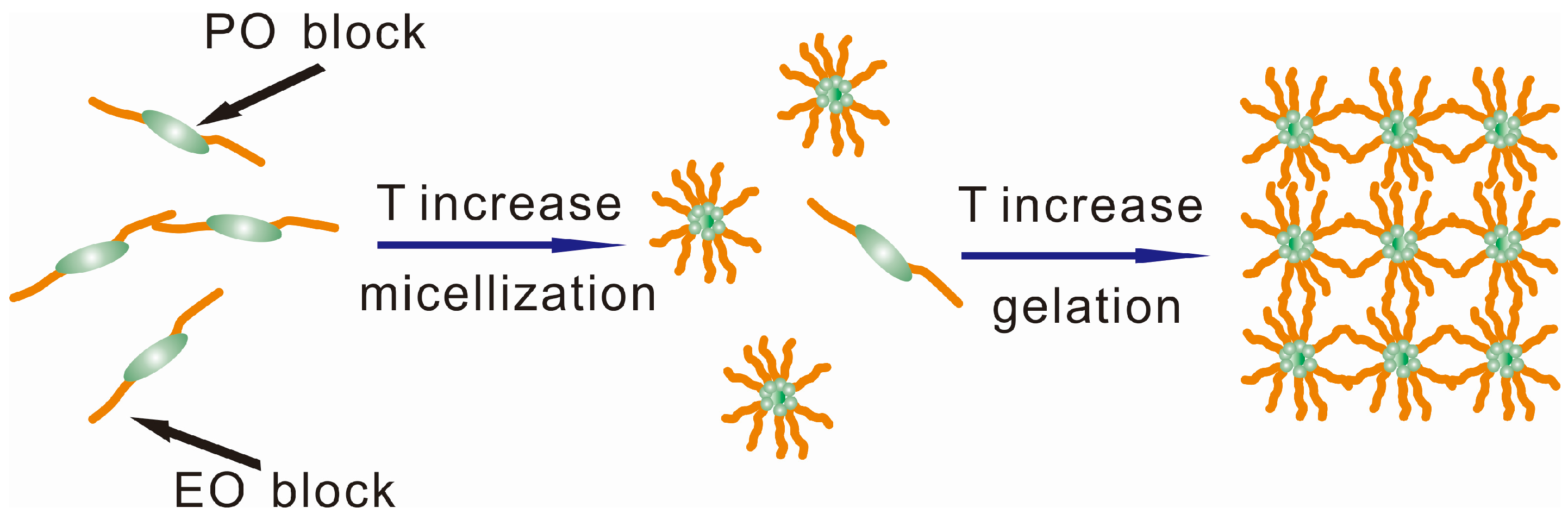
  96. Jia, L.; Guo, C.; Yang, L.; Xiang, J.; Tang, Y.; Liu, C.; Liu, H. Mechanism of PEO–PPO–PEO micellization in aqueous solutions studied by two-dimensional correlation FTIR spectroscopy. J. Colloid Interface Sci. 2010, 345, 332–337. [Google Scholar] [CrossRef] [PubMed]
  97. Jiang, J.; Malal, R.; Li, C.; Lin, M.Y.; Colby, R.H.; Gersappe, D.; Rafailovich, M.H.; Sokolov, J.C.; Cohn, D. Rheology of thermoreversible hydrogels from multiblock associating copolymers. Macromolecules 2008, 41, 3646–3652. [Google Scholar] [CrossRef]
  98. Dumortier, G.; Grossiord, J.L.; Agnely, F.; Chaumeil, J.C. A review of poloxamer 407 pharmaceutical and pharmacological characteristics. Pharm. Res. 2006, 23, 2709–2728. [Google Scholar] [CrossRef] [PubMed]
  99. Alexandridis, P.; Hatton, T.A. Poly (ethylene oxide)/poly (propylene oxide)/poly (ethylene oxide) block copolymer surfactants in aqueous solutions and at interfaces: Thermodynamics, structure, dynamics, and modeling. Colloid Surface A 1995, 96, 1–46. [Google Scholar] [CrossRef]
  100. Alonso, C.; Marti, M.; Martinez, V.; Rubio, L.; Parra, J.L.; Coderch, L. Antioxidant cosmeto-textiles: Skin assessment. Eur. J. Pharm. Biopharm. 2013, 84, 192–199. [Google Scholar] [CrossRef] [PubMed]
  101. Lin, M.F.; Chen, H.J.; Chen, C.J.; Chen, Y.C.; Chen, C.M. Process of Making Natural Tourmaline Fiber and Filter. U.S. Patent 8491825B2, 23 July 2013. [Google Scholar]
  102. Hipler, U.C.; Elsner, P.; Fluhr, J.W. Antifungal and antibacterial properties of a silver-loaded cellulosic fiber. J. Biomed. Mater. Res. B Appl. Biomater. 2006, 77, 156–163. [Google Scholar] [CrossRef] [PubMed]
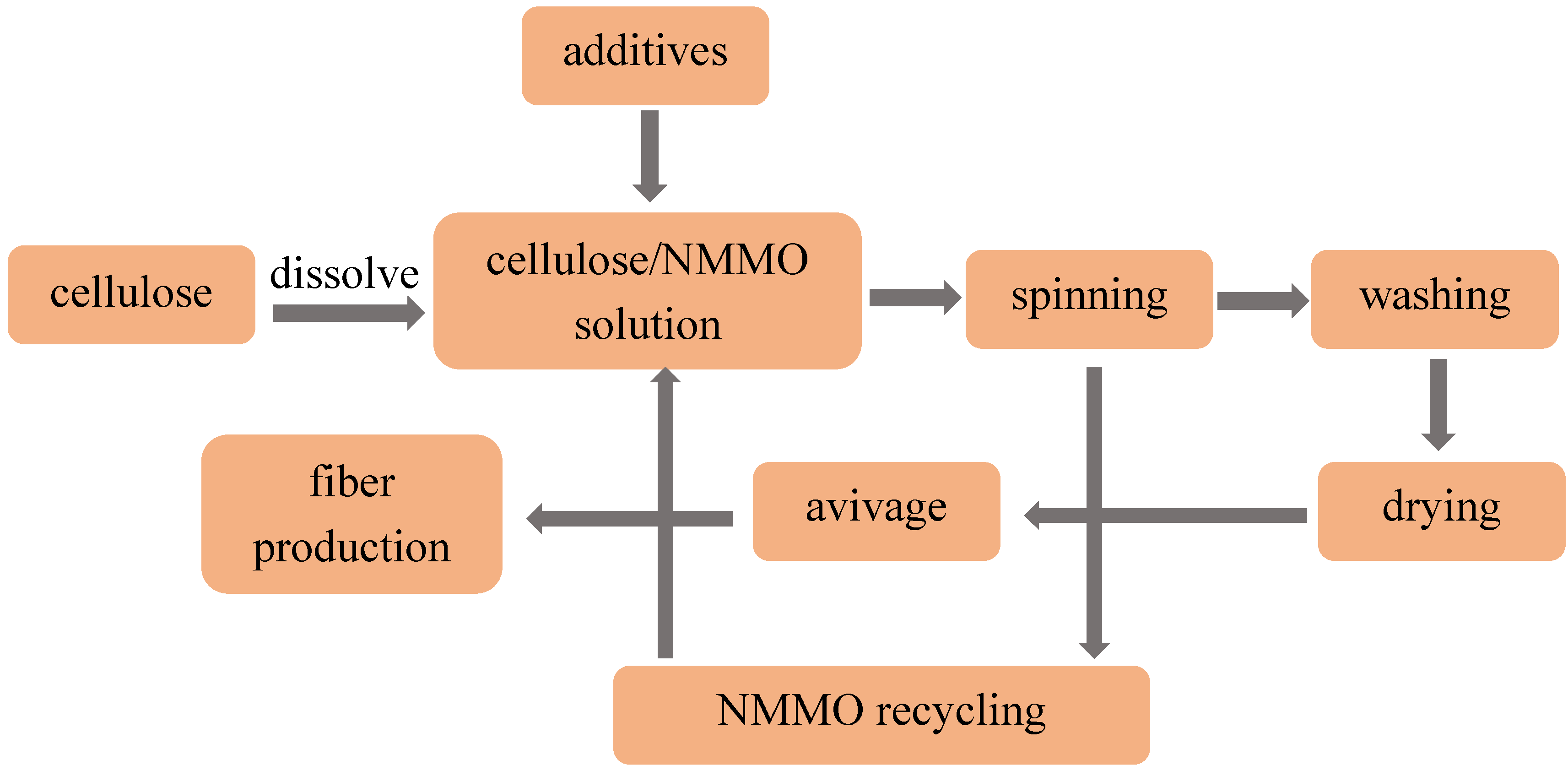
  103. Noé, P.; Chanzy, H. Swelling of Valonia cellulose microfibrils in amine oxide systems. Can. J. Chem. 2008, 86, 520–524. [Google Scholar] [CrossRef]
  104. Rosenau, T.; Potthast, A.; Sixta, H.; Kosma, P. The chemistry of side reactions and byproduct formation in the system NMMO/cellulose (Lyocell process). Prog. Polym. Sci. 2001, 26, 1763–1837. [Google Scholar] [CrossRef]
  105. Smartfiber AG. Available online: http://www.smartfiber.info/technology/innovation (accessed on 3 May 2016).
  106. Smartcel Sensitive. Available online: http://www.swicofil.com/smartfiber_smartcel_sensitive.html (accessed on 3 May 2016).
  107. Smartfiber AG. Available online: http://www.smartfiber.info/ (accessed on 3 May 2016).
  108. Leusch, A.; Holan, Z.R.; Volesky, B. Biosorption of heavy metals (Cd, Cu, Ni, Pb, Zn) by chemically-reinforced biomass of marine algae. J. Chem. Technol. Biotechnol. 1995, 62, 279–288. [Google Scholar] [CrossRef]
  109. AATCC Evalation Procedure 5-2011, Fabric Hand: Guidleine; American Association of Textile Chemists and Colorists: Research Triangle Park, NC, USA, 2012; pp. 388–390.
  110. Wiegand, C.; Hipler, U.C.; Boldt, S.; Strehle, J.; Wollina, U. Skin-protective effects of a zinc oxide-functionalized textile and its relevance for atopic dermatitis. Clin. Cosmet. Investig. Dermatol. 2013, 6, 115–121. [Google Scholar] [PubMed]
  111. Smartfiber AG. Available online: http://www.smartfiber.info/smartcel-sensitive/fiber (accessed on 3 May 2016).
  112. Yadav, A.; Prasad, V.; Kathe, A.A.; Raj, S.; Yadav, D.; Sundaramoorthy, C.; Vigneshwaran, N. Functional finishing in cotton fabrics using zinc oxide nanoparticles. Bull. Mater. Sci. 2006, 29, 641–645. [Google Scholar] [CrossRef]
  113. Chatterjee, S.; Salaun, F.; Campagne, C. Development of multilayer microcapsules by a phase coacervation method based on ionic interactions for textile applications. Pharmaceutics 2014, 6, 281–297. [Google Scholar] [CrossRef] [PubMed]
  114. Ghosh, S.K. Functional Coatings and Microencapsulation: A General Perspective. In Functional Coatings: By Polymer Microencapsulation; Ghosh, S.K., Ed.; Wiley-VCH Verlag GmbH & Co. KGaA: Weinheim, Germany, 2006; pp. 1–28. [Google Scholar]
  115. Bansode, S.S.; Banarjee, S.K.; Gaikwad, D.D.; Jadhav, S.L.; Thorat, R.M. Microencapsulation: A review. Int. J. Pharm. Sci. Rev. Res. 2010, 1, 38–43. [Google Scholar]
  116. Nelson, G. Application of microencapsulation in textiles. Int. J. Pharm. 2002, 242, 55–62. [Google Scholar] [CrossRef]
  117. Parys, M.V. Smart Textiles Using Microencapsulation Technology. In Functional Coatings; Ghosh, S.K., Ed.; WILEY-VCH Verlag GmbH & Co. KGaA: Weinheim, Germany, 2006; pp. 221–258. [Google Scholar]
  118. Azizi, N.; Chevalier, Y.; Majdoub, M. Isosorbide-based microcapsules for cosmeto-textiles. Ind. Crops Prod. 2014, 52, 150–157. [Google Scholar] [CrossRef]
  119. Wang, J.M.; Xu, X.W.; Zhou, Y. The Anti-virus Properties of Nano-Chinese Medicine Microcapsule Treated Fabrics. JFBI 2008, 1, 193–200. [Google Scholar] [CrossRef]
  120. Liu, J.; Liu, C.; Liu, Y.; Chen, M.; Hu, Y.; Yang, Z. Study on the grafting of chitosan-gelatin microcapsules onto cotton fabrics and its antibacterial effect. Colloids Surf. B 2013, 109, 103–108. [Google Scholar] [CrossRef] [PubMed]
  121. Hassan, M.M.; Sunderland, M. Antimicrobial and insect-resist wool fabrics by coating with microencapsulated antimicrobial and insect-resist agents. Prog. Org. Coat. 2015, 85, 221–229. [Google Scholar] [CrossRef]
  122. Hui, P.C.; Wang, W.Y.; Kan, C.W.; Zhou, C.E.; Ng, F.S.; Wat, E.; Zhang, V.X.; Chan, C.L.; Lau, C.B.; Leung, P.C. Preparation and characterisation of chitosan microcapsules loaded with Cortex Moutan. Int. J. Biol. Macromol. 2013, 55, 32–38. [Google Scholar] [CrossRef] [PubMed]
  123. Zhou, J.; Ritter, H. Cyclodextrin functionalized polymers as drug delivery systems. Polym. Chem. 2010, 1, 1552–1559. [Google Scholar] [CrossRef]
  124. Abdel-Halim, E.S.; Al-Deyab, S.S.; Alfaifi, A.Y. Cotton fabric finished with β-cyclodextrin: Inclusion ability toward antimicrobial agent. Carbohydr. Polym. 2014, 102, 550–556. [Google Scholar] [CrossRef] [PubMed]
  125. Tonelli, A.E. Cyclodextrins as a means to nanostructure and functionalize polymers. J. Incl. Phenom. Macrocycl. 2007, 60, 197–202. [Google Scholar] [CrossRef]
  126. Issazadeh-Baltorki, H.; Khoddami, A. Cyclodextrin-coated denim fabrics as novel carriers for ingredient deliveries to the skin. Carbohydr. Polym. 2014, 110, 513–517. [Google Scholar] [CrossRef] [PubMed]
  127. Bajpai, M.; Gupta, P.; Bajpai, S.K. Silver(I) ions loaded cyclodextrin-grafted-cotton fabric with excellent antimicrobial property. Fiber Polym. 2010, 11, 8–13. [Google Scholar] [CrossRef]
  128. Radu, C.D.; Salariu, M.; Avadanei, M.; Ghiciuc, C.; Foia, L.; Lupusoru, E.C.; Ferri, A.; Ulea, E.; Lipsa, F. Cotton-made cellulose support for anti-allergic pajamas. Carbohydr. Polym. 2013, 95, 479–486. [Google Scholar] [CrossRef] [PubMed]
  129. Samchenko, Y.; Ulberg, Z.; Korotych, O. Multipurpose smart hydrogel systems. Adv. Colloid Interface Sci. 2011, 168, 247–262. [Google Scholar] [CrossRef] [PubMed]
  130. Patel, A.; Mequanint, K. Hydrogel Biomaterials. In Biomedical Engineering-Frontiers and Challenges; Fazel, R., Ed.; Intechopen: Rijeka, Croatia, 2011; pp. 275–296. [Google Scholar]
  131. Hoffman, A.S. Hydrogels for biomedical applications. Adv. Drug Deliv. Rev. 2012, 64, 18–23. [Google Scholar] [CrossRef]
  132. Chen, Q.; Zhu, L.; Zhao, C.; Wang, Q.; Zheng, J. A robust, one-pot synthesis of highly mechanical and recoverable double network hydrogels using thermoreversible sol-gel polysaccharide. Adv. Mater. 2013, 25, 4171–4176. [Google Scholar] [CrossRef] [PubMed]
  133. Peppas, N.A.; Hilt, J.Z.; Khademhosseini, A.; Langer, R. Hydrogels in Biology and Medicine: From Molecular Principles to Bionanotechnology. Adv. Mater. 2006, 18, 1345–1360. [Google Scholar] [CrossRef]
  134. Yang, H.; Zhu, H.; Hendrix, M.M.; Lousberg, N.J.; de with, G.; Esteves, A.C.; Xin, J.H. Temperature-triggered collection and release of water from fogs by a sponge-like cotton fabric. Adv. Mater. 2013, 25, 1150–1154. [Google Scholar] [CrossRef] [PubMed]
  135. Hu, J.; Meng, H.; Li, G.; Ibekwe, S.I. A review of stimuli-responsive polymers for smart textile applications. Smart Mater. Struct. 2012, 21, 053001. [Google Scholar] [CrossRef]
  136. Ter Schiphorst, J.; van den Broek, M.; de Koning, T.; Murphy, J.N.; Schenning, A.P.H.J.; Esteves, A.C.C. Dual light and temperature responsive cotton fabric functionalized with a surface-grafted spiropyran–NIPAAm-hydrogel. J. Mater. Chem. A 2016, 4, 8676–8681. [Google Scholar] [CrossRef]
  137. Hu, J.; Lu, J. Smart polymers for textile applications. In Smart Polymers and their Applications; Woodhead Publishing Limited: Cambridge, UK, 2014; p. 14. [Google Scholar]
  138. Sun, T.; Wang, G.; Feng, L.; Liu, B.; Ma, Y.; Jiang, L.; Zhu, D. Reversible switching between superhydrophilicity and superhydrophobicity. Angew. Chem. Int. Ed. Engl. 2004, 43, 357–360. [Google Scholar] [CrossRef] [PubMed]
  139. Jiang, C.; Wang, Q.; Wang, T. Thermoresponsive PNIPAAm-modified cotton fabric surfaces that switch between superhydrophilicity and superhydrophobicity. Appl. Surf. Sci. 2012, 258, 4888–4892. [Google Scholar] [CrossRef]
  140. Bashari, A.; Hemmatinejad, N.; Pourjavadi, A. Surface modification of cotton fabric with dual-responsive PNIPAAm/chitosan nano hydrogel. Polym. Adv. Technol. 2013, 24, 797–806. [Google Scholar] [CrossRef]
  141. Gras, S.L.; Mahmud, T.; Rosengarten, G.; Mitchell, A.; Kalantar-Zadeh, K. Intelligent control of surface hydrophobicity. Chem. Phys. Chem. 2007, 8, 2036–2050. [Google Scholar] [CrossRef] [PubMed]
  142. Duval, A.; Lange, H.; Lawoko, M.; Crestini, C. Modification of Kraft Lignin to Expose Diazobenzene Groups: Toward pH- and Light-Responsive Biobased Polymers. Biomacromolecules 2015, 16, 2979–2989. [Google Scholar] [CrossRef] [PubMed]
  143. Liu, B.; Hu, J.; Meng, Q. Nonwoven supported temperature-sensitive poly (N-isopropylacrylamide)/polyurethane copolymer hydrogel with antibacterial activity. J. Biomed. Mater. Res. B 2009, 89, 1–8. [Google Scholar] [CrossRef] [PubMed]
  144. Ribeiro, M.P.; Espiga, A.; Silva, D.; Baptista, P.; Henriques, J.; Ferreira, C.; Silva, J.C.; Borges, J.P.; Pires, E.; Chaves, P. Development of a new chitosan hydrogel for wound dressing. Wound Repair Regen. 2009, 17, 817–824. [Google Scholar] [CrossRef] [PubMed]
  145. Wang, X.; Hu, H.; Yang, Z.; He, L.; Kong, Y.; Fei, B.; Xin, J.H. Smart hydrogel-functionalized textile system with moisture management property for skin application. Smart Mater. Struct. 2014, 23, 1–11. [Google Scholar] [CrossRef]
  146. Peppas, N.A.; Bures, P.; Leobandung, W.; Ichikawa, H. Hydrogels in pharmaceutical formulations. Eur. J. Pharm. Biopharm. 2000, 50, 27–46. [Google Scholar] [CrossRef]
  147. Cerchiara, T.; Bigucci, F.; Luppi, B. Hydrogel Vehicles for Hydrophilic Compounds. In Percutaneous Penetration Enhancers Chemical Methods in Penetration Enhancement: Drug Manipulation Strategies and Vehicle Effects; Dragicevic, N., Maibach, H.I., Eds.; Springer: Berlin/Heidelberg, Germany, 2015; pp. 285–298. [Google Scholar]
  148. Kircik, L.; Del Rosso, J. A novel hydrogel vehicle formulated for the treatment of atopic dermatitis. J. Drugs Dermatol. 2007, 6, 718–722. [Google Scholar] [PubMed]
  149. Kircik, L.H. The effect of desonide hydrogel on pruritus associated with atopic dermatitis. J. Drugs Dermatol. 2014, 13, 725–728. [Google Scholar] [PubMed]
  150. Üstündağ Okur, N.; Çağlar, E.Ş.; Arpa, M.D.; Karasulu, H.Y. Preparation and evaluation of novel microemulsion-based hydrogels for dermal delivery of benzocaine. Pharm. Dev. Technol. 2016, 22, 500–510. [Google Scholar] [CrossRef] [PubMed]
  151. Nayak, A.; Babla, H.; Han, T.; Das, D.B. Lidocaine carboxymethylcellulose with gelatine co-polymer hydrogel delivery by combined microneedle and ultrasound. Drug Deliv. 2016, 23, 668–679. [Google Scholar] [CrossRef] [PubMed]
  152. Yapar, E.A.; Ýnal, Ö. Poly (ethylene oxide)–poly (propylene oxide)-based copolymers for transdermal drug delivery: An overview. Trop. J. Pharm. Res. 2013, 11, 855–866. [Google Scholar] [CrossRef]
  153. Arora, A.; Prausnitz, M.R.; Mitragotri, S. Micro-scale devices for transdermal drug delivery. Int. J. Pharm. 2008, 364, 227–236. [Google Scholar] [CrossRef] [PubMed]
  154. Donnelly, R.F.; Singh, T.R.R.; Morrow, D.I.; Woolfson, A.D. Transdermal drug delivery-microneedle. In Microneedle-Mediated Transdermal and Intradermal Drug Delivery, 1st ed.; John Wiley & Sons: New York, NY, USA, 2012; pp. 1–19. [Google Scholar]
  155. Prausnitz, M.R.; Langer, R. Transdermal drug delivery. Nat. Biotechnol. 2008, 26, 1261–1268. [Google Scholar] [CrossRef] [PubMed]
  156. Cevc, G.; Vierl, U. Nanotechnology and the transdermal route: A state of the art review and critical appraisal. J. Control. Release 2010, 141, 277–299. [Google Scholar] [CrossRef] [PubMed]
  157. Alexander, A.; Dwivedi, S.; Ajazuddin; Giri, T.K.; Saraf, S.; Saraf, S.; Tripathi, D.K. Approaches for breaking the barriers of drug permeation through transdermal drug delivery. J. Control. Release 2012, 164, 26–40. [Google Scholar] [CrossRef] [PubMed]
  158. Heilmann, S.; Kuchler, S.; Wischke, C.; Lendlein, A.; Stein, C.; Schafer-Korting, M. A thermosensitive morphine-containing hydrogel for the treatment of large-scale skin wounds. Int. J. Pharm. 2013, 444, 96–102. [Google Scholar] [CrossRef] [PubMed]
  159. Ishii, Y.; Nakae, T.; Sakamoto, F.; Matsuo, K.; Matsuo, K.; Quan, Y.S.; Kamiyama, F.; Fujita, T.; Yamamoto, A.; Nakagawa, S.; Okada, N. A transcutaneous vaccination system using a hydrogel patch for viral and bacterial infection. J. Control. Release 2008, 131, 113–120. [Google Scholar] [CrossRef] [PubMed]
  160. Ajazuddin; Alexander, A.; Khichariya, A.; Gupta, S.; Patel, R.J.; Giri, T.K.; Tripathi, D.K. Recent expansions in an emergent novel drug delivery technology: Emulgel. J. Control. Release 2013, 171, 122–132. [Google Scholar]
  161. Jose, A.; Mandapalli, P.K.; Venuganti, V.V. Liposomal hydrogel formulation for transdermal delivery of pirfenidone. J. Liposome Res. 2016, 26, 139–147. [Google Scholar] [CrossRef] [PubMed]
  162. Rajan, R.; Jose, S.; Mukund, V.P.B.; Vasudevan, D.T. Transferosomes—A vesicular transdermal delivery system for enhanced drug permeation. J. Adv. Pharm. Technol. Res. 2011, 2, 138–143. [Google Scholar] [CrossRef] [PubMed]
  163. Jeong, B.; Kim, S.W.; Bae, Y.H. Thermosensitive sol–gel reversible hydrogels. Adv. Drug Deliv. Rev. 2012, 64, 154–162. [Google Scholar] [CrossRef]
  164. Li, Y.; Rodrigues, J.; Tomas, H. Injectable and biodegradable hydrogels: Gelation, biodegradation and biomedical applications. Chem. Soc. Rev. 2012, 41, 2193–2221. [Google Scholar] [CrossRef] [PubMed]
  165. Buwalda, S.J.; Boere, K.W.; Dijkstra, P.J.; Feijen, J.; Vermonden, T.; Hennink, W.E. Hydrogels in a historical perspective: From simple networks to smart materials. J. Control. Release 2014, 190, 254–273. [Google Scholar] [CrossRef] [PubMed]
  166. Ahmed, E.M. Hydrogel: Preparation, characterization, and applications: A review. J. Adv. Res. 2015, 6, 105–121. [Google Scholar] [CrossRef] [PubMed]
  167. Schwartz, J.; Moreno, E.; Fernandez, C.; Navarro-Blasco, I.; Nguewa, P.A.; Palop, J.A.; Irache, J.M.; Sanmartin, C.; Espuelas, S. Topical treatment of L. major infected BALB/c mice with a novel diselenide chitosan hydrogel formulation. Eur. J. Pharm. Sci. 2014, 62, 309–316. [Google Scholar] [CrossRef] [PubMed]
  168. Kwon, S.S.; Kong, B.J.; Park, S.N. Physicochemical properties of pH-sensitive hydrogels based on hydroxyethyl cellulose–hyaluronic acid and for applications as transdermal delivery systems for skin lesions. Eur. J. Pharm. Biopharm. 2015, 92, 146–154. [Google Scholar] [CrossRef] [PubMed]
  169. Kabanov, A.V.; Vinogradov, S.V. Nanogels as pharmaceutical carriers: Finite networks of infinite capabilities. Angew. Chem. Int. Ed. Engl. 2009, 48, 5418–5429. [Google Scholar] [CrossRef] [PubMed]
  170. Gonçalves, C.; Pereira, P.; Gama, M. Self-assembled hydrogel nanoparticles for drug delivery applications. Materials 2010, 3, 1420–1460. [Google Scholar] [CrossRef]
  171. Mavuso, S.; Marimuthu, T.; Choonara, Y.E.; Kumar, P.; Toit, L.C.D.; Pillay, V. A review of polymeric colloidal nanogels in transdermal drug delivery. Curr. Pharm. Des. 2015, 21, 2801–2813. [Google Scholar] [CrossRef] [PubMed]
  172. Panyam, J.; Sahoo, S.K.; Prabha, S.; Bargar, T.; Labhasetwar, V. Fluorescence and electron microscopy probes for cellular and tissue uptake of poly(d,l-lactide-co-glycolide) nanoparticles. Int. J. Pharm. 2003, 262, 1–11. [Google Scholar] [CrossRef]
  173. Sultana, S.S.; Parveen, P.; Rekha, M.S.; Deepthi, K.; Sowjanya, C.; Devi, A.S. Emulgel-a novel surrogate approach for transdermal drug delivery system. Ind. Am. J. Pharm. Res. 2014, 4, 5250–5265. [Google Scholar]
  174. Naga Sravan Kumar Varma, V.; Maheshwari, P.V.; Navya, M.; Reddy, S.C.; Shivakumar, H.G.; Gowda, D.V. Calcipotriol delivery into the skin as emulgel for effective permeation. Saudi Pharm. J. 2014, 22, 591–599. [Google Scholar] [CrossRef] [PubMed]
  175. Jeengar, M.K.; Rompicharla, S.V.; Shrivastava, S.; Chella, N.; Shastri, N.R.; Naidu, V.G.; Sistla, R. Emu oil based nano-emulgel for topical delivery of curcumin. Int. J. Pharm. 2016, 506, 222–236. [Google Scholar] [CrossRef] [PubMed]
  176. Khullar, R.; Kumar, D.; Seth, N.; Saini, S. Formulation and evaluation of mefenamic acid emulgel for topical delivery. Saudi Pharm. J. 2012, 20, 63–67. [Google Scholar] [CrossRef] [PubMed]
  177. Carafa, M.; Marianecci, C.; di Marzio, L.; Rinaldi, F.; Meo, C.; Matricardi, P.; Alhaique, F.; Coviello, T. A new vesicle-loaded hydrogel system suitable for topical applications: Preparation and characterization. J. Pharm. Pharm. Sci. 2011, 14, 336–346. [Google Scholar] [CrossRef] [PubMed]
  178. Escobar-Chávez, J.J. Nanocarriers for transdermal drug delivery. Skin 2012, 19, 3–17. [Google Scholar] [CrossRef]
  179. Hubbell, J.A.; Chilkoti, A. Nanomaterials for drug delivery. Science 2012, 337, 303–305. [Google Scholar] [CrossRef] [PubMed]
Figure 1. Wet-wrap dressing. Reprinted from Ref. [19] with permission. Copyright 2006 John Wiley & Sons, Inc.
Coatings 07 00082 g001
Figure 2. (A, ×100) Photo of anion fabrics and (B) SEM of doped polyester filaments with fine crusted tourmaline power (white circle). Reprinted from Ref. [29] with permission. Copyright 2012 the Korean Dermatological Association and the Korean Society for Investigative Dermatology.
Coatings 07 00082 g002
Figure 3. (A) Scanning electron micrographs of fabricated microcapsules and (B) coated fabrics. Reprinted from Ref. [91] with permission. Copyright 2013 Elsevier.
Coatings 07 00082 g003
Figure 4. (A) Schematic diagram of hydrogel functionalized fabric with dual-functions; and (B) Poloxamer-based hydrogel. Reprinted from Ref. [92] with permission. Copyright 2016 Nature Publishing Group.
Coatings 07 00082 g004
Figure 5. Schematic representation of the gelation mechanism of Poloxamer 407 in water. Adapted from Ref. [98] with permission. Copyright 2006 Springer.
Coatings 07 00082 g005
Figure 6. Manufacturing method of additives loaded seaweed-based cellulosic fiber. Adapted from Ref. [104] with permission. Copyright 2001Elsevier.
Coatings 07 00082 g006
Figure 7. Zinc loaded seaweed-based cellulosic textiles (Smartcel TM Sensitive). Reprinted from Ref. [110] with the permission.
Coatings 07 00082 g007
Figure 8. Antibacterial finishing of cotton fabrics by pad-dry-cure. Adapted from Ref. [51] with permission. Copyright 2011 Elsevier.
Coatings 07 00082 g008
Figure 9. Chemical structure (a) and 3-dimensional structure (b) of cyclodextrins. Reprinted from Ref. [123] with permission. Copyright 2010 Royal Society of Chemistry.
Coatings 07 00082 g009
Figure 10. (a) Shows the molecular structures of N-isopropyl acrylamide (NIPAAm) and Poly (N-isopropyl acrylamide) (PNIPAAm) and (b) illustrates the switch between a hydrophilic and hydrophobic PNIPAAm-modified surface. PNIPAAm chains are shown forming intermolecular hydrogen bonds with water molecules at a temperature below the (Lower Critical Solution Temperature) LCST (left) and forming intermolecular hydrogen bonds between C=O and N–H groups at temperatures above the LCST (right). Adapted from Ref. [138] with permission. Copyright 2004 John Wiley & Sons, Inc.
Coatings 07 00082 g010
Table 1. Antibacterial agents used for antibacterial textiles therapy in AD treatment.
TypesExamplesReferences
Metal nanoparticlestitania nanoparticle[59]
silver nanoparticle[60,61]
zinc oxide nanoparticle[62,63]
copper nanoparticles[62,64]
Organic antibacterialscitric acid[65]
citrus grandis osbeck extract[66,67]
Table 2. Commonly used prescriptions and the component herbal medicines for AD treatment.
Herbs FormulaIngredientsReferences
Huangbai Zicao Diyu creamCortex phellodendri 50 g; Radix arnebiae 50 g; Radix sanguisorbae 100 g[81]
Huanglian Qingdai ointmentRhizoma coptidis 0.6 g; Indigo naturalis 0.3 g; Cortex phellodendri 0.3 g; Alum 0.3 g; Borneolum syntheticum; Urea cream 40 g[81]
Xiao-Feng-San (XFS)Radix saposhnikovia 2.5 mg; Herba schizonepetae 2.5 mg; Radix angelicae sinensis 2.5 mg; Radix rehmanniae 2.5 mg; Radix sophorae flavescentis 2.5 mg; Rhizoma atractylodis 2.5 mg; Periostracum cicdae 2.5 mg; Linum usitatissimum 2.5 mg; Rhizoma anemarrhenae 2.5 mg; Gypsum fibrosum 2.5 mg; Caulis clematidis armandii 1.25 mg; Radix glycyrrhizae 1.25 mg; Fructus arctii 2.5 mg[81,82]
TCHM capsulesFlos lonicerae 2 g; Herba menthae 1 g; Cortex moutan 2 g; Rhizoma atractylodis 2 g; Cortex phellodendri 2 g[83,84]
ByakkokaninjintoGypsum fibrosum; Anemarrhenae rhizome; Glycyrrhizae radix; Ginseng radix; Oryzae fructus[85,86]
Juzen-Taiho-toAstragali Radix; Cinnamomi Cortex, Rehmanniae Radix; Paeoniae Radix; Cnidii Rhizoma; Atractylodis Lanceae Rhizoma; Angelicae Radix; Ginseng Radix; Hoelen; Glycyrrhizae Radix[85,87]
KeishibukuryoganCinnamomi cortex; Paeoniae Radix; Moutan cortex; Persicae semen; Hoelen[85,88]
Gami-Cheongyeul-Sodok-EumAngelica gigas; Astragalus membranaceus; Atractylodes japonica; Coptis japonica; Forsythia virdissima; Glycyrrhiza uralensis; Lonicera japonica; Portulaca oleracea; Scutellaria baicalensis[89]

