Article

Preparation of Silver-Loaded Antibacterial Agent Using Sodium Titanate Nanotubes and Its Strengthening and Antifungal Effect on Wooden Cultural Relics

Capital Museum, Beijing 100045, China
Coatings 2026, 16(5), 508; https://doi.org/10.3390/coatings16050508
Submission received: 23 March 2026 / Revised: 11 April 2026 / Accepted: 17 April 2026 / Published: 22 April 2026
(This article belongs to the Special Issue Innovations in Functional Coatings for Wood Processing)

Abstract

In this paper, we utilized sodium titanate as a substrate to fabricate a supported antifungal repair agent capable of inhibiting fungi through the release of silver ions, and applied it to the preservation and restoration of wooden materials. The structural and material properties of sodium titanate were characterized using transmission electron microscopy (TEM), scanning electron microscopy (SEM), and adsorption kinetic modeling. Furthermore, its effectiveness in wood restoration as well as its antifungal performance were evaluated. Results indicate that the synthesized sodium titanate exhibits a distinctive tubular structure, with a diameter of approximately 12 nm, a pore size of 7 nm, and a specific surface area as high as 310.91 m2/g. The abundant ion exchange active sites on the material surface provide conditions for the loading of silver ions. At 25 °C, the maximum adsorption capacity for silver ions reaches 515.5 mg/g, with an adsorption amount accounting for 34.0 wt.%. When combined with polyvinyl alcohol (PVA) for reinforcing wooden materials, it significantly increases the packing density of the reinforcing agent, ultimately enhancing the compressive strength of wood from 155.0 MPa to 412.2 MPa. Furthermore, owing to the antifungal effect of silver ions, the treated wood demonstrates effective resistance against the growth of Aspergillus niger.

1. Introduction

Wooden artifacts are commonly unearthed in archaeological excavations and possess significant historical value. Currently, restoration efforts for wooden artifacts target both water-saturated and dry wood. For dry wooden artifacts, commonly used materials include beeswax, collagen, and epoxy resin [1,2]. For some wooden artifacts retrieved from water, the restoration process must account for deformation and cracking caused by moisture loss, making it necessary to use hydration ingredients for filling. While such materials exhibit good compatibility with wood and can help slow aging and degradation, their reinforcement effects remain limited, especially given the shrinkage and deformation that occur as organic materials dehydrate.
It is worth noting that due to the acidic or alkaline conditions of the burial environment and the prolonged exposure to natural erosion, such cultural relics often exhibit decay upon excavation. To display the original appearance of the historic wooden architecture, a series of new repair agents have been attempted by restorers for application in wood restoration. For example, Li [3] et al. used a pectin-sawdust composite material for the restoration, thereby ensuring the compatibility of the restoration material with the historical wood. Glastrup [4] et al. investigated the effect of using PEG as a filler for wood restoration and found that the polyethylene glycol (PEG) polymer still maintained a relatively intact structure after 30 years. This work has verified that organic polymer materials possess excellent durability when used as wood fillers. However, it should be noted that these decayed woods often contain microorganisms. If left untreated, these organisms can continue to damage the artifacts after restoration. Therefore, when selecting restoration materials for wooden artifacts, it is crucial not only to enhance their structural strength but also to ensure long-term antibacterial efficacy [5].
To prevent further degradation during long-term preservation, developing highly effective, durable, and safe antibacterial materials has become an important research focus in materials science and cultural heritage conservation. This study proposes a mixed reinforcing material incorporating sodium titanate nanotubes loaded with silver ions as a filler, combined with polyvinyl alcohol (PVA) as a reinforcing agent, to create an antibacterial composite. The study investigates the effects of adding sodium titanate powder on the compressive strength and antifungal efficacy of wood. It is hoped that this research will provide valuable insights for the development of reinforcing materials for dry wooden artifacts.

2. Materials and Methods

2.1. Preparation of Sodium Titanate

Disperse 0.2 g of TiO2 (20 nm) into 40 mL of deionized water and stir thoroughly. Then, add 16 g of NaOH to the mixture. Subsequently, transfer the solution to a 50 mL Teflon-lined reaction vessel and conduct a hydrothermal reaction at 120 °C for 24 h. After the reaction is complete, collect the powder by filtration and wash it with a large amount of deionized water until the pH of the solution is neutral. Finally, dry the product to obtain one-dimensional sodium titanate nanotubes (STN) [6].

2.2. Loading with Ag+ Ions

Using the prepared sodium titanate nanotubes as a substrate, a silver-loaded antifungal agent was synthesized via an impregnation method. First, 0.03 g of sodium titanate powder was placed in 500 mL of deionized water at a temperature of 298 K. Then, 500 mL of a 10 mmol/L Ag+ metal ion solution was added. After mixing and stirring for 1 h, the powder was collected and rinsed with deionized water to remove any residual metal ions on the surface, resulting in the silver-loaded antifungal agent.
The corresponding dynamic adsorption process was carried out at an Ag+ ion concentration of 223.5 mg/L. Samples were then collected using a filter at predetermined time intervals, and the solution concentration was measured by ICP. The experiment was performed in triplicate, and the results were averaged.

2.3. Filling and Reinforcement of Wood

Add 0.05 g of the silver-loaded antifungal agent to 20 mL of deionized water and stir continuously during the ultrasonic-assisted dispersion process. Once the mixture is uniformly dispersed, add 4 g of polyvinyl alcohol (PVA, Mn = 600,000) to the solution and stir for 24 h until the PVA is completely dissolved, forming a viscous soaking solution. Then, immerse the fixed-size pear wood into the soaking solution, apply a vacuum to 0.1 kPa, and maintain the pressure for 30 min. After soaking, remove the wood and dry it in an oven at 60 °C.

2.4. Characterization

X-ray diffraction (XRD) patterns were obtained using a SmartLab 9KW X-ray diffractometer (RigakuCorporation, Japan) with a Cu cathode (Cu Kα1, λ = 1.54056 Å) at a scanning rate of 10° 2θ/s, operated at 40 kV and 40 mA. The morphology and structure of the synthesized samples were characterized using a field emission scanning electron microscope (FESEM, Regulus 8100, Hitachi, Japan). The samples were pre-dispersed on rectangular silicon wafers with a side length of 10 mm and coated with approximately 10 nm of gold using a Hummer 6.2 sputtering system (Anatech Ltd., Hayward, CA, USA). Nitrogen adsorption–desorption isotherms were measured at 77.2 K using an ASP-2460 (Micromeritics Instrument Corp., GA, USA). Prior to testing, the powder was pressed into pellets with a diameter of 6 mm under a pressure of 5 MPa and evacuated at 373 K to remove gases. The specific surface area (SBET) was determined using the multipoint BET method from adsorption data within a relative pressure range (P/P0) of 0.05 to 0.3. The pore volume and average pore diameter were calculated at a relative pressure of nitrogen adsorption volume (P/P0) of 0.994. The mechanical properties of the samples were characterized using a universal testing machine (model WDD-5) manufactured by Guance Testing Equipment Co., Ltd. (Beijing, China) The experiment was repeated five times, and the results were averaged.

2.5. Antifungal Activity

Considering that the corrosion of wood materials is mainly caused by fungal microorganisms, in this experiment, we used fungal bacteria as the experimental microorganisms. The experimental fungus were collected and purified from the Tripitaka Editions using a bacteria collector. The operation included the preparation of nutrient broth/nutrient agar, activation of strains, purification of strains, preservation of strains by freezing, and preparation of fungal suspensions [7]. All glassware used in the experiment, such as evaporating dishes and inoculation loops, was sterilized by dry heat sterilization; specifically, after thorough cleaning, the glassware was wrapped in newspaper and incubated in an oven at 160 °C for 2 h. The culture medium used was potato starch agar.
  • Preparation of fungal suspension
A high-concentration microbial suspension of the third-generation Talaromyces funiculosus species was obtained by rinsing the agar plates with sterile water. The optical density (OD) of the fungal suspension was measured at a wavelength of 600 nm using a UV-1800 UV-Vis spectrophotometer manufactured by Shimadzu (Kyoto, Japan). The concentration of the fungal suspension was adjusted to achieve an OD600 of 0.1 using sterile water.
To calculate the optical density, the following formula was used:
OD 600   = l o g 10 ( 1 trans ) .
The formula OD600 refers to the absorbance of the sample at 600 nm, while trans refers to the transmittance of the sample at this wavelength. When OD600 = 0.1, the corresponding bacterial concentration in the suspension is 107 CFU/mL.
The antifungal performance of the materials was qualitatively analyzed using the antibacterial ring method, while quantitative analysis was performed using the shake flask method, where Trichoderma species are co-cultured with the antifungal agent [8]. The concentration for the antibacterial ring test is 107  CFU/mL. The specific steps are as follows.
Take 100 μL of a fungal suspension with a concentration of 107 CFU/mL and evenly spread it on a potato starch agar plate using a spreader to create a culture plate for the antibacterial ring experiment. Use a punch to cut out circular paper disks with a diameter of 6 mm from sterile neutral filter paper. Disperse the sample into sterile water to form a suspension with a concentration of 0.2 g/mL. Add 100 μL of the suspension dropwise onto each circular paper disk, repeating this process three times, then dry the disks to form antibacterial samples containing the loaded powder. Place the antifungal samples flat on the culture plate and incubate in a 28 °C incubator for 24 h.

3. Results and Discussion

3.1. Characterization of Sodium Titanate

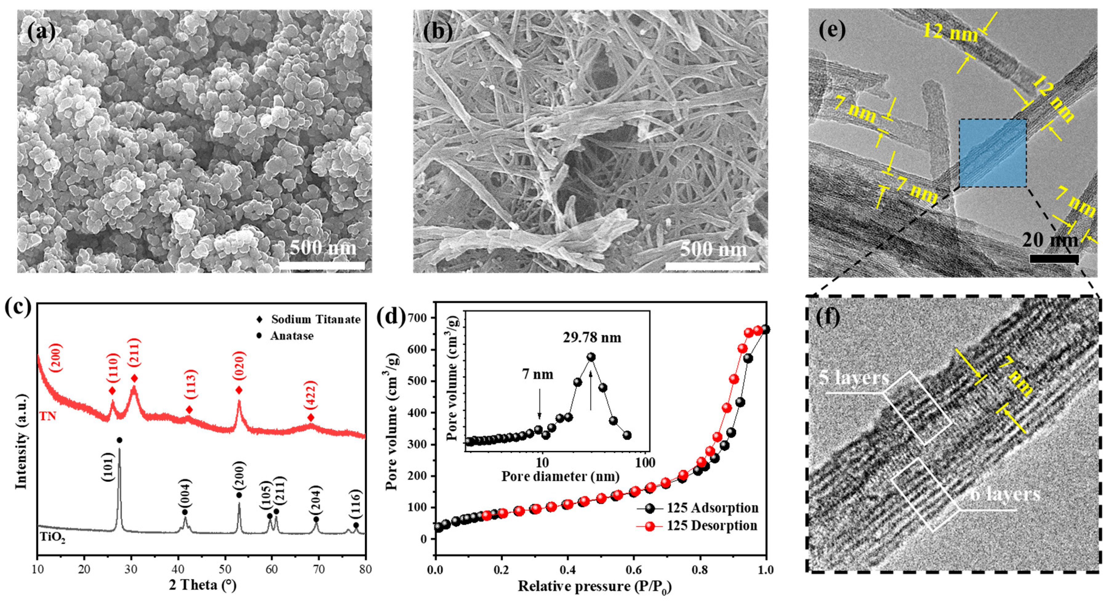
As shown in Figure 1a, the scanning electron microscope (SEM) image of commercially available TiO2 reveals uniformly sized particles with an average diameter of approximately 25 nm. XRD analysis indicates that the crystal phase of this material is primarily in the anatase form of TiO2. After hydrothermal treatment, the morphology of the particles undergoes significant changes. As observed in Figure 1b, the resulting products adopt a one-dimensional fibrous structure with lengths exceeding 500 nm. The corresponding XRD pattern displays multiple broad diffraction peaks, which are positioned at 2θ~9.25°, 2θ~24.31°, 2θ~28.30°, 2θ~48.24°, and 2θ~61.37°. These peak positions align well with the characteristic reflections of titanate nanotubes reported in the literature, confirming that the hydrothermally derived product is sodium titanate [9]. Furthermore, the diffraction pattern obtained after the reaction shows no significant intensity of the diffraction peaks corresponding to the original TiO2 crystal phase. This indicates that nearly all of the TiO2 had been consumed during the reaction process, suggesting that the conversion rate of the material is close to 100%. The high conversion efficiency further confirms the effectiveness of the hydrothermal treatment in completely transforming TiO2 into sodium titanate, underscoring the success of the synthetic approach.
From the TEM image in Figure 1e, it can be seen that the sample after hydrothermal reaction exhibits a hollow one-dimensional tubular structure, with an inner diameter of 7 nm and an outer diameter of approximately 12 nm, which is consistent with the sizes observed in the SEM images. The number of layers in the tubular walls varies; notably, the right side has one more layer than the opposite side. It has been reported that this phenomenon occurs because the sodium titanate structure is formed through the curling of the (001) crystal face of TiO2. This unique layered structure contributes to the material’s properties and potential applications [9]. The N2 adsorption–desorption isotherm in Figure 1d similarly displays characteristics typical of mesoporous materials [10]. The adsorption curve shows a relatively gradual increase during the low-pressure adsorption phase, followed by a steep rise in adsorption amount at higher pressures. This behavior can be attributed to the capillary condensation of adsorbates within the mesopores once relative pressure reaches a certain threshold. After all capillary condensation has concluded, adsorption occurs primarily on surfaces with lower surface energy, resulting in a plateau in the adsorption isotherm. Due to the occurrence of capillary condensation, a hysteresis phenomenon is observed in the desorption curve, where the adsorption and desorption curves do not overlap, leading to the formation of a hysteresis loop. This behavior is consistent with typical type IV isotherms [11]. Analysis of the pore size distribution reveals the presence of two distinct pore sizes: 7 nm and 29.7 nm. The 7 nm pore size corresponds to the dimensions of the hollow structure within the material, while the 29.7 nm size originates from the arrangement and stacking of the one-dimensional structures in three-dimensional space [12]. According to the Brunauer–Emmett–Teller (BET) results, the specific surface area of the material is as high as 310.91 m2/g, indicating its potential for applications that benefit from high surface areas, such as catalysis, adsorption, and energy storage. It is noteworthy that the sample in Figure 1f displays clear lattice fringes, indicating good crystallinity of the material. However, the XRD spectra show broad diffraction peaks. This phenomenon can be attributed to the small crystal size of the tubular TiO2, which limits the coherent diffraction region of the X-rays. Additionally, the one-dimensional directional growth of the crystals contributes to the broadening of the Miller indices peaks. As a result, the diffraction peaks observed in the X-ray diffraction pattern appear as broad peaks, reflecting the less pronounced crystallinity of the material. This broad peak characteristic is common in nanoscale materials and highlights the influence of particle size and growth orientation on their crystallographic properties [13].

3.2. Characterization of Antimicrobial Agents

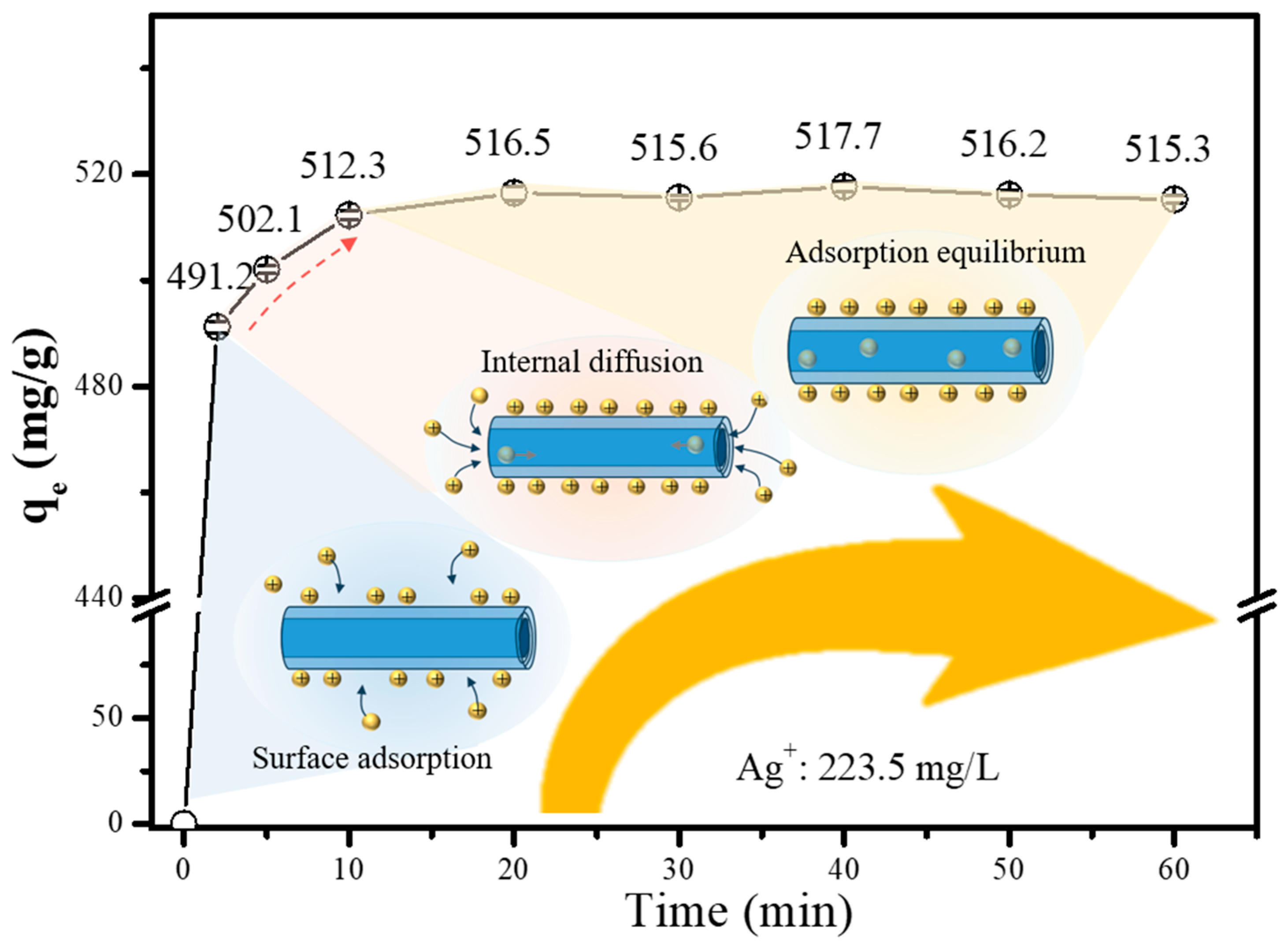
From the above discussion, it can be seen that the sodium titanate nanotubes prepared by hydrothermal synthesis have a hollow one-dimensional tubular structure, and the material has a very large specific surface area. Utilizing this characteristic, we attempted to apply it as a substrate to load silver ions. Figure 2 presents the adsorption kinetics of silver ions on STN at 25 °C, with an initial silver ion concentration of 223.5 mg/L. Upon initial contact between the adsorbent and adsorbate, the amount of adsorbed silver ions increased rapidly, reaching over 95% of the saturation adsorption capacity within just 2 min. This rapid uptake is mainly due to the abundance of exposed active sites on the surface of the sodium titanate nanotubes. Subsequently, within the period between 2 and 10 min, due to the unique tubular structure of the substrate, silver ions diffused into the interior of the material. The adsorption process is significantly influenced by the rate of diffusion. Therefore, a slow growth phenomenon appeared during stage b of the adsorption curve. After the adsorption time reached 10 min, the adsorption amount basically no longer changed with the extension of the adsorption time, and the adsorption reached equilibrium. The saturated adsorption capacity was reached at 515.5 mg/g, corresponding to a loading rate of 34.0 wt.%. This value is significantly higher than those reported in previous studies (compared with the figures shown in Table 1), which can be primarily ascribed to the large specific surface area (310.91 m2/g) of the tubular sodium titanate prepared via hydrothermal reaction.
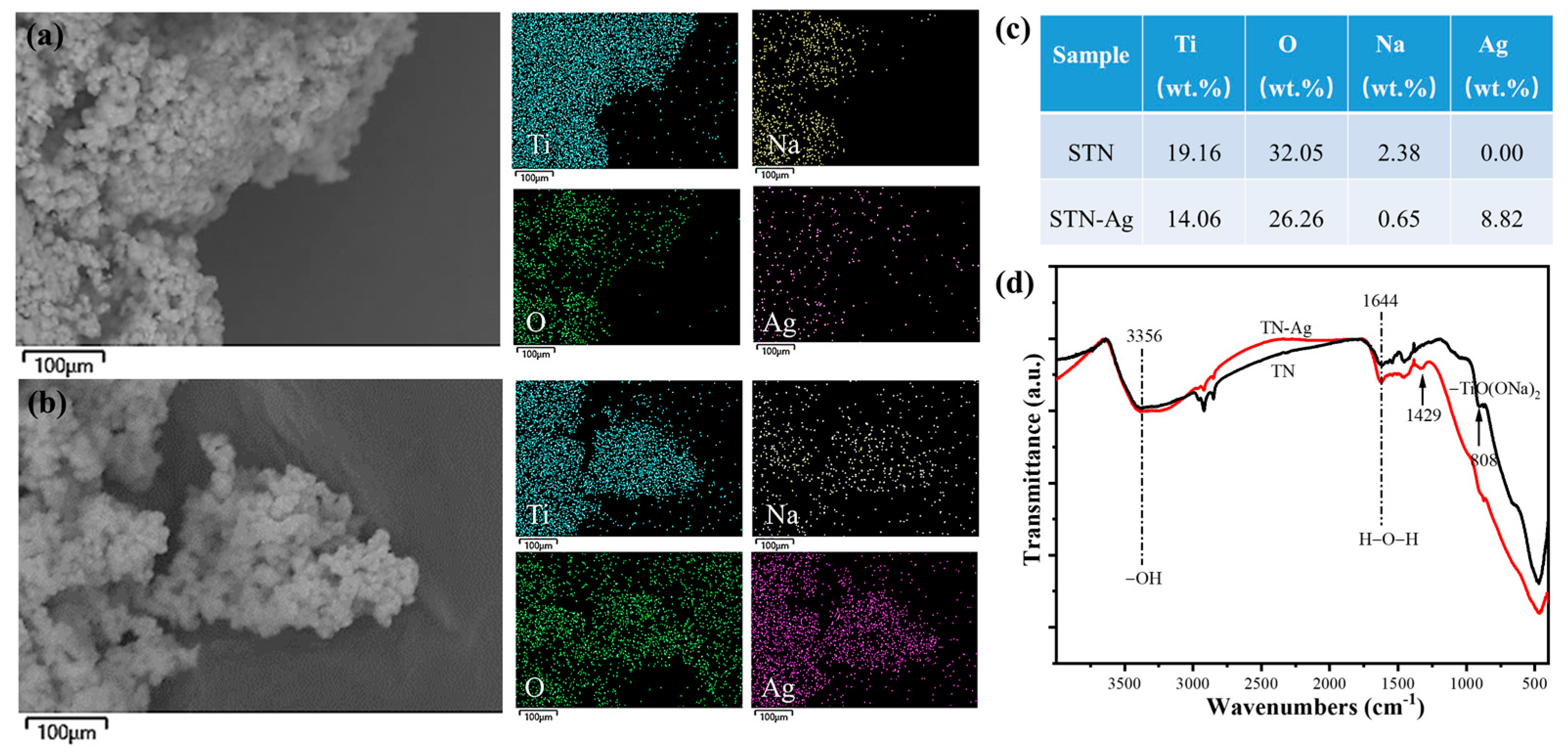
The adsorption of metal ions was characterized using energy dispersive spectroscopy (EDS) and Fourier transform infrared spectroscopy (FTIR). As shown in Figure 3a, prior to adsorption, the EDS analysis detected only titanium (Ti), oxygen (O), and sodium (Na) in the sodium titanate (STN) material, consistent with its typical composition [19]. After the adsorption of Ag+ ions, the silver content in the material increased from 0% to 8.82%, with a uniform distribution of Ag observed across the STN surface. Furthermore, as the Ag+ content rose, a continuous decrease in sodium (Na) content was noted. This correlation confirms that the adsorption mechanism of Ag+ ions proceeds primarily through ion exchange, wherein Ag+ replaces Na+ in the material structure [20]. Fourier transform infrared spectroscopy was employed to analyze the adsorption mode of the material. As can be seen from Figure 3d, after the adsorption of Ag+ ions, the characteristic peak of -TiO(ONa)2 at 808 cm−1 in the sample disappeared, and a new peak appeared at 1429 cm−1 [21]. The appearance of new peaks indicates the generation of new functional groups, which corresponds to the position of Na+ ions in -TiO(ONa)2 replaced by Ag+ to form a new -TiO(OAg)2 functional group [22]. In addition, there were still -OH and water adsorption peaks in the material, indicating that the ion exchange process does not affect other functional groups [23]. This is because the H+ ion has a lower energy when combined with STN, so the Ag+ ion tends to replace Na+ ions rather than the H+ ions.
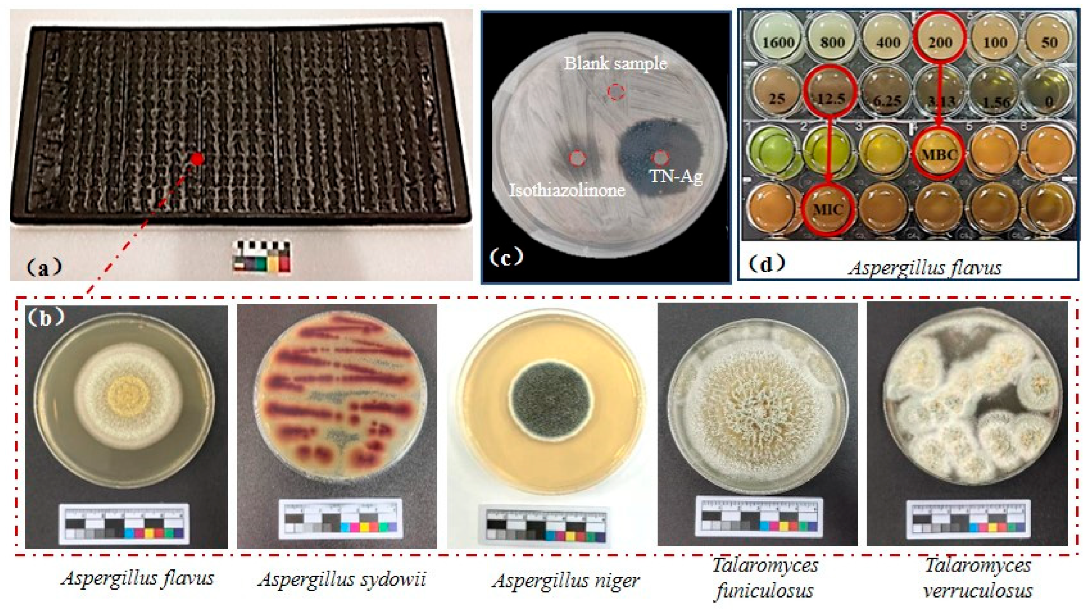
Colonies were isolated from the Tripitaka printing blocks using a microbial collector and then purified. The analysis reveals that the microorganisms present on the printing blocks mainly include Aspergillus niger, Aspergillus flavus, Aspergillus sydowii, and Talaromyces funiculosus, among others. Based on the relatively typical corrosion of wood by fungus, the collected Talaromyces funiculosus was used as the experimental specimen. As shown in Figure 4c, when STN was co-cultured with Talaromyces funiculosus, fungal growth was observed surrounding the sample, which indicates that STN itself lacks the capability to induce bacterial inactivation [24]. After adsorption of Ag+ ions, a distinctly sterile zone, the so-called fungistatic zone, appears around the STN-Ag sample [25]. The diameter of the inhibition zone was approximately 30 mm, forming a sharp contrast with the STN sample, confirming that STN-Ag possesses the ability to inhibit fungal growth. According to existing literature, for silver-loaded antibacterial agents with ion-exchange functionality, their antifungal effect primarily stems from the ion exchange between H+ ions generated during fungal respiration and Ag+ ions in the agent, which promotes the release of Ag+ ions. The released silver ions then bind to fungal proteins, rendering them inactive [26]. Furthermore, its antifungal effect is superior to that of the commonly used antifungal agent (isothiazolinone (3%)) for wooden cultural relics. This proves the excellent antifungal properties of this new material [27]. The antifungal efficacy was quantitatively analyzed using the minimum inhibitory concentration (MIC) and minimum bactericidal concentration (MBC). As shown in Figure 4d, the MIC of STN-Ag was 12.5 mg/L, while the MBC was 200 mg/L, demonstrating significant antifungal efficacy [28].

3.3. Reinforcement of Wood

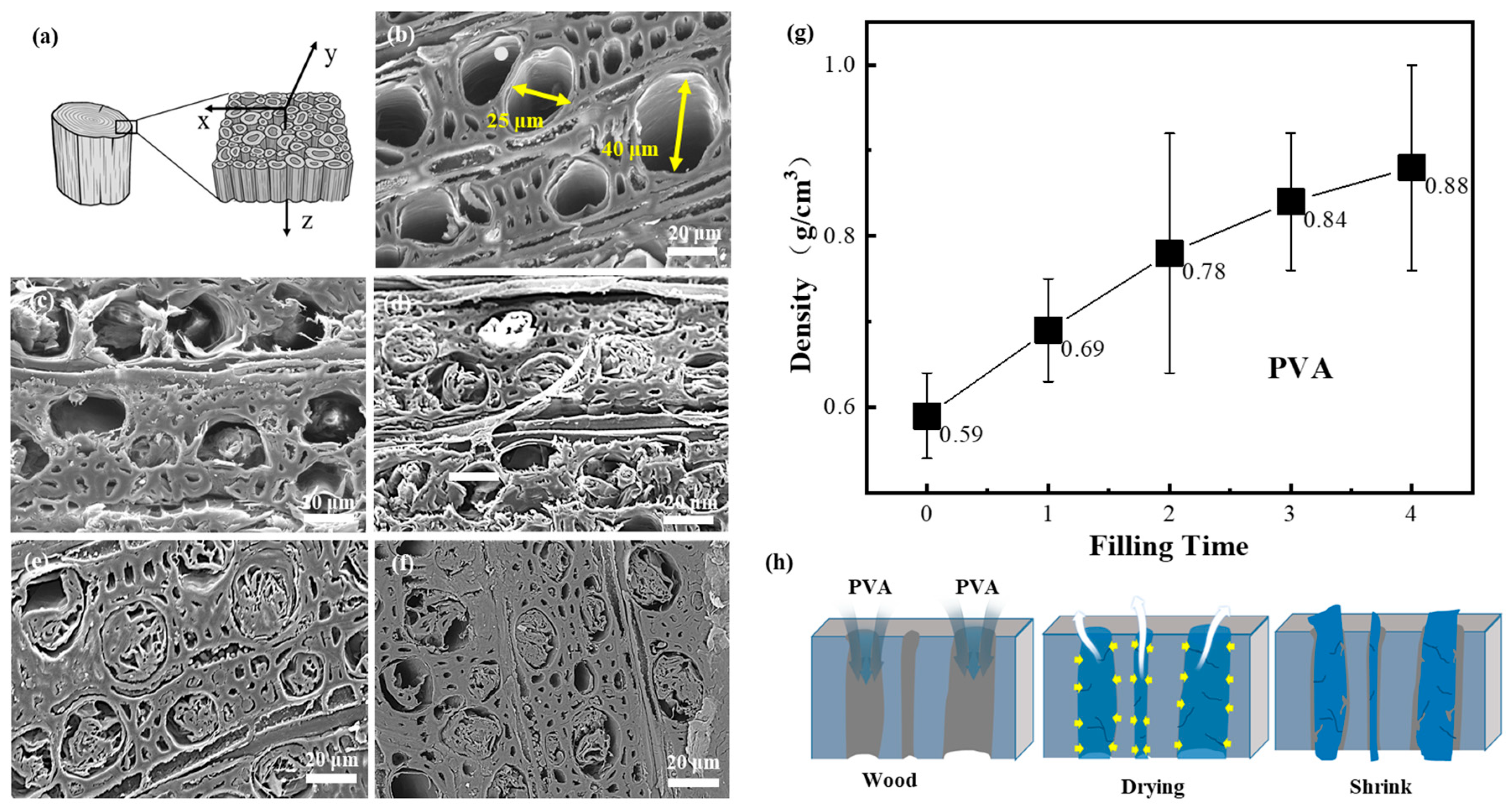
The reinforcement and microbial protection of wood have significant application value in fields such as construction and cultural relic restoration. As shown in Figure 5a, wood is a natural polymer material with an anisotropic and porous structure, which provides conditions for the reinforcement of wood. Given that polyvinyl alcohol is a water-soluble organic polymer, its water-soluble nature allows it to be removed by water washing after being filled into wood pores, making it a promising material for wood restoration. As shown in Figure 5, when polyvinyl alcohol is used as a medium to fill wood pores, it penetrates the cavities under pressure differential and capillary action so that a large amount of filler can be seen in the wood pores. The compressive strength of wood also increased from 155.0 MPa to 213.9 MPa (as shown in Figure 6g). However, due to the shrinkage of polyvinyl alcohol during the drying process, the resulting density was 0.69 g/cm3, representing only a 16.95% increase compared to that of natural wood (as shown in Figure 5g) [29]. While these voids can theoretically be reduced via multiple filling cycles, after four filling cycles, the wood density only increased from 0.59 g/cm3 to 0.88 g/cm3, and its compressive strength rose from 155.0 MPa to 259.2 MPa, suggesting that the improvement was not substantial. This limitation arises because the dried polyvinyl alcohol absorbs moisture from the subsequent filling solution, causing the existing filler to re-expand and thereby impede the infiltration of new filler (Figure 5h). Consequently, relying solely on polyvinyl alcohol as a filler proves to be an unreliable approach for achieving complete densification.
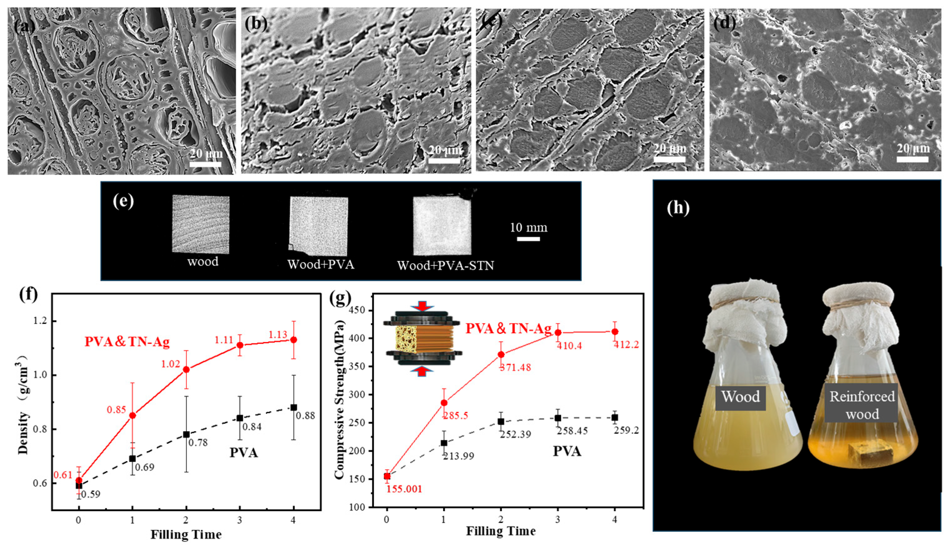
When STN-Ag is used as an antifungal agent and added to the polyvinyl alcohol matrix, as can be seen from Figure 6, the filling effect of the filler on the wood is improved [31]. The resulting composite achieved a density of 0.85 g/cm3 after one filling process, marking a 23.19% increase compared to wood filled with pure polyvinyl alcohol (0.59 g/cm3). SEM analysis revealed that although minor voids persisted after a single filling cycle, their size was substantially reduced, indicating improved densification. It is worth noting that after multiple filling cycles, the void filling rate shown by the CT reached over 75%, and the final density of the wood was 1.13 g/cm3. As shown in Figure 6a, since STN-Ag is an inorganic nanomaterial, it does not undergo volume changes due to water absorption or dehydration. When incorporated into the polyvinyl alcohol solution, STN-Ag acted as a structural support, inhibiting PVA shrinkage and thereby reducing volumetric changes during drying [32]. In addition, based on the volume effect of filling rate and the synergistic effect of inorganic nanomaterial modification, the mechanical properties of the wood were also significantly improved. For instance, after the first reinforcement cycle, the compressive strength increased from 155.0 megapascals to 285.5 megapascals, which was a 33.5% increase compared to pure polyvinyl alcohol. By the third cycle, the compressive strength reached 412.2 megapascals, which was 2.6 times higher than that of untreated wood.
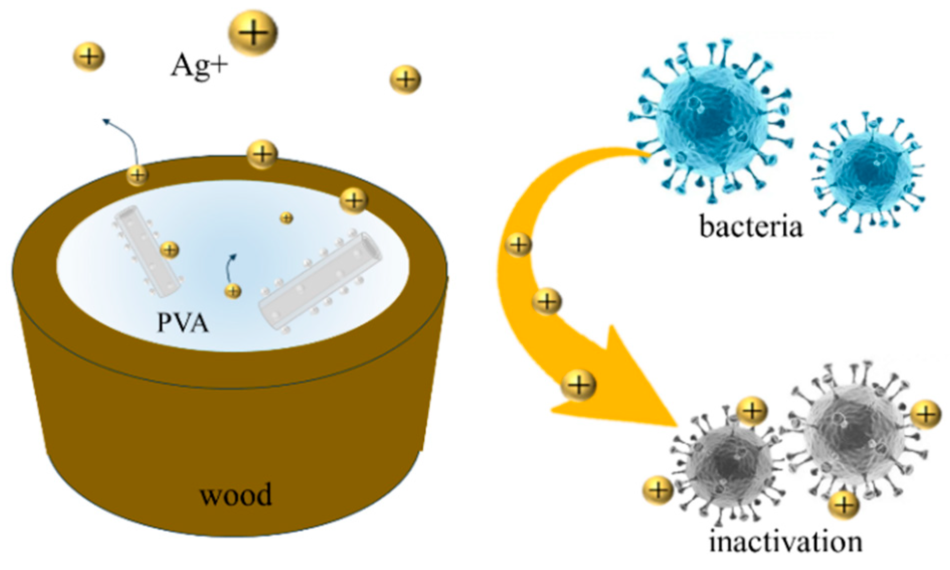
The inhibitory effect of PVA/STN-Ag enhanced wood on mold was tested by using the co-culture method. As shown in Figure 6h, when untreated wood was immersed in a nutrient solution containing Talaromyces funiculosus, the solution became turbid after 48 h due to extensive fungal growth. In contrast, reinforced wood suppressed mold proliferation, maintaining clarity in the solution over extended periods. This is because when a large number of microorganisms appeared in the section, the respiration of the microorganisms changed the pH value of the section. This antifungal mechanism is illustrated in Figure 7. The weak effect prompted the silver ions in STN-Ag to undergo an ion exchange process with the H+ ions in the environment, thereby releasing silver ions with antifungal effects [7]. These results confirm the excellent anti-mold efficacy of the reinforced wood [33].

4. Conclusions

This study proposes the use of polyvinyl alcohol (PVA) as a consolidating agent, and the combination of sodium titanate nanotubes loaded with silver ions (Ag+) as an antifungal additive, to prepare a wood consolidating agent with antifungal properties. Experimental results indicate that while PVA effectively improves the mechanical performance of dried wooden artifacts, its significant shrinkage during the drying process limits consolidation efficacy. However, the incorporation of sodium titanate-silver composite powder as an antifungal agent not only enhances the mechanical strength of consolidated wood but also imparts anti-mold functionality. This method is feasible for reinforcing wood and protecting wooden materials from decay, and can provide valuable insight for technological innovations in building and cultural relic protection.

Funding

This research received no external funding.

Institutional Review Board Statement

Not applicable.

Informed Consent Statement

Not applicable.

Data Availability Statement

All data generated or analyzed during this study are included in the published article.

Conflicts of Interest

The author declares no conflicts of interest.

References

  1. Fahey, L.M.; Nieuwoudt, M.K.; Harris, P. Using near infrared spectroscopy to predict the lignin content and monosaccharide compositions of Pinus radiata wood cell walls. Int. J. Biol. Macromol. Struct. Funct. Interact. 2018, 113, 507–514. [Google Scholar] [CrossRef]
  2. Wu, M.; Jin, J.; Cai, C.; Shi, J.; Cai, J. Effects of impregnation combined heat treatment on the pyrolysis behavior of poplar wood. PLoS ONE 2020, 15, e0229907. [Google Scholar] [CrossRef] [PubMed]
  3. Li, K.; Han, K.; Liu, X.; Ruan, Y.; Li, X.; Zhao, G.; Li, Y.; Luo, Y. Pectin-wood shaving composite for filling wormholes in decorative paintings of historic wooden architecture. J. Cult. Herit. 2025, 74, 120–129. [Google Scholar] [CrossRef]
  4. Glastrup, J.; Shashoua, Y.; Egsgaard, H.; Mortensen, M.N. Degradation of PEG in the warship Vasa. Macromol. Symp. 2010, 238, 22–29. [Google Scholar] [CrossRef]
  5. Broda, M.; Hill, C. Conservation of Waterlogged Wood—Past, Present and Future Perspectives. Forests 2021, 12, 1193. [Google Scholar] [CrossRef]
  6. Priya, S.; Robichaud, J.; Methot, M.-C.; Balaji, S.; Ehrman, J.M.; Su, B.-L.; Djaoued, Y. Transformation of microporous titanium glycolate nanorods into mesoporous anatase titania nanorods by hot water treatment. J. Mater. Sci. 2009, 44, 6470–6483. [Google Scholar] [CrossRef]
  7. Dong, X.; Huang, J.; Li, H.; Ge, C.; Ren, X. Construction of one-dimensional pn heterojunction TiO2/CuO composite with hierarchical structure and its dual efficient inactivation of Escherichia coli. J. Mater. Sci. 2024, 59, 11480–11496. [Google Scholar] [CrossRef]
  8. Adnan, R.; Mezher, M.; Abdallah, A.; Awad, R.; Khalil, M. Synthesis, characterization, and antibacterial activity of Mg-doped CuO nanoparticles. Molecules 2023, 28, 103. [Google Scholar] [CrossRef]
  9. Iani, I.M.; Teodoro, V.; Marana, N.L.; Coleto, U.; Sambrano, J.R.; Simoes, A.Z.; Teodoro, M.D.; Longo, E.; Perazolli, L.A.; Amoresi, R.A.C.; et al. Cation-exchange mediated synthesis of hydrogen and sodium titanates heterojunction: Theoretical and experimental insights toward photocatalyic mechanism. Appl. Surf. Sci. 2021, 538, 148137. [Google Scholar] [CrossRef]
  10. Chen, Q.; Du, G.H.; Zhang, S.; Peng, L.M. The structure of trititanate nanotubes. Acta Crystallogr. 2010, 58, 587–593. [Google Scholar] [CrossRef]
  11. Umek, P.; Korošec, R.C.; Jančar, B.; Dominko, R.; Arčon, D. The influence of the reaction temperature on the morphology of sodium titanate 1D nanostructures and their thermal stability. J. Nanosci. Nanotechnol. 2007, 7, 3502–3508. [Google Scholar] [CrossRef] [PubMed]
  12. Rajeswari, M.; Vanasundari, K.; Mahalakshmi, G.; Ponnarasi, P. Design and Fabrication of High Performance Visible Light Driven H2 Production of N-doped TiO2 Nanotubes Incorporated 2D MoS2 Nanosheets Heterojunction Photocatalyst. J. Clust. Sci. 2023, 34, 2941–2949. [Google Scholar] [CrossRef]
  13. Amy, L.; Favre, S.; Gau, D.L.; Faccio, R. The effect of morphology on the optical and electrical properties of sodium titanate nanostructures. Appl. Surf. Sci. 2021, 555, 149610. [Google Scholar] [CrossRef]
  14. Binay, M.I.; Kirdeciler, S.K.; Akata, B. Development of antibacterial powder coatings using single and binary ion-exchanged zeolite A prepared from local kaolin. Appl. Clay Sci. 2019, 182, 105251. [Google Scholar] [CrossRef]
  15. Banafti, S.; Jahanshahi, M.; Peyravi, M.; Khalili, S. Controllable release activity of antibacterial Ag/SBA-16 cage-like synthesized by one-pot method. Microporous Mesoporous Mater. 2020, 299, 110107. [Google Scholar] [CrossRef]
  16. Salim, M.M.; Malek, N.A.N.N. Characterization and antibacterial activity of silver exchanged regenerated NaY zeolite from surfactant-modified NaY zeolite. Mater. Sci. Eng. C-Mater. Biol. Appl. 2016, 59, 70–77. [Google Scholar] [CrossRef]
  17. Dlugosz, O.; Banach, M. Kinetic, isotherm and thermodynamic investigations of the adsorption of Ag+ and Cu2+ on vermiculite. J. Mol. Liq. 2018, 258, 295–309. [Google Scholar] [CrossRef]
  18. Li, S.; Wang, Q.T.; Yu, H.Q.; Ben, T.; Xu, H.J.; Zhang, J.C.; Du, Q.Y. Preparation of effective Ag-loaded zeolite antibacterial materials by solid phase ionic exchange method. J. Porous Mater. 2018, 25, 1797–1804. [Google Scholar] [CrossRef]
  19. Bitonto, L.; Volpe, A.; Pagano, M.; Bagnuolo, G.; Mascolo, G.; La Parola, V.; Di Leo, P.; Pastore, C. Amorphous boron-doped sodium titanates hydrates: Efficient and reusable adsorbents for the removal of Pb2+ from water. J. Hazard. Mater. 2017, 324, 168–177. [Google Scholar] [CrossRef]
  20. Motlochova, M.; Slovak, V.; Plizingrova, E.; Lidin, S.; Subrt, J. Highly-efficient removal of Pb(ii), Cu(ii) and Cd(ii) from water by novel lithium, sodium and potassium titanate reusable microrods. Rsc Adv. 2020, 10, 3694–3704. [Google Scholar] [CrossRef]
  21. Yang, X.T.; Guo, N.; Yu, Y.; Li, H.Y.; Xia, H.; Yu, H.W. Synthesis of magnetic graphene oxide-titanate composites for efficient removal of Pb(II) from wastewater: Performance and mechanism. J. Environ. Manag. 2020, 256, 109943. [Google Scholar] [CrossRef]
  22. Shi, Q.S.; Tan, S.Z.; Yang, Q.H.; Jiao, Z.P.; Ouyang, Y.S.; Chen, Y.B. Preparation and characterization of antibacterial Zn2+-exchanged montmorillonites. J. Wuhan Univ. Technol.-Mater. Sci. Ed. 2010, 25, 725–729. [Google Scholar] [CrossRef]
  23. Preda, S.; Anastasescu, C.; Balint, I.; Umek, P.; Sluban, M.; Negrila, C.C.; Angelescu, D.G.; Bratan, V.; Rusu, A.; Zaharescu, M. Charge separation and ROS generation on tubular sodium titanates exposed to simulated solar light. Appl. Surf. Sci. 2019, 470, 1053–1063. [Google Scholar] [CrossRef]
  24. Bayisa, T.; Deressa, G.; Feyisa, Z.; Inki, L.G.; Gupta, N.K. In-situ synthesis of CuO/TiO2 nanocomposite onto amine modified cotton fabric for antibacterial durability and UV protection. J. Nat. Fibers 2024, 21, 2346120. [Google Scholar] [CrossRef]
  25. Hao, L.; Ju, P.; Zhang, Y.; Sun, C.J.; Dou, K.P.; Liao, D.K.; Zhai, X.F.; Lu, Z.X. Novel plate-on-plate hollow structured BiOBr/Bi2MoO6 p-n heterojunctions: In-situ chemical etching preparation and highly improved photocatalytic antibacterial activity. Sep. Purif. Technol. 2022, 298, 121666. [Google Scholar] [CrossRef]
  26. Dong, X.; Lv, X.; Huang, J.; Chang, Y.; Ren, X.; Ge, C. Preparation of one-dimensional hierarchical sodium titanate under mild conditions and its potential application in recyclable Ag+-loaded antimicrobials. Colloids Surf. A Physicochem. Eng. Asp. 2023, 676, 8. [Google Scholar] [CrossRef]
  27. Shi, H.; Fu, L.; Xue, L.; Lu, H.; Zhou, Q. Enhancing antibacterial performances of PVDF hollow fibers by embedding Ag-loaded zeolites on the membrane outer layer via co-extruding technique. Compos. Sci. Technol. 2014, 96, 1–6. [Google Scholar] [CrossRef]
  28. Hrenovic, J.; Milenkovic, J.; Ivankovic, T.; Rajic, N. Antibacterial activity of heavy metal-loaded natural zeolite. J. Hazard. Mater. 2012, 201, 260–264. [Google Scholar] [CrossRef]
  29. Sun, Z.; Zuo, Y.; Li, P.; Wu, Y.; Wang, Z.; Li, X.; Lyu, J. Hyperbranched organic-inorganic co-modification improves the strength, dimensional stability, and thermal stability of poplar wood. Ind. Crops Prod. 2023, 191, 115923. [Google Scholar] [CrossRef]
  30. Zhu, M.; Li, T.; Davis, C.S.; Yao, Y.; Dai, J.; Wang, Y.; Alqatari, F.; Gilman, J.W.; Hu, L. Transparent and haze wood composites for highly efficient broadband light management in solar cells. Nano Energy 2016, 26, 332–339. [Google Scholar] [CrossRef]
  31. Liu, X.M.; Zhang, X.; Long, K.; Zhu, X.; Yang, S. PVA wood adhesive modified with sodium silicate cross-linked copolymer. In Proceedings of the 2012 International Conference on Biobase Material Science and Engineering; IEEE: New York, NY, USA, 2013; pp. 108–111. [Google Scholar]
  32. Riggio, M.; Sandak, J.; Sandak, A.; Pauliny, D.; Babinski, L. Analysis and prediction of selected mechanical/dynamic properties of wood after short and long-term waterlogging. Constr. Build. Mater. 2014, 68, 444–454. [Google Scholar] [CrossRef]
  33. Zhang, Y.; Bi, X.; Li, P.; Wu, Y.; Yuan, G.; Li, X.; Zuo, Y. Sodium silicate/magnesium chloride compound-modified Chinese fir wood. Wood Sci. Technol. 2021, 55, 1781–1794. [Google Scholar] [CrossRef]
Figure 1. (a) Morphology of TiO2 before the reaction; (b) morphology of sodium titanate after the reaction; (c) XRD patterns before and after the hydrothermal reaction; (d) N2 adsorption–desorption curves; (e) transmission electron microscope (TEM) image of sodium titanate; (f) magnified view of the transmission electron microscope image.
Coatings 16 00508 g001
Figure 2. Dynamic adsorption process.
Coatings 16 00508 g002
Figure 3. (a,b) The distribution of elements STN and STN-Ag, respectively, (c) the statistical table of each element content, and (d) the Fourier infrared spectrum of the sample.
Coatings 16 00508 g003
Figure 4. Antifungal effect diagrams of the (a) Tripitaka edition, (b) microbial isolation effect, (c) fungistatic zone and (d) minimum bacteriostatic concentration and minimum bactericidal concentration.
Coatings 16 00508 g004
Figure 5. Reinforcement effect of PVA on wood. (a) The cellular structure of wood, (b) the hole size of pure wood, (cf) SEM images of wood with different filling times, (g) density of different wood samples, and (h) hole formation process during drying [30].
Coatings 16 00508 g005
Figure 6. Reinforcement effect on wood after adding antifungal agent (a) once, (b) twice, (c) thrice, and (d) four times. (e) CT images of filled-effect, (f) density of wood samples in different fillings, (g) compressive strength of enhanced wood, and (h) antifungal activity diagram.
Coatings 16 00508 g006
Figure 7. Antifungal mechanism.
Coatings 16 00508 g007
Table 1. Silver content of Ag-loaded antibacterial prepared by different processes.
AbsorbentLoading MethodAdsorbing Capacity
(wt.%)
Ref.
Zeolite ASolution ion-exchange5.3[14]
SBA-16One-step hydrothermal synthesis6.7[15]
Na-Y zeoliteSolution ion-exchange for 16 h9.0[16]
VermiculiteSolution ion-exchange for 60 min6.8[17]
ZeoliteSolid phase ion-exchange23.45[18]
Disclaimer/Publisher’s Note: The statements, opinions and data contained in all publications are solely those of the individual author(s) and contributor(s) and not of MDPI and/or the editor(s). MDPI and/or the editor(s) disclaim responsibility for any injury to people or property resulting from any ideas, methods, instructions or products referred to in the content.