Review

Recent Trends in Gelatin Electrospun Nanofibers: Advances in Fabrication, Functionalization, and Applications

by
Bruna Silva de Farias
1,*,
Anelise Christ Ribeiro
1,
Débora Pez Jaeschke
1,
Eduardo Silveira Ribeiro
1,
Janaína Oliveira Gonçalves
2,
Ricardo Freitas Vergara
1,
Sibele Santos Fernandes
1,
Daiane Dias
1,
Tito Roberto Sant’Anna Cadaval Jr.
1 and
Luiz Antonio de Almeida Pinto
1
1
School of Chemistry and Food, Federal University of Rio Grande (FURG), Rio Grande 96203-900, Brazil
2
Department of Civil and Environmental, Universidad de la Costa CUC, Calle 58 #55-66, Barranquilla 080002, Colombia
*
Author to whom correspondence should be addressed.
Coatings 2025, 15(9), 1110; https://doi.org/10.3390/coatings15091110
Submission received: 17 August 2025 / Revised: 14 September 2025 / Accepted: 19 September 2025 / Published: 21 September 2025
(This article belongs to the Special Issue Advances and Trends in Bio-Based Electrospun Nanofibers)

Abstract

Bio-based nanofibers are gaining increasing attention in nanotechnology owing to their high surface area, interconnected porosity, and capacity to incorporate bioactive compounds. Among natural polymers, gelatin is particularly attractive because of its abundance, low cost, biodegradability, and versatile physicochemical properties. When processed by electrospinning, gelatin combines its amphiphilic nature with the structural advantages of nanofibers, enabling efficient interactions with a wide range of molecules. Nevertheless, pure gelatin nanofibers have drawbacks, such as poor mechanical strength and high-water solubility. To address these limitations, strategies including polymer blending, chemical and physical crosslinking, and multilayer biomaterials have been developed, resulting in improved stability, functionality, and application-specific performance. Therefore, this review summarizes recent advances in the fabrication and functionalization of gelatin nanofibers, highlighting how processing parameters and gelatin source influence electrospinning outcomes and fiber properties. Key applications are also discussed, with emphasis on biomedical, food, environmental, and biosensing. Therefore, gelatin nanofibers represent a sustainable and versatile biomaterial with high potential for advanced technological applications.

Graphical Abstract

1. Introduction

Polymeric nanofibers emerge as one of the most innovative solutions in the field of nanotechnology, particularly due to their structural characteristics that can provide unique properties. Nanofibers are defined as continuous fibers with a reduced diameter, typically ranging from 50 to 500 nm. This configuration results in a high specific surface area and interconnected porosity, as well as allowing for easy surface modifications [1]. These characteristics favor applications where the interaction between the material and the external environment (cellular, chemical, or biological) is crucial, such as in tissue engineering, advanced filtration, controlled drug release, and sensors [2].
The predominant technique for producing nanofibers is electrospinning, which combines operational simplicity and efficiency, low cost and the ability to produce continuous and uniform nanofibers on a laboratory or semi-industrial scale. The electrospinning method involves applying a high-voltage electric field to a polymer solution, which, when ejected from the needle, forms a polarized fluid jet that stretches and solidifies into continuous fibers upon reaching the polarized collector [3]. Process parameters influence the diameter, uniformity, and final morphology of the nanofibers [4]. Furthermore, electrospinning can be used for the development of more complex structures, such as core–shell fibers, multilayers, or composites, enabling the incorporation of drugs, enzymes, or nanoparticles, thus expanding the potential for specific applications [5].
Polymeric nanofibers can be classified into two main types: synthetic and biological. Synthetic nanofibers, such as those produced from polylactic acid (PLA) or polycaprolactone (PCL), are valued for their good mechanical properties and stability; however, they may have limitations in terms of biocompatibility and environmental impact [6]. In contrast, biological nanofibers, obtained from natural polymers, such as collagen, gelatin, chitosan, and hyaluronic acid, have attracted significant attention due to characteristics including biodegradability, sustainability, renewability, and the presence of reactive functional groups [7]. This versatility makes them increasingly explored in areas, such as wound dressings, sustainable packaging, detection devices, and adsorption of organic pollutants [8].
Among natural polymers, gelatin stands out as a strategic option due to its abundance, affordability, and bioactive properties. Gelatin is a heterogeneous mixture of polypeptides derived from the controlled hydrolysis of collagen. Industrial production typically involves pre-treatment using acid (Type A), base (Type B), or a combination of both, followed by thermal extraction processes [9,10]. During collagen hydrolysis, hydrogen bonds stabilizing the triple-helix are disrupted, and peptide bonds, along with other covalent crosslinks, are cleaved. This process yields α-chains, as well as β and γ aggregates composed of two or three polypeptide chains, respectively. The β and γ aggregates originate from crosslinked α-chains that were not completely separated during hydrolysis. The specific operating conditions used during gelatin production, such as temperature, pH, processing time, and hydrolysis method, affect the extent of chain cleavage and, consequently, the relative amounts of α-chains and β/γ aggregates. This, in turn, determines the molecular weight distribution of the gelatin and influences its physicochemical properties. Indeed, gelatin has the ability to partially reconstitute collagen-like triple helix structures, usually, around 25–35 °C, forming junction zones that enhance intra- and intermolecular interactions. Above this temperature, the helices dissociate, and the gelatin assumes a random coil conformation in solution [6,11,12,13].
The ability of gelatin to form junction zones upon cooling is strongly influenced by its amino acid composition and molecular weight. Proline and hydroxyproline, abundant in mammalian gelatin, but present at lower levels in fish gelatin, especially from cold-water species, can restrict chain flexibility and stabilize collagen-like triple helices through hydrogen bonding, thereby enhancing junction zone formation and increasing gel strength [14,15]. The molecular weight, determined by the relative proportions of α-chains and β and γ aggregates, depends on both the collagen source and extraction conditions. Higher proline/hydroxyproline content can protect the native structure during processing, preserving more β and γ aggregates and leading to higher molecular weight gelatin. Longer chains and aggregates provide more interaction sites for helix reconstitution, whereas low molecular weight fractions with fewer stabilizing imino acids form fewer and weaker junction zones, resulting in softer gels and lower Bloom strength [6,14,16].
Among gelatin-based biomaterials, electrospun gelatin nanofibers (GNFs) have garnered significant attention due to their high surface area, porosity, and tunable morphology. These structural features, combined with the amphiphilic nature of gelatin, containing both polar and nonpolar functional groups, enable effective interactions with a broad range of molecules [17,18]. As a result, GNFs can provide a versatile biomaterial for multiple applications, including wound dressings in biomedical applications, active packaging in food applications, biosorbents for the removal of organic pollutants in environmental applications and substrates for immobilizing enzymes in electroanalytical and biosensing applications [19,20,21,22]. Therefore, this review aims to discuss how the physicochemical properties of gelatin affect the electrospinning process and to highlight its wide range of applications.

2. Methodology

A structured bibliographic search was conducted to identify the most relevant publications on gelatin-based electrospun nanofibers. Searches were performed in PubMed, ScienceDirect, and Google Scholar. Combinations of keywords and Boolean operators were used, including the following: gelatin nanofibers AND electrospinning, crosslink OR cross-link, blending, functionalization, bovine gelatin, porcine gelatin, cold-water fish gelatin, warm-water fish gelatin, water stability, mechanical, degradation OR erosion, antimicrobial, antiviral, drug release, tissue engineering, wound healing, food packaging, smart packaging, intelligent packaging, monolayer packaging, bilayer packaging, pollutants, air filtration, ion detection, dye removal, heavy metal removal, biosensor, piezoelectric, colorimetric, and electrochemical. A saturation criterion was applied as the stopping rule; the search was concluded when additional queries and citation chaining no longer yielded substantively new information. Inclusion criteria were as follows: (i) direct relevance to fabrication, modification, characterization, or applications of gelatin-based electrospun nanofibers; (ii) peer-reviewed articles, reviews, or book chapters in English; and (iii) publications within 2000–2025 to capture both foundational and recent advances.

3. Electrospinning of Gelatin Nanofibers

The electrospinning technique involves the ejection of a charged polymer solution jet under a high-voltage electric field. When the applied electrostatic force overcomes the solution’s surface tension, a Taylor cone forms at the spinneret tip, from which a fine jet is expelled and drawn toward a grounded collector (Figure 1). During this process, the jet undergoes stretching and whipping, reducing its diameter and forming nanofibers. Therefore, operational and environmental parameters should be studied and controlled in order to achieve the required conditions for nanofibers development. Furthermore, to achieve stable fiber formation, the gelatin solution must exhibit appropriate physicochemical characteristics [23,24].
The molecular weight of gelatin, encompassing α-chains as well as β and γ aggregates, is relevant in electrospinning performance. This parameter is influenced by both the collagen source and the hydrolysis method employed. Type A gelatin, produced via acid hydrolysis, generally preserves a higher proportion of intact α-chains and aggregates, resulting in molecular weights usually ranging from 90 to 100 kDa or higher. In contrast, Type B gelatin, derived from alkaline hydrolysis, undergoes more extensive peptide bond cleavage, leading to a broader distribution toward lower molecular weight, typically between 40 and 90 kDa [25,26,27]. Despite its importance, the molecular weight of gelatin remains underexplored in comparison to other processing parameters, such as concentration, solvent type, and electrospinning conditions.
Farias et al. compared the development of nanofibers using porcine, bovine, and fish gelatin, with acetic acid (30% v/v) as the solvent [6]. The molecular weights of porcine, bovine, and fish gelatin were 98.4, 48.8, and 24.8 kDa, respectively, reflecting differences attributed to the distinct raw sources and extraction processes used for each gelatin. Moreover, the electrospinning behavior of bovine, porcine, and fish gelatin showed differences due to their molecular weight variations. For bovine gelatin, fiber formation began at 20% w/v, but at this concentration, a mixture of droplets and fibers was observed, indicating incomplete polymer chain entanglement; uniform fibers were only achieved at higher concentrations (25%–30% w/v). In contrast, porcine gelatin, which has a higher molecular weight, was able to form continuous and uniform fibers already at 20% w/v, without the presence of droplets, highlighting its superior chain entanglement ability at lower concentrations. Fish gelatin, presenting the lowest molecular weight among the three, required the highest concentration to form fibers, with fiber formation beginning only at 25% w/v and uniform, droplet-free fibers observed at 35% w/v, reflecting the need for greater polymer concentration to achieve sufficient chain entanglement.
Another important solution parameter is the viscosity. This parameter must be high enough to resist Rayleigh instabilities, but not so high as to block the spinneret. Viscosity is closely linked to both concentration, amino acid composition and molecular weight. At low viscosities, insufficient chain entanglement leads to bead formation or spray-like deposition, as previously discussed. As viscosity increases, fibers become more uniform in diameter. Moreover, the concentration plays an important role. Below the minimum spinnability threshold, polymer chains lack sufficient overlap and entanglement, resulting in bead-rich morphologies. As concentration increases, fiber uniformity improves until a plateau is reached. Excessive concentration, however, can increase viscosity beyond the optimal range, leading to uneven, coarse fibers or spinneret clogging [4,28]. Nevertheless, the temperature of gelatin solution also affects the intermolecular interactions, as well as the presence of junction zones, thereby influencing the viscosity of the solution. In mammalian gelatins, the high junction zone density can lead to more viscous solutions until 35 °C, potentially requiring higher applied voltages or solvent modifications to achieve stable fiber formation. In contrast, cold-water fish gelatin, with its lower proline/hydroxyproline content and reduced junction zone stability at room temperature, produces less viscous, more flexible solutions. This reduced viscosity, combined with its lower molecular weight, facilitates electrospinning of cold-water gelatin solution.
The properties of the solvent, such as surface tension, boiling point, dielectric constant, polarity, density, and volatility, play an important role in the electrospinning process. An ideal solvent should evaporate rapidly to ensure fiber solidification before the jet reaches the collector. Solvents with high boiling points or low volatility can cause bead formation or fiber fusion due to insufficient drying. In addition, solvent polarity influences both the conductivity of the solution and the solubility of gelatin [29,30,31]. This protein readily dissolves in water due to its molecular structure, which includes polar side chains, amino groups at the N-terminal, and carboxylic acid groups at the C-terminal. These functional groups form hydrogen bonds and electrostatic interactions with water molecules, enhancing solubility in aqueous media. This enables gelatin to be dissolved in water, a green, non-toxic solvent [15,18].
However, water’s high surface tension (~72.8 mN/m) typically hinders fiber formation during electrospinning, often requiring the use of acetic acid to dissolve gelatin, which has a lower surface tension (~27.6 mN/m), to facilitate fiber formation. Nevertheless, cold-water fish gelatin can be electrospun directly from aqueous solutions. This capability is attributed to its lower molecular weight, reduced proline and hydroxyproline content (~15%–20%), and lower Bloom strength (~40–100 g), compared to bovine, porcine, and warm-water fish gelatins, which typically contain higher imino acid levels (~25%–33%) and exhibit Bloom strengths of ~200–490 g [10,32,33,34,35,36,37]. These characteristics result in lower viscosity and weaker intermolecular interactions, which reduce the impact of water’s high surface tension and enable successful nanofiber formation without the need for organic solvents. Therefore, further studies should focus on elucidating how processing parameters in gelatin production influence the amino acid profile, molecular weight, viscosity, and Bloom strength, with the aim of enhancing the electrospinnability of aqueous gelatin solutions. Furthermore, the physicochemical properties of gelatin derived from different cold-water fish species should be carefully investigated to optimize their suitability for nanofiber production.

4. Physicochemical Properties of Gelatin Nanofibers

Table 1 presents a comprehensive overview of how different electrospun gelatin-based nanofiber systems are formulated, and characterized with respect to their physicochemical properties. The amphiphilic nature of gelatin, containing both hydrophilic and hydrophobic functional groups, makes it particularly suitable for structuring oil-rich systems, such as oleogels. In conventional oleogel development, indirect approaches have commonly employed water-soluble proteins and polysaccharides to form gel networks through methods, such as solvent exchange, emulsion templating, or foam templating. While effective, these techniques often result in stiff, inflexible matrices and involve complex fabrication steps that hinder their practical application. Electrospinning, in contrast, can provide a more versatile platform by producing nanofibers with high surface area and tunable morphology, enabling improved interactions within oleogel matrices. While traditional 2D nanofiber mats cannot form fully interconnected 3D networks, recent advances demonstrate that combining aerogel matrices with short electrospun gelatin fibers can enhance the mechanical resilience and functional performance of oleogels. These hybrid systems exhibit moderate flexibility, allowing deformation under stress without fracturing, although they are generally less flexible than fully polymeric gels [38].
On the other hand, in most applications, gelatin used as the sole polymer in electrospinning presents significant processing challenges. The hydrophilic nature of gelatin, combined with lack of crystallinity and its heterogeneous molecular structure, results in nanofibers with poor structural integrity and low mechanical strength. Moreover, gelatin nanofibers (GNFs) are highly soluble in aqueous environments, which restricts their use in applications where water stability is essential. To mitigate these limitations, strategies including polymer blending, chemical or physical modifications, and various crosslinking techniques have been employed to enhance both the spinnability and functional properties of gelatin-based nanofibers [39].

4.1. Crosslinking Strategies for Gelatin Nanofibers

One of the major strategies to overcome the high solubility and low mechanical stability of GNFs is the use of crosslinkers. Table 2 shows a comprehensive overview of different electrospun gelatin-nanofibers systems after post-treatment. Indeed, different crosslinking approaches, chemical, natural, enzymatic, and polysaccharide-based, have been reported, each presenting distinct mechanisms and impacts on the physicochemical properties of the nanofibers. Chemical crosslinkers, such as glutaraldehyde (GTA) and 1,4-butanediol diglycidyl ether (BDDGE) react with the free amine groups of gelatin to form covalent bonds, thereby improving physicochemical properties. For example, GNFs treated with GTA vapor displayed increased thermal stability, while BDDGE crosslinking at higher concentrations and times resulted in fibers with stable diameters and enhanced strength [40,41]. Although effective, these reagents may raise concerns regarding cytotoxicity, particularly for biomedical or food-related applications.
Natural crosslinkers, such as genipin, provide a safer alternative. Genipin reacts with the primary amine groups of gelatin through the formation of stable heterocyclic structures, leading to nanofibers with higher decomposition temperatures (>250 °C) and improved mechanical performance [42]. Compared to GTA, genipin is less toxic, which makes it attractive for pharmaceutical and biomedical applications.
Enzymatic crosslinkers, particularly microbial transglutaminase (mTG), catalyze the formation of ε-(γ-glutamyl) lysine isopeptide bonds between gelatin chains. This process reduces chain mobility, increases glass transition temperature, and enhances the overall thermal stability of nanofibers [43]. Polysaccharide-based crosslinking can also be achieved through Schiff base reactions, in which aldehyde groups from oxidized xanthan gum interact with free amine groups of gelatin to form imine linkages. This strategy was shown to improve tensile strength (up to 13.2 MPa), reduce solubility, and enhance the antioxidant and antibacterial activity of the nanofibers [44].
In addition, composite-assisted stabilization has been reported, where inorganic fillers (e.g., metal–organic frameworks, ZnO nanoparticles) or biopolymers (e.g., chitosan) synergistically reinforce the network, complementing crosslinking by providing additional stabilization or bioactive functionalities [38,45,46].
Overall, crosslinking represents a critical step for tailoring the physicochemical and thermal properties of GNFs. While chemical crosslinkers ensure strong covalent bonding, their toxicity must be carefully considered. Natural and enzymatic agents, on the other hand, offer safer and more sustainable alternatives, whereas polysaccharide-based approaches provide multifunctional properties. The choice of crosslinking strategy should therefore be guided by the intended end-use, balancing stability, functionality, and biocompatibility.

4.2. Morphology, Thermal and Mechanical Properties

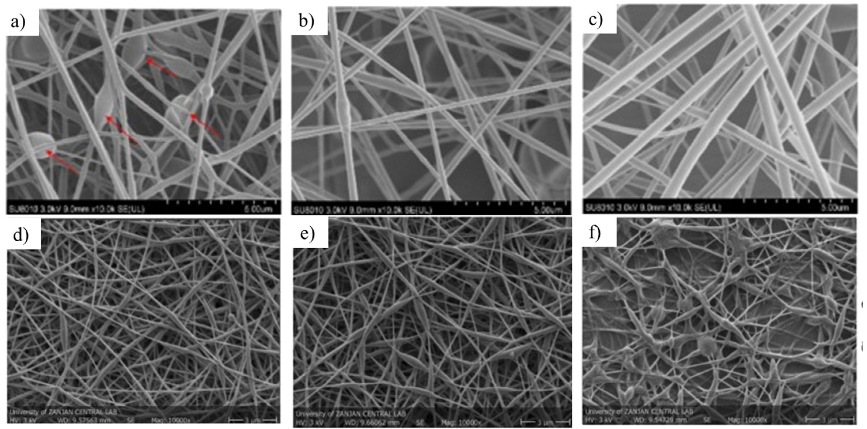
The SEM images in Figure 2 highlight how the type and proportion of the polymers strongly influence the morphology of gelatin-based nanofibers. In the wheat gluten/gelatin system, pure wheat gluten fibers (a) exhibited irregular morphology with multiple beads (indicated by arrows), whereas pure gelatin fibers (b) showed a more uniform structure but still with some defects. Interestingly, the blended formulation (16:9, c) resulted in smoother and more continuous fibers, suggesting that the miscibility between the two proteins can reduce surface tension instabilities during electrospinning.
For the PVA/gelatin system, distinct trends were also observed. At a higher PVA content (8:2, d), fibers appeared thinner, smooth, and well-distributed, while increasing the gelatin fraction (7:3, e) slightly decreased uniformity but still maintained bead-free structures. At the 5:5 ratio (f), the network became denser and more interconnected, with visible junctions between fibers, which may enhance mechanical integrity but could also alter porosity and permeability.
While some of these approaches have resulted in composite gelatin systems with markedly improved functionality, they still face challenges in replacing synthetic polymers, particularly with respect to mechanical properties, moisture and gas barrier capabilities, and controlled delivery of active compounds. In this context, surface modification of GNFs using plasma methods has recently gained attention. Treatments with helium or nitrogen plasma can increase surface functionality, such as hydrophilicity and reactive group density, without affecting the bulk fiber structure. These modifications improve cell adhesion and proliferation, making them particularly useful for tissue engineering scaffolds, including skin regeneration applications [49].
Moreover, multilayer systems have gained attention. By combining distinct polymer layers, each tailored for specific functionalities, such as structural support, moisture resistance, or active compound encapsulation, these architectures enable synergistic enhancements in performance, resulting in materials that are both robust and multifunctional [47]. For example, Talib Al-Sudani et al. [50] developed bilayer electrospun films using gelatin and polymethyl-methacrylate that exhibited a 61.8% reduction in water vapor permeability and enhanced tensile strength, which was attributed to the outer hydrophobic layer. Duan et al. [51] designed multilayer scaffolds incorporating curcumin in gelatin/chitosan nanofibers, enhancing both the mechanical property and bioactive delivery capacity of the system. Moreover, Zhang et al. [42] reported a core–shell nanofiber configuration that enabled prolonged release of ciprofloxacin, suggesting potential in food packaging and biomedical coatings.
Across the studies presented in Table 1 and Table 2, several key improvements were observed. Enhanced thermal stability was reported in systems incorporating compounds, such as poly-sulphonic acid diphenyl aniline, cerium-doped hydroxyapatite, chitosan, curcumin, propolis, or lycopene, primarily due to crosslinking reactions and reinforcement with nanoparticles or bioactive compounds. Tg and Tm values of gelatin-based electrospun nanofibers vary widely depending on the type of composite and modification strategy. For instance, gelatin/wheat gluten fibers exhibit Tg values between 67 and 75 °C and Tm ranging from 124 to 148 °C, whereas gelatin/PVA blends present much higher transitions, with Tg between 104 and 126 °C and Tm up to 265 °C, reflecting the stabilizing effect of PVA. Lycopene incorporation in multilayer gelatin systems also increases Tg (63–89 °C), while enzymatic crosslinking with mTG results in higher glass transitions compared to neat gelatin. Even more pronounced effects are observed for core–shell gelatin/gum arabic fibers crosslinked with genipin, which retain thermal stability with Tm close to 395 °C. In general, the addition of nanoparticles or reinforcing agents (e.g., Ce-HA, ZnO, PSDA, chitosan/curcumin) consistently enhances both Tg and Tm, indicating improved resistance to thermal degradation.
Significant gains in mechanical performance, including increased tensile strength, Young modulus, and elongation at break were achieved through chemical and enzymatic crosslinkers (such as glutaraldehyde, genipin, Schiff base, and microbial transglutaminase) as well as reinforcing agents, including chitosan, PCL, and oxidized xanthan gum. For instance, a formulation combining oxidized xanthan gum and propolis reached a tensile strength of 13.2 MPa, approximately ten times greater than that of pure gelatin.
In terms of barrier properties, several formulations effectively reduced water solubility and water vapor permeability, particularly those involving hydrophobic additives or crosslinking strategies, such as shellac, cerium-doped hydroxyapatite, polymethyl-methacrylate, and metal–organic frameworks. A notable example is a bilayer film that achieved a 28.5% decrease in solubility and a 61.8% reduction in water vapor permeability [47]. Morphological improvements, such as the formation of continuous fibers with fewer beads, were observed following adjustments in formulation and the incorporation of emulsifiers. The inclusion of anthocyanins, for example, facilitated a morphological transition from bead-like structures to uniform fibers [48].

4.3. Bioactive Compound Encapsulation and Functional Properties

From the studies presented in Table 1 and Table 2, many systems loaded with bioactive compounds, including quercetin, curcumin, eugenol, ciprofloxacin, and lycopene, demonstrated sustained release profiles, which were closely associated with crosslinking density and the structural design of the fibers, such as core–shell or emulsion-based configurations [18,46,51,52]. Modifications to the electrospinning solution rheological behavior, such as increased conductivity and elasticity induced by agents, such as genipin, also contributed to improved fiber formation and performance [42]. Finally, enhanced antioxidant, antimicrobial, and mucoadhesive properties were frequently reported, reinforcing the applicability of these gelatin-based nanofibers in both biomedical and food-related contexts.

4.4. Overall Trends in Physicochemical Improvements

From the studies summarized in Table 1 and Table 2, some tendencies can be identified. Thermal stability of GNFs is generally enhanced by covalent crosslinking (e.g., GTA, genipin, mTG) and by the incorporation of reinforcing agents, such as nanoparticles or bioactive compounds. Mechanical strength, usually low in pure gelatin fibers, can be increased several-fold through crosslinking or blending with polymers like chitosan, PCL, or PMMA. Barrier properties, particularly water solubility and vapor permeability, are effectively reduced by hydrophobic additives and multilayer designs. Morphological control (bead-free, continuous fibers) is strongly linked to polymer miscibility and emulsifier use, which in turn improves reproducibility and stability. In addition, vapor-induced phase separation, non-solvent-induced phase separation, thermally induced phase separation, and selective removal are strategies that can be further explored to obtain porous morphologies. Finally, encapsulation of bioactives not only adds antioxidant or antimicrobial functions but also contributes to sustained release and structural reinforcement. Overall, these trends show that physicochemical performance can be systematically tuned by combining crosslinking, blending, and nanofiller strategies according to the intended application. Despite these improvements, some drawbacks have been reported, including reduced flexibility of the fibers after incorporation of rigid nanoparticles, possible cytotoxicity of certain inorganic fillers, and the need for complex optimization when combining gelatin with synthetic polymers such as PVA. These aspects highlight the trade-off between enhanced physicochemical performance and processing or biocompatibility limitations.
Table 1. Overview of physicochemical improvements in gelatin-based electrospun nanofibers and composite systems without post-processing treatment.
Nanofiber MaterialPreparation MethodPost-Processing TreatmentPhysicochemical EffectsThermal Stability Ref.
Bilayer film of balangu seed mucilage–gelatin (base layer) and polyvinyl acetate (PVA)–gelatin nanofiber layer with Fe3O4 nanoparticlesCasting for base layer; electrospinning of PVA–gelatin layer with Fe3O4 dispersed by sonication-The electrospun layer improved mechanical strength, hydrophobicity, and barrier properties. It decreased moisture content (37.4%), solubility (28.5%), swelling (35%), water vapor permeability (61.8%) and oxygen permeability (31.5%).Tg and Tm increased with the increase in PVA content in the fibers (Tg: 104–126 °C
Tm: 245–265 °C).
[47]
Polyamide/gelatin nanofibers with cerium-doped hydroxyapatite (Ce-HA)Electrospinning of PA/gelatin solution with Ce-HA nanoparticles (prepared by precipitation)-Cerium doping increased hydrophilicity (contact angle from 38° to 0°), degradation rate, and mechanical properties.-[53]
Anthocyanin-enriched wheat gluten/gelatin electrospun nanofiber filmsElectrospinning from wheat gluten/gelatin solutions (dissolution + stirring)-Compared with pure wheat gluten, an adjusted ratio of the wheat gluten to gelatin (16:9) improved physicochemical properties, reducing viscosity from 3.56 to 2.75 Pa·s and increasing conductivity from 1.53 to 2.06 mS/cm. SEM showed a transition from bead-like to fibrous morphology (691.6 nm diameter). The films exhibited improved thermal properties, and increased water solubility from 38% to 45%.Gelatin confers better thermal stability than wheat gluten (Tg: 67–75 °C; Tm: 124–148 °C).[48]
Silk fibroin/gelatin nanofibers Electrospinning from silk fibroin/gelatin blends (various ratios) in formic acid-The addition of gelatin to silk fibroin increased fiber diameter, hydrophilicity, and mass loss, but decreased Young’s modulus, tensile strength, and porosity.-[54]
Gelatin/poly(sulphonic acid diphenyl aniline) (PSDA) nanofibersElectrospinning of gelatin in 1% acetic acid with PSDA (10%–20%)-Improved thermal stability, storage modulus, and oxidation current with PSDA content.Thermal stability gradually increases as the PSDA ratio increases in Gel/PSDA nanofibers (Tg and Tm data not available)[39]
Gelatin/xanthan gum nanofibers with chitin and black barberry anthocyaninsElectrospinning from gelatin (12 g) and xanthan gum (1 g) in ethanol/acetic acid/water (45:45:10)-The nanofibers containing chitin and anthocyanins presented: improved thermal stability; decreased crystallinity, tensile strength, solubility, and water vapor permeability; enhanced antioxidant properties.
compared to the straight nanofibers. Other properties of the nanofibers containing chitin and anthocyanins, including tensile strength, water vapor permeability, moisture content, and water solubility, were significantly lower than the straight nanofibers.
The increase in chitin and anthocyanins concentration increased the thermal stability of the nanofibers (Tg and Tm data not available)[46]
Lycopene-loaded gelatin nanofibers (tri-layer structure)Gelatin (30% w/v) dissolved in 30% acetic acid, with lycopene (2% w/v) and Tween 80® (0.5%) added; emulsion prepared by low-energy emulsification (40 °C, 1000 rpm); -Lycopene–gelatin nanofibers had average diameter of 139 ± 29 nm and showed improved structural stability and crystallinity. Tri-layer system enhanced molecular interactions among gelatin, chitosan, and lycopene, increasing lycopene bioaccessibility to 28.5%.Tg was lower in gelatin nanofibers compared to lycopene-loaded electrospun nanofibers (Tg: 63–89 °C).[18]
Polymethyl-methacrylate (PMMA)/gelatin nanofibers with propolisElectrospinning of PMMA/Gelatin (70:30); propolis added at 10%–50% w/v-Homogeneous morphology; increased diameter with propolis; highest wettability (∼70°) and water vapor transmission rate (∼250 g/m2·24 h) for PMMA70/Gel30.-[50]
PLGA/gelatin nanofibers with quercetin and ciprofloxacinElectrospinning of PLGA (15%) and gelatin (15%) in HFIP; DMSO used to solubilize quercetin and ciprofloxacin-High water absorption; drug-loaded scaffolds exhibited slower degradation due to hydrogen bonding with gelatin.-[55]
Eugenol-loaded gelatin nanofibersElectrospinning of gelatin (2 g) and eugenol (0.16 mg) in acetic acid/ethanol/water (3:2:3)-Smooth, uniform fibers; increased fiber diameter (∼125 nm) due to eugenol encapsulation.-[52]
Gelatin/chitosan nanofibers with curcuminElectrospinning from chitosan and gelatin in acetic acid; curcumin added at 0.1%–0.3%-Diameter ∼160–180 nm; enhanced tensile strength and thermal stability at 0.2% curcumin; improved antioxidant and antimicrobial properties.The addition of curcumin decreased thermal stability (Tg and Tm data not available).[51]
Ethyl cellulose/poly caprolactone/gelatin nanofibersElectrospinning from ethyl cellulose/poly caprolactone/gelatin (70:20:10 or 70:10:20) in chloroform/ethanol; ZnO (3%) and zataria multiflora essential oil (ZEO) (10%–50%) added The ethyl cellulose/poly caprolactone/gelatin/ZEO/ZnO nanofiber exhibited uniform morphology with a mean diameter of 362–467 nm. The material presented improved thermal and mechanical properties: young’s modulus (437.49 ± 18), tensile strength (7.88 ± 0.7), elongation at break (5.02 ± 0.6) and water contact angle (61.13 ± 0.5).The addition of ZnO enhanced thermal stability (Tg and Tm data not available).[56]
Table 2. Overview of physicochemical improvements in gelatin-based electrospun nanofibers and composite systems with post-processing treatment.
Nanofiber MaterialPreparation MethodPost-Processing TreatmentPhysicochemical EffectsThermal Stability Ref.
Gelatin nanofibersElectrospinning of gelatin solutionCrosslinking via glutaraldehyde (25% v/v) vaporCrosslinking improved water resistivity and thermal stability of the material. MEV analyses show morphological changes due to hydrophilicity. TGA analyses show that weight loss increased after cross linking. Controlled drug release modulated by pH and crosslinking time.Crosslinking increased thermal stability (Tg and Tm data not available).[41]
Gelatin/camellia oil oleogelsElectrospinning of gelatin-based spinning solutionsCrosslinking Adding gelatin nanofiber in electrospun fiber-based oleogels enhanced oil binding capacity (up to 79.3%) and thixotropic recovery (83.2%). Crosslinking reduced free fatty acid release (final: 50.3%) and stabilized structure.Crosslinking increased thermal stability (Tg and Tm data not available). [38]
PVA/gelatin (PG) nanofibers with Cu-based metal–organic frameworks (MOFs) Electrospinning of PG solution with MOFs (stirring + sonication)Crosslinking via glutaraldehyde vaporPure PG nanofibers had highest water uptake (∼349%). MOF-loaded PG fibers showed reduced water uptake but higher swelling capacity.The incorporation of MOFs enhanced thermal stability nanofibers (Tg and Tm data not available). [45]
Quercetin-loaded gelatin nanofibers with shellac coatingElectrospinning from gelatin solution (20% w/v) + quercetin (2.5%–7.5%) in acetic acid; shellac solution (30% w/v) stirred overnightShellac coatingUniform nanofibers (∼206 nm); pH-responsive wettability due to shellac; stable in gastrointestinal tract; quercetin release: 4.75%–12.54%.The addition of quercetin and shellac increased the thermal stability of nanofibers (Tg and Tm data not available).[46]
Gelatin/oxidized xanthan gum nanofibers with propolisElectrospinning of gelatin/oxidized xanthan gum mixture with propolis; precise method not detailedSchiff base crosslinkingThe fibers with more oxidized xanthan gum exhibited tensile strength up to 13.2 MPa (10× higher than neat gelatin); lower water vapor permeability and water solubility; higher porosity, antioxidant and antibacterial activity. The increase on oxidized xanthan gum concentration increased thermal stability (Tg and Tm data not available).[44]
Core–shell PVA/gelatin nanofibers crosslinked with microbial transglutaminase (mTG)Core–shell electrospinning; gelatin phase crosslinked with mTG (0.5%–4%)mTG crosslinking Nanofibers cross-linked with mTG maintained fiber morphology at optimal mTG concentration and time; improved stability.Crosslinking increased thermal stability (Tg: 70–90 °C).[43]
Core–shell gelatin/gum arabic nanofibers from O/W emulsionEmulsion electrospinning; genipin added (5% w/w) and allowed to crosslink for 0–24 hGenipin crosslinkingThe crosslinking increased viscosity and elasticity of emulsion. It also led to thicker, more stable fibers; decomposition only above 250 °C, associated with the chemical bonds formed between primary amines on the protein chains.Crosslinking did not affect thermal stability (Tm: 392–395 °C).[42]
Gelatin nanofibers crosslinked with 1,4-butanediol diglycidyl ether (BDDGE) Electrospinning of gelatin followed by in situ crosslinking BDDGE crosslinking (2%–6%, up to 72 h at 37 °C)Fibers with 4% and 6% BDDGE (72 h) showed high crosslinking and stable diameters (339 ± 91 nm and 276 ± 88 nm). 4% BDDGE gave the best balance of crosslinking and mechanical strength.-[40]

5. Biomedical Applications

Electrospun gelatin nanofibers (GNFs) represent a cutting-edge development in biomaterials for biomedical applications, leveraging their intrinsic properties, such as biocompatibility, biodegradability, low cost, and the abundant presence of cell recognition sequences (like the arginine-glycine-aspartate, RGD sequence). These sequences actively promote cell adhesion and proliferation. Although pure gelatin may present challenges related to its high-water solubility and modest mechanical properties, as mentioned above, strategic combination with other polymers, the incorporation of nanomaterials, and the application of post-processing treatments, such as crosslinking can optimize its performance. These enhancements position GNFs as essential components in critical areas of regenerative medicine and diagnostics.

5.1. Tissue Engineering Scaffolds

Electrospun gelatin nanofibers (GNFs) are widely employed as scaffolds in tissue engineering because they closely recapitulate extracellular matrix (ECM) architecture while providing high porosity, large surface area, and abundant cell-recognition motifs that promote adhesion, proliferation, and lineage-specific differentiation. By tailoring fiber diameter, orientation, and crosslinking strategy (carbodiimide chemistry, genipin, or photo-crosslinking), GNFs provide tunable mechanical property and degradation kinetics that can be matched to the target tissue. In cardiac applications, electrically conductive hybrids, such as polyaniline–gelatin systems and gelatin/oligoaniline/PVA composites, introduce physiologically relevant charge transport that enhances cardiomyocyte coupling and contractile function [1,57]. Coaxial designs combining furfuryl–gelatin with polycaprolactone (PCL) or integrating poly(glycerol sebacate) with gelatin improve elasticity and fatigue resistance while maintaining cytocompatibility, supporting robust cardiomyocyte proliferation and alignment [58]. Gelatin methacryloyl (GelMA) scaffolds have been used to encapsulate human induced pluripotent stem cells (hiPSCs) and support their maturation into functional cardiac tissue. The incorporation of reduced graphene oxide (rGO) into GelMA matrices further augments electrical conductivity and mechanical strength, which is particularly important for cardiac tissue engineering, as enhanced conductivity facilitates the propagation of electrical signals between cardiomyocytes, thereby accelerating their maturation, improving beat synchrony, and promoting the formation of functional myocardial tissue [58,59].
In the vascular domain, PLCL/gelatin composites enable fine control of compliance, suture retention, and burst pressure through gelatin content and crosslinking, facilitating endothelialization and smooth muscle cell organization [60]. Pro-angiogenic strategies have also been realized using GelMA/silk-fibroin methacrylate (SFMA) nanofibers loaded with fingolimod, promoting neovascularization and coupling angiogenesis with osteogenesis in bone-defect settings [61]. For bone regeneration, PCL/gelatin nanofibers, with or without bioactive glass, guide bone formation and provide controllable degradation suited to the stages of osteogenesis [62]. Mineral-reinforced gelatin composites, including hydroxyapatite and chitosan, enhance mesenchymal stem cell proliferation, osteogenic differentiation, and matrix mineralization [63]. The addition of β-tricalcium phosphate and rGO [64] or bioactive glass [65] increases compressive properties and bioactivity while conferring antibacterial functionality, supporting guided bone regeneration under contamination risk. In nerve repair, PCL/gelatin scaffolds support neurite outgrowth and Schwann cell activity, with aligned GNFs providing contact guidance to direct axonal extension [66].
For cartilage engineering, co-electrospun PLA/PLLA with gelatin yields biomimetic, viscoelastic scaffolds that maintain chondrocyte phenotype, enhance glycosaminoglycan deposition, and approximate the compressive behavior of native cartilage [67]. Applications in delicate ocular tissues further underscore GNF versatility: nanofiber-reinforced gelatin hydrogels with high optical transparency and cornea-like modulus have shown promising epithelialization and stromal cell responses [68]. In the posterior segment, gelatin/chitosan scaffolds and regenerated silk/PCL/gelatin membranes have been evaluated as supportive substrates for retinal pigment epithelium, demonstrating biocompatibility, barrier formation, and sustained cell viability [69]. Collectively, these examples illustrate how GNF-based scaffolds integrate ECM-mimetic architecture with programmable mechanics, degradation, and bioactivity, including conductivity, angiogenic and osteoinductive cues, and antimicrobial additivities, to address the diverse requirements of tissue engineering. Remaining translational priorities include standardized sterilization compatible with collagen-derived matrices, control of batch-to-batch variability, quantitative in vivo degradation-remodeling maps, and long-term safety/efficacy in relevant preclinical models to enable consistent clinical performance.

5.2. Wound Healing Materials

Electrospun gelatin nanofibers (GNFs) are powerful wound-dressing platforms because they closely emulate the skin’s extracellular matrix (ECM) while offering a highly porous, interconnected architecture that supports gas exchange, wicks exudate, and acts as a physical barrier against microbial infiltration. Their abundant bioactive motifs (RGD-containing sequences) enhance keratinocyte and fibroblast adhesion and migration, while the tunable degradation of collagen-derived backbones enables temporal alignment with the wound-healing cascade (hemostasis → inflammation → proliferation → remodeling). In practice, GNFs can be engineered-via polymer blending, nanoparticle loading, crosslinking, and drug incorporation-to simultaneously deliver antimicrobial protection, immunomodulation, angiogenic cues, moisture balance, and mechanical protection, thereby accelerating re-epithelialization and improving tissue quality at closure. A consistent finding across studies is the capacity of GNFs to accelerate wound closure while preserving a moist, oxygen-permeable microenvironment. Hybrid scaffolds, such as PHB/gelatin mats [70] illustrate how blending a slowly degrading, mechanically reinforcing biopolyester with gelatin yields controlled degradation and robust handling alongside bioactivity, collectively improving re-epithelialization dynamics. Likewise, gelatin/keratin nanofibers [71] leverage keratin’s intrinsic cell-binding domains to heighten epithelial migration and proliferation, achieving approximately 93.5% closure in vivo, while supporting viable cell infiltration. Silk fibroin/gelatin systems capitalize on silk’s tensile resilience and controlled hydration to reduce wound area more rapidly and form a thicker, more mature neo-epidermis, indicating improved structural and barrier restoration.
GNFs are also being tailored for challenging clinical scenarios, such as chronic and diabetic wounds, where impaired angiogenesis, excessive protease activity, oxidative stress, and persistent inflammation delay healing. “In situ gelling” electrospun dressings based on OCMC/PVA–gelatin [72] exemplify how hybrid chemistries can combine rapid conformability to irregular wound beds, high antibacterial efficacy, and complete resorption (reported within 21 days) with cytocompatibility, features particularly desirable for complex ulcers. Drug-loaded platforms extend this therapeutic reach: PLGA/GNFs loaded with liraglutide [73] provide sustained release of a GLP-1 analog known to enhance angiogenesis and re-epithelialization, thereby accelerating closure in diabetic models where vascularization is a bottleneck.
For dermal regeneration and burn care, both intrinsic and added functionalities matter. Modified gelatin systems, such as GelMA [74], although often formulated as hydrogels rather than fibers, highlight the versatility of gelatin chemistry to regulate inflammation, support cell proliferation, and promote neovascularization; the same design logic translates to electrospun GNFs via photo- or chemical crosslinking to tune stiffness, water stability, and degradation. Antimicrobial nanocomposites further expand the repertoire of GNFs: chitosan/gelatin fibers incorporating Ag-ZnO nanoparticles [75] deliver broad-spectrum antibacterial activity while maintaining high fibroblast migration, critical for granulation and closure, whereas PVA/GNFs with silver nanoparticles [57] improve the microscopic architecture of the repaired skin and impede microbial ingress. These examples underscore the importance of balancing antimicrobial potency with cytocompatibility and avoiding prolonged silver ion excess that could impede remodeling.
Exudate management and barrier function are equally central to clinical performance. The fibrous network and wettability of GNFs facilitate capillary-driven uptake and redistribution of wound fluid, minimizing maceration while preserving moisture needed for cell migration. Purposefully engineered composites, such as gelatin blended with quaternized PMVE/MA and ZnO nanoparticles [76], deliver rapid epithelial coverage (reported near 99% wound healing by day 10) in tandem with intrinsic antimicrobial effects from cationic/quaternary moieties and metal-oxide additives. Cellulose acetate/gelatin/hydroxyapatite systems [1] combine high swelling capacity with bacteriostatic properties; hydroxyapatite can contribute to ionic exchange and local pH modulation, while reinforcing the mat, thereby improving fluid handling without sacrificing conformability.
From a materials-engineering perspective, the clinical readiness of GNFs hinges on a few practical levers. Crosslinking (EDC/NHS, genipin, UV/photo-crosslinkable methacrylates) is often necessary to improve wet stability and tailor degradation kinetics, but residual reagents and by-products must be carefully controlled to avoid cytotoxicity. Moisture vapor transmission rate, tensile resilience in the hydrated state, and peel forces during dressing changes should match wound location and exudate burden to prevent secondary trauma. Incorporating bioactive payloads, antibiotics, metal nanoparticles, growth factors, cytokine-mimicking peptides, antioxidants, or pro-angiogenic small molecules, benefits from controlled release profiles that maintain therapeutic windows without burst toxicity. Finally, sterility assurance, shelf stability, and batch-to-batch reproducibility are essential for translation, especially for collagen-derived materials that can exhibit source variability. Overall, electrospun gelatin-based dressings unify ECM-mimetic topology, bioactivity, and modular functionality to meet the multifactorial demands of wound care-from fast epithelial closure in acute wounds to vascularization and immunomodulation in chronic and diabetic ulcers, and infection control and moisture management in burns and highly exudative lesions. The studies cited [1,57,70,71,72,73,74,75,76] collectively demonstrate that tailoring polymer blends, crosslink chemistry, and therapeutic payloads within GNF architectures can yield dressings that not only close wounds faster but also reconstruct higher-quality tissue, with fewer infections and complications.

5.3. Drug Delivery Systems

Electrospun gelatin nanofibers (GNFs) provide a bioinspired, versatile platform for localized and programmable drug delivery by combining high surface area, tunable porosity, and abundant functional groups that collectively enable efficient loading, protection of labile therapeutic agents, and fine control over release kinetics and tissue targeting in clinically relevant microenvironments. Rapid, sustained, or multiphasic profiles can be engineered by tuning fiber diameter and alignment, crosslinking density (physical or chemical), hydrophobicity via blends (for example, with chitosan, PCL, or PLGA), and architecture (blend versus core–shell; hybrids with nanoparticles). Indeed, in biodegradable systems, such as GNFs, drug release kinetics are often directly coupled to the degradation kinetics of the gelatin matrix. As the fibers degrade through hydrolysis or enzymatic action, the loss of structural integrity alters diffusion pathways, pore connectivity, and surface area, thereby accelerating or modulating drug release. Conversely, stable or highly crosslinked fibers degrade more slowly, sustaining release over extended periods. In many cases, release profiles reflect a combination of Fickian diffusion, polymer relaxation, and matrix erosion, which can be described by models, such as Higuchi, Korsmeyer–Peppas, Hixson–Crowell, or Weibull equations. For fast action, especially antibacterial or analgesic-systems, such as GNFs with sodium bicarbonate create microenvironments that disfavor bacterial growth, while gelatin-chitosan nanofibers loaded with lidocaine combine intrinsic antimicrobial effects with local analgesia [1].
When ultrarapid release of small hydrophilic molecules is desired (for example, to exploit an early therapeutic window or to prime a wound bed), cold-water fish gelatin GNFs loaded with caffeine have shown near-immediate delivery, illustrating how composition, fiber morphology, and rapid water-induced swelling can be leveraged to achieve distinct degradation–release profiles [77]. Indeed, this behavior aligns with the intrinsic characteristics of cold-water fish gelatin, as described above, including its lower molecular weight, reduced proline and hydroxyproline content, and lower Bloom strength. These features weaken intermolecular interactions and promote rapid water-induced swelling, thereby accelerating matrix degradation and drug diffusion. For more complex regimens, GNFs accommodate antibiotics and antineoplastics with tunable lag time, burst magnitude, and sustained phases: incorporation of cefradine and 5-fluorouracil into PLGA/GNFs demonstrates how polymer composition and fiber architecture can be harnessed for infection control or localized chemotherapy [78].
Targeting can be further improved by formats that interface directly with tissue or by affinity-based strategies: GelMA microneedle patches loaded with galunisertib enable minimally invasive depot formation with sustained in situ release for cardiac anti-fibrotic therapy, reducing systemic exposure and conferring anatomical specificity [79]. Hybrid and multifunctional constructs expand performance by embedding nanoporous or stimulus-responsive phases within the fibrous matrix: PCL/GNFs containing mesoporous silica nanoparticles act as high-capacity reservoirs with controllable diffusion pathways effective against acute and chronic infection; in parallel, an injectable gelatin/hyaluronic acid/lysozyme nanofibrils (LNFs)/gold nanoparticle hydrogel combines electrical and antioxidant properties with controlled release of a model drug, suggesting routes to modulate redox state, inflammation, and cell signaling [80]. Taken together, rational integration of matrix chemistry, nanofibrous architecture, degradation behavior and payload engineering allows gelatin-based electrospun platforms to deliver antimicrobials, anesthetics, antibiotics, and anti-fibrotics with application-matched kinetics and targeting, advancing localized therapies for wounds, cardiovascular disease, and beyond [1,77,78,79,80].

5.4. Antibacterial/Antiviral Functionalities

The antimicrobial properties of GNFs are pivotal for preventing and controlling infections in biomedical uses, especially in wound dressings and implantable surfaces. Beyond acting as a passive scaffold, gelatin can serve as an efficient carrier for a wide array of antimicrobial agents, and its intrinsic performance can be enhanced by chemical or hybrid modifications that amplify microbe–material interactions, regulate release, and improve microenvironmental compatibility [1]. In practice, electrospun gelatin-based systems combine high surface area, interconnected porosity, and tunable chemistry to deliver rapid bacterial burden reduction, sustained biofilm suppression, and, when needed, coverage that includes antiviral activity.
A series of formulations demonstrates broad-spectrum efficacy in clinically relevant models. Parham et al. reported nearly 99% wound closure within 10 days using gelatin/PMVE/MA/zinc oxide nanoparticle nanofibers, coupling antimicrobial action with pro-healing cues. Complementarily, gelatin incorporated with silver nanoparticles (AgNPs) exhibited dose-dependent antibacterial activity against Pseudomonas aeruginosa and Staphylococcus aureus [81], underscoring how nanoparticle loading can be tuned to balance potency with cytocompatibility.
GNFs also perform as robust vehicles for established antimicrobials and phytochemicals, enabling localized dosing and mitigating systemic exposure. PCL/gelatin methacryloyl nanofibers loaded with the antibiotic cephalexin provided effective, controlled release while protecting against Staphylococcus aureus and Escherichia coli [82] illustrating how hybrid synthetic-natural matrices can stabilize cargo and sustain therapeutic levels at the site of infection. Likewise, cellulose acetate/GNFs incorporating berberine exhibited antibacterial activity [71], suggesting the platform’s compatibility with alkaloid antimicrobials and potential synergy with other agents in multidrug or multimodal strategies.
Mechanistic studies further clarify how composition and architecture translate into efficacy. Gelatin/dendrimer nanofibers demonstrated antibacterial activity against E. coli and S. aureus while maintaining high biocompatibility [83], highlighting the role of cationic dendrimer architectures in membrane disruption and bacterial killing. In the same vein, gelatin–rhodamine–chlorhexidine systems showed potent activity against S. aureus and P. aeruginosa [83], with chlorhexidine providing broad-spectrum action and the conjugation strategy supporting localized retention and reduced washout. Together, these examples emphasize the importance of charge interactions, controlled diffusion, and sustained presence of actives within the wound microenvironment, which is rich in proteases and exudate and often supports biofilm formation.
Importantly, antiviral activity can be engineered into gelatin platforms. Incorporation of silver nanoparticles confers antiviral properties in addition to antibacterial effects, broadening the applicability of gelatin-based nanofibers to settings that demand comprehensive microbial control across bacteria and viruses [84]. Such functionality is particularly relevant for dressings, barrier layers for implant interfaces, and high-contact surfaces where mixed microbial challenges are common.
Design considerations link these outcomes to practical performance. Fiber diameter and alignment influence surface contact and microbial adhesion, while crosslinking density modulates water uptake, drug diffusion, and structural stability over the intended wear time. Hybridization with synthetic polymers (for mechanical robustness) and the inclusion of inorganic nanoparticles or cationic polymers (for rapid kill and anti-biofilm action) enable multiphasic strategies: an initial burst to reduce planktonic load, followed by sustained release to suppress biofilm regrowth. Agent selection can target complementary mechanisms—membrane disruption (cationic dendrimers, chlorhexidine), protein and DNA interactions (berberine), oxidative stress (AgNPs, ZnO), and cell-wall synthesis inhibition (cephalexin)—to reduce resistance risk while maintaining cytocompatibility. Translationally, it remains essential to balance antimicrobial potency with host safety, verify efficacy in complex media and biofilm models, and ensure that sterilization, storage, and dressing change intervals preserve the intended release profile. Taken together, the evidence shows that gelatin-based electrospun systems can deliver broad-spectrum antibacterial and antiviral performance through intelligent material design and payload integration, achieving rapid bacterial reduction, sustained suppression, and meaningful wound-healing benefits across diverse pathogens and application scenarios [1,71,81,82,83,84,85,86]. Building upon the mechanistic understanding of how GNFs achieve antimicrobial properties, Table 3 consolidates recent findings, showcasing diverse material modifications and their specific contributions to microbial control.
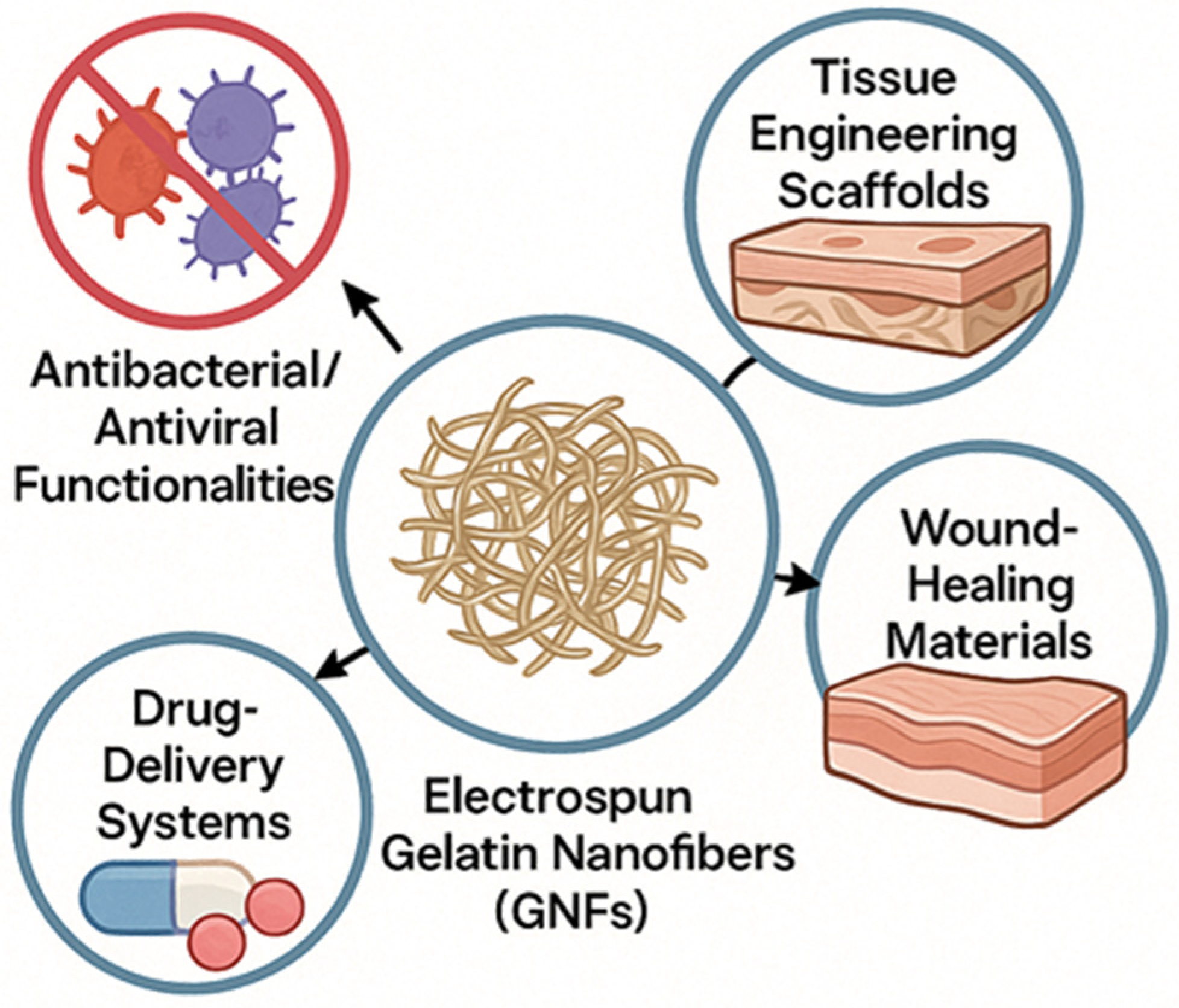
To provide a concise overview of the diverse applications and functionalities of electrospun GNFs discussed in this section, Table 4 summarizes key compositions, their primary biomedical application categories, and the general results observed across various studies. Complementarily, Figure 3 provides an integrated visual synthesis of how material properties and design strategies map onto applications in regenerative medicine, drug delivery, and infection control.

6. Food and Packaging Applications

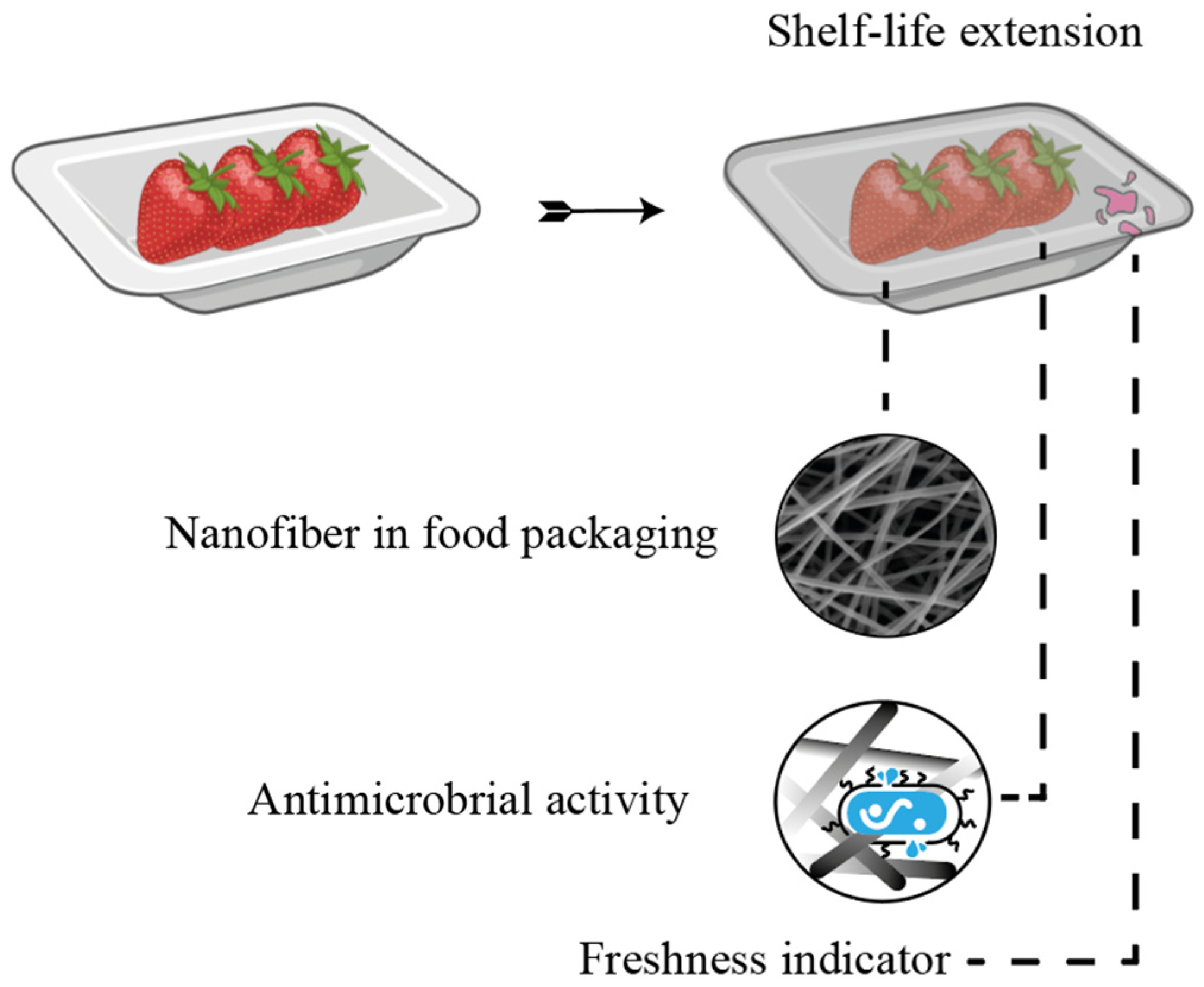
Electrospinning has emerged as a promising technique for developing active and smart food packaging, particularly because it allows the incorporation of bioactive compounds into nanofibers with a high specific surface area, as shown in Figure 4. This characteristic enhances the functional activity of packaging, such as antioxidant and antimicrobial properties, contributing to the extension of food shelf life [21,87]. As previously explained, the main advantage of electrospinning is the high surface area of the formed nanofibers, which provides a larger active surface area available for interactions with microorganisms, promoting more efficient bacterial inactivation. Furthermore, electrospun fibers have a uniform and continuous structure, which favors the controlled release of functional compounds [87].
However, gelatin, despite being an attractive biopolymer for food applications, has limitations when used alone in food packing, such as low mechanical strength, low water barrier, and lack of sufficient bioactivities [21,87]. Therefore, strategies, such as the incorporation of other biopolymers and bioactive compounds have been explored. For example, Gulzar et al. [88] developed gelatin/chitosan nanofibers incorporated with tannic acid and chitooligosaccharides, which were electrospun onto a polylactic acid (PLA) film. The smooth, bead-free nanofibers acted as antioxidants and antimicrobials, in addition to improving the mechanical properties of the films. Other studies reinforce the mechanisms of action of these structures. Liu et al. [89] demonstrated that nanofiber films cause bacterial cell death through two distinct mechanisms: cell aggregation, promoted by gelatin and chitosan, and cell membrane damage, induced by chitosan and ε-polylysine.
Furthermore, the encapsulation of natural compounds by electrospinning has shown good results. Zhou et al. [90] developed gelatin nanofibers (GNFs) with encapsulated angelica essential oil with good hydrophobic properties, significant antioxidant activity, and inhibitory effect against bacteria, suggesting potential for food packaging. Yilmaz et al. [52] found that electrospinning is an effective technique for encapsulating eugenol in GNFs, being a new biodegradable active packaging material with good antibacterial effects.
Practical applications of these packages have also been reported in the shelf life of Asian seabass slices [87], shrimp freshness detection [48], chicken breasts [91,92], and shelf-life extension of strawberries [93]. These studies based on GNFs are versatile, effective, and promising both for extending shelf life and for visually indicating food quality. Therefore, the electrospinning of food-grade biopolymers incorporated with bioactive compounds represents a promising approach for the development of innovative active packaging, with the potential to increase food safety, quality, and shelf life. Several studies have explored the use of GNFs combined with biopolymers, such as chitosan [51,89,94], zein [92,93], cellulose acetate [95], and PLA [94], as well as bioactive compounds such as eugenol [95], curcumin [51], perillaldehyde [92], anthocyanins [48], and essential oils [93] to develop packaging films with active and/or smart properties.
Therefore, Table 5 presents the development of active and smart packaging using GNFs. Among the studies presented in Table 5, most used the conventional electrospinning technique, only Shi et al. [95] used the coaxial electrospinning technique. Furthermore, it is possible to see that there was a significant reduction in several pathogenic bacteria, an increase in antioxidant activity, and an improvement in the characteristics of the films. The main function of these films was in relation to antibacterial activity, with significant efficacy against pathogens such as Escherichia coli, Salmonella enteritidis, Staphylococcus aureus, and Listeria monocytogenes, promoting everything from growth inhibition to cell death through membrane damage.
In addition to their antimicrobial activity, the films also exhibited antioxidant properties, with high DPPH (2,2-diphenyl-1-picrylhydrazyl) activity, and improved physicochemical characteristics, such as thermal stability, mechanical strength, nanofiber network structure, water vapor permeability, and moisture retention. In smart packaging, the films were able to monitor food quality and deterioration, exhibiting color changes sensitive to gases, such as ammonia and acetic acid, associated with the degradation of animal products [48].
Electrospinning has been widely used in the development of active layers in composite films for food packaging [21]. Considering the limitations of conventional films in terms of protection and functionality, most double-layer films combine GNFs with PLA [21,87,88]. In these cases, the active layer is deposited on the surface of a PLA film, where the PLA acts as an outer protective layer, while the electrospun GNFs form the inner layer, in direct contact with food, providing functional activity, such as antimicrobial properties [21]. In addition to PLA, several materials with different hydrophobicities, including Balangu seed mucilage and chitosan, have been used in the preparation of multilayer active films, combined with eugenol [21], tannic acid and chitooligosaccharides (Gulzar et al., 2022), nisin [87], and Fe3O4 nanoparticles [47].
Table 6 presents the active packaging developed using GNFs through bilayer systems. Smart packaging using GNFs in bilayer films has not yet been found in the literature. The bilayer systems presented in Table 6 demonstrated high antibacterial efficacy, with significant inhibition zones against E. coli, S. aureus, L. monocytogenes, P. aeruginosa, and other relevant foodborne pathogens, reaching inhibitions of up to 32 mm. Furthermore, they exhibited high antioxidant activity, notably a DPPH scavenging capacity of over 90% and a scavenging capacity for ABTS (2,2′-azino-bis(3-ethylbenzothiazoline-6-sulfonic acid)) and DPPH radicals that was significantly higher than that of the control films. The mechanical and functional properties of the films improved, including increased strength, reduced water vapor permeability, enhanced thermal stability, improved surface hydrophobicity, and efficient encapsulation of the active compounds.
Comparing the studies presented in Table 5 and Table 6, bilayer systems demonstrated superior performance in several functional and structural aspects, reflecting their greater potential for advanced applications in active and smart packaging. The combination of a protective layer (such as PLA) with an active layer of GNFs enabled the development of more robust, functional packaging with controlled release of bioactive compounds, making them more suitable for advanced and active packaging.

7. Environmental Applications

With the rapid advancement of urbanization and the consequent increase in industrial activities, there has been a significant rise in the presence of emerging contaminants in the environment. Many of these compounds, which are not yet fully identified or regulated, have raised growing concern due to their potential adverse effects on human health and ecosystems, with long-term consequences that are still not fully understood [96,97,98].
These toxic compounds, when released into the environment, exhibit high toxicity, particularly in aquatic ecosystems, where they can impair the reproductive capacity of organisms, induce oxidative stress, and promote bioaccumulation along the food chain. Consequently, water resources serve as receivers of a wide range of organic and inorganic pollutants, including pharmaceutical compounds, heavy metals, ions, dyes, oils, and others [99,100].
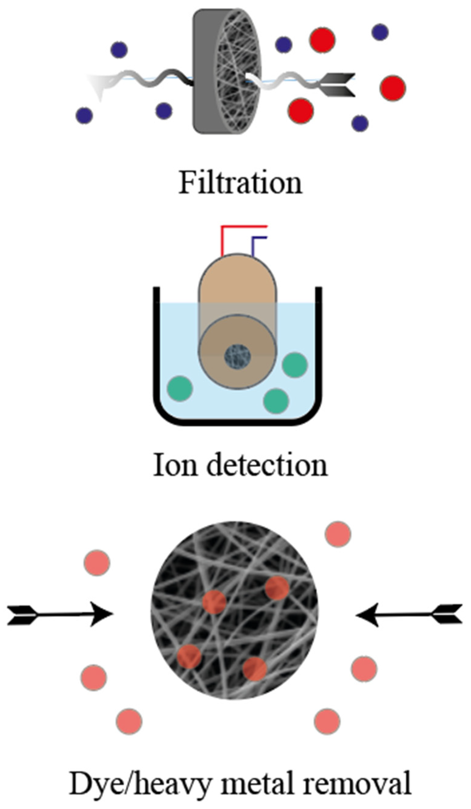
To mitigate this impact, various techniques and materials have been developed to remediate this situation [101,102,103]. In this context, nanotechnology has gained prominence for enabling the synthesis of materials with unique characteristics, facilitating the removal of these emerging contaminants. Accordingly, electrospun nanofibers stand out due to their distinctive morphological features, such as a three-dimensional porous structure, a high surface-to-volume ratio, and functional versatility, including the ability to incorporate active agents [104,105]. These properties make electrospun nanofibers promising for environmental applications, particularly as adsorbents, filtration membranes, ion detection systems in water, electrochemical electrodes, and efficient systems for the removal of dyes and heavy metals (Figure 5) [106,107,108]. Therefore, it is worth highlighting that a relevant aspect of electrospun nanofibers is their ability to have their composition and morphology tailored to meet the specific requirements of each type of pollutant to be removed. This versatility, such as the use of synthetic polymers, supports both large-scale applications and targeted solutions for complex effluents. For example, sulfonated polyethersulfone nanofiber membranes incorporating silver nanoparticles stabilized with plant extract (AgNPs) were developed by Talukder et al. [109]. The authors employed a green synthesis approach for AgNPs, eliminating the need for chemical agents. Moreover, these compounds enhanced the pollutant adsorption capacity and improved both the efficiency of adsorption and the interaction with contaminants. The nanofibers exhibited a high adsorption capacity for Pb2+ and Cd2+ ions, with rapid removal kinetics and excellent reusability, maintaining performance over 10 adsorption–desorption cycles.
However, there is still a search for more sustainable polymers for the removal of these pollutants, and research has therefore focused on biopolymers, such as chitosan (CS), gelatin (GA), collagen (COL), alginate (ALG), polyvinyl alcohol (PVA), and polyhydroxyalkanoates (PHAs), due to their biodegradability, biocompatibility, and abundance in nature [109,110,111,112]. Ahmadijokani et al. [110] showed that the incorporation of UiO-66-NH2 into PVA/chitosan nanofibers increased the adsorption of the anionic dyes methyl red (384 mg g−1) and methyl orange (454 mg g−1), surpassing the values obtained for the cationic dyes malachite green (133 mg g−1) and methylene blue. UiO-66 maintained high selectivity for anionic dyes even after 12 months in water. Therefore, these results indicate a high potential for the removal of contaminants in aqueous media, in addition to highlighting the ability to maintain this performance over extended periods.
A recent trend in the field is the use of natural polymers, such as gelatin, for the production of these nanofibers. Gelatin stands out due to its biocompatibility and biodegradability, making it an even more sustainable and environmentally friendly option, and its potential applications in contaminant removal are highlighted in Table 7. A notable example is the study by Yang et al. [113], which developed electrospun gelatin-polycaprolactone nanofibers for the rapid and selective detection of copper (Cu2+) and chromium (Cr3+) ions in wastewater. The authors highlighted the effectiveness of gelatin as a matrix for interacting with heavy metal ions. Therefore, this functionalization capability for the detection and potential removal of heavy metals is particularly relevant given their high toxicity and widespread occurrence in industrial effluents, with chromium being one of the most hazardous heavy metals to human health and the environment.
In addition to detection, gelatin has also been employed in composite materials for the active removal of pollutants. Wang and Shi [114], for example, developed composite aerogels made of PVA nanofibers and gelatin/graphene oxide. These aerogels were designed with a superhydrophobic and superoleophilic surface, enabling efficient oil-water separation. Therefore, the material’s properties, such as high porosity, contributed to its high selective oil adsorption capacity, indicating the potential of gelatin as an active component in the removal of oily contaminants.
However, GNFs are still scarcely explored and applied in the removal of aqueous contaminants, mainly due to their low gelation temperature and high-water solubility. Although they present a promising feature, high specific surface area resulting from their reduced fiber diameter, in the range of hundreds of nanometers, these structures remain structurally unstable in aqueous environments, which limits their efficiency and durability in water treatment [115].
Table 7. Summary of recent advancements in gelatin-based nanofibers for environmental applications.
MaterialApplicationFiltration Efficiency (%)Adsorption CapacityReusabilityReference
Gelatin/PCLWater Detection---[113]
Gelatin/PVA/Graphene OxideOil/Water Separation---[114]
Gelatin/Calcium AlginateWater Adsorption-1937 mg g−1 (Methylene Blue)4 cycles[116]
Fish GelatinWater Adsorption-60 mg g−1 (Methylene Blue)6 cycles[117]
Cellulose Nanofibers/GelatinAir Filtration>90% (PM2.5)--[118]
Gelatin/β−CDAir Filtration>95%High (VOCs)-[119]
EC/Gelatin/β-CD/CurcuminMultifunctional Air Filtration>99.25% (0.3 µm)442 µg g−1 (Formaldehyde)-[120]
Gelatin/ZIF-67Uranium Removal-612.24 mg g−1 -[121]
A promising alternative to overcome this limitation was demonstrated by Ma et al. [116] who developed composite nanofibers of gelatin and calcium alginate through electrospinning. Gelatin acted as the structural component, mechanically reinforcing the fibers, while alginate, crosslinked with calcium ions (Ca2+), formed a stable and insoluble three-dimensional network capable of maintaining the integrity of the nanofibers even after prolonged contact with water. In addition, high adsorption capacities were achieved for methylene blue (1937 mg g−1) along with excellent reusability, favored by the electrostatic repulsion generated by protonated amine groups in the gelatin.
Following a more sustainable and environmentally friendly approach, a recent study stands out for presenting another solution to address the challenge of aqueous instability, the development of nanofibers produced from fish gelatin [15]. Kim et al. [117] developed fish GNFs via the Maillard reaction using glucose-6-phosphate as a natural crosslinker, which not only preserved the integrity of the nanofibers during water contact, but also introduced phosphate groups onto the adsorbent surface, thereby enhancing interactions between the nanofibers and cationic dyes. Consequently, the authors improved the mechanical stability of the material while addressing the key limitation of adsorbent durability in aqueous environments. As a result, the membrane achieved a methylene blue adsorption capacity of approximately 60 mg g−1 in just 15 min, retaining over 84% efficiency after six adsorption/desorption cycles and exhibiting more than 90% biodegradation within five days.
These findings highlight the potential for further research into such innovative approaches, reinforcing the role of sustainable strategies for aqueous contaminant removal. In another example, Furuno et al. [122] produced GNFs crosslinked via horseradish peroxidase, resulting in water-insoluble structures that maintained their morphology and stability, positioning this material as a promising adsorbent and even as an advanced air filtration medium.
In addition to the detection and removal of contaminants from liquid effluents, gelatin has also proven to be highly promising in the separation of atmospheric pollutants due to its ability to combine high filtration efficiency, low cost, and biodegradability. In a recent study, Laitinen and Liimatainen [118] developed composite cryogels of cellulose nanofibers reinforced with gelatin for the production of high-performance air filters. The addition of a small amount of gelatin (only 0.5%) not only doubled the mechanical strength of the material but also significantly improved the filtration efficiency to over 90%, ensuring the separation of fine particles (PM2.5) with high porosity (>98.8%) and low pressure drop. The use of 100% renewable materials and the low manufacturing cost highlight this approach as a promising and sustainable solution for the development of advanced air-purification filters.
GNFs can also be used to create “green” and biodegradable filters, offering a robust, low-resistance solution for respiratory protection against air pollution. Kadam et al. [119] produced electrospun gelatin/β-cyclodextrin nanofibers as a dual-function filtering medium for respiratory masks. These nanofibers not only captured aerosols with high filtration efficiency (>95%) but also adsorbed hazardous volatile organic compounds (VOCs), such as xylene and benzene. The authors reported adsorption capacities significantly higher than those of commercial masks and attributed this enhanced VOC adsorption performance to the high specific surface area of the nanofibers and the protein-based structure of gelatin.
Considering the multifunctionality of these nanofibers, Shao et al. [120] synthesized a nanofibrous membrane composed entirely of biomaterials—gelatin, β-cyclodextrin, and curcumin—embedded in an ethylcellulose matrix. This multifunctional material, produced via electrospinning, achieved a high filtration efficiency of 99.25% for 0.3 µm particles, exhibited strong antibacterial activity against E. coli and S. aureus, and demonstrated effective formaldehyde adsorption. The study emphasizes that gelatin, in combination with the other components, enhanced both the formaldehyde adsorption capacity and the antibacterial performance, while supporting the development of lightweight, high-performance, and advanced air filtration systems.
Overall, these advances demonstrate that gelatin-based nanofibers, especially when combined with other functional biomaterials, provide a versatile and sustainable framework for the development of high-performance air filters capable of simultaneously targeting particles, microorganisms, and gaseous pollutants.

8. Biosensing Applications

The functional versatility of nanofibers allows their implementation in different sensor mechanisms, such as piezoelectric, colorimetric, and electrochemical, according to recent literature. Liu and co-workers [123] developed an electronic skin (e-skin) applying gelatin nanofibers (GNFs) and carbonizing them at 800 °C in an inert atmosphere (Argon) to develop graphene oxide films, in order to act as a piezoresistive material, converting pressure into an electric signal. The nanofibers were produced using a 40% solution of porcine skin gelatin in formic acid, under 17 kV with a 17 cm syringe–collector distance. The produced nanofibers achieved diameters ranging from 300 to 600 nm, which were collected over a Cu film. The resulting nanofiber/film exhibited a failure tensile stress of 35 MPa, much higher than other conventional films and hydrogels. This characteristic was determinant in the decision to develop a gelatin nanofiber/film. However, to be applied as an e-skin, the proposed nanofiber/film needed to be conductive, justifying the carbonization of the material. In this context, the work aimed to combine the mechanical strength of GNFs with the electrical conductivity of a carbonized material, while preserving the fibrous architecture and mechanical strength as much as possible and overcoming the intrinsically insulating nature of GNFs. Finally, the stretchable sensor, with a 710 ± 100 nm of thickness was applied, using two layers of the developed sensor with a layer of Polydimethylsiloxane in the middle. This final structure permitted the two layers of the stretchable sensor to be loosely contacted, reacting to the applied pressure, causing resistance variation. The developed e-skin achieved a pressure sensitivity around 100 kPa in the range of 0–150 Pa, much more sensitivity over conventional reported e-skins. The proposed e-skin was successfully applied for sound recognition, respiratory monitoring, pulse beating and apexcardiogram recording, making it a promising solution for non-invasive monitoring process.
A similar approach was proposed by Ghosh and co-workers [124], who proposed an e-skin to detect and discriminate static human physiological signals based on the same piezoelectric principle, discussed above. In order to develop an e-skin with high flexibility, low cost, large area-to-volume ratio, light weight and environmental safety, once again GNFs were chosen. In this case, the nanofibers were produced under 1.5 kV cm−1 applying fish gelatin (40%, v/v) in a mixture composed by formic acid and N, N-dimethylformamide. Posteriorly, the nanofibers were dried to complete solvent removal and crosslinked with glutaraldehyde (50%), resulting in nanofibers with average diameter around 700 nm. The mechanosensitivity achieved by the nanofibers was ~0.8 V kPa−1 with excellent operational stability (over 108,000 cycles) and anti-fatigue (over 6 months) properties. Finally, the nanofibers were placed between two copper nickel electrodes and encapsulated with polydimethylsiloxane to be applied as an e-skin sensor, showing applicability in monitoring blood pressure pulse waves to vibrations on the throat associated with phonation without using any invasive approaches.
On the other hand, Kiefer and co-workers [125] proposed a different approach using porcine skin GNFs to develop a multifunctional electrochemical sensor-capacitor. Porcine skin gelatin and glucose (10:1) were dissolved using glacial acetic acid (99%) and applied at the electrospinning process under 17.5 kV with a 14.5 cm syringe–collector distance, resulting in nanofibers with an average diameter of 0.8 ± 0.06 µm. Due to the large surface area-to-volume ratio, the authors choose the nanofibers to act as a support matrix to immobilize polypyrrole, as recognition element. The electro-chemo-mechanical characterization of this composite reveals its multifunctionality as an actuator for energy storage and as a sensor. Therefore, it could be applied as a smart patches or smart textile devices where these nanofibers combined with polypyrrole can stand out due to its multifunctionality.
Owing to their large surface area-to-volume ratio, nanofibers are excellent candidates to serve as support matrices for the immobilization of different agents that can function as biorecognition elements in the sensor context. In this context, Teepoo and co-workers [22] produced nanofibers under 20 kV at a 20 cm syringe–collector distance using a chitosan–gelatin (60:40) solution in acetic acid (60%), resulting in continuous fiber with an average diameter of 80 nm. The nanofiber was collected directly in the surface of a graphite electrode and applied glutaraldehyde to crosslink the nanofiber to horseradish peroxidase. The peroxidase immobilized in the electrode can catalyze the reduction of hydrogen peroxide (H2O2) by electron transferring and the electrode can measure this phenomenon generating a measurable current proportional to the H2O2 concentration. Interestingly, the authors also compared the efficiency of using these nanofibers as a support matrix for horseradish peroxidase against a film produced using the same constituent proportions. The results exhibited that the large surface area-to-volume ratio from the nanofibers was able to immobilize more horseradish peroxidase than the film produced, which resulted in higher enzymatic activity and, consequently, a stronger signal for the electrode to operate on. The proposed sensor showed excellent linearity in the range from 0.1 to 1.7 mM, with a correlation coefficient of 0.9996, sensitivity of 44 µA mM−1 cm−2 and a detection limit of 0.05 mM.
A different approach for electrode modification using nanofibers was proposed by Deng and co-workers [126]. The nanofibers were produced using 10 kV at a 12 cm syringe–collector distance, with a solution composed by 60 mg of gelatin, 140 mg of poly(lactide-co-glycolide) dissolved in 2 mL of 2,2,2-trifluoroethanol and added 20 mg of carbon nanotubes. The developed nanofibers achieved an average diameter of 127 ± 20 nm, with high porosity, were applied on the surface of a carbon fiber electrode for dopamine detection. In this context, GNFs are described to have an antifouling capability to avoid nonspecific protein adsorption, providing an excellent platform for electrostatic interaction specific for dopamine, with the advantages of high hydrophilicity and good biocompatibility. However, GNFs can swell and lose their fiber structure in contact with water, to prevent this scenario, poly(lactide-co-glycolide) was chosen for crosslinking. Modifying electrode surfaces often involves adding a conductive component to facilitate electron transfer between the analyte and the electrode, thereby enhancing the measurable current response, which justifies the incorporation of carbon nanotubes into the system. Finally, the developed sensor was successfully applied to determine dopamine in human blood with high sensitivity (0.30 nA/μM) and good linearity in the range from 5 to 25 mM for dopamine hydrochloride (R = 0.995).
Another approach consists of the application of nanofibers in the development of fluorescent sensors. Yang and co-workers [113] developed nanofibers composed of gelatin, poly-ε-caprolactone and 1,4-dihydroxyanthraquinone, in different proportions, using formic acid as solvent for the rapid detection of Cu2+ and Cr3+ as a fluorescent sensor. It is important to highlight that each component in this system plays an important role. In this scenario, gelatin was chosen due to its hydrophilicity and biodegradability. The poly-ε-caprolactone was incorporated to enhance mechanical robustness and stability of the developed nanofibers and the 1,4-dihydroxyanthraquinone acted as cation recognizing element by changing its fluorescence upon interaction with target cations. The electrospinning process was performed at 20 kV with an 18 cm syringe–collector distance, achieving nanofibers with an average diameter of 305.5 ± 8.9 nm. The developed sensor exhibited high sensitivity and selectivity being able to detect Cu2+ ions in the range from 4.8 × 10⁻⁶ to 6.5 × 10⁻⁶ M and Cr3+ in the range from 2.7 × 10⁻⁶ to 4.3 × 10⁻⁶ M.
The analysis of different sensors based on GNFs highlights the immense potential of these materials in creating advanced, sensitive, sustainable, and adaptable devices for multiple applications. Whether through piezoelectric, piezoresistive, fluorescent, or electrochemical mechanisms, GNFs can provide remarkable performance in terms of sensitivity, response time, stability, and selectivity, even in challenging environments, such as biological fluids or wastewater. Their ease of processing via electrospinning, combined with the ability to incorporate functional or conductive molecules and their compatibility with flexible substrates, such as polydimethylsiloxane, further expands their integration into wearable and portable platforms. Beyond technical advantages, the use of biodegradable and renewable materials reinforces the pursuit of more ecological and sustainable solutions. Collectively, gelatin nanofiber-based sensors embody a convergence of advanced technology, sustainability, and functionality, addressing the emerging demand for precise, portable, and secure detection systems.

9. Conclusions

This review explored the physicochemical properties of gelatin and their influence on electrospinning, with particular emphasis on how processing conditions and the type of raw source affect solution properties, such as molecular weight, amino acid composition, viscosity, and concentration. Indeed, cold-water fish gelatin has been highlighted for its ability to be electrospun directly from aqueous solutions, a capability attributed to its lower molecular weight, reduced proline and hydroxyproline content, and lower Bloom strength compared to bovine, porcine, and warm-water fish gelatins. Moreover, the inherent properties of gelatin, abundance, affordability, biodegradability, and the presence of both polar and apolar functional groups, combined with the key features of nanofibers, including high surface area, porosity, and tunable morphology, make electrospun gelatin nanofibers highly attractive for applications in biomedicine, food, environmental remediation, and biosensing. However, one of their major drawbacks is high water solubility, which limits use in many practical applications. To address this, strategies such as polymer blending, chemical and physical modifications, and diverse crosslinking techniques have been employed to enhance the functional performance of gelatin nanofibers. In addition, multilayer systems, in which distinct layers are tailored for functionalities, such as structural reinforcement, moisture resistance, or active compound encapsulation, have enabled synergistic improvements in performance, leading to materials that are robust and multifunctional. Moving forward, further studies should focus on improving these properties by incorporating bio-based compounds in blends and layered systems, as well as studying new green crosslinking approaches to achieve a more sustainable process.

Author Contributions

Conceptualization, B.S.d.F.; methodology, B.S.d.F., A.C.R., D.P.J., E.S.R., J.O.G., R.F.V., S.S.F., D.D., T.R.S.C.J. and L.A.d.A.P.; resources, B.S.d.F.; data curation, B.S.d.F.; writing—original draft preparation, B.S.d.F., A.C.R., D.P.J., E.S.R., J.O.G., R.F.V., S.S.F., D.D., T.R.S.C.J. and L.A.d.A.P.; writing—review and editing, B.S.d.F. and E.S.R.; visualization, B.S.d.F. All authors have read and agreed to the published version of the manuscript.

Funding

This research was funded by the Coordenação de Aperfeiçoamento de Pessoal de Nível Superior (CAPES)/Brazil-Finance Code 001 and Fundação de Amparo à Pesquisa do Estado do Rio Grande do Sul (FAPERGS), grant number 25/2551-0000820-4.

Institutional Review Board Statement

Not applicable.

Informed Consent Statement

Not applicable.

Data Availability Statement

Data are contained within the article.

Conflicts of Interest

The authors declare no conflicts of interest.

References

  1. El-Seedi, H.R.; Said, N.S.; Yosri, N.; Hawash, H.B.; El-Sherif, D.M.; Abouzid, M.; Abdel-Daim, M.M.; Yaseen, M.; Omar, H.; Shou, Q.; et al. Gelatin Nanofibers: Recent Insights in Synthesis, Bio-Medical Applications and Limitations. Heliyon 2023, 9, e16228. [Google Scholar] [CrossRef]
  2. Gavande, V.; Nagappan, S.; Seo, B.; Lee, W.K. A Systematic Review on Green and Natural Polymeric Nanofibers for Biomedical Applications. Int. J. Biol. Macromol. 2024, 262, 130135. [Google Scholar] [CrossRef]
  3. Majidi, S.S.; Slemming-Adamsen, P.; Hanif, M.; Zhang, Z.; Wang, Z.; Chen, M. Wet Electrospun Alginate/Gelatin Hydrogel Nanofibers for 3D Cell Culture. Int. J. Biol. Macromol. 2018, 118, 1648–1654. [Google Scholar] [CrossRef]
  4. Ribeiro, E.S.; de Farias, B.S.; Sant’Anna Cadaval Junior, T.R.; de Almeida Pinto, L.A.; Diaz, P.S. Chitosan–Based Nanofibers for Enzyme Immobilization. Int. J. Biol. Macromol. 2021, 183, 1959–1970. [Google Scholar] [CrossRef]
  5. Lu, W.; Ma, M.; Xu, H.; Zhang, B.; Cao, X.; Guo, Y. Gelatin Nanofibers Prepared by Spiral-Electrospinning and Cross-Linked by Vapor and Liquid-Phase Glutaraldehyde. Mater. Lett. 2015, 140, 1–4. [Google Scholar] [CrossRef]
  6. de Farias, B.S.; Rizzi, F.Z.; Ribeiro, E.S.; Diaz, P.S.; Sant’Anna Cadaval Junior, T.R.; Dotto, G.L.; Khan, M.R.; Manoharadas, S.; de Almeida Pinto, L.A.; dos Reis, G.S. Influence of Gelatin Type on Physicochemical Properties of Electrospun Nanofibers. Sci. Rep. 2023, 13, 15195. [Google Scholar] [CrossRef]
  7. Sharma, G.K.; Jalaja, K.; Ramya, P.R.; James, N.R. Electrospun Gelatin Nanofibres—Fabrication, Cross-Linking and Biomedical Applications: A Review. Biomed. Mater. Devices 2023, 1, 553–568. [Google Scholar] [CrossRef]
  8. Yıkar, E.; Demir, D.; Bölgen, N. Electrospinning of Gelatin Nanofibers: Effect of Gelatin Concentration on Chemical, Morphological and Degradation Characteristics. Turk. J. Eng. 2021, 5, 171–176. [Google Scholar] [CrossRef]
  9. Su, X.N.; Khan, M.F.; Xin-Ai; Liu, D.L.; Liu, X.F.; Zhao, Q.L.; Cheong, K.L.; Zhong, S.Y.; Li, R. Fabrication, Modification, Interaction Mechanisms, and Applications of Fish Gelatin: A Comprehensive Review. Int. J. Biol. Macromol. 2025, 288, 138723. [Google Scholar] [CrossRef] [PubMed]
  10. Silva, R.S.G.; Bandeira, S.F.; Pinto, L.A.A. Characteristics and Chemical Composition of Skins Gelatin from Cobia (Rachycentron Canadum). LWT 2014, 57, 580–585. [Google Scholar] [CrossRef]
  11. Miyawaki, O.; Omote, C.; Matsuhira, K. Thermodynamic Analysis of Sol-Gel Transition of Gelatin in Terms of Water Activity in Various Solutions. Biopolymers 2015, 103, 685–691. [Google Scholar] [CrossRef]
  12. Amirrah, I.N.; Lokanathan, Y.; Zulkiflee, I.; Wee, M.F.M.R.; Motta, A.; Fauzi, M.B. A Comprehensive Review on Collagen Type I Development of Biomaterials for Tissue Engineering: From Biosynthesis to Bioscaffold. Biomedicines 2022, 10, 2307. [Google Scholar] [CrossRef]
  13. Jafari, H.; Lista, A.; Siekapen, M.M.; Ghaffari-Bohlouli, P.; Nie, L.; Alimoradi, H.; Shavandi, A. Fish Collagen: Extraction, Characterization, and Applications for Biomaterials Engineering. Polymers 2020, 12, 2230. [Google Scholar] [CrossRef]
  14. Haug, I.J.; Draget, K.I.; Smidsrød, O. Physical and Rheological Properties of Fish Gelatin Compared to Mammalian Gelatin. Food Hydrocoll. 2004, 18, 203–213. [Google Scholar] [CrossRef]
  15. Kwak, H.W.; Shin, M.; Lee, J.Y.; Yun, H.; Song, D.W.; Yang, Y.; Shin, B.S.; Park, Y.H.; Lee, K.H. Fabrication of an Ultrafine Fish Gelatin Nanofibrous Web from an Aqueous Solution by Electrospinning. Int. J. Biol. Macromol. 2017, 102, 1092–1103. [Google Scholar] [CrossRef]
  16. Zou, Y.; Chen, X.; Lan, Y.; Yang, J.; Yang, B.; Ma, J.; Cheng, M.; Wang, D.; Xu, W. Find Alternative for Bovine and Porcine Gelatin: Study on Physicochemical, Rheological Properties and Water-Holding Capacity of Chicken Lungs Gelatin by Ultrasound Treatment. Ultrason. Sonochemistry 2024, 109, 107004. [Google Scholar] [CrossRef]
  17. Huang, C.; Tang, J.; Chen, X.; Zeng, X.; Zhong, W.; Pang, J.; Wu, C. Novel Electrospun Gelatin Nanofibers Loaded with Purple Potato Anthocyanin and Syringic Acid for Multifunctional Food Packaging. Foods 2024, 13, 2538. [Google Scholar] [CrossRef]
  18. de Farias, B.S.; Rizzi, F.Z.; Silva, P.P.; Ribeiro, E.S.; Diaz, P.S.; Cadaval Junior, T.R.S.A.; de Almeida Pinto, L.A. Tri–Layer Nanostructured Lycopene Delivery System Based on Chitosan Nanospheres and Gelatin Nanofibers. Food Res. Int. 2025, 220, 117099. [Google Scholar] [CrossRef]
  19. Topçu, A.A. The Adsorption Performance of Magnetic Gelatin Nanofiber for Orange G Removal. Polym. Bull. 2023, 80, 1017–1029. [Google Scholar] [CrossRef]
  20. İnal, M.; Mülazımoğlu, G. Production and Characterization of Bactericidal Wound Dressing Material Based on Gelatin Nanofiber. Int. J. Biol. Macromol. 2019, 137, 392–404. [Google Scholar] [CrossRef] [PubMed]
  21. Li, M.; Yu, H.; Xie, Y.; Guo, Y.; Cheng, Y.; Qian, H.; Yao, W. Fabrication of Eugenol Loaded Gelatin Nanofibers by Electrospinning Technique as Active Packaging Material. LWT 2021, 139, 110800. [Google Scholar] [CrossRef]
  22. Teepoo, S.; Dawan, P.; Barnthip, N. Electrospun Chitosan-Gelatin Biopolymer Composite Nanofibers for Horseradish Peroxidase Immobilization in a Hydrogen Peroxide Biosensor. Biosensors 2017, 7, 47. [Google Scholar] [CrossRef]
  23. de Farias, B.S.; Sant’Anna Cadaval Junior, T.R.; de Almeida Pinto, L.A. Chitosan-Functionalized Nanofibers: A Comprehensive Review on Challenges and Prospects for Food Applications. Int. J. Biol. Macromol. 2019, 123, 210–220. [Google Scholar] [CrossRef]
  24. Haider, A.; Haider, S.; Kang, I.K. A Comprehensive Review Summarizing the Effect of Electrospinning Parameters and Potential Applications of Nanofibers in Biomedical and Biotechnology. Arab. J. Chem. 2018, 11, 1165–1188. [Google Scholar] [CrossRef]
  25. Derkach, S.R.; Kuchina, Y.A.; Baryshnikov, A.V.; Kolotova, D.S.; Voron’ko, N.G. Tailoring Cod Gelatin Structure and Physical Properties with Acid and Alkaline Extraction. Polymers 2019, 11, 1724. [Google Scholar] [CrossRef] [PubMed]
  26. Zhang, T.; Xu, J.; Zhang, Y.; Wang, X.; Lorenzo, J.M.; Zhong, J. Gelatins as Emulsifiers for Oil-in-Water Emulsions: Extraction, Chemical Composition, Molecular Structure, and Molecular Modification. Trends Food Sci. Technol. 2020, 106, 113–131. [Google Scholar] [CrossRef]
  27. Vaca Chávez, F.; Hellstrand, E.; Halle, B. Hydrogen Exchange and Hydration Dynamics in Gelatin Gels. J. Phys. Chem. B 2006, 110, 21551–21559. [Google Scholar] [CrossRef]
  28. Okutan, N.; Terzi, P.; Altay, F. Affecting Parameters on Electrospinning Process and Characterization of Electrospun Gelatin Nanofibers. Food Hydrocoll. 2014, 39, 19–26. [Google Scholar] [CrossRef]
  29. Cooper, C.J.; Mohanty, A.K.; Misra, M. Electrospinning Process and Structure Relationship of Biobased Poly(Butylene Succinate) for Nanoporous Fibers. ACS Omega 2018, 3, 5547–5557. [Google Scholar] [CrossRef]
  30. Al-Abduljabbar, A.; Farooq, I. Electrospun Polymer Nanofibers: Processing, Properties, and Applications. Polymers 2022, 15, 65. [Google Scholar] [CrossRef]
  31. Ahmadi Bonakdar, M.; Rodrigue, D. Electrospinning: Processes, Structures, and Materials. Macromol 2024, 4, 58–103. [Google Scholar] [CrossRef]
  32. Ninan, G.; Joseph, J.; Aliyamveettil, Z.A. A Comparative Study on the Physical, Chemical and Functional Properties of Carp Skin and Mammalian Gelatins. J. Food Sci. Technol. 2014, 51, 2085–2209. [Google Scholar] [CrossRef]
  33. Chandra, M.V.; Shamasundar, B.A. Texture Profile Analysis and Functional Properties of Gelatin from the Skin of Three Species of Fresh Water Fish. Int. J. Food Prop. 2015, 18, 572–584. [Google Scholar] [CrossRef]
  34. Rather, J.A.; Akhter, N.; Ashraf, Q.S.; Mir, S.A.; Makroo, H.A.; Majid, D.; Barba, F.J.; Khaneghah, A.M.; Dar, B.N. A Comprehensive Review on Gelatin: Understanding Impact of the Sources, Extraction Methods, and Modifications on Potential Packaging Applications. Food Packag. Shelf Life 2022, 34, 100945. [Google Scholar] [CrossRef]
  35. Derkach, S.R.; Voron’ko, N.G.; Kuchina, Y.A.; Kolotova, D.S. Modified Fish Gelatin as an Alternative to Mammalian Gelatin in Modern Food Technologies. Polymers 2020, 12, 3051. [Google Scholar] [CrossRef] [PubMed]
  36. Gómez-Guillén, M.C.; Turnay, J.; Fernández-Díaz, M.D.; Ulmo, N.; Lizarbe, M.A.; Montero, P. Structural and Physical Properties of Gelatin Extracted from Different Marine Species: A Comparative Study. Food Hydrocoll. 2002, 16, 25–34. [Google Scholar] [CrossRef]
  37. Ninan, G.; Jose, J.; Abubacker, Z. Preparation and Characterization of Gelatin Extracted from the Skins of Rohu (Labeo Rohita) and Common Carp (Cyprinus Carpio). J. Food Process. Preserv. 2011, 35, 143–161. [Google Scholar] [CrossRef]
  38. Li, J.; Qin, Z.; Jiang, Q.; Chen, M.; Li, Y.; Zou, Y.; Zhang, H. Development of Gelatin/Camellia Oil Oleogels as Fat Substitutes via a Novel Strategy: Covalent Crosslinking of Electrospun Nanofiber. Food Hydrocoll. 2025, 166, 111340. [Google Scholar] [CrossRef]
  39. Huner, A. Fast-Dissolving Electroactive Drug Delivery Systems: Gelatin/Poly(Sulphonic Acid Diphenyl Aniline) Electrospun Nanofibers. Food Biosci. 2024, 61, 104924. [Google Scholar] [CrossRef]
  40. Dias, J.R.; Baptista-Silva, S.; de Oliveira, C.M.T.; Sousa, A.; Oliveira, A.L.; Bártolo, P.J.; Granja, P.L. In Situ Crosslinked Electrospun Gelatin Nanofibers for Skin Regeneration. Eur. Polym. J. 2017, 95, 161–173. [Google Scholar] [CrossRef]
  41. Laha, A.; Yadav, S.; Majumdar, S.; Sharma, C.S. In-Vitro Release Study of Hydrophobic Drug Using Electrospun Cross-Linked Gelatin Nanofibers. Biochem. Eng. J. 2016, 105, 481–488. [Google Scholar] [CrossRef]
  42. Zhang, C.; Wang, P.; Li, J.; Zhang, H.; Weiss, J. Characterization of Core-Shell Nanofibers Electrospun from Bilayer Gelatin/Gum Arabic O/W Emulsions Crosslinked by Genipin. Food Hydrocoll. 2021, 119, 106854. [Google Scholar] [CrossRef]
  43. Sengor, M.; Ozgun, A.; Gunduz, O.; Altintas, S. Aqueous Electrospun Core/Shell Nanofibers of PVA/Microbial Transglutaminase Cross-Linked Gelatin Composite Scaffolds. Mater. Lett. 2020, 263, 127233. [Google Scholar] [CrossRef]
  44. Yavari Maroufi, L.; Norouzi, R.; Ramezani, S.; Ghorbani, M. Novel Electrospun Nanofibers Based on Gelatin/Oxidized Xanthan Gum Containing Propolis Reinforced by Schiff Base Cross-Linking for Food Packaging. Food Chem. 2023, 416, 135806. [Google Scholar] [CrossRef]
  45. Kenawy, E.-R.; Kamoun, E.A.; Ghaly, Z.S.; Shokr, A.-B.M.; El-Moslamy, S.H.; Abou-Elyazed, A.S. Copper and Fluorine-MOFs Loaded-Electrospun PVA/Gelatin Nanofibers for Enhancing the Antimicrobial Activity of Topical Wound Dressings: MOFs Synthesis and Spinning Conditions Optimization. Mater. Chem. Phys. 2025, 332, 130303. [Google Scholar] [CrossRef]
  46. Li, S.F.; Hu, T.G.; Wu, H. Development of Quercetin-Loaded Electrospun Nanofibers through Shellac Coating on Gelatin: Characterization, Colon-Targeted Delivery, and Anticancer Activity. Int. J. Biol. Macromol. 2024, 277, 134204. [Google Scholar] [CrossRef] [PubMed]
  47. Navaei, F.; Zandi, M.; Ganjloo, A.; Dardmeh, N. Fabrication of Magnetic Nanoparticle-Enhanced Bi-Layer Films: Fe3O4-Loaded Electrospun PVA/Gelatin Nanofibers on a Balangu Seed Mucilage-Gelatin Base. Ind. Crops Prod. 2025, 229, 121037. [Google Scholar] [CrossRef]
  48. Chen, M.; Yan, T.; Jiang, Q.; Li, J.; Ali, U.; Miao, Y.; Farooq, S.; Hu, Y.; Zhang, H. Real-Time Shrimp Freshness Detection by Anthocyanin-Enriched Wheat Gluten/Gelatin Electrospun Nanofiber Films. Food Chem. 2025, 469, 142542. [Google Scholar] [CrossRef]
  49. Mozaffari, A.; Parvinzadeh Gashti, M.; Alimohammadi, F.; Pousti, M. The Impact of Helium and Nitrogen Plasmas on Electrospun Gelatin Nanofiber Scaffolds for Skin Tissue Engineering Applications. J. Funct. Biomater. 2024, 15, 326. [Google Scholar] [CrossRef]
  50. Talib Al-Sudani, B.; Mahmoudi, E.; Adnan Shaker Al-Naymi, H.; Al-Musawi, M.H.; Al-Talabanee, I.B.N.; Ramezani, S.; Ghorbani, M.; Mortazavi Moghadam, F. Antibacterial and Wound Healing Performance of a Novel Electrospun Nanofibers Based on Polymethyl-Methacrylate/Gelatin Impregnated with Different Content of Propolis. J. Drug Deliv. Sci. Technol. 2024, 95, 105641. [Google Scholar] [CrossRef]
  51. Duan, M.; Sun, J.; Huang, Y.; Jiang, H.; Hu, Y.; Pang, J.; Wu, C. Electrospun Gelatin/Chitosan Nanofibers Containing Curcumin for Multifunctional Food Packaging. Food Sci. Hum. Wellness 2023, 12, 614–621. [Google Scholar] [CrossRef]
  52. Yilmaz, M.T.; Hassanein, W.S.; Alkabaa, A.S.; Ceylan, Z. Electrospun Eugenol-Loaded Gelatin Nanofibers as Bioactive Packaging Materials to Preserve Quality Characteristics of Beef. Food Packag. Shelf Life 2022, 34, 100968. [Google Scholar] [CrossRef]
  53. Ekram, B.; Mousa, S.M.; El-Bassyouni, G.T.; Abdel-Hady, B.M.; Moaness, M. Novel Highly Proliferative Electrospun Cerium-Doped Hydroxyapatite/Polyamide/Gelatin Nanofibers for Guided Bone Regeneration Application. Mater. Chem. Phys. 2025, 342, 130975. [Google Scholar] [CrossRef]
  54. Dadras Chomachayi, M.; Solouk, A.; Akbari, S.; Sadeghi, D.; Mirahmadi, F.; Mirzadeh, H. Electrospun Nanofibers Comprising of Silk Fibroin/Gelatin for Drug Delivery Applications: Thyme Essential Oil and Doxycycline Monohydrate Release Study. J. Biomed. Mater. Res. A 2018, 106, 1092–1103. [Google Scholar] [CrossRef] [PubMed]
  55. Ajmal, G.; Bonde, G.V.; Mittal, P.; Pandey, V.K.; Yadav, N.; Mishra, B. PLGA/Gelatin-Based Electrospun Nanofiber Scaffold Encapsulating Antibacterial and Antioxidant Molecules for Accelerated Tissue Regeneration. Mater. Today Commun. 2023, 35, 105633. [Google Scholar] [CrossRef]
  56. Beikzadeh, S.; Hosseini, S.M.; Mofid, V.; Ramezani, S.; Ghorbani, M.; Ehsani, A.; Mortazavian, A.M. Electrospun Ethyl Cellulose/Poly Caprolactone/Gelatin Nanofibers: The Investigation of Mechanical, Antioxidant, and Antifungal Properties for Food Packaging. Int. J. Biol. Macromol. 2021, 191, 457–464. [Google Scholar] [CrossRef]
  57. Sojobi, J.W.; Bankole, O.L.; Ulelu, D.C.; Fayomi, O.S.I. Examination into Nanomaterials, Nanofibers and Nanoparticles Advances for Wound Healing and Scaffolds in Tissue Engineering Application: A Review. Results Surf. Interfaces 2025, 18, 100380. [Google Scholar] [CrossRef]
  58. Shin, S.R.; Zihlmann, C.; Akbari, M.; Assawes, P.; Cheung, L.; Zhang, K.; Manoharan, V.; Zhang, Y.S.; Yüksekkaya, M.; Wan, K.; et al. Reduced Graphene Oxide-GelMA Hybrid Hydrogels as Scaffolds for Cardiac Tissue Engineering. Small 2016, 12, 3677–3689. [Google Scholar] [CrossRef]
  59. Kerscher, P.; Kaczmarek, J.A.; Head, S.E.; Ellis, M.E.; Seeto, W.J.; Kim, J.; Bhattacharya, S.; Suppiramaniam, V.; Lipke, E.A. Direct Production of Human Cardiac Tissues by Pluripotent Stem Cell Encapsulation in Gelatin Methacryloyl. ACS Biomater. Sci. Eng. 2017, 3, 1499–1509. [Google Scholar] [CrossRef]
  60. Jeong, S.I.; Lee, A.-Y.; Lee, Y.M.; Shin, H. Electrospun Gelatin/Poly(L-Lactide-Co-ε-Caprolactone) Nanofibers for Mechanically Functional Tissue-Engineering Scaffolds. J. Biomater. Sci. Polym. Ed. 2008, 19, 339–357. [Google Scholar] [CrossRef]
  61. Yang, J.; Deng, C.; Shafiq, M.; Li, Z.; Zhang, Q.; Du, H.; Li, S.; Zhou, X.; He, C. Localized Delivery of FTY-720 from 3D Printed Cell-Laden Gelatin/Silk Fibroin Composite Scaffolds for Enhanced Vascularized Bone Regeneration. Smart Mater. Med. 2022, 3, 217–229. [Google Scholar] [CrossRef]
  62. Ren, K.; Wang, Y.; Sun, T.; Yue, W.; Zhang, H. Electrospun PCL/Gelatin Composite Nanofiber Structures for Effective Guided Bone Regeneration Membranes. Mater. Sci. Eng. C 2017, 78, 324–332. [Google Scholar] [CrossRef] [PubMed]
  63. Wu, E.; Huang, L.; Shen, Y.; Wei, Z.; Li, Y.; Wang, J.; Chen, Z. Application of Gelatin-Based Composites in Bone Tissue Engineering. Heliyon 2024, 10, e36258. [Google Scholar] [CrossRef]
  64. Cabral, C.S.D.; de Melo-Diogo, D.; Ferreira, P.; Moreira, A.F.; Correia, I.J. Reduced Graphene Oxide–Reinforced Tricalcium Phosphate/Gelatin/Chitosan Light-Responsive Scaffolds for Application in Bone Regeneration. Int. J. Biol. Macromol. 2024, 259, 129210. [Google Scholar] [CrossRef] [PubMed]
  65. Shams, M.; Nezafati, N.; Hesaraki, S.; Azami, M. Gelatin-Containing Functionally Graded Calcium Sulfate/Bioactive Glass Bone Tissue Engineering Scaffold. Ceram. Int. 2024, 50, 31700–31717. [Google Scholar] [CrossRef]
  66. Omidian, H. Advancing Gelatin-Polycaprolactone Hybrids for Multifunctional Biomedical Applications. J. Bioact. Compat. Polym. 2025, 40, 303–335. [Google Scholar] [CrossRef]
  67. Zhang, X.; Wu, Y.; Pan, Z.; Sun, H.; Wang, J.; Yu, D.; Zhu, S.; Dai, J.; Chen, Y.; Tian, N.; et al. The Effects of Lactate and Acid on Articular Chondrocytes Function: Implications for Polymeric Cartilage Scaffold Design. Acta Biomater. 2016, 42, 329–340. [Google Scholar] [CrossRef]
  68. Orash Mahmoudsalehi, A.; Soleimani, M.; Stalin Catzim Rios, K.; Ortega-Lara, W.; Mamidi, N. Advanced 3D Scaffolds for Corneal Stroma Regeneration: A Preclinical Progress. J. Mater. Chem. B 2025, 13, 5980–6020. [Google Scholar] [CrossRef]
  69. Wang, Z.; Zhang, M.; Liu, L.; Mithieux, S.M.; Weiss, A.S. Polyglycerol Sebacate-based Elastomeric Materials for Arterial Regeneration. J. Biomed. Mater. Res. A 2024, 112, 574–585. [Google Scholar] [CrossRef]
  70. Palani, N.; Vijayakumar, P.; Monisha, P.; Ayyadurai, S.; Rajadesingu, S. Electrospun Nanofibers Synthesized from Polymers Incorporated with Bioactive Compounds for Wound Healing. J. Nanobiotechnol. 2024, 22, 211. [Google Scholar] [CrossRef]
  71. Samadian, H.; Salehi, M.; Farzamfar, S.; Vaez, A.; Ehterami, A.; Sahrapeyma, H.; Goodarzi, A.; Ghorbani, S. In Vitro and in Vivo Evaluation of Electrospun Cellulose Acetate/Gelatin/Hydroxyapatite Nanocomposite Mats for Wound Dressing Applications. Artif. Cells Nanomed. Biotechnol. 2018, 46, 964–974. [Google Scholar] [CrossRef]
  72. Hivechi, A.; Yousefmoumji, H.; Bahrami, S.H.; Brouki Milan, P. Fabrication and Characterization of in Situ Gelling Oxidized Carboxymethyl Cellulose/Gelatin Nanofibers for Wound Healing Applications. Int. J. Biol. Macromol. 2025, 298, 140033. [Google Scholar] [CrossRef]
  73. Yusuf Aliyu, A.; Adeleke, O.A. Nanofibrous Scaffolds for Diabetic Wound Healing. Pharmaceutics 2023, 15, 986. [Google Scholar] [CrossRef] [PubMed]
  74. Guo, A.; Zhang, S.; Yang, R.; Sui, C. Enhancing the Mechanical Strength of 3D Printed GelMA for Soft Tissue Engineering Applications. Mater. Today Bio 2024, 24, 100939. [Google Scholar] [CrossRef] [PubMed]
  75. Naveedunissa, S.; Meenalotchani, R.; Manisha, M.; Ankul Singh, S.; Nirenjen, S.; Anitha, K.; Harikrishnan, N.; Prajapati, B.G. Advances in Chitosan Based Nanocarriers for Targetted Wound Healing Therapies: A Review. Carbohydr. Polym. Technol. Appl. 2025, 11, 100891. [Google Scholar] [CrossRef]
  76. Chhabra, H.; Deshpande, R.; Kanitkar, M.; Jaiswal, A.; Kale, V.P.; Bellare, J.R. A Nano Zinc Oxide Doped Electrospun Scaffold Improves Wound Healing in a Rodent Model. RSC Adv. 2016, 6, 1428–1439. [Google Scholar] [CrossRef]
  77. Kwak, H.W.; Woo, H.; Kim, I.-C.; Lee, K.H. Fish Gelatin Nanofibers Prevent Drug Crystallization and Enable Ultrafast Delivery. RSC Adv. 2017, 7, 40411–40417. [Google Scholar] [CrossRef]
  78. Razzaq, A.; Khan, Z.; Saeed, A.; Shah, K.; Khan, N.; Menaa, B.; Iqbal, H.; Menaa, F. Development of Cephradine-Loaded Gelatin/Polyvinyl Alcohol Electrospun Nanofibers for Effective Diabetic Wound Healing: In-Vitro and In-Vivo Assessments. Pharmaceutics 2021, 13, 349. [Google Scholar] [CrossRef]
  79. Chen, H.; Fan, L.; Peng, N.; Yin, Y.; Mu, D.; Wang, J.; Meng, R.; Xie, J. Galunisertib-Loaded Gelatin Methacryloyl Hydrogel Microneedle Patch for Cardiac Repair after Myocardial Infarction. ACS Appl. Mater. Interfaces 2022, 14, 40491–40500. [Google Scholar] [CrossRef]
  80. Carvalho, T.; Bártolo, R.; Pedro, S.N.; Valente, B.F.A.; Pinto, R.J.B.; Vilela, C.; Shahbazi, M.-A.; Santos, H.A.; Freire, C.S.R. Injectable Nanocomposite Hydrogels of Gelatin-Hyaluronic Acid Reinforced with Hybrid Lysozyme Nanofibrils-Gold Nanoparticles for the Regeneration of Damaged Myocardium. ACS Appl. Mater. Interfaces 2023, 15, 25860–25872. [Google Scholar] [CrossRef]
  81. Loan Khanh, L.; Thanh Truc, N.; Tan Dat, N.; Thi Phuong Nghi, N.; van Toi, V.; Thi Thu Hoai, N.; Ngoc Quyen, T.; Thi Thanh Loan, T.; Thi Hiep, N. Gelatin-Stabilized Composites of Silver Nanoparticles and Curcumin: Characterization, Antibacterial and Antioxidant Study. Sci. Technol. Adv. Mater. 2019, 20, 276–290. [Google Scholar] [CrossRef]
  82. Bakhsheshi-Rad, H.R.; Ismail, A.F.; Aziz, M.; Akbari, M.; Hadisi, Z.; Daroonparvar, M.; Chen, X.B. Antibacterial Activity and in Vivo Wound Healing Evaluation of Polycaprolactone-Gelatin Methacryloyl-Cephalexin Electrospun Nanofibrous. Mater. Lett. 2019, 256, 126618. [Google Scholar] [CrossRef]
  83. Dongargaonkar, A.A.; Bowlin, G.L.; Yang, H. Electrospun Blends of Gelatin and Gelatin–Dendrimer Conjugates as a Wound-Dressing and Drug-Delivery Platform. Biomacromolecules 2013, 14, 4038–4045. [Google Scholar] [CrossRef]
  84. Anitha, S.; Nadar, N.R.; Shivakumar, S.; Sharma, S.C.; Siddharth, P.; Surekha, V.V.; Lenka, R.; Rajadurai, S. Harnessing Natural Polymers and Nanoparticles: Synergistic Scaffold Design for Improved Wound Healing. Hybrid. Adv. 2025, 8, 100381. [Google Scholar] [CrossRef]
  85. Parham, S.; Kharazi, A.Z.; Bakhsheshi-Rad, H.R.; Ghayour, H.; Ismail, A.F.; Nur, H.; Berto, F. Electrospun Nano-Fibers for Biomedical and Tissue Engineering Applications: A Comprehensive Review. Materials 2020, 13, 2153. [Google Scholar] [CrossRef]
  86. Canafístula, F.V.C.; Oliveira, M.X.; Araújo, A.J.; Filho, J.D.B.M.; Sá, R.E.; Araujo-Nobre, A.R.; Araújo, S.S.M.; Ribeiro, F.O.S.; Jorge, R.J.B.; Oliveira, A.C.X.; et al. Gelatin-Guar Gum Hydrogel for Topical Application: Cytotoxicity, Antibacterial Activity Against Mrsa, and Non-Irritant Characteristics. Eur. Polym. J. 2025, 235, 114059. [Google Scholar] [CrossRef]
  87. Gulzar, S.; Tagrida, M.; Prodpran, T.; Benjakul, S. Antimicrobial Film Based on Polylactic Acid Coated with Gelatin/Chitosan Nanofibers Containing Nisin Extends the Shelf Life of Asian Seabass Slices. Food Packag. Shelf Life 2022, 34, 100941. [Google Scholar] [CrossRef]
  88. Gulzar, S.; Tagrida, M.; Nilsuwan, K.; Prodpran, T.; Benjakul, S. Electrospinning of Gelatin/Chitosan Nanofibers Incorporated with Tannic Acid and Chitooligosaccharides on Polylactic Acid Film: Characteristics and Bioactivities. Food Hydrocoll. 2022, 133, 107916. [Google Scholar] [CrossRef]
  89. Liu, F.; Liu, Y.; Sun, Z.; Wang, D.; Wu, H.; Du, L.; Wang, D. Preparation and Antibacterial Properties of ε-Polylysine-Containing Gelatin/Chitosan Nanofiber Films. Int. J. Biol. Macromol. 2020, 164, 3376–3387. [Google Scholar] [CrossRef] [PubMed]
  90. Zhou, Y.; Miao, X.; Lan, X.; Luo, J.; Luo, T.; Zhong, Z.; Gao, X.; Mafang, Z.; Ji, J.; Wang, H.; et al. Angelica Essential Oil Loaded Electrospun Gelatin Nanofibers for Active Food Packaging Application. Polymers 2020, 12, 299. [Google Scholar] [CrossRef] [PubMed]
  91. Wang, X.; Wang, Z.; Sun, Z.; Wang, D.; Liu, F. A Novel Gelatin/Chitosan-Based “Sandwich” Antibacterial Nanofiber Film Loaded with Perillaldehyde for the Preservation of Chilled Chicken. Food Chem. 2025, 465, 142025. [Google Scholar] [CrossRef]
  92. Wang, D.; Sun, J.; Li, J.; Sun, Z.; Liu, F.; Du, L.; Wang, D. Preparation and Characterization of Gelatin/Zein Nanofiber Films Loaded with Perillaldehyde, Thymol, or ɛ-Polylysine and Evaluation of Their Effects on the Preservation of Chilled Chicken Breast. Food Chem. 2022, 373, 131439. [Google Scholar] [CrossRef]
  93. Wu, X.; Liu, Z.; He, S.; Liu, J.; Shao, W. Development of an Edible Food Packaging Gelatin/Zein Based Nanofiber Film for the Shelf-Life Extension of Strawberries. Food Chem. 2023, 426, 136652. [Google Scholar] [CrossRef]
  94. Liu, Y.; Wang, D.; Sun, Z.; Liu, F.; Du, L.; Wang, D. Preparation and Characterization of Gelatin/Chitosan/3-Phenylacetic Acid Food-Packaging Nanofiber Antibacterial Films by Electrospinning. Int. J. Biol. Macromol. 2021, 169, 161–170. [Google Scholar] [CrossRef]
  95. Shi, Y.; Cao, X.; Zhu, Z.; Ren, J.; Wang, H.; Kong, B. Fabrication of Cellulose Acetate/Gelatin-Eugenol Core–Shell Structured Nanofiber Films for Active Packaging Materials. Colloids Surf. B Biointerfaces 2022, 218, 112743. [Google Scholar] [CrossRef] [PubMed]
  96. Gonçalves, J.O.; Leones, A.R.; de Farias, B.S.; da Silva, M.D.; Jaeschke, D.P.; Fernandes, S.S.; Ribeiro, A.C.; Cadaval, T.R.S.; Pinto, L.A.d.A. A Comprehensive Review of Agricultural Residue-Derived Bioadsorbents for Emerging Contaminant Removal. Water 2025, 17, 2141. [Google Scholar] [CrossRef]
  97. Sultana, M.; Rownok, M.H.; Sabrin, M.; Rahaman, M.H.; Alam, S.M.N. A Review on Experimental Chemically Modified Activated Carbon to Enhance Dye and Heavy Metals Adsorption. Clean. Eng. Technol. 2022, 6, 100382. [Google Scholar] [CrossRef]
  98. Wang, Z.; Bin Kang, S.; Yun, H.J.; Won, S.W. Efficient Removal of Arsenate from Water Using Electrospun Polyethylenimine/Polyvinyl Chloride Nanofiber Sheets. React. Funct. Polym. 2023, 184, 105514. [Google Scholar] [CrossRef]
  99. Varghese, A.G.; Paul, S.A.; Latha, M.S. Remediation of Heavy Metals and Dyes from Wastewater Using Cellulose-Based Adsorbents. Environ. Chem. Lett. 2019, 17, 867–877. [Google Scholar] [CrossRef]
  100. Kollarahithlu, S.C.; Balakrishnan, R.M. Adsorption of Pharmaceuticals Pollutants, Ibuprofen, Acetaminophen, and Streptomycin from the Aqueous Phase Using Amine Functionalized Superparamagnetic Silica Nanocomposite. J. Clean. Prod. 2021, 294, 126155. [Google Scholar] [CrossRef]
  101. Gonçalves, J.O.; Strieder, M.M.; Silva, L.F.O.; Dos Reis, G.S.; Dotto, G.L. Advanced Technologies in Water Treatment: Chitosan and Its Modifications as Effective Agents in the Adsorption of Contaminants. Int. J. Biol. Macromol. 2024, 270, 132307. [Google Scholar] [CrossRef] [PubMed]
  102. Di Marcantonio, C.; Chiavola, A.; Dossi, S.; Cecchini, G.; Leoni, S.; Frugis, A.; Spizzirri, M.; Boni, M.R. Occurrence, Seasonal Variations and Removal of Organic Micropollutants in 76 Wastewater Treatment Plants. Process Saf. Environ. Prot. 2020, 141, 61–72. [Google Scholar] [CrossRef]
  103. Choi, K.J.; Kim, S.G.; Kim, C.W.; Park, J.K. Removal Efficiencies of Endocrine Disrupting Chemicals by Coagulation/Flocculation, Ozonation, Powdered/Granular Activated Carbon Adsorption, and Chlorination. Korean J. Chem. Eng. 2006, 23, 399–408. [Google Scholar] [CrossRef]
  104. Dong, Y.; Jaleh, B.; Ashrafi, G.; Kashfi, M.; Rhee, K.Y. Mechanical Properties of the Hybrids of Natural (Alginate, Collagen, Chitin, Cellulose, Gelatin, Chitosan, Silk, and Keratin) and Synthetic Electrospun Nanofibers: A Review. Int. J. Biol. Macromol. 2025, 312, 143742. [Google Scholar] [CrossRef]
  105. Li, G.; Jankowski, W.; Kujawa, J.; Yalcinkaya, B.; Yalcinkaya, F.; Balogh-Weiser, D.; Tóth, G.; Ender, F.; Sepsik, N.; Kujawski, W. Recent Advances in the Preparation and Applications in Separation Processes of Electrospun Nanofiber-Based Materials. J. Environ. Chem. Eng. 2025, 13, 115174. [Google Scholar] [CrossRef]
  106. Salehi, M.; Sharafoddinzadeh, D.; Mokhtari, F.; Esfandarani, M.S.; Karami, S.; Salehi, M.; Sharafoddinzadeh, D.; Mokhtari, F.; Esfandarani, M.S.; Karami, S. Electrospun Nanofibers for Efficient Adsorption of Heavy Metals from Water and Wastewater. Clean Technol. Recycl. 2021, 1, 1–33. [Google Scholar] [CrossRef]
  107. Panthi, G.; Park, M.; Kim, H.Y.; Lee, S.Y.; Park, S.J. Electrospun ZnO Hybrid Nanofibers for Photodegradation of Wastewater Containing Organic Dyes: A Review. J. Ind. Eng. Chem. 2015, 21, 26–35. [Google Scholar] [CrossRef]
  108. Radoor, S.; Karayil, J.; Jayakumar, A.; Siengchin, S. Efficient Removal of Dyes, Heavy Metals and Oil-Water from Wastewater Using Electrospun Nanofiber Membranes: A Review. J. Water Process Eng. 2024, 59, 104983. [Google Scholar] [CrossRef]
  109. Talukder, M.E.; Pervez, M.N.; Jianming, W.; Stylios, G.K.; Hassan, M.M.; Song, H.; Naddeo, V.; Figoli, A. Ag Nanoparticles Immobilized Sulfonated Polyethersulfone/Polyethersulfone Electrospun Nanofiber Membrane for the Removal of Heavy Metals. Sci. Rep. 2022, 12, 5814. [Google Scholar] [CrossRef] [PubMed]
  110. Ahmadijokani, F.; Mohammadkhani, R.; Ahmadipouya, S.; Shokrgozar, A.; Rezakazemi, M.; Molavi, H.; Aminabhavi, T.M.; Arjmand, M. Superior Chemical Stability of UiO-66 Metal-Organic Frameworks (MOFs) for Selective Dye Adsorption. Chem. Eng. J. 2020, 399, 125346. [Google Scholar] [CrossRef]
  111. Gönen, S.Ö.; Erol Taygun, M.; Küçükbayrak, S. Evaluation of the Factors Influencing the Resultant Diameter of the Electrospun Gelatin/Sodium Alginate Nanofibers via Box–Behnken Design. Mater. Sci. Eng. C 2016, 58, 709–723. [Google Scholar] [CrossRef]
  112. Ahmetoglu, U.; Gungor, M.; Kilic, A. Alginate/Gelatin Blend Fibers for Functional High-Performance Air Filtration Applications. Int. J. Biol. Macromol. 2025, 294, 139389. [Google Scholar] [CrossRef]
  113. Yang, J.; Yuan, Y.; Zhang, R.; Liu, C.; Zhu, D.; Ji, D. Rapid Detection of Cu2+ and Cr3+ Ions in Industrial Wastewater Using Gelatin-Poly-ε-Caprolactone Nanofiber Fluorescent Sensors. J. Mol. Liq. 2025, 418, 126718. [Google Scholar] [CrossRef]
  114. Wang, J.; Shi, Q. Poly(Vinyl Alcohol) Nanofiber Incorporated Graphene Oxide/Gelatin Composite Aerogels Modified by Chemical Vapor Deposition with Superwetting Character for Efficient Separation of Oil and Water. Int. J. Biol. Macromol. 2025, 306, 141719. [Google Scholar] [CrossRef] [PubMed]
  115. Ghorani, B.; Emadzadeh, B.; Rezaeinia, H.; Russell, S.J. Improvements in Gelatin Cold Water Solubility after Electrospinning and Associated Physicochemical, Functional and Rheological Properties. Food Hydrocoll. 2020, 104, 105740. [Google Scholar] [CrossRef]
  116. Ma, Y.; Qi, P.; Ju, J.; Wang, Q.; Hao, L.; Wang, R.; Sui, K.; Tan, Y. Gelatin/Alginate Composite Nanofiber Membranes for Effective and Even Adsorption of Cationic Dyes. Compos. B Eng. 2019, 162, 671–677. [Google Scholar] [CrossRef]
  117. Kim, J.; Jung, S.; Kim, Y.; Yoon, J.; Kim, H.; Jin, H.J.; Kwak, H.W. Eco-Friendly Fish Gelatin Nanofibrous Membrane for Effective Cationic Dye Removal. Surf. Interfaces 2025, 72, 107096. [Google Scholar] [CrossRef]
  118. Laitinen, O.; Liimatainen, H. Gelatin-Reinforced Cellulose Nanofiber Composite Cryogels for Effective Separation of Small Particulate Matter in Air. Mater. Des. 2024, 238, 112654. [Google Scholar] [CrossRef]
  119. Kadam, V.; Truong, Y.B.; Schutz, J.; Kyratzis, I.L.; Padhye, R.; Wang, L. Gelatin/β–Cyclodextrin Bio–Nanofibers as Respiratory Filter Media for Filtration of Aerosols and Volatile Organic Compounds at Low Air Resistance. J. Hazard. Mater. 2021, 403, 123841. [Google Scholar] [CrossRef]
  120. Shao, Z.; Shen, R.; Gui, Z.; Xie, J.; Jiang, J.; Wang, X.; Li, W.; Guo, S.; Liu, Y.; Zheng, G. Ethyl Cellulose/Gelatin/β–Cyclodextrin/Curcumin Nanofibrous Membrane with Antibacterial and Formaldehyde Adsorbable Capabilities for Lightweight and High–Performance Air Filtration. Int. J. Biol. Macromol. 2024, 254, 127862. [Google Scholar] [CrossRef] [PubMed]
  121. Dai, W.; Dai, Y.; Fang, C.; Xu, L.; Wang, Y.; Zhou, H.; Zhong, X.; Li, Z.; Tao, Q. Electrospun Gelatin Nanofibers in Situ Composite with ZIF-67 for Eco-Friendly and Efficient Uranium Removal. J. Radioanal. Nucl. Chem. 2024, 333, 3841–3855. [Google Scholar] [CrossRef]
  122. Furuno, K.; Wang, J.; Suzuki, K.; Nakahata, M.; Sakai, S. Gelatin-Based Electrospun Fibers Insolubilized by Horseradish Peroxidase-Catalyzed Cross-Linking for Biomedical Applications. ACS Omega 2020, 5, 21254–21259. [Google Scholar] [CrossRef] [PubMed]
  123. Liu, Y.; Meng, F.; Zhou, Y.; Mugo, S.M.; Zhang, Q. Graphene Oxide Films Prepared Using Gelatin Nanofibers as Wearable Sensors for Monitoring Cardiovascular Health. Adv. Mater. Technol. 2019, 4, 1900540. [Google Scholar] [CrossRef]
  124. Ghosh, S.K.; Adhikary, P.; Jana, S.; Biswas, A.; Sencadas, V.; Gupta, S.D.; Tudu, B.; Mandal, D. Electrospun Gelatin Nanofiber Based Self-Powered Bio-e-Skin for Health Care Monitoring. Nano Energy 2017, 36, 166–175. [Google Scholar] [CrossRef]
  125. Kiefer, R.; Otero, T.F.; Harjo, M.; Le, Q.B. Chemically Polymerized Polypyrrole on Glucose-Porcine Skin Gelatin Nanofiber as Multifunctional Electrochemical Actuator-Sensor-Capacitor. Polymers 2025, 17, 631. [Google Scholar] [CrossRef]
  126. Deng, Z.; Bao, D.; Jiang, L.; Zhang, X.; Xi, W.; Zheng, W.; Xu, X. A Low Fouling and High Biocompatibility Electrochemical Sensor Based on the Electrospun Gelatin-PLGA-CNTs Nanofibers for Dopamine Detection in Blood. J. Appl. Polym. Sci. 2024, 141, e55969. [Google Scholar] [CrossRef]
Figure 1. Schematic representation of an electrospinning setup.
Coatings 15 01110 g001
Figure 2. SEM images of nanofibers: feed solutions with wheat gluten to gelatin volume ratios of (a) 25:0; (b) 0:25; and (c) 16:9; feed solutions with different PVA and gelatin ratios: (d) 8:2; (e) 7:3; (f) 5:5. Adapted from Navaei et al. [47] and Chen et al. [48].
Coatings 15 01110 g002
Figure 3. Design map of electrospun gelatin nanofibers linking properties and modifications to antimicrobial mechanisms and biomedical applications.
Coatings 15 01110 g003
Figure 4. Schematic representation of food and packaging applications of electrospun gelatin nanofibers.
Coatings 15 01110 g004
Figure 5. Schematic representation of gelatin nanofiber and their main environmental applications.
Coatings 15 01110 g005
Table 3. Modifications and Antimicrobial Control Methods in Gelatin Nanofibers for Biomedical Applications.
Gelatin Nanofiber Composition/ModificationAntimicrobial Agent/MechanismAntimicrobial Activity/Observed EffectReference
Gelatin/PMVE/MA/Zinc oxide nanoparticlesZinc oxide nanoparticles (ZnO)Antimicrobial action combined with pro-healing cues; nearly 99% wound healing within 10 days.[85]
Gelatin incorporated with Silver Nanoparticles (AgNPs)Silver Nanoparticles (AgNPs)Dose-dependent antibacterial activity against Pseudomonas aeruginosa and Staphylococcus aureus.[81]
PLGA/Gelatin methacryloyl loaded with cephalexinCephalexin (antibiotic)Effective, controlled release and protection against Staphylococcus aureus and Escherichia coli.[82,84]
Cellulose acetate/gelatin nanofibers with berberineBerberine (alkaloid)Antibacterial activity.[71]
Gelatin/Dendrimer nanofibersCationic dendrimer architecturesAntibacterial activity against E. coli and S. aureus via membrane disruption; high biocompatibility.[83]
Gelatin–rhodamine–chlorhexidine systemsChlorhexidinePotent activity against S. aureus and P. aeruginosa; broad-spectrum action; localized retention.[83]
Gelatin with Silver Nanoparticles (AgNPs)Silver Nanoparticles (AgNPs)Additional antiviral properties to antibacterial effects.[84]
Table 4. Summary of electrospun gelatin nanofibers in biomedical applications.
Main Gelatin Nanofiber Type/CompositionKey Biomedical Application CategoriesKey Results/Activities (Summary)References
Pure and Coaxial Gelatin Nanofibers Tissue Engineering (Vascular, Bone, Cardiac, Neural), Wound Healing.Mimic ECM, support cell adhesion and proliferation, promote angiogenesis and dermal regeneration. Modulable mechanical properties and degradation rate.[1,62]
Gelatin Methacrylate (GelMA) and its variants Tissue Engineering (Cardiac, Bone), Targeted Drug Delivery.High biocompatibility, induce cell differentiation (hiPSCs into cardiomyocytes), improve electrical conductivity and mechanical strength, allow sustained and targeted drug release.[58,59,61,79]
Gelatin Composites with Natural Polymers Wound Healing (Chronic, Diabetic), Tissue Engineering (Bone, Dermal), Antibacterial/Antiviral Functionalities.High antibacterial efficacy (MRSA, P. aeruginosa), promote cell migration and accelerated healing, improve mechanical properties and biocompatibility. Structural versatility.[67,72]
Gelatin Composites with Nanoparticles Antibacterial/Antiviral Functionalities, Drug Delivery, Tissue Engineering (Bone, Vascular).Broad-spectrum antibacterial/antiviral, controlled ion/drug release, improve mechanical properties (compressive strength, toughness), promote osteogenesis, vascularization, and cytocompatibility.[64,76,81]
Gelatin Nanofibers for Drug Delivery Controlled Drug Release, Wound Healing, Infection Treatment.Adaptable release profiles (rapid, sustained), drug protection, pain reduction, angiogenesis, potential for diabetic wound and infection treatment.[77,78,80]
Other Specialized Compositions Diverse Tissue Engineering, Wound Healing, Specific Functionalities.Promote cell proliferation and high wound closure, exhibit antibacterial activity and biocompatibility, improve stem cell retention and survival.[83]
Table 5. Monolayer films based on gelatin nanofibers.
PackagingNanofiber Film CompositionMain FunctionMain ResultsOthers ResultReference
ActiveGelatin (7.2%), chitosan (1.2%), and ε-polylysine
(0.15%)
Antibacterial activityReduction in E. coli S7, K. pneumoniae B6, S. enteritidis H4, Pseudomonas aeruginosa M5, S. aureus G, and L. monocytogenes L1Increased thermal stability and decreased permeability to water vapor and oxygen of the film[89]
Gelatin (8%), chitosan (1.6%), and PLA (2%)Antibacterial activityReduction of more than 4 log CFU/mL of S. enterica Enteritidis and S. aureus in 30 minImproved nanofiber network structure and morphology, heat capacity, moisture content, water solubility, and water vapor permeability[94]
Gelatin (30%), cellulose acetate (18%), and eugenol (10%)Antibacterial activityInhibition of greater than 60% of the growth of E. coli and S. aureus within 24 hEncapsulation efficiency is greater than 70%, with greater tensile strength and improved thermal stability[95]
Gelatin (18%), zein (2%), glucose (5%), cinnamaldehyde (1.378%), Tween 80 (1.5%), and thymol (0.32%)Antibacterial activityDPPH inhibitory activity of 99.9%; and inhibitory effects against E. coli with a bacteriostatic ratio of 67.5%, S. aureus, and L. monocytogenes with an antibacterial ratio of 100%Keeps strawberries fresh for up to 7 days[93]
Gelatin (12.5%), zein (2.5%), and perillaldehyde (0.05%)Antibacterial activityLarger zones of inhibition for S. aureus (14.3 mm) and S. enteritidis (15.2 mm)The film absorbed the water from the blood that overflowed from the chilled chicken breast and slowed down the deterioration of the meat[92]
SmartGelatin (25%), wheat gluten protein (25%), and blueberry-derived anthocyanin (22.7%)Fresh quality monitoringThe film color ranged from white to red, with superior color stability under different temperatures and storage conditions, and a sensitive color response to acetic acid and ammonia gasImproved thermal stability and mechanical properties were observed with high gelatin content[48]
Active and smartGelatin (25%), chitosan (3%), and curcumin (0.3%)Protect and monitor the freshness of foods of animal originDPPH inhibitory activity of 51.2%; and inhibitory effects for S. aureus (17.3 mm), and E. coli (16.1 mm)Color changes from yellow to reddish orange in the presence of ammonia[51]
DPPH = 2,2-diphenyl-1-picrylhydrazyl; PLA = 3-phenyllactic acid.
Table 6. Active packaging using gelatin nanofibers through bilayer systems.
Bilayer FilmsMain FunctionMain ResultsOthers ResultReference
FilmNanofiber Film Composition
PLA (4%) and nanomaterial (MgO and ZnO, 0.08%)Gelatin (30%), and eugenol
(0.0125%)
Antibacterial and antioxidant activitiesHighest antioxidant activity (32.99 mg DPPH/g dry weight), radical scavenging activity (43.80%), and significant microbial growth inhibition with CFU of about 3 log units (E. coli) and 2.5 log units (S. aureus) lower than the controlHigh encapsulation efficiencies and loading capacity for eugenol[21]
PLA (5%)Gelatin (20%), chitosan (3%), tannic acid (0.1%), and chitooligosaccharides (0.1%)Antibacterial and antioxidant activitiesLarger zones of inhibition for S. aureus (12.3 mm), L. monocytogenes (14.6 mm), E. coli (19.3 mm), and P. aeruginosa (18.0 mm); and higher DPPH (71.9 mmol TE/g sample) and ABTS (95.4 mmol TE/g sample) radical scavenging activitiesIncreased mechanical resistance and decreased permeability to water vapor and light transmission[88]
PLA (5%)Gelatin (20%), chitosan (3%), and nisin (0.4%)Antibacterial activityLarger zones of inhibition for S. aureus (20.3 mm), L. monocytogenes (21.7 mm), E. coli (14.3 mm), and P. aeruginosa (12.7 mm) The TBARS limit was only exceeded after the 12th day, and TVB after the 9th day of storage[87]
Balangu seed mucilage (4%) and gelatin (2%)Gelatin (2%), PVA (8%), and Fe3O4 nanoparticles (4%)Antibacterial and antioxidant activitiesDPPH inhibitory activity greater than 90%; and significant increase in antimicrobial activity against E. coli (18.2 mm), S. aureus (32.1 mm), C. albicans (14.4 mm), and A. niger (13.2 mm)Increased the strength, barrier properties, and hydrophobicity of the film surface, and reduced moisture content, water solubility, and swelling rate[47]
Gelatin (1.6%) and chitosan (0.3%)Gelatin (4%), Tween 80 (1.6%), and perillaldehyde (2%)Antibacterial activitiesIncrease in antimicrobial activity against P. lundensis (15.9 mm), A. hydrophila (17.5 mm), and B. thermosphacta (17.6 mm)Increased shelf life of chilled chicken from 4 to 10 days[91]
ABTS = 2,2′-azino-bis(3-ethylbenzothiazoline-6-sulfonic acid)); DPPH = 2,2-diphenyl-1-picrylhydrazyl; PLA = 3-phenyllactic acid; PVA = polyvinyl alcohol; TBARS = thiobarbituric acid reactive substances; TVB = total volatile base.
Disclaimer/Publisher’s Note: The statements, opinions and data contained in all publications are solely those of the individual author(s) and contributor(s) and not of MDPI and/or the editor(s). MDPI and/or the editor(s) disclaim responsibility for any injury to people or property resulting from any ideas, methods, instructions or products referred to in the content.