Article

Preparation of Tea Tree Essential Oil@Chitosan-Arabic Gum Microcapsules and Its Effect on the Properties of Waterborne Coatings

1
Co-Innovation Center of Efficient Processing and Utilization of Forest Resources, Nanjing Forestry University, Nanjing 210037, China
2
College of Furnishings and Industrial Design, Nanjing Forestry University, Nanjing 210037, China
*
Author to whom correspondence should be addressed.
Coatings 2025, 15(9), 1105; https://doi.org/10.3390/coatings15091105
Submission received: 14 August 2025 / Revised: 10 September 2025 / Accepted: 18 September 2025 / Published: 20 September 2025
(This article belongs to the Section Functional Polymer Coatings and Films)

Abstract

Furniture surfaces are prone to the accumulation of bacteria, fungi and other micro-organisms, especially in humid environments such as kitchens and bathrooms. The antimicrobial treatment of coatings has been demonstrated to enhance the performance of wood, prolong its service life, and improve hygiene and safety. Consequently, by investigating the most effective preparation process for antimicrobial microcapsules and incorporating them into the coating, the coating can be endowed with antimicrobial properties, thereby expanding its application range. Microcapsules were prepared using a composite wall material consisting of chitosan (CS) and Arabic gum (AG), with tea tree essential oil (TTO) serving as the core material. The best CS-AG coated TTO microcapsules were prepared when the core–wall ratio was 1.2:1, the emulsifier concentration was 2%, the pH was 3, and the mass ratio of AG to CS (mAG:mCS) was 3:1. The mAG:mCS was identified as the most significant factor affecting the microcapsule yield and encapsulation rate. With the increase in mAG:mCS, the antimicrobial rate of the coating against Escherichia coli (E. coli) exhibited a trend of first rising and then falling, while the antimicrobial rate against Staphylococcus aureus (S. aureus) demonstrated a trend of first rising, then falling, and then rising again. The colour difference (ΔE) and gloss exhibited an overall downward trend, the light loss rate demonstrated a fluctuating upward trend, and the roughness exhibited a trend of first falling and then rising. The visible light band transmittance exhibited minimal variation, ranging from 86.43% to 92.76%. Microcapsule 14# (mAG:mCS = 3:1) demonstrated remarkable antimicrobial properties (E. coli 65.55%, S. aureus 73.29%), exceptional optical characteristics (light transmittance 92.12%, 60° gloss 24.0 GU), and notable flexibility (elongation at break 18.10%, modulus 0.10 GPa). The waterborne coating was modified by microcapsule technology, thus endowing the coating with antimicrobial properties and concomitantly broadening the scope of application of antimicrobial microcapsules.

1. Introduction

Wood is naturally porous [1,2,3]. Influences such as differences in the control of process parameters during the coating process make it inevitable that pores exist on the coating surface at a microscopic scale [4,5,6]. It has been established that these pores have the potential to become sites for the adsorption of airborne bacteria, viruses and other microbial carriers, as well as aerosol particles [7,8]. Furniture may be subjected to various loads during use and can be easily damaged [9,10,11,12,13,14]. Furnishings are susceptible to the accumulation of bacteria, fungi and other microorganisms, particularly in humid environments such as kitchens and bathrooms [15,16]. The antimicrobial treatment of wood has been shown to have a number of benefits, including enhanced performance, extended service life, improved hygiene and safety, maintained aesthetic and structural integrity, and adaptation to diverse environments [17,18,19,20]. In the context of contemporary societal trends towards a healthier lifestyle [21], there has been a discernible increase in the popularity of furniture that has undergone antibacterial treatment. This development can be seen as a response to consumers’ demands for products that contribute to a healthy home environment, in accordance with the principles of sustainable development [22,23,24,25,26]. Waterborne coatings have been shown to exhibit significant advantages, including environmental friendliness, safety, and ease of construction [27,28,29]. Furthermore, there has been a steady progression in terms of enhanced weather resistance, adhesion, and aesthetic appeal. This progression has led to a gradual shift in market preference towards this particular coating. Waterborne coatings have been shown to possess a variety of special functions for wood, including antibacterial, anti-mould, fire-prevention, flame-retardant and self-cleaning properties [30,31]. The current research status of antimicrobial agents is characterised by multidisciplinary intersection and rapid development [32]. With the aggravation of antibiotic resistance problems worldwide, the development of new antimicrobial agents has become a hot spot of scientific research. Commonly used antimicrobial agents include nanomaterials, natural compounds, polymers, inorganic agents, and composites [33,34]. Nevertheless, a considerable number of antimicrobial materials (for instance, nanosilver) have the potential to pose risks to the environment and human health. Moreover, these materials are characterised by a number of drawbacks, including their inability to persist and stabilise their antimicrobial properties, their high development costs, their high energy consumption, and their unfriendliness to the environment [35,36]. Consequently, the practical implementation of this technology in the coating field remains encumbered by numerous constraints, rendering its large-scale industrialisation highly improbable. It is imperative to undertake a more comprehensive programme of basic and applied research to address these challenges. Medical environments are highly concentrated with microorganisms and prone to harbouring drug-resistant bacteria [37,38]. Improper control measures can lead to cross-infection, posing a particular threat to immunocompromised patients. Certain antimicrobial coatings physically disrupt microbial cell membranes, making them less likely to induce bacterial resistance [39]. These coatings can be used long term in high-risk areas to reduce bacterial transmission. Microbial contamination is a primary cause of food spoilage and foodborne illnesses. Applying antimicrobial coatings to the inner surfaces of packaging inhibits the proliferation of residual microorganisms [40,41]. Incorporating antimicrobial agents into surface coatings in public spaces provides long-term suppression of environmental microbial growth, significantly reducing the likelihood of viral transmission through contact, especially during peak flu seasons [42]. As the demand for “health, safety, and sustainability” continues to rise, the importance of antimicrobial coatings will become increasingly prominent, with their application scenarios expanding further.
The microencapsulation process can be delineated into two fundamental components: the shell material, which functions as the carrier medium, and the volatile active ingredient, which is protected by the shell material [43]. The stable encapsulation of volatile functional substances is achieved through the encapsulation effect of microencapsulation technology, which provides unique functions in different applications. The application of microencapsulation technology has been demonstrated to enhance the performance of coatings. Chitosan (CS) materials are extensively utilised due to their favourable biocompatibility, environmental sustainability and antimicrobial properties. Through the flexible design and process optimisation of natural polymer wall materials, efficient encapsulation and performance regulation of functional core materials have been achieved, especially in the domains of antibacterial, slow-release and antioxidant properties. Nevertheless, when Arabic gum (AG) is employed as the wall material, its inherent hydrophilicity, solubility in acidic environments, and susceptibility to biodegradation render the formed microcapsules poorly stabilised during long-term preservation [44,45]. Zhang et al. [46] used peppermint essential oil as a core material in the study of silica/chitosan composite microcapsules for bifunctional fabric coating. The microcapsules showed 99.99% and 93.10% inhibition of Escherichia coli (E. coli) and Staphylococcus aureus (S. aureus), respectively. Liu et al. [47] prepared CS–gelatin microcapsules encapsulated with patchouli oil by a composite co-gelation method. The results showed that the antimicrobial activity of the fabric against S. aureus and E. coli could reach about 65% even after 25 washes. AG has been demonstrated to exhibit excellent hydrophilicity and lipophilicity, which affords it emulsifying properties. Consequently, upon uniform mixing with the oil phase, it is capable of forming a stable O/W emulsion system. In recent years, AG has been extensively utilised in the domain of microencapsulation, particularly through the synergistic action of the complex coalescence method in conjunction with cationic polymers, such as AG, to formulate coating materials that exhibit exceptional performance. This has led to a significant expansion in its application potential as a functional material. In a study on optimised microcapsules of essential oil of lime peel based on CS/alginate and its application as an antimicrobial textile, the microcapsules were found to be inhibitory against S. aureus, E. coli and S. pneumoniae [48]. In a study on peroxidative stability and antimicrobial activity conferred by gum Arabic/maltodextrin microcapsule technology to piperine seed oil, piperine seed oil (PSO) was microencapsulated by spray drying method [49]. PSOP was found to have inhibitory activity against S. aureus, Pseudomonas aeruginosa and Enterococcus faecalis. In the study on the effect of maltodextrin and gum Arabic on the encapsulation efficiency of biologically active compounds from sugarcane bagasse, it was found that the encapsulated compounds had antimicrobial properties against E. coli, S. aureus, and modified yeast SGS1 [50]. Chang et al. [51] developed self-repairing antimicrobial microcapsules using thistle oil and flaxseed oil as core materials and CS as wall material, which were applied to walnut plywood. The capsules effectively repaired artificially simulated daily scratch damage within 7 days. At a microcapsule content of 10.0%, they exhibited antimicrobial properties against E. coli and S. aureus. Ma et al. [52] prepared microcapsules using wood oil as the core material and urea–formaldehyde resin as the wall structure. Microcapsules at varying concentrations were incorporated into water-based coatings and applied to form self-repairing wood coatings. The results demonstrated that adding microcapsules at a 6% concentration significantly improved the coating’s repair rate to over 30% while enhancing its mechanical properties. Liu et al. [53] prepared microcapsules using a composite auxiliary method, with gelatin and gum Arabic as wall materials and peppermint essential oil as the core material. When incorporated into wax oil for coating wood products, these microcapsules imparted significant fragrance-releasing capability and enhanced thermal stability to the treated wood.
Among the various wall materials for microencapsulation, AG is regarded as optimal due to its inherent, non-toxic, biocompatible and biodegradable properties. AG, a natural and inexpensive substance, is characterised by a high water solubility and low viscosity, as well as its good thermal stability and solubility with most biopolymers. Tea tree essential oil (TTO) is characterised by a wide-ranging antimicrobial spectrum and potent antimicrobial activity. In comparison with other natural plant essential oils, TTO has been shown to possess notable antimicrobial properties. Despite its volatility, the primary active constituent of TTO, namely terpenen-4-ol, has been observed to demonstrate a high degree of stability, which enables it to effectively penetrate and destroy the outer and cell membranes of bacteria [54,55]. It has been demonstrated that, under conditions of effective antimicrobial concentration, the irritant and toxic properties of the substance in question are comparatively diminished in comparison to those of cinnamon, clove and other essential oils [56]. However, the majority of bioactive ingredients are volatile and emit an irritating odour. Consequently, the encapsulation of TTO in microcapsules represents a significant method of enhancing its volatility and reducing its irritancy. Taguchi orthogonal array (L9) is considered one of the most efficient methods for multivariate optimisation to obtain a target response [57]. This method is mainly used to design experimental space with appropriate variables for a small number of experiments [58].
In this study, the Taguchi orthogonal array methodology was employed to enhance the efficiency of tea tree essential oil @ chitosan–Arabic gum microcapsules (TTO@CS-AG microcapsules). An experimental space was designed, consisting of four factors: core–wall ratio (A), mAG:mCS (B), emulsifier concentration (C), and pH (D). Nine experimental runs were conducted with the array (34). The optimal preparation process of TTO@CS-AG microcapsules was obtained by analysing the yield, encapsulation rate, and morphology of the microcapsules. TTO@CS-AG microcapsules were incorporated into waterborne topcoat at a concentration of 5% to prepare the coating. The primary objective of this study was to investigate the antimicrobial efficacy of E. coli and S. aureus against the coating and to assess the impact on an overall performance. The present study investigated the antimicrobial activity of TTO@CS-AG microcapsules on E. coil and S. aureus and their effect on the overall performance of the coating, with a view to providing new ideas for the development of high-performance antimicrobial coatings.

2. Methods and Test Materials

2.1. Test Materials

The materials and equipment necessary for the preparation of TTO@CS-AG microcapsules, along with the testing instruments employed, are enumerated in Table 1 and Table 2. A coating preparation involved the utilisation of silicone moulds, with dimensions of 50 mm × 20 mm × 10 mm and 25 mm × 76 mm, for the coating process. The coating is a Dulux waterborne topcoat and the main ingredients include unsaturated polyester, polyurethane acrylate, acrylic–styrene copolymer, polyacrylate, and polyester acrylate. The monomeric molecular weight of chitosan is 161.2, with an acetylation degree of 80.0%–95.0%.

2.2. Preparation of Microcapsules and Experimental Design

Orthogonal experiments were designed as illustrated in Table 3. The core–wall ratio, mAG:mCS, emulsifier concentration, and solution pH of the microcapsules were controlled. The specific conditions for the preparation of the nine microcapsules are shown in Table 4. Orthogonal tests were performed to analyse TTO@CS-AG microcapsules prepared under nine different conditions. An assessment was conducted to determine the degree of influence exerted by each factor, and the key factors that exerted the greatest influence were identified. A one-factor test was conducted in order to ascertain the most effective preparation process for TTO@CS-AG microcapsules. The mAG:mCS, which has a large relative influence, was designated as a variable, and the three factors of core–wall ratio, emulsifier concentration, and pH were designated as fixed factors for the single factor test.
Core material preparation method: An exact quantity of Arabic gum was measured out and then dissolved in deionised water. The reaction was carried out with a magnetic stirrer at 45 °C and 800 rpm for 30 min to obtain a solution of Arabic gum. A precise quantity of tea tree essential oil was added in a drop-by-drop manner, with the total mass of Arabic gum, deionised water, and tea tree essential oil amounting to 100 g. The reaction was permitted to persist for a duration of 10 min, thereby yielding the tea tree essential oil–Arabic gum solution. The mass sum of Tween-80 and deionised water was precisely 100 g. The Tween-80 solution was added dropwise to the gum acacia–tea tree essential oil solution. The temperature of the magnetic stirrer was set at 45 °C and the speed was 1200 rpm. Following a 60 min emulsification process, ultrasonication was conducted for a duration of 10 min, followed by an additional 30 min of emulsification to achieve the core emulsion.
Wall material solution preparation: An appropriate amount of acetic acid was initially dissolved in deionised water to create an acidic solution with a concentration of 1% (w/v). Subsequently, 1 g of chitosan was accurately measured and added to the acetic acid solution. The mixture was then subjected to magnetic stirring to ensure complete dissolution. The temperature of the magnetic stirrer was set at 45 °C, and the speed was set at 600 rpm. The reaction was carried out for a period of one hour in order to obtain the wall chitosan solution.
Preparation of TTO@CS-AG microcapsules: The core emulsion was placed in a magnetic stirrer set at 600 rpm, and the wall chitosan solution was then added dropwise. The drop acceleration was meticulously regulated to ensure full contact with the core emulsion. Acetic acid was added to adjust the pH. The positively charged CS molecules exhibit strong adsorption onto the surface of the negatively charged AG-stabilised tea tree essential oil droplets, driven by electrostatic attraction. The chains of CS molecules interact electrostatically with the carboxyl groups on the chains of AG molecules and bind tightly at the interface of the core droplets to form a primary wall that wraps around the core. Following a period of 30 min dedicated to the microencapsulation process, deionised water should be added to the solution in order to reach a volume of 400 mL. The magnetic stirrer temperature should be set to 20 °C and the microencapsulation process should be continued for a duration of 30 min. A quantity of 0.4 g of STPP was meticulously weighed and subsequently introduced into 19.6 g of deionised water, which was then added dropwise to the TTO@CS-AG solution. STPP has been demonstrated to have the capacity to further cure, strengthen, and stabilise the wall structure through the process of ionic cross-linking of its multivalent anions with the positively charged amino groups of CS. The cross-linking reaction was performed for a period of 3 h. The materials utilised in the orthogonal test are enumerated in Table 5.
Spray drying: The solution of TTO@CS-AG after the cross-linking reaction was permitted to stand for a period of 12 h and then spray-dried. The temperature of the spray dryer was set at 125 °C, and the feed rate was 200 mL/h. The powder in the spray dryer was collected to obtain TTO@CS-AG microcapsules.
The maximum influencing factor for the preparation of TTO@CS-AG microcapsules was obtained as mAG:mCS by analysing the encapsulation and yield of nine microcapsules. The experiment of a single factor was set up with this as the variable, and the gradient was set as 1:1, 1.5:1, 2:1, 2.5:1, 3:1, 3.5:1. Microcapsules were prepared based on the dosages set out in Table 6. The pivotal factors that exerted influence on the microencapsulation process were enhanced and improved.

2.3. Preparation of Waterborne Coatings with TTO@CS-AG Microcapsules

The prepared single-factor microcapsules were added to the water-based topcoat at a concentration of 5%. In accordance with the stipulated methodology [59], the standard coating weight for substrate surfaces ranges from 60 g/m2 to 80 g/m2, applied in 4 to 6 layers. In consideration of the practical operating conditions, simulation, and loss compensation, the test utilised a coating weight of 400 g/m2. The dry film thickness was measured using a micrometer, and the resultant coating system was found to be approximately 100 μm thick. Subsequent to levelling, the coating was placed in an oven at 55 °C for a duration of approximately 30 min, in order to facilitate the drying process. Once the coating film had attained a stable mass, the sample was carefully demoulded from the silicone mould in order to assess its antibacterial properties, optical performance, and mechanical properties. Furthermore, the formulated coatings were applied to transparent glass plates in order to evaluate the coating film’s surface roughness.

2.4. Test and Characterisation

2.4.1. Yield and Encapsulation Rate Testing

The yield is defined as the total mass of all the ingredients, denoted as M1. The mass of the resulting microcapsules after drying is denoted as M2. The formula for calculating the yield is given in Formula (1).
Yield = M2/M1 × 100%
The encapsulation rate was determined by subjecting microcapsules of mass P1 to a grinding and soaking process in ethanol for a duration of 48 h. Subsequent to filtration, the microcapsules were placed in an oven at a temperature of 60 °C and dried to a constant mass P2. The encapsulation rate was calculated using the following Equation (2):
Encapsulation rate = [(P1P2)/P1] × 100%

2.4.2. Microscopic Morphology

The observation of the morphology of microcapsules was conducted using a Zeiss optical microscope (OM) (AX10, Carl Zeiss AG, Jena, Germany). In the course of the experiment, an appropriate amount of the prepared microcapsules was taken and a 20× microscope lens was selected for the observation and documentation of the microscopic morphology of the microcapsules. The microscopic morphology of the microcapsules and the prepared coating were analysed using scanning electron microscopy (SEM). In the context of the scanning electron microscope, the prepared microcapsules and coating are initially affixed separately to the sample stage. The specimen to be examined is then coated with gold and positioned in a designated location. The adjustment of magnification and focal length is crucial for optimising the image. The microscopic morphology of the sample to be tested should be observed and recorded. The scanning electron microscope images of the microcapsules were imported into the Origin 2025 software, and the particle size distribution of the microcapsules was processed, counted, and calculated.

2.4.3. Chemical Composition Test

The chemical composition of the core and wall materials, the microcapsules, and the prepared antimicrobial coating was analysed using Fourier transform infrared spectroscopy (FTIR), in accordance with ISO 20579-3:2021 [60]. In the context of infrared testing, a microcapsule sample, weighing between 1 and 2 milligrams, was meticulously amalgamated and pulverised with 200 to 300 milligrams of desiccated potassium bromide (KBr) powder in a mortar composed of agate. The microcapsule powder was then compressed into thin discs using a powder tablet press. The chemical composition of both the microcapsules and the prepared coating was characterised by means of an infrared spectrometer.

2.4.4. Antimicrobial Performance Test

According to the test specification of GB/T 21866-2008 [61], E. coli and S. aureus were selected as the test strains for the antimicrobial performance test. Firstly, the live bacteria operation was carried out and the strains were transferred to the culture medium, where they were cultured for 18–20 h. A nutritional broth culture solution was prepared and 1–2 bacterial rings were scraped off using a sterile inoculation ring. The test bacterial suspension was then diluted to a concentration of 106 cfu/mL for backup. The bacterial suspension was dripped onto the specimen and a sterilised polyethylene film was placed flat on its surface. The specimen was placed in a Petri dish and incubated at a constant temperature and humidity for 24 h. An elution solution of 0.85% NaCl was prepared. Then, 2 mL of the eluate that had been rinsed through the coating and polyethylene wall film was transferred to a medium and incubated for 48 h at a constant temperature and humidity.
According to GB/T 4789.2-2022 [62], the number of colonies in the culture medium was determined and recorded using a colony counter. The formula for the antimicrobial rate was as in Equation (3), and B indicates the average number of colonies recovered after 48 h for pure wood coating samples; C indicates the average number of bacteria recovered after 48 h for antimicrobial coating samples, and the unit is CFU/sheet.
Antimicrobial rate = [(BC)/B] × 100%

2.4.5. Optical Performance Test

As stated in GB/T 11186.3-1989 [63], the colour value of the coating surface was analysed quantitatively using a colour difference metre. The brightness difference in the coating is denoted by ΔL, the red–green difference by Δa, and the yellow–blue difference by Δb. In order to obtain L, a, and b, three measurements were averaged. To achieve a more perceptually consistent colour difference assessment, this study employs the CIEDE2000 formula (ΔE00) for calculating colour differences.
The glossiness of the coating was evaluated in accordance with the provisions outlined in GB/T 4893.6-2013 [64]. The formula is shown in Equation (4). The rate of loss of light from the coating is designated GL, the gloss of the coating without the addition of microcapsules is G0, and the gloss of the coating with the addition of microcapsules is G1.
GL = [(G0G1)/G0] × 100%
In accordance with the provisions stipulated in ISO 2813:2014 [65], the transmittance of the coating was subjected to rigorous testing using a UV spectrophotometer. Initially, baseline calibration of the spectrophotometer was conducted. The test wavelength range extends from 380 nm to 780 nm, encompassing the entire visible light spectrum. The transmittance is defined as the ratio of the remaining light intensity to the incident light intensity after passing through the sample. Transmittance is calculated using Formula (5). The i(λ) is defined as the standard radiation intensity of sunlight, and r(λ) is defined as the value of the reflectance obtained after the test.
transmission   rate = 380 780 r ( λ ) i ( λ ) d ( λ ) 380 780 i λ d λ × 100 %

2.4.6. Mechanical Performance Test

Roughness: Tests were conducted utilising a roughness tester in accordance with ISO 25178-601:2025 [66]. Following the adjustment of the probe in contact with the coating, the roughness test was conducted and the data were recorded.
Tensile test: In accordance with the provisions stipulated in ASTM D2370-16 (2021) [67], a universal mechanical testing machine was employed to assess the tensile properties of the coating. The formula for calculating the elongation at break is shown in Equation (6). In this equation, L0 denotes the initial length of the coating, while L is the length of the coating at the moment of breakage. The tensile strength of the coating, denoted by σ, is calculated as outlined in Equation (7). The width of the coating, b, and its thickness, d, are utilised in the calculation. The tensile modulus of coating E is calculated as outlined in Equation (8).
ϵ = L L 0 L 0 × 100 %
σ = P b × d
E = σ ϵ
The symbols “+” and “−” indicate significant increases and decreases, respectively, compared to the control group (0). A value of 0 indicates no significant difference from the control group, with a change magnitude < 5%. Additionally, the number of symbols (+, ++, +++, +++) represents the magnitude of change (5%–20%, 20%–40%, 40%–60%, and >60%, respectively). The opposite is also true.

3. Results and Discussion

3.1. Analysis of Yield and Encapsulation Rate of TTO@CS-AG Microcapsules

Nine samples of TTO@CS-AG microcapsules were obtained using a four-factor, three-level orthogonal test. Table 7 shows the analysis of the yield of the obtained TTO@CS-AG microcapsules from the orthogonal test. The 5# microcapsule had the highest output (3.54 g), followed by the 8# microcapsule (2.66 g), and then the 4# microcapsule (2.53 g). This was followed by 1# microcapsules with 2.01 g. Comparing the mean results indicates that the optimal levels are A2, B1, C1 and D1, that is, a core–wall ratio of 1.2:1, mAG:mCS ratio of 2:3, an emulsifier concentration of 2%, and pH of 3.
According to the polar deviation results, the sequential order of the microcapsule yield influencing factors is mAG:mCS > core–wall ratio > emulsifier concentration > pH. The variance values of the four influencing factors and the polar deviation values are consistent, and none of the four factors were significant.
Table 8 shows the orthogonal test analysis of encapsulation rate of the resulting TTO@CS-AG microcapsules. The encapsulation rate of microcapsules 1#, 7# and 9# was found to be 70%, which is indicative of a higher level of encapsulation. The 3# microcapsules exhibited the lowest encapsulation rate of 45%. A comparison of the mean results yielded the conclusion that the optimum level was A3 B1 C2 D1, that is, a core–wall ratio of 1.5:1, mAG:mCS of 2:3, an emulsifier concentration of 3%, and pH of 3. Based on the results of the extreme variance, the factors influencing the microcapsule encapsulation rate were determined to be pH > mAG:mCS > core–wall ratio > emulsifier concentration. The values of variance and extreme variance of the four factors exhibited identical statistical characteristics, and none of the four influencing factors were found to be significant.
The study objective was to develop a one-factor test that would integrate the superior preparation process parameters of yield and encapsulation rate from an orthogonal test. In the event of an excess of core material, residual material will be left unobscured by the wall material, resulting in wastage. Consequently, the microcapsule core–wall ratio was established at 1.2:1, the emulsifier concentration was set at 2%, and the pH was set at 3.
As illustrated in Table 9, the results of the yield and encapsulation rate of six microcapsules in a one-way test are presented. The highest yield was recorded for 11# microcapsule with 36.34%, followed by 13# microcapsule with 35.19%. The encapsulation rate was found to be highest in the 12# microcapsules, with a rate of 66.7%. The 10# microcapsules and 13# microcapsules had an encapsulation rate of 63.3%. It was observed that an increase in mAG:mCS resulted in a decline in both the yield and the encapsulation rate of the microcapsules. However, the disparity in the encapsulation rate remained minimal. The impact of mAG:mCS on the encapsulation rate of microcapsules was found to be negligible. Instead, mAG:mCS exerted an influence on the state of charge distribution within the microcapsule system. In circumstances where the AG content is elevated, the inability to establish an effective charge balance can result in a diminution of electrostatic interactions between the polymers. This, in turn, can have a deleterious effect on the microcapsule moulding process.

3.2. Micro-Morphological Analysis of TTO@CS-AG Microcapsules

3.2.1. Microcosmographic Analysis of Microcapsules

As illustrated in Figure 1, the microscopic morphology of TTO@CS-AG microcapsules was prepared by means of an orthogonal test. Figure 1A–I corresponds to microcapsule samples 1# to 9#. Microscopic analysis reveals the presence of two distinct materials within the microcapsules: a core material and a wall material, with the core material being covered by the wall material. The white highlights within the structure represent the core material TTO, while the black circles denote the wall materials CS and AG. It is evident that microcapsules 1#, 6# and 7# demonstrate superior morphology. The microcapsules exhibited increased uniformity in dispersion, reduced agglomeration, and a near-circular morphology, accompanied by a discernible shell–core configuration. Microscopic analysis revealed that microcapsules 3# and 5# exhibited a reduced tendency to form rounded shell–nucleus structures. As illustrated in Figure 2, the microcapsule micrographs were obtained under the one-factor test. The 12# microcapsules exhibited optimal morphology, characterised by a greater number of uniformly dispersed microcapsules and a discernible shell–core structure. The 10# microcapsules contained a reduced number of microcapsules. At this time, the mAG:mCS is 1:1, the content of AG is lower, and the concentration of wall material is lower. These factors have a detrimental effect on the total amount of product and reduce the efficiency. However, the highlights of the symbolic shell–core structure are more obvious, and the encapsulation rate is relatively high.
As illustrated in Figure 3, the SEM images of microcapsules at low magnification are presented under the one-factor test conditions. Figure 4 presents the distribution of microcapsule particle sizes. It was observed that all six microcapsules exhibited a spherical shape; however, their roundness and surface smoothness were found to be inadequate. When the ratio of mAG:mCS was 1:1, the distribution of microcapsules was more uniform, with the majority of the particle sizes ranging from 5 to 8 μm, and the predominant microcapsules having particle sizes of 6–7 μm. When the ratio of mAG:mCS was 1.5:1, the microcapsules were agglomerated, and the particle size was predominantly distributed between 5 and 7 μm. When the ratio of mAG:mCS was 2:1, the surface of the microcapsules was found to be smooth and uniformly distributed, with the majority of particles ranging in size from 5 to 7 μm. When the ratio of mAG:mCS was 2.5:1, the particle size of microcapsules exhibited significant variation, with the majority distributed between 6 and 8 μm. When the ratio of mAG:mCS was 3:1, the microcapsules exhibited a more uniform distribution, a smoother surface texture, and a particle size distribution primarily ranging from 5 to 8 μm, with the majority of microcapsules measuring between 6 and 7 μm. At a ratio of mAG:mCS of 3.5:1, adhesion was observed on the surface of microcapsules, with the particle size distribution primarily concentrated between 6 and 8 μm. In general, 12# and 13# microcapsules exhibited a more uniform distribution, a relatively concentrated particle size distribution, and a relatively well-maintained microcapsule morphology. This is due to the mAG:mCS being a more appropriate measure, with the positive and negative charges being close to neutralisation. It has been demonstrated that the presence of AG excess, or excess negative charge, results in the dissolution of the complex. Furthermore, it has been determined that the formation of a capsule is a difficult process. Furthermore, the CS is not excessive, as evidenced by the absence of excess positive charge and the formation of flocculent precipitation, as opposed to regular spherical microcapsules.

3.2.2. Analysis of the Chemical Composition of Microcapsules

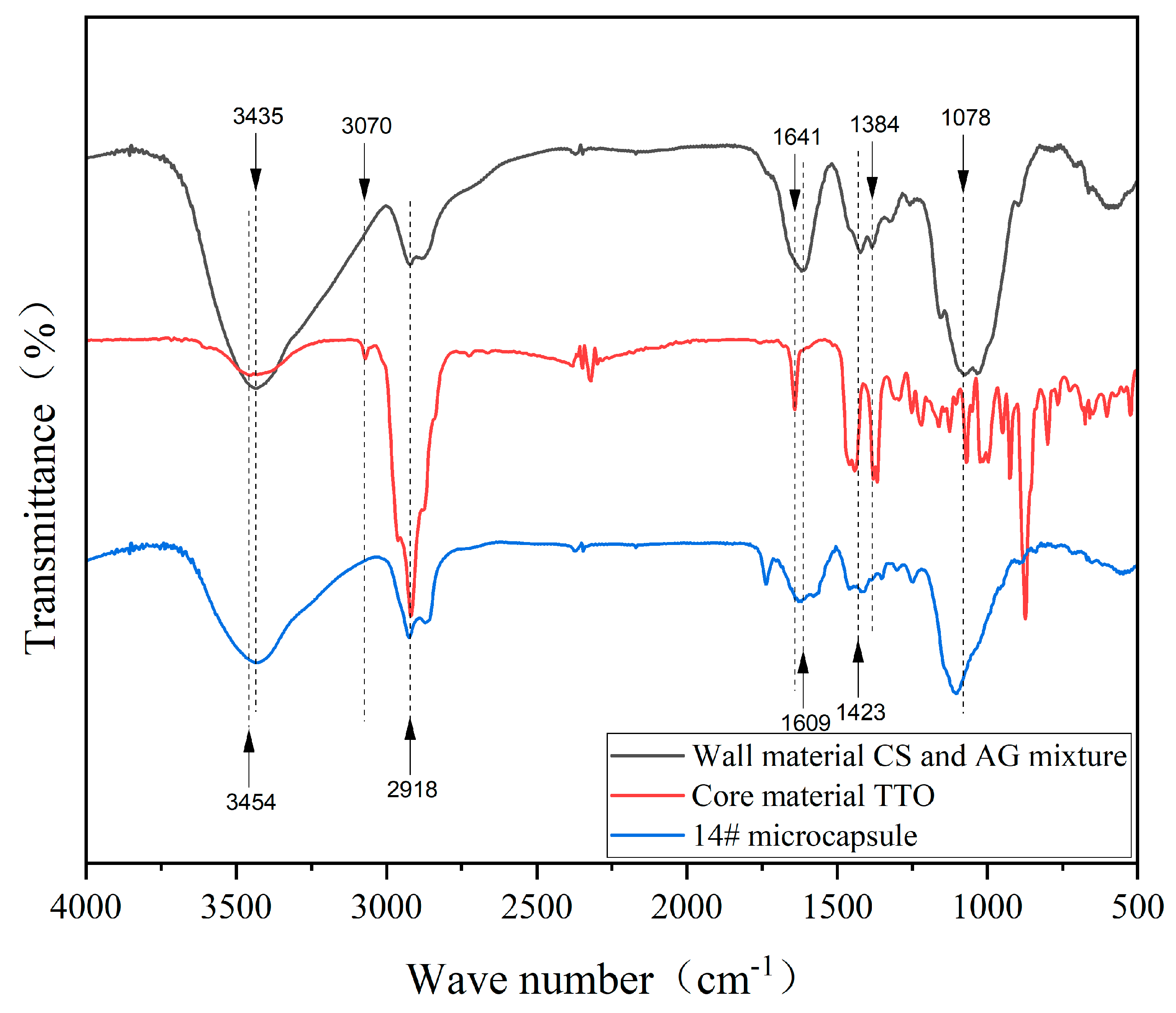
As illustrated in Figure 5, the infrared spectra of wall material CS and AG, TTO, and 14# microcapsule are shown. In the wall IR spectrum, the characteristic band located at 3436 cm−1 is the absorption band of the stretching vibration of the hydroxyl group in the CS molecule. The 1078 cm−1 value has been identified as the stretching vibration of -C-O. The N-H bending vibration of amide II was detected at 1384 cm−1. The asymmetric and symmetric stretching of the carboxylate of AG, -COO, was detected at 1609 cm−1 and 1423 cm−1 [68]. In the infrared spectrogram of the core, the absorption bands located at 3454–3070 cm−1 are attributed to the stretching vibrations of the hydroxyl group and the symmetric and asymmetric stretching vibrations of the N-H bond present in the amino group. In addition, the presence of strong unsaturated C-H vibrational absorption bands at 2918 cm−1 is notable. The presence of an unsaturated C=C vibrational absorption band at 1641 cm−1 was identified. Infrared spectral analysis of TTO@CS-AG microcapsules demonstrated that the characteristic absorption bands corresponded to the standard spectra of CS-AG and TTO. This finding suggests that the core material TTO and the CS-AG wall material exhibit a favourable chemical compatibility, resulting in their effective coexistence and interaction within the microcapsule system.

3.3. Morphology and Chemical Composition Analysis of TTO@CS-AG Microencapsulated Waterborne Coating

A comparative analysis of the SEM images of the microcapsules reveals that the 12#, 13#, and 14# microcapsules exhibit superior morphology and reduced agglomeration compared to the 10#, 11#, and 15# microcapsules. Consequently, the microcapsules exhibiting superior morphology were selected for incorporation into the waterborne paint, thereby preparing the coating. Figure 6 presents the SEM images of the coating prepared from 12#, 13#, and 14# microcapsules with 5% addition content. The surface of the coating exhibited a flat and smooth appearance in the absence of microcapsules. However, following the incorporation of microcapsules, the surface of the coating exhibited elevated structures. Nonetheless, it was challenging to quantitatively ascertain the discrepancy in surface roughness through direct observation of the topographical features depicted in the SEM images. The presence of agglomerates was observed on the surface of the coating, indicating the incorporation of 12# microcapsules. The addition of 13# microcapsules resulted in the formation of corrugated undulations, while the incorporation of 14# microcapsules led to relatively uniform raised structures on the coating surface. This phenomenon can be attributed to the presence of more irregular agglomerates in 12# microcapsules, which possess relatively large particle sizes. As the percentage of CS increases, the rigidity of the molecular chain leads to uneven drying and shrinkage of the coating containing microcapsules. This results in wrinkles, thereby reducing the surface flatness of the prepared coating. In order to quantitatively compare the relative surface elevation variations, a statistical analysis of the grey values was performed across regions in Figure 6 using ImageJ 1.54g software (n = 4). The Mean Grey Value results are presented in Table 10. The following samples were analysed: sample 0#, 129.64; sample 12#, 146.44; sample 13#, 134.84; sample 14#, 137.73. This finding aligns with the observed trends in surface roughness measurements.
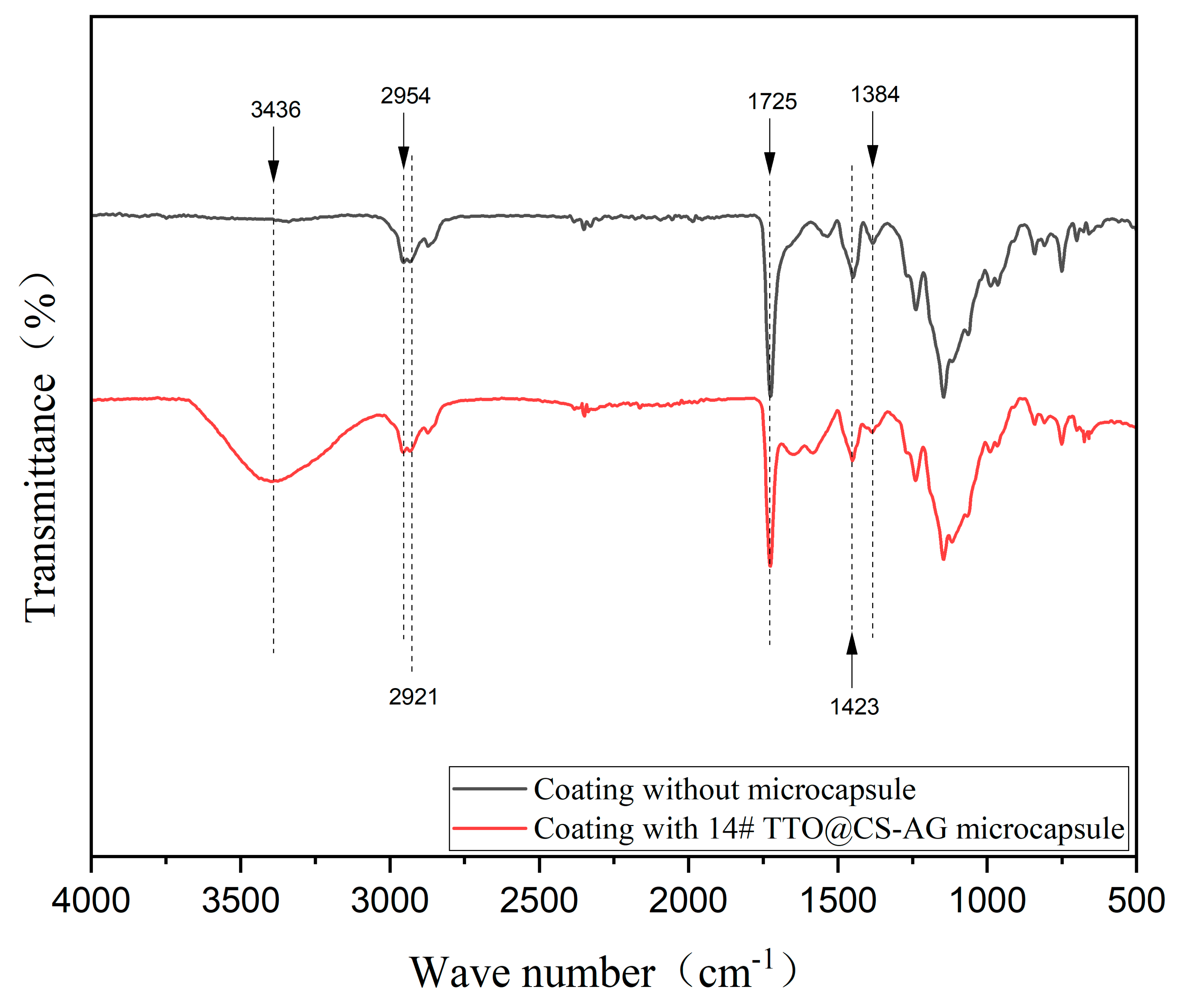
As illustrated in Figure 7, the infrared spectra of the coating with 14# TTO@CS-AG microcapsule added and the coating without microcapsule are shown. The stretching vibration band of the O-H bond was observed at 3436 cm−1, a finding that is consistent in both the waterborne coating system and the TTO fraction. The absorption band at 2954 cm−1 was attributed to the bending vibration of the C-H bond in the coating. The characteristic band at 1725 cm−1 corresponded to the stretching vibration of the carbonyl group (C=O) in the coating molecule. Furthermore, the absorption band at 2921 cm−1 can be attributed to the -CH2 stretching vibration in the CS molecule. The wavenumber of 1384 cm−1 corresponds to the bending vibration of the N-H group of amide II. The symmetric stretching of the carboxylate of AG-COO appears at 1423 cm−1. This finding demonstrates that the wall and core components of the microcapsules persist following their incorporation into the waterborne topcoat, thereby confirming the absence of any chemical reaction between the microcapsules and the waterborne topcoat.

3.4. Analysis of Antimicrobial Properties of Microcapsules with Different mAG:mCS on Coating

Table 11 shows the number of colonies recovered from the coating surface and the antimicrobial rate when microcapsules with various mAG:mCS ratios were added. Figure 8 presents a representative colony recovery diagram, while Figure 9 shows the trend diagram for the antimicrobial rate. The presence of microcapsules in paint coatings has been demonstrated to significantly reduce the number of recovered colonies of the two tested bacteria (E. coli and S. aureus) in comparison to the pure paint coating devoid of microcapsules (0#). This finding serves to confirm that the incorporation of microcapsules has enhanced the overall antimicrobial performance of the paint coating to a certain extent. The trend in the antimicrobial rate with the mAG:mCS ratio exhibited a significant strain dependence. The antimicrobial rate of E. coli demonstrated a tendency towards an accelerated increase, subsequently followed by a substantial decrease, attaining a maximum of 65.55% at a mAG:mCS ratio of 3.0:1 (14#). The efficacy was found to be the least pronounced at the lowest ratio of 1:1 (10#), exhibiting a reduction to 28.32%. It is noteworthy that the rate was instead reduced to 38.21% at the highest ratio of 3.5:1 (15#). The antimicrobial rate of S. aureus showed a fluctuating increasing trend. In contrast, S. aureus exhibited a fluctuating upward trend, with its overall antimicrobial level consistently surpassing that of E. coli. The lowest recorded value was 1:1 (10#), at 63.36%, while the highest was 3.5:1 (15#), at 77.47%. Additionally, a higher value was observed at the intermediate ratio of 3.0:1 (14#), at 73.29%. The trend of antimicrobial rates was steeper for E. coli than for S. aureus, suggesting that it is more sensitive to high mAG:mCS ratios, which may stem from the differential response of its Gram-negative outer membrane structure. The higher antimicrobial rates of all samples against S. aureus are consistent with specific inhibition of AG carboxylated Fe3+, Mg2+, and other plasma interfering with membrane protein function and the greater sensitivity of the Gram-positive bacterium S. aureus to this mechanism [69,70]. The paradoxical phenomenon that the 15# sample (mAG:mCS = 3.5:1) was the most effective against S. aureus (77.47%) but the least effective against E. coli (38.21%) may be attributed to the fact that the high ratio of AG altered the physicochemical properties of the microcapsule, which was not conducive to the effective antimicrobial activity against E. coli. Taken together, the mAG:mCS ratio is a key factor in regulating the antimicrobial performance and there is a strain dependence of the effect, and Sample 14# (mAG:mCS = 3.0:1) showed the best performance in balancing the antimicrobial effects of the two bacteria. The antimicrobial rate of 14# against E. coli (65.55%) was significantly higher than that of 15# (38.21%), with a 31% reduction in standard deviation, confirming that the 3:1 ratio is more stable against Gram-negative bacteria. While the antimicrobial rate of 15# against S. aureus (77.47%) was the highest, the antimicrobial rate of both 15# and 14# were high, indicating that the inhibitory effect on Gram-positive bacteria remains stable at high ratios.

3.5. Analysis of Microcapsules with Different mAG:mCS on the Optical Properties of Coating

3.5.1. Colour Difference Performance Analysis

The chromaticity parameters (L, a, b) and colour difference in the coating are shown in Table 12. The incorporation of microcapsules resulted in a marginal decrease in the L (lightness) of the coating. As the mAG:mCS ratio increased, the L of the coating demonstrated an inconsistent significant change trend, while a (red/green chromaticity) and b (yellow/blue chromaticity) exhibited a significant downward trend. The colour difference (ΔE) exhibited a marked downward trend. As the mAG:mCS ratio increased, the ΔE value decreased gradually from a maximum of 7.59 (sample 10#) to a minimum of 3.30 (sample 14#), with sample 15# showing a slight increase. The smallest colour difference (ΔE = 3.30) was exhibited by Sample 14#, yet it still represented a clearly visible difference. The variation in colour difference may be attributed to two potential factors: firstly, the potential agglomeration of microcapsules within the coating layer could impede its uniform dispersion, thereby affecting the flatness and homogeneity of the coating film surface. This phenomenon tends to amplify the colour difference, particularly in cases where dispersion is inadequate [71]. Conversely, an increase in AG addition has been observed to decrease the refractive index of the microcapsule wall material, thereby bringing it closer to that of the resin matrix [72]. This reduction in the refractive index has been shown to help reduce light scattering at the interface, thus leading to a reduction in colour difference.

3.5.2. Gloss and Loss Performance Analysis

As illustrated in Table 13, the gloss and light loss rate of the coating are presented. As demonstrated in Figure 10, the trend in changes in the gloss and light loss rate of the coating is evident, with the presented data including error bars. The incorporation of microcapsules resulted in a reduction in the coating gloss and an enhancement of its gloss dissipation when compared with the coating devoid of microcapsules. As the incident angle was adjusted to 20°, it was observed that the overall trend of gloss decreased in proportion to the increase in the mAG:mCS ratio (from 9.2 GU for 10# to 6.0 GU for 15#), while the corresponding trend of gloss loss increased (from 43.21% for 10# to 62.96% for 15#). Samples 10# (mAG:mCS = 1:1) and 11# (mAG:mCS = 1.5:1) exhibited a relatively high gloss (9.2 GU, 8.7 GU) and low light loss rate (43.21%, 46.30%), respectively. At an incidence angle of 60°, the gloss level exhibited fluctuating changes in conjunction with the increase in mAG:mCS. When the ratio of mAG:mCS was 3:1 (14#), the gloss of the coating reached its maximum value (24.00 GU). Concurrently, an increase in mAG:mCS was observed to be accompanied by fluctuations in the light loss rate of the coating. The minimum light gloss rate of the coating was 18.09% (14#). At an incidence angle of 85°, the gloss of the coating exhibited fluctuations in response to the increase in mAG:mCS. When the ratio of mAG:mCS was 3:1 (14#), the gloss of the coating reached its maximum value (29.20 GU). As the mAG:mCS ratio increased, the light gloss rate of the coating exhibited a decline, followed by an uptick. The light gloss rate of the coating was minimised to 21.30% through the incorporation of 14# microcapsules. When the mAG:mCS ratio was 2.5:1 (13#) and 3:1 (14#), the composite optical properties exhibited superior optical properties at 60° and 85° incidence angles. This phenomenon can be attributed to the fact that the refractive index of the AG and CS composite wall ratio is closely aligned with that of the acrylic resin at this particular juncture, thereby minimising interfacial scattering. The dispersion of microcapsules within the coating system, particularly the occurrence of localised agglomeration, contributes to the enhancement of microscopic unevenness on the coating surface. Increased roughness has been shown to result in enhanced light scattering, leading to a substantial reduction in gloss [73]. The gloss and light gloss rate of the coating are the result of a synergistic interaction between two factors. Firstly, there is a change in surface roughness due to the introduction of microcapsules. Secondly, there is a degree of wall–substrate refractive index matching. The results of this study underscore the significance of optimising the dispersion of microcapsules in coatings, with a view to minimising agglomeration and surface roughness. This is essential for achieving the desired optical properties of coatings containing functional microcapsules.

3.5.3. Visible Light Transmission Rate Analysis

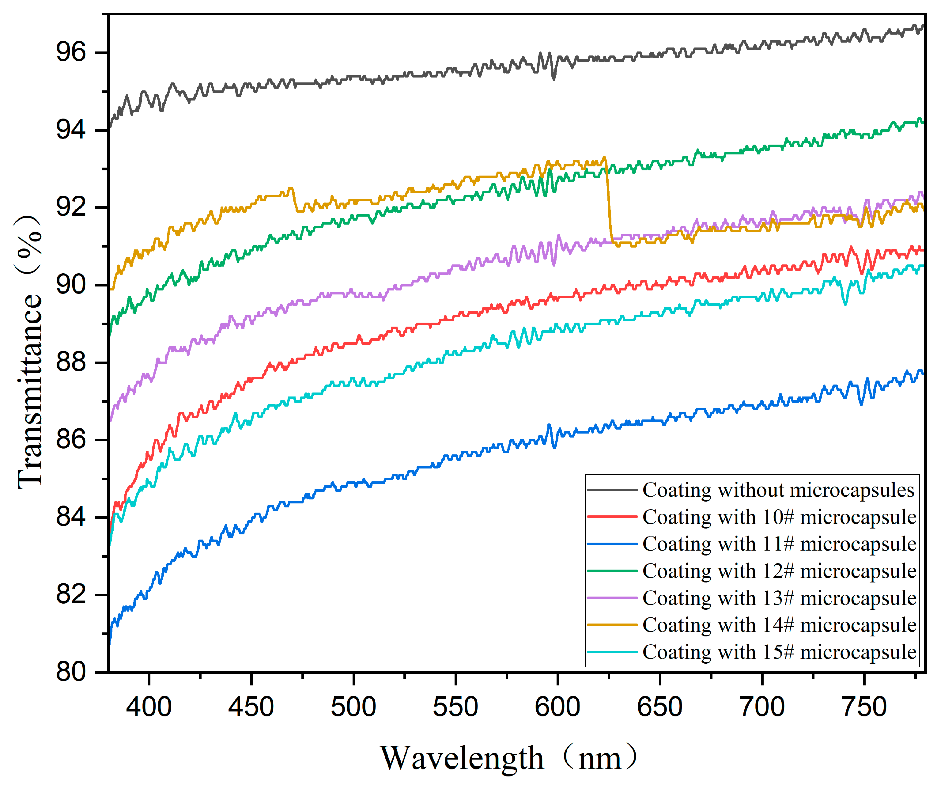
As illustrated in Table 14, the light transmittance of coatings prepared from microcapsules with varying mAG:mCS ratios is demonstrated. Figure 11 shows the transmittance curves of these coatings. The investigation yielded the finding that the visible light transmittance of the paint coatings containing microcapsules was reduced in comparison with those devoid of microcapsules. This finding serves to confirm the hypothesis that the introduction of microcapsules into the paint coatings results in a reduction in the light transmittance. It was observed that as the mAG:mCS ratio increased, the coating visible light transmittance exhibited a fluctuating trend. Samples 12# (mAG:mCS = 2:1) and 14# (mAG:mCS = 3:1) demonstrated relatively high transmittance levels of 91.01% and 92.12%, respectively. Sample 11# (mAG:mCS = 3.5:1) exhibited the lowest transmittance (86.43%), thereby indicating that its light transmission performance was comparatively deficient. This phenomenon can be attributed to the agglomeration of the white opaque powder-like microcapsules introduced to the waterborne coating. The agglomerates have been shown to increase the light scattering cross-section and exacerbate the optical unevenness both inside and on the surface of the coating, thus reducing the light transmission rate. AG has a highly branched molecular structure, which, in theory, may have an effect on the microstructure of the formed coating. The molecular structure of AG, as well as the significant light scattering effect produced by the microcapsule particles and their agglomerations, resulted in lower light transmittance than that of the pure coating for all microcapsule-containing samples. The transmittance curve of sample 14# (mAG:mCS = 3:1) exhibits a substantial discontinuity at 625 nm. This phenomenon may be attributed to the formation of localised agglomerates or interfacial structures of the microcapsule or its wall material in the coating matrix under this particular formulation, which may produce stronger resonance, scattering, or interference effects at a particular wavelength (625 nm) [74,75].

3.6. Analysis of Microcapsules with Different mAG:mCS on Mechanical Properties of Coating

3.6.1. Roughness Performance Analysis

Table 15 shows the effect of different mAG:mCS microcapsule compositions on the surface roughness (Ra) of the coating, based on four independent repetitions. The roughness (Ra) of all microcapsule-containing paint coatings increased significantly in comparison with the pure coating devoid of added microcapsules, thus confirming the hypothesis that the addition of microcapsules increases the unevenness of the coating surface. As mAG:mCS increased, the coating roughness (Ra) exhibited a non-monotonic trend of decreasing and then increasing, commencing from 10# and 11# (high values > 0.6 μm), decreasing to a lower level (0.419 μm–0.428 μm) in the interval from 12# to 14#, and then increasing again at 15# (0.579 μm). This phenomenon may be attributed to the disparate molecular properties of CS and AG, and their compatibility effects during the coalescence process. CS is a linear rigid molecule, while AG is a highly branched flexible polymer [76,77]. It has been established that both of these phases are prone to microphase separation when coalescing to form the microcapsule wall. This is due to the difference in molecular conformation and hydrophilicity, resulting in an inhomogeneous wall structure, which in turn makes the roughness (Ra) larger [78].

3.6.2. Mechanical Properties Analysis

The effect of TTO@CS-AG microcapsules on the mechanical properties of the coating is shown in Table 16 and Figure 12. As the mAG:mCS ratio increased from 1:1 to 3.5:1, the coating elongation at break exhibited an “increasing–decreasing” pattern. In the case of mAG:mCS 2.5:1 (13#), the elongation at break of the coating reached a maximum value of 18.54%. When the ratio of mAG:mCS was 1:1 (10#), the elongation at break of the coating reached a minimum of 4.73%. The coatings prepared from 10#, 13# and 14# microcapsules exhibited specific elastic deformation regions on the stress–strain curve. The stress of the coating-absent microcapsule addition was 4.86 MPa, and the maximum tensile strength of the coating was generally reduced by microcapsule addition. Among them, the coatings with 11# and 15# microcapsule additions exhibited relatively high tensile strength and good tensile resistance. The modulus of elasticity of the coatings without the addition of the microcapsule was found to be 0.23 GPa, and the modulus of elasticity of most of the coatings increased after the addition of microcapsule. However, the coatings with 13# and 14# microcapsule additions exhibited a reduced modulus of elasticity (0.16 GPa and 0.10 GPa, respectively), indicating that the incorporation of microcapsules enhances the flexibility and reduces the stiffness of the coatings at these specific ratios.

4. Conclusions

The most significant factor in the preparation of TTO@CS-AG microcapsules is mAG:mCS, and the sample of TTO@CS-AG microcapsules with the optimum yield and encapsulation rate is 12# microcapsule according to the test. The microcapsules exhibit a more protuberant surface, a more uniform distribution, an evident shell–core structure, and the particle size is predominantly distributed between 5 and 7 μm. The optimal process was determined as follows: a core–wall ratio of 1.2:1, ab emulsifier concentration of 2%, a pH of 3, and a mAG:mCS of 2:1. Six microcapsule samples were incorporated into the waterborne coating at the concentration of 5% to prepare the coating film. The FTIR analysis confirmed that TTO was encapsulated within the core of the film and that no chemical change occurred subsequent to the addition of TTO to the coating film. For E. coli, the antimicrobial rate of the coating exhibited a single-peak trend with increasing mAG:mCS (maximum at 14#: 65.55%), and the elevated mAG:mCS (15#) diminished the effect (38.21%). For S. aureus, the antimicrobial rate of the coating demonstrated a fluctuating upward trend. The colour difference in the coating exhibited a decreasing trend with increasing mAG:mCS. The gloss of the coating was found to decrease with increasing mAG:mCS at a 20° incidence angle. The 14# specimen demonstrated the optimal performance of 24.0 GU and 29.2 GU at 60° and 85°. The transmittance of the coating in the visible band was found to decrease with the addition of the microcapsule. The incorporation of microcapsules resulted in an increase in roughness ranging from 3 to 7 times, with 14# exhibiting the least significant increase (0.422 μm). The elongation at break was found to be “peaked”, with 13# reaching the peak (18.54%). The 14# microcapsules (mAG:mCS = 3:1) demonstrated remarkable efficacy in terms of the antimicrobial properties (E. coli of 65.55%, S. aureus of 73.29%), the optical characteristics (light transmittance 92.12%, 60° gloss 24.0 GU), and the flexibility (elongation at break 18.10%, modulus 0.10 GPa). The experiments did not quantify the slow-release properties of the microcapsules, and further validation of antimicrobial durability is required.

Author Contributions

Conceptualization, methodology, validation, resources, data management, N.Z.; supervision, writing—review and editing, Y.Z.; formal analysis and investigation, X.Y. and J.L. All authors have read and agreed to the published version of the manuscript.

Funding

This project was partly supported by the Qing Lan Project and the Natural Science Foundation of Jiangsu Province (BK20201386).

Institutional Review Board Statement

Not applicable.

Informed Consent Statement

Not applicable.

Data Availability Statement

Data are contained within the article.

Conflicts of Interest

The authors declare no conflicts of interest.

References

  1. Zhou, J.; Xu, W. Optimizing the Interface Compatibility of Transparent Wood for Green Phase-Change Thermal Storage. Wood Sci. Technol. 2025, 59, 45. [Google Scholar] [CrossRef]
  2. Yang, Y.; Xu, W.; Liu, X.; Wang, X. Study on Permeability of Cunninghamia Lanceolata Based on Steam Treatment and Freeze Treatment. Wood Res. 2021, 66, 721–731. [Google Scholar] [CrossRef]
  3. Wu, S.; Tao, X.; Xu, W. Thermal Conductivity of Poplar Wood Veneer Impregnated with Graphene/Polyvinyl Alcohol. Forests 2021, 12, 777. [Google Scholar] [CrossRef]
  4. Liu, X.; Cui, R.; Shi, J.; Jiang, Q.; Gao, J.; Wang, Z.; Li, X. Separation and microencapsulation of antibacterial compounds from wood vinegar. Process Biochem. 2021, 110, 275–281. [Google Scholar] [CrossRef]
  5. Bi, W.; Li, H.; Hui, D.; Gaff, M.; Lorenzo, R.; Corbi, I.; Ashraf, M. Effects of chemical modification and nanotechnology on wood properties. Nanotechnol. Rev. 2021, 10, 978–1008. [Google Scholar] [CrossRef]
  6. Liu, Q.; Gao, D.; Xu, W. Effect of Paint Process on the Performance of Modified Poplar Wood Antique. Coatings 2021, 11, 1174. [Google Scholar] [CrossRef]
  7. Susi, P.; Aktuganov, G.; Himanen, J.; Korpela, T. Biological control of wood decay against fungal infection. J. Environ. Manag. 2011, 92, 1681–1689. [Google Scholar] [CrossRef] [PubMed]
  8. Duan, X.; Liu, S.; Huang, E.; Shen, X.; Wang, Z.; Li, S.; Jin, C. Superhydrophobic and antibacterial wood enabled by polydopamine-assisted decoration of copper nanoparticles. Colloids Surf. A Physicochem. Eng. Asp. 2020, 602, 125145. [Google Scholar] [CrossRef]
  9. Hu, W.; Yang, Z.; Shi, N.; Yu, X. Experimental study on effects of the selected load parameters on fatigue life of the mortise-and-tenon furniture joint. Wood Mater. Sci. Eng. 2025, 19, 1–7. [Google Scholar] [CrossRef]
  10. Wang, C.; Li, J.; Wang, T.; Wang, X. Additive manufacturing of furniture corner guards based on thermoplastic polyurethane filament. BioResources 2025, 20, 5398–5406. [Google Scholar] [CrossRef]
  11. Liu, Y.; Hu, W.; Kasal, A.; Erdil, Y.Z. The state of the art of biomechanics applied in ergonomic furniture design. Appl. Sci. 2023, 13, 12120. [Google Scholar] [CrossRef]
  12. Xu, X.; Zhang, M.; Yue, X.; Xiong, X. Design of furniture mortise-and-tenon joints: A review of mechanical properties and design recommendations. Wood Mater. Sci. Eng. 2025, 1–15. [Google Scholar] [CrossRef]
  13. Yu, R.; Liu, Y.; Konukcu, C.A.; Hu, W. A method of simulating seat load for numerical analysis of wood chair structure. Wood Res. 2024, 69, 432–444. [Google Scholar] [CrossRef]
  14. Hu, W.; Yu, R.; Yang, P. Characterizing roughness of wooden mortise and tenon considering effects of measured position and assembly condition. Forests 2024, 15, 1584. [Google Scholar] [CrossRef]
  15. Fu, S.; Xiong, X.; Wan, R.; Zhang, M.; Xu, X. The Development and Future Challenges of China’s Furniture Industry. Drewno 2025, 68, 199709. [Google Scholar] [CrossRef]
  16. Hu, W.; Fu, W.; Zhao, Y. Optimal design of the traditional Chinese wood furniture joint based on experimental and numerical method. Wood Res. 2024, 69, 50–59. [Google Scholar] [CrossRef]
  17. Chang, C.W.; Lu, K.T. Synthesis and antibacterial activity of metal-containing ultraviolet-cured wood floor coatings. Polymers 2021, 13, 3022. [Google Scholar] [CrossRef]
  18. Xu, W.; Fang, X.Y.; Han, J.T.; Wu, Z.H. Effect of Coating Thickness on Sound Absorption Property of Four Wood Species Commonly Used for Piano Soundboards. Wood Fiber Sci. 2020, 52, 28–43. [Google Scholar] [CrossRef]
  19. Liu, Q.Q.; Gao, D.; Xu, W. Effect of Sanding Processes on the Surface Properties of Modified Poplar Coated by Primer Compared with Mahogany. Coatings 2020, 10, 856. [Google Scholar] [CrossRef]
  20. Liu, Q.Q.; Gao, D.; Xu, W. Effect of Polyurethane Non-Transparent Coating Process on Paint Film Performance Applied on Modified Poplar. Coatings 2022, 12, 39. [Google Scholar] [CrossRef]
  21. Hu, T.; Yuan, F.; Zhou, C.; Kaner, J. Effect of Innovative Office Furniture on Enhancing Employee Active Health. BioResources 2025, 20, 5200–5213. [Google Scholar] [CrossRef]
  22. Broda, M. Natural compounds for wood protection against fungi—A review. Molecules 2020, 25, 3538. [Google Scholar] [CrossRef]
  23. Liu, Q.Q.; Gu, Y.T.; Xu, W.; Lu, T.; Li, W.J.; Fan, H.B. Compressive Properties of Green Velvet Material Used in Mattress Bedding. Appl. Sci. 2021, 11, 11159. [Google Scholar] [CrossRef]
  24. Wan, R.Y.; Xiong, X.Q.; Fu, S.J.; Xiong, D.J.; Xu, X.T. Life cycle assessment and optimization scenarios of sofas: A case study in China. Int. J. Life Cycle Assess. 2025, 30, 2020–2036. [Google Scholar] [CrossRef]
  25. Wang, C.; Li, J.Y.; Wang, T.Y.; Chu, Q.; Wang, X.W. Fused deposition 3D printing of bonsai tree guiding mold based on acrylonitrile-butadiene-styrene copolymer. BioResources 2024, 19, 5839–5846. [Google Scholar] [CrossRef]
  26. Wang, C.; Li, J.Y.; Wang, T.Y.; Chu, Q.; Wen, S.Q. Design and Rapid Prototyping of Packaging Liner for Rosewood Craft based on Gyroid Infill Structure. BioResources 2025, 20, 842–851. [Google Scholar] [CrossRef]
  27. Abolhasani, H.; Farzi, G.; Davoodi, A.; Vakili-Azghandi, M.; Das, O.; Neisiany, R.E. Development of self-healable acrylic water-based environmental-friendly coating as an alternative to chromates coatings. Prog. Org. Coat. 2023, 176, 107402. [Google Scholar] [CrossRef]
  28. Han, K.; Liu, J.; Hao, F.; Wang, J.; Yuan, J.; Pan, Z.; Pan, M. An adaptive waterborne fluorocarbon coatings with Anti-Flashing Rust, Antibiofouling, and Self-Repairing properties. Chem. Eng. J. 2024, 495, 153644. [Google Scholar] [CrossRef]
  29. Wu, A.; Sun, Y.; Wang, Z.; Cao, G.; Zhang, H. Polyaniline/cellulose nanocrystal nanorods integrated into waterborne polyurethane coatings for enhanced corrosion resistance. Colloids Surf. A Physicochem. Eng. Asp. 2025, 713, 136531. [Google Scholar] [CrossRef]
  30. Mastouri, A.; Efhamisisi, D.; Tarmian, A.; Boukherroub, R.; Lexa, M.; Karami, E.; Frigione, M. Sustainable superhydrophobic and self-cleaning wood via wax within Epoxy/PDMS nano-composite coatings: Durability related to surface morphology. Prog. Org. Coat. 2024, 186, 107951. [Google Scholar] [CrossRef]
  31. Barthwal, S.; Uniyal, S.; Barthwal, S. Nature-inspired superhydrophobic coating materials: Drawing inspiration from nature for enhanced functionality. Micromachines 2024, 15, 391. [Google Scholar] [CrossRef]
  32. Zubko, M.K. Towards sustainable antimicrobials from plants: Some ways to abridge current methodological approaches. Sustain. Mater. Technol. 2024, 39, e00801. [Google Scholar] [CrossRef]
  33. Mercan, D.A.; Niculescu, A.G.; Grumezescu, A.M. Nanoparticles for antimicrobial agents delivery—An up-to-date review. Int. J. Mol. Sci. 2022, 23, 13862. [Google Scholar] [CrossRef] [PubMed]
  34. Zhang, H.L.; Li, Y.X. Antibacterial and Antimalarial Therapeutic Agents: A Patent Perspective. Recent Adv. Inflamm. Allergy Drug Discov. 2024, 18, 57–66. [Google Scholar] [CrossRef] [PubMed]
  35. Xiao, Q.Q.; Zhu, J.N.; Fang, T.; Peng, R.Y.; Chen, J.Y.; Liu, K.L.; Ceng, Y.S.; Yuan, M.; Hu, Y.R. Preparation and characterization of nano silver antibacterial gel for gynecolog. Ferroelectrics 2024, 618, 2249–2256. [Google Scholar] [CrossRef]
  36. Serwecińska, L. Antimicrobials and antibiotic-resistant bacteria: A risk to the environment and to public health. Water 2020, 12, 3313. [Google Scholar] [CrossRef]
  37. La Fauci, V.; Costa, G.B.; Genovese, C.; Palamara, M.A.R.; Alessi, V.; Squeri, R. Effect of Drug-Resistant Bacteria on Hands of Healthcare Workers and in the Patient Area: An Environmental Survey in Southern Italy’s Hospital. Rev. Española Quimioter. 2019, 32, 303. [Google Scholar]
  38. Nath, S.; Sinha, A.; Singha, Y.S.; Dey, A.; Bhattacharjee, N.; Deb, B. Effect of Prevalence of Antibiotic-Resistant, Toxic Metal-Tolerant and Biofilm-Forming Bacteria in Hospital Surroundings. Environ. Anal. Health Toxicol. 2020, 35, 2020018. [Google Scholar] [CrossRef]
  39. Cloutier, M.; Mantovani, D.; Rosei, F. Effect of Antibacterial Coatings: Challenges, Perspectives, and Opportunities. Trends Biotechnol. 2015, 33, 637–652. [Google Scholar] [CrossRef]
  40. Fu, Y.; Dudley, E.G. Effect of Antimicrobial-Coated Films as Food Packaging: A Review. Compr. Rev. Food Sci. Food Saf. 2021, 20, 3404–3437. [Google Scholar] [CrossRef]
  41. Valdés, A.; Ramos, M.; Beltrán, A.; Jiménez, A.; Garrigós, M.C. Effect of State of the Art of Antimicrobial Edible Coatings for Food Packaging Applications. Coatings 2017, 7, 56. [Google Scholar] [CrossRef]
  42. Yong, L.X.; Calautit, J.K. Effect of A Comprehensive Review on the Integration of Antimicrobial Technologies onto Various Surfaces of the Built Environment. Sustainability 2023, 15, 3394. [Google Scholar] [CrossRef]
  43. Mehta, N.; Kumar, P.; Verma, A.K.; Umaraw, P.; Kumar, Y.; Malav, O.P.; Sazili, A.Q.; Domínguez, R.; Lorenzo, J.M. Microencapsulation as a Noble Technique for the Application of Bioactive Compounds in the Food Industry: A Comprehensive Review. Appl. Sci. 2022, 12, 1424. [Google Scholar] [CrossRef]
  44. Wang, J.; Zhuang, S. Chitosan—Based Materials: Preparation, Modification and Application. J. Clean. Prod. 2022, 355, 131825. [Google Scholar] [CrossRef]
  45. Lobel, B.T.; Baiocco, D.; Al-Sharabi, M.; Routh, A.F.; Zhang, Z.; Cayre, O.J. Current Challenges in Microcapsule Designs and Microencapsulation Processes: A Review. ACS Appl. Mater. Interfaces 2024, 16, 40326–40355. [Google Scholar] [CrossRef]
  46. Zhang, H.; Wang, Z.; Qiu, H.; Xu, B.; Chen, K. Design and fabrication of silica/chitosan composite microcapsules for bifunctional fabric coating. Int. J. Biol. Macromol. 2025, 309, 143018. [Google Scholar] [CrossRef]
  47. Liu, J.; Liu, C.; Liu, Y.; Chen, M.; Hu, Y.; Yang, Z. Study on the grafting of chitosan–gelatin microcapsules onto cotton fabrics and its antibacterial effect. Colloids Surf. B Biointerfaces 2013, 109, 103–108. [Google Scholar] [CrossRef]
  48. Indriyani, N.N.; Al-Anshori, J.; Wahyudi, T.; Nurzaman, M.; Nurjanah, S.; Permadi, N.; Julaeha, E. An optimized chitosan/alginate-based microencapsulation of lime peel essential oil and its application as an antibacterial textile. J. Biomater. Sci. Polym. 2024, 35, 989–1007. [Google Scholar] [CrossRef]
  49. Karaaslan, M.; Şengün, F.; Cansu, Ü.; Başyiğit, B.; Sağlam, H.; Karaaslan, A. Gum arabic/maltodextrin microencapsulation confers peroxidation stability and antimicrobial ability to pepper seed oil. Food Chem. 2021, 337, 127748. [Google Scholar] [CrossRef]
  50. Velazquez-Martinez, V.; Valles-Rosales, D.; Rodriguez-Uribe, L.; Holguin, O.; Quintero-Quiroz, J.; Reyes-Jaquez, D.; Delgado, E. Antimicrobial, shelf-life stability, and effect of maltodextrin and gum arabic on the encapsulation efficiency of sugarcane bagasse bioactive compounds. Foods 2021, 10, 116. [Google Scholar] [CrossRef]
  51. Chang, Y.; Wu, Z.; Liu, E. Effect of Fabrication of Chitosan-Encapsulated Microcapsules Containing Wood Wax Oil on Antibacterial Self-Healing Wood Coatings. Ind. Crops Prod. 2024, 222, 119438. [Google Scholar] [CrossRef]
  52. Ma, L.; Xu, S. Effect of Investigation on the Restoration Properties of Wood Oil Microcapsules in Wood Coatings. Prog. Org. Coat. 2024, 197, 108853. [Google Scholar] [CrossRef]
  53. Liu, Y.; Li, L.; Zhao, A.; Song, X.; Wei, L.; Fang, M.; Zhu, X. Effect of Sustainable Fragrance Release Wax Oil Coating for Wood Substrate Based on Peppermint Essential Oil Microcapsules. Ind. Crops Prod. 2024, 208, 117848. [Google Scholar] [CrossRef]
  54. An, P.; Yang, X.; Yu, J.; Qi, J.; Ren, X.; Kong, Q. α-terpineol and terpene-4-ol, the critical components of tea tree oil, exert antifungal activities in vitro and in vivo against Aspergillus niger in grapes by inducing morphous damage and metabolic changes of fungus. Food Control 2019, 98, 42–53. [Google Scholar] [CrossRef]
  55. Mondello, F.; Fontana, S.; Scaturro, M.; Girolamo, A.; Colone, M.; Stringaro, A.; Ricci, M.L. Terpinen-4-ol, the main bioactive component of tea tree oil, as an innovative antimicrobial agent against Legionella pneumophila. Pathogens 2022, 11, 682. [Google Scholar] [CrossRef]
  56. Yadav, E.; Kumar, S.; Mahant, S.; Khatkar, S.; Rao, R. Tea tree oil: A promising essential oil. J. Essent. Oil Res. 2017, 29, 201–213. [Google Scholar] [CrossRef]
  57. Egbosiuba, T.C.; Abdulkareem, A.S.; Tijani, J.O.; Ani, J.I.; Krikstolaityte, V.; Srinivasan, M.; Veksha, A.; Lisak, G. Taguchi optimization design of diameter-controlled synthesis of multi walled carbon nanotubes for the adsorption of Pb(II) and Ni(II) from chemical industry wastewater. Chemosphere 2021, 266, 128937. [Google Scholar] [CrossRef]
  58. Moralı, U.; Demiral, H.; Şensöz, S. Optimization of activated carbon production from sunflower seed extracted meal: Taguchi design of experiment approach and analysis of variance. J. Clean. Prod. 2018, 189, 602–611. [Google Scholar] [CrossRef]
  59. QB/T 4373-2012; Surface Coating of Furniture—Technical Specification for Application of Waterborne Wood Coatings. Ministry of Industry and Information Technology of the People’s Republic of China: Beijing, China, 2012.
  60. ISO 20579-3:2021; Surface Chemical Analysis—Sample Handling, Preparation and Mounting Part 3: Biomaterials. International Standard Published: Geneva, Switzerland, 2021.
  61. GB/T 21866-2008; Test Method for Antibacterial Property and Antibacterial Effect of Antibacterial Coatings (Paint Films). Standardization Administration of the People’s Republic of China: Beijing, China, 2008.
  62. GB/T 4789.2-2022; Microbiological Examination of Food Hygiene—Aerobic Plate Count. Standardization Administration of the People’s Republic of China: Beijing, China, 2022.
  63. GB/T 11186.3-1989; Method for Measuring the Color of Painted Film—Part 3: Calculation of Color Difference. Standardization Administration of the People’s Republic of China: Beijing, China, 1989.
  64. GB/T 4893.6-2013; Physical and Chemical Performance Tests of the Paint Film on the Surface of Furniture—Part 6: Method for Determining Gloss. Standardization Administration of the People’s Republic of China: Beijing, China, 2013.
  65. ISO 2813: 2014; Paints and Varnishes—Determination of Gloss Value at 20°, 60° and 85°. International Standard Published: Geneva, Switzerland, 2014.
  66. ISO 25178-601:2025; Geometrical Product Specifications (GPS)—Surface Texture: Areal. International Standard Published: Geneva, Switzerland, 2025.
  67. ASTM D2370-16 (2021); Standard Test Method for Tensile Properties of Organic Coatings. American Society of Testing Materials: West Conshohocken, PA, USA, 2021.
  68. John Kennedy, J.P.K.; Muthuramalingam, J.B.; Balasubramanian, V.K.; Balakrishnan, M.; Murugan, K.; Ponnuchamy, K. Controlled Release of Urea Using Negatively Charged Polysaccharides. Polym. Adv. Technol. 2024, 35, e6508. [Google Scholar] [CrossRef]
  69. Ganie, S.A.; Naik, R.A.; Mir, T.A.; Ali, A.; Mazumdar, N.; Li, Q. Effect of Tailored Functionalization of Gum Arabic Iron (II) Complexes on Synthesis, Characterization and Dwindling of Antianemic Approach via In Vivo Studies. J. Polym. Environ. 2023, 31, 2448–2461. [Google Scholar] [CrossRef]
  70. Guan, Y.; Zhong, Q. Effect of Gum Arabic and Fe2+ on Synergistically Improving the Heat and Acid Stability of Norbixin at pH 3.0–5.0. J. Agric. Food Chem. 2014, 62, 12668–12677. [Google Scholar] [CrossRef]
  71. Pedaballi, S.; Li, C.C.; Song, Y.J. Dispersion of microcapsules for the improved thermochromic performance of smart coatings. RSC Adv. 2019, 9, 24175–24183. [Google Scholar] [CrossRef]
  72. Akram, S.; Bao, Y.; Butt, M.S.; Shukat, R.; Afzal, A.; Huang, J.Y. Fabrication and characterization of gum arabic-and maltodextrin-based microcapsules containing polyunsaturated oils. J. Sci. Food Agric. 2021, 101, 6384–6394. [Google Scholar] [CrossRef]
  73. Maleki, A.; Vafaeenezhad, H.; Eslami-Farsani, R. Polystyrene microcapsules containing linseed oil and SiC nanoparticles as a lubricant additive for boosting the self-healing and self-lubricating efficiency of epoxy coatings. Tribol. Int. 2024, 193, 109428. [Google Scholar] [CrossRef]
  74. Wu, Q.; Li, W.; Yan, X. Effect of Microcapsules on Mechanical, Optical, Self-Healing and Electromagnetic Wave Absorption Properties in Waterborne Wood Paint Coatings. Coatings 2023, 13, 1478. [Google Scholar] [CrossRef]
  75. Bollhorst, T.; Rezwan, K.; Maas, M. Effect of Colloidal Capsules: Nano-and Microcapsules with Colloidal Particle Shells. Chem. Soc. Rev. 2017, 46, 2091–2126. [Google Scholar] [CrossRef] [PubMed]
  76. Cunha, R.A.; Soares, T.A.; Rusu, V.H.; Pontes, F.J.; Franca, E.F.; Lins, R.D. The molecular structure and conformational dynamics of chitosan polymers: An integrated perspective from experiments and computational simulations. In The Complex World of Polysaccharides; IntechOpen: London, UK, 2012. [Google Scholar]
  77. Mohamed, S.A.; Elsherbini, A.M.; Alrefaey, H.R.; Adelrahman, K.; Moustafa, A.; Egodawaththa, N.M.; Sabra, S.A. Gum Arabic: A commodity with versatile formulations and applications. Nanomaterials 2025, 15, 290. [Google Scholar] [CrossRef] [PubMed]
  78. Tasios, N.; Samin, S.; Van Roij, R.; Dijkstra, M. Microphase separation in oil-water mixtures containing hydrophilic and hydrophobic ions. Phys. Rev. Lett. 2017, 119, 218001. [Google Scholar] [CrossRef] [PubMed]
Figure 1. OM images of TTO@CS microcapsules: (AI), respectively, represent 9 kinds of microcapsules prepared by orthogonal test.
Coatings 15 01105 g001
Figure 2. OM images of TTO@CS-AG microcapsules: (AF), respectively, represent 6 kinds of microcapsules prepared by single-factor test.
Coatings 15 01105 g002
Figure 3. SEM images of TTO@CS-AG microcapsules at low magnification: (AF) represent 10# to 15#, respectively.
Coatings 15 01105 g003
Figure 4. Particle size distribution of TTO@CS-AG microcapsules: (AF) represent 10# to 15#, respectively.
Coatings 15 01105 g004
Figure 5. FTIR of TTO, CS and TTO@CS-AG microcapsule.
Coatings 15 01105 g005
Figure 6. SEM image of the prepared coating containing TTO@CS-AG microcapsules: (A) Not added, (B) 12#, (C) 13#, (D) 14#.
Coatings 15 01105 g006
Figure 7. FTIR of coating without microcapsule and addition of 14# microcapsule coating.
Coatings 15 01105 g007
Figure 8. Colony recovery of coating, against E. coli: microcapsule (A) not added, (BG), respectively, represent 10# to 15#; against S. aureus: (H) not added, (IN), respectively, represent 10# to 15#.
Coatings 15 01105 g008
Figure 9. Variation trend in antimicrobial rate of coating.
Coatings 15 01105 g009
Figure 10. The trend graphs of variations about coating: (A) glossiness, (B) light loss rate.
Coatings 15 01105 g010
Figure 11. Transmission curves of coating.
Coatings 15 01105 g011
Figure 12. Stress–strain diagram of coating.
Coatings 15 01105 g012
Table 1. Preparation of TTO@CS-AG microcapsule raw materials and reagents list.
Test MaterialsPurityManufacturer
Tea tree essential oil-Wuhan Huaxiang Biotechnology Co., Ltd., Wuhan, China
ChitosanARSinopharm Chemical Reagent Co., Ltd., Shanghai, China
Arabic gumARTianjin Kermel Chemical Reagent Co., Ltd., Tianjin, China
Tween-80ARSinopharm Chemical Reagent Co., Ltd., Shanghai, China
SDBSARShandong Xinjucheng Chemical Technology Co., Ltd., Jinan, China
Acetic acidARShandong Chengkai New Material Co., Ltd., Linyi, China
Sodium tripolyphosphateARTianjin Huasheng Chemical Reagent Co., Ltd., Tianjin, China
Dulux topcoat-Nanjing Jinyou Biotechnology Co., Ltd., Nanjing, China
Nutrient agar medium-Zhongshan Baimicrobial Technology Co., Ltd., Zhongshan, China
Nutrient broth medium-Zhongshan Baimicrobial Technology Co., Ltd., Zhongshan, China
Sodium chlorideARSinopharm Chemical Reagent Co., Ltd., Shanghai, China
E. coli-Beijing Baocang Biotechnology Co., Ltd., Beijing, China
S. aureus-Beijing Baocang Biotechnology Co., Ltd., Beijing, China
Table 2. Test instruments for TTO@CS-AG microcapsules.
EquipmentModelManufacturer
High-precision balanceBSA323SSartorius Scientific Instruments Co., Ltd., Beijing, China
Heat-collecting magnetic stirrerDF-101SChuangyuan Instrument Manufacturing Co., Ltd., Zhengzhou, China
Spray dryerJA-PWGZ100Hangzhou Feiyue Instrument Co., Ltd., Hangzhou, China
Blast drying ovenDHG-9423AShanghai Jinghong Experimental Equipment Co., Ltd., Shanghai, China
Scanning electron microscopeM360-HK822Shenzhen Oswald Optical Instrument Co., Ltd., Shenzhen, China
High-precision gloss metreYG268Shenzhen 3nh Technology Co., Ltd., Shenzhen, China
Fourier transform infrared spectrometerAgilent 5500Agilent Technologies (China) Co., Ltd., Beijing, China
Constant temperature and
humidity box
THA150Nanjing Jinyou Biotechnology Co., Ltd., Nanjing, China
High-precision spectrophotometerDC-23DShandong Yusuo Chemical Technology Co., Ltd., Linyi, China
Ultraviolet spectrophotometerUV-2600iShimadzu (China) Co., Ltd., Shanghai, China
Universal mechanical testing machineAGS-XShimadzu Manufacturing, Kyoto, Japan
Roughness metreJ8-4CShanghai Taiming Optical Instrument Co., Ltd., Shanghai, China
Ultrasonic emulsifierXU-JY92-IINShanghai Huxi Industry Co., Ltd., Shanghai, China
Circulating water vacuum pumpSHZ-D (III)Shaoxing Xiniu Instrument Technology Co., Ltd., Shaoxing, China
Colony counterXK97-AHangzhou Qiwei Instrument Co., Ltd., Hangzhou, China
Table 3. Factors influencing the orthogonal test of TTO@CS-AG microcapsules.
LevelsFactor A
Core–Wall Ratio
Factor B
mAG:mCS
Factor C
Emulsifier Concentration (%)
Factor D
pH
11.0:12:323
21.2:12:134
31.5:17:345
Table 4. Preparation conditions of TTO@CS-AG microcapsules by orthogonal test.
Sample (#)Factor AFactor BFactor CFactor D
11.0:12:323
21.0:12:134
31.0:17:345
41.2:12:335
51.2:12:143
61.2:17:324
71.5:12:344
81.5:12:125
91.5:17:333
Table 5. List of materials for microencapsulation orthogonal test.
Sample (#)CS-AG (% w/w)TTO (% w/w)Tween-80 (% w/w)STPP (% w/w)Acetic Acid (% v/v)
11.251.251.000.201.00
22.502.251.500.201.00
32.503.002.000.201.00
41.251.501.500.201.00
52.503.002.000.201.00
62.503.001.000.201.00
71.251.882.000.201.00
82.503.751.000.201.00
92.503.751.500.201.00
Table 6. Test material for TTO@CS-AG microcapsule single factor test.
Sample (#)Core–Wall Ratio (TTO:CS-AG)CS (% w/w)AG (% w/w)TTO (% w/w)STPP (% w/w)Acetic Acid (% v/v)
101.2:10.500.501.200.201.00
111.2:10.500.751.500.201.00
121.2:10.501.001.800.201.00
131.2:10.501.252.100.201.00
141.2:10.501.502.400.201.00
151.2:10.501.752.700.201.00
Table 7. Analysis of the results of an orthogonal test of microcapsule yield.
Sample (#)Factor A
Core–Wall Ratio
Factor B
mAG:mCS
Factor C
Emulsifier Concentration (%)
Factor D
pH
Output (g)Yield (%)
11.0:12:3232.0123.93
21.0:12:1342.3217.31
31.0:17:3451.6310.58
41.2:12:3352.5325.56
51.2:12:1433.5423.14
61.2:17:3242.5017.36
71.5:12:3442.3620.26
81.5:12:1252.6618.16
91.5:17:3331.8811.12
k117.27323.25019.81719.397
k222.02019.53717.99718.310
k316.51313.02017.99318.10
R5.50710.2301.8231.297
LevelB > A > C > D
Best LevelA2B1C1D1
Best ProcessA2 B1 C1 D1
Deviation Sum of Squares53.432160.9096.6372.906
Free Degree2222
F-Ratio0.9552.8750.1190.052
F-Critical Value4.4604.4604.4604.460
Significance
Table 8. Analysis of encapsulation rate of orthogonal test results of TTO@CS-AG microcapsules.
Sample (#)Factor A
Core–Wall Ratio
Factor B
mAG:mCS
Factor C
Emulsifier Concentration (%)
Factor D
pH
Encapsulation Rate (%)
11.0:12:32370
21.0:12:13460
31.0:17:34545
41.2:12:33560
51.2:12:14365
61.2:17:32450
71.5:12:34470
81.5:12:12555
91.5:17:33370
k158.33366.66758.33368.333
k258.33360.00063.33360.000
k365.00055.00060.00053.333
R6.66711.6675.00015.000
LevelD > B > A > C
Best LevelA3B1C2D1
Best ProcessA3 B1 C2 D1
Deviation Sum of Squares88.889205.55638.889672.22
Free Degree2222
F-Ratio0.5291.2230.2312.017
F-Critical Value4.4604.4604.4604.460
Significance
Table 9. The yield and encapsulation rate of TTO@CS-AG microcapsules by single-factor test.
Sample (#)mAG:mCSYield (%)Encapsulation Rate (%)
101:127.7263.3
111.5:136.3453.3
122:117.2466.7
132.5:135.1963.3
143:131.9860.0
153.5:112.8660.0
Table 10. The average grey value of the coating.
Sample (#)Mean Grey ValueEvaluation
0129.640
12146.44+
13134.840
14137.73+
Table 11. The actual number of recovered viable colonies and antimicrobial rate of E. coli and S. aureus on the coating surface.
Sample (#)E. coli
(CFU/Piece)
Antimicrobial Rate
(%)
EvaluationS. Aureus
(CFU/Piece)
Antimicrobial Rate
(%)
Evaluation
0212 191
1015228.32++7063.36++++
1113934.43++5571.20++++
1210948.56+++4974.33++++
138758.96+++5770.15++++
147265.55++++5173.29++++
1513138.21++4277.47++++
Note: The symbol “++” indicates that the effect has significantly increased by 20% to 40% compared to the control group, “+++” represents an increase of 40% to 60%, and “++++” indicates an increase of more than 60%.
Table 12. The chromaticity value and colour difference in coating.
Sample (#)Chromaticity ValueΔEEvaluation
Lab
085.21−0.115.93
1083.111.4116.937.59+
1181.770.8316.697.56+
1282.410.2714.866.40+
1384.48−0.2610.553.490
1483.85−0.5610.143.300
1584.18−0.5811.434.090
Note: The symbol “+” indicates an increase of 5% to 20% compared to the control group.
Table 13. The glossiness and light loss rate of coating.
Sample (#)Glossiness (GU)Light Loss Rate (%)Evaluation
20°60°85°20°60°85°
016.2029.3037.10
109.2019.7021.2043.2132.7642.86--
118.7018.8022.0046.3035.8440.70---
127.9019.1024.8051.2334.8133.15--
138.6020.1029.0046.9131.4021.83--
145.5024.0029.2066.0518.0921.30--
156.0018.3021.2062.9637.5442.86---
Note: The symbol “--” indicates that the effect is reduced by 20% to 40% compared to the control group, while “---” represents a reduction of 40% to 60%.
Table 14. The visible light transmission rate of coating.
Sample (#)Transmittance (%)Evaluation
095.830
1089.77-
1186.43-
1292.760
1391.01-
1492.120
1588.69-
Note: The symbol “-” indicates a reduction of 5% to 20% compared to the control group.
Table 15. The average surface roughness of coating.
Sample (#)Average Surface Roughness (Ra, µm)Evaluation
00.0800
100.618----
110.605----
120.428----
130.419----
140.422----
150.579----
Note: The symbol “----” indicates that the effect is more than 60% lower than that of the control group.
Table 16. Mechanical properties analysis of coatings.
Sample (#)Elongation (%)Evaluationσ (MPa)EvaluationE (GPa)Evaluation
021.0404.8600.230
104.73----3.00--0.63++++
117.41----4.41-0.60++++
1211.63---3.66--0.31++
1318.54-2.90---0.16--
1418.10-1.74----0.10---
156.29----4.30-0.68++++
Note: The symbol “-” indicates a reduction of 5%–20% compared to the control group, “--” indicates a reduction of 20%–40% compared to the control group, “---” indicates a reduction of 40%–60%, and “----” indicates a reduction of >60%. The symbol “++” indicates an increase of 20%–40% compared to the control group, and “++++” indicates an increase of >60%.
Disclaimer/Publisher’s Note: The statements, opinions and data contained in all publications are solely those of the individual author(s) and contributor(s) and not of MDPI and/or the editor(s). MDPI and/or the editor(s) disclaim responsibility for any injury to people or property resulting from any ideas, methods, instructions or products referred to in the content.