Article

Temperature-Dependent Performance of Thermally Oxidized Zr2.5Nb Alloy for Orthopedic Implants: Mechanical Properties, Wear Resistance, and Biocompatibility

1
Air Force Logistics Academy, Xuzhou 221000, China
2
School of Materials Science and Physics, China University of Mining and Technology, Xuzhou 221116, China
3
Hubei Engineering Institute, Huangshi 435003, China
4
Shanghai Key Laboratory of Advanced High-Temperature Materials & Precision Forming, School of Materials Science and Engineering, Shanghai JiaoTong University, Shanghai 200240, China
*
Authors to whom correspondence should be addressed.
Coatings 2025, 15(8), 940; https://doi.org/10.3390/coatings15080940
Submission received: 7 July 2025 / Revised: 6 August 2025 / Accepted: 9 August 2025 / Published: 11 August 2025

Abstract

This study investigates the critical influence of oxidation temperature on the intrinsic characteristics and surface properties of thermally oxidized Zr2.5Nb alloy. The resulting oxide layers were characterized by scanning electron microscopy (SEM), X-ray diffraction (XRD), X-ray photoelectron spectroscopy (XPS), surface hardness, and nanoindentation. The tribological behavior of the untreated and thermally oxidized Zr2.5Nb alloy was evaluated via reciprocating ball-on-disc wear tests under a load of 29.4 N. MC3T3-E1 cells were employed to assess the biocompatibility. The results show that oxide layers primarily composed of m-ZrO2 formed on the alloy surface, with thickness increasing from 2.43 µm to 13.59 µm as the oxidation temperature rose from 500 °C to 700 °C. However, this thickness increase was accompanied by elevated defect density. Compared to the untreated alloy, thermally oxidized samples exhibited significantly enhanced hardness and wear resistance. Notably, oxidation at 600 °C produced a dense 5.31 µm oxide layer with optimal structural integrity, achieving an 85% reduction in wear rate and a superior MC3T3-E1 cell relative activity of 123.07 ± 6.02%. These findings provide foundational data for developing zirconium-based implants with improved stability.

Graphical Abstract

1. Introduction

The projected demand for total hip arthroplasty (THA) in the United States shows exponential growth with population aging, driving THA procedures to an estimated 1.4 million annual cases by 2040 [1]. While contemporary THA demonstrates 15-year survival rates reaching 90% in international registry data, the long-term endurance of bearing surfaces in hip prostheses remains a persistent clinical challenge [2]. With the use of prostheses, the bearing surfaces and supporting materials inevitably generate wear particles. These particles and their by-products could elicit local and systemic biological responses in the host within a unique microenvironment, involving multiple cell types such as macrophages, osteoblasts, mesenchymal stem cells, and osteoclasts, which collectively drive chronic inflammation and foreign body reactions [3,4]. Ultimately, this disruption of bone homeostasis may lead to implant loosening and osteolysis, requiring revision surgery. Existing bearing surface combinations, such as metal-on-metal (MoM), metal-on-polyethylene (MoP), ceramic-on-polyethylene (CoP), and ceramic-on-ceramic (CoC), each face unique limitations [5,6]. Though initially lauded for their durability, MoM bearings have seen restricted use due to adverse biological reactions triggered by metallic wear particles [7]. MoP bearings remain widely used, but issues like osteolysis associated with polyethylene wear debris have also emerged [8]. Advanced alternatives like CoP and CoC bearings demonstrate improved wear resistance, yet remain constrained by ceramic fracture risks and squeaking during articulation [9,10]. These limitations are most pronounced in patients under 60 years of age, where current bearing surface materials struggle to simultaneously meet the critical demands of mechanical integrity and biocompatibility [11]. Addressing these challenges requires a renewed focus on developing bearing surface materials that can provide exceptional wear resistance, maintain mechanical integrity under complex loading conditions, and exhibit superior biocompatibility to ensure long-term implant performance.
Zirconium-based alloys have emerged as promising metallic implant biomaterials owing to their superior corrosion resistance, biocompatibility, and MRI compatibility [12]. When subjected to controlled oxidation, these alloys can develop ceramic-like surface properties while retaining the toughness of their metallic core, positioning them as a reliable alternative to conventional titanium or cobalt–chromium alloys for hip prosthesis-bearing surfaces [13,14]. In particular, Oxinium™, a proprietary material developed by Smith & Nephew, has demonstrated successful clinical application in THA. It is essentially an oxidized zirconium based on Zr2.5Nb alloy. Unlike conventional metallic implant biomaterials that form passive oxide layers through environmental exposure, Oxinium™ uniquely develops a 5–7 μm thick zirconia (ZrO2) layer during a specialized thermal oxidation processing step in heated air at 600 °C. This engineered oxide layer exhibits 4900-fold lower volumetric wear in laboratory wear testing compared with cobalt–chromium alloy, while maintaining the substrate’s fracture resistance [15]. This is a critical advantage under the complex loading conditions encountered in human gait. In addition, Oxinium™ generates less wear debris than cobalt–chromium alloy when articulating with ultra-high molecular weight polyethylene (UHMWPE) and highly cross-linked polyethylene (HXLPE), making it a promising option for improving the longevity of hip prosthesis, particularly in younger, more active patients [16]. However, the intrinsic and surface properties of oxidized zirconium remain incompletely understood due to the sparse literature and limited long-term clinical tracking data. This knowledge gap has raised concerns among clinicians regarding the long-term stability of zirconium-based implants.
This study systematically investigates the thermal oxidation behavior of Zr2.5Nb alloy for THA applications. Thermal oxidation was performed at 500 °C, 600 °C, and 700 °C for 6 h in air. The resulting samples were characterized to evaluate temperature-dependent property variations, including phase composition, oxide layer morphology, mechanical properties via hardness and nanoindentation testing, wear resistance through tribological testing, and biocompatibility. These results will provide foundational data for developing zirconium-based implants with enhanced stability.

2. Materials and Methods

2.1. Materials and Sample Preparation

The Zr2.5Nb alloy used in this work was purchased from Shaanxi Zhuohangxin Metal Material Co., Ltd. (Baoji, China), and its chemical composition is presented in Table 1. Samples with dimensions of 10 × 10 × 5 mm and ϕ25 mm × 5 mm were fabricated via wire cutting. Prior to thermal oxidation treatment, the samples were ground using SiC sandpapers from 180# to 3000# and then polished with silica solution to achieve mirror-finish surfaces. Afterward, all specimens were ultrasonically cleaned in acetone and subjected to hot-air drying.

2.2. Thermal Oxidation Treatment

Thermal oxidation treatments were performed on Zr2.5Nb alloy using a tube furnace (GSL-1400X, HF-Kejing, Hefei, China) under a static air atmosphere. The oxidation of Zr2.5Nb alloy generally follows the Arrhenius equation. Research by Arima et al. [17] indicates that the oxidation kinetics of Zr2.5Nb alloy below 1000 °C adhere to the parabolic rate law, with the parabolic rate constant increasing as temperature rises. Additionally, our preliminary optimization studies showed that oxidation temperatures below 500 °C produced a thin and loose oxide layer, whereas temperatures above 700 °C caused excessive grain growth in the alloy substrate. Therefore, 500 °C, 600 °C, and 700 °C were selected to investigate the critical influence of oxidation temperature on the intrinsic and surface properties of thermally oxidized Zr2.5Nb alloy, each held for 6 h. The temperature was precisely controlled using a K-type thermocouple positioned near the samples and a PID controller integrated into the furnace, ensuring accuracy within ±1 °C. A constant heating and cooling rate of 5 °C/min was maintained throughout the process. Figure 1 schematically illustrates this thermal oxidation process. Based on the treatment temperatures, the resulting thermally oxidized Zr2.5Nb alloy samples are designated as OxZr500, OxZr600, and OxZr700.

2.3. Microstructure and Phase Identification Characterization

The microstructure of the untreated and thermally oxidized Zr2.5Nb alloy samples was examined via scanning electron microscopy (SEM; SU3500, Hitachi, Tokyo, Japan). The phase composition was analyzed by X-ray diffraction (XRD; D8 Advance, Bruker, Ettlingen, Germany) using Cu Kα radiation at 40 kV/30 mA, scanning 2θ angles from 20° to 90°, while the chemical states were examined by X-ray photoelectron spectroscopy (XPS; EscaLab 250Xi, Thermo Scientific, Waltham, MA, USA) with monochromatic Al Kα radiation (1486.6 eV).

2.4. Mechanical and Tribological Properties Evaluations

Vickers hardness tests were performed on the untreated and thermally oxidized Zr-2.5Nb alloy samples using a hardness tester (187.5E, THBRVP, Beijing, China) with a normal load of 0.98 N applied for 10 s. Three indentations were made at different positions, and the average value was calculated. Nanoindentation tests on the thermally oxidized Zr2.5Nb samples were conducted using a nanoindentation instrument (Tribo Indenter, Hysitron, Minneapolis, MN, USA) equipped with a diamond Berkovich indenter. A maximum load of 2000 μN was applied for 5 s during the indentation, with loading and unloading times each set to 5 s. Indentations were performed along the depth direction from the surface toward the substrate to characterize the hardness gradient across the oxide layer.
The tribological performance of the untreated and thermally oxidized Zr2.5Nb alloy samples was evaluated using a ball-on-plate tribometer (UMT-2, CETR, Santa Barbara, CA, USA) under the lubrication of 25% calf serum. Zirconia balls (Shanghai Unite Technology Co., Ltd., Shanghai, China) with a diameter of 10 mm served as counterfaces under a normal load of 29.4 N. Tests employed a 5 mm stroke length at 1 Hz frequency for 120 min. Before and after the tribological tests, each sample was ultrasonically cleaned in distilled water and ethanol, and then dried with hot air. During testing, the friction coefficient was continuously recorded. Weight loss was measured using an electronic analytical balance with an accuracy of ±1 mg and converted to volume loss using the density of the alloy. The wear rate was calculated from the volume loss, and the relationship between wear rate and wear volume was expressed by Equation (1):
W = V R × S
where W is the wear rate; ΔV is the volume decreased; R is the load force, and S is the sliding distance. In order to verify the accuracy of the results, all experiments were performed three times under identical conditions.

2.5. Biocompatibility Testing

MC3T3-E1 cells (Procell Biotechnology Co., Ltd., Wuhan, China) were used to evaluate the cytocompatibility of the untreated and thermally oxidized Zr2.5Nb alloy samples. Cells were cultured in DMEM supplemented with 10% fetal bovine serum (FBS) in an incubator maintained at 37 °C with 5% CO2 and 95% humidity. The culture medium was changed three times weekly. At 80%–90% confluence, cells were trypsinized with 0.25% trypsin and centrifuged for passaging. Prior to cell seeding, all 5 mm-diameter samples were sterilized by ultraviolet irradiation for 45 min and placed in 24-well plates. For testing, 4 × 10⁴ cells/well were seeded onto the samples in 24-well plates and incubated at 37 °C in 5% CO2. Three parallel replicates were prepared for both the control group and each sample group. After 1, 4, and 7 days of incubation, cell proliferation was assessed using CCK-8 assays, and absorbance was measured at 450 nm. Moreover, cells were stained with Calcein-AM/PI after 7 days of incubation. Bright-field and Calcein-AM/PI fluorescence images were acquired using a laser scanning confocal microscope (CK58, Olympus, Tokyo, Japan).

3. Results and Discussion

3.1. Microstructure and Phase Identification

Figure 2 shows the cross-sectional SEM images of the thermally oxidized Zr2.5Nb alloy samples. It can be seen that the oxide layers were formed on the surface of the Zr2.5Nb alloy, and the oxide layer thicknesses of the OxZr500, OxZr600, and OxZr700 were 2.43, 5.31, and 13.59 μm, respectively, revealing temperature-dependent oxide layer formation. Due to the chemical bonding during thermal oxidation, the oxide layers were closely bonded to the substrate. However, microstructural defects, including pores and cracks, were observed in the oxide layers. Especially for the OxZr700 sample, it exhibited extensive lateral cracks propagating along the oxide/metal interface and longitudinal cracks penetrating the oxide layer, which was consistent with the report by Duriez et al. [18] on Zircaloy-4 alloy. As discussed by Ding et al. [19], those defects originated from excessive growth stress as well as thermal expansion coefficient mismatches between the metallic substrate and oxide layer during the cooling process. In addition, the volume change caused by the phase transition from the m-ZrO2 phase to the t-ZrO2 phase can also exacerbate crack propagation [20]. Such microcracks could potentially accelerate fatigue failure or stress corrosion cracking under cyclic loading in biomedical environments [21,22].
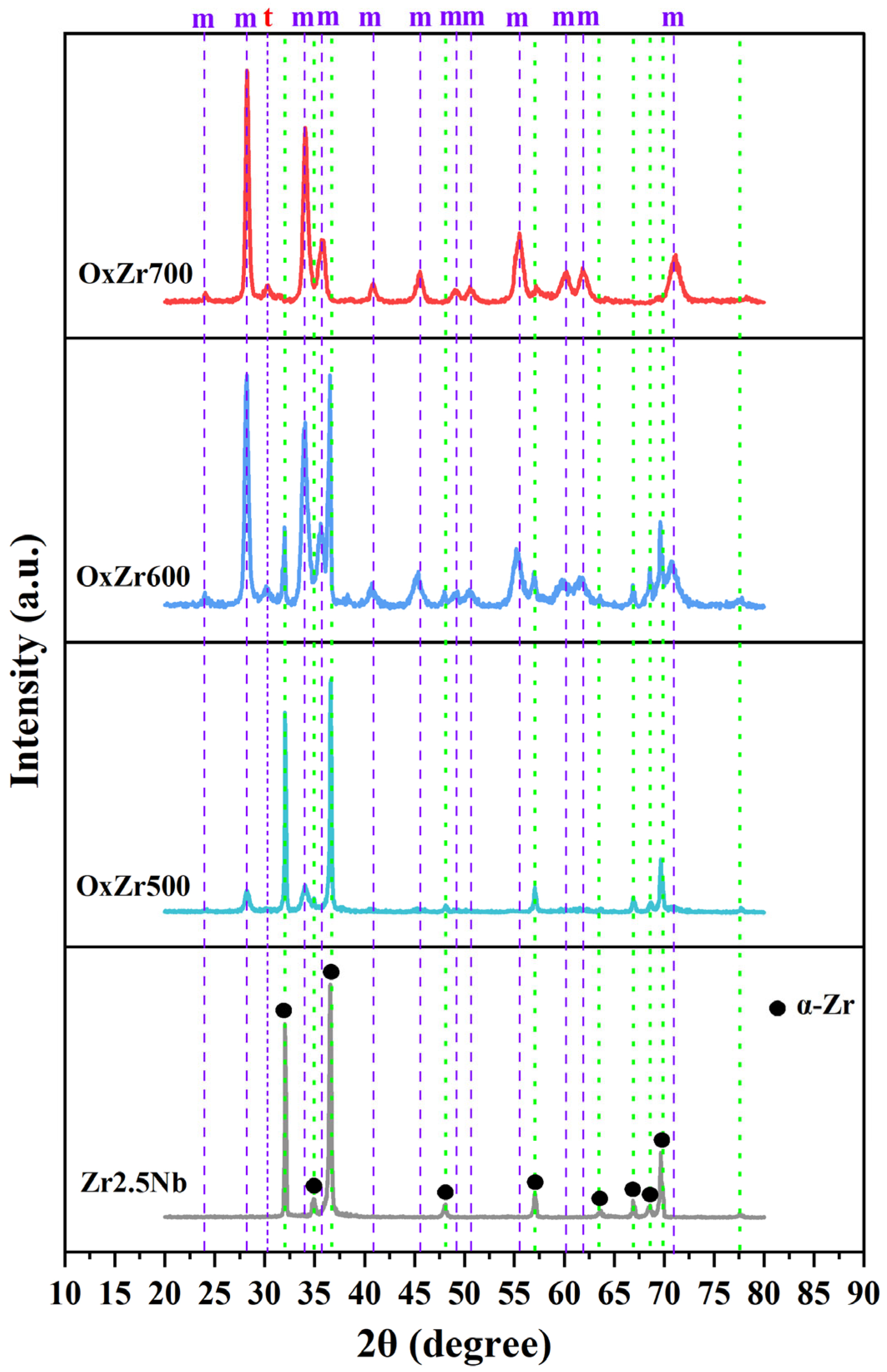
Figure 3 presents the XRD diffraction patterns of the untreated and thermally oxidized Zr2.5Nb alloy samples. It was found that the untreated Zr2.5Nb alloy consisted of α-Zr phase (ICDD PDF No. 97-004-3700) with a hexagonal close-packed crystal structure. The α-Zr phase diffraction peaks are marked by green dashed lines. For the OxZr500 sample, the diffraction peaks coinciding with the green dashed lines exhibited high intensities, and the peaks at 28.21° (−111) and 34.05° (020) that belong to the m-ZrO2 phase (ICDD PDF No. 97-004-1010) were observed. This demonstrated the formation of an oxide layer composed of the m-ZrO2 phase, but with limited thickness (Figure 2a). When the thermal oxidation temperature was increased to 600 °C, the diffraction peaks of α-Zr remained present but showed significantly reduced intensities. Meanwhile, m-ZrO2 peak intensities increased substantially. This indicates enhanced oxide formation (Figure 2b), attributed to improved oxygen dissolution efficiency at 600 °C. In addition, the peak at 30.18°, corresponding to the t-ZrO2 phase (ICDD PDF No. 97-008-5322), began emerging at this temperature. According to the Zr-O binary phase diagram, a phase transformation from m-ZrO2 to t-ZrO2 typically occurs above 1205 °C [23]. However, Yang et al. [24] proposed that compressive stress might facilitate transformation at relatively low temperatures. During the 600–700 °C oxidation process, high compressive stress stabilized the t-ZrO2 phase, and it disappeared when the temperature exceeded 800 °C. Following thermal oxidation at 700 °C, α-Zr diffraction peaks on the Zr2.5Nb alloy surface were nearly absent. The oxide layer (Figure 2c) predominantly comprised m-ZrO2, whose diffraction peaks were marked using purple dashed lines.
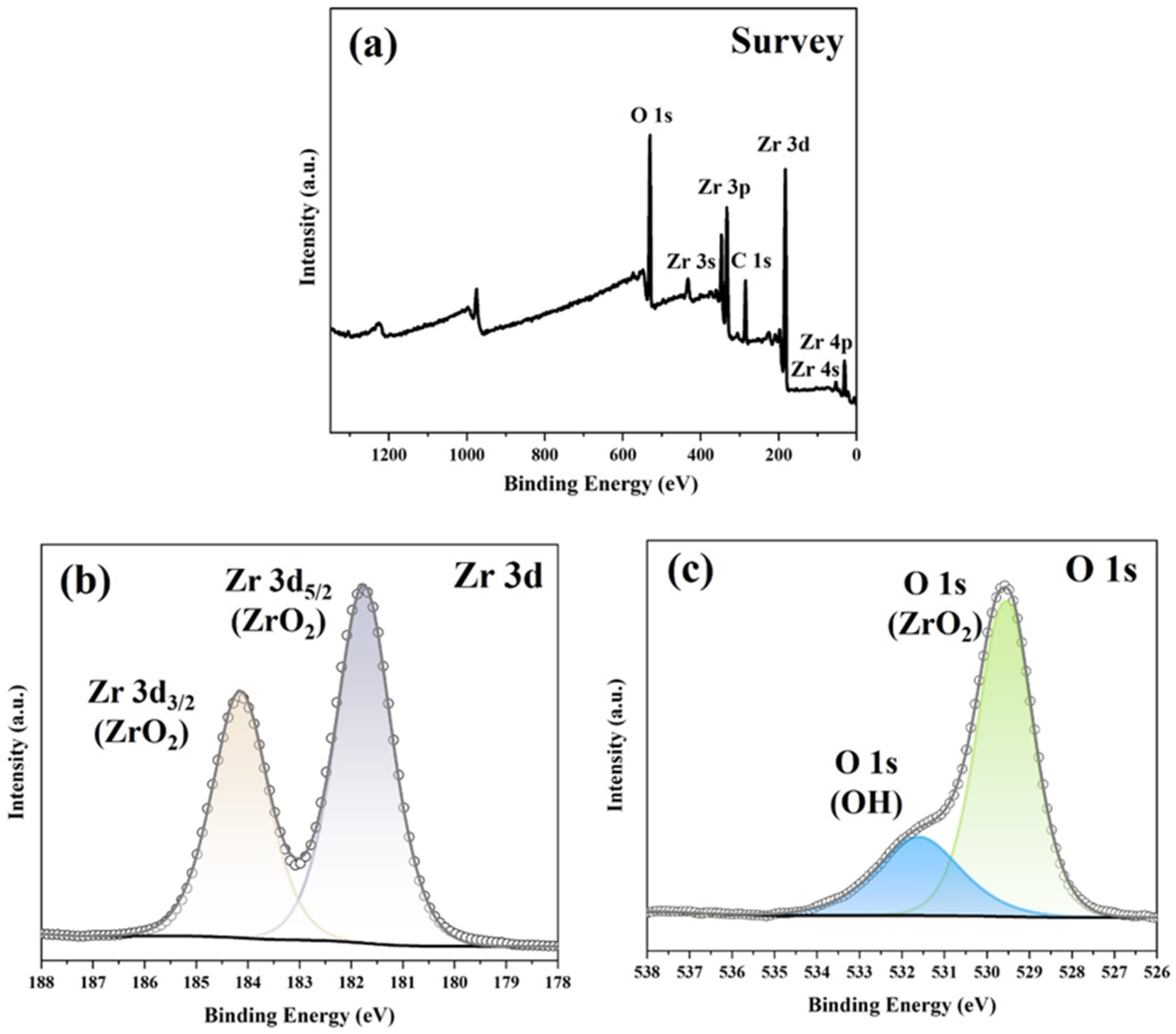
XPS was conducted to characterize the surface composition of the oxide layers. As revealed by XRD, oxide layers formed on the surface of the Zr2.5Nb alloy subjected to 500–700 °C thermal oxidation treatments, and the cross-sectional SEM images confirmed that their thickness ranged from 2.43 to 13.59 μm. Given the intrinsic 2–5 nm detection depth of XPS, only the outermost surface regions of each sample were probed. Consequently, nearly identical XPS spectral features were observed across samples oxidized between 500 and 700 °C. To preclude redundancy, detailed analysis herein focuses only on the OxZr600 sample. The C 1s peak at 284.8 eV served for energy calibration. Figure 4a displays wide-range XPS spectra for the OxZr600 sample, revealing Zr 3d and O 1s core-level signals in the oxide layer. Narrow scans of each core-level spectrum are presented in Figure 4b,c. Peak deconvolution employed a Gaussian–Lorentzian function via Thermo Avantage (v5.9931). Due to spin–orbit coupling, the Zr 3d peak splits into Zr 3d5/2 (181.76 eV) and Zr 3d3/2 (184.15 eV) [25]. These peak positions align with ZrO2 reference values in the NIST database. The O 1s core-level spectrum in Figure 4c resolved into two peaks. The 529.51 eV peak corresponds to the Zr–O bond in ZrO2, whereas the 531.61 eV peak indicates hydroxyl groups.

3.2. Mechanical Properties

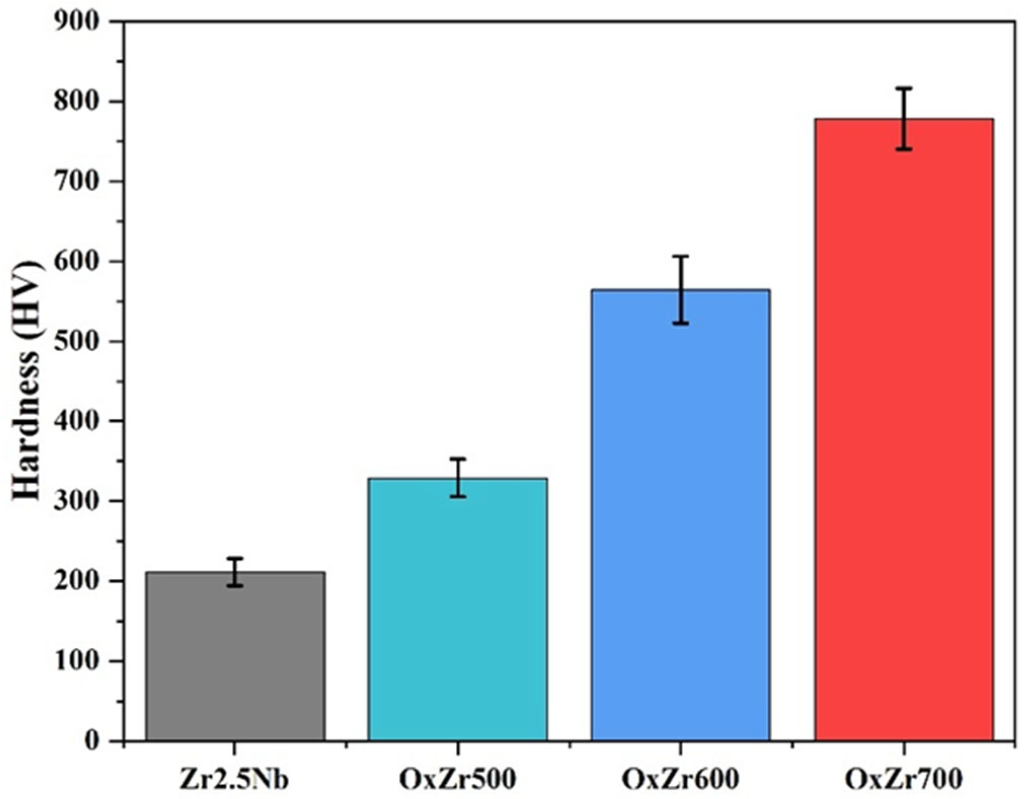
Figure 5 displays the surface hardness of the untreated and thermally oxidized Zr2.5Nb alloy samples. The surface hardness of the Zr2.5Nb alloy without thermal oxidation treatment was 244.24 ± 17.13 HV, while the thermally oxidized samples exhibited elevated hardness due to oxide layer formation. The results demonstrate that the surface hardness of the OxZr500, OxZr600, and OxZr700 samples increased to 329.25 ± 23.69, 564.53 ± 42.01, and 778.28 ± 37.85 HV, respectively. This enhancement correlated directly with oxide layer thickening at higher thermal oxidation temperatures.
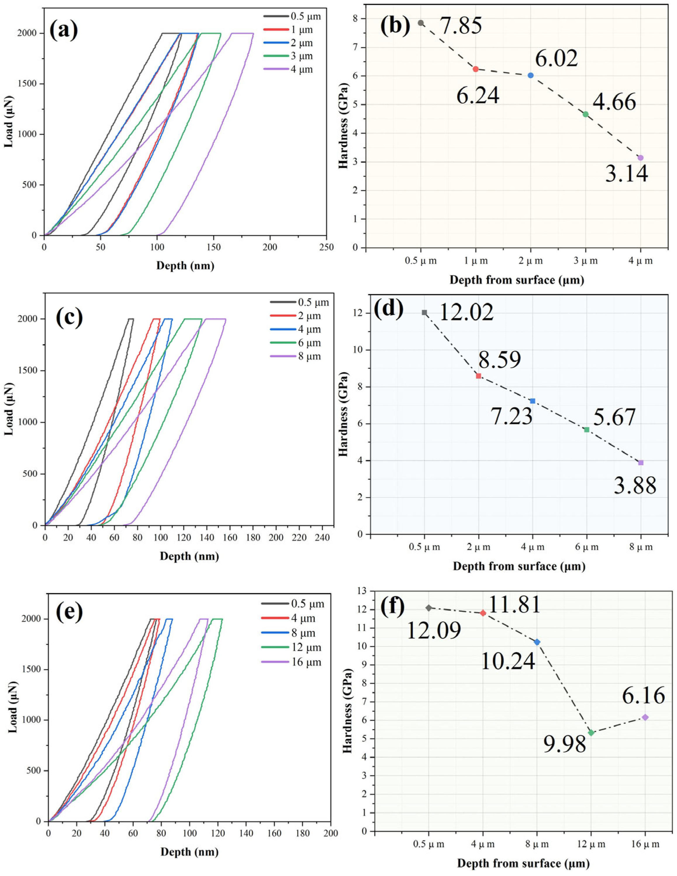
Figure 6a,c,e presents load–displacement curves for the thermally oxidized samples. When the oxidation temperature rose from 500 °C to 700 °C, the unloading curve slopes progressively steepened, indicating reduced elastic recovery in the oxide layers [26]. This behavior corroborated increased formation of the ZrO2 phase at the elevated temperatures, aligning with the XRD results. Figure 6b,d,f displays cross-sectional nanohardness profiles. At 0.5 μm depth, the OxZr500, OxZr600, and OxZr700 samples exhibited nanohardness values of 7.85, 12.01, and 12.09 GPa, respectively. Subsequently, all samples displayed characteristic nanohardness gradients decreasing with depth. Oxide layer growth on zirconium alloys is governed by oxygen diffusion. Increased thickness could constrict downward oxygen diffusion channels, thereby establishing an oxygen concentration gradient [27]. This elemental gradient corresponded directly to the cross-sectional nanohardness gradient. Notably, the OxZr700 sample showed a sharp nanohardness reduction near 12 μm depth, which could be attributed to the formation and propagation of lateral cracks (Figure 2c) above the oxide/metal interface. Additionally, the nanohardness at the fifth depth in all sample positions exceeded that of the substrate (2.88 GPa), despite the measured points being located beneath the oxide layers. This should be ascribed to the formation of oxygen solid-solution-strengthened zones beneath the oxide layer, even though they were not visible in SEM. Based on the above analysis, the OxZr600 sample exhibited homogeneous, dense, hard oxide layers with a near-linear nanohardness gradient, collectively validating 600 °C as optimal for enhancing Zr2.5Nb alloy mechanical performance. Nevertheless, subsequent tribological and biocompatibility assessments remain essential to evaluate its suitability as an artificial joint material.

3.3. Tribological Behavior

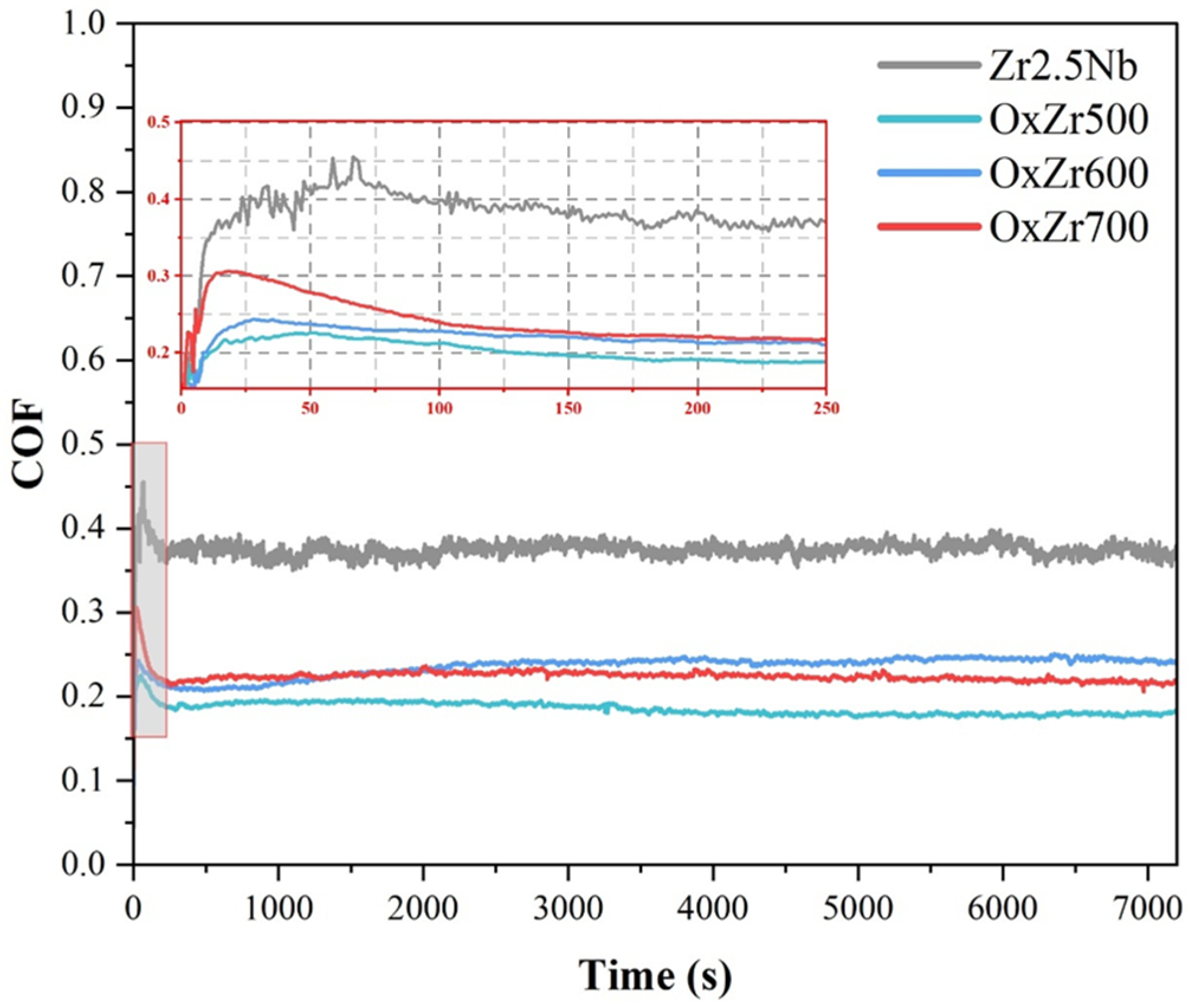
Figure 7 shows the friction coefficient curves of the untreated and thermally oxidized Zr2.5Nb alloy samples under the lubrication of 25% calf serum. During the running-in stage, all samples demonstrated rising friction coefficients, but the thermally oxidized samples showed substantially smaller increases than the untreated Zr2.5Nb alloy, as shown in the inset. The thickness of the naturally formed oxide film on the Zr2.5Nb surface is usually in the range of nanometers to sub-micrometers, which is readily damaged under load and too thin to effectively protect the substrate. When the friction pairs transitioned from zirconia ball against oxide film to zirconia ball against bare alloy, an abrupt friction coefficient shift occurred. Conversely, the thermally oxidized samples developed hard oxide layers that could resist zirconia ball intrusion and suppress asperity deformation/adhesive wear, thus reducing both the magnitude of friction coefficient increase and running-in duration. After the running-in stage, the friction coefficients of all samples gradually decreased and entered stable friction stages with values of about 0.38 (untreated Zr2.5Nb alloy), 0.18 (OxZr500), 0.25 (OxZr600), and 0.22 (OxZr700). Notably, the untreated Zr2.5Nb alloy demonstrated substantial friction coefficient fluctuations throughout the friction process. For the untreated Zr2.5Nb alloy, due to its softness and poor wear resistance, wear particles were generated by scratching and adhesive wear. These particles accumulated at friction interfaces, thus enlarging actual contact areas and elevating the friction coefficient. Simultaneously, serum solution scoured particles toward wear scar ends, reducing actual contact areas and friction coefficient. Thus, this fluctuation could be attributed to the dynamic balance between the continuous accumulation and removal of wear particles. In contrast, with the protection of the hard oxide layers, only the asperities on the surfaces of the thermally oxidized Zr2.5Nb alloy samples were smoothed by the zirconia ball, maintaining stable friction coefficients. In addition, since the proteins in the calf serum promoted the formation of tribofilm, the friction coefficient could be further stabilized [28].
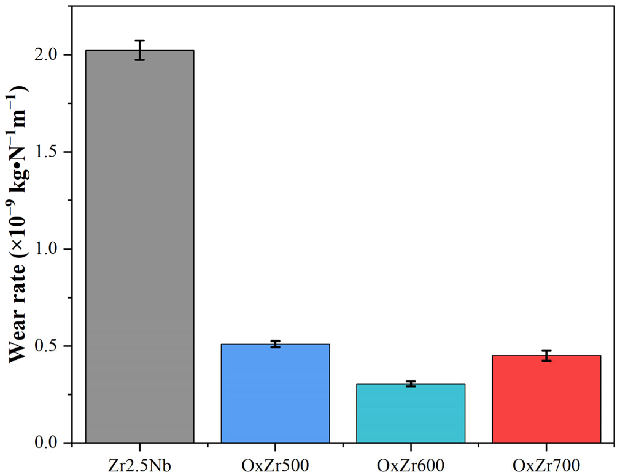
The wear rates of the untreated and thermally oxidized Zr2.5Nb alloy samples under the lubrication of 25% calf serum are shown in Figure 8. The wear rates of the untreated Zr2.5Nb alloy, OxZr500, OxZr600, and OxZr700 samples were 2.06 ± 0.05, 0.51 ± 0.02, 0.30 ± 0.01, and 0.45 ± 0.03 × 10⁻⁹ kg·N−1 m−1, respectively. Among all the thermally oxidized samples, the OxZr500 sample exhibited the highest wear rate due to the inadequate oxide layer thickness and hardness, while the OxZr700 sample demonstrated the second highest wear rate attributable to the existence of defects in the oxide layer. The superior oxide layer structural integrity of the OxZr600 sample was reflected in its wear resistance, with the lowest wear rate under the same friction conditions, which was 85% lower than that of the untreated Zr2.5Nb alloy.

3.4. Biocompatibility

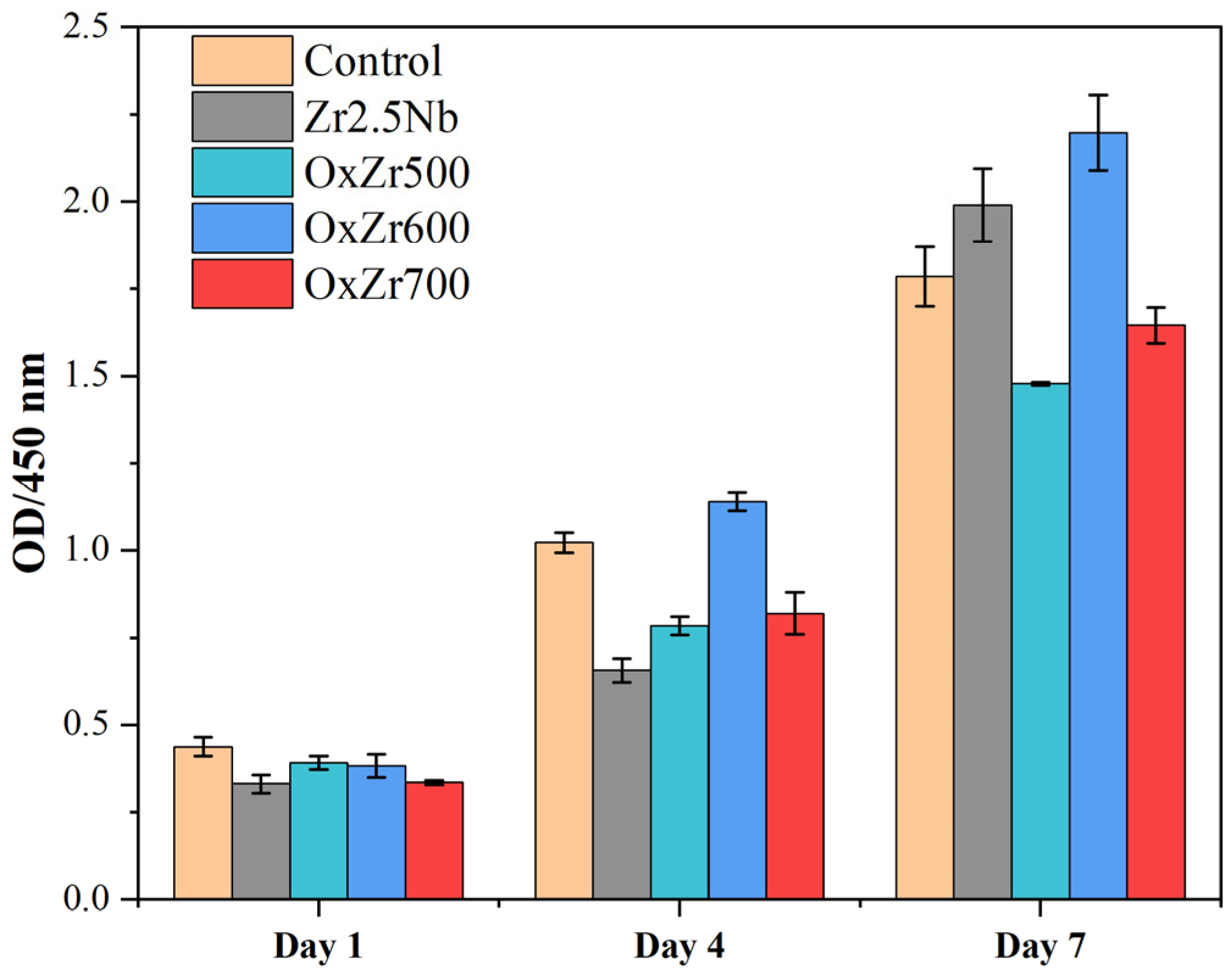
MC3T3-E1 cells were employed to evaluate the biocompatibility of the untreated and thermally oxidized Zr2.5Nb alloy samples. Cell proliferation was quantified by optical density (OD) measurements using CCK-8 assays. Figure 9 depicts the proliferation of the MC3T3-E1 cells cultured on the untreated and thermal oxidized Zr2.5Nb alloy samples after 1, 4, and 7 days. All samples exhibited a significant increase in OD values with prolonged culture time, indicating no significant cytotoxic effects on the MC3T3-E1 cell growth and proliferation.
The relative viability of the cultured cells can be calculated from the OD value of each sample group using Equation (2):
R e l a t i v e   v i a b i l i t y % = O D 450 s a m p l e O D 450 c o n t r o l × 100 %
where OD450sample is the optical density of the experimental group, and OD450control is the optical density of the control group.
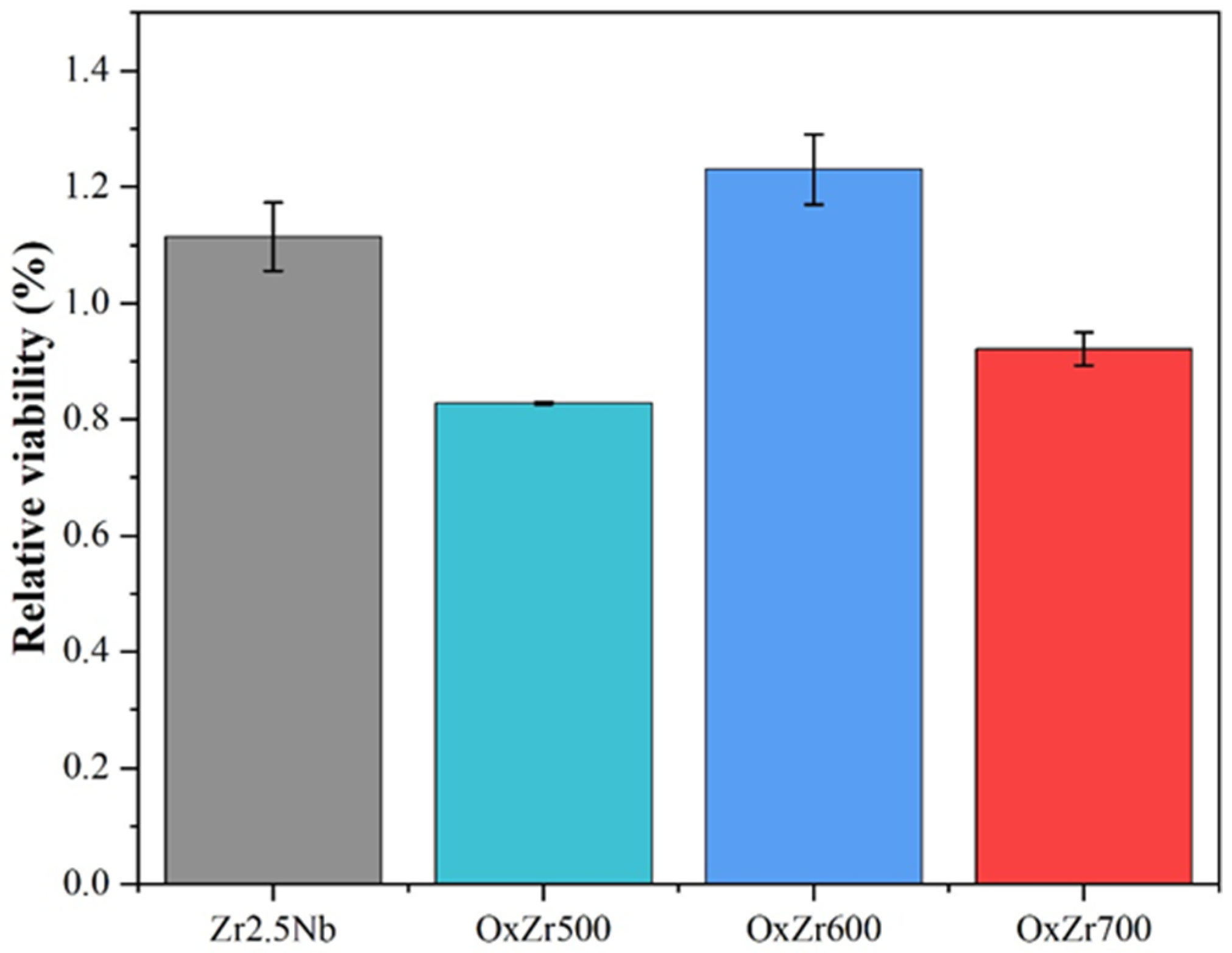
Figure 10 shows the relative viability of the MC3T3-E1 cells cultured on the untreated and thermally oxidized Zr2.5Nb alloy samples after 7 days. The relative viability of cells on the untreated Zr2.5Nb alloy, OxZr500, OxZr600, and OxZr700 samples were 111.46 ± 5.87%, 82.82 ± 0.25%, 123.07 ± 6.02%, and 92.12 ± 2.84%, respectively. Cytotoxicity was evaluated based on the ISO 10993-5: 2018 standard, which specifies that relative viability ≥ 70% indicates cytotoxicity grades 0 or 1, confirming non-toxicity [29]. Therefore, it can be concluded that the thermally oxidized Zr2.5Nb alloy samples maintained excellent biocompatibility.
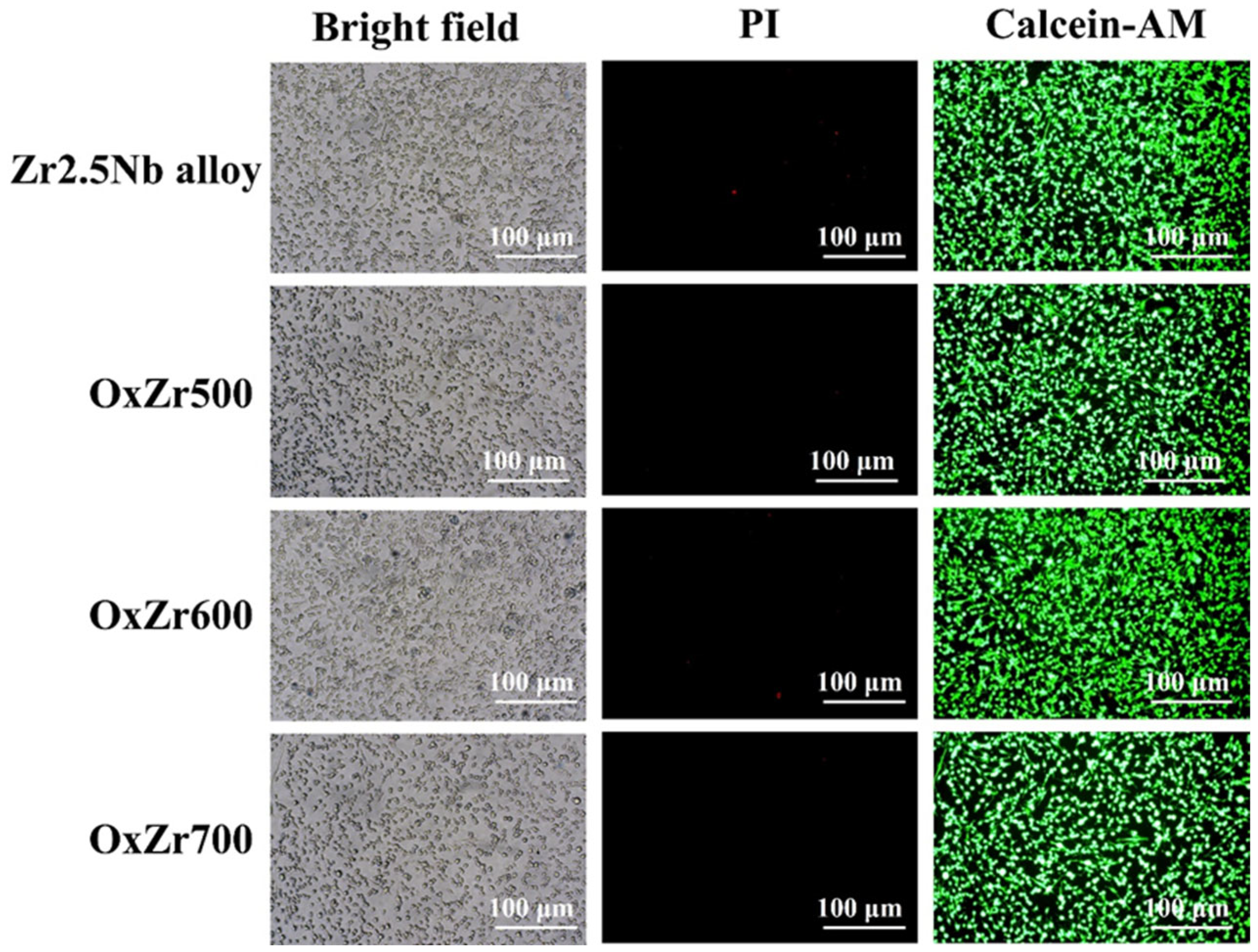
Figure 11 displays bright-field and Calcein-AM/PI staining images of the MC3T3-E1 cells cultured on the untreated and thermally oxidized Zr2.5Nb alloy samples after 7 days. All samples exhibited uniform cell distribution with visible filamentous pseudopodia extension and no aggregation or morphological abnormalities. These findings confirm that the tested materials support MC3T3-E1 cell adhesion and proliferation without any adverse effects. Remarkably, sporadic fluorescent-bright spots in the OxZr600 sample correlated with localized Calcein-AM uptake, indicating elevated metabolic activity, which aligned with the highest OD value at 7 days in CCK-8 assays.

4. Conclusions

This study systematically investigates the effect of oxidation temperature on the properties of the thermally oxidized Zr2.5Nb alloy. The microstructure, composition, mechanical properties, tribological behavior, and biocompatibility of the oxide layers have been investigated to reach the following conclusions:
(1)
Oxide layers, primarily composed of m-ZrO2, formed on the surface of the Zr2.5Nb alloy. Increasing the temperature accelerated the growth of the oxide layer by enhancing oxygen diffusion, resulting in thicknesses that increased from 2.43 µm at 500 °C to 5.31 µm at 600 °C, and further to 13.59 µm at 700 °C. Nevertheless, this growth coincided with an increase in defect density;
(2)
Compared to the untreated Zr2.5Nb alloy, the thermally oxidized samples exhibited significantly enhanced mechanical properties. Specifically, the surface hardness increased from the original 244.24 ± 17.13 HV to 329.25 ± 23.69, 564.53 ± 42.01, and 778.28 ± 37.85 HV for samples oxidized at 500 °C, 600 °C, and 700 °C, respectively. Additionally, all oxidized samples displayed characteristic nanohardness gradients that decreased with depth;
(3)
The thermally oxidized samples all exhibited significantly improved wear resistance. In particular, the Zr2.5Nb alloy sample oxidized at 600 °C, owing to its optimal structural integrity and uniformity, showed a wear rate reduced by 85% compared to the untreated Zr2.5Nb alloy. Furthermore, this sample also possessed the best biocompatibility, as evidenced by the relative viability of MC3T3-E1 cells cultured on it after 7 days, reaching 123.07 ± 6.02%.
In summary, this work has illuminated an economically viable strategy for fabricating high-performance zirconium-based implants through optimizing thermal oxidation temperature parameters, thereby enhancing their structural integrity and functional attributes in biomedical applications. Future research will explore the intrinsic correlations between phase transformations and mechanical properties, as well as tribological properties, and further conduct in vivo assessments.

Author Contributions

Conceptualization, Y.X. and X.S.; methodology, X.S. and Y.L.; validation, Y.X. and H.S.; formal analysis, T.H.; investigation, Y.X. and H.S.; data curation, H.S.; writing—original draft preparation, Y.X.; writing—review and editing, X.S.; supervision, H.X. and Y.L.; project administration, H.S.; funding acquisition, H.X. All authors have read and agreed to the published version of the manuscript.

Funding

This project is supported by the State Key Laboratory of Advanced Technology for Materials Synthesis and Processing (Wuhan University of Technology) (No. 2025-KF-8), and jointly supported by Hubei Provincial Natural Science Foundation and Huangshi of China (Nos. 2023AFD009 and 2025AFD023).

Institutional Review Board Statement

Not applicable.

Informed Consent Statement

Not applicable.

Data Availability Statement

Data will be made available upon request.

Conflicts of Interest

The authors declare no conflicts of interest.

References

  1. Singh, J.A.; Yu, S.; Chen, L.; Cleveland, J.D. Rates of Total Joint Replacement in the United States: Future Projections to 2020–2040 Using the National Inpatient Sample. J. Rheumatol. 2019, 46, 1134–1140. [Google Scholar] [CrossRef] [PubMed]
  2. Clar, C.; Leitner, L.; Koutp, A.; Hauer, G.; Rasic, L.; Leithner, A.; Sadoghi, P. The worldwide survival rate of total hip arthroplasties is improving: A systematic comparative analysis using worldwide hip arthroplasty registers. EFORT Open Rev. 2024, 9, 745–750. [Google Scholar] [CrossRef] [PubMed]
  3. Li, J.X.; Wen, J.P.; Ou, S.J.; Qu, Y.D.; Xia, C.L.; Li, W.J.; Yang, Y.; Liu, J.B.; Yang, R.; Zeng, W.; et al. Wear particles-induced periprosthetic osteolysis from a new perspective based on single-cell RNA-seq: Crosstalk of osteoblast PANoptosis and macrophage efferocytosis in osteoimmunology microenvironment. Chem. Eng. J. 2025, 515, 163509. [Google Scholar] [CrossRef]
  4. Hao, X.; Jiang, B.; Wu, J.; Xiang, D.; Xiong, Z.; Li, C.; Li, Z.; He, S.; Tu, C.; Li, Z. Nanomaterials for bone metastasis. J. Control. Release 2024, 373, 640–651. [Google Scholar] [CrossRef]
  5. Li, Z. Advancements of biomaterial in hip replacement technology incorporating ceramic materials. J. Orthop. 2025, 62, 27–35. [Google Scholar] [CrossRef]
  6. Morano, C.; Garofalo, S.; Bertuccio, P.; Sposato, A.; Zappone, I.; Pagnotta, L. A Comprehensive Literature Review of Total Hip Arthroplasty (THA): Part 1-Biomaterials. J. Funct. Biomater. 2025, 16, 179. [Google Scholar] [CrossRef]
  7. Knecht, C.; Polakof, L.; Behrens, J.; Goodman, S.B. Wear debris in metal-on-metal bearings and modular junctions What have we learned from the last decades? Orthopadie 2023, 52, 206–213. [Google Scholar]
  8. Alvarez-Vera, M.; Ortega, J.A.; Ortega-Ramos, I.A.; Hdz-Garcia, H.M.; Munoz-Arroyo, R.; Diaz-Guillen, J.C.; Acevedo-Davila, J.L.; Hernandez-Rodriguez, M.A.L. Tribological and microstructural characterization of laser microtextured CoCr alloy tested against UHMWPE for biomedical applications. Wear 2021, 477, 203819. [Google Scholar] [CrossRef]
  9. Kim, H.S.; Lee, H.J.; Lee, S.J.; Yoo, J.J. Ceramic-on-Ceramic or Metal-on-Polyethylene: The Bearing of Choice after Ceramic Component Fracture in Total Hip Arthroplasty along with Concise Follow-Up of the Previous Cohort. Orthop. Surg. 2023, 15, 2864–2871. [Google Scholar] [CrossRef]
  10. Abdelghafour, K.M.; Jaques, A.; Shah, N. Ceramic-on-ceramic bearings: Can we avoid the complications? Long-term outcomes of big head CoC bearings in primary total hip arthroplasty. Eur. J. Orthop. Surg. Traumatol. 2025, 35, 80. [Google Scholar] [CrossRef] [PubMed]
  11. Merola, M.; Affatato, S. Materials for Hip Prostheses: A Review of Wear and Loading Considerations. Materials 2019, 12, 495. [Google Scholar] [CrossRef]
  12. Nasiri-Tabrizi, B.; Basirun, W.J.; Walvekar, R.; Yeong, C.H.; Phang, S.W. Exploring the potential of intermetallic alloys as implantable biomaterials: A comprehensive review. Biomater. Adv. 2024, 161, 213854. [Google Scholar] [CrossRef]
  13. Good, V.; Widding, K.; Hunter, G.; Heuer, D. Oxidized zirconium—A potentially longer lasting hip implant. Mater. Des. 2005, 26, 618–622. [Google Scholar] [CrossRef]
  14. Kurtz, S.M.; Kocagoez, S.; Arnholt, C.; Huet, R.; Ueno, M.; Walter, W.L. Advances in zirconia toughened alumina biomaterials for total joint replacement. J. Mech. Behav. Biomed. Mater. 2014, 31, 107–116. [Google Scholar] [CrossRef]
  15. Leto, A.; Zhu, W.; Matsubara, M.; Pezzotti, G. Bioinertness and fracture toughness evaluation of the monoclinic zirconia surface film of Oxinium femoral head by Raman and cathodoluminescence spectroscopy. J. Mech. Behav. Biomed. Mater. 2014, 31, 135–144. [Google Scholar] [CrossRef]
  16. DesJardins, J.D.; Burnikel, B.; LaBerge, M. UHMWPE wear against roughened oxidized zirconium and CoCr femoral knee components during force-controlled simulation. Wear 2008, 264, 245–256. [Google Scholar] [CrossRef]
  17. Arima, T.; Miyata, K.; Idemitsu, K.; Inagaki, Y. Oxidation properties of Zr-Nb alloys at 973–1273 K in air. Prog. Nucl. Energy 2009, 51, 307–312. [Google Scholar] [CrossRef]
  18. Duriez, C.; Drouan, D.; Pouzadoux, G. Reaction in air and in nitrogen of pre-oxidised Zircaloy-4 and M5™ claddings. J. Nucl. Mater. 2013, 441, 84–95. [Google Scholar] [CrossRef]
  19. Ding, Y.; Liu, S.; Xia, C.; Zou, X.; Liu, D.; Wang, Y.; Yang, T.; Li, Q. Thermal oxidation of novel Zr-Ti-Al-V alloy with high strength and toughness and its influence on the corrosion behavior. Surf. Coat. Technol. 2021, 423, 127576. [Google Scholar] [CrossRef]
  20. Pawar, V.; Weaver, C.; Jani, S.C. Physical characterization of a new composition of oxidized zirconium—2.5 wt% niobium produced using a two step process for biomedical applications. Appl. Surf. Sci. 2011, 257, 6118–6124. [Google Scholar] [CrossRef]
  21. Sun, Y.; Hu, W.; Zhang, S.; Lu, Y.; Wang, J.; Ma, G.; Lin, J.; Hosseinkhani, S.; Ma, J.; Wang, Q. Corrosion fatigue behavior of porous Cu-bearing Ti alloy fabricated by selective laser melting. J. Mater. Res. Technol. 2023, 23, 1630–1643. [Google Scholar] [CrossRef]
  22. Rao, Q.; Zhang, J.; Chen, Y.; Yang, Y.; Chen, X.; Liu, D.; Zhu, R.; Li, A.; Lv, Y.; Zheng, S. Research Progress of the Coatings Fabricated onto Titanium and/or Titanium Alloy Surfaces in Biomaterials for Medical Applications for Anticorrosive Applications. Coatings 2025, 15, 599. [Google Scholar] [CrossRef]
  23. Abriata, J.P.; Garcés, J.; Versaci, R. The O−Zr (Oxygen-Zirconium) system. Bull. Alloy Phase Diagr. 1986, 7, 116–124. [Google Scholar] [CrossRef]
  24. Yang, F.; Zhao, X.; Xiao, P. In Situ Measurement of Stresses and Phase Compositions of the Zirconia Scale During Oxidation of Zirconium by Raman Spectroscopy. Oxid. Met. 2013, 81, 331–343. [Google Scholar] [CrossRef]
  25. Wong, Y.H.; Cheong, K.Y. Thermal oxidation and nitridation of sputtered Zr thin film on Si via N2O gas. J. Alloys Compd. 2011, 509, 8728–8737. [Google Scholar] [CrossRef]
  26. Luo, Y.; Chen, W.; Tian, M.; Teng, S. Thermal oxidation of Ti6Al4V alloy and its biotribological properties under serum lubrication. Tribol. Int. 2015, 89, 67–71. [Google Scholar] [CrossRef]
  27. Brachet, J.C.; Rouesne, E.; Ribis, J.; Guilbert, T.; Urvoy, S.; Nony, G.; Toffolon-Masclet, C.; Le Saux, M.; Chaabane, N.; Palancher, H.; et al. High temperature steam oxidation of chromium-coated zirconium-based alloys: Kinetics and process. Corros. Sci. 2020, 167, 108537. [Google Scholar] [CrossRef]
  28. Qi, J.; Guan, D.; Nutter, J.; Wang, B.; Rainforth, W.M. Insights into tribofilm formation on Ti-6Al-4V in a bioactive environment: Correlation between surface modification and micro-mechanical properties. Acta Biomater. 2022, 141, 466–480. [Google Scholar] [CrossRef]
  29. Guan, J.; Jiang, X.; Xiang, Q.; Yang, F.; Liu, J. Corrosion and tribocorrosion behavior of titanium surfaces designed by electromagnetic induction nitriding for biomedical applications. Surf. Coat. Technol. 2021, 409, 126844. [Google Scholar] [CrossRef]
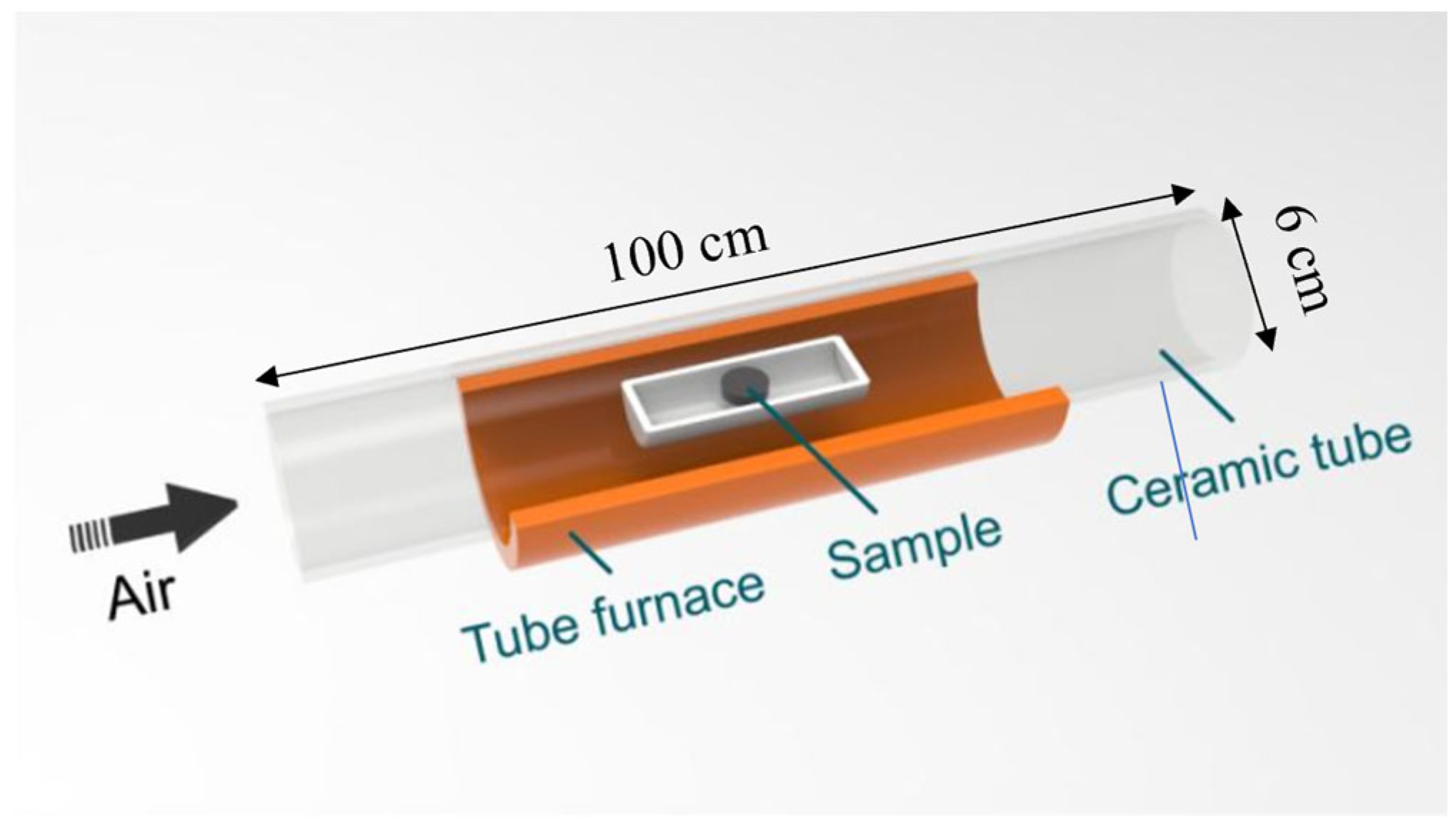
Figure 1. Schematic diagram of the thermal oxidation process.
Coatings 15 00940 g001
Figure 2. Cross-sectional SEM images of the thermally oxidized Zr2.5Nb alloys: (a) OxZr500; (b) OxZr600; (c) OxZr700.
Coatings 15 00940 g002
Figure 3. XRD patterns of the untreated and thermally oxidized Zr2.5Nb alloy samples.
Coatings 15 00940 g003
Figure 4. XPS spectra of the Oxzr600 sample: (a) survey; (b) Zr 3d; and (c) O 1s.
Coatings 15 00940 g004
Figure 5. Surface hardness of the untreated and thermally oxidized Zr2.5Nb alloy samples.
Coatings 15 00940 g005
Figure 6. Load–displacement curves and cross-sectional nanohardness profiles of the thermally oxidized Zr2.5Nb alloy samples: (a,b) OxZr500; (c,d) OxZr600; (e,f) OxZr700.
Coatings 15 00940 g006
Figure 7. Friction coefficient curves of the untreated and thermally oxidized Zr2.5Nb alloy samples under the lubrication of 25% calf serum.
Coatings 15 00940 g007
Figure 8. Wear rate of the untreated and thermally oxidized Zr2.5Nb alloy samples under the lubrication of 25% calf serum.
Coatings 15 00940 g008
Figure 9. Proliferation of the MC3T3-E1 cells cultured on the untreated and thermally oxidized Zr2.5Nb alloy samples after 1, 4, and 7 days.
Coatings 15 00940 g009
Figure 10. Relative activity of the MC3T3-E1 cells cultured on the untreated and thermally oxidized Zr2.5Nb alloy samples after 7 days.
Coatings 15 00940 g010
Figure 11. Bright-field and Calcein-AM/PI staining images of the MC3T3-E1 cells cultured on the untreated and thermally oxidized Zr2.5Nb alloy samples after 7 days.
Coatings 15 00940 g011
Table 1. Composition of the applied Zr2.5Nb alloy in mass percent.
NbHfFeOCHZr
2.26%1.90%0.015%0.032%<0.010%<0.010%Blance
Disclaimer/Publisher’s Note: The statements, opinions and data contained in all publications are solely those of the individual author(s) and contributor(s) and not of MDPI and/or the editor(s). MDPI and/or the editor(s) disclaim responsibility for any injury to people or property resulting from any ideas, methods, instructions or products referred to in the content.