Review

Plasma Electrolytic Oxidation (PEO) Coatings for Biomedical Implants: A Review on Enhancing Antibacterial Efficacy Through Controlled Antibiotic Release

by
Maryam Molaei
1,
Masoud Atapour
2,* and
Ehsan Mohammadi Zahrani
3,*
1
Department of Materials Engineering, Hamedan University of Technology, Hamedan 65169-13733, Iran
2
Department of Materials Engineering, Isfahan University of Technology, Isfahan 84156-83111, Iran
3
Department of Materials Science and Engineering, Faculty of Engineering, Ferdowsi University of Mashhad, Mashhad 91775-1111, Iran
*
Authors to whom correspondence should be addressed.
Coatings 2025, 15(8), 925; https://doi.org/10.3390/coatings15080925 (registering DOI)
Submission received: 15 July 2025 / Revised: 29 July 2025 / Accepted: 5 August 2025 / Published: 8 August 2025

Abstract

The use of biomedical implants has significantly enhanced patient survival rates and overall quality of life. However, bacterial infections caused by bacterial adhesion and the subsequent formation of biofilm on the surface of the implants are challenging clinical issues, leading to implant failure and high social and economic costs. Modification of the surface of the implants with antibacterial coatings is a promising technique to address implant-associated bacterial infection problems. One strategy to fabricate bactericidal antibacterial coatings is to load antibacterial agents, like antibiotics—the most important type of antibacterial drug for killing or inhibiting the growth of bacteria—at therapeutic doses into the coatings and subsequently release them, ideally in a controlled way. Plasma electrolytic oxidation (PEO) is a simple, affordable, and eco-friendly method to produce high-performance, multifunctional coatings with desired antibacterial properties. This review examines the antibacterial activity of antibiotic-loaded PEO coatings, offering valuable insights for the development of novel, high-performance antibacterial coatings that meet clinical requirements.

1. Introduction

Implantation refers to the partial or complete insertion of any object or material into the body for therapeutic, curative, replacement, or research purposes [1]. Recent advancements in materials science have facilitated the use of diverse implants in orthopedics, cardiovascular, dentistry, and numerous other areas [2]. Implantable medical devices have been highly successful in improving the quality of life for millions each year [3]. However, implant infections caused by pathogenic bacteria during surgery or the time spent in the body are still among the most prevalent and severe clinical complications, resulting in poor functional outcomes, implant failure, chronic osteomyelitis, or even death, posing a significant risk to human health and economic issues [4,5,6,7]. The formation of biofilms in over 80% of clinical bacterial infections is an important challenge in implantation. Biomolecules of exopolysaccharides, matrix proteins, and extracellular deoxyribonucleic acid (DNA) are the main components of the biofilms [8]. The process of biofilm formation is a multifaceted and complex phenomenon, which can be divided into four distinct sequential steps, as depicted in Figure 1.
The initial stage of biofilm formation involves the bacterial cells’ reversible attachment to the implant surface. Various physical and chemical factors, including steric interactions, electrostatic interactions, van der Waals forces, and the involvement of specialized adhesion proteins, control this process. As the biofilm forms, the bacterial cells transition from a reversible adhesion to a more permanent, non-reversible colonization of the implant surface. This change is associated with a series of cellular and molecular interactions. Key mechanisms involved in this stage include the production of specialized adhesion proteins, the formation of protein appendages, and the formation of an extracellular polymeric substance (EPS) matrix. The third critical step is the biofilm development and maturation. During this stage, the bacteria that have successfully colonized the surface begin to develop into intricate microcolonies. Concurrent with this growth, the microorganisms produce EPS, primarily composed of polysaccharides and other macromolecules. This EPS matrix is essential for the structural and functional organization of the maturing biofilm [9].
Common approaches for treating implant-associated infections include antibiotic therapy, revision surgery, and debridement. However, these methods are only effective at preventing bacterial growth or eliminating bacteria before biofilm formation. After a biofilm is established, bacteria undergo notable metabolic changes. This results in the thickening of the biofilm and improves the protection against the host’s immune response as well as antibacterial agents [10]. Therefore, to effectively prevent implant-associated infections, it is necessary to develop novel strategies that specifically target biofilm formation.

2. Antibacterial Coatings

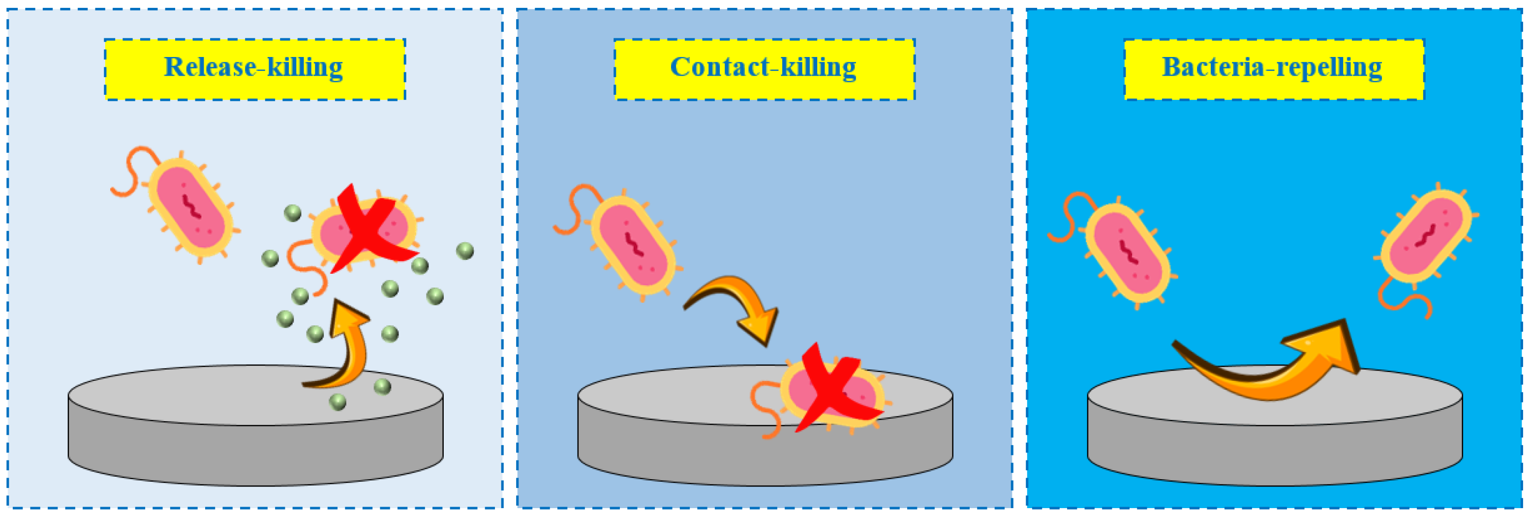
Surface modification of implants with antibacterial coatings is a promising and effective preoperative strategy to prevent or reduce implant-associated bacterial infections. Coatings can eliminate the initial attachment of bacteria and hinder subsequent biofilm formation on the implant surface. The coating generally involves creating an additional layer on the implant surface without disrupting the overall performance of the material [11,12]. The surface properties of the implant, such as surface charge, topography, and wettability, can control the absorption of proteins and, as a result, the adhesion of bacteria [13]. Therefore, changing the topography and altering the chemistry of the implant surface with antibacterial coatings can prevent or reduce bacterial adhesion [14]. Moreover, coatings can improve other required surface characteristics of bone implants, including their resistance to wear and corrosion [15,16]. The idea of modifying implant surfaces through antibacterial coatings has gained significant attention in recent years. According to the mechanism by which they fight bacteria, antibacterial coatings are classified into two types: bactericidal (active), and bacteriostatic (passive). The bactericidal category can be further divided into release-killing and contact-killing types. Release-killing coatings compromise the structural integrity of bacterial cells or membranes by gradually releasing antibacterial agents over time.
In contrast, contact-killing antibacterial coatings physically interact with bacteria through their anionic or cationic compounds. Bacteriostatic coatings can inhibit or minimize the adhesion of microorganisms to the surface of the implant by utilizing the inherent properties of the materials found in the coating to repel bacteria [17]. Figure 2 shows schematic illustrations of different types of antibacterial coatings.

3. Antibacterial Coatings: Release-Killing Strategies

Among antibacterial coatings, release-killing bactericidal coatings are ideal candidates for the surface modification of implants because they can eliminate surface-adhered bacteria and provide a high local concentration of bactericidal agents to kill planktonic microbes. These coatings release antibacterial agents (e.g., antibiotics, nanoparticles, metal ions, and metal oxides) gradually at therapeutic doses in a controlled manner through the degradation of the coatings and the breaking of bonds between the coatings and the antibacterial agents, effectively killing bacteria both close to and far from the surface [15,18]. An appropriate concentration and optimal release of bactericidal agents are two crucial parameters for release-killing antibacterial coatings [19]. One of the main aspects of antibacterial coatings is the release rate of antibacterial agents, which needs to be well controlled to avoid toxic effects or sublethal doses [15,18]. High release of bactericidal agents on and around the surface of the implant, as well as in the surrounding host tissues, can lead to a decrease in antibacterial activity and an increased risk of postsurgical contaminations [15,20]. It is well known that the first 24 h after implantation involve a short-term release of bactericidal agents to ensure the antibacterial success of an implant. However, a long-term release capacity (from days to months) is required to treat diseases or infections that take a longer time to cure or eliminate [21]. Therefore, it is essential to develop coatings that allow for controlled release rates to maintain an appropriate concentration of antibacterial agents that meet the bactericidal requirements of implants throughout their use. Up to now, a broad range of surface coating techniques, such as cold spray [21], dip-coating [22], physical vapor deposition (PVD) [23], electrophoretic deposition (EPD) [17], and plasma electrolytic oxidation (PEO) [24], have been developed to fabricate antibacterial coatings on various biomaterials. Among these methods, PEO is a low-cost, environmentally friendly, and high-voltage plasma-assisted anodic oxidation process that has recently drawn significant attention for modifying the surfaces of metallic implants with antibacterial coatings [25,26,27,28].

4. PEO Process

PEO is a promising surface treatment technique used to enhance the wear resistance, corrosion resistance, biocompatibility, and antibacterial properties of some metals and alloys such as titanium [29,30], magnesium [31,32], zirconium [33], zinc [34,35], and copper [36]. PEO, which evolved from the anodizing process, can produce a hard, ceramic-like oxide layer with strong adhesion, owing to the combined effects of chemical, electrochemical, and plasma-driven reactions [37]. This process results in the formation of a coating with a porous outer layer and a dense, compact inner layer, typically ranging in thickness from 1 to 100 µm [38]. PEO coatings are formed through the oxidation of the substrate in an aqueous electrolyte, driven by a series of localized electrical discharges. These discharges manifest as numerous short-lived sparks, resulting from the localized electrical breakdown of the growing oxide layer [39,40]. The porous surface morphology of PEO coatings is attributed to plasma emissions and gas release during discharge events, with pore sizes ranging from the nanoscale to several micrometers. These porous structures become especially beneficial in medical applications by enhancing drug delivery, cellular integration, and osseointegration. Moreover, the rough surface of the PEO coatings plays a crucial role in promoting bone tissue formation through improved mechanical interlocking and increased surface area [41,42,43]. PEO coatings are composed of species derived from both the electrolyte and substrate, with proportions depending on the discharge conditions [44]. For instance, the primary constituent of PEO coatings on magnesium alloys is typically MgO [45,46,47,48], whereas on titanium alloys it is TiO2 (in the rutile and anatase phases) [49]. The simultaneous inward migration of oxygen ions and outward migration of metal ions at the metal–electrolyte interface causes the growth of oxides. The most commonly used electrolytes in the PEO process are those based on phosphate, aluminate, and silicate solutions [50]. Research has shown that adding calcium and phosphorus (Ca–P), the main components of human bone, to the PEO electrolyte encourages the formation of Ca–P compounds on the coating surface, enhancing osteoblast attachment and growth, ultimately improving the biocompatibility of the implants [42]. The morphology, composition, and thickness of PEO coatings can be modified by controlling the system’s working parameters, such as voltage, current density, duration time, and the electrolytic solution [51]. Therefore, optimizing the processing parameters during the PEO process is important, as these parameters influence the characteristics of plasma discharges and, consequently, the quality of the PEO coatings [52].

5. Antibacterial PEO Coatings: Strategies, Efficacy, and Applications

Antibacterial PEO coatings have recently gained attention in the surface modification of metallic implants due to the increasing need for antimicrobial surfaces. The primary strategy for preparing antibacterial PEO coatings is doping antibacterial metallic or oxide particles into the electrolyte solution, which is also the most straightforward method. For example, Thukkaram et al. [53] used Ag nanoparticles to fabricate antibacterial PEO coatings on pure titanium. They found that the PEO coatings synthesized in an electrolyte with Ag nanoparticles—which release Ag+ ions that can contact and kill bacteria—exhibited remarkable antibacterial action against Staphylococcus aureus (S. aureus) and Escherichia coli (E. coli) compared to the coatings formed in an Ag nanoparticles-free electrolyte. S. aureus and E. coli are common bacteria that are responsible for 34% of infections related to orthopedic implants [54]. S. aureus is a Gram-positive bacterium recognized for involvement in infections that occur after surgery, whereas E. coli is a Gram-negative bacterium that represents a common source of bacterial contamination in surgical environments [55]. Liu et al. [56] prepared ZnO nanoparticle-doped antibacterial PEO coatings on magnesium alloy. The high antibacterial activity of these coatings against E. coli was attributed to the release of Zn2+ ions, which could penetrate the walls of bacteria and disintegrate them.
Nanoparticles are strong antibacterial materials because of their small size as well as their high area-to-volume ratio. These characteristics enhance nanoparticles’ ability to penetrate and interact with bacterial membranes, thereby improving their antibacterial effect. Metal nanoparticles can release metal ions both in solution and on their surfaces, enabling them to adhere to the bacterial cell walls via electrostatic interactions. The atoms on the surface of metal or metal oxide nanoparticles, known as surface active centers, can interact with bacteria through donor–acceptor interactions. Metal ions can influence different targets within bacterial cells, such as enzymes, cell membranes, and DNA molecules. However, it is essential to highlight that metal nanoparticles could cause cellular toxicity [57,58].
Bacterial adhesion and growth are strongly influenced by key features of PEO coatings, including their chemical composition, porosity, surface roughness, and hydrophilic nature. Increased porosity and surface roughness facilitate rapid bacterial adhesion. Surface wettability also plays a key role in bacterial adhesion. Hydrophilic surfaces, due to their strong hydrogen-bonded water layers, act as physical and energetic barriers that hinder bacterial attachment. As a result, highly hydrophilic coatings have been explored as practical strategies to repel bacteria. Additionally, surface properties such as wettability influence protein adsorption, which in turn, affects bacterial colonization and biofilm formation [59,60,61].
PEO coatings are porous materials with an open pore structure, making them particularly suitable for further surface modification and functionalization for biomedical applications. Micropores can act as reservoirs for incorporating different biologically active agents, including antibiotics [62]. Antibiotics are crucial for preventing and treating bacterial infections related to implants, and they have recently been utilized as antibacterial agents in the development of antibacterial PEO coatings. Several techniques, such as immersion [45,47,49,63], impregnation [48], dip-coating [64,65], hydrothermal methods [46], and EPD [66], can be employed to fix antibiotics in the PEO coatings. Dip-coating is a versatile technique that can be easily applied to a wide range of substrates, including those with extensive surface areas. It offers high efficiency for industrial-scale processes and enables the production of coatings with excellent uniformity.
Nevertheless, one drawback is that the resulting coatings may occasionally exhibit poor adhesion to the substrate [67]. The hydrothermal method is relatively straightforward and enables precise control over coating composition, microstructure, and surface morphology, making it suitable for coating complex and irregularly shaped surfaces. Moreover, coatings produced via this technique typically exhibit strong interfacial adhesion and high density. However, the process necessitates elevated temperatures and pressures, requiring expensive autoclave systems and raising safety concerns. It also tends to be time-consuming, difficult to scale up, and incompatible with substrates that are sensitive to heat [68]. EPD offers several advantages, including relatively low equipment costs, the ability to operate at room temperature, and short processing times. It also allows for precise control over coating morphology and thickness, and can utilize both aqueous and organic suspensions. EPD is compatible with a wide variety of materials, such as polymers, bioceramics, metals, and their composites. However, a key limitation of EPD is its reliance on a direct current (DC) power supply [17]. This review discusses the antibacterial activity of PEO coatings loaded with antibiotics. First, the properties of each antibiotic are described, followed by an examination of their release behavior into physiological solutions and their effects on the antibacterial activity of PEO coatings. Table 1 summarizes the experimental details of the PEO processes used to prepare the antibacterial coatings. This review aims to provide a valuable reference for designing and developing antibacterial coatings resistant to implant-associated pathogenic infections for clinical use.

6. Antibiotic-Loaded PEO Coatings: Mechanisms and Strategies

Antibiotics are natural or synthetic compounds used to prevent or treat implant infections. They can be categorized as bactericidal, which kill bacteria, or bacteriostatic, which prevent the growth of bacteria [69]. Antibiotics interfere with bacterial cellular growth and essential molecular processes by inhibiting the synthesis of cell walls, disrupting the cell membrane, inhibiting protein synthesis, disrupting the synthesis and function of nucleic acids, or interfering with metabolic pathways [70]. As previously mentioned, antibiotics can be embedded as antibacterial agents into PEO coatings to kill bacteria upon their release. Amoxicillin, cefazolin, ciprofloxacin, doxycycline, levofloxacin, rifampicin, tetracycline, and vancomycin are antibiotics that have been used in PEO coatings. The properties of each group of antibiotics are provided in Table 2.
The most important factors to consider in designing antibiotic-loaded antibacterial PEO coatings for effectively treating implant-associated infections are (i) tailoring the pore structure of the coatings to match the molecular weight of each specific antibiotic to facilitate efficient drug incorporation, ensure sustained release, and prevent issues such as premature burst release or molecule entrapment; (ii) choosing the most effective antibiotic to inhibit the growth of antibiotic-resistant bacteria around the implant; (iii) releasing the antibiotic at effective concentrations to prevent the formation of biofilms that may develop resistance to antibiotics, leading to prolonged infections on the surface of the implant; and (iv) terminating antibiotic release at a suitable time to prevent the emergence of antibiotic-resistant strains [15].

6.1. Results

6.1.1. Amoxicillin

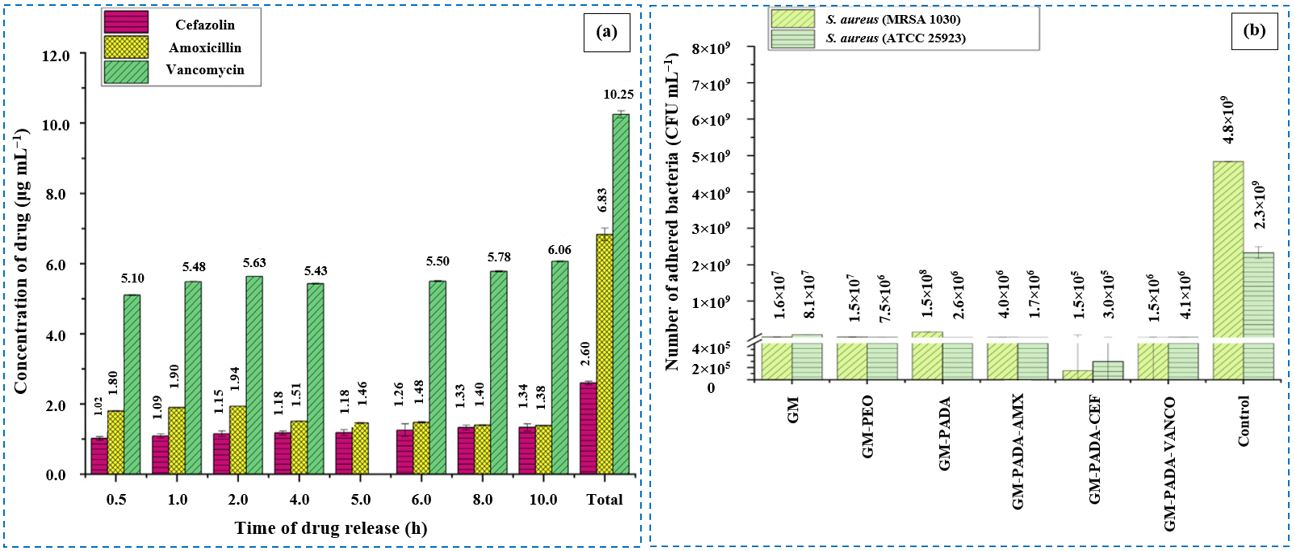
Amoxicillin is a generic antibiotic that belongs to the beta-lactam family of antibiotics and is used to treat a variety of infections, including skin infections, pneumonia, and urinary tract infections in humans. It exhibits an antibacterial action against a wide range of Gram-positive and Gram-negative bacterial pathogens by inhibiting cross-linking between peptidoglycan chains in the cell walls of sensitive bacteria. Peptidoglycan is a biopolymer present in all bacteria that protects them against external environmental factors, especially osmotic pressure. In addition to its use in humans, amoxicillin is also employed for the prevention and treatment of diseases in animals [71,72,73,74]. Leśniak-Ziółkowska et al. [65] found that dip-coated samples containing amoxicillin showed the most effective inhibition against S. aureus, outperforming those loaded with cefazolin or vancomycin in terms of the inhibition zone size. Figure 3a shows the concentration of drug released from the PEO/poly(adipic anhydride) coatings that contain amoxicillin, cefazolin, or vancomycin deposited on titanium alloys into phosphate-buffered saline (PBS) solutions at different degradation times. The results showed that the amoxicillin concentration released from the coating after 0.5 h was 1.80 μg mL−1, which then slightly decreased to 1.38 μg mL−1 after 10 h. The antibacterial effect of the PEO/poly(adipic anhydride)@amoxicillin coating against the clinical strain of methicillin-resistant S. aureus (MRSA) was significantly higher than that against the S. aureus reference strain. As clinical strains typically have higher resistance to antibiotic action compared to reference strains, this result could be constructive.

6.1.2. Cefazolin

Cefazolin is a first-generation cephalosporin antibiotic used to treat endocarditis, sepsis, inflammation of the peritoneum, urinary tract infections, bone infections, and both upper and lower respiratory tract infections. Cefazolin is generally effective against Gram-positive bacterial pathogens, such as S. aureus, but it faces resistance from Gram-negative bacterial pathogens, such as E. coli. Its antibacterial action works by inactivating penicillin-binding proteins in the inner membrane of the bacterial cell wall and interfering with the cross-linking of peptidoglycan chains, ultimately leading to cell lysis [75,76,77]. Leśniak-Ziółkowska et al. [65] observed that after 24 h, the PEO coating containing cefazolin had fewer adhered S. aureus and MRSA bacteria on its surface compared to the dip-coated samples loaded with amoxicillin and vancomycin (Figure 3b). This result was correlated with the concentration of the drug loaded into the polymer layer and the different molecular weights of the drugs, which may affect their diffusion rates into the solution.

6.1.3. Ciprofloxacin

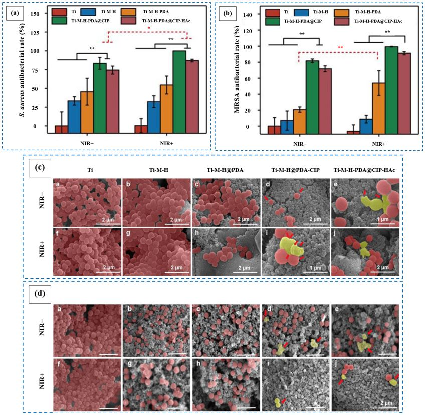
Ciprofloxacin is a second-generation synthetic quinolone antibiotic typically used for the treatment of a variety of bacterial infections affecting the urinary tract, respiratory system, skin, gastrointestinal system, abdominal region, bones, and joints [78]. It is active against a broad spectrum of Gram-positive and Gram-negative bacteria and can be prescribed for both humans and animals [79,80,81]. Its antibacterial action works primarily by inhibiting type II bacterial topoisomerase enzymes, including DNA gyrase, which plays a crucial role in DNA replication, recombination, and repair [80]. Additionally, ciprofloxacin can bind to topoisomerase IV. This interaction traps protein–DNA complexes, leading to DNA damage that triggers cell death and disrupts the normal process of DNA replication [78]. Li et al. [63] examined the antibacterial effect of ciprofloxacin-loaded PEO/hydrothermal@mesoporous polydopamine nanoparticle coatings against S. aureus and MRSA, with the drug incorporated into the PEO coatings via immersion. They observed (as shown in Figure 4a,b) that the antibacterial rate of the coating under near-infrared light (NIR) irradiation against S. aureus and MRSA was about 99.9 and 99.4%, respectively. However, the antibacterial rate of coating covered with sodium hyaluronate catechol was lowered for the two types of bacteria to 87.1 and 91.2%, respectively, due to the weak antibacterial effect of sodium hyaluronate catechol and its barrier effect that hindered the explosive release of ciprofloxacin. When treated with NIR irradiation, the ciprofloxacin release from coatings was enhanced, resulting in a significant reduction in bacterial adhesion and growth. The scanning electron microscopy (SEM) images of the bacteria colony morphology in Figure 4c,d indicate that the bacteria adhered to the PEO/hydrothermal coating surface exhibited a smooth surface, and displayed a typical spherical form. After NIR irradiation, the number of bacteria on the surface of PEO/hydrothermal@mesoporous polydopamine nanoparticles-ciprofloxacin coating, without and with sodium hyaluronate catechol, was significantly reduced. Meanwhile, the structural integrity of the bacterial membrane was compromised, resulting in wrinkles (marked with red arrows) appearing on the surface of the bacteria.

6.1.4. Doxycycline

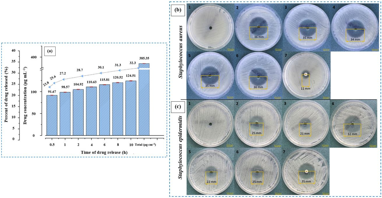
Doxycycline is a semi-synthetic antibiotic made from oxytetracycline, which is a second-generation tetracycline antibiotic. It is used to treat pneumonia and other respiratory tract infections, skin and eye infections, and infections in the lymphatic, digestive, reproductive, and urinary systems. Doxycycline is better at penetrating cell membranes, has a longer half-life, and causes less kidney toxicity compared to other tetracyclines [82,83,84]. It enters bacteria by diffusing through their membranes or by porin transport as a metal ion doxycycline complex. Once inside, the complex breaks apart, which allows doxycycline to pass through the inner cytoplasmic membrane by diffusion. Inside the cell, it prevents bacterial protein production by binding to the 30S ribosomal subunit, which blocks the interaction with aminoacyl-transfer RNA (tRNA) [83]. Doxycycline is also used as a veterinary antibiotic in animals [85]. Kazek-Kęsik et al. [64] examined the effect of doxycycline on the growth of S. aureus and Staphylococcus epidermidis (S. epidermidis) on titanium alloy surfaces coated with PEO/poly (DL-lactide-co-glycolide)@doxycycline, where doxycycline was loaded into the PEO coatings using the dip-coating technique. Figure 5a shows the doxycycline concentration released from the coating into the artificial saliva for different times of exposure. It is seen that after 30 min, more than 23% of the doxycycline, equal to 91.67 μg mL−1, was released from the coating. This concentration was enough to prevent bacterial growth, as observed by large inhibition zones (36 and 25 mm, respectively) in Figure 5b,c. When the concentration of doxycycline reached 110.63 μg mL−1, there was no notable change in the inhibition zone for S. aureus. However, S. epidermidis showed more sensitivity, and its inhibition zone was larger. After 4 h of bacterial culture, the drug’s effectiveness slightly decreased due to a change in the solution’s pH. As the drug concentration increased with more extended immersion, its activity against bacteria also improved. Finally, after 10 h of immersion, 32.3% of doxycycline was released into the solution.

6.1.5. Levofloxacin

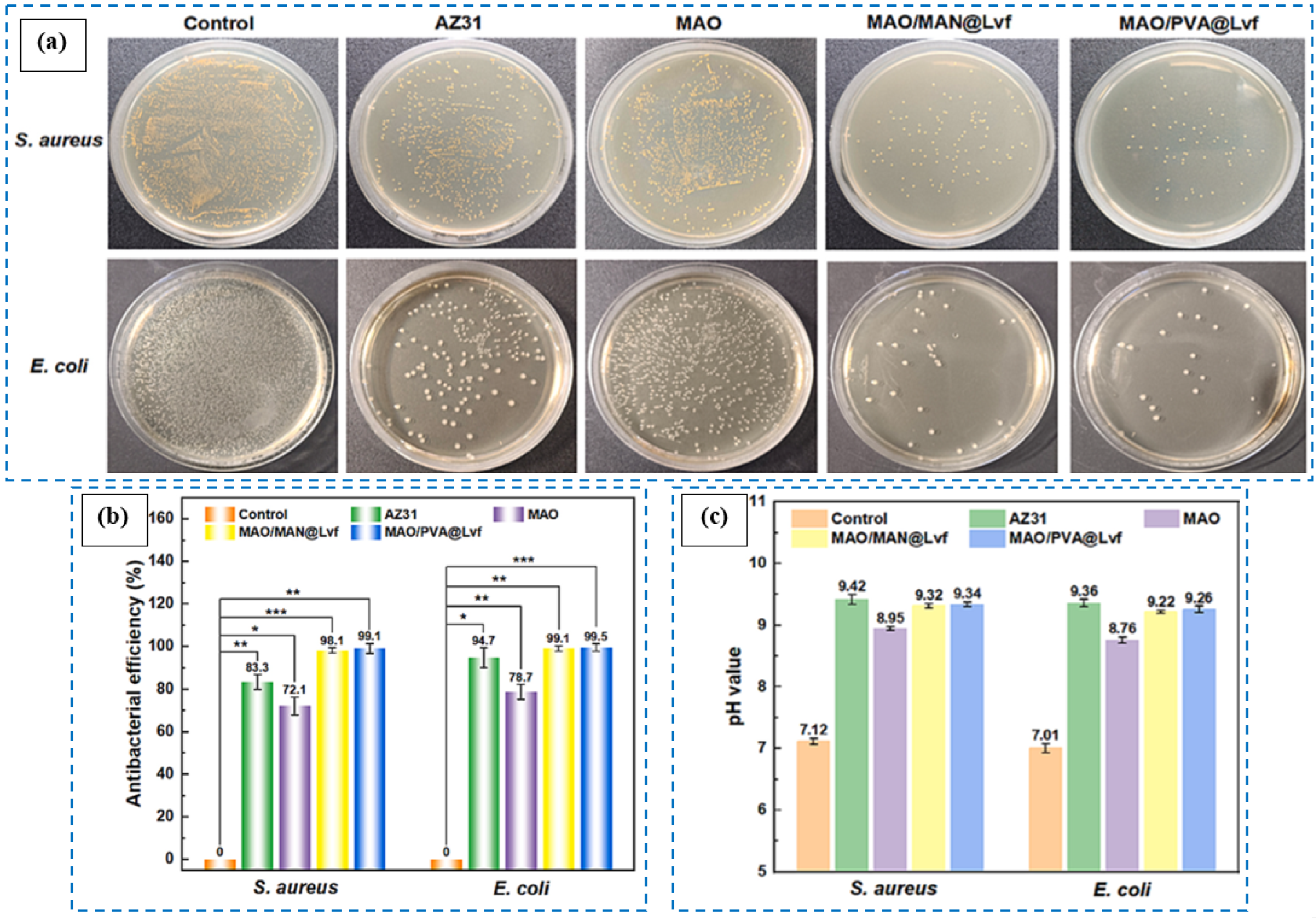
Levofloxacin is a synthetic third-generation fluoroquinolone antibiotic used for the treatment of bacterial infections of the sinuses, kidneys, prostate gland, skin, and bladder. Levofloxacin has several benefits, including reduced cross-resistance to chemicals, a low necessary dosage, excellent tissue penetration, strong antibacterial effects, and minimal side effects. It works against a wide range of Gram-positive and Gram-negative bacteria. In Gram-positive bacteria, levofloxacin primarily targets topoisomerase IV, while in Gram-negative bacteria, it targets DNA gyrase [46,86,87,88,89]. Chen et al. [46] reported that PEO/mannitol@levofloxacin and PEO/polyvinyl alcohol@levofloxacin coatings, both loaded with levofloxacin via a hydrothermal technique, demonstrated excellent antibacterial activity against S. aureus and E. coli. The antibacterial test results in Figure 6a,b show that, while the bare PEO coating exhibited a slight antibacterial effect, both the PEO coatings loaded with levofloxacin had significant antibacterial action due to the release of levofloxacin, the release of Mg2+ cations, and the alkalinity of corrosion. It is known that excessive release of Mg2+ ions could inhibit the growth of bacteria. Moreover, the isoelectric point of most bacteria is approximately pH 4. When the solution’s pH exceeds 4, the bacterial cell acquires a negative charge, making it easier to rupture in an alkaline environment. Therefore, the high pH values of the extracted bacterial solutions for the PEO coatings with levofloxacin could suppress bacterial growth and proliferation (Figure 6c).
Mannitol is a safe sugar alcohol that helps reduce the risk of severe kidney failure following a transplant. It is an effective evaporative agent and osmotic diuretic. Mannitol can help regulate intracranial pressure and maintain kidney function in certain patients who have experienced vascular or cardiac surgery or have jaundice. Pharmaceutical polyvinyl alcohol is a biocompatible polymer characterized by its longer molecular chains than mannitol. It is commonly utilized in ophthalmology, wound dressings, and tissue engineering. The small molecules of mannitol create many reactive sites, which allow for the adsorption of ions and the accumulation of corrosion byproducts. As a result, the drug loading capacity of the PEO/mannitol@levofloxacin coating and its drug release rate during coating degradation after 12 h of immersion in PBS solution were relatively lower than those of the PEO/polyvinyl alcohol@levofloxacin coating. As a result, the inhibition rates for the two bacteria with the PEO/polyvinyl alcohol@levofloxacin coating, due to its higher drug release capacity, were greater (99.1 and 99.5%, respectively) than those for the PEO/mannitol@levofloxacin coating (98.1 and 99.1%, respectively).

6.1.6. Rifampicin

Rifampicin or rifampin is a semi-synthetic antibiotic derived from rifamycin B. It exerts a bactericidal effect on Mycobacteria and Gram-positive aerobic bacteria by inhibiting their DNA-dependent ribonucleic acid (RNA) polymerase [90]. Due to its broad-spectrum activity, good bioavailability, and minimal resistance barrier, rifampicin is often used with other antimicrobial agents to treat infections caused by organisms other than Mycobacteria [91]. For instance, rifampicin helps treat osteomyelitis because it can penetrate osteoblasts and remain active within these cells. It can also penetrate biofilms and maintain its effectiveness there. When antimicrobial therapies are stopped, persister cells may reactivate, leading to recurrent infections. Rifampicin is particularly effective against these persister cells in biofilms, outperforming other available antibiotics [92]. Furthermore, rifampicin can be utilized to treat prosthesis-associated infections that are caused by bacteria adhering to surfaces and forming biofilms [91]. Bigham et al. [49] incorporated rifampicin into PEO coatings on pure titanium via immersion and observed that the release of rifampicin from the PEO/ordered mesoporous magnesium silicate@rifampicin coating effectively inhibited S. aureus proliferation on the coating surface after 24 h.

6.1.7. Tetracycline

Tetracyclines belong to a class of antibiotics that are commonly utilized for the prophylaxis and treatment of bacterial infections in both humans and animals [93,94]. They exhibit potent antibacterial activity against both Gram-positive and Gram-negative bacterial pathogens by inhibiting protein biosynthesis through the prevention of aminoacyl-tRNA attachment to the ribosomal acceptor site [95,96,97]. Typical tetracyclines include tetracycline, oxytetracycline, chlortetracycline, and doxycycline [98]. Bakhsheshi-Rad et al. [45] reported that tetracycline, loaded into PEO/monticellite coatings on magnesium alloy via immersion, was released over time and effectively inhibited the growth of S. aureus and E. coli on the coating surface after 24 h. However, its effect was lower against E. coli than against S. aureus due to the dichotomy of cellular wall structures between the Gram-negative bacterium E. coli and the Gram-positive bacterium S. aureus.

6.1.8. Vancomycin

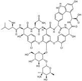
Vancomycin is a glycopeptide antibiotic commonly utilized for the treatment of bacterial infections caused by Gram-positive pathogens [99,100]. It inhibits bacterial synthesis through three main mechanisms: (i) inhibition of peptidoglycan synthesis, (ii) modification of the permeability of the cell membrane, and (iii) disruption of RNA synthesis in the cytoplasm [101]. However, vancomycin is an antibiotic with a narrow spectrum. Numerous bacteria can overcome vancomycin’s bactericidal properties and develop resistance due to multiple mutations in their chromosomal genes that influence the synthesis of their cell walls [102]. Huang et al. [66] reported that vancomycin, loaded into PEO coatings via EPD, was released from the coatings and significantly affected the survival rates of S. aureus and E. coli. The results in Figure 7a show that, compared to the PEO/boron nitride nanosheets@chitosan and PEO/vancomycin@chitosan coatings, the PEO/boron nitride nanosheets-vancomycin@chitosan composite coating demonstrated a remarkable inhibitory action on the growth of both S. aureus and E. coli. This improvement was due to the combined antibacterial actions of boron nitride nanosheets, vancomycin, and chitosan.
The antibacterial effect of boron nitride nanosheets arises from reactive oxygen species (ROS) and the physical contact of the nanosheets with bacteria. Due to their strong hydrophobic properties, boron nitride nanosheets readily extract phospholipids from the lipid bilayer; thus, the sharp edges of the nanosheets disrupt the bacterial cell membrane and penetrate the interior of the bacteria, leading to bacterial death. Additionally, boron nitride nanosheets stimulate the production of intracellular ROS due to the presence of unsaturated boron atoms (B radicals) at the edge of the nanosheets. Chitosan interacts with bacterial cell membranes via its surface functional amino groups and cations under specific conditions. The electrostatic interaction between chitosan and bacterial cells results in the leakage of intracellular substances, ultimately leading to bacterial cell death. Although the effect of vancomycin on the antibacterial activity of the coatings was lower against E. coli than against S. aureus, the complementary antibacterial actions of boron nitride nanosheets and chitosan compensated for the limitations of vancomycin’s bactericidal effect on E. coli. These interactions caused the leakage of intracellular nucleic acids and proteins in E. coli by compromising the bacterial cell membrane integrity, leading to bacterial cell death. The antibacterial mechanism of PEO coatings loaded with boron nitride nanosheets, vancomycin, and chitosan is illustrated in Figure 7b.

6.2. Discussion

Bacterial infections associated with medical implants pose significant clinical and economic challenges. Surface modification of implants with antibacterial coatings is a promising and effective preoperative strategy to prevent or reduce implant-associated bacterial infections by eliminating initial bacterial attachment and hindering subsequent biofilm formation on the implant surface. Release-killing coatings, a class of bactericidal coatings, compromise the structural integrity of bacterial cells or membranes by gradually releasing antibacterial agents in a controlled manner at therapeutic doses over time. The effectiveness of release-killing antibacterial coatings largely depends on maintaining an appropriate concentration and achieving an optimal release profile of the bactericidal agents. Release-killing coatings typically incorporate antibiotics, metal nanoparticles, nitric oxide, and various other biocidal agents, which are embedded within the coatings to provide sustained antimicrobial activity [61]. PEO is an affordable and eco-friendly technique that has recently gained considerable interest for enhancing the surfaces of metallic implants by applying antibacterial coatings. Micropores within PEO coatings serve as effective reservoirs for loading various biologically active substances, including antibiotics, using multiple incorporation methods. Common antibiotics incorporated into PEO coatings include amoxicillin, cefazolin, ciprofloxacin, doxycycline, levofloxacin, rifampicin, tetracycline, and vancomycin, all of which contribute to enhanced antibacterial properties of the implant surface.
The release kinetics of antibiotics from PEO coatings are crucial for maintaining effective antibacterial activity. Amoxicillin experienced a modest decline in release after 10 h [65], while tetracycline followed a two-phase pattern: an initial burst and a sustained release [45]. Ciprofloxacin displayed long-term release with stable levels and no burst effect [47]. In contrast, vancomycin underwent a rapid drop in concentration by day 7, then released slowly until day 28 [48]. Gaining insight into these mechanisms enables the precise tailoring of drug delivery systems to meet specific clinical needs. The type of antibiotic used in PEO coatings significantly influences antibacterial effectiveness. Cefazolin-coated surfaces showed fewer adhered S. aureus and MRSA bacteria compared to those loaded with amoxicillin and vancomycin. This difference can be attributed to variations in drug concentration within the polymer layer and the molecular weights of the antibiotics, which affect their diffusion rates and, ultimately, their ability to inhibit bacterial adhesion [65].
The antibacterial efficacy of antibiotics also varies depending on the bacterial strain. Amoxicillin showed significantly higher antibacterial activity against a clinical strain of MRSA compared to the standard S. aureus reference strain [65]. Doxycycline demonstrated greater effectiveness against S. epidermidis, as evidenced by a larger inhibition zone [64]. Similarly, tetracycline showed more potent activity against Gram-positive S. aureus compared to Gram-negative E. coli [45]. Vancomycin’s antibacterial effect was also less pronounced against E. coli than against S. aureus [66]. These differences highlight the importance of considering bacterial species and their structural characteristics when selecting antibiotics for coating applications. Enhancing the efficacy of antibiotics through synergistic combinations with functional materials is a promising strategy in combating resistant bacterial strains. For instance, the integration of boron nitride nanosheets and chitosan significantly improved the antibacterial activity of vancomycin against E. coli. This combination helped compensate for vancomycin’s limited effectiveness against E. coli [66]. Compared to mannitol, polyvinyl alcohol, due to its longer molecular chains, exhibited superior levofloxacin drug loading capacity and a more controlled release rate [46]. Moreover, under specific conditions such as NIR light-induced photochemotherapy, the release of ciprofloxacin was improved [63]. Therefore, multiple factors must be carefully considered in the design of antibacterial PEO coatings incorporating antibiotics.

7. Conclusions and Future Prospects

Recently, PEO coatings loaded with antibiotics have emerged as a promising and effective surface modification strategy to prevent bacterial infections associated with orthopedic and dental implants. These coatings inhibit bacterial adhesion and colonization on the implant surface by facilitating the localized and controlled release of antibiotics, which effectively kill bacteria, both near and far from the surface. The success of these coatings is governed by the careful selection of an appropriate antibiotic and the controlled release of precise doses within an optimal timeframe. The comparison of various antibiotic-loaded PEO coatings highlights distinct differences in drug release profiles and antibacterial efficacy, primarily influenced by the type of antibiotic, the coating method, and any additional functional components. Dip-coated samples containing amoxicillin exhibited a rapid initial release and demonstrated vigorous antibacterial activity, particularly against MRSA strains. Cefazolin-loaded PEO coatings showed low bacterial adhesion after 24 h, likely due to their slow diffusion rates. Coatings incorporating ciprofloxacin demonstrated near-complete bacterial eradication under NIR irradiation, emphasizing the benefits of photo-responsive systems. Doxycycline-loaded coatings also showed a high initial release and significant inhibition of S. aureus and S. epidermidis. Levofloxacin-loaded coatings achieved broad-spectrum antibacterial effects due to a synergistic mechanism involving drug release, Mg2+ ion release, and alkaline pH. Rifampicin and tetracycline, introduced via immersion, showed effective inhibition against S. aureus, though tetracycline was less effective against E. coli. Notably, vancomycin-loaded PEO coatings, especially when combined with boron nitride nanosheets and chitosan, provided the most potent antibacterial activity against both Gram-positive and Gram-negative bacteria through combined chemical and physical mechanisms. While significant progress has been made in the development of antibiotic-loaded antibacterial PEO coatings, this research is still mainly in the preclinical stages. Much work remains to be done to ensure the safety and maximize the efficacy of these coatings for practical applications. Therefore, future research should focus on several key areas including (i) assessing the cytotoxicity and potential undesirable effects of antibiotics on cells, (ii) evaluating the long-term antibacterial effects of the coatings, (iii) testing the antibacterial activity of the coatings under real in vivo conditions, (iv) conducting in-depth studies on the mechanisms of bactericidal action of the coatings, and (v) investigating the combined effects of two or more different types of antibiotics on the antibacterial properties of the coatings.

Author Contributions

Conceptualization, M.M., M.A. and E.M.Z.; methodology, M.M., M.A. and E.M.Z.; formal analysis, M.M. and M.A.; investigation, M.M., M.A. and E.M.Z.; resources, M.A.; data curation, M.A.; writing—original draft preparation, M.M.; writing—review and editing, M.A. and E.M.Z.; visualization, M.A.; supervision, M.A.; project administration, M.A.; funding acquisition, M.A. and E.M.Z. All authors have read and agreed to the published version of the manuscript.

Funding

This research received no external funding.

Conflicts of Interest

The authors declare no conflicts of interest.

References

  1. Chopra, D.; Gulati, K.; Ivanovski, S. Understanding and Optimizing the Antibacterial Functions of Anodized Nano-Engineered Titanium Implants. Acta Biomater. 2021, 127, 80–101. [Google Scholar] [CrossRef]
  2. Pemmada, R.; Shrivastava, A.; Dash, M.; Cui, K.; Kumar, P.; Ramakrishna, S.; Zhou, Y.; Thomas, V.; Nanda, H.S. Science-Based Strategies of Antibacterial Coatings with Bactericidal Properties for Biomedical and Healthcare Settings. Curr. Opin. Biomed. Eng. 2023, 25, 100442. [Google Scholar] [CrossRef]
  3. Gimeno, M.; Pinczowski, P.; Pérez, M.; Giorello, A.; Martínez, M.Á.; Santamaría, J.; Arruebo, M.; Luján, L. A Controlled Antibiotic Release System to Prevent Orthopedic-Implant Associated Infections: An in Vitro Study. Eur. J. Pharm. Biopharm. 2015, 96, 264–271. [Google Scholar] [CrossRef] [PubMed]
  4. Wang, N.; Ma, Y.; Shi, H.; Song, Y.; Guo, S.; Yang, S. Mg-, Zn-, and Fe-Based Alloys With Antibacterial Properties as Orthopedic Implant Materials. Front. Bioeng. Biotechnol. 2022, 10, 888084. [Google Scholar] [CrossRef]
  5. Sathishkumar, G.; Gopinath, K.; Zhang, K.; Kang, E.T.; Xu, L.; Yu, Y. Recent Progress in Tannic Acid-Driven Antibacterial/Antifouling Surface Coating Strategies. J. Mater. Chem. B 2022, 10, 2296–2315. [Google Scholar] [CrossRef]
  6. Zhang, T.; Liu, Q.; Meng, F.; Hou, Y.; Leung, M.K.H.; Wen, Y.; Zhang, Q. Recent Advances in Stimuli-Responsive Antibacterial Coatings: Bacteria-Killing and Releasing Mechanism, Design Strategies, and Potential Applications. Prog. Org. Coat. 2024, 186, 107923. [Google Scholar] [CrossRef]
  7. Ordikhani, F.; Tamjid, E.; Simchi, A. Characterization and Antibacterial Performance of Electrodeposited Chitosan-Vancomycin Composite Coatings for Prevention of Implant-Associated Infections. Mater. Sci. Eng. C 2014, 41, 240–248. [Google Scholar] [CrossRef]
  8. Wang, H.; Xiong, C.; Yu, Z.; Zhang, J.; Huang, Y.; Zhou, X. Research Progress on Antibacterial Coatings for Preventing Implant-Related Infection in Fractures: A Literature Review. Coatings 2022, 12, 1921. [Google Scholar] [CrossRef]
  9. Rao, Q.; Weng, L.; Zhang, J.; Liu, D.; Zhang, W.; Chen, S.; Chen, J.; Li, X.; Qiu, H.; Cao, Y.; et al. Research Progress in Superhydrophobic Titanium-Based Implants for Antibacterial Applications. Coatings 2023, 13, 419. [Google Scholar] [CrossRef]
  10. Wang, M.; Zheng, Y.; Yin, C.; Dai, S.; Fan, X.; Jiang, Y.; Liu, X.; Fang, J.; Yi, B.; Zhou, Q.; et al. Recent Progress in Antibacterial Hydrogel Coatings for Targeting Biofilm to Prevent Orthopedic Implant-Associated Infections. Front. Microbiol. 2023, 14, 1343202. [Google Scholar] [CrossRef]
  11. Zhang, R.; Han, B.; Liu, X. Functional Surface Coatings on Orthodontic Appliances: Reviews of Friction Reduction, Antibacterial Properties, and Corrosion Resistance. Int. J. Mol. Sci. 2023, 24, 6919. [Google Scholar] [CrossRef]
  12. Forysenkova, A.A.; Fadeeva, I.V.; Deyneko, D.V.; Gosteva, A.N.; Mamin, G.V.; Shurtakova, D.V.; Davydova, G.A.; Yankova, V.G.; Antoniac, I.V.; Rau, J.V. Polyvinylpyrrolidone—Alginate—Carbonate Hydroxyapatite Porous Composites for Dental Applications. Materials 2023, 16, 4478. [Google Scholar] [CrossRef] [PubMed]
  13. Escobar, A.; Muzzio, N.; Moya, S.E. Antibacterial Layer-by-Layer Coatings for Medical Implants. Pharmaceutics 2021, 13, 16. [Google Scholar] [CrossRef] [PubMed]
  14. Romanò, C.L.; Scarponi, S.; Gallazzi, E.; Romanò, D.; Drago, L. Antibacterial Coating of Implants in Orthopaedics and Trauma: A Classification Proposal in an Evolving Panorama. J. Orthop. Surg. Res. 2015, 10, 157. [Google Scholar] [CrossRef] [PubMed]
  15. Chen, X.; Zhou, J.; Qian, Y.; Zhao, L.Z. Antibacterial Coatings on Orthopedic Implants. Mater. Today Bio 2023, 19, 100586. [Google Scholar] [CrossRef]
  16. Yang, X.; Yu, Q.; Wang, X.; Gao, W.; Zhou, Y.; Yi, H.; Tang, X.; Zhao, S.; Gao, F.; Tang, X. Progress in the Application of Spray-Type Antibacterial Coatings for Disinfection. Trends Food Sci. Technol. 2023, 135, 131–143. [Google Scholar] [CrossRef]
  17. Hadzhieva, Z.; Boccaccini, A.R. Recent Developments in Electrophoretic Deposition (EPD) of Antibacterial Coatings for Biomedical Applications—A Review. Curr. Opin. Biomed. Eng. 2022, 21, 100367. [Google Scholar] [CrossRef]
  18. Zhao, C.; Zhou, L.; Chiao, M.; Yang, W. Antibacterial Hydrogel Coating: Strategies in Surface Chemistry. Adv. Colloid Interface Sci. 2020, 285, 102280. [Google Scholar] [CrossRef]
  19. Akshaya, S.; Rowlo, P.K.; Dukle, A.; Nathanael, A.J. Antibacterial Coatings for Titanium Implants: Recent Trends and Future Perspectives. Antibiotics 2022, 11, 1719. [Google Scholar] [CrossRef]
  20. Akay, S.; Yaghmur, A. Recent Advances in Antibacterial Coatings to Combat Orthopedic Implant-Associated Infections. Molecules 2024, 29, 1172. [Google Scholar] [CrossRef]
  21. Wei, T.; Qu, Y.; Zou, Y.; Zhang, Y.; Yu, Q. Exploration of Smart Antibacterial Coatings for Practical Applications. Curr. Opin. Chem. Eng. 2021, 34, 100727. [Google Scholar] [CrossRef]
  22. Kayani, Z.N.; Intizar, T.; Riaz, S.; Naseem, S. Antibacterial, Magnetic and Dielectric Properties of Nano-Structured V Doped TiO2 Thin Films Deposited by Dip Coating Technique. Mater. Chem. Phys. 2021, 267, 124659. [Google Scholar] [CrossRef]
  23. Ali, L.A.; Dikici, B.; Aslan, N.; Yilmazer, Y.; Sen, A.; Yilmazer, H.; Niinomi, M. In-Vitro Corrosion and Surface Properties of PVD-Coated β-Type TNTZ Alloys for Potential Usage as Biomaterials: Investigating the Hardness, Adhesion, and Antibacterial Properties of TiN, ZrN, and CrN Film. Surf. Coat. Technol. 2023, 466, 129624. [Google Scholar] [CrossRef]
  24. Fattah-alhosseini, A.; Molaei, M.; Attarzadeh, N.; Babaei, K.; Attarzadeh, F. On the Enhanced Antibacterial Activity of Plasma Electrolytic Oxidation (PEO) Coatings That Incorporate Particles: A Review. Ceram. Int. 2020, 46, 20587–20607. [Google Scholar] [CrossRef]
  25. Nikoomanzari, E.; Fattah-alhosseini, A.; Pajohi Alamoti, M.R.; Keshavarz, M.K. Effect of ZrO2 Nanoparticles Addition to PEO Coatings on Ti–6Al–4V Substrate: Microstructural Analysis, Corrosion Behavior and Antibacterial Effect of Coatings in Hank’s Physiological Solution. Ceram. Int. 2020, 46, 13114–13124. [Google Scholar] [CrossRef]
  26. Molaei, M.; Fattah-alhosseini, A.; Nouri, M.; Mahmoodi, P.; Navard, S.H.; Nourian, A. Enhancing Cytocompatibility, Antibacterial Activity and Corrosion Resistance of PEO Coatings on Titanium Using Incorporated ZrO2 Nanoparticles. Surf. Interfaces 2022, 30, 101967. [Google Scholar] [CrossRef]
  27. Fattah-alhosseini, A.; Molaei, M.; Nouri, M.; Babaei, K. Antibacterial Activity of Bioceramic Coatings on Mg and Its Alloys Created by Plasma Electrolytic Oxidation (PEO): A Review. J. Magnes. Alloys 2022, 10, 81–96. [Google Scholar] [CrossRef]
  28. Mozafarnia, H.; Fattah-Alhosseini, A.; Chaharmahali, R.; Nouri, M.; Keshavarz, M.K.; Kaseem, M. Corrosion, Wear, and Antibacterial Behaviors of Hydroxyapatite/MgO Composite PEO Coatings on AZ31 Mg Alloy by Incorporation of TiO2 Nanoparticles. Coatings 2022, 12, 1967. [Google Scholar] [CrossRef]
  29. Makurat-Kasprolewicz, B.; Ossowska, A. Recent Advances in Electrochemically Surface Treated Titanium and Its Alloys for Biomedical Applications: A Review of Anodic and Plasma Electrolytic Oxidation Methods. Mater. Today Commun. 2023, 34, 105425. [Google Scholar] [CrossRef]
  30. Noorbakhsh Nezhad, A.H.; Mohammadi Zahrani, E.; Mousavinasab, S.F.; Alfantazi, A.M. A Comprehensive Review of Additively Manufactured Biomedical Titanium Alloys for Bone Tissue Engineering: Biocorrosion, Biomechanical, and Biological Properties. J. Mater. Res. Technol. 2025, 36, 9113–9157. [Google Scholar] [CrossRef]
  31. Nikoomanzari, E.; Fattah-alhosseini, A. Maximizing the Potential Applications of Plasma Electrolytic Oxidation Coatings Produced on Mg-Based Alloys in Anti-Corrosion, Antibacterial, and Photocatalytic Targeting through Harnessing the LDH/PEO Dual Structure. J. Magnes. Alloys 2024, 12, 2674–2694. [Google Scholar] [CrossRef]
  32. Rahmati, M.; Mohammadi Zahrani, E.; Atapour, M.; Noorbakhsh Nezhad, A.H.; Hakimizad, A.; Alfantazi, A.M. In Situ Synthesis and Electrochemical Corrosion Behavior of Plasma Electrolytic Oxidation Coating Containing an Osteoporosis Drug on AZ31 Magnesium Alloy. Mater. Chem. Phys. 2024, 315, 128983. [Google Scholar] [CrossRef]
  33. Attarzadeh, N.; Ramana, C.V. Plasma Electrolytic Oxidation Ceramic Coatings on Zirconium (Zr) and Zr-Alloys: Part-Ii: Properties and Applications. Coatings 2021, 11, 620. [Google Scholar] [CrossRef]
  34. Shishir, R.; Nasiruddin, U.; Manojkumar, P.; Ponnilavan, V.; Lokeshkumar, E.; Rama Krishna, L.; Rameshbabu, N. Development of Bioactive Ceramic Composite Coating with Bactericidal Property on Zn–1Mg Alloy by Plasma Electrolytic Oxidation for Temporary Orthopaedic Implant Applications. Ceram. Int. 2024, 50, 15538–15550. [Google Scholar] [CrossRef]
  35. Chen, Z.; Liu, X.; Cheng, Z.; Tan, X.; Xiang, Y.; Li, J.; Zhang, Y.; Lu, Z.; Kang, E.T.; Xu, L.; et al. Degradation Behavior, Biocompatibility and Antibacterial Activity of Plasma Electrolytic Oxidation Treated Zinc Substrates. Surf. Coat. Technol. 2023, 455, 129234. [Google Scholar] [CrossRef]
  36. Fattah-alhosseini, A.; Molaei, M.; Kaseem, M. A Review on the Plasma Electrolytic Oxidation (PEO) Process Applied to Copper and Brass. Surf. Interfaces 2024, 46, 104179. [Google Scholar] [CrossRef]
  37. Atapour, M.; Blawert, C.; Zheludkevich, M.L. The Wear Characteristics of CeO2 Containing Nanocomposite Coating Made by Aluminate-Based PEO on AM 50 Magnesium Alloy. Surf. Coat. Technol. 2019, 357, 626–637. [Google Scholar] [CrossRef]
  38. Fathyunes, L.; Hosseini, M.; Khalil-Allafi, J. In-Vitro Biocompatibility, Antibacterial Activity, and Corrosion Resistance of HA-TiO2/ZnO Coating Fabricated by Plasma Electrolytic Oxidation on Ti6Al4V. Mater. Today Commun. 2024, 40, 109443. [Google Scholar] [CrossRef]
  39. Curran, J.A.; Clyne, T.W. Porosity in Plasma Electrolytic Oxide Coatings. Acta Mater. 2006, 54, 1985–1993. [Google Scholar] [CrossRef]
  40. Dunleavy, C.S.; Golosnoy, I.O.; Curran, J.A.; Clyne, T.W. Characterisation of Discharge Events during Plasma Electrolytic Oxidation. Surf. Coat. Technol. 2009, 203, 3410–3419. [Google Scholar] [CrossRef]
  41. Rizwan, M.; Alias, R.; Zaidi, U.Z.; Mahmoodian, R.; Hamdi, M. Surface Modification of Valve Metals Using Plasma Electrolytic Oxidation for Antibacterial Applications: A Review. J. Biomed. Mater. Res. Part A 2018, 106, 590–605. [Google Scholar] [CrossRef]
  42. Molaei, M.; Fattah-alhosseini, A.; Nouri, M.; Nourian, A. Systematic Optimization of Corrosion, Bioactivity, and Biocompatibility Behaviors of Calcium-Phosphate Plasma Electrolytic Oxidation (PEO) Coatings on Titanium Substrates. Ceram. Int. 2022, 48, 6322–6337. [Google Scholar] [CrossRef]
  43. Pesode, P.; Barve, S. Biocompatibility of Plasma Electrolytic Oxidation Coated Titanium Alloy for Biomedical Applications. Bionanoscience 2025, 15, 232. [Google Scholar] [CrossRef]
  44. Shi, Y.; Xue, Z.; Li, P.; Yang, S.; Zhang, D.; Zhou, S.; Guan, Z.; Li, Y.; Wang, L.N. Surface Modification on Biodegradable Zinc Alloys. J. Mater. Res. Technol. 2023, 25, 3670–3687. [Google Scholar] [CrossRef]
  45. Bakhsheshi-Rad, H.R.; Hamzah, E.; Ismail, A.F.; Aziz, M.; Najafinezhad, A.; Daroonparvar, M. Synthesis and In-Vitro Performance of Nanostructured Monticellite Coating on Magnesium Alloy for Biomedical Applications. J. Alloys Compd. 2019, 773, 180–193. [Google Scholar] [CrossRef]
  46. Chen, H.B.; Cui, L.Y.; Liu, W.N.; Zhang, S.C.; Wang, Y.; Li, S.Q.; Liu, C.B.; Zeng, R.C. In Vitro Degradation, Antibacterial Activity and Drug Release of Levofloxacin-Loaded Coatings on Mg Alloy AZ31: The Effect of Chain Length. Prog. Org. Coat. 2024, 189, 108327. [Google Scholar] [CrossRef]
  47. Xue, K.; Liang, L.X.; Cheng, S.C.; Liu, H.P.; Cui, L.Y.; Zeng, R.C.; Li, S.Q.; Wang, Z.L. Corrosion Resistance, Antibacterial Activity and Drug Release of Ciprofloxacin-Loaded Micro-Arc Oxidation/Silane Coating on Magnesium Alloy AZ31. Prog. Org. Coat. 2021, 158, 106357. [Google Scholar] [CrossRef]
  48. Nadaraia, K.V.; Mashtalyar, D.V.; Piatkova, M.A.; Pleshkova, A.I.; Imshinetskiy, I.M.; Gerasimenko, M.S.; Belov, E.A.; Kumeiko, V.V.; Kozyrev, D.N.; Fomenko, K.A.; et al. Antibacterial HA-Coatings on Bioresorbable Mg Alloy. J. Magnes. Alloys 2024, 12, 1965–1985. [Google Scholar] [CrossRef]
  49. Bigham, A.; Saudi, A.; Rafienia, M.; Rahmati, S.; Bakhtiyari, H.; Salahshouri, F.; Sattary, M.; Hassanzadeh-Tabrizi, S.A. Electrophoretically Deposited Mesoporous Magnesium Silicate with Ordered Nanopores as an Antibiotic-Loaded Coating on Surface-Modified Titanium. Mater. Sci. Eng. C 2019, 96, 765–775. [Google Scholar] [CrossRef]
  50. Pesode, P.; Barve, S. Surface Modification of Titanium and Titanium Alloy by Plasma Electrolytic Oxidation Process for Biomedical Applications: A Review. Mater. Today Proc. 2021, 46, 594–602. [Google Scholar] [CrossRef]
  51. Ocampo, R.A.; Galvis, O.; Castaño, J.G.; Robledo, S.; Echeverría, F.; Echeverry-Rendón, M. Functionalization of Modified Titanium by Plasma Electrolytic Oxidation with Antibiotic and Cell Adhesion Promoters to Improve Osseointegration Processes. Surf. Coat. Technol. 2024, 481, 130680. [Google Scholar] [CrossRef]
  52. Kaseem, M.; Dikici, B. Optimization of Surface Properties of Plasma Electrolytic Oxidation Coating by Organic Additives: A Review. Coatings 2021, 11, 374. [Google Scholar] [CrossRef]
  53. Thukkaram, M.; Cools, P.; Nikiforov, A.; Rigole, P.; Coenye, T.; Van Der Voort, P.; Du Laing, G.; Vercruysse, C.; Declercq, H.; Morent, R.; et al. Antibacterial Activity of a Porous Silver Doped TiO2 Coating on Titanium Substrates Synthesized by Plasma Electrolytic Oxidation. Appl. Surf. Sci. 2020, 500, 144235. [Google Scholar] [CrossRef]
  54. Xu, D.; Lu, Z.; Wang, T.; Wang, S.; Jiang, Y.; Xu, Z.; Bi, Z.; Geng, S. Novel Ti-Based Alloys Prepared with Different Heat Treatment Strategies as Antibacterial Biomedical Implants. Mater. Des. 2021, 205, 109756. [Google Scholar] [CrossRef]
  55. Husak, Y.; Ma, J.; Wala-Kapica, M.; Leśniak, K.; Babilas, D.; Blacha-Grzechnik, A.; Dulski, M.; Gawecki, R.; Matuła, I.; Dercz, G.; et al. Antibacterial Coatings on Magnesium Formed via Plasma Electrolytic Oxidation in CuO Suspension. Mater. Chem. Phys. 2024, 323, 129627. [Google Scholar] [CrossRef]
  56. Liu, D.; Deng, J.; Liu, Z.; Jin, J.; Bi, Y.; Yang, J.; Zhou, S. Effects of ZnO Nanoparticles Addition to Plasma Electrolytic Oxidation Coatings on Magnesium Alloy: Microstructure, in Vitro Corrosion and Antibacterial Properties. J. Mater. Res. 2022, 37, 2897–2909. [Google Scholar] [CrossRef]
  57. Ferreres, G.; Ivanova, K.; Ivanov, I.; Tzanov, T. Nanomaterials and Coatings for Managing Antibiotic-Resistant Biofilms. Antibiotics 2023, 12, 310. [Google Scholar] [CrossRef]
  58. Shabatina, T.I.; Vernaya, O.I.; Melnikov, M.Y. Hybrid Nanosystems of Antibiotics with Metal Nanoparticles—Novel Antibacterial Agents. Molecules 2023, 28, 1603. [Google Scholar] [CrossRef]
  59. Molaei, M.; Fattah-alhosseini, A.; Nouri, M.; Mahmoodi, P.; Nourian, A. Incorporating TiO2 Nanoparticles to Enhance Corrosion Resistance, Cytocompatibility, and Antibacterial Properties of PEO Ceramic Coatings on Titanium. Ceram. Int. 2022, 48, 21005–21024. [Google Scholar] [CrossRef]
  60. Olmo, J.A.-D.; Ruiz-Rubio, L.; Pérez-Alvarez, L.; Sáez-Martínez, V.; Vilas-Vilela, J.L. Antibacterial Coatings for Improving the Performance of Biomaterials. Coatings 2020, 10, 139. [Google Scholar] [CrossRef]
  61. Zhao, L.; Chu, P.K.; Zhang, Y.; Wu, Z. Antibacterial Coatings on Titanium Implants. J. Biomed. Mater. Res. Part B Appl. Biomater. 2009, 91, 470–480. [Google Scholar] [CrossRef] [PubMed]
  62. Ponomarev, V.A.; Orlov, E.A.; Malikov, N.A.; Tarasov, Y.V.; Sheveyko, A.N.; Permyakova, E.S.; Kuptsov, K.A.; Dyatlov, I.A.; Ignatov, S.G.; Ilnitskaya, A.S.; et al. Ag(Pt) Nanoparticles-Decorated Bioactive yet Antibacterial Ca- and P-Doped TiO2 Coatings Produced by Plasma Electrolytic Oxidation and Ion Implantation. Appl. Surf. Sci. 2020, 516, 146068. [Google Scholar] [CrossRef]
  63. Li, D.; Wang, D.; He, Y.; Tao, B.; Liu, X.; Yang, Y.; Tan, L.; Zhang, Y.; Hu, J.; Yang, W.; et al. A HAase/NIR Responsive Surface on Titanium Implants for Treating Bacterial Infection and Improving Osseointegration. J. Mater. Sci. Technol. 2023, 143, 93–106. [Google Scholar] [CrossRef]
  64. Kazek-Kęsik, A.; Nosol, A.; Płonka, J.; Śmiga-Matuszowicz, M.; Student, S.; Brzychczy-Włoch, M.; Krok-Borkowicz, M.; Pamuła, E.; Simka, W. Physico-Chemical and Biological Evaluation of Doxycycline Loaded into Hybrid Oxide-Polymer Layer on Ti–Mo Alloy. Bioact. Mater. 2020, 5, 553–563. [Google Scholar] [CrossRef]
  65. Leśniak-Ziółkowska, K.; Śmiga-Matuszowicz, M.; Blacha-Grzechnik, A.; Student, S.; Brzychczy-Włoch, M.; Krok-Borkowicz, M.; Pamuła, E.; Simka, W.; Kazek-Kęsik, A. Antibacterial and Cytocompatible Coatings Based on Poly(Adipic Anhydride) for a Ti Alloy Surface. Bioact. Mater. 2020, 5, 709–720. [Google Scholar] [CrossRef]
  66. Huang, W.; Mei, D.; Zhong, Y.; Li, J.; Zhu, S.; Chen, Y.; Wang, L.; Guan, S. The Enhanced Antibacterial Effect of BNNS_Van@CS/MAO Coating on Mg Alloy for Orthopedic Applications. Colloids Surf. B Biointerfaces 2023, 221, 112971. [Google Scholar] [CrossRef]
  67. Wu, X.; Wyman, I.; Zhang, G.; Lin, J.; Liu, Z.; Wang, Y.; Hu, H. Preparation of Superamphiphobic Polymer-Based Coatings via Spray- and Dip-Coating Strategies. Prog. Org. Coat. 2016, 90, 463–471. [Google Scholar] [CrossRef]
  68. Ling, L.; Cai, S.; Li, Q.; Sun, J.; Bao, X.; Xu, G. Recent Advances in Hydrothermal Modification of Calcium Phosphorus Coating on Magnesium Alloy. J. Magnes. Alloys 2022, 10, 62–80. [Google Scholar] [CrossRef]
  69. Majdi, C.; Meffre, P.; Benfodda, Z. Recent Advances in the Development of Bacterial Response Regulators Inhibitors as Antibacterial and/or Antibiotic Adjuvant Agent: A New Approach to Combat Bacterial Resistance. Bioorg. Chem. 2024, 150, 107606. [Google Scholar] [CrossRef]
  70. Kaiser, K.G.; Delattre, V.; Frost, V.J.; Buck, G.W.; Phu, J.V.; Fernandez, T.G.; Pavel, I.E. Nanosilver: An Old Antibacterial Agent with Great Promise in the Fight against Antibiotic Resistance. Antibiotics 2023, 12, 1264. [Google Scholar] [CrossRef]
  71. Aryee, A.A.; Han, R.; Qu, L. Occurrence, Detection and Removal of Amoxicillin in Wastewater: A Review. J. Clean. Prod. 2022, 368, 133140. [Google Scholar] [CrossRef]
  72. Sodhi, K.K.; Kumar, M.; Singh, D.K. Insight into the Amoxicillin Resistance, Ecotoxicity, and Remediation Strategies. J. Water Process Eng. 2021, 39, 101858. [Google Scholar] [CrossRef]
  73. Araújo Júnior, A.G.; Costa, M.L.V.A.; Silva, F.R.P.; Arcanjo, D.D.R.; Moura, L.F.A.D.; Oliveira, F.A.A.; Soares, M.J.S.; Quelemes, P.V. Amoxicillin-Resistant Streptococci Carriage in the Mouths of Children: A Systematic Review and Meta-Analysis. Pathogens 2022, 11, 1114. [Google Scholar] [CrossRef]
  74. Hervin, V.; Roy, V.; Agrofoglio, L.A. Antibiotics and Antibiotic Resistance—Mur Ligases as an Antibacterial Target. Molecules 2023, 28, 8076. [Google Scholar] [CrossRef] [PubMed]
  75. Dastneshan, A.; Rahiminezhad, S.; Naderi Mezajin, M.; Nouri Jevinani, H.; Akbarzadeh, I.; Abdihaji, M.; Qahremani, R.; Jahanbakhshi, M.; Asghari Lalami, Z.; Heydari, H.; et al. Cefazolin Encapsulated UIO-66-NH2 Nanoparticles Enhance the Antibacterial Activity and Biofilm Inhibition against Drug-Resistant S. Aureus: In Vitro and in Vivo Studies. Chem. Eng. J. 2023, 455, 140544. [Google Scholar] [CrossRef]
  76. Valiulin, S.V.; Onischuk, A.A.; Baklanov, A.M.; Dubtsov, S.N.; An’kov, S.V.; Shkil, N.N.; Nefedova, E.V.; Plokhotnichenko, M.E.; Tolstikova, T.G.; Dolgov, A.M.; et al. Aerosol Inhalation Delivery of Cefazolin in Mice: Pharmacokinetic Measurements and Antibacterial Effect. Int. J. Pharm. 2021, 607, 121013. [Google Scholar] [CrossRef] [PubMed]
  77. Munir, M.U.; Ihsan, A.; Javed, I.; Ansari, M.T.; Bajwa, S.Z.; Bukhari, S.N.A.; Ahmed, A.; Malik, M.Z.; Khan, W.S. Controllably Biodegradable Hydroxyapatite Nanostructures for Cefazolin Delivery against Antibacterial Resistance. ACS Omega 2019, 4, 7524–7532. [Google Scholar] [CrossRef]
  78. Khwaza, V.; Mlala, S.; Aderibigbe, B.A. Advancements in Synthetic Strategies and Biological Effects of Ciprofloxacin Derivatives: A Review. Int. J. Mol. Sci. 2024, 25, 4919. [Google Scholar] [CrossRef]
  79. Shariati, A.; Noei, M.; Chegini, Z. Bacteriophages: The Promising Therapeutic Approach for Enhancing Ciprofloxacin Efficacy against Bacterial Infection. J. Clin. Lab. Anal. 2023, 37, e24932. [Google Scholar] [CrossRef]
  80. Sodhi, K.K.; Singh, D.K. Insight into the Fluoroquinolone Resistance, Sources, Ecotoxicity, and Degradation with Special Emphasis on Ciprofloxacin. J. Water Process Eng. 2021, 43, 102218. [Google Scholar] [CrossRef]
  81. Crapnell, R.D.; Adarakatti, P.S.; Banks, C.E. Electroanalytical Overview: The Measurement of Ciprofloxacin. Sens. Diagn. 2023, 3, 40–58. [Google Scholar] [CrossRef]
  82. Ghasemi, K.; Ghasemi, K. A Brief Look at Antitumor Effects of Doxycycline in the Treatment of Colorectal Cancer and Combination Therapies. Eur. J. Pharmacol. 2022, 916, 174593. [Google Scholar] [CrossRef] [PubMed]
  83. Feitosa, R.C.; Ishikawa, E.S.A.; da Silva, M.F.A.; da Silva-Júnior, A.A.; Oliveira-Nascimento, L. Five Decades of Doxycycline: Does Nanotechnology Improve Its Properties? Int. J. Pharm. 2022, 618, 121655. [Google Scholar] [CrossRef]
  84. Cárdenas Sierra, R.S.; Zúñiga-Benítez, H.; Peñuela, G.A. Elimination of Cephalexin and Doxycycline under Low Frequency Ultrasound. Ultrason. Sonochem. 2021, 79, 105777. [Google Scholar] [CrossRef]
  85. Aniagor, C.O.; Igwegbe, C.A.; Ighalo, J.O.; Oba, S.N. Adsorption of Doxycycline from Aqueous Media: A Review. J. Mol. Liq. 2021, 334, 116124. [Google Scholar] [CrossRef]
  86. Rizzo, S.; Gambini, G.; De Vico, U.; Rizzo, C.; Kilian, R. A One-Week Course of Levofloxacin/Dexamethasone Eye Drops: A Review on a New Approach in Managing Patients After Cataract Surgery. Ophthalmol. Ther. 2022, 11, 101–111. [Google Scholar] [CrossRef]
  87. Sitovs, A.; Sartini, I.; Giorgi, M. Levofloxacin in Veterinary Medicine: A Literature Review. Res. Vet. Sci. 2021, 137, 111–126. [Google Scholar] [CrossRef]
  88. Assaf, H.F.; Shamroukh, A.A.; Rabie, E.M.; Khodari, M. Green Synthesis of CaO Nanoparticles Conjugated with L-Methionine Polymer Film to Modify Carbon Paste Electrode for the Sensitive Detection of Levofloxacin Antibiotic. Mater. Chem. Phys. 2023, 294, 127054. [Google Scholar] [CrossRef]
  89. Saya, L.; Malik, V.; Gautam, D.; Gambhir, G.; Balendra; Singh, W.R.; Hooda, S. A Comprehensive Review on Recent Advances toward Sequestration of Levofloxacin Antibiotic from Wastewater. Sci. Total Environ. 2022, 813, 152529. [Google Scholar] [CrossRef]
  90. Khadka, P.; Dummer, J.; Hill, P.C.; Katare, R.; Das, S.C. A Review of Formulations and Preclinical Studies of Inhaled Rifampicin for Its Clinical Translation. Drug Deliv. Transl. Res. 2023, 13, 1246–1271. [Google Scholar] [CrossRef]
  91. Lee, C.Y.; Huang, C.H.; Lu, P.L.; Ko, W.C.; Chen, Y.H.; Hsueh, P.R. Role of Rifampin for the Treatment of Bacterial Infections Other than Mycobacteriosis. J. Infect. 2017, 75, 395–408. [Google Scholar] [CrossRef] [PubMed]
  92. Zhang, Y.; Wang, S.; Liu, M.; Yao, S.; Fang, S.; Cheng, H.; Chen, Q. Adjunctive Rifampin Therapy for Diabetic Foot Osteomyelitis: A Protocol for Systematic Review and Meta-Analysis. Medicine 2020, 99, E20375. [Google Scholar] [CrossRef] [PubMed]
  93. Li, J.; Qin, Y.; Zhao, C.; Zhang, Z.; Zhou, Z. Tetracycline Antibiotics: Potential Anticancer Drugs. Eur. J. Pharmacol. 2023, 956, 175949. [Google Scholar] [CrossRef] [PubMed]
  94. Abbasnia, A.; Zarei, A.; Yeganeh, M.; Sobhi, H.R.; Gholami, M.; Esrafili, A. Removal of Tetracycline Antibiotics by Adsorption and Photocatalytic-Degradation Processes in Aqueous Solutions Using Metal Organic Frameworks (MOFs): A Systematic Review. Inorg. Chem. Commun. 2022, 145, 109959. [Google Scholar] [CrossRef]
  95. Fiaz, A.; Zhu, D.; Sun, J. Environmental Fate of Tetracycline Antibiotics: Degradation Pathway Mechanisms, Challenges, and Perspectives. Environ. Sci. Eur. 2021, 33, 64. [Google Scholar] [CrossRef]
  96. Rusu, A.; Buta, E.L. The Development of Third-Generation Tetracycline Antibiotics and New Perspectives. Pharmaceutics 2021, 13, 2085. [Google Scholar] [CrossRef]
  97. Rong, M.; Huang, Y.; Lin, C.; Lai, L.; Wu, Y.; Niu, L. Recent Advances in Optical Sensing for Tetracycline Antibiotics. TrAC Trends Anal. Chem. 2024, 178, 117839. [Google Scholar] [CrossRef]
  98. Zhang, X.; Cai, T.; Zhang, S.; Hou, J.; Cheng, L.; Chen, W.; Zhang, Q. Contamination Distribution and Non-Biological Removal Pathways of Typical Tetracycline Antibiotics in the Environment: A Review. J. Hazard. Mater. 2024, 463, 132862. [Google Scholar] [CrossRef]
  99. Cafaro, A.; Stella, M.; Mesini, A.; Castagnola, E.; Cangemi, G.; Mattioli, F.; Baiardi, G. Dose Optimization and Target Attainment of Vancomycin in Children. Clin. Biochem. 2024, 125, 110728. [Google Scholar] [CrossRef]
  100. Haseeb, A.; Alqurashi, M.K.; Althaqafi, A.S.; Alsharif, J.M.; Faidah, H.S.; Bushyah, M.; Alotaibi, A.F.; Elrggal, M.E.; Mahrous, A.J.; Abuhussain, S.S.A.; et al. A Systematic Review on Clinical Safety and Efficacy of Vancomycin Loading Dose in Critically Ill Patients. Antibiotics 2022, 11, 409. [Google Scholar] [CrossRef]
  101. Cheng, X.; Ma, J.; Su, J. An Overview of Analytical Methodologies for Determination of Vancomycin in Human Plasma. Molecules 2022, 27, 7319. [Google Scholar] [CrossRef]
  102. Asia, S.; Ejaz, M.; Syed, M.A.; Jackson, C.R.; Sharif, M.; Faryal, R. Epidemiology of Staphylococcus Aureus Non-Susceptible to Vancomycin in South Asia. Antibiotics 2023, 12, 972. [Google Scholar] [CrossRef]
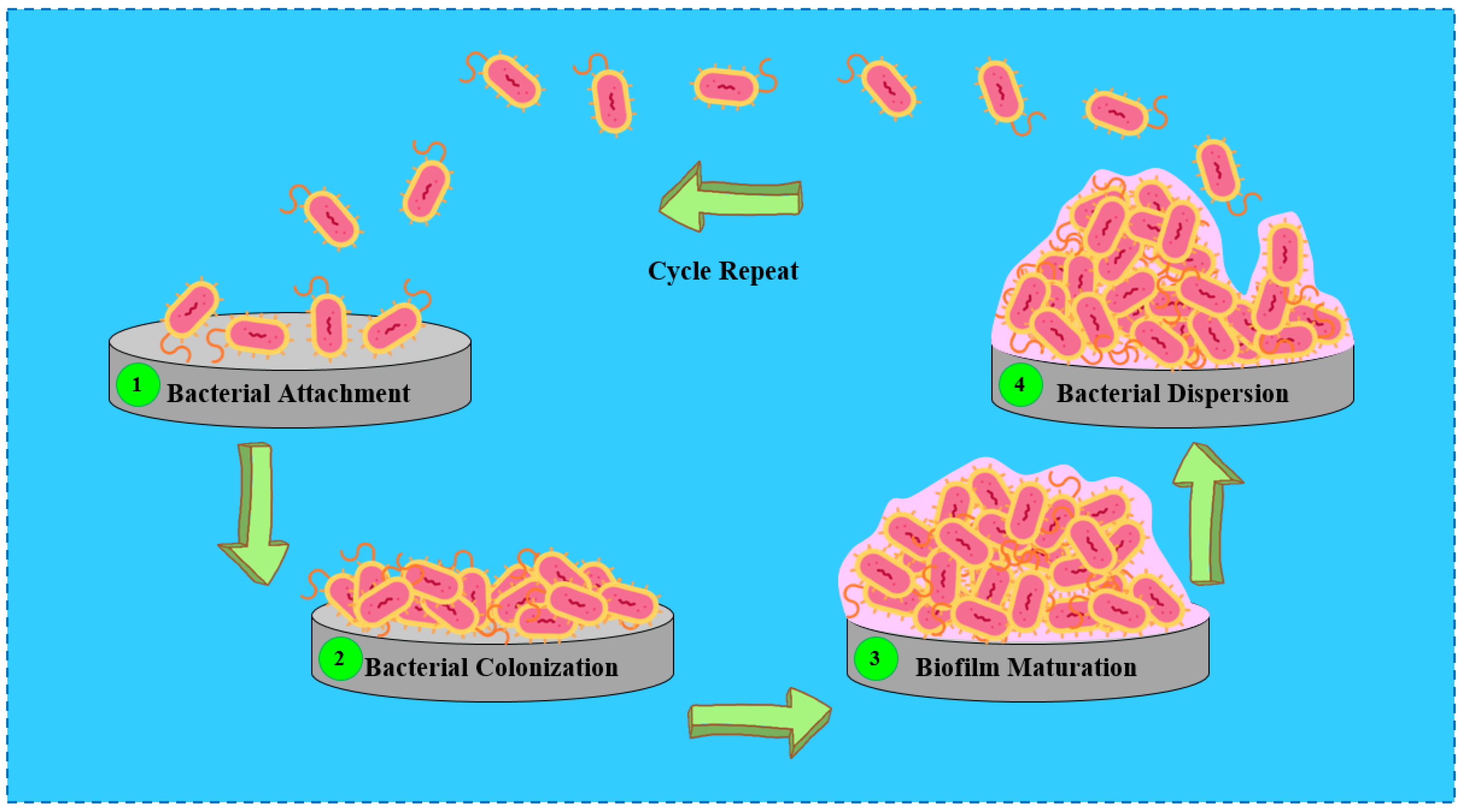
Figure 1. Schematic illustration of the main stages involved in the development of biofilms.
Coatings 15 00925 g001
Figure 2. Strategies for the design of antibacterial coatings.
Coatings 15 00925 g002
Figure 3. (a) Released drugs’ concentrations during various exposure times, and (b) adhesion tests of S. aureus bacteria (ATCC 25923 and MRSA 1030) on investigated surfaces of GM (titanium alloy), GM-PEO (PEO coating), GM-PADA (PEO/poly(adipic anhydride) coating), and GM-PADA_AMX, GM-PADA_CEF, and GM-PADA_VANCO (PEO/poly(adipic anhydride) coatings loaded with amoxicillin, cefazolin, and vancomycin, respectively) [65].
Coatings 15 00925 g003
Figure 4. (a,b) Antibacterial effect of samples on S. aureus and MRSA bacteria, without or with near-infrared light (NIR) irradiation treatment (n = 3, * p < 0.05, ** p < 0.01), and (c,d) scanning electron microscopy images of S. aureus and MRSA colonies cultured on respective samples: (a,f) Ti (titanium substrate), (b,g) Ti-M-H (PEO/hydrothermal coating), (c,h) Ti-M-H@PDA (PEO/hydrothermal@mesoporous polydopamine nanoparticles coating), (d,i) Ti-M-H@PDA-CIP (PEO/hydrothermal@mesoporous polydopamine nanoparticles-ciprofloxacin coating), and (e,j) Ti-M-H-PDA@CIP-HAc (PEO/hydrothermal@mesoporous polydopamine nanoparticles-ciprofloxacin-sodium hyaluronate catechol coating) (The red arrows show the compromised structural integrity of the bacterial membrane) [63]. (With permission from Ref. [63]; License Number: 6070691128829, License date: 16 July 2025).
Coatings 15 00925 g004
Figure 5. (a) The doxycycline concentration released from the PEO/poly (DL-lactide-co-glycolide)@doxycycline coating into artificial saliva, and inhibition zones of (b) S. aureus and (c) S. epidermidis after culture with (1) artificial saliva, and (2–6) the doxycycline released from the coating into (7) the artificial body fluid after (2–6) 1, 2, 4, 6, and 12 h, respectively [64].
Coatings 15 00925 g005
Figure 6. (a) Images of Petri dishes of S. aureus and E. coli, (b) the antibacterial efficiency, and (c) the pH of specimens after immersing into bacterial solutio for AZ31 (magnesium alloy), MAO (PEO coating), MAO/MAN@Lvf (PEO/mannitol@levofloxacin coating), and MAO/PVA@Lvf (PEO/polyvinyl alcohol@levofloxacin coating) (n = 3, * p < 0.05, ** p < 0.01, *** p < 0.001) [46]. (With permission from Ref. [46]; License Number: 6070680705705, License date: 16 July 2025).
Coatings 15 00925 g006
Figure 7. (a) Antibacterial ability of specimens against S. aureus and E. coli (* p < 0.05, ** p < 0.01, and *** p < 0.001), and (b) schematic diagram of antibacterial mechanism (MAO: PEO coating, BNNS: boron nitride nanosheets, CS: chitosan, Van: vancomycin) [66]. (With permission from Ref. [66]; License Number: 6070701188767, License date: 16 July 2025).
Coatings 15 00925 g007
Table 1. The experimental procedures of the PEO processes.
SubstratePEO Process ParametersPEO Coatings CharacteristicsRef
Pore Diameter (µm)Thickness (µm)Roughness (µm)
AZ31 MgElectrolyte: 10 g L−1 NaOH and 8 g L−1 phytic acid, current: 1 A, frequency: 500 Hz, duty cycle: 20%, time: 2.5 min0.63 ± 0.194.37 ± 0.953.96 ± 0.37[46]
Mg–Mn–CeElectrolyte: 25 g L−1 C3H7CaO6P, 5 g L−1 NaF, and 7 g L−1 Na2SiO3; voltage: 380 V, frequency: 300 Hz, time: 100 s 73–953.6 ± 0.3[48]
TiElectrolyte: β-glycerophosphate disodium and calcium acetate monohydrate, voltage: 420 V, time: 6 min  0.387[63]
Mg–Zn–Y–Nd–ZrElectrolyte: 54 g L−1 Na3PO4.12H2O, 2 g L−1 NaOH, and 6 mL L−1 C3H8O3; voltage: 200 V; frequency: 800 Hz; time: 4 min1–3 0.019[66]
AZ31 MgElectrolyte: 10 g L−1 NaOH and 8 g L−1 phytic acid, voltage: 220–250 V, current density: 0.008–0.016 A cm−2, duty cycle: 50%, time: 10 min0.1–0.33.67 ± 0.810.8048 ± 0.008[47]
Ti–15MoElectrolyte: 0.1 M Ca(H2PO2)2, voltage: 300 V, current density: 100 mA cm−2, time: 5 min  0.5 ± 0.09[64]
Ti–Ta–Zr–NbElectrolyte: 0.1 M Ca(H2PO2)2, voltage: 300 V, current density: 150 mA cm−2, time: 5 min 8.54–11.201.24 ± 0.35[65]
Pure TiElectrolyte: 8 g L−1 sodium carbonate and 4 g L−1 potassium hydroxide, current density: 1.5 mA cm−2, time: 10 min 4 [49]
Mg–Ca–ZnElectrolyte: 10 g L−1 Na2SiO3.9H2O, 8 g L−1 KF.2H2O, and 1 g L−1 KOH; voltage: 350 V; current density: 2 A cm−2; time: 10 min 12 ± 0.5 [45]
Table 2. The properties of antibiotics used in antibacterial PEO coatings.
AntibioticFamilyChemical FormulaChemical StructureMolar Mass (g mol−1)
AmoxicillinAminopenicillinC16H19N3O5SCoatings 15 00925 i001365.40
CefazolinCephalosporinC14H14N8O4S3Coatings 15 00925 i002454.50
CiprofloxacinFluoroquinoloneC17H18FN3O3Coatings 15 00925 i003331.34
DoxycyclineTetracyclineC22H24N2O8Coatings 15 00925 i004444.44
LevofloxacinFluoroquinoloneC18H20FN3O4Coatings 15 00925 i005361.37
RifampicinRifamycinC43H58N4O12Coatings 15 00925 i006822.95
TetracyclineTetracyclineC22H24N2O8Coatings 15 00925 i007444.44
VancomycinGlycopeptideC66H75Cl2N9O24Coatings 15 00925 i0081449.27
Disclaimer/Publisher’s Note: The statements, opinions and data contained in all publications are solely those of the individual author(s) and contributor(s) and not of MDPI and/or the editor(s). MDPI and/or the editor(s) disclaim responsibility for any injury to people or property resulting from any ideas, methods, instructions or products referred to in the content.