Article

Unveiling the Role of Hydrogel Stiffness Threshold in Schwann Cell Context: Regulating Adhesion Through TRIP6 Gene Expression

1
Key Laboratory of Neuroregeneration of Jiangsu and Ministry of Education, Nantong University, Nantong 226001, China
2
Coinnovation Center of Neuroregeneration, Nantong University, Nantong 226001, China
3
Laboratory Medicine Center, The Sixth People’s Hospital of Nantong (Affiliated Nantong Hospital of Shanghai University), Nantong 226001, China
4
Fuyang People’s Hospital, Fuyang 236000, China
*
Authors to whom correspondence should be addressed.
These authors contributed equally to this work.
Coatings 2025, 15(7), 753; https://doi.org/10.3390/coatings15070753
Submission received: 23 May 2025 / Revised: 21 June 2025 / Accepted: 23 June 2025 / Published: 25 June 2025

Abstract

Adhesion between Schwann cells (SCs, a type of glial cell in the peripheral nervous system) and their underlying substrates is a fundamental process that holds critical importance for the proper functioning of the peripheral nervous system. Conducting further in-depth research into the adhesion mechanisms of nerve cells is of paramount significance, as it can pave the way for the development of highly effective biomaterials and facilitate the repair of nerve injuries. Thyroid Receptor Interaction Protein 6 (TRIP6), a member of the ZYXIN family of LIM domain-containing proteins, serves as a key component of focal adhesions. It plays a pivotal role in regulating a diverse array of cellular responses, including the reorganization of the actin cytoskeleton and cell adhesion. Accumulated data indicate that RSC96 cells (rat Schwann cells), which are rat Schwann cells, exhibit integrin-based mechanosensitivity during the initial phase of adhesion, specifically within the first 24 h. This enables the cells to sense and respond to alterations in matrix stiffness. The results of immunofluorescence staining experiments revealed intriguing findings. An increase in matrix stiffness not only led to significant changes in the morphological parameters of RSC96 ells, such as circularity, aspect ratio, and cell spreading area, but also enhanced the expression levels of TRIP6, focal adhesion kinase (FAK), and vinculin within these cells. These changes collectively promoted the adhesion of RSC96 cells to the matrix. Furthermore, when TRIP6 expression was silenced in RSC96 cells cultured on hydrogels, a notable decrease in the expression of both FAK and vinculin was observed. This, in turn, had a detrimental impact on cell adhesion. In summary, the present study strongly suggests that TRIP6 may play a crucial role in promoting the adhesion of RSC96 cells to polyacrylamide hydrogels with varying stiffness. This research not only offers a fresh perspective on the study of the integrin-mediated force regulation of cell adhesion but also lays a solid foundation for potential applications in tissue engineering, regenerative medicine, and other related fields.

1. Introduction

In the peripheral nervous system, the influence of the matrix elasticity of tissue engineering materials on the behavior of nerve cells is of great significance [1]. SCs (Schwann cells, a type of glial cell in the peripheral nervous system) are the most important glial cells in the peripheral nervous system, which have various biological functions such as myelin formation, nerve signal transmission, neurotrophic factor secretion, the support and promotion of axon growth, and playing a key role in nerve injury repair and nerve function repair [2,3]. Peripheral nerve injury causes not only biochemical changes in tissue structure, but also mechanical changes affecting SCs. SCs can mechanically sense the hardness of the extracellular matrix, and the mechanical sensitivity of SCs can regulate their morphology and migration behavior [4,5]. After peripheral nerve injury, they can promote neuron axon extension, SC adhesion, and the movement migration of the peripheral nerve, which will be more conducive to nerve regeneration and repair.
Many aspects of cell growth are regulated by the spatial and mechanical properties of their microenvironment [6]. Using experimental methods to regulate the spatial constraints of cells during culture, studies have shown that the imposed boundary conditions can profoundly affect cell adhesion. Cell adhesion to the extracellular matrix plays an important role in cell migration [7]. Transmembrane integrins in focal adhesion (FA) undergo cycles of matrix attachment, cytoskeletal recruitment, contractility induction, and decomposition [8]. Neonatal FAs form at or near the edge of the pro-cell and mature into larger FAs located in the middle and rear of the migrating cell. Newborn FAs are responsible for most of the contractile force, while mature FAs delay the rate of cell translocation and generate signals for cell survival and transcriptional activation [7].
Studies have shown that mechanical stimulation can regulate cell adhesion by regulating the cell–substrate connection [9].The change in basal stiffness is essentially a mechanical stimulus for cells. The key link in how substrate stiffness affects cells is how cells perceive mechanical changes in the extracellular microenvironment, convert external mechanical stimuli into biochemical signals, and transmit them into cells [10]. Cytoskeleton (CSK) and integrin proteins play a crucial role in sensing the mechanical properties of the extracellular matrix microenvironment and transmitting mechanical signals into the cell [11]. In addition, integrins play a key role in regulating intracellular adhesion-related proteins [12]. Thus, adherent cells can sense and respond to mechanical signals from the matrix in their microenvironment. However, despite extensive research on mechanosensing, a comprehensive understanding of the bioinformatics that control cell adhesion and substrate surface mechanical signaling is still lacking.
Zyxin and related proteins play an essential role in the cellular perception of FA recombination induced by mechanical signals from the external microenvironment. Zyxin, a protein that contains LIM domains, is recruited to adhesion and actin stress fibers in a force-sensitive manner and plays an important role in cell morphology, adhesion, skeletal development, and cell mechanical response to growth at different matrix stiffness [13,14,15,16]. Another Zyxin-associated protein found in mature FAs, Thyroid Receptor Interaction Protein 6 (TRIP6), also known as Zyxin-associated protein-1 (ZRP-1), is a convergent protein belonging to the Zyxin family of LIM proteins and is localized primarily to cytoplasmic or focal adhesive plaques. The ability to shuttle between cytoplasm and nuclear compartments plays a key role in cytoskeletal recombination and FAs [17]. Stimulated by lysophosphatidic acid (LPA), TRIP6 indirectly binds to components of the focal complex, including paxillin, p130cas, FAK, and c-Src [18]. TRIP6 and its relatives LPP and Zyxin are directed to “integrin adherents” by myosin II-dependent recruitment [19]. Both TRIP6 and LPP (but not Zyxin) can interact with supervisin at large focal adhesions [7]. Therefore, TRIP6 is characterized as an aptamer protein, which can bind to a variety of proteins, sense the mechanical signals of the external microenvironment, and participate in the regulation of a variety of cellular responses, especially actin cytoskeletal recombination, cell adhesion, and migration.
On the basis of the matrix hardness hydrogels prepared by the researchers in the previous work [20,21], we prepared polyacrylamide/chitosan composite hydrogels with different elastic moduli to explore the effects of hydrogels with different matrix elastic moduli on nerve cell morphology and FAs, as well as the change characteristics of TRIP6 expression during this process, and analyzed the effects of TRIP6 on nerve cell morphology on the surface of the material by interfering TRIP6 expression to explore the possible regulatory role of TRIP6 in the biological behavior of cells sensing and responding to different matrix elasticity.

2. Materials and Methods

2.1. Materials

Acrylamide and N-N-methylenebisacrylamide were obtained from Sigma-Aldrich, St. Louis, MO, USA. Anhydrous ethanol and paraformaldehyde were obtained from Shanghai Zhenxing Chemical Factory, Shanghai, China. Chitosan was from Nantong Xingcheng Biotechnology, Nantong, China. Phosphate buffered saline (PBS) was from Hyclone Limited Co., Beijing, China. Ammonium persulfate, penicillin–streptomycin (PS), and CCK8 Cell Proliferation and Toxicity Kit were from Shanghai Biyuntian Biotechnology, Shanghai, China. Dulbecco’s modified Eagle’s medium (DMEM) and fetal bovine serum (FBS) were from Gibco, Waltham, MA, USA. The 0.25% trypsin EDTA was from Invitrogen, (Carls Bard, CA, USA). Anti-TRIP6 antibody, anti-FAK antibody, ghost pen cyclic peptide, anti-vinculin antibody, and DAPI were obtained from Abcam (Cambridge, UK). Primers used in all experiments were prepared by a microtiter deionized water purification unit.

2.2. Preparation of PAM/CS Hydrogels with Different Moduli

In this study, PAM/CS composite hydrogels with different elastic moduli were constructed by varying the concentration of bisacrylamide through in situ free radical polymerization reactions. The mixtures were prepared with 29% acrylamide, 0.1%, 0.5%, 1%, 2%, and 3% bisacrylamide, 10% ammonium persulfate, and 5% chitosan solutions. For chitosan solution, 5 g of chitosan powder was weighed and poured into a beaker, 100 mL of bis-distilled water containing 2% acetic acid was added and stirred, and the mixture was placed in a 60 °C oven overnight to fully dissolve. The mixture was then added to the grinder and placed in an oven at 55 °C to induce gel polymerization for 1 h. After gelation, the hydrogels were removed from the Petri dishes and soaked in PBS to remove the uncrosslinked toxic monomer acrylamide.

2.3. Measurement of the Elastic Modulus of Hydrogels

The elastic modulus of all hydrogels was measured using a universal testing machine (UTM, TFW-58, Shanghai Tuoyi Technology Co., Ltd., Shanghai, China). It is a well-established and widely recognized instrument for mechanical property testing in materials science research. The hydrogel samples were prepared into standard cylindrical shapes with a diameter of 8 mm and a height of 6 mm. During the measurement, the samples were placed between the two clamps of the UTM to ensure good contact between the samples and the clamps. Then, a uniaxial compression test was conducted at a constant strain rate of 1 mm/min. The force–displacement data were continuously recorded by the built-in software of the UTM. The elastic modulus was calculated from the linear region of the stress–strain curve obtained from the test data. The stress was calculated by dividing the applied force by the original cross-sectional area of the sample, and the strain was calculated by dividing the displacement by the original height of the sample.

2.4. Cell Culture and Transfection

The rat Schwann cell line RSC96 cells were purchased from the National Identification Cell Culture Preservation Center of China. RSC96 cells were cultured in DMEM medium supplemented with 15%FBS and 1% antibiotics (100 bits/mL PS). RSC96 cells were incubated in a humidified atmosphere with 5% CO2 at 37 °C. Cell transfection with Super-Fectin (Ruibo Suzhou Pharmaceutical Co., Ltd., Suzhou, China) was performed according to the manufacturer’s instructions.

2.5. Cell Viability Assay

The effect of hydrogel on cell viability was determined by the CCK-8 method. The materials of each group were cut into circles with a diameter of 1 cm. They were then sterilized by UV for 30 min, soaked in 75% alcohol for 30 min, and subsequently washed three times - first with sterile water and then with PBS, for 15 min each time. Finally, they were put into 24-well plates for spare use. Add 1 mL of DMEM basic medium to each well of the plate, put the soaked plate into the 5% CO2 and 37 °C constant-temperature cell culture incubator, and incubate for 12 h. At the same time, inoculate the RSC96 rat Schwann cell line on the hydrogel in the 24-well plate and incubate the cells for 48 h. The medium used was DMEM complete medium containing 10%FBS + 1%PS. Next, 300 µL of working solution was added to each well and incubated for 4 h at 37 °C in a constant-temperature cell culture incubator, protected from light; then, 100 µL of working solution was pipetted from each well of the 24-well plate into a 96-well plate for the measurement of absorbance values at 450 nm. The cell viability was calculated as follows: Cell viability (%) = Absexp/Abscon × 100%, where Absexp is the absorbance of the experimental group and Abscon is the absorbance of the control group, when all the values were compared with those of the blank control group (cells cultured in the well plates without the material alone).

2.6. Cell Proliferation Assay

Cultivate the cells on hydrogels of different elastic moduli in 24-well plates and process the cells after 48 h of incubation; prepare a 2× EdU working solution in serum-free medium with 10 mM EdU solution; pre-warm the 2× EdU solution and add the 2× EdU solution to an equal volume of medium containing the experimental cells to obtain a 1× EdU solution; add 0.2 mL of Click reaction mixture to each well. Add 0.2 mL of Click reaction mixture to each well, gently shake the plate to ensure that the reaction mixture can evenly cover the sample, and configure the system as required; react for 1–2 h away from light; aspirate the Click reaction solution, and wash it with washing solution for 3 times, each time for 3–5 min. For DNA staining, perform the preparation of DAPI solution: Dilute DAPI (1000×) in PBS at the ratio of 1:1000, and keep it away from light; add 1× DAPI solution 1 mL to each well, and keep it away from light; add 1× DAPI solution 1 mL to each well at room temperature. For solution 1ml, incubate for about 10 min at room temperature and keep away from light; add PBS to each well to wash 1~3 times; pick up the picture.

2.7. Cell Adhesion Assay

To test the differences in adhesion on the surface of hydrogels with different moduli of elasticity, RSC96 cells were inoculated on 1.5 kPa, 8.0 kPa,13.75 kPa, 27.67 kPa, and 39 kPa PAM/CS hydrogels and incubated for 48 h. Cells were allowed to attach to the PAM/CS hydrogels for 48 h in an incubator at 37 °C, 5% CO2. Petri dishes were washed 3 times with PBS to remove non-adherent cells. The number of remaining cells attached to the gel was imaged using a fluorescence microscope (Leica Microsystems, Wetzlar, Germany). Ten different image fields (1.5 mm2 area) were selected for each gel, and the number of cells was counted for each substrate (in triplicate) using ImageJ software (ij150-win-jre6-32-bit, NIH, Bethesda, MD, USA). Data were exported to Origin Pro 8.0 (Origin Lab Corporation, Northampton, MA, USA) for analysis and graphic production.

2.8. Immunofluorescence Staining Experiment

After 48 h of incubation, RSC96 cells were fixed in 4% paraformaldehyde solution for 2 h. The cells were then washed twice with PBS for 5 min each time. The blocking solution was then added to each sample. The samples were sealed and incubated at 4 °C for 1.1 h, after which the blocking solution was discarded. Hydrogels were incubated with primary antibodies TRIP6, FAK, and vinculin at 4 °C overnight. The hydrogel was then washed with PBS 3 times for 5 min each. After that, it was incubated with secondary antibodies Alexa-488 (1:500; Abcam, Cambridge, MA, USA) and CY3 (1:500), protected from light at 37 °C for 2 h. Cells were then treated with Hoechst 33,342 for 15 min. Finally, the samples were washed three times with PBS and visualized by laser copolymer fluorescence microscopy (Leica, Wetzlar, Germany).

2.9. Real-Time Quantitative Polymerase Chain Reaction

RNA was extracted from RSC96 cells using the Trizol reagent. cDNA was then obtained from RNA using the Takara kit (Invitrogen, Carlsbad, CA, USA). The sequences of the primers required in this experiment are listed: trip6-rat-qpcr-F-1: tccctttcacagtggatgcc; trip6-rat-qpcr-R-1: cctcacacttgtagcagcca. Materials were purchased from Kingsley Biotechnology, Wuhan, China.

2.10. Statistical Analysis

Unless otherwise stated, experimental results are the mean ± standard deviation of three independent experiments. All data were analyzed using Graphpad Prism 8.0, Image J, and Origin 2019b software. Results are expressed as mean ± standard deviation, differences between two groups were statistically determined by t-test, multiple groups were compared two-by-two using one-way ANOVA statistics, ns indicates no statistical difference, * p < 0.05.

3. Results and Discussion

3.1. Modulation of Cell Adhesion by PAM/CS Hydrogels with Different Stiffness

Cell adhesion is an important step in enabling adherent cells to thrive and perform normal biological functions in the living body [22].We varied the bisacrylamide concentration (0.1%, 0.5%, 1%, 2%, and 3%) and conducted mechanical tests on the hydrogels using a universal testing machine. The measured elastic moduli were 1.5 kPa, 8.0 kPa, 13.75 kPa, 27.47 kPa, and 39 kPa in sequence. We examined the ability of RSC96 cells to adhere to polyacrylamide/chitosan hydrogels with constant biochemical composition but varying stiffness (1.5kPa, 8.0kPa,13.75 kPa, 27.47 kPa, and 39 kPa). Cell adhesion was studied by incubating cells on different substrate stiffness hydrogels for 4, 24, and 48 h. Figure 1A shows the macroscopic image of the different hydrogels. Figure 1B shows white light images of RSC96 cells adhering to a surface at 1.5 kPa to 39 kPa with incubation times ranging from 4 to 48 h. Figure 1C–E shows that, on average, approximately 100 cells per square millimeter adhered to the 1.5 kPa surface during the first 4 h of culture. This is significantly lower compared to the polyacrylamide/chitosan hydrogels with higher modulus (13.75 kPa, 27.47 kPa and 39 kPa), suggesting that RSC96 cells are able to sense mechanical signals during the initial stages of adhesion. The number of RSC96 cells attached to the 13.75 kPa surface increased as the incubation time increased. In addition, the percentage of adherent cells increased significantly with increasing matrix hardness, with the highest percentage of adherent cells at a matrix hardness of 13.75 kPa, which then began to decrease as the percentage of adherent cells matrix stiffness increased.

3.2. Substrate Stiffness Controls RSC96 Cell Phenotype and Proliferative Capacity In Vitro

The morphology of single cells depends on the balance between external biomechanical forces and internal intercellular forces, and the level of intracellular forces is proportional to the elastic nature of the surrounding extracellular matrix (ECM) [23]. This suggests that the morphology of the cell is influenced by a combination of the elasticity and biomechanics of the substrate. Therefore, it is assumed that the specific shape of RSC96 cells can be influenced according to the rigidity of the substrate. On substrates with higher stiffness (8.0 to 39 kPa), RSC98 cells spread over a larger area and exhibited protruding growth (Figure 2A). The morphology of RSC96 cells was analyzed using ImageJ software. We found that hydrogels showed stiffness-dependent effects on the growth of RSC96 cells, such as circularity, aspect ratio, and cell spreading (as shown in Figure 2A–D). Thus, our experimental results indicate that RSC96 cells respond to substrate elasticity by changing their morphology and shape.
Cell morphology is an important marker of cell proliferation and plays an important role in regulating cell growth, physiological functions, and specific functions [24]. To this end, we investigated different matrix stiffnesses 1.5 kPa to 39 kPa to explore their effects on the proliferative effects of RSC96 cells cultured in vitro. The proliferative capacity and cell viability of RSC96 cells were assessed using EDU and Cell Counting Kit-8 (CCK-8) assays. The results showed that the cell viability of RSC96 cells grown on a soft substrate (1.5 kPa) was 1.7-fold (Figure 3A,B) and 2-fold (Figure 3C) lower than that of cells grown on 13.75 kPa, indicating that neuronal cell viability was increased on a substrate stiffness of 13.75 kPa. According to the EDU and CCK8 results, when the hardness of the substrate did not exceed 13.75 kPa, we observed that the proliferation capacity and cell viability of the cells were proportional to the hardness of the substrate; when the hardness of the substrate exceeded 13.75 kPa, the proliferation capacity and cell viability of the cells began to decrease. Collectively, these results suggest that matrix stiffness modulates the proliferative capacity and cell viability of RSC96 cells.

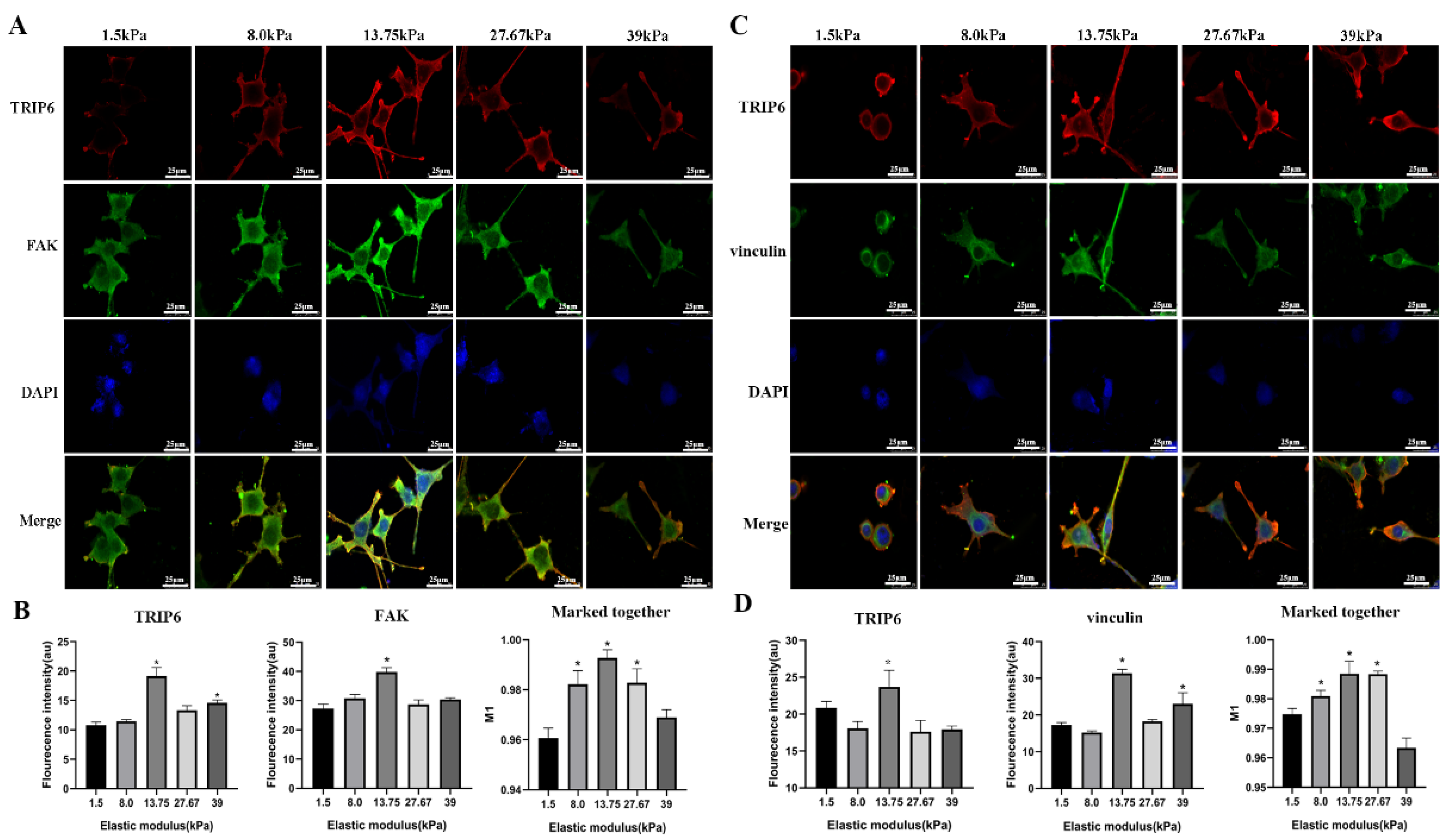
3.3. Important Relevance of TRIP6 for Matrix Stiffness-Induced RSC9 Cell Adhesion

TRIP6 is a member of the Zyxin family of LIM structural-domain proteins and is a focal adhesion component that regulates a variety of cellular responses, such as actin cytoskeleton reorganization and cell adhesion [25]. Cell adhesion is dependent on the formation of focal adhesions (FAs), specialized structures that act as bridges between the cytoskeleton and the surrounding matrix. These complex structures of FA consist of transmembrane integrins and various intracellular proteins, such as FAK and vinculin, that function as potential mechanosensors [26,27,28,29,30]. Studies have shown that FAK is involved in the regulation of various cellular processes and is an important molecule in the regulation of cell adhesion, migration, and signal transduction [26]. Therefore, to assessTRIP6, FAK, and vinculin expression, adherent RSC96 cells were fluorescently stained when cultured on polyacrylamide/chitosan hydrogels. Figure 4A–D shows that, when the substrate hardness did not exceed 13.75 kPa, as the hardness of the polyacrylamide/chitosan hydrogel increased leading to an increase in the expression of TRIP6, the level of FAK was elevated and the expression of vinculin was also increased, which was directly proportional to the hardness of the substrate; when the hardness of the substrate was more than 13.75 kPa, the expression of TRIP6, FAK, and vinculin levelled off or began to decline. In response to external mechanical stimuli, proteins such as vinculin undergo changes in conformation and subcellular localization and act as receptors for extracellular stress signals. The co-localization of TRIP6, FAK, and vinculin was also strongest at 13.75 kPa. Taken together, the results of the present study suggest that the increased stiffness of polyacrylamide/chitosan hydrogels may enhance the adhesion of RSC96 cells by up-regulating the expression of the focal adhesion protein TRIP6, which enhances the adhesion of RSC96 cells.

3.4. Inhibition of TRIP6 Expression Attenuated RSC96 Cell Adhesion on 1.5 kPa and 13.75 kPa Substrates

To further validate the role of TRIP6 in the adhesion of RSC96 cells on hydrogels of different stiffness, we tested whether small interferences inhibiting the expression of TRIP6 could further attenuate the adhesion of substrate-paired RSC96 cells. To determine the role of TRIP6 in the adhesion of RSC96 cells grown on hydrogels, we used siRNA to inhibit TRIP6 expression. The small interfering RNAs used to interfere with TRIP6 expression were used for the validation of results (Figure 5A). Among them, si-TRIP6-2 had the highest knockdown efficiency among the three siRNAs tested (Figure 5A). We used si-TRIP6-2 in RSC96 cells to provide a useful tool for testing the function of TRIP6 in RSC96 cell adhesion. The results of CCK8 assay and EdU assay showed that the inhibition of TRIP6 expression reduced the survival of RSC96 cells grown on hydrogels (Figure 5B,C). We found that the inhibition of TRIP6 downregulated the adhesion effect of RSC96 cells grown on hydrogels (Figure 5D–F). As shown in Figure 5D–F, the results of cellular immunofluorescence staining indicated that the inhibition of TRIP6 expression significantly reduced the expression of vinculin and FAK and decreased the cell adhesion of RSC96 cells on both soft and stiffness hydrogels compared with the control group. These results suggest that TRIP6 expression may be involved in the effect of hydrogel matrix hardness on RSC96 cell viability and may play a crucial role in mediating the response of RSC96 cells to matrix stiffness-driven cell adhesion.

4. Conclusions

Based on our findings that polyacrylamide/chitosan hydrogels with varying stiffnesses significantly influence RSC96 cell adhesion through the involvement of TRIP6 as a potential biomechanical sensor, several promising future research directions emerge. Firstly, in terms of material development, a further optimization of the hydrogel composition and stiffness range is warranted. We could explore the incorporation of other biopolymers or bioactive molecules into the hydrogel matrix to fine-tune its mechanical and biological properties. For instance, adding growth factors or extracellular matrix components might enhance the cell–hydrogel interactions and promote more specific cellular responses, which could be beneficial for applications in tissue engineering and regenerative medicine. Secondly, from a cellular and molecular biology perspective, a deeper understanding of the signaling pathways downstream from TRIP6 activation is crucial. Future studies could employ advanced techniques such as single-cell RNA sequencing and proteomics to identify the key genes and proteins involved in the TRIP6-mediated response to substrate stiffness. This would not only provide a more comprehensive picture of the molecular mechanisms but also help in identifying potential therapeutic targets for diseases related to abnormal cell adhesion and migration. Thirdly, in the context of clinical applications, long-term in vivo studies are essential to evaluate the safety and efficacy of the hydrogel-based cell adhesion modulation strategy. We could investigate the performance of the optimized hydrogels in animal models of nerve injury or other relevant diseases to assess their ability to promote cell growth, migration, and functional recovery. Additionally, exploring the feasibility of large-scale production and quality control methods for the hydrogels would be necessary for their eventual clinical translation.

Author Contributions

F.L. (Fang Liu): Writing—original draft, Project administration. M.X.: Writing—original draft, Conceptualization, Methodology. Y.C.: Writing—original draft, Software. C.J.: Writing—original draft, Conceptualization, Methodology. W.W.: Writing—original draft, Supervision. F.L. (Feng Li): Writing—original draft, Conceptualization, Methodology. Y.L.: Formal analysis, Software, Investigation, Funding acquisition. Y.Y.: Writing—review and editing, Project administration, Visualization. J.H.: Writing—review and editing, Investigation, Software, Funding acquisition. All authors have read and agreed to the published version of the manuscript.

Funding

The authors gratefully acknowledge financial support from the National Natural Science Foundation of China (32371416) and Fuyang Municipal Health Commission Fund (FY2023-004).

Institutional Review Board Statement

Not applicable.

Informed Consent Statement

Not applicable.

Data Availability Statement

Data are contained within the article.

Conflicts of Interest

The authors declare that they have no known competing financial interests or personal relationships that might influence the work reported in this paper.

References

  1. Rosso, G.; Wehner, D.; Schweitzer, C.; Mollmert, S.; Sock, E.; Guck, J.; Shahin, V. Matrix stiffness mechanosensing modulates the expression and distribution of transcription factors in Schwann cells. Bioeng. Transl. Med. 2022, 7, e10257. [Google Scholar] [CrossRef] [PubMed]
  2. Bunge, M.B.; Monje, P.V.; Khan, A.; Wood, P.M. From transplanting Schwann cells in experimental rat spinal cord injury to their transplantation into human injured spinal cord in clinical trials. Prog. Brain Res. 2017, 231, 107–133. [Google Scholar]
  3. Monje, P.V. Schwann Cell Cultures: Biology, Technology and Therapeutics. Cells 2020, 9, 1848. [Google Scholar] [CrossRef]
  4. Jessen, K.R.; Mirsky, R. The origin and development of glial cells in peripheral nerves. Nat. Rev. Neurosci. 2005, 6, 671–682. [Google Scholar] [CrossRef] [PubMed]
  5. Jessen, K.R.; Arthur-Farraj, P. Repair Schwann cell update: Adaptive reprogramming, EMT, and stemness in regenerating nerves. Glia. 2019, 67, 421–437. [Google Scholar] [CrossRef] [PubMed]
  6. Belaadi, N.; Pernet, L.; Aureille, J.; Chadeuf, G.; Rio, M.; Vaillant, N.; Vitiello, E.; Lafanechere, L.; Loirand, G.; Guilluy, C. SUN2 regulates mitotic duration in response to extracellular matrix rigidity. Proc. Natl. Acad. Sci. USA 2022, 119, e2116167119. [Google Scholar] [CrossRef]
  7. Takizawa, N.; Smith, T.C.; Nebl, T.; Crowley, J.L.; Palmieri, S.J.; Lifshitz, L.M.; Ehrhardt, A.G.; Hoffman, L.M.; Beckerle, M.C.; Luna, E.J. Supervillin modulation of focal adhesions involving TRIP6/ZRP-1. J. Cell Biol. 2006, 174, 447–458. [Google Scholar] [CrossRef]
  8. Ridley, A.J.; Schwartz, M.A.; Burridge, K.; Firtel, R.A.; Ginsberg, M.H.; Borisy, G.; Parsons, J.T.; Horwitz, A.R. Cell migration: Integrating signals from front to back. Science 2003, 302, 1704–1709. [Google Scholar] [CrossRef]
  9. Discher, D.E.; Janmey, P.; Wang, Y.L. Tissue cells feel and respond to the stiffness of their substrate. Science 2005, 310, 1139–1143. [Google Scholar] [CrossRef]
  10. Sun, Z.; Guo, S.S.; Fassler, R. Integrin-mediated mechanotransduction. J. Cell Biol. 2016, 215, 445–456. [Google Scholar] [CrossRef]
  11. Elosegui-Artola, A.; Trepat, X.; Roca-Cusachs, P. Control of Mechanotransduction by Molecular Clutch Dynamics. Trends Cell Biol. 2018, 28, 356–367. [Google Scholar] [CrossRef]
  12. Seetharaman, S.; Etienne-Manneville, S. Integrin diversity brings specificity in mechanotransduction. Biol. Cell 2018, 110, 49–64. [Google Scholar] [CrossRef] [PubMed]
  13. Yoshigi, M.; Hoffman, L.M.; Jensen, C.C.; Yost, H.J.; Beckerle, M.C. Mechanical force mobilizes zyxin from focal adhesions to actin filaments and regulates cytoskeletal reinforcement. J. Cell Biol. 2005, 171, 209–215. [Google Scholar] [CrossRef]
  14. Colombelli, J.; Besser, A.; Kress, H.; Reynaud, E.G.; Girard, P.; Caussinus, E.; Haselmann, U.; Small, J.V.; Schwarz, U.S.; Stelzer, E.H. Mechanosensing in actin stress fibers revealed by a close correlation between force and protein localization. J. Cell Sci. 2009, 122, 1665–1679. [Google Scholar] [CrossRef]
  15. Oakes, P.W.; Wagner, E.; Brand, C.A.; Probst, D.; Linke, M.; Schwarz, U.S.; Glotzer, M.; Gardel, M.L. Optogenetic control of RhoA reveals zyxin-mediated elasticity of stress fibres. Nat. Commun. 2017, 8, 15817. [Google Scholar] [CrossRef] [PubMed]
  16. Hoffman, L.M.; Jensen, C.C.; Chaturvedi, A.; Yoshigi, M.; Beckerle, M.C. Stretch-induced actin remodeling requires targeting of zyxin to stress fibers and recruitment of actin regulators. Mol. Biol. Cell 2012, 23, 1846–1859. [Google Scholar] [CrossRef] [PubMed]
  17. Lin, V.T.; Lin, F.T. TRIP6: An adaptor protein that regulates cell motility, antiapoptotic signaling and transcriptional activity. Cell Signal. 2011, 23, 1691–1697. [Google Scholar] [CrossRef]
  18. Lai, Y.J.; Li, M.Y.; Yang, C.Y.; Huang, K.H.; Tsai, J.C.; Wang, T.W. TRIP6 regulates neural stem cell maintenance in the postnatal mammalian subventricular zone. Dev. Dyn. 2014, 243, 1130–1142. [Google Scholar] [CrossRef]
  19. Schiller, H.B.; Fassler, R. Mechanosensitivity and compositional dynamics of cell-matrix adhesions. EMBO Rep. 2013, 14, 509–519. [Google Scholar] [CrossRef]
  20. Liu, F.; Xu, J.; Liu, A.; Wu, L.; Wang, D.; Han, Q.; Zheng, T.; Wang, F.; Kong, Y.; Li, G.; et al. Development of a polyacrylamide/chitosan composite hydrogel conduit containing synergistic cues of elasticity and topographies for promoting peripheral nerve regeneration. Biomater. Sci. 2022, 10, 4915–4932. [Google Scholar] [CrossRef]
  21. Gu, Y.; Ji, Y.; Zhao, Y.; Liu, Y.; Ding, F.; Gu, X.; Yang, Y. The influence of substrate stiffness on the behavior and functions of Schwann cells in culture. Biomaterials 2012, 33, 6672–6681. [Google Scholar] [CrossRef]
  22. Xu, H.; Duan, S.; Hu, Y.; Ding, X.; Xu, F.J. Rapid Regulation of Cardiomyocytes Adhesion on Substrates with Varied Modulus via Mechanical Cues. Biomacromolecules 2023, 24, 5847–5858. [Google Scholar] [CrossRef]
  23. Uynuk-Ool, T.; Rothdiener, M.; Walters, B.; Hegemann, M.; Palm, J.; Nguyen, P.; Seeger, T.; Stockle, U.; Stegemann, J.P.; Aicher, W.K.; et al. The geometrical shape of mesenchymal stromal cells measured by quantitative shape descriptors is determined by the stiffness of the biomaterial and by cyclic tensile forces. J. Tissue Eng. Regen. Med. 2017, 11, 3508–3522. [Google Scholar] [CrossRef] [PubMed]
  24. Melica, M.E.; La Regina, G.; Parri, M.; Peired, A.J.; Romagnani, P.; Lasagni, L. Substrate Stiffness Modulates Renal Progenitor Cell Properties via a ROCK-Mediated Mechanotransduction Mechanism. Cells 2019, 8, 1561. [Google Scholar] [CrossRef] [PubMed]
  25. Shukla, S.; Haenold, R.; Urbanek, P.; Frappart, L.; Monajembashi, S.; Grigaravicius, P.; Nagel, S.; Min, W.K.; Tapias, A.; Kassel, O.; et al. TRIP6 functions in brain ciliogenesis. Nat. Commun. 2021, 12, 5887. [Google Scholar] [CrossRef] [PubMed]
  26. Cheng, B.; Wan, W.; Huang, G.; Li, Y.; Genin, G.M.; Mofrad, M.R.K.; Lu, T.J.; Xu, F.; Lin, M. Nanoscale integrin cluster dynamics controls cellular mechanosensing via FAKY397 phosphorylation. Sci. Adv. 2020, 6, eaax1909. [Google Scholar] [CrossRef]
  27. Critchley, D.R. Biochemical and structural properties of the integrin-associated cytoskeletal protein talin. Annu. Rev. Biophys. 2009, 38, 235–254. [Google Scholar] [CrossRef]
  28. Bays, J.L.; DeMali, K.A. Vinculin in cell-cell and cell-matrix adhesions. Cell Mol. Life Sci. 2017, 74, 2999–3009. [Google Scholar] [CrossRef]
  29. Demali, K.A. Vinculin—A dynamic regulator of cell adhesion. Trends Biochem. Sci. 2004, 29, 565–567. [Google Scholar] [CrossRef]
  30. Kanchanawong, P.; Shtengel, G.; Pasapera, A.M.; Ramko, E.B.; Davidson, M.W.; Hess, H.F.; Waterman, C.M. Nanoscale architecture of integrin-based cell adhesions. Nature 2010, 468, 580–584. [Google Scholar] [CrossRef]
Figure 1. (A) The macroscopic image of the different hydrogels. (B) Phase contrast microscopy images of RSC96 cells on the surface of hydrogel with elastic modulus of 1.5 kPa to 39 kPa. Scale bar, 50 μm. (CE) Number of RSC96 cells adhering to polyacrylamide/chitosan hydrogels 1.5 kPa to 39 kPa after 4, 24, and 48 h of incubation, respectively (n = 5, (C): 13.7 kPa group * p = 0.012; 27.67 kPa group * p = 0.005; 39 kPa group * p = 0.002; (D): 13.7 kPa group * p = 0.0298; (E): * p < 0.05; ** p < 0.001).
Coatings 15 00753 g001
Figure 2. Substrate stiffness affects the morphology of somatic RSC96 cells. (A) Representative immunofluorescence images of RSC96 cells grown on hydrogels with Young’s modulus of 1.5 kPa to 39 kPa. (Bi,Ci,Di) Quantification of single-cell roundness, (BiBiii,CiCiii,DiDiii) cell area and (Biii,Ciii,Diii) ratio of long to short axis. At least 70 cells were analyzed from two independent experiments. Statistical analyzes were performed using ANOVA and Tukey post hoc tests. Bar = 25 µm. (Bi): * p < 0.001; (Bii): 8.0 kPa group, 13.75 kPa group, and 27.67 kPa group, * p < 0.001; 39 kPa group, * p = 0.0002; (Biii): 8.0 kPa group, * p = 0.0002; 13.75 kPa group, * p < 0.0001; 27.67 kPa group, * p = 0.0038; 39 kPa group, * p = 0.0012. (Ci): * p < 0.001; (Cii): 8.0 kPa group, * p = 0.002; 13.75 kPa group, 27.67 kPa group, and 39 kPa group, * p < 0.001. (Di): * p < 0.001; (Dii): 13.7 kPa group * p = 0.002.
Coatings 15 00753 g002
Figure 3. Substrate stiffness modulates the proliferative capacity of RSC96 cells in vitro. (A,B) EDU proliferation assay analysis of RSC96 cells after 48 h of growth on polyacrylamide/chitosan hydrogel substrates of different stiffness. Data are expressed as mean ± standard error. 13.75 kPa group, * p < 0.0001; 27.67 kPa group, * p = 0.0001; 39 kPa group, * p = 0.0035. (C) CCK8 assay analysis of RSC96 cells after 48 h of culture on substrates of different hardness. Data are expressed as mean ± standard error (replicates n = 6) * p < 0.001 using one-way ANOVA combined with Tukey’s post hoc test. Bars = 250 µm.
Coatings 15 00753 g003
Figure 4. (A,C) Laser confocal images of RSC96 cells after fluorescence staining of FAK and vinculin on polyacrylamide/chitosan hydrogels of different hardness. Scale bar: 25 μm. (B) Relative fluorescence intensity statistics of FAK and (D) vinculin. Statistical significance is * p < 0.05. (B) FAK: 13.75 kPa group, * p = 0.0035; (D) TRIP6: 13.75 kPa group, * p = 0.0169; vinculin: 13.75 kPa group, * p < 0.0001; 39 kPa group, * p = 0.038.
Coatings 15 00753 g004
Figure 5. Inhibition of TRIP6 expression affects the adhesion of RSC96 cells on hydrogels. (A) RT-PCR analysis of TRIP6 in control, si-TRIP6-1, si-TRIP6-2, and si-TRIP6-3. * p < 0.001. (B) Analysis of CCK8 experimental results after TRIP6 inhibition. * p = 0.032. (C) Analysis of EdU experimental results after TRIP6 inhibition. (D) Laser confocal images of RSC96 cells after fluorescent staining of TRIP6, vinculin, and FAK on polyacrylamide/chitosan hydrogels of different stiffness. Scale bar: 25 μm. (E,F) Statistics of relative fluorescence intensity of FAK and vinculin on 1.5 kPa and 13.75 kPa hydrogels after small interference with TRIP6. Statistical significance is * p < 0.05 (F: vinculin, * p = 0.034).
Coatings 15 00753 g005
Disclaimer/Publisher’s Note: The statements, opinions and data contained in all publications are solely those of the individual author(s) and contributor(s) and not of MDPI and/or the editor(s). MDPI and/or the editor(s) disclaim responsibility for any injury to people or property resulting from any ideas, methods, instructions or products referred to in the content.

