Article

Enhanced Corrosion Resistance and Cytocompatibility of Magnesium Alloys with Mg(OH)2/Polydopamine Composite Coatings for Orthopedic Applications

1
Department of Materials Science and Engineering, Jinzhong University, Jinzhong 030619, China
2
Shanxi Bethune Hospital, Shanxi Academy of Medical Sciences, Third Hospital of Shanxi Medical University, Tongji Shanxi Hospital, Taiyuan 030032, China
*
Author to whom correspondence should be addressed.
Coatings 2025, 15(6), 729; https://doi.org/10.3390/coatings15060729
Submission received: 28 April 2025 / Revised: 26 May 2025 / Accepted: 16 June 2025 / Published: 18 June 2025
(This article belongs to the Special Issue Deposition-Based Coating Solutions for Enhanced Surface Properties)

Abstract

:
A critical barrier to the clinical translation of biodegradable magnesium (Mg)-based materials lies in their rapid degradation rate in physiological environment, which leads to premature structural failure and compromised cytocompatibility. Micro-arc oxidation (MAO) coatings offer preliminary corrosion mitigation for Mg alloys, while their inherent structural porosity compromises long-term durability in physiological environment. To address this limitation, we developed a hierarchical coating system consisting of a dense Mg(OH)2 interlayer (MAO/HT) superimposed on the MAO-treated substrate, followed by a functional polydopamine (PDA) topcoat to create a MAO/HT/PDA composite architecture. The surface characteristics and crystalline structures of these coatings were systematically characterized using field-emission scanning electron microscopy (FE-SEM), energy-dispersive X-ray spectroscopy (EDS), and X-ray diffraction (XRD). The corrosion resistance and interfacsial stability in physiological environment were quantitatively assessed through electrochemical analyses and long-term immersion tests in simulated body fluid (SBF). The cytocompatibility of the coatings was assessed by directly culturing osteoblast on the coated samples. The results reveal that the Mg(OH)2 film possesses a bulk-like structure and effectively seals the micro-pores of the MAO coating. The current density of MAO/HT/PDA sample decreases by two orders of magnitude compared to that of MAO sample, indicating excellent corrosion resistance. The PDA layer not only acts as a strong barrier to improve the corrosion performance of the coating but also helps maintain the stability of the coating, thus delaying coating destruction in SBF. Moreover, the osteoblast culture results suggest that the MAO/HT/PDA coating promotes cell spread and proliferation noticeably compared to both the MAO and MAO/HT coatings. This study provides compelling evidence that the Mg(OH)2/PDA composite coating is biodegradable and offers outstanding protection for micro-arc oxidized magnesium. As a result, it holds great promise for significant applications in the field of orthopedic medicine.

1. Introduction

Magnesium (Mg) has been proposed as a viable option for orthopedic implants in the field of biomedical materials due to its biodegradable and bioabsorbable properties as well as its exceptional mechanical properties closer to those of human bone [1,2,3]. Taking advantage of their unique biodegradability, it is anticipated that patients could avoid a secondary surgery procedure for implant removal once the completion of remodeling process [4,5]. However, the limited corrosion resistance of Mg presents a significant obstacle to its clinical application [6,7]. It is widely recognized that the degradation of Mg implants results in hydrogen evolution and pH elevation in the surrounding tissue, thereby diminishing Mg’s biocompatibility through the formation of gas pockets and deterring cell adhesion to the implant surface [8,9]. Furthermore, Mg implants may lose their inherent mechanical properties before tissue reconstruction is achieved [10,11]. Thus, it is imperative to suppress the rapid degradation of Mg in physiological environment before clinical application [12].
Surface modification is one of the most feasible ways to tackle the aforementioned challenges [13,14,15]. Commonly employed surface modification technologies for Mg include chemical conversion, micro-arc oxidation (MAO), self-assembly, and so on [16,17,18]. In comparison to alternative coatings, the porous MAO coatings on Mg exhibit excellent adhesion strength and considerable thickness [19,20]. Additionally, MAO coatings demonstrate a slow rate of degradation during the initial stage of implantation, followed by an accelerated degradation during the subsequent few weeks, which aligns well with the process of bone reconstruction [21]. However, numerous micro-pores exist in the structure of MAO coating, leading to the penetration of aggressive ions and severe corrosion [22,23]. Thus, sealing these pores is necessary to achieve long-time corrosion protective of the coating to Mg substrate [24,25]. In recent years, studies have explored a large number of agents, such as calcium–phosphorus compound, polybutadiene, and polylactic acid for deposition onto the MAO coating [26,27,28]. Among these, the nontoxic Mg(OH)2 layer is preferred in the clinical areas due to its accessibility, excellent biocompatibility, superior adhesion, and degradation properties. Li et al. employed a simple hydrothermal method to fabricate rod-like Mg(OH)2 layers on MAO-coated Mg, effectively sealing the micro-pores and reducing the corrosion current densities of MAO samples from 6.59 × 10⁻⁶ A/cm2 to 5.50 × 10⁻⁷ A/cm2 [29]. Similarly, Zeng et al. prepared a sheet-like Mg(OH)2 layer on MAO-coated Mg in an alkaline electrolyte containing ethylenediamine tetraacetic acid disodium (EDTA-2Na), resulting in a one order of magnitude decrease in corrosion current density from 3.72 × 10⁻⁷ to 5.69 × 10⁻⁸ A/cm2 [30].
Although the corrosion resistance of MAO coating is improved following the growth of Mg(OH)2 layer, the Mg(OH)2-MAO composite coating is susceptible to cracking or damage subsequent to immersion in simulated body fluid [30]. The rapid destruction of the coatings indicates that the long-term corrosion protection of the present single Mg(OH)2 coating is inadequate for in vivo applications. Thus, it is necessary to introduce another agent with exceptional biocompatibility to fabricate Mg(OH)2-based composite coatings. Polydopamine (PDA), a bio-inspired polymer known for its hydrophilic adhesive properties due to the presence of catechol and amine functional groups, can be easily adhered to the surface of biomaterials through the oxidative self-polymerization of dopamine in an alkaline environment [31,32,33]. PDA has been frequently employed in the surface modification of Mg, Ti and other biomaterials, either as a selective separation layer, an interlayer in thin film or a support for nanoparticle anchoring. For instance, Zhou et al. created a polydopamine-induced hydroxyapatite coating using hydrothermal treatment. Their results demonstrated that the polydopamine-induced hydroxyapatite coating not only exhibited a more compact structure and lower corrosion rate than the pure hydroxyapatite coating, but also enhanced the proliferation, adhesion, and spread of osteoblasts [34]. Chung et al. modified a titanium implant with a Sr-containing polydopamine (PDA) coating to promote its antibacterial ability and osseointegration ability [35]. Wu et al. developed a combined sequential bio-interfacial release system with a basic fibroblast growth factor (bFGF) and bone morphogenetic protein 2 (BMP-2) using a PDA coating on the Ti surface to promote angiogenesis and osteogenesis [36]. These studies confirm that PDA not only provides superior corrosion resistance for Mg but also possesses sericin-like properties that promote cell adhesion.
Although dopamine has been widely used in surface modification of biomaterials, few studies have explored its application as a coating for medical magnesium surfaces to simultaneously enhance corrosion resistance and cytocompatibility. In this work, we aimed to prepare a Mg(OH)2/PDA composite coating on MAO-coated Mg to simultaneously enhance its corrosion resistance, stability in physiological environment, and cytocompatibility. The morphology, structure, corrosion resistance, and cytocompatibility of the Mg(OH)2/PDA composite coating were characterized and evaluated, in which the uncoated MAO sample and Mg(OH)2-coated MAO sample were set as control.

2. Material and Methods

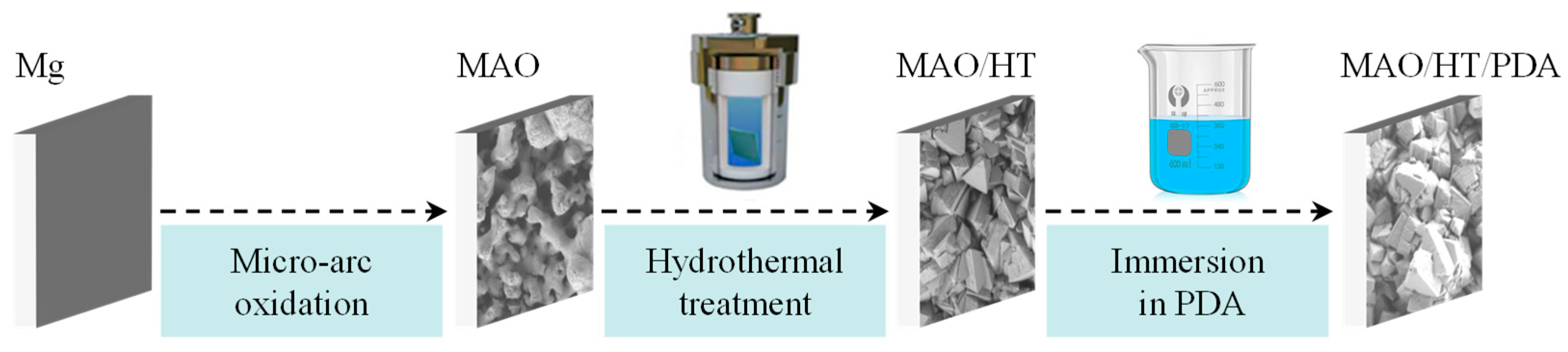
2.1. Sample Preparation

Pure (99.9%, Shenyang Jiabei Commercial Trading Co., Ltd., Shenyang, China) Mg disks with a size of φ 8 mm × 4 mm were sequentially polished with silicon carbide abrasive paper of 600, 1000, and 2000 grit. Afterwards, they were rinsed in ultrasonication baths containing ethanol. The polished disks were then sealed with resin (Wuxi Fitesi Electronic Technology Co., Ltd., Wuxi, China), leaving only one surface exposed for further processing. The MAO technique was employed using a pulsed power supply (MAO-1, Chengdu Siruike Electrical Appliances Co., Ltd., Chengdu, China) under a constant current mode with a duty cycle of 10%, pulse frequency of 1000 Hz, and electric current density of 0.1 A/cm2 for a duration of 3 min. The electrolyte used in the MAO process consisted of distilled water containing 8 g/L NaOH, 5 g/L Na3PO4, and 3 g/L KF. Following the MAO treatment, the samples were ultrasonically cleaned in deionized water and referred to as MAO samples. To fabricate a Mg(OH)2 layer on the MAO coating, a hydrothermal treatment (HT) was performed. The MAO samples were immersed in a Teflon-lined autoclave filled with distilled water and subjected to HT at 90 °C for 30 min. The resulting samples after HT were labeled as MAO/HT. In order to create a polydopamine (PDA) layer on the MAO/HT coating, the MAO/HT samples were immersed in a 1 mL solution of dopamine hydrochloride (pH 8.5, 2 mg/mL) overnight without agitation in darkness. After this process, the samples were thoroughly rinsed with distilled water. All reagents were of analytical grade and supplied by Tianjin Tianli Chemical Reagent Co., Ltd., Tianjin, China. The samples coated with PDA were denoted as MAO/HT/PDA. The preparation process of MAO/HT/PDA coating is presented in Figure 1.

2.2. Sample Characterization

The surface and cross morphologies of the coatings were observed by scanning electronic microscope (SEM, JEM-7001F, JEOL, Tokyo, Japan). Prior to observation, the specimens were gold-sputtered (30 s) to minimize surface charging. The surface pore size distribution and cross-sectional thickness of the MAO coating were quantified through ImageJ software (version number 1.53) based on SEM images. The elemental distributions were analyzed utilizing an energy-dispersive spectrometer (EDS) coupled with the SEM. The phase structure of the coatings was identified through an X-ray powder diffractometer (XRD, Bruker D8 Advance, Ettlingen, Germany, Cu target, incident wavelength λ = 0.154 nm) with a scanning rate of 4°/min over a diffraction angle (2θ) range of 10–90°.

2.3. Corrosion Resistance Evaluation

The corrosion resistance of the coated samples was evaluated by electrochemical measurement. The electrochemical impedance spectroscopy (EIS) and potentiodynamic polarization were measured using an electrochemical workstation (CHI600E, Chenhua, Shanghai, China) in simulated body fluid (SBF) at 37 °C. The electrochemical test system and detailed information of the measurement were referenced from our previous publication [22,23]. Prior to immersion tests, the samples were sealed by silastic exposing only one working side. The ratio of the SBF volume (mL) to the sample surface area (cm2) was fixed at 100 mL/cm2. The experiments above were repeated at least three times to ensure the accuracy and repeatability of the data. Meanwhile, immersion tests were performed to evaluate the long-term corrosion protection performance according to ASTM-G31-72 [37].

2.4. In Vitro Cytocompatibility

MC3T3-E1 pre-osteoblasts were acquired from the Institute of Biochemistry and Cell Biology of Chinese Academy of Sciences (Shanghai, China). The cells were cultivated in a complete culture medium (CCM), prepared by adding 10% fetal bovine serum (FBS), 1% penicillin, and 1% streptomycin into α-MEM at 37 °C with 5% CO2 atmosphere.
The in vitro cytocompatibility of the MAO, MAO/HT, and MAO/HT/PDA samples was assessed by directly cultivating the cells on the surface of the samples. The samples were sealed with Silastic exposing only one functional side, sterilized using 75% alcohol and positioned in a 24-well plate. The cells were then seeded on the sample surfaces at a density of 2 × 10⁴/mL and incubated for 1, 3, and 5 days. At the end of each time point, the CCM was removed and the samples were gently rinsed thrice with PBS. Subsequently, the cells were stained with 40 μL of a working solution from the esteemed Live/Dead viability/cytotoxicity kit (Invitrogen, Waltham, MA, USA) for a duration of 40 min, then meticulously observed and recorded under the lens of a fluorescence microscope (C2plus, Nikon, Tokyo, Japan). The MTT assay was employed to evaluate cell proliferation. The cell-adherent samples were further incubated for an additional 4 h at 37 °C in a mixture medium comprising 900 μL of CCM and 100 μL of MTT solution. Following this, 1 mL of dimethyl sulfoxide (DMSO) was added into each well, ensuring adequate dissolution of the formazan on the surface of the samples. The subsequent DMSO solution was then transferred to a 96-well plate, and the absorbance was meticulously quantified using a microplate reader (Infinite F50, TECAN, Männedorf, Switzerland). In order to ensure the reliability of the results, each test was conducted in triplicate.
Cell morphology was carefully observed using scanning electron microscopy (SEM) after a 2-day cultivation period, followed by fixation in a 4% paraformaldehyde solution and dehydration utilizing a gradient ethanol series (40, 60, 80, 100, 100 vol.%). Fluorescence staining of actin and cell nucleus was performed with a staining kit (Chemicon International, Temecula, CA, USA) as follows. After 48 h of culture, the cell seeded samples were fixed with 4% paraformaldehyde solution and then washed gently using PBS. Subsequently, the cells were stained with 50 μL FITC (5 μL/mL) for a duration of 40 min. After washing gently using PBS, the cell-attached samples were stained with 50 μL DAPI (1 μL/mL) for another 15 min. The fluorescence images of actin and cell nucleus were recorded under the lens of a fluorescence microscope (C2plus, Nikon).

3. Results and Discussion

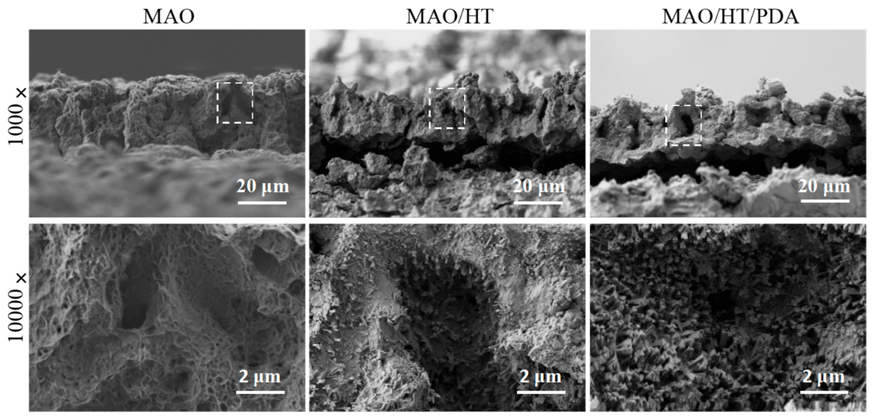
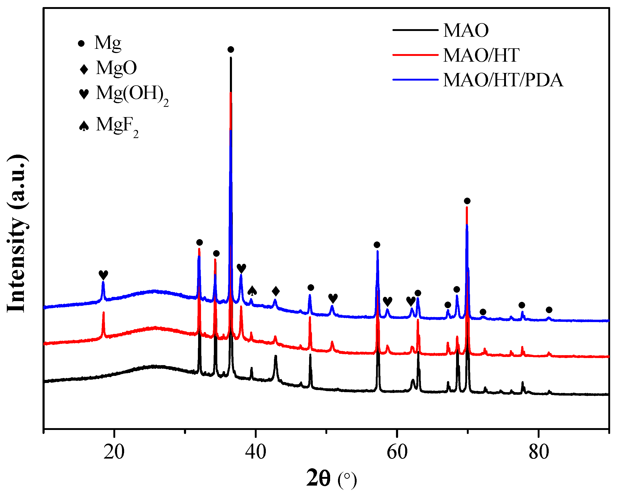
3.1. Coating Character

Figure 2A displays the surface morphologies of the MAO, MAO/HT, and MAO/HT/PDA coatings. The MAO coating demonstrates a distinctive porous microstructure characterized by uniformly distributed pores ranging in diameter from 0.5 to 2 μm. Upon HT, the surface pores become entirely encapsulated by randomly oriented bulk-like crystals, which present a size range of 1 to 2 μm. Subsequent modification with PDA does not generate any discernible changes in the morphology of the MAO/HT coating, except for the appearance of PDA nanoparticles onto the bulk-like crystals. EDS analysis (Figure 2B) elucidates that the MAO coating is composed of Mg, O, P, and F elements, whereas the MAO/HT coating solely comprises Mg and O elements, with an atomic ratio of approximately 1:2. The presence of C is observed on the MAO/HT/PDA coating, indicating the adhesion of PDA onto the MAO/HT coating following immersion in PDA.
Figure 3 portrays the cross-sectional morphology of the MAO, MAO/HT, and MAO/HT/PDA coatings. All the coatings have a thickness of approximately 32 μm. It can be observed that the MAO coating exhibits a discontinuous nature due to the formation of micro-pores during the MAO process. Following HT, needle-like crystals grow in the inner wall of the pores. The modification with PDA elicits negligible alterations in the cross-sectional morphology of the MAO/HT coating, suggesting that PDA adheres only to the surface of the MAO/HT coating.
Figure 4 shows the XRD patterns of the MAO, MAO/HT, and MAO/HT/PDA samples. In the pattern of the MAO sample, characteristic peaks corresponding to Mg (JCPDS no. 35-0821), MgO (JCPDS no. 30-0794), and MgF2 (JCPDS no. 38-0882) are observed, while the characteristic peaks of Mg(OH)2 emerge after HT, indicating the transformation of MgO into Mg(OH)2 during HT. No significant discrepancies are manifested between the XRD patterns of the MAO/HT and MAO/HT/PDA samples, which could be attributed to the relatively thin layer or amorphous crystallinity of the PDA prepared using this polymerization method [34,35].

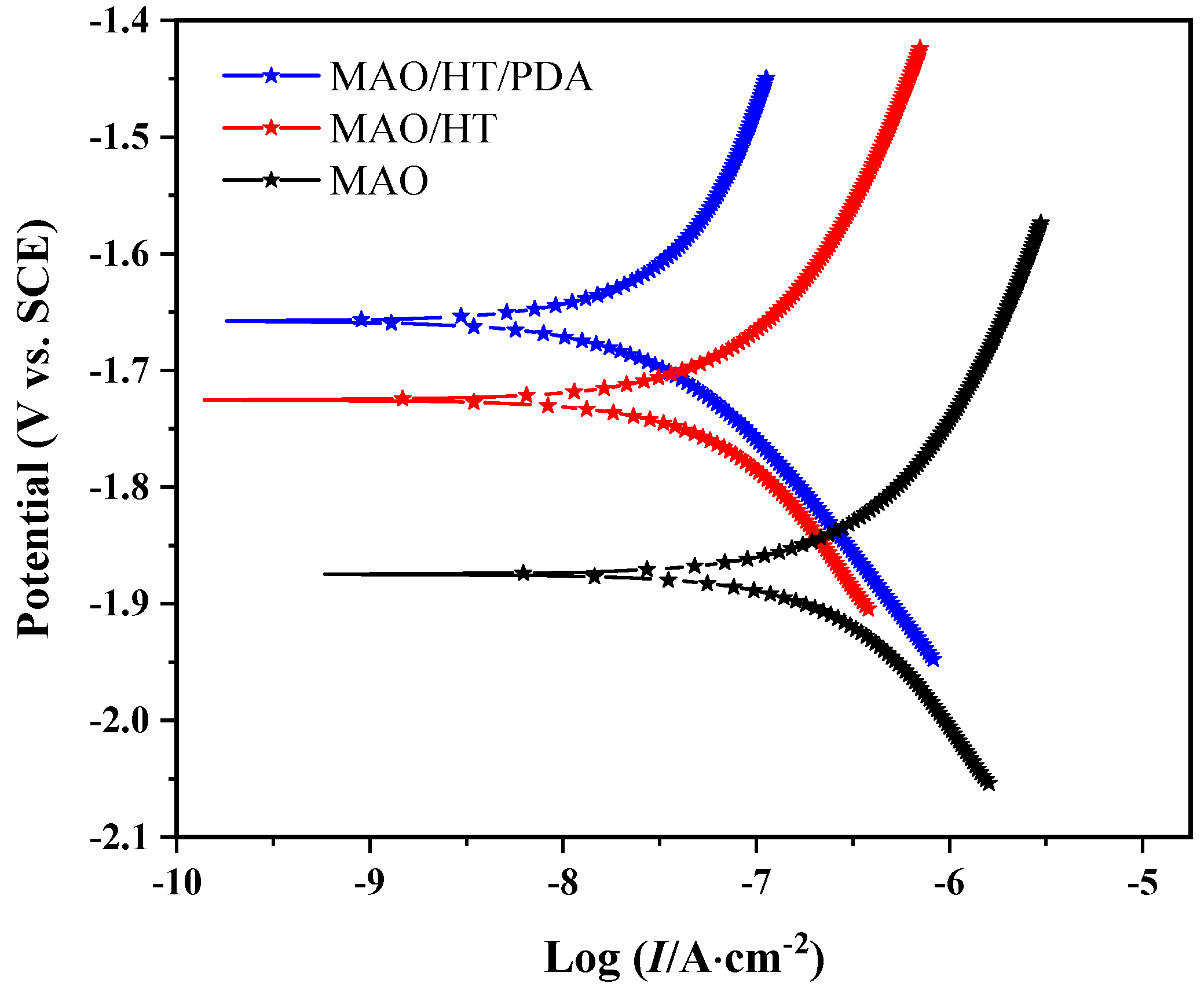
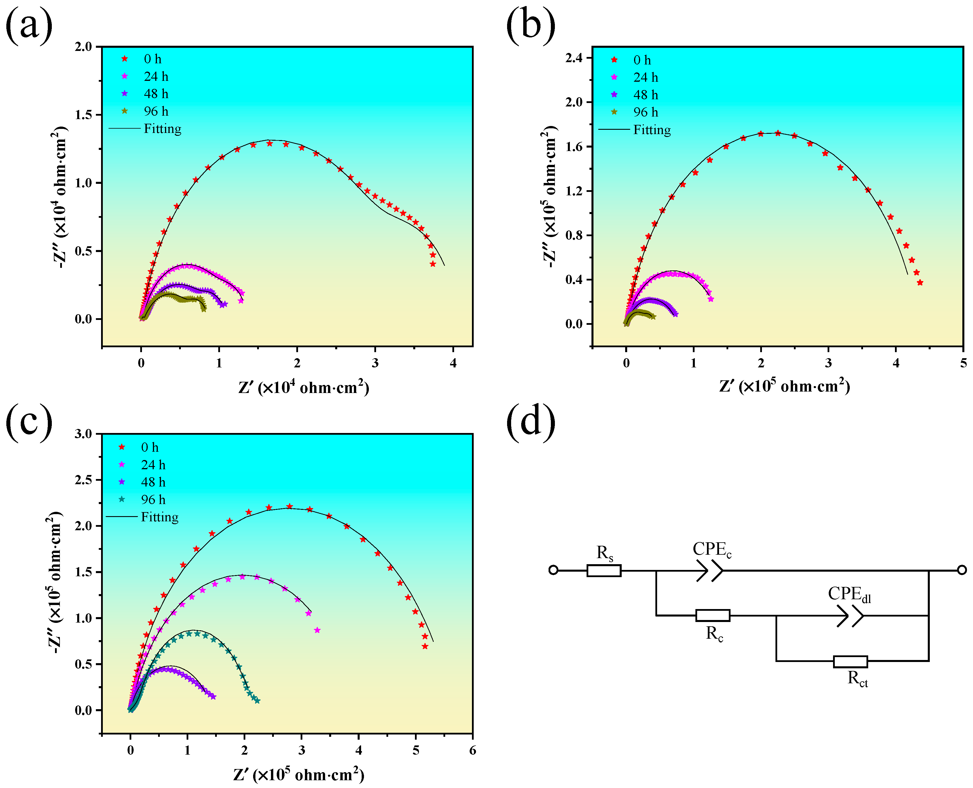
3.2. Corrosion Resistance

Figure 5 displays the polarization curves of all samples measured in SBF at 37 °C. Several critical electrochemical parameters derived from these curves, including corrosion potential (Ecorr) and corrosion current density (Icorr), are listed in Table 1. The Ecorr for the MAO/HT/PDA sample is more positive in comparison to the other two samples, with the MAO sample exhibiting the most negative value. Compared to the MAO sample (−1.87 V), the MAO/HT (−1.73 V) and MAO/HT/PDA (−1.65 V) samples demonstrate a positive shift of 140 mV and 220 mV, respectively. Conversely, the Icorr of MAO/HT/PDA sample (2.51 × 10⁻⁷ A·cm−2) is lower than that of the other two samples. This suggests that the MAO/HT/PDA sample exhibits significantly improved anti-corrosion performance. In fact, it has long been established that fabricating a Mg(OH)2 coating on Mg surfaces can significantly enhance the substrate’s corrosion resistance [29]. However, the PDA layer dooes not offer any protection against corrosion for Mg [38]. In this work, we found that Mg(OH)2/PDA composite coatings significantly improve the anti-corrosion performance of MAO-coated Mg, which can be attributed to the protective properties of the compact Mg(OH)2 layer adhered to the surface of the MAO coating, while the PDA layer acts as an additional physical barrier against external liquid media.

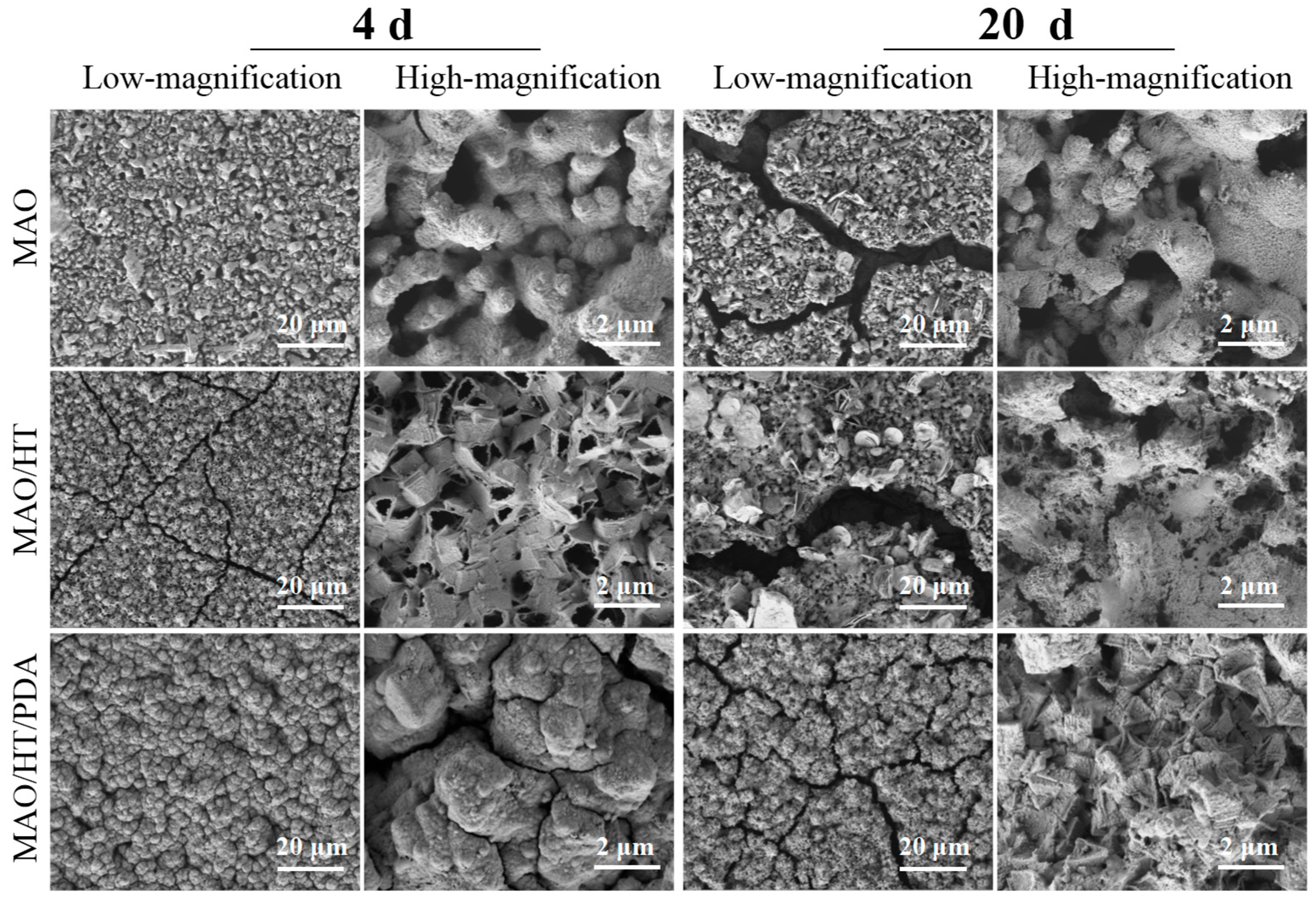
3.3. Degradation Behavior In Vitro

Figure 6 illustrates the surface morphology of the samples after immersion in SBF for 4 and 20 days. Minimal coating destruction is observed on the MAO sample after 4 days, whereas coarse cracks with a width exceeding 2 μm develop on the MAO coating after 20 days of immersion. Notably, numerous micro-cracks exclusively manifest along the MAO/HT coating after 4 days of immersion, accompanied by the detachment of bulk-like Mg(OH)2 from the porous MgO layer. Upon extending the immersion period to 20 days, wide cracks emerge, and the outer layer of Mg(OH)2 is completely removed, exposing the inner layer of porous MgO. The above results are in accordance with the previous study that Mg(OH)2 is prone to dissolve in a physiological environment and thus presents poor stability [30]. In contrast, the MAO/HT/PDA coating exhibits a reduced number of cracks after 4 days of immersion. Extending the soaking duration to 20 days leads to an augmentation in crack formation. It is worth noting that the crack width is considerably narrower compared to that of the MAO and MAO/HT coatings. Additionally, only a minimal amount of Mg(OH)2 bulks is detached from the coating, ensuring complete coverage of the inner layer of porous MgO, which is a sign that the PDA layer enhances the stability of MAO/HT coating in a physiological environment.
Figure 7 demonstrates the EIS plots of MAO, MAO/HT, and MAO/HT/PDA samples immersed in SBF solution for various durations. It is evident that all the samples exhibit two distinctive loops in their plots. The high-frequency capacitive loop corresponds to the penetration of the SBF into the coatings, while the low-frequency loop is associated with the charge transfer reaction at the interface of the coating and Mg [22,23]. Initially, all the tested coatings demonstrate the largest loop sizes, indicating the highest level of corrosion resistance. However, as the immersion time increases, the radii of the capacitive semicircles in the Nyquist plots gradually decrease, suggesting a reduction in corrosion resistance. To further elucidate the contribution of the coatings to corrosion resistance, the EIS plots are fitted using an equivalent circuit, as shown in Figure 7d. In this circuit, Rs is the resistance of the SBF solution and Qc and Rc represent the capacity and resistance of the coatings, respectively. Qc is a constant-phase element defined by capacitance CPEc and power index number n, which accounts for the surface inhomogeneities of the coatings. Cdl and Rt denote the electric double-layer capacity at the coating/substrate interface and the charge transfer resistance during electrochemical reaction, respectively [22,23].
The fitting results match well with the experimental measurements. As summarized in Table 2, both coating resistance (Rc) and charge transfer resistance (Rct) exhibit a consistent hierarchy across all testing intervals: MAO <MAO/HT < MAO/HT/PDA. This trend indicates that the MAO/HT/PDA composite coating provides superior corrosion protection for Mg substrates compared to the other two coatings. Both resistance parameters show gradual decreases with prolonged immersion time, albeit with distinct degradation patterns among different coatings. Notably, the MAO coating exhibits significant resistance deterioration, with its initial Rc value of 5.98 × 10³ Ω·cm2 decreasing by approximately 75% after 96 h of immersion. This dramatic reduction suggests substantial structural degradation of the MAO coating during prolonged electrolyte exposure. As illustrated in Figure 6, the MAO/HT coating demonstrates distinct degradation characteristics during prolonged immersion. While showing moderate Rc value reduction (12%) within the initial 24 h, it undergoes substantial deterioration with a 62% decrease after 96 h of exposure, primarily attributable to the dissolution of its outer Mg(OH)2 layer. Notably, despite this accelerated degradation in later stages, the MAO/HT system maintains Rc values two orders of magnitude higher than conventional MAO coatings throughout the 96 h test period, confirming its superior protective capability. The MAO/HT/PDA composite coating exhibits exceptional stability, demonstrating the most gradual Rc reduction rate (≤0.8%/h) among all tested systems. This performance enhancement stems from PDA’s effective barrier function that significantly retards electrolyte penetration towards the underlying MAO/HT structure.

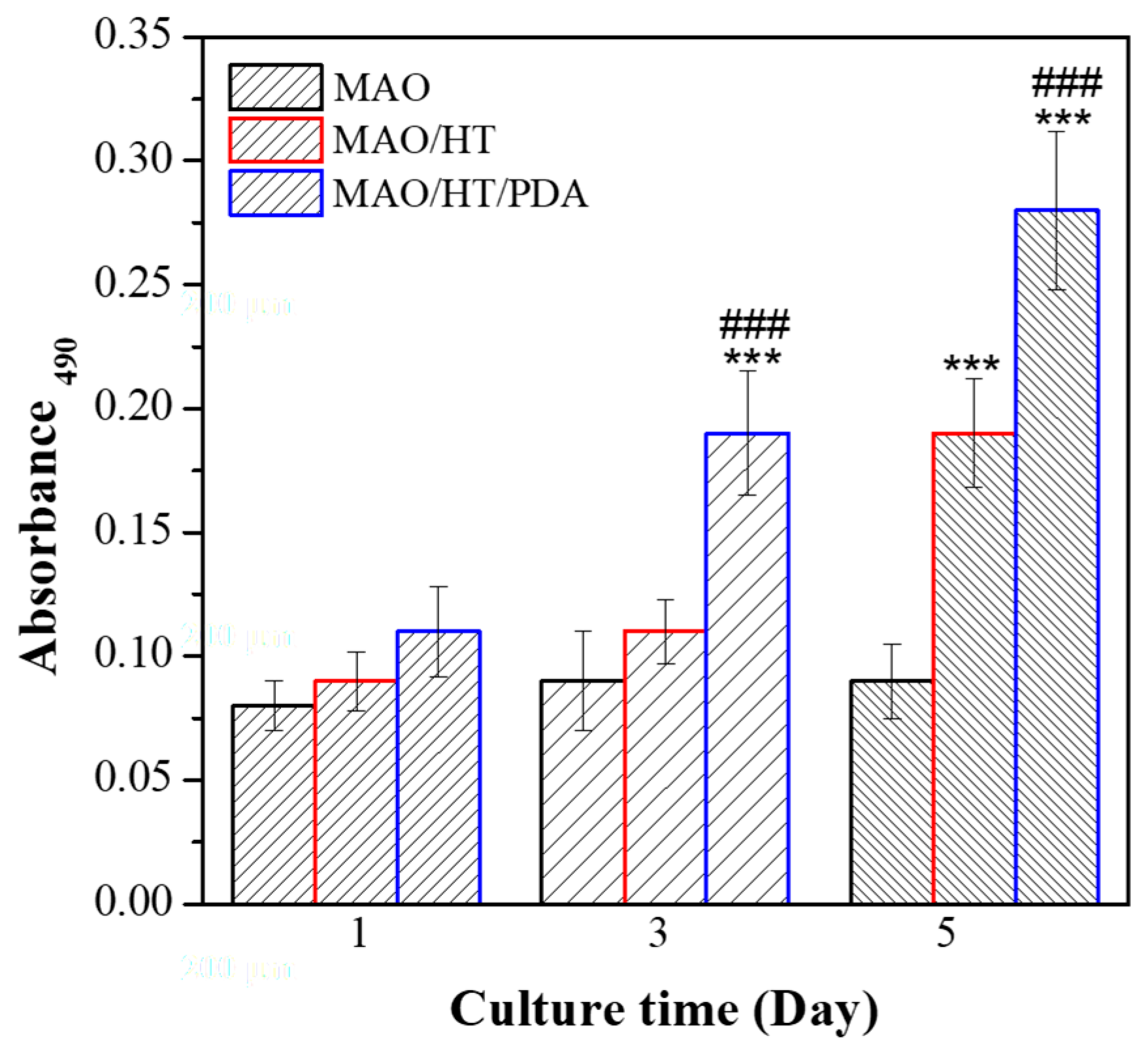
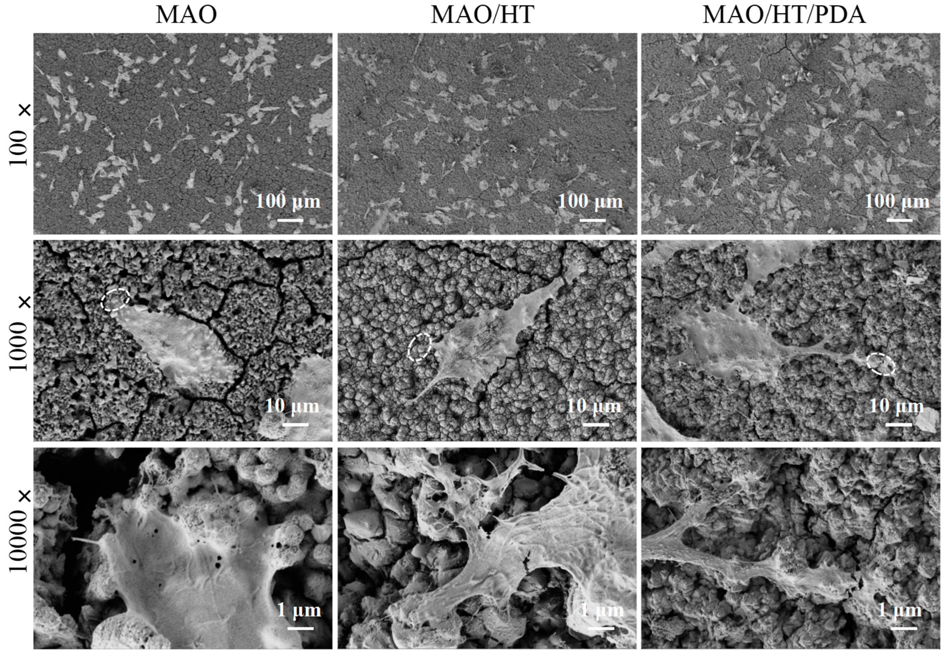
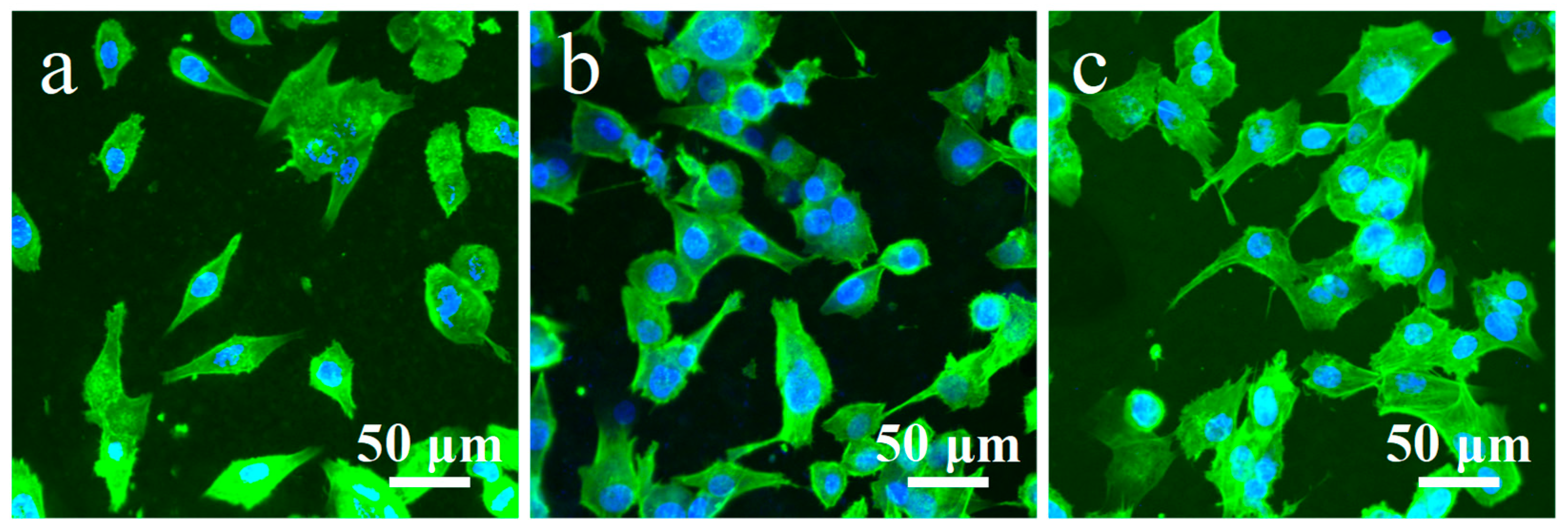
3.4. Cytocompatibility

The MTT assay is employed to determine cell viability and proliferation on MAO, MAO/HT, and MAO/HT/PDA samples. Figure 8 shows the resulting MTT results of MC3T3-E1 cells incubated on different samples for 1, 3, and 5 days. Generally, all the samples can support cell proliferation throughout the whole culture period. However, the obtained cell quantity derived from MTT results were in the following order: MAO < MAO/HT < MAO/HT/PDA at each measurement time point, confirming that MAO/HT/PDA coating exhibits better cytocompatibility than MAO and MAO/HT coatings.
Figure 9 shows the SEM morphologies of MC3T3-E1 cells after 2 days culture on all the sample surfaces. Low-magnification images show that cells on all the sample surfaces demonstrate the typical polygonal shape of osteoblasts, while spread more on the MAO/HT/PDA than that on the MAO and MAO/HT surfaces. Medium-magnification images indicate that cells on MAO coating spread pseudopodia, while filopodia extension are more obvious on the MAO and MAO/HT surfaces. Moreover, the micro-cracks on the MAO coating may be responsible for its poor spreading. High-magnification images show that cells attach tightly with all the tested coatings.
Figure 10 shows the actin (green)-nucleus (blue)-staining florescence images of MC3T3-E1 after 48 h of culture on the sample surfaces. More cells can be observed on the surface of MAO/HT and MAO/HT/PDA than that on the MAO surface. The cells attached on all the samples have well-organized actin cytoskeleton and present normal and health morphologies, especially on the MAO/HT/PDA sample. Most of the cells on the MAO/HT/PDA sample significantly spread with a larger area and have radially oriented actin bundles that fill the cytoplasm, indicating that the MC3T3-E1 are more favorable for spreading on the MAO/HT/PDA sample.
Many previous studies have suggested that a lower corrosion resistance lead to a higher pH value caused by the corrosion of Mg-based implants and finally reduces cell viability. Therefore, the MAO/HT/PDA sample with enhanced corrosion resistance is expected to have higher cytocompatibility than that of MAO and MAO/HT samples. This has been demonstrated by the current MTT results. In the electrochemical test, the Icorr value of the MAO/HT/PDA sample shifts towards noble direction than that of MAO and MAO/HT samples. In addition, at each of immersion periods (0–96 h), the Rct value of the MAO/HT/PDA sample is higher than that of MAO and MAO/HT samples. The results of electrochemical measurements agree well with the MTT results, demonstrating the improvement of degradation rate and cytocompatibility of Mg by the MAO/HT/PDA coating.
In the interaction between biomaterials and cells, cell adhesion and spreading are extremely important because the cells must have appropriate adhesion and spreading to the biomaterial surface before differentiation. MC3T3-E1 cultured on the surface of MAO/HT/PDA coating shows the largest spreading area among three groups, showing that the MAO/HT/PDA coating on Mg provides an interface that is more favorable for MC3T3-E1 to attach and spread than MAO and MAO/HT coatings, which may include three major mechanisms. One mechanism is linked to degradation of the samples. As stated above, the degradation of Mg-based biomaterials leads to a higher pH value surrounding and superfluous Mg2+. The previous studies showed that Mg2+ could promote cell adhesion and spread because it is a necessary metallic ion for the assembly of actin, the binding of integrin, and the regulation of cyclic adenosine monophosphate [39]. However, excess concentration of Mg2+ (more than 10 mM) undermined cell spread due to cytotoxicity [11]. The other mechanism is associated with the destruction of the coatings. As shown in Figure 7, an abundance of micro-cracks exclusively manifests along the MAO and MAO/HT coatings after 2 days of immersion in CCM. The dynamic destruction process occurring on the surface of the MAO and MAO/HT could be too aggressive for MC3T3-E1 to attach and spread. In contrast, the MAO/HT/PDA coating experiences less destruction, which can produce little adverse effect on the adhesion and spreading of destruction. The third mechanism is related to the direct positive regulation of PDA on cell spreading. It has been reported that PDA coating prepared on Ti greatly enhanced the expression of vinculin of MC3T3-E1 and facilitated cell spreading [40,41].

4. Conclusions

In this study, a dense bulk-like Mg(OH)2 layer was prepared on micro-arc-oxidized magnesium through hydrothermal treatment. Subsequently, a PDA layer was successfully fabricated on the Mg(OH)2 layer to fabricate a Mg(OH)2/PDA composite coating. Compared with the MAO coating, the corrosion density of the composite-coated magnesium reduced from 1.99 × 10⁻⁵ A·cm−2 to 2.51 × 10⁻⁷ A·cm−2, and the charge transfer resistance was improved by two orders of magnitude, demonstrating that the corrosion resistance of micro-arc-oxidized magnesium was greatly improved after its the application. The outer layer of PDA delayed composite coating cracking and peeling off during the immersion period and thus prolonged the protection period. Overall, the composite coating experienced less destruction than the Mg(OH)2 coating and exhibited better corrosion resistance. Owing to its excellent corrosion resistance, the Mg(OH)2/PDA composite coating was shown to greatly enhance the proliferation and spreading of MC3T3-E1. This study demonstrates that the Mg(OH)2/PDA composite coating is biodegradable and provides excellent protection to the micro-arc oxidized magnesium.

Author Contributions

Conceptualization, W.Y.; Methodology, C.L. and B.L.; Validation, C.L.; Investigation, C.L.; Data curation, B.L.; Writing—original draft, C.L. and W.Y.; Writing—review & editing, B.L.; Supervision, B.L. and W.Y.; Funding acquisition, B.L. and W.Y. All authors have read and agreed to the published version of the manuscript.

Funding

The authors acknowledge the financial support provided by the Fundamental Research Program of Shanxi Province (No. 202303021222325), the National Natural Science Foundation of Shanxi Province (No. 202103021224306), the Shanxi Province Science and Technology Innovation Young Talents Team for Lightweight Material Modification and Application (No. 202204051001005), and the Research Fund for Doctoral Program of Jinzhong University and Shanxi Province Technology Innovation Center of New Multi-functional Glass.

Institutional Review Board Statement

Not applicable.

Informed Consent Statement

Not applicable.

Data Availability Statement

Data are contained within the article.

Conflicts of Interest

The authors declare no conflict of interest.

References

  1. Wu, Z.; Hu, J.; Yu, L.; Wang, K.; Zhang, W.; Fan, H.; Deng, Z.; Wu, J.; Wang, K. Corrosion behavior investigation of gallium coating on magnesium alloy in simulated body fluid. J. Mater. Res. Technol. 2023, 27, 225–236. [Google Scholar] [CrossRef]
  2. Aghamohammadi, H.; Rabiee, S.M.; Hosseinipour, S.J.; Jamaati, R. Effect of microstructure and texture of AZ31 magnesium alloy substrate on nucleation and growth of biomimetic calcium phosphate coating. J. Mater. Res. Technol. 2023, 27, 5154–5164. [Google Scholar] [CrossRef]
  3. Iqbal, F.; Ali, A.; Naveed, M.; Ikram, F.; Fatima, H. Hydrothermal deposition of high strength biocompatible magnesium phosphate coating through in situ conversion of AZ91D-3Ca magnesium substrate. Surf. Coat. Tech. 2023, 457, 129301. [Google Scholar] [CrossRef]
  4. Li, X.; Hu, J.; Yu, Z.; Liu, M.; Xiao, X.; Qin, G.; Yang, L.; Zhang, E. A study on the in vitro and in vivo degradation behaviour and biocompatibility of a Mg-Mn-Zn alloy with PLLA and Micro arc oxidation composite coating. Surf. Coat. Tech. 2023, 471, 129894. [Google Scholar] [CrossRef]
  5. Nachtsheim, J.; Ma, S.Y.; Burja, J.; Batič, B.Š.; Markert, B. Tuning the long-term corrosion behaviour of biodegradable WE43 magnesium alloy by PEO coating. Surf. Coat. Tech. 2023, 474, 130115. [Google Scholar] [CrossRef]
  6. Peng, Y.F.; Wan, S.; Liao, B.K.; Guo, X. Preparation and properties of multi-function composite coating on AZ91D magnesium alloy with wave-assimilation and anti-corrosion performance. Surf. Coat. Tech. 2024, 478, 130464. [Google Scholar] [CrossRef]
  7. Wang, Y.; Wang, T.L.; Liu, S.B.; Wei, Z.; Zhang, T.C.; Yuan, S. Bilayer diatomite-based composite coatings with superhydrophobic and self-healing properties for enhanced anticorrosion of AZ31B magnesium alloys. Surf. Coat. Tech. 2024, 489, 131151. [Google Scholar] [CrossRef]
  8. Wang, X.; Liu, X.; Dai, Y.; She, J.; Zhang, D.; Qi, F.; Wei, W.; Ouyang, X. A novel Ca-Mg-P/PDA composite coating of Mg alloys to improve corrosion resistance for orthopedic implant materials. Surf. Coat. Tech. 2023, 471, 129920. [Google Scholar] [CrossRef]
  9. Witecka, A.; Valet, S.; Basista, M.; Boccaccini, A.R. Electrophoretically deposited high molecular weight chitosan/bioactive glass composite coatings on WE43 magnesium alloy. Surf. Coat. Tech. 2021, 418, 127232. [Google Scholar] [CrossRef]
  10. Zhao, Z.; Zong, L.S.; Liu, C.D.; Li, X.; Wang, C.; Liu, W.; Cheng, X.; Wang, J.; Jian, X. A novel Mg(OH)2/MgFx(OH)1−x composite coating on biodegradable magnesium alloy for coronary stent application. Corros. Sci. 2022, 208, 110627. [Google Scholar] [CrossRef]
  11. Zhang, Y.; Yan, K.; Xu, W.; Wang, C.; Qian, K.; Shao, Y.; Jiang, G.; Liu, H.; Ju, J.; Höche, D.; et al. Localized accelerated degradation of magnesium: A new insight into the mechanism of its biomedical degradation. Corros. Sci. 2024, 237, 112335. [Google Scholar] [CrossRef]
  12. Barzegari, M.; Mei, D.; Lamaka, S.; Geris, L. Computational modeling of degradation process of biodegradable magnesium biomaterials. Corros. Sci. 2021, 190, 109674. [Google Scholar] [CrossRef]
  13. Yang, C.; Cai, H.; Cui, S.H.; Huang, J.; Zhu, J.; Wu, Z.; Ma, Z.; Fu, R.K.; Sheng, L.; Tian, X.; et al. A zinc-doped coating prepared on the magnesium alloy by plasma electrolytic oxidation for corrosion protection. Surf. Coat. Tech. 2022, 433, 128148. [Google Scholar] [CrossRef]
  14. Chen, Y.N.; Wu, L.; Yao, W.H.; Wu, J.; Xie, Z.; Yuan, Y.; Jiang, B.; Pan, F. In situ growth of Mg-Zn-Al LDHs by ZIF-8 carrying Zn source and micro-arc oxidation integrated coating for corrosion and protection of magnesium alloys. Surf. Coat. Tech. 2022, 451, 129032. [Google Scholar] [CrossRef]
  15. He, H.; Li, K.; Luo, W.; Jiao, Z.; Ai, F.; Zhou, K.; Cao, C. Structure design, preparation and performance of a novel composite coating on medical magnesium-zinc alloy. Surf. Coat. Tech. 2022, 443, 128643. [Google Scholar] [CrossRef]
  16. Chen, Y.H.; Wu, L.; He, X.X.; Yao, W.; Wu, J.; He, Y.; Zhou, Y.; Yuan, Y.; Wang, J.; Xie, Z.; et al. Corrosion resistance study of ZIF-67/Co-Fe LDHs composite coating on the surface of AZ31 magnesium alloy micro-arc oxidation film. J. Alloys Compd. 2024, 989, 174278. [Google Scholar] [CrossRef]
  17. Liu, Y.X.; Duan, J.Q.; Zhang, J.L.; Guo, X.G. Active corrosion protection of micro-arc oxidation-based composite coating on magnesium alloy: Multiple roles of ionic liquid modified layered double hydroxide. Ceram. Int. 2024, 50, 26160–26170. [Google Scholar] [CrossRef]
  18. Tang, C.; Pan, J.; Wu, J.; Zhao, X.; Lei, J.; Li, L.; Pan, F. Surroundings-adaptive coating enabling robustness of magnesium alloys. Colloid Surf. A 2024, 696, 134385. [Google Scholar] [CrossRef]
  19. Yao, W.H.; Wu, L.; Wang, J.F.; Jiang, B.; Zhang, D.; Serdechnova, M.; Shulha, T.; Blawert, C.; Zheludkevich, M.L.; Pan, F. Micro-arc oxidation of magnesium alloys: A review. J. Mater. Sci. Technol. 2022, 118, 158–180. [Google Scholar] [CrossRef]
  20. Wang, J.; Dou, J.H.; Wang, Z.C.; Hu, C.; Liu, J.; Yu, H.; Chen, C. Corrosion resistance and biodegradability of micro-arc oxidation coatings with the variable sodium fluoride concentration on ZM21 magnesium alloys. J. Alloys Compd. 2023, 962, 171172. [Google Scholar] [CrossRef]
  21. Fischerauer, S.F.; Kraus, T.; Wu, X.; Tangl, S.; Sorantin, E.; Hänzi, A.C.; Löffler, J.F.; Uggowitzer, P.J.; Weinberg, A.M. In vivo degradation performance of micro-arc-oxidized magnesium implants: A micro-CT study in rats. Acta Biomater. 2013, 9, 5411–5420. [Google Scholar] [CrossRef]
  22. Li, C.L.; Yao, X.H.; Hang, R.Q.; Zhang, X.Y. Facile preparation of nanostructured octacalcium phosphate coatings on micro-arc oxidized magnesium with different functionalities for bone repair application. Colloid Surf. B 2021, 197, 111426. [Google Scholar] [CrossRef] [PubMed]
  23. Li, C.L.; Yao, X.H.; Hang, R.Q.; Huang, X.B.; Hang, R.Q. Corrosion behavior and cytocompatibility of nanostructured hydroxyapatite hydrothermally grown on porous MgO coatings with different P contents on magnesium. Mater. Lett. 2020, 264, 127136. [Google Scholar] [CrossRef]
  24. Liu, Y.P.; Cheng, X.; Wang, X.Y.; Sun, Q.; Wang, C.X.; Di, P.; Lin, Y. Micro-arc oxidation-assisted sol-gel preparation of calcium metaphosphate coatings on magnesium alloys for bone repair. Mat. Sci. Eng. C 2021, 131, 112491. [Google Scholar] [CrossRef]
  25. Wu, W.X.; Wang, W.P.; Lin, H.C. A study on corrosion behavior of micro-arc oxidation coatings doped with 2-aminobenzimidazole loaded halloysite nanotubes on AZ31 magnesium alloys. Surf. Coat. Tech. 2021, 416, 127116. [Google Scholar] [CrossRef]
  26. Wang, T.X.; Xu, Y.C.; Liu, Z.Q.; Li, G.Y.; Guo, Y.T.; Lian, J.S.; Zhang, Z.H.; Ren, L.Q. A chitosan/polylactic acid composite coating enhancing the corrosion resistance of the bio-degradable magnesium alloy. Prog. Org. Coat. 2023, 178, 107469. [Google Scholar] [CrossRef]
  27. Alabbasi, A.; Liyanaarachchi, S.; Bobby Kannan, M. Polylactic acid coating on a biodegradable magnesium alloy: An in vitro degradation study by electrochemical impedance spectroscopy. Thin Solid Films 2012, 520, 6841–6844. [Google Scholar] [CrossRef]
  28. Beyzavi, A.H.; Azadi, M.; Azadi, M.; Dezianian, S.; Talebsafa, V. Bio-polymer coatings fabricated on AM60 magnesium alloys by fused deposition modeling 3D-printing to investigate electrochemical behavior. Mat. Lett. 2023, 337, 133935. [Google Scholar] [CrossRef]
  29. Li, B.; Han, Y.; Qi, K. Formation mechanism, degradation behavior, and cytocompatibility of a nanorod-shaped HA and pore-sealed MgO bilayer coating on magnesium. ACS Appl. Mater. Interfaces 2014, 6, 18258–18274. [Google Scholar] [CrossRef]
  30. Li, C.Y.; Fan, X.L.; Zeng, R.C.; Cui, L.Y.; Li, S.Q.; Zhang, F.; He, Q.K.; Bobby Kannan, M.; Jiang, H.W.; Chen, D.C.; et al. Corrosion resistance of in-situ growth of nano-sized Mg(OH)2 on micro-arc oxidized magnesium alloy AZ31-Influence of EDTA. J. Mater. Sci. Technol. 2019, 35, 1088–1098. [Google Scholar] [CrossRef]
  31. Sareło, P.; Sobieszczańska, B.; Wysokińska, E.; Gąsior-Głogowska, M.; Kałas, W.; Podbielska, H.; Wawrzyńska, M.; Kopaczyńska, M. In vitro examinations of the anti-inflammatory interleukin functionalized polydopamine based biomaterial as a potential coating for cardiovascular stents. Biocybern. Biomed. Eng. 2023, 43, 369–385. [Google Scholar] [CrossRef]
  32. Peng, M.X.; Zhang, Q.C.; Zhang, M.N.; Yang, Z.W.; Wan, Y.Z.; Deng, X.Y.; Luo, H.L. Dealloying and polydopamine/silver coating on NiTi alloy for improved antibacterial activity. Mater. Chem. Phys. 2023, 305, 127939. [Google Scholar] [CrossRef]
  33. Liu, Y.H.; Jin, G.C.; Miao, H.; Xu, S.A. Preparation and characterization of polydopamine and n-butyl methacrylate copolymer coatings on titanium-nickel alloy stents. RSC Adv. 2024, 14, 15240–15248. [Google Scholar] [CrossRef]
  34. Zhou, Z.W.; Zheng, B.; Gu, Y.P.; Shen, C.; Wen, J.; Meng, Z.B.; Chen, S.P.; Ou, J.; Qin, A.M. New approach for improving anticorrosion and biocompatibility of magnesium alloys via polydopamine intermediate layer-induced hydroxyapatite coating. Surf. Interfaces 2020, 19, 100501. [Google Scholar] [CrossRef]
  35. Chung, J.J.; Chen, C.C.; Ding, S. Strontium-polydopamine hybrid coating to improve antibacterial ability and osseointegration of titanium implants. Mater. Chem. Phys. 2024, 312, 128629. [Google Scholar] [CrossRef]
  36. Wu, J.P.; Liu, Y.T.; Cao, Q.D.; Yu, T.; Zhang, J.; Liu, Q.Y.; Yang, X.Y. Growth factors enhanced angiogenesis and osteogenesis on polydopamine coated titanium surface for bone regeneration. Mater. Des. 2020, 196, 109162. [Google Scholar] [CrossRef]
  37. ASTM G31-72(2004); Standard Practice for Laboratory Immersion Corrosion Testing of Metals. ASTM International: West Conshohocken, PA, USA, 2004.
  38. Carangelo, A.; Acquesta, A.; Monetta, T. In-vitro corrosion of AZ31 magnesium alloys by using a polydopamine coating. Bioact. Mater. 2019, 4, 71–78. [Google Scholar] [CrossRef]
  39. Jiang, W.S.; Tian, Q.M.; Vuong, T.; Shashaty, M.; Gopez, C.; Sanders, T.; Liu, H.N. Comparison study on four biodegradable polymer coatings for controlling magnesium degradation and human endothelial cell adhesion and spreading. ACS Biomater.-Sci. Eng. 2017, 3, 936–950. [Google Scholar] [CrossRef]
  40. Guarise, C.; Maglio, M.; Sartori, M.; Galesso, D.; Barbera, C.; Pavan, M.; Martini, L.; Giavaresi, G.; Sambri, V.; Fini, M. Titanium implant coating based on dopamine-functionalized sulphated hyaluronic acid: In vivo assessment of biocompatibility and antibacterial efficacy. Mat. Sci. Eng. C 2021, 128, 112286. [Google Scholar] [CrossRef]
  41. Wang, M.Y.; Wang, C.X.; Zhang, Y.; Lin, Y. Controlled release of dopamine coatings on titanium bidirectionally regulate osteoclastic and osteogenic response behaviors. Mat. Sci. Eng. C 2021, 129, 112376. [Google Scholar] [CrossRef] [PubMed]
Figure 1. Schematic illustration for creation of MAO/HT/PDA coating on Mg.
Coatings 15 00729 g001
Figure 2. Surface morphologies (A) and EDS results (B) of MAO, MAO/HT, and MAO/HT/PDA coatings.
Coatings 15 00729 g002
Figure 3. Cross morphologies of MAO, MAO/HT, and MAO/HT/PDA coatings, 10,000× images are enlarged views of the dashed box in corresponding 1000× images.
Coatings 15 00729 g003
Figure 4. XRD patterns of the MAO, MAO/HT, and MAO/HT/PDA samples.
Coatings 15 00729 g004
Figure 5. Polarization curves of MAO, MAO/HT, and MAO/HT/PDA samples in SBF.
Coatings 15 00729 g005
Figure 6. Surface morphologies of MAO, MAO/HT, and MAO/HT/PDA coatings after immersion in SBF for 4 and 20 days.
Coatings 15 00729 g006
Figure 7. Nyquist plots of the MAO (a), MAO/HT (b), and MAO/HT/PDA (c) samples in SBF for different immersion times; (d) equivalent circuit for fitting impedance data.
Coatings 15 00729 g007
Figure 8. MTT results of MC3T3-E1 after culturing for different times on sample surfaces, *** p < 0.001 vs. MAO, ### p < 0.001 vs. MAO/HT.
Coatings 15 00729 g008
Figure 9. Morphologies of MC3T3-E1 after cultured on MAO, MAO/HT, and MAO/HT/PDA coatings for 2 days. The high-resolution images represent the amplification of the dotted area in the corresponding low resolution images.
Coatings 15 00729 g009
Figure 10. Fluorescence images of MC3T3-E1 cells for actin (green) and nucleus (blue) after 48 h of culture on MAO (a), MAO/HT, (b) and MAO/HT/PDA (c).
Coatings 15 00729 g010
Table 1. Corrosion potentials (Ecorr) and corrosion current densities (Icorr) obtained from the polarization curves of MAO, MAO/HT, and MAO/HT/PDA samples.
SamplesMAOMAO/HTMAO/HT/PDA
Ecorr (V vs. SCE)−1.87−1.73−1.65
Icorr (A·cm−2)1.99 × 10−51.28 × 10−62.51 × 10⁻⁷
Table 2. EIS-fitted Rc and Rct values of the equivalent circuits for MAO, MAO/HT, and MAO/HT/PDA samples.
TimeMAOMAO/HTMAO/HT/PDA
Rc (Ω·cm2)Rct (Ω·cm2)Rc (Ω·cm2)Rct (Ω·cm2)Rc (Ω·cm2)Rct (Ω·cm2)
05.98 × 1037.11 × 1037.25 × 1044.32 × 1048.56 × 1044.82 × 104
244.72 × 1032.27 × 1036.53 × 1043.75 × 1046.31 × 1044.04 × 104
483.25 × 1031.72 × 1033.75 × 1041.78 × 1045.38 × 1042.80 × 104
962.55 × 1031.53 × 1031.02 × 1049.85 × 1034.21 × 1042.01 × 104
Disclaimer/Publisher’s Note: The statements, opinions and data contained in all publications are solely those of the individual author(s) and contributor(s) and not of MDPI and/or the editor(s). MDPI and/or the editor(s) disclaim responsibility for any injury to people or property resulting from any ideas, methods, instructions or products referred to in the content.

