Article

Development of Iron-Modified Cotton Material: Surface Characterization, Biochemical Activity, and Cytotoxicity Assessment

1
Łukasiewicz Research Network—Lodz Institute of Technology, Marii Sklodowskiej-Curie 19/27, 90-570 Łódź, Poland
2
Circular Economy Center, Environmental Protection Engineering Research Group, Łukasiewicz Research Network—Lodz Institute of Technology, Brzezińska 5/15, 92-103 Łódź, Poland
3
Institute of Molecular Biology and Biotechnology, University of Lahore, 96RR+39P UOL, Defence Rd., Lahore 54600, Punjab, Pakistan
4
Department of Molecular Genetics, Faculty of Biology and Environmental Protection, University of Lodz, Pomorska 141/143, 90-236 Łódź, Poland
5
Department of General Biochemistry, Faculty of Biology and Environmental Protection, University of Lodz, Pomorska 141/143, 90-236 Łódź, Poland
*
Author to whom correspondence should be addressed.
Coatings 2025, 15(6), 663; https://doi.org/10.3390/coatings15060663
Submission received: 28 April 2025 / Revised: 26 May 2025 / Accepted: 28 May 2025 / Published: 30 May 2025

Abstract

Cotton, commonly used in wound care, has limitations such as quick saturation and wound adhesion, prompting surface modifications. In our studies, iron, which promotes platelet aggregation and coagulation, was deposited onto cotton via direct current (DC) magnetron sputtering. Thus, the biochemical properties of cotton fabrics were enhanced. Microscopic analyses revealed uniform iron coating on the fibers, and biochemical tests, such as activated partial thromboplastin time (aPTT) and prothrombin time (PT), showed that the modification did not affect the material’s coagulation activity. Measurements with the thiobarbituric acid (TBA) method (TBARS) showed that iron-modified cotton had antioxidant activity by lowering lipid peroxidation, which can be beneficial for better wound healing and lower infection risk. Moreover, our analysis showed the absence of cyto- and genotoxic properties against normal peripheral blood mononuclear cells (PBM cells). It was found that tested fabrics did not directly interact with DNA.

1. Introduction

Excessive bleeding as a result of traumatic wounds is one of the most common reasons for prehospital deaths worldwide [1,2]. It is essential to develop effective hemostatic measures to minimize the mortality associated with the massive loss of blood [2].
Cotton (COT) is still currently the most commonly applied topical wound dressing material used to stop bleeding [2,3,4]. The wide use of cotton for wound treatment is due to its safety, low cost, and stability [2,3,4]. Cotton exhibits breathability, high blood absorbency, and lack of allergenic response [2,3,4]. The hemostatic effect of cotton is associated with platelet adhesion in contact with the cotton fiber network and high liquid blood absorption, which helps clot formation [2]. The capability of cotton to absorb blood is caused by its hydrophilicity, porosity, as well as its capillary effect occurring between the weaved fibers [2].
The excessive absorption of blood by cotton dressing may cause undesirable blood loss [2], leading to quick saturation and the necessity of frequent changes of the dressing, thus generating high medical costs [2,4]. Moreover, cotton adheres to the wound, causing tissue damage [4]. Therefore, much effort is dedicated to developing more advanced hemostatic wound dressings [3,4]. This can be achieved by the modification of the cotton surface in order to enhance its hemostatic efficiency.
Among different solutions, the most commonly applied approaches include cotton modifications with biopolymers, such as chitosan or alginate, or with various clotting activators, such as kaolin [3,4,5,6,7,8]. Another method proposed by the researchers involves the surface immobilization of transition metals, which stimulate the activation of the intrinsic pathway of coagulation [9]. These elements include copper and zinc, which the authors previously studied [10,11,12,13].
Apart from the above-mentioned metals, iron is also believed to exhibit some hemostatic effects. This is due to the fact that iron ions promote platelet adhesion and aggregation [14]. According to the literature, Fe3+ can also be responsible for thrombosis [15]. Moreover, some studies have suggested that the Fe3+ oxidation property results in the formation of quinone groups, which enhance the antibacterial effect, which is crucial for preventing wound infection [16,17]. Caili et al. have demonstrated that the Fe3+ modified biomimetic PLLA cotton-like mat shows superior hemostatic performance due to the ability of Fe3+ to form fibrinolysis-resistant fibrinogen and stimulate the coagulation cascade [16]. Similarly, Long et al. have shown that iron oxide-kaolinite nanocomposites were characterized by the shortest bleeding time and the best wound-healing performance [18]. This was due to the synergistic effect of kaolin, which activates the intrinsic coagulation cascade, and α-Fe2O3, which induces the aggregation of red blood cells and clotting [18]. It has also been shown that iron oxide particles are capable of the activation of the contact system [19]. Moreover, the iron oxide nanoparticles may be applied in order to provoke rapid wound closure and repair [20].
Additionally, iron and its compounds are well-known for their UV-protective properties [21,22]. This is of the utmost importance since excessive UV radiation is harmful to human skin [21,22]. It may result in severe skin damage, skin ageing, skin cancer, as well as DNA damage and suppression of the immune system [21,22]. Therefore, UV shielding is desirable in the case of wound dressings in order to ensure protection of the wounded skin [21].
While cotton dressings are often used in clinical or indoor settings, they exhibit poor UV protection due to the inability of cellulose to absorb UV [23]. This is crucial since the ability to shield against UV radiation may be beneficial in outdoor environments or in the treatment of superficial wounds exposed to sunlight. UV exposure has been shown to impair the wound healing process by increasing oxidative stress, suppressing immune responses, and delaying re-epithelialization [24,25]. Therefore, UV-shielding properties may offer an added layer of protection in relevant clinical or field applications.
Nevertheless, excessive iron in human organisms may cause tissue damage by generating reactive oxygen species (ROS) through the Fenton reaction [26,27]. Therefore, this work focuses on the complex characterization of biochemical properties of cotton fabric modified with iron coating via direct current (DC) magnetron sputtering. The study involved the assessment of the anticoagulant effects as well as lipid peroxidation. The biological properties of COT and cotton-iron (COT-Fe) composites were investigated by analyzing the effects of post-incubation mixtures on PBM cells, used as a model for normal human cells. Cell viability and DNA integrity were assessed following exposure to these mixtures. Utilizing post-incubation mixtures was considered the most physiologically relevant approach to evaluate COT’s interaction with cells. This study provides evidence for the potential of COT-Fe composites as components in medical dressings, capable of lowering lipid peroxidation without influencing the coagulation cascade, cell viability, and DNA integrity.

2. Materials and Methods

2.1. Materials

Low-melting-point (LMP) and normal-melting-point (NMP) agarose, phosphate-buffered saline (PBS), 4′,6-diamidino-2-phenylindole (DAPI), resazurin sodium salt, and hydrogen peroxide (H2O2) were purchased from Sigma-Aldrich (St. Louis, MO, USA).

2.2. Methods

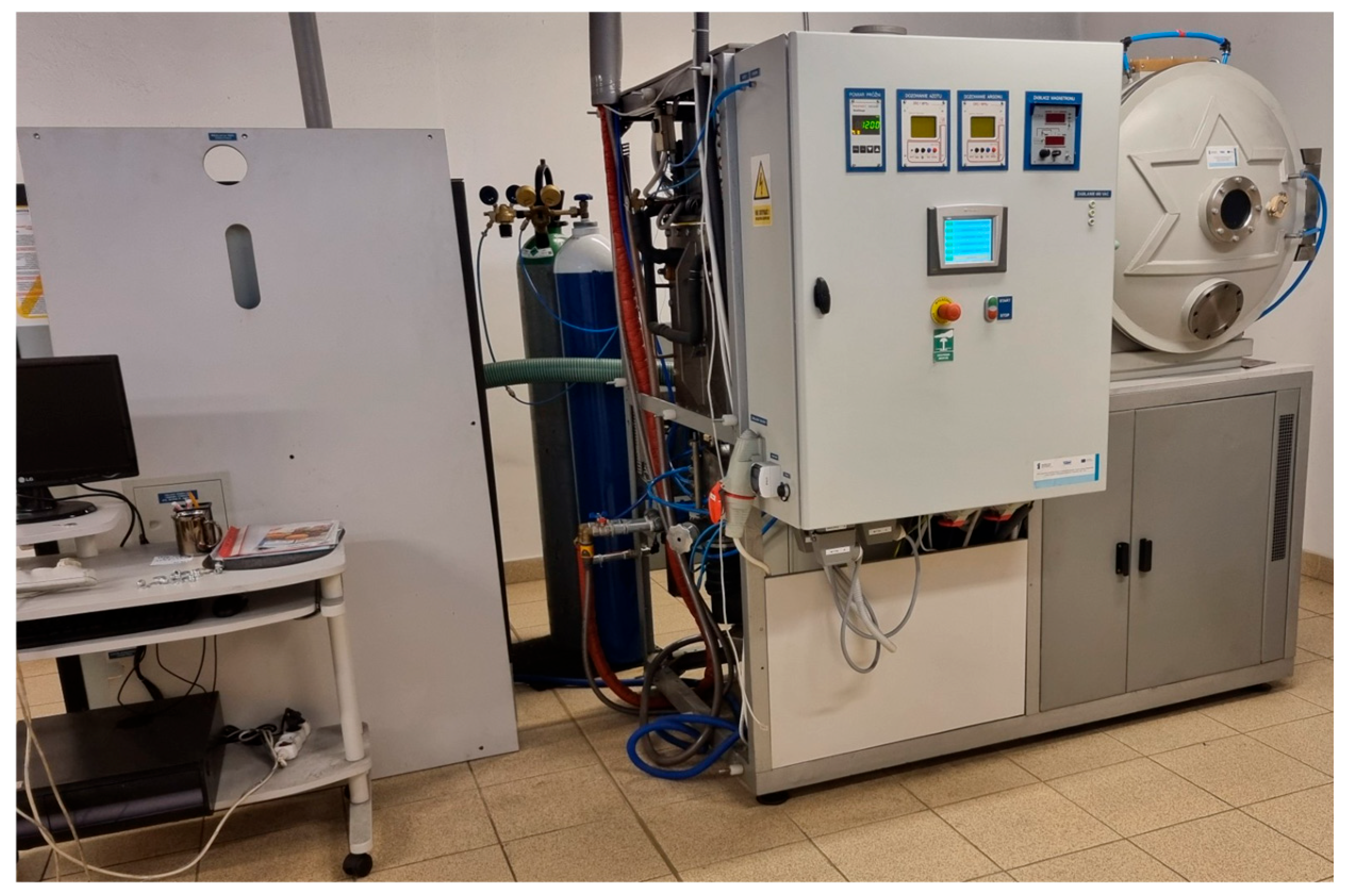
2.2.1. Modification of Material

In this study, medical cotton fabric with a plain weave and a qualitative composition: cotton (100% w/w), surface mass: 200 g/m2, warps/10 cm: 214, wefts/10 cm: 170 (Andropol S.A., Andrychów, Poland) was used. The fabric was functionalized using a direct current (DC) magnetron sputtering method in order to obtain a uniform coating on the surface. For that purpose, an iron target purchased from Testbourne Ltd. (Basingstoke, UK) with 99.99% purity and a DC magnetron sputtering system developed by P.P.H. Jolex S.C. (Czestochowa, Poland) were applied (Figure 1). The target-substrate distance was set to 15 cm, the working atmosphere was argon, and the ambient pressure was 2.4 × 10⁻³ mbar. To obtain different concentrations of iron, the sputtering deposition time was varied: 10 and 30 min. The power discharge was 0.5 kW and 1 kW, while the power density was 0.7 W/cm2.

2.2.2. Determination of Iron

The iron concentration was evaluated by atomic absorption spectrometry with flame excitation (FAAS) after the sample mineralization. In order to do that, a single-module Magnum II microwave mineralizer (Ertec, Wroclaw, Poland) was used. Then, the FAAS measurements were carried out using a Thermo Scientific Thermo Solar M6 (LabWrench, Midland, ON, Canada) spectrometer, which was equipped with coded lamps with a single-element hollow cathode, a 100 mm titanium burner, and D2 deuterium lamp for background correction. The total iron content (bulk iron content) was calculated using Equation (1):
M = C × V m
where:
  • M is total iron content (bulk iron content) (mg/kg);
  • C is the metal concentration in the tested solution (mg/L);
  • V is the volume of the sample solution (mL);
  • M is the mass of the mineralized sample (g).

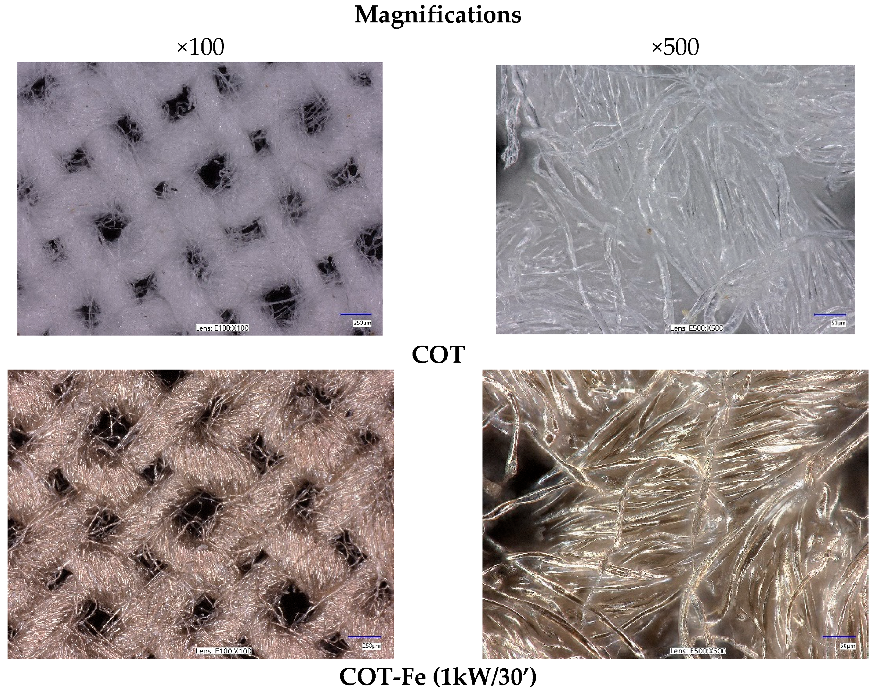
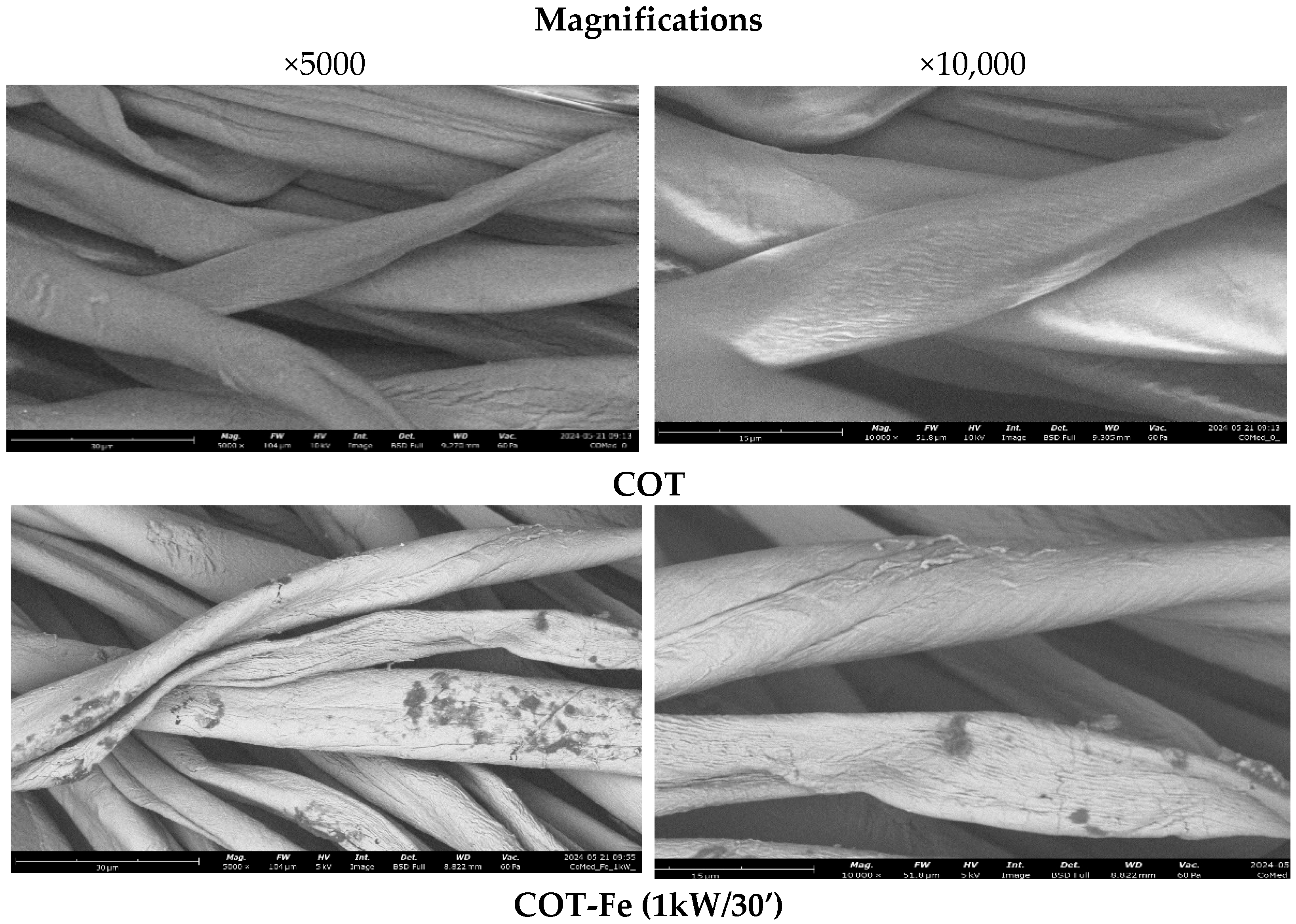
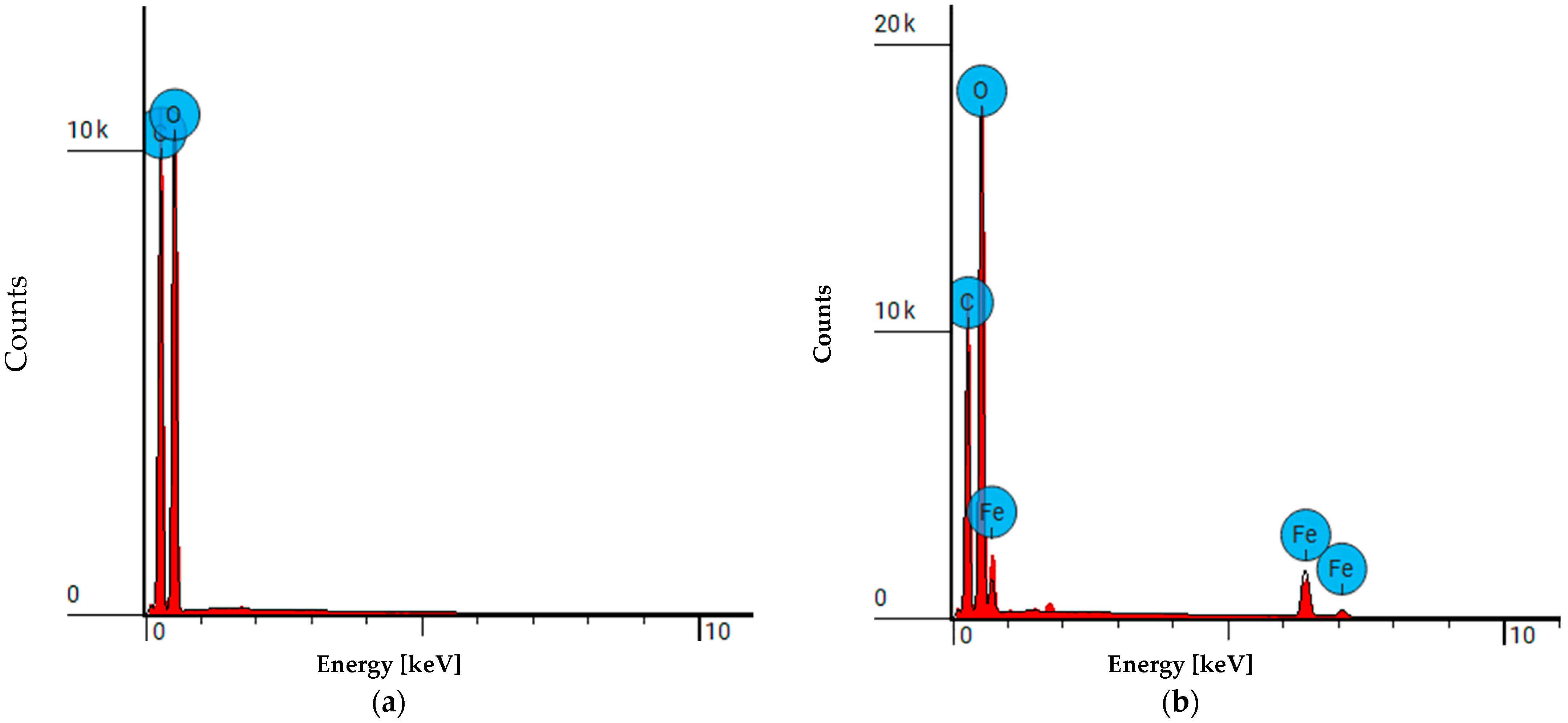
2.2.3. Microscopic Analysis

The surface morphology of the obtained samples was evaluated using a digital VHX-7000N microscope (Keyence, Osaka, Japan) at magnifications of 100× and 500×. Additionally, scanning electron microscopy (SEM) investigations were performed by means of the Phenom ProX G6 microscope (Thermo Fisher Scientific, Waltham, MA, USA). The observations were carried out using back-scattered electron imaging under a low vacuum (60 Pa) and an electron acceleration voltage of 15 keV. The applied magnification was 5000× and 10,000×.

2.2.4. Specific Surface Area

The Brunauer–Emmet–Teller method (BET) was employed in order to assess the total pore volume, and specific surface area of the cotton-Fe samples. For that purpose, the Autosorb-1 analyzer (Quantachrome Instruments, Boynton Beach, FL, USA) was used with liquid nitrogen as a sorption agent. Prior to the measurements, the samples were dehumidified for 24 h at 105 °C and degassed at room temperature afterwards. Approximately 2 g of each sample was used for each analysis.

2.2.5. UV-VIS Analysis and Determination of the Protective Properties Against UV Radiation

The alterations in physical properties, such as transmittance [%T], of the samples resulting from the applied modifications, were examined by means of a V-550 UV-VIS spectrophotometer (Jasco, Tokyo, Japan) equipped with an integrating sphere attachment. Measurements were conducted in the wavelength range of 200–800 nm. This same instrument was employed to evaluate the ultraviolet protection factor (UPF) of the samples, following the EN 13758-1:2002 standard [28]. The UPF was calculated using the equation:
U P F = 290 400 E ( λ ) ε ( λ ) d ( λ ) 290 400 E ( λ ) ε ( λ ) T ( λ ) d ( λ )
where:
  • E(λ) is solar irradiance;
  • ε(λ) is the erythema action spectrum (a measure of the harmfulness of UV radiation for human skin);
  • Δλ is the wavelength interval of the measurements;
  • T(λ) is the spectral transmittance at wavelength λ.
The results were measured in triplicate and presented as a mean value with ± deviation approximately 6%. The final UPF value for each sample was calculated as the arithmetic average of the individual UPF values, adjusted by the statistical error related to the number of measurements, with a 95% confidence interval.

2.2.6. Air Permeability and Fabric’s Structure

The air permeability of cotton (COT) and cotton coated with iron (COT-Fe) was assessed in pursuance of the EN ISO 9237:1995 standard [29]. For that purpose, the FX 3300 permeability tester from TEXTEST AG (Klimatest, Wrocław, Poland) was used. The applied air pressure was equal to 100 and 200 Pa, while the area of the tested sample was 10 cm2.
The analysis of the fabric’s structure included the assessment of the thickness, surface mass, as well as the number of warps and wefts per cm. The thickness was measured according to the EN ISO 5084:1996 standard [30] using the Universal thickness tester D-1000-C-0918 (Hans Schmidt & Co GmbH, Waldkraiburg, Germany). The applied pressure was equal to 1 kPa, while the feeler area was 20 cm2. The surface mass was evaluated in accordance with the EN 12127:1997 standard [31] by means of the Sartorius Laboratory Balance (Sartorius GmbH, Göttingen, Germany).
Before the assessment of the air permeability and fabric’s structure, all samples were acclimatized for 24 h under the following conditions: temperature 20 °C ± 2.0 °C, and relative air humidity 65% ± 4.0% according to the EN ISO 139:2005/A1:2011 standard [32].
Measurement uncertainty is expressed in accordance with the recommendations of the European Co-operation for Accreditation EA-4/16 document [33] as an expanded uncertainty “U” at 95% confidence level and coverage factor k = 2.

2.2.7. Biochemical Properties

The biochemical properties of the samples were measured using the activated partial thromboplastin time (aPTT) and prothrombin time (PT). To assess the blood coagulation parameters, the Coag4D coagulometer (Diagon Kft, Budapest, Hungary) was used. A square piece of each sample, approximately 1 mg, was incubated for 15 min at 37 °C in 200 µL of plasma. The plasma was obtained from the commercially available standard human blood lyophilized plasma, i.e., Dia-CONT I (Diagon Kft, Budapest, Hungary). To perform the aPTT measurements, the Dia-PTT reagent and 0.025 M CaCl2 solution (Diagon Kft, Budapest, Hungary) were used (according to the manufacturer’s instructions). The Dia-PT reagent (Diagon Kft, Budapest, Hungary) was used for the PT measurements. Our earlier works described the procedure in detail [10,11,12,13].

2.2.8. Thiobarbituric Acid Reactive Substances (TBARS)

Lipid peroxidation in blood plasma was investigated using the thiobarbituric acid (TBA) method (TBARS) in the presence of 0.1 M hydrogen peroxide to determine if metallic iron or its passivation could cause oxidative stress via the Fenton reaction or have an antioxidant effect. Lipid peroxidation, the oxidation of unsaturated fatty acids, produces peroxides and aldehydes like malondialdehyde (MDA), which reacts with TBA. The MDA–TBA complex is measured spectrophotometrically at 535 nm. Plasma (0.5 mL) was incubated with 2.5 mg of the material and 0.1 M H2O2 for 30 min, followed by a TBARS reaction with TCA and TBA. Samples were heated and centrifuged, and the absorbance was measured at 535 nm. TBARS concentration was calculated in µM using the molar absorption coefficient (ε = 156,000 M−1cm−1) with a three-fold dilution of the sample.

2.2.9. Influence on PBM Cells

Preparation of Fabrics for Assessment of Biological Properties
The impact of cotton (COT) and cotton fabrics with iron (COT-Fe) on cells was investigated by incubating cell cultures with post-incubation mixtures from COT and COT-Fe fabric fragments. The preparation of fabrics was performed in a similar manner as described previously [34]. These fragments (1 cm2) were incubated in RPMI medium for 24 h, and the resulting solutions were filtered with a 0.2 µm filter to ensure sterility before being added to cells in a 1:1 ratio. Cell viability and DNA damage were subsequently assessed.
Cell Culture
Peripheral blood mononuclear cells (PBM cells) were isolated from buffy coats of healthy, non-smoking donors (Blood Bank, Lodz, Poland) according to a previously published protocol [35]. Informed consent was obtained from all subjects involved in the study. Briefly, buffy coat blood was diluted 1:1 with PBS and layered onto a Lymphosep gradient (Cytogen, Zgierz, Poland). Following centrifugation (2200 RPM, 20 min, minimal acceleration/deceleration), PBM cells were collected and washed three times with 1% PBS before resuspension in RPMI 1640 medium. The University of Lodz Research Ethics Committee approved this study (12/KEBN-UŁ/I/2024-2025).
Cell Viability Resazurin Assay
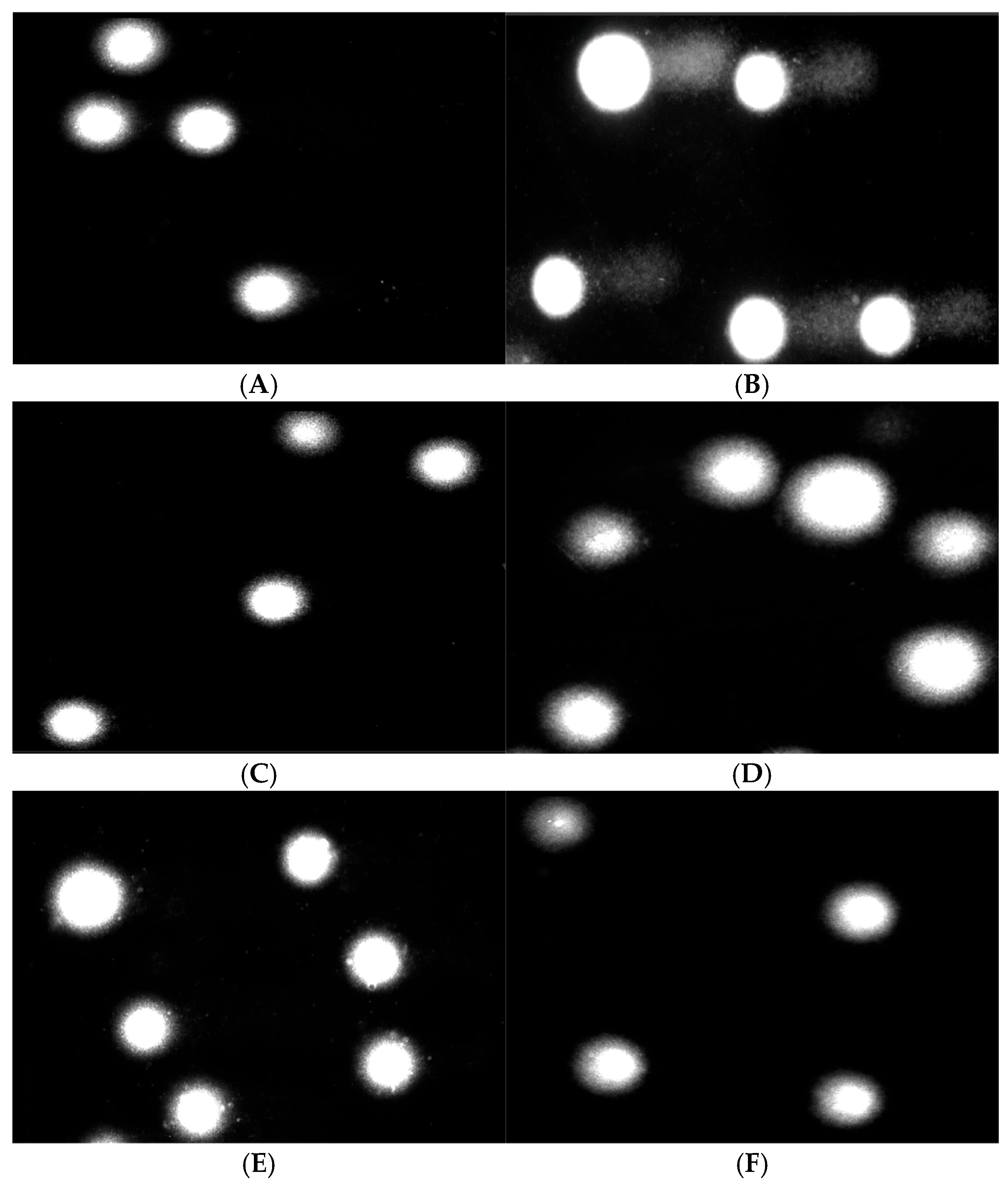
Cell viability was assessed using a resazurin assay, following the protocol of O’Brien et al. [36]. Briefly, resazurin solution (in sterile PBS) was added to 5 × 10⁴ PBM cells, incubated for 24 or 48 h (37 °C, 5% CO2) with post-incubation mixtures of COT and COT-Fe. Negative controls comprised RPMI 1640 medium. After incubation with resazurin for 2 h, fluorescence was measured (BioTek Synergy HT, Agilent, Santa Clara, CA, USA, λex = 530/25 nm, λem = 590/35 nm). Results were expressed as a percentage of control fluorescence.
DNA Damage
PBM cells (7.5 × 10⁴ cells/well) were incubated with post-incubation mixtures of COT and COT-Fe for 24 or 48 h at 37 °C in a 5% CO2 atmosphere. RPMI 1640 medium, prepared identically to the post-incubation mixtures, served as the negative control. A positive control was included, consisting of cells exposed to 25 µM hydrogen peroxide (H2O2) for 15 min on ice. Following treatment, PBM cells were washed and resuspended in RPMI medium.
DNA damage was assessed using the alkaline comet assay, following the protocol of Tokarz et al. [37]. Briefly, a suspension of treated PBM cells in 0.75% low-melting-point (LMP) agarose was layered onto microscope slides pre-coated with 0.5% normal-melting-point (NMP) agarose. Cells were lysed for 1 h at 4 °C in a buffer containing 2.5 M NaCl, 0.1 M EDTA, 10 mM Tris, and 1% Triton X-100 (pH 10). Slides were then subjected to DNA unwinding in a solution of 300 mM NaOH and 1 mM EDTA (pH > 13) for 20 min. Electrophoresis was performed in 30 mM NaOH and 1 mM EDTA (pH > 13) at 4 °C (buffer temperature ≤ 12 °C) for 20 min at 0.73 V/cm (28 mA). Following electrophoresis, slides were washed, stained with 2 µg/mL DAPI, and coverslipped. The entire procedure was conducted under minimized light to prevent additional DNA damage.
Comets were visualized at 200× magnification using a Nikon Eclipse fluorescence microscope equipped with a COHU 4910 video camera, a UV-1A filter block, and Lucia-Comet v. 6.0 image analysis software (Laboratory Imaging, Prague, Czech Republic). Fifty randomly selected comets per sample were analyzed, and DNA damage was quantified as the mean percentage of DNA in the comet tail.
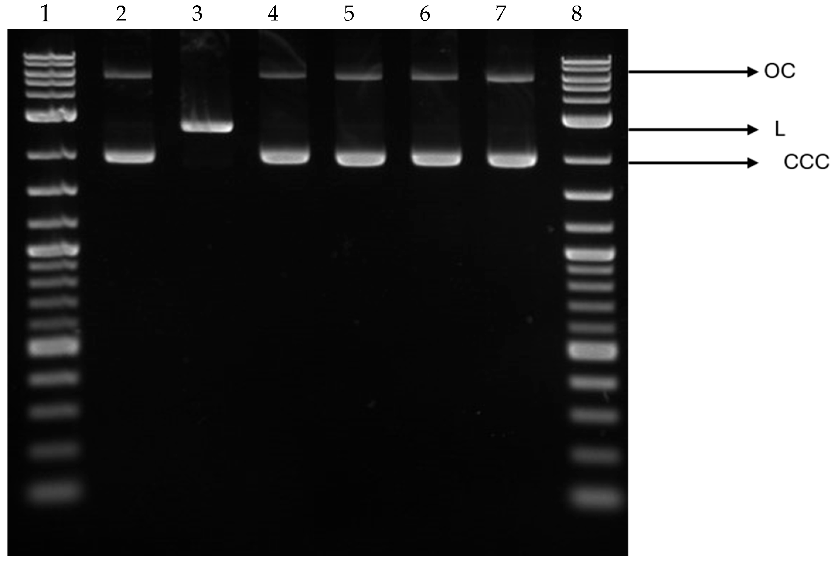
Plasmid Relaxation Assay
To investigate the effects of cotton and cotton-iron composite materials on plasmid DNA, 1 cm2 pieces of each material were placed in separate wells of a 6-well plate and incubated with 3 mL of ultrapure water for 24 h at 37 °C in a 5% CO2 environment. Following incubation, the resulting solutions were filtered through a 0.2 µm filter to ensure sterility. These filtered extracts were then combined with plasmid DNA at a 1:1 ratio to evaluate their impact on plasmid conformation.
A plasmid relaxation assay was carried out according to the method described by Juszczak et al. [38]. The pUC19 plasmid was isolated from DH5α E. coli cells using the AxyPrep Plasmid Miniprep Kit (Axygen, Corning, NY, USA), following the manufacturer’s instructions. The concentration and purity of the isolated plasmid were assessed by measuring the A260/A280 ratio and by agarose gel electrophoresis. Native pUC19 predominantly exists in a supercoiled (CCC) form, which exhibits high electrophoretic mobility. To generate a linear (L) form, the plasmid was digested with the restriction enzyme PstI (New England Biolabs, Frankfurt, Germany). The differing topological structures of the CCC and L forms result in their distinct electrophoretic mobilities. Plasmid DNA (50 ng/µL) was incubated with the COT and COT-Fe composite post-incubation mixtures for 24 h. Subsequently, the samples were subjected to 2% agarose gel electrophoresis, stained with ethidium bromide, and visualized under UV light (302 nm). Images were captured using a CCD camera and analyzed with GeneTools v. 4.3.9.0. software (Syngene, Cambridge, UK). A 1 kb DNA ladder (GeneRuler 1 kb DNA Ladder, Thermo Scientific, Waltham, MA, USA) was included in the electrophoresis to determine DNA fragment sizes.

3. Results and Discussion

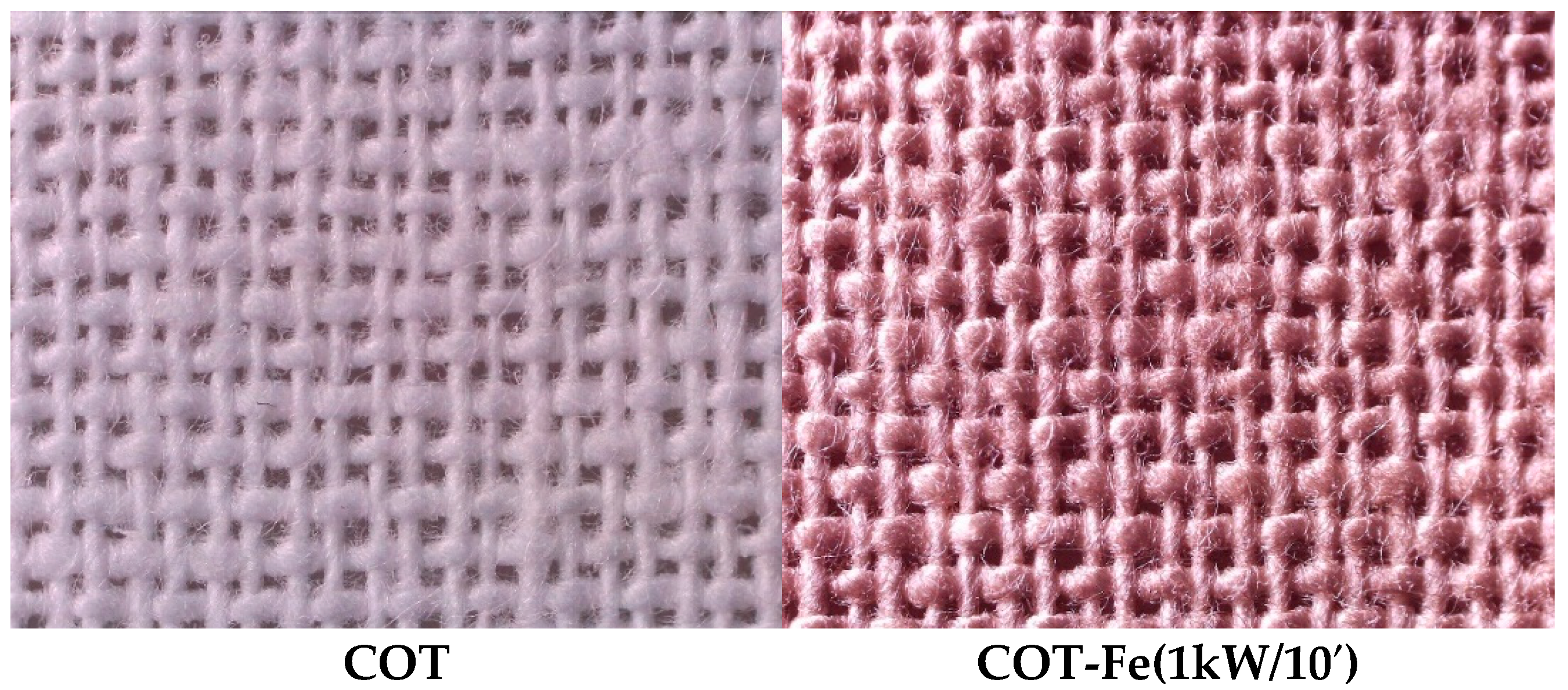
3.1. Microscopic Analysis

Figure 2 shows the morphology of the sample surface before and after the modification, as observed using optical microscopy at various magnifications. The images clearly show the successful application of the iron coating, as evidenced by the noticeable color change. The metal layer is visible on the fabric’s outer and inner fibers, extending deep into the material. Further analysis of the iron-coated cotton fabric, shown in Figure 3, highlights significant changes in the surface structure.
SEM analysis of the cotton fibers revealed typical structural features, such as elongated, narrow, and parallel fibers with smooth surfaces and uniform architecture. Understanding the microstructure of cotton fibers is vital for evaluating how the modification process affects their physical and chemical properties. SEM analysis of COT-Fe materials produced through magnetron sputtering confirmed the uniform metal deposition on the fiber surface. SEM can also detect structural changes in the fibers, such as damage or misalignment. The structural modifications observed in the fibers after metal coating indicate that the process has altered their microstructure. SEM examination of the COT-Fe samples also enabled a qualitative assessment of chemical composition using energy-dispersive X-ray spectroscopy (EDS), which allowed the identification of the chemical elements present in the samples (Figure 4).

3.2. FAAS Analysis

The obtained results were accurately recorded and summarized in Table 1, enabling a detailed analysis of iron content in the tested samples and facilitating conclusions regarding the effectiveness of the sputtering process on cotton materials.
It can be observed that with the increasing deposition time and sputtering power, the Fe concentration also increases. However, it is worth noting that the highest difference in the Fe content is due to the change in the deposition time. Only a slight difference in the Fe content is observed for the samples with the same deposition time but different sputtering power. It can be concluded that the higher deposition time results in a thicker iron coating on the surface of the cotton material and, thus, higher Fe content.
Although the literature indicates that pore volume may affect blood-material interactions and clot formation [39,40], in our study, no direct relationship between the reduced pore volume and changes in coagulation activity was observed. The aPTT and PT assays (Section 3.6) showed no significant variation between samples despite notable differences in pore structure. This suggests that, under the tested conditions, iron modification influenced surface properties without impairing or enhancing hemostatic functionality.

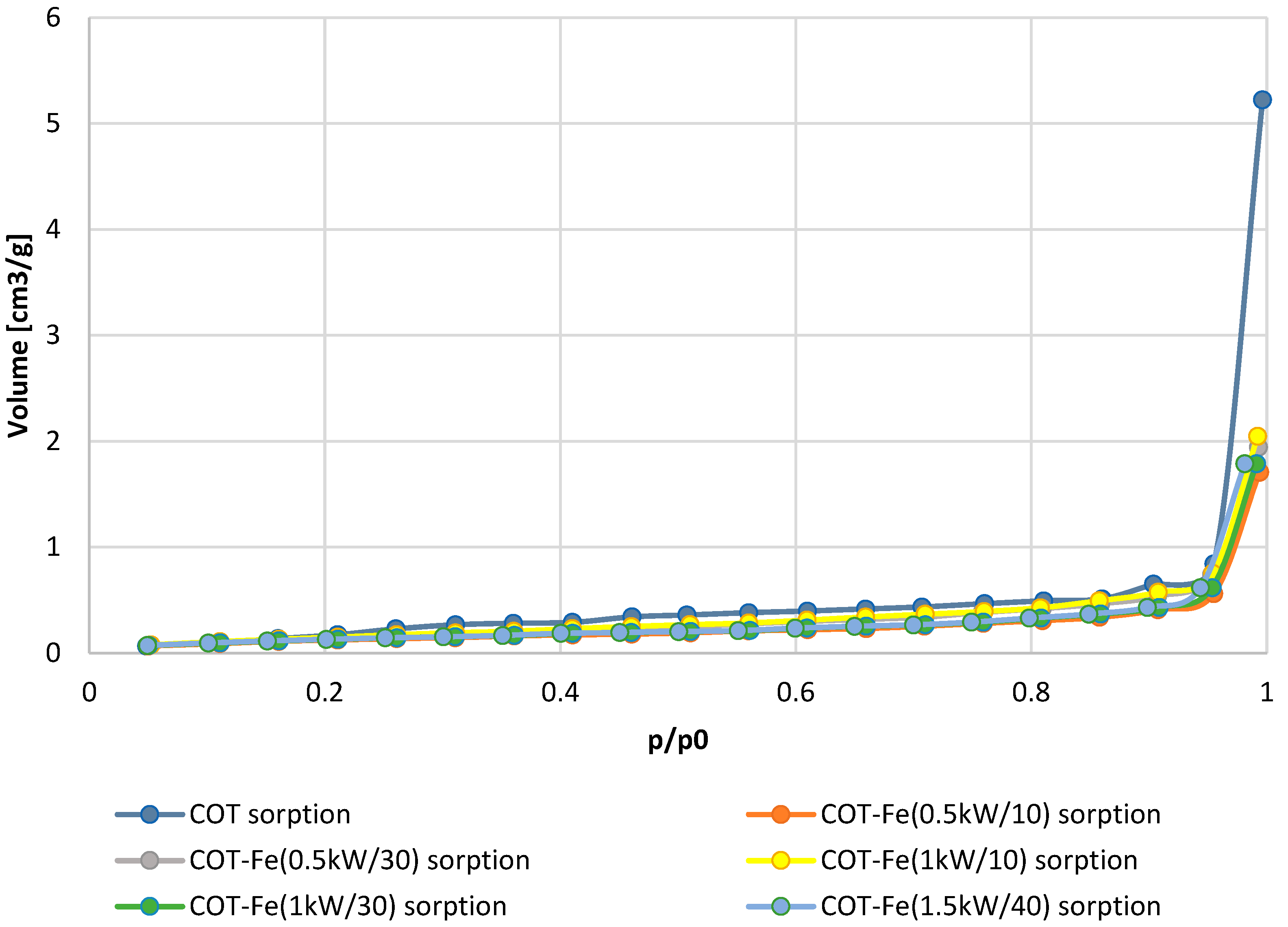
3.3. Specific Surface Area

The obtained N2 adsorption isotherms for the investigated samples are presented in Figure 5, while the calculated specific surface area and total pore volume are given in Table 1. The results have shown that the coating of the cotton material with iron using the magnetron sputtering process causes a significant change in the analyzed parameters. First of all, a notable decrease (over 50%) in the total pore volume is observed. The higher the deposition time (i.e., the higher Fe content), the lower the total pore volume. At the same time, there is only a slight difference in the total pore volume depending on the sputtering power (for the samples with the same deposition time). The lowering of the total pore volume may be due to the fact that the iron coating is filling the pores of the cotton material. The higher the deposition time, the thicker the iron coating, and thus, more pores are filled with iron, causing the lower total pore volume. A similar effect was observed in our previous works [10,11,12,13]. Such a trend is also observed in the specific surface area. The deposition of the iron coating causes a decrease in the specific surface area. The higher the Fe concentration (i.e., the higher deposition time and sputtering power), the lower the specific surface area. This is caused by the deposition of iron on the surface of the cotton, which results in a smoother surface due to the filling of the pores.
Figure 5 shows that the quantity of the adsorbate increases exponentially with the rising pressure for all of the samples. For lower pressures, there is a slow, gradual increase in the amount of the adsorbed N2, followed by an exponential increase for higher pressures, and then reaching saturation level at p/p0 = 1. The resulting absorption isotherms resemble a hyperbolic curve without a distinct “knee”, which allows for their classification as type III in accordance with IUPAC guidelines [41]. Type III isotherms are characteristic of nonporous and/or macroporous materials and are typically observed when interactions between the adsorbent and adsorbate are relatively weak [41]. The absence of a distinct “knee” (that is associated with the formation of a well-defined monolayer) suggests that the sorption process is entirely governed by a multilayer mechanism [41].

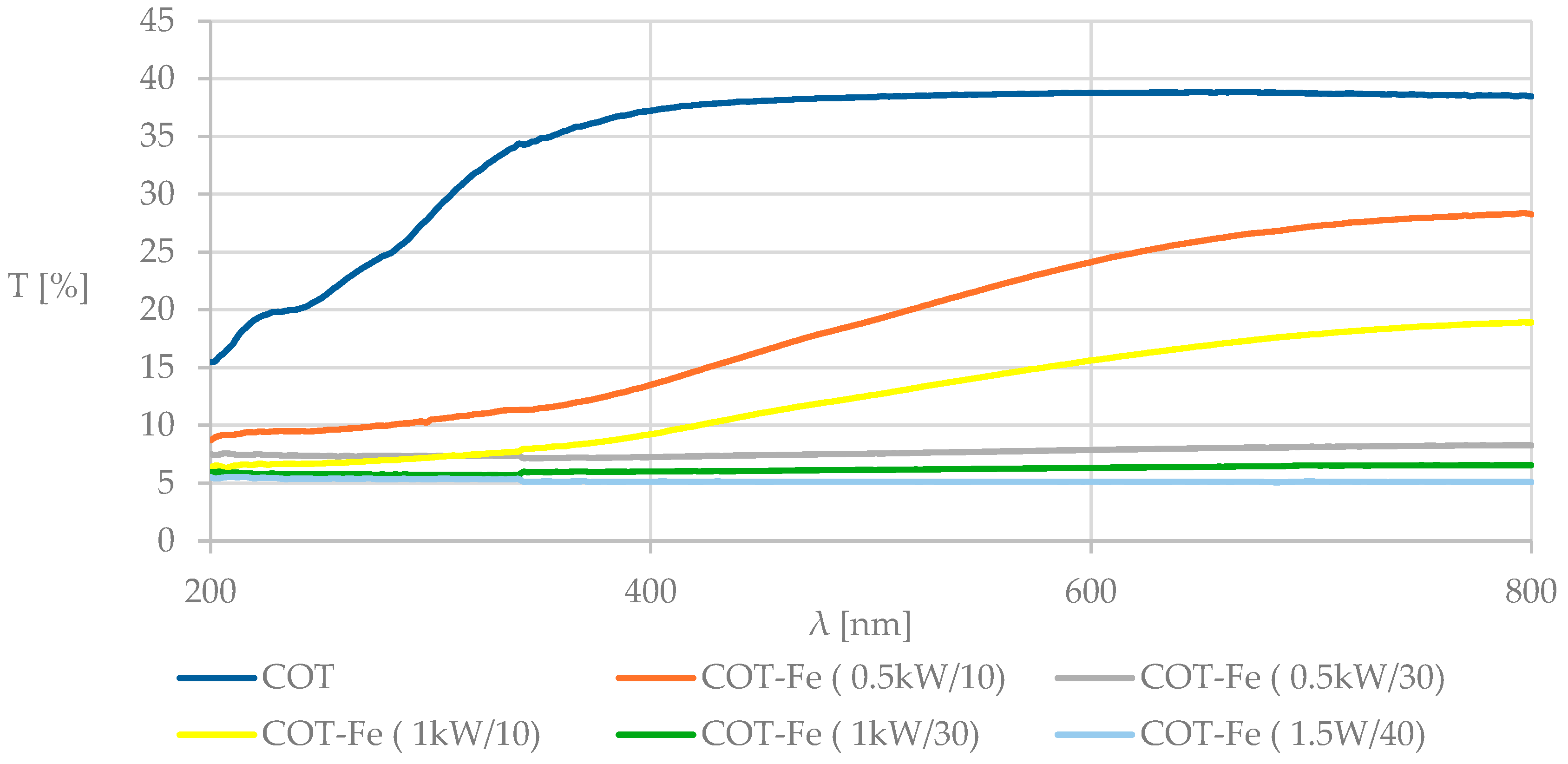
3.4. UV-VIS Analysis and Determination of the Protective Properties Against UV Radiation

Figure 6 displays the spectrophotometric transmittance spectra obtained for COT, COT-Fe (0.5kW/10′), COT-Fe (0.5kW/30′), COT-Fe (1kW/10′), and COT-Fe (1kW/30′) samples in the wavelength range of λ = 200–800 nm.
The transmittance spectra of the modified COT samples show significant changes in their macrostructure when compared to the unmodified COT, as evidenced by a decrease in transmittance across the entire measurement range. While both the unmodified and modified samples displayed similar spectral features throughout the range, the reduced spectral transmission can be attributed to the additional iron layer on the surface of the modified samples. Importantly, the Fe content in the modified samples plays a significant role in influencing the transmission levels, particularly in the 250–450 nm range, where a noticeable decrease in transmission occurs. Table 2 presents a comparison of the average value of transmittance (%T) and the determined UPF values for the samples after the modification in relation to unmodified samples.
The modification improved the protective properties against UV radiation, as indicated by the enhanced barrier performance (UPF: 3.29 (COT) → 18.22 (COT-Fe (1.5kW/40′))). This confirms that the surface modification provides effective UV protection, in compliance with the EN 13758-1:2002 standard. Although controlled, “low-dose” UVB irradiation may support wound healing by promoting vitamin D synthesis and exerting antimicrobial effects, excessive and uncontrolled exposure, especially in the UVA and UVB range, can hinder tissue regeneration by inducing oxidative stress, inflammation, and DNA damage [21,22]. Since wounds are typically covered, providing a UV barrier is desirable to minimize these risks. The significant improvement in UPF confirms that the iron coating offers additional protection for wounded skin in exposed conditions.

3.5. Air Permeability and Fabric’s Structure

Table 3 shows the results of the determination of the air permeability of the investigated samples, as well as the thickness and surface mass. Figure 7 displays the structure of the unmodified cotton fabric (COT) and the structure of the exemplary sample after the modification with an iron coating (COT-Fe (1kW/10′)).
As may be observed in Figure 7, the analyzed cotton samples are plain weave fabrics. The analysis of the structure of the fabric prior to and after the modification using the magnetron sputtering of iron shows that there are no significant changes observed. For each of the measured parameters, the differences between the average value for the unmodified sample and the subsequently modified samples ranged from 1 to 6%, which indicates that the applied modification does not affect the fabric structure.
Similarly, there is no substantial difference in the air permeability of the tested samples. This may be explained by the fact that while iron coating affects the structure of the individual cotton fibers, it does not alter the structure of the fabric itself. As a result, the plain weave structure is preserved, which ensures good air permeability.

3.6. Biochemical Properties

This study evaluated the effect of iron coating on cotton fabric, applied through DC magnetron sputtering, on coagulation activity and biochemical properties related to clotting. The analysis focused on aPTT and PT, which are key assays for assessing coagulation (Figure 8). Both the aPTT and PT are key assays used to determine the functionality of the coagulation system, particularly in evaluating the intrinsic and extrinsic pathways, respectively.
The data shows no significant changes in the coagulation times for the modified cotton samples compared to the control (unmodified cotton), indicating that the surface modification via iron coating does not affect the clotting ability of the cotton material. The results show that the iron coating has no influence on the physiological process of blood plasma coagulation. Our findings are important because they support the premise that the modification does not disrupt the balance of coagulation.
Although the aPTT and PT assays revealed no significant systemic changes in coagulation time, it is important to note that blood coagulation at the wound site is not solely dependent on the cascade activation. Surface properties of wound dressings—such as topography, chemical composition, and charge—can influence platelet adhesion, activation, and aggregation. The literature suggests that iron ions (especially Fe3+) may promote local platelet activation and thrombus formation via interaction with platelet surface proteins and oxidative processes [14,15]. Therefore, the improved hemostatic potential of COT-Fe may arise from localized effects at the material-blood interface rather than systemic modulation of coagulation factors.

3.7. TBARS

Table 4 shows the obtained TBARS results. The TBARS concentration in the presence of iron is lower than in the samples without iron at the respected concentration.
In FeO-coated cotton, H2O2 may decompose more quickly into water and oxygen without leading to the generation of OH radical and, thus, further significant lipid peroxidation. At higher H2O2 concentrations, this decomposition may occur even more rapidly. The iron on the cotton surface may have been partially passivated, which may further prevent the creation of hydroxyl radicals in the presence of hydrogen peroxide, especially at higher concentrations. The results suggest that H2O2 reactions, in the presence of iron, may lead to effects such as scavenging of hydroxyl with the decomposition of H2O2 or the formation of by-products which prevent lipid peroxidation. The TBARS method indicates that the iron-coated cotton surfaces display antioxidant properties, as a decrease in lipid peroxidation was observed compared to the unmodified cotton. Metallic iron modification may play a role in mitigating oxidative stress, which accompanies inflammation and tissue damage after trauma and during the wound healing process. However, such antioxidant activity material modification is not related to any adverse effects on the blood’s coagulation pathways, reinforcing the hypothesis that the iron coating is neutral for physiological mechanisms involved in clot formation.

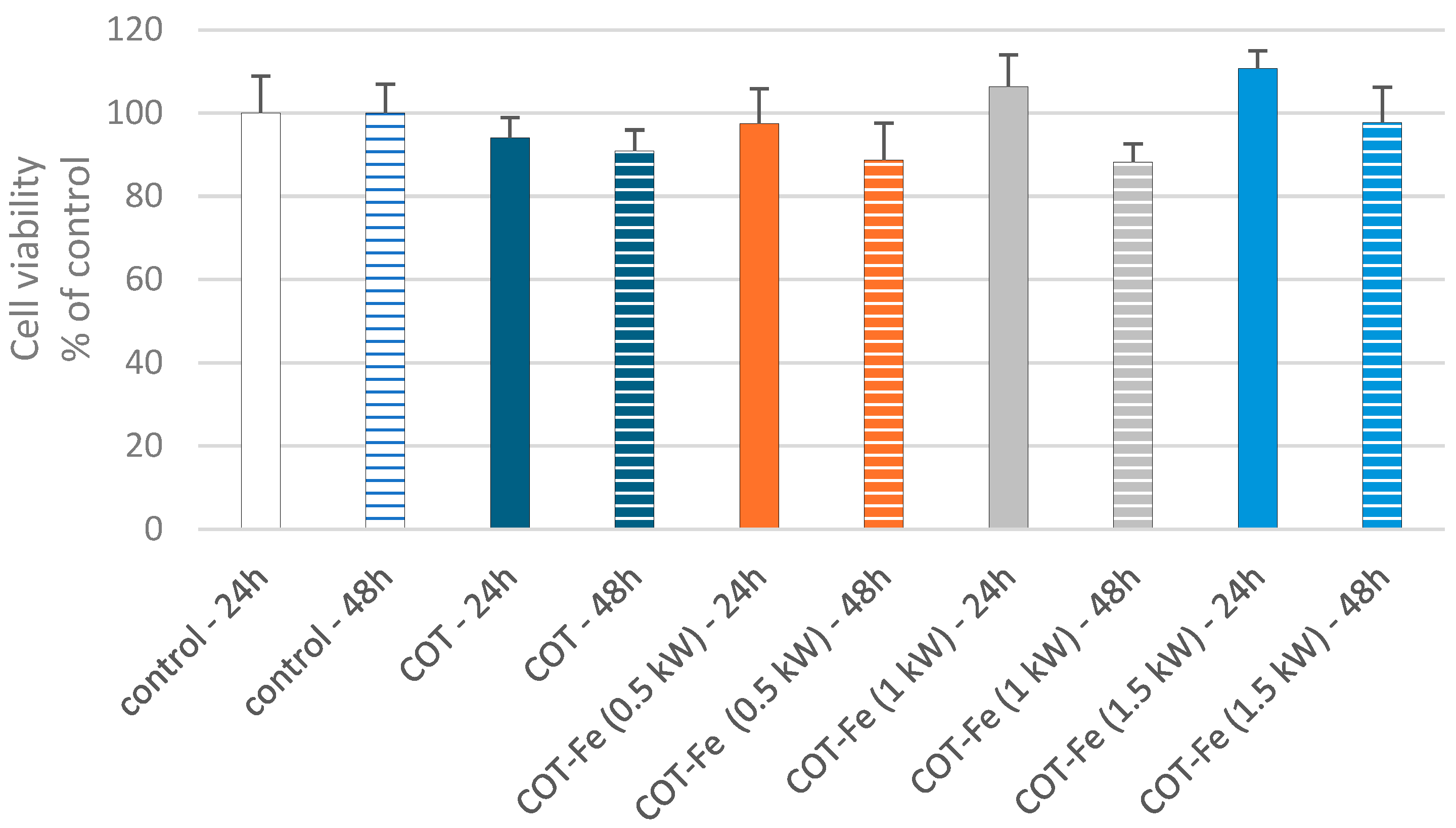
3.8. Effect of COT-Fe Samples on the Viability of PBM Cells

Cell viability following exposure to cotton and cotton-iron, post-incubation mixtures were evaluated using the resazurin reduction assay. This assay utilizes resazurin, a dye that acts as an indicator of cellular metabolic activity. In viable cells, mitochondrial redox reactions convert the nonfluorescent, dark blue resazurin into fluorescent resorufin, which appears pink at 570 nm or red at neutral pH. Our results exhibited that the incubation of PBM cells with COT and COT-Fe post-incubation mixtures did not deteriorate cell viability after 24 and 48 h (Figure 9). The results imply that cotton and cotton-iron fabrics lack cytotoxic properties against PBM cells. These findings suggest that iron in cotton fabric did not induce a Fenton reaction, increasing reactive oxygen species (ROS) levels with potential cytotoxic effects [42]. The Fenton reaction generates highly reactive hydroxyl radicals, which are capable of damaging essential biomolecules like DNA, proteins, and lipids [43,44].

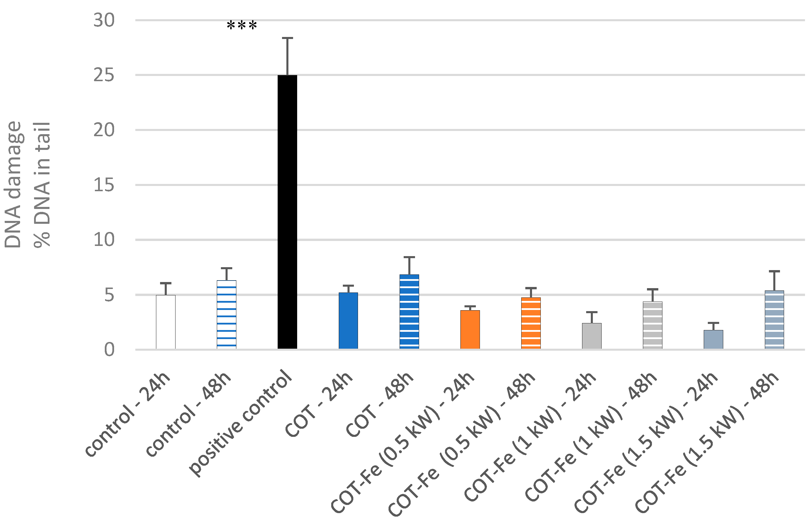
3.9. Effect of COT-Fe Samples on the DNA Damage in PBM Cells

The comet assay provides a sensitive and straightforward method for assessing DNA integrity in viable cells, enabling the detection of single- and double-strand breaks and alkali-labile lesions in DNA [45]. Significant DNA damage in PBM cells incubated with 25 µM H2O2 (positive control) was observed (Figure 10). Cotton and cotton-iron post-incubation mixtures, regardless of the power of sputtering, did not exhibit genotoxic potential against PBM cells after 24 and 48 h of incubation (Figure 10). These findings suggest that the power of sputtering is not associated with the release of iron in amounts that can induce Fenton’s reaction. Our results confirm the nontoxic nature of Fe-containing materials. Investigations into the biological effects of Mg0.1-γ-Fe2O3 (mPEG-silane) 0.5 nanoparticles revealed an absence of DNA strand breaks and oxidative DNA damage in A549 and BEAS-2B cells, with no observed cytotoxicity, despite a minor elevation in intracellular reactive oxygen species [46]. Concurrently, studies on Zn-3Cu alloys modified with trace Fe demonstrated significant improvements in biocompatibility and antibacterial efficacy in EA.hy926 and A7r5 cells without compromising hemocompatibility, thereby suggesting the potential of these alloys as candidate materials for vascular stent fabrication [47]. Images of comets obtained from fluorescence microscopy confirmed results from the comet assay (Figure 11).
The versatility of iron-containing materials, encompassing nanoparticles and polymeric matrices, has led to their intensive investigation as imaging agents, biocatalysts, biosensors in the biomedical field, drug delivery systems, and antibacterial therapeutics [48]. We have developed and characterized novel poly (lactide) nonwoven materials, fabricated through melt blowing and subsequently modified via iron magnetron sputtering, which expand the scope of potential biomedical applications of iron materials to include wound and injury treatment. Our results show that tested COT and COT-Fe post-incubation mixtures did not exhibit genotoxic potential against normal PBM cells. This proves their potentially safe use of dressing.

3.10. Effect of COT-Fe Samples on the pUC19 Plasmid Conformation

The plasmid relaxation assay investigated the potential for direct interactions between cotton or cotton-iron post-incubation mixtures and DNA. Electrophoretic mobility shift analysis (EMSA) revealed that the pUC19 plasmid, isolated from DH5α E. coli cells, exhibited a predominant supercoiled conformation (CCC), characterized by its high electrophoretic mobility. To create a control with different electrophoretic mobility, the plasmid was linearized by overnight incubation with the restriction enzyme PstI at 37 °C, resulting in the linear (L) form (Figure 12). Both COT and COT-Fe, regardless of sputtering power, did not interact with plasmid DNA. We did not observe single- or double-strand DNA breaks. Our results showed the absence of spontaneous interaction of the post-incubation mixture with DNA. This finding could be associated with the absence of iron ion release.

4. Conclusions

The surface modification of cotton fabric with iron via DC magnetron sputtering successfully improved the biochemical properties, including its antioxidant activity, without compromising its coagulation performance. Microscopic analysis confirmed uniform iron deposition on the cotton fibers, and biochemical tests showed that the iron coating does not influence the clotting times (aPTT and PT), causing no coagulation changes. Moreover, the iron-coated cotton demonstrates antioxidant properties, as indicated by reduced lipid peroxidation in TBARS analysis, which can potentially aid in mitigating oxidative stress during wound healing. The obtained results showed the lack of cyto- and genotoxic potential of tested cotton-iron fabrics against normal PBM cells. No effect on cell viability and lack of DNA damage allow us to consider that tested fabrics are safe for normal cells. Additionally, COT and COT-Fe fabrics did not directly interact with DNA, which suggests a lack of iron ion release. As well as that, it was proved that the modification with iron leads to excellent barrier properties against UV radiation, which may be beneficial in terms of wound dressings. Further research is needed to evaluate the long-term effects of this modification on wound healing.

Author Contributions

M.H.K. developed the concept and designed experiments, analyzed data, and wrote the paper; Z.M. developed the concept and designed experiments, performed experiments, analyzed data, and wrote the paper; A.K. analyzed data and wrote the paper; J.J.C. analyzed data; A.P. performed experiments; E.S. analyzed data; S.A.R.S. analyzed data; M.J. performed experiments and analyzed data; K.W. analyzed data; M.B.P. analyzed data and wrote the paper. All authors have read and agreed to the published version of the manuscript.

Funding

This research was partly conducted within the National Science Centre (Poland), project M-ERA.NET 2022, No. 2022/04/Y/ST4/00157.

Institutional Review Board Statement

The study was carried out in accordance with the Declaration of Helsinki and approved by the University of Lodz Research Ethics Committee (12/KEBN-UŁ/I/2024-2025), approved on 17 December 2024.

Informed Consent Statement

Not applicable.

Data Availability Statement

The data are included in the text.

Acknowledgments

The authors would like to thank Agnieszka Lisiak-Kucińska for their technical contributions.

Conflicts of Interest

The authors declare no conflicts of interest.

Abbreviations

The following abbreviations are used in this manuscript:
DCDirect current
aPTTActivated partial thromboplastin time
PTProthrombin time
TBARSLinear dichroism
PLLAPoly-L-lactic acid
ROSReactive oxygen species
UPFUltraviolet protection factor
SEMScanning electron microscopy
BETBrunauer–Emmet–Teller method
TBAThiobarbituric acid
TCATrichloroethanoic acid
MDAMalondialdehyde
TBARSThiobarbituric acid reactive substances
EDSEnergy-dispersive X-ray spectroscopy
COTCotton sample
COT-FeCotton-iron sample
IUPACInternational Union of Pure and Applied Chemistry
PBM cellsPeripheral blood mononuclear cells
PBSPhosphate Buffered Saline
DAPI4′,6-diamidino-2-phenylindole
LMPLow-melting-point agarose
NMPNormal-melting-point agarose

References

  1. Kauvar, D.S.; Lefering, R.; Wade, C.E. Impact of Hemorrhage on Trauma Outcome: An Overview of Epidemiology, Clinical Presentations, and Therapeutic Considerations. J. Trauma 2006, 60, S3–S11. [Google Scholar] [CrossRef] [PubMed]
  2. He, H.; Zhou, W.; Gao, J.; Wang, F.; Wang, S.; Fang, Y.; Gao, Y.; Chen, W.; Zhang, W.; Weng, Y.; et al. Efficient, Biosafe and Tissue Adhesive Hemostatic Cotton Gauze with Controlled Balance of Hydrophilicity and Hydrophobicity. Nat. Commun. 2022, 13, 552. [Google Scholar] [CrossRef] [PubMed]
  3. Granados, A.; Pleixats, R.; Vallribera, A. Recent Advances on Antimicrobial and Anti-Inflammatory Cotton Fabrics Containing Nanostructures. Molecules 2021, 26, 3008. [Google Scholar] [CrossRef] [PubMed]
  4. Shahriari-Khalaji, M.; Alassod, A.; Nozhat, Z. Cotton-Based Health Care Textile: A Mini Review. Polym. Bull. 2022, 79, 10409–10432. [Google Scholar] [CrossRef]
  5. Raval, A.A.; Patel, V.N.; Pancholi, U.V. In-Vitro Analysis of Hemostatic Cotton Khadi Dressing with Kaolin/Chitosan-Based Composition and Its Application as Prospective Wound Dressing. bioRxiv 2022. [Google Scholar] [CrossRef]
  6. Wang, Y.; Zhou, P.; Xiao, D.; Zhu, Y.; Zhong, Y.; Zhang, J.; Sui, X.; Feng, X.; Xu, H.; Mao, Z. Chitosan-Bound Carboxymethylated Cotton Fabric and Its Application as Wound Dressing. Carbohydr. Polym. 2019, 221, 202–208. [Google Scholar] [CrossRef]
  7. Yu, L.; Shang, X.; Chen, H.; Xiao, L.; Zhu, Y.; Fan, J. A Tightly-Bonded and Flexible Mesoporous Zeolite-Cotton Hybrid Hemostat. Nat. Commun. 2019, 10, 1932. [Google Scholar] [CrossRef]
  8. Edwards, J.V.; Prevost, N.; Yager, D.; Nam, S.; Graves, E.; Santiago, M.; Condon, B.; Dacorta, J. Antimicrobial and Hemostatic Activities of Cotton-Based Dressings Designed to Address Prolonged Field Care Applications. Mil. Med. 2021, 186, 116–121. [Google Scholar] [CrossRef]
  9. Mutch, N.J.; Waters, E.K.; Morrissey, J.H. Immobilized Transition Metals Stimulate Contact Activation and Drive Factor XII-Mediated Coagulation. J. Thromb. Haemost. 2012, 10, 2108–2115. [Google Scholar] [CrossRef]
  10. Mrozińska, Z.; Ponczek, M.; Kaczmarek, A.; Boguń, M.; Sulak, E.; Kudzin, M.H. Blood Coagulation Activities of Cotton–Alginate–Copper Composites. Mar. Drugs 2023, 21, 625. [Google Scholar] [CrossRef]
  11. Mrozińska, Z.; Kudzin, M.H.; Ponczek, M.B.; Kaczmarek, A.; Król, P.; Lisiak-Kucińska, A.; Żyłła, R.; Walawska, A. Biochemical Approach to Poly(Lactide)–Copper Composite—Impact on Blood Coagulation Processes. Materials 2024, 17, 608. [Google Scholar] [CrossRef] [PubMed]
  12. Mrozińska, Z.; Kaczmarek, A.; Świerczyńska, M.; Juszczak, M.; Kudzin, M.H. Biochemical Behavior, Influence on Cell DNA Condition, and Microbiological Properties of Wool and Wool–Copper Materials. Materials 2024, 17, 2878. [Google Scholar] [CrossRef] [PubMed]
  13. Mrozińska, Z.; Ponczek, M.B.; Kaczmarek, A.; Świerczyńska, M.; Kudzin, M.H. Activity in the Field of Blood Coagulation Processes of Poly(Lactide)-Zinc Fiber Composite Material Obtained by Magnetron Sputtering. Coatings 2024, 14, 666. [Google Scholar] [CrossRef]
  14. Praticò, D.; Pasin, M.; Barry, O.P.; Ghiselli, A.; Sabatino, G.; Iuliano, L.; FitzGerald, G.A.; Violi, F. Iron-Dependent Human Platelet Activation and Hydroxyl Radical Formation: Involvement of Protein Kinase C. Circulation 1999, 99, 3118–3124. [Google Scholar] [CrossRef]
  15. Nechipurenko, D.Y.; Receveur, N.; Yakimenko, A.O.; Shepelyuk, T.O.; Yakusheva, A.A.; Kerimov, R.R.; Obydennyy, S.I.; Eckly, A.; Léon, C.; Gachet, C.; et al. Clot Contraction Drives the Translocation of Procoagulant Platelets to Thrombus Surface. Arterioscler. Thromb. Vasc. Biol. 2019, 39, 37–47. [Google Scholar] [CrossRef]
  16. Lv, C.; Zhou, X.; Wang, P.; Li, J.; Wu, Z.; Jiao, Z.; Guo, M.; Wang, Z.; Wang, Y.; Wang, L.; et al. Biodegradable Alginate-Based Sponge with Antibacterial and Shape Memory Properties for Penetrating Wound Hemostasis. Compos. Part B Eng. 2022, 247, 110263. [Google Scholar] [CrossRef]
  17. Wang, L.; Zhang, X.; Yang, K.; Fu, Y.V.; Xu, T.; Li, S.; Zhang, D.; Wang, L.-N.; Lee, C.-S. A Novel Double-Crosslinking-Double-Network Design for Injectable Hydrogels with Enhanced Tissue Adhesion and Antibacterial Capability for Wound Treatment. Adv. Funct. Mater. 2020, 30, 1904156. [Google Scholar] [CrossRef]
  18. Long, M.; Zhang, Y.; Huang, P.; Chang, S.; Hu, Y.; Yang, Q.; Mao, L.; Yang, H. Emerging Nanoclay Composite for Effective Hemostasis. Adv. Funct. Mater. 2018, 28, 1704452. [Google Scholar] [CrossRef]
  19. Simberg, D.; Zhang, W.-M.; Merkulov, S.; McCrae, K.; Park, J.-H.; Sailor, M.J.; Ruoslahti, E. Contact Activation of Kallikrein–Kinin System by Superparamagnetic Iron Oxide Nanoparticles In Vitro and In Vivo. J. Control. Release 2009, 140, 301–305. [Google Scholar] [CrossRef]
  20. Meddahi-Pellé, A.; Legrand, A.; Marcellan, A.; Louedec, L.; Letourneur, D.; Leibler, L. Organ Repair, Hemostasis, and In Vivo Bonding of Medical Devices by Aqueous Solutions of Nanoparticles. Angew. Chem. Int. Ed. 2014, 53, 6369–6373. [Google Scholar] [CrossRef]
  21. Cunha, C.S.; Castro, P.J.; Sousa, S.C.; Pullar, R.C.; Tobaldi, D.M.; Piccirillo, C.; Pintado, M.M. Films of Chitosan and Natural Modified Hydroxyapatite as Effective UV-Protecting, Biocompatible and Antibacterial Wound Dressings. Int. J. Biol. Macromol. 2020, 159, 1177–1185. [Google Scholar] [CrossRef] [PubMed]
  22. Bai, Y.; Zhao, Y.; Li, Y.; Xu, J.; Fu, X.; Gao, X.; Mao, X.; Li, Z. UV-Shielding Alginate Films Crosslinked with Fe3+ Containing EDTA. Carbohydr. Polym. 2020, 239, 115480. [Google Scholar] [CrossRef] [PubMed]
  23. Abd El-Hady, M.M.; Farouk, A.; Saeed, S.E.-S.; Zaghloul, S. Antibacterial and UV Protection Properties of Modified Cotton Fabric Using a Curcumin/TiO2 Nanocomposite for Medical Textile Applications. Polymers 2021, 13, 4027. [Google Scholar] [CrossRef]
  24. Yin, H.; Xu, L.; Porter, N.A. Free Radical Lipid Peroxidation: Mechanisms and Analysis. Chem. Rev. 2011, 111, 5944–5972. [Google Scholar] [CrossRef]
  25. Guo, S.; Dipietro, L.A. Factors Affecting Wound Healing. J. Dent. Res. 2010, 89, 219–229. [Google Scholar] [CrossRef]
  26. Day, S.M.; Duquaine, D.; Mundada, L.V.; Menon, R.G.; Khan, B.V.; Rajagopalan, S.; Fay, W.P. Chronic Iron Administration Increases Vascular Oxidative Stress and Accelerates Arterial Thrombosis. Circulation 2003, 107, 2601–2606. [Google Scholar] [CrossRef]
  27. Kozlova, E.; Sherstyukova, E.; Sergunova, V.; Kozlov, A.; Gudkova, O.; Inozemtsev, V.; Chernysh, A. The Toxic Influence of Excess Free Iron on Red Blood Cells in the Biophysical Experiment: An In Vitro Study. J. Toxicol. 2022, 2022, 7113958. [Google Scholar] [CrossRef]
  28. EN 13758-1:2002; Textiles. Solar UV Protective Properties. Method of Test for Apparel Fabrics. International Organization for Standardization: Geneva, Switzerland, 2002.
  29. ISO 9237:1995; Textiles—Determination of the Permeability of Fabrics to Air. International Organization for Standardization: Geneva, Switzerland, 1995.
  30. EN ISO 5084:1996; Textiles. Determination of Thickness of Textiles and Textile Products. International Organization for Standardization: Geneva, Switzerland, 1996.
  31. EN 12127:1997; Textiles—Fabrics—Determination of Mass per Unit Area Using Small Samples. The European Committee for Standardization (CEN): Brussels, Belgium, 1997.
  32. EN ISO 139:2005; Textiles—Standard Atmospheres for Conditioning and Testing. Amendment 1. Organization for Standardization: Geneva, Switzerland, 2005.
  33. Kaals, R.; Priel, M.; Bremser, W.; D’Emilio, S.; Bosch, W.; Bich, W.; Vilalta, E.; Averlant, P.; Bièvre, P.J.D.; Fisicaro, P.; et al. EA Guideline on the Expression of Uncertainty in Quantitative Testing for Up-to-Date Information. In Proceedings of the EALC Meeting, Oslo, Norway, 7–9 September 2003. [Google Scholar] [CrossRef]
  34. Mrozińska, Z.; Świerczyńska, M.; Juszczak, M.; Woźniak, K.; Kudzin, M.H. Evaluation of Antimicrobial Activity, Hemostatic Efficacy, Blood Coagulation Dynamics, and DNA Damage of Linen–Copper Composite Materials. J. Compos Sci. 2025, 9, 30. [Google Scholar] [CrossRef]
  35. Kluska, M.; Juszczak, M.; Wysokiński, D.; Żuchowski, J.; Stochmal, A.; Woźniak, K. Kaempferol derivatives isolated from Lens culinaris Medik. reduce DNA damage induced by etoposide in peripheral blood mononuclear cells. Toxicol. Res. 2019, 8, 896–907. [Google Scholar] [CrossRef]
  36. O’Brien, J.; Wilson, I.; Orton, T.; Pognan, F. Investigation of the Alamar Blue (resazurin) fluorescent dye for the assessment of mammalian cell cytotoxicity. Eur. J. Biochem. 2000, 267, 5421–5426. [Google Scholar] [CrossRef]
  37. Tokarz, P.; Piastowska-Ciesielska, A.; Kaarniranta, K.; Blasiak, J. All-Trans Retinoic Acid Modulates DNA Damage Response and the Expression of the VEGF-A and MKI67 Genes in ARPE-19 Cells Subjected to Oxidative Stress. Int. J. Mol. Sci. 2016, 17, 898. [Google Scholar] [CrossRef] [PubMed]
  38. Juszczak, M.; Das, S.; Kosińska, A.; Rybarczyk-Pirek, A.J.; Wzgarda-Raj, K.; Tokarz, P.; Vasudevan, S.; Chworos, A.; Woźniak, K.; Rudolf, B. Piano-stool ruthenium(ii) complexes with maleimide and phosphine or phosphite ligands: Synthesis and activity against normal and cancer cells. Dalton Trans. 2023, 52, 4237–4250. [Google Scholar] [CrossRef] [PubMed]
  39. Sood, A.; Granick, M.S.; Tomaselli, N.L. Wound Dressings and Comparative Effectiveness Data. Adv. Wound Care 2014, 3, 511–529. [Google Scholar] [CrossRef] [PubMed]
  40. Torres-Sanchez, C.; Al Mushref, F.R.A.; Norrito, M.; Yendall, K.; Liu, Y.; Conway, P.P. The Effect of Pore Size and Porosity on Mechanical Properties and Biological Response of Porous Titanium Scaffolds. Mater. Sci. Eng. C 2017, 77, 219–228. [Google Scholar] [CrossRef]
  41. Thommes, M.; Kaneko, K.; Neimark, A.V.; Olivier, J.P.; Rodriguez-Reinoso, F.; Rouquerol, J.; Sing, K.S.W. Physisorption of Gases, with Special Reference to the Evaluation of Surface Area and Pore Size Distribution (IUPAC Technical Report). Pure Appl. Chem. 2015, 87, 1051–1069. [Google Scholar] [CrossRef]
  42. Toyama, T.; Kobayashi, M.; Rubiy’atno; Morikawa, M.; Mori, K. Sulfamethoxazole removal and fuel-feedstock biomass production from wastewater in a phyto-Fenton process using duckweed culture. Chemosphere 2024, 361, 142592. [Google Scholar] [CrossRef]
  43. Fernando, P.D.S.M.; Ko, D.O.; Piao, M.J.; Kang, K.A.; Herath, H.M.U.L.; Hyun, J.W. Protective effect of luteolin against oxidative stressmediated cell injury via enhancing antioxidant systems. Mol. Med. Rep. 2024, 30, 121. [Google Scholar] [CrossRef]
  44. Huang, Y.; Liu, H.; Zhao, Y.; Chen, H.; Li, Q.; Li, X.; Hua, S.; Cao, D.; Chang, Y. Disrupting redox homeostasis for tumor therapy based on PDT/chemo/ferroptosis therapeutic hybrid liposomes. RSC Adv. 2024, 14, 20152–20162. [Google Scholar] [CrossRef]
  45. Singh, N.P.; McCoy, M.T.; Tice, R.R.; Schneider, E.L. A simple technique for quantitation of low levels of DNA damage in individual cells. Exp. Cell Res. 1988, 175, 184–191. [Google Scholar] [CrossRef]
  46. Sikorska, M.; Ruzycka-Ayoush, M.; Rios-Mondragon, I.; Longhin, E.M.; Meczynska-Wielgosz, S.; Wojewodzka, M.; Kowalczyk, A.; Kasprzak, A.; Nowakowska, J.; Sobczak, K.; et al. Lack of cytotoxic and genotoxic effects of mPEG-silane coated iron(III) oxide nanoparticles doped with magnesium despite cellular uptake in cancerous and noncancerous lung cells. Toxicol. Vitro 2024, 99, 105850. [Google Scholar] [CrossRef]
  47. Yue, R.; Niu, J.; Li, Y.; Ke, G.; Huang, H.; Pei, J.; Ding, W.; Yuan, G. In vitro cytocompatibility, hemocompatibility and antibacterial properties of biodegradable Zn-Cu-Fe alloys for cardiovascular stents applications. Mater. Sci. Eng. C 2020, 113, 111007. [Google Scholar] [CrossRef]
  48. Peng, X.; Xu, L.; Zeng, M.; Dang, H. Application and Development Prospect of Nanoscale Iron Based Metal-Organic Frameworks in Biomedicine. Int. J. Nanomed. 2023, 18, 4907–4931. [Google Scholar] [CrossRef]
Figure 1. DC magnetron sputtering system.
Coatings 15 00663 g001
Figure 2. Optical microscopy images (magnifications: ×100, ×500) of the surface structure of samples before and after the modification processes.
Coatings 15 00663 g002
Figure 3. SEM images of COT and COT-Fe samples. Magnification: 5000×; 10,000×.
Coatings 15 00663 g003
Figure 4. EDS spectra of COT sample and COT-Fe samples: (a) COT sample, (b) COT-Fe.
Coatings 15 00663 g004
Figure 5. Obtained N2 adsorption isotherms for COT; COT-Fe (0.5kW/10′); COT-Fe (0.5kW/30′); COT-Fe (1kW/10′); COT-Fe (1kW/30′) samples.
Coatings 15 00663 g005
Figure 6. Comparison of the transmittance spectra [%T] for the same composites COT, COT-Fe (0.5kW/10′), COT-Fe (0.5kW/30′), COT-Fe (1kW/10′), and COT-Fe (1kW/30′), recorded over the 200–800 nm wavelength range.
Coatings 15 00663 g006
Figure 7. The structure of the unmodified COT fabric and COT-Fe (1kW/10′) sample after the modification with iron coating.
Coatings 15 00663 g007
Figure 8. Effect of studied cotton composites on aPTT (a), PT (b): COT; COT-Fe (0.5kW/10′); COT-Fe (0.5kW/30′); COT-Fe (1kW/10′); COT-Fe (1kW/30′); COT-Fe (1.5kW/40′); and C—plasma control sample: plasma not exposed to composites. Results are shown as mean (×), median (horizontal line), range (bars), and interquartile range (box).
Coatings 15 00663 g008
Figure 9. Effect of cotton (COT) and cotton-iron (COT-Fe (0.5kW), COT-Fe (1kW), COT-Fe (1.5kW)) post-incubation mixtures on PBM cells viability after 24 and 48 h incubation. Results are shown as mean results from six repeats. Error bars denote SD.
Coatings 15 00663 g009
Figure 10. Effect of cotton (COT) and cotton-iron (COT-Fe (0.5kW), COT-Fe (1kW), COT-Fe (1.5kW)) post-incubation mixtures on PBM cells DNA damage after 24 and 48 h incubation. Results are shown as mean results from 100 comets. Error bars denote SEM; *** p < 0.001.
Coatings 15 00663 g010
Figure 11. Effect of medium (A), 25 µM hydrogen peroxide (B), cotton Cot (C), and cotton-iron Cot-Fe (0.5kW) (D), Cot-Fe (1kW) (E), Cot-Fe (1.5kW) (F), post-incubation mixtures on PBM cells DNA damage after 24 h incubation.
Coatings 15 00663 g011
Figure 12. Plasmid relaxation assay. pUC19 plasmid was incubated for 24 h (37 °C) with cotton and cotton-iron post-incubation mixtures (COT-Fe (0.5kW), COT-Fe (1kW), COT-Fe (1.5kW)), then was separated on a 2% agarose gel, stained with ethidium bromide and visualized in UV light. Line 1—DNA ladder; line 2—pUC19 plasmid (the supercoiled form, CCC); line 3—pUC19 plasmid incubated with restrictase PstI (the linear form, L); lines 4–7—pUC19 plasmid incubated with COT, COT-Fe (0.5kW), COT-Fe (1kW), COT-Fe (1.5kW), respectively; line 8—DNA ladder. OC—the open circular form of plasmid DNA.
Coatings 15 00663 g012
Table 1. Iron content analysis and surface property testing results, including specific surface area and total pore volume.
Sample NameFe Concentration [g/kg]Total Pore Volume
[cm3/g]
Specific
Surface Area
[m2/g]
COT07.125 × 10−30.7216
COT-Fe (0.5kW/10′)0.6383.331 × 10−30.7122
COT-Fe (0.5kW/30′)2.1872.643 × 10−30.6130
COT-Fe (1kW/10′)1.0433.249 × 10−30.7085
COT-Fe (1kW/30′)3.5622.411 × 10−30.6059
COT-Fe (1.5kW/40′)20.412.118 × 10−30.5155
Table 2. UPF values for modified cotton fabric samples.
COTCOT-Fe
(0.5kW/10′)
COT-Fe
(0.5kW/30′)
COT-Fe
(1kW/10′)
COT-Fe
(1kW/30′)
COT-Fe
(1.5kW/40′)
UPF3.299.2713.629.9716.7518.22
average %T,
λ = 290–400 nm
31.8110.566.879.025.754.72
Table 3. Air permeability, thickness, and surface mass for the examined COT and COT-Fe samples.
Thickness [mm]Surface Mass [g/m2]Air Permeability
[mm/s]
100 Pa200 Pa
COT0.46 ± 0.02183 ± 2802 ± 101300 ± 20
COT-Fe (0.5kW/10′)0.47 ± 0.02184 ± 1841 ± 121353 ± 16
COT-Fe (0.5kW/30′)0.47 ± 0.02184 ± 1847 ± 191363 ± 38
COT-Fe (1kW/10′)0.48 ± 0.02184 ± 1842 ± 101358 ± 16
COT-Fe (1kW/30′)0.47 ± 0.02185 ± 2829 ± 341368 ± 60
COT-Fe (1.5kW/40′)0.47 ± 0.02184 ± 2840 ± 111358 ± 25
Table 4. Concentrations in different sample preparations (TBARS).
CCOT-Fe
(1.5kW/40′)
COT-Fe
(1kW/30′)
COT-Fe
(1kW/10′)
COT-Fe
(0.5kW/30′)
COT-Fe
(0.5kW/10′)
COT
0.6150.7050.7050.7050.3210.4230.692TBARS (nM)
Disclaimer/Publisher’s Note: The statements, opinions and data contained in all publications are solely those of the individual author(s) and contributor(s) and not of MDPI and/or the editor(s). MDPI and/or the editor(s) disclaim responsibility for any injury to people or property resulting from any ideas, methods, instructions or products referred to in the content.

