Article

Development of Chitosan-Coated Tung Oil Microcapsules with Antioxidants from Bamboo Leaves for Enhanced Antimicrobial Waterborne Coatings

1
Co-Innovation Center of Efficient Processing and Utilization of Forest Resources, Nanjing Forestry University, Nanjing 210037, China
2
College of Furnishings and Industrial Design, Nanjing Forestry University, Nanjing 210037, China
*
Author to whom correspondence should be addressed.
Coatings 2025, 15(5), 517; https://doi.org/10.3390/coatings15050517
Submission received: 27 February 2025 / Revised: 10 April 2025 / Accepted: 21 April 2025 / Published: 25 April 2025
(This article belongs to the Special Issue Innovations in Functional Coatings for Wood Processing)

Abstract

:
Antibacterial microcapsules were prepared by using a compound of chitosan with an antioxidant of bamboo leaves (AOB) as the wall material and tung oil as the core material. The microcapsules were modified by adding them to waterborne coatings, and the modified waterborne coatings were coated onto Basswood samples. The performance of the obtained coatings was then characterised through a comparative analysis. The investigation focused on the effect of varying percentages of chitosan and AOB in microcapsules with a constant core-to-wall ratio on the performance of the waterborne on the surface of Basswood. The core-to-wall ratio of the microcapsules was established at 1:2, with the ratios of chitosan and AOB in the walls fixed at 9:1, 8:2, and 7:3, respectively. The results demonstrated that the gloss, impact resistance, and hardness of the coatings exhibited an increase with increasing ratios of AOB under varying Mchitosan:MAOB (MC:MA) conditions. Conversely, the adhesion exhibited a decrease with an increase in AOB. The colour difference value exhibited minimal change. The self-healing rate of the coating exhibited an initial increase, followed by a subsequent decrease, in response to the increasing AOB concentration. The antimicrobial effect was optimised at a ratio of 9:1 for the combination of chitosan and AOB. The coating of Basswood containing 1.0% microcapsules and 9:1 MC:MA demonstrated superior performance, exhibiting a gloss of 9.7 GU, a colour difference ΔE of 31.03, a hardness of HB, an adhesion rating of grade 1, an impact resistance of grade 4, a self-healing rate of 19.09%, and a noteworthy antimicrobial effect against both Escherichia coli and Staphylococcus aureus.

1. Introduction

In recent years, a number of different industries have become involved in antimicrobial research. There has been a considerable interest in the use of antimicrobial materials in a number of different fields, including medicine, food packaging, construction, and textiles. A chitosan is a polysaccharide derived from chitin. It is biocompatible, biodegradable, non-toxic, and exhibits excellent membrane formation properties [1,2,3]. This makes chitosan an ideal carrier material that can be used to coat and protect active ingredients [4,5,6]. Antioxidants of bamboo leaves (AOB) are a specific type of flavonoid that occurs naturally in bamboo leaves. They possess a range of biological activities, including antioxidant, anti-inflammatory, and antibacterial properties [7,8,9]. It is polar and hydrophilic, readily soluble in hot water, methanol, ethanol, acetone, and other solvents [10,11,12,13,14]. Flavonoids are currently the subject of intensive study due to their numerous beneficial properties. These include safety, lack of odour, affordability, natural origin, nutritional value, and multifunctionality [15]. The hydrophobic aromatic ring structure of AOB has been shown to insert itself into the phospholipid bilayer of the bacterial cell membrane. This has been demonstrated to cause a disturbance in the orderly arrangement of the cell membrane, resulting in an increase in its permeability. This, in turn, has been observed to lead to the leakage of intracellular substances and, ultimately, the death of cells. The phenolic hydroxyl group, a constituent of the flavonoid molecule, has the capacity to undergo partial dissociation within the solution, resulting in adsorption on the negatively charged bacterial surface through electrostatic action. This process has the potential to destabilise the membrane potential. Consequently, the incorporation of AOB into waterborne coatings has the potential to enhance their antimicrobial properties.
In the 1930s, researchers developed a class of minute, spherical particles comprising a compact outer shell and a variety of substances within the shell, which could exist in either a solid or liquid state. The particles were designated “microcapsules” due to their overall structural resemblance to a capsule. Due to their wall-core structure, microcapsules are capable of encapsulating core materials with specific functions [16,17,18]. Long et al. employed sodium tripolyphosphate (STPP) as a cross-linking agent in the investigation of slow-release tea tree essential oil-chitosan microcapsules, and prepared tea tree essential oil-chitosan (CS) microcapsules by the ionic gelation method. The structural and functional properties of tea tree essential oil microcapsules were investigated, and the release pattern of tea tree essential oil microcapsules in different food simulation systems was analysed in order to obtain polymer-nanosilver composites [19]. Huang et al. conducted a study to examine the preparation, characterisation, and evaluation of the antibacterial activity of chitosan–cinnamon essential oil microcapsules. These microcapsules were microencapsulated with chitosan as the wall material. The encapsulation rate was employed as an indicator to optimise the preparation process through one-factor experiments and response surface methodology. The properties were then characterised and evaluated for their antibacterial activity against food-borne pathogens [20]. The development of microcapsules represents a significant scientific accomplishment, with a diverse array of preparation techniques and disciplinary contributions.
Tung oil is notable for its absence of volatile organic compounds (VOCs), a feature that distinguishes it from synthetic resins. This characteristic renders it harmless to the environment and the human body, thus aligning with the growing demand for green building materials and eco-friendliness. The utilisation of tung oil as a coating for wooden structures dates back to ancient Chinese traditions, where it has been employed for centuries to preserve the integrity of wood. The chemical composition of tung oil is such that it is rich in α-tungstic acid, which, through oxidative polymerisation reactions in the air, forms a dense three-dimensional mesh cross-linking film. It has been demonstrated that this process enhances the self-healing capabilities of the coating, while concurrently impeding the ingress of moisture and extending the durability of the wood. This is achieved by curtailing the proliferation of fungi and bacteria through the isolation of moisture.
Wooden furniture faces various challenges in daily use [21,22,23]. In addition to strengthening the structural strength of wooden furniture, coatings can, to some extent, protect furniture from negative environmental factors [24,25]. At present, coatings are endowed with self-healing [26], antibacterial [27], flame-retardant properties [28], and more through various means [29,30]. Wood is categorised as a form of renewable biomass material, comprising a combination of three primary components: lignin, cellulose, and hemicellulose. It is evident that the presence of these ingredients renders the timber susceptible to infestation by pests and diseases, including bacterial and fungal growth [31,32,33]. Wood antibacterial treatment represents a pivotal method of prolonging the service life of wood, enhancing its utilisation, and conserving resources [34,35,36]. There are also many modification methods in the field of colour-changing coatings, such as 3D printing and structural colour, which have been introduced to construct colour-changing coatings [37,38,39]. While using various methods to modify the surface of furniture, attention should also be paid to maintaining the performance of the coating itself [40,41]. Among these methods, microcapsules have the characteristics of a low cost and simple preparation. Many fossil-based and biobased microcapsules have also long been widely used in various aspects of people’s daily lives [42,43].
The disparity in polarity between film-forming substances (e.g., acrylic emulsions, polyurethane dispersions) and natural antimicrobial agents in waterborne coatings has the potential to result in uneven dispersion. Furthermore, the antimicrobial components are susceptible to aggregation or migration to the coating, thereby reducing the effective concentration. The porous nature of wood may result in the rapid release and loss of antimicrobial agents, particularly in humid environments, and the hydrophilicity of waterborne may accelerate the dissolution of antimicrobial components, thereby reducing the duration of the antimicrobial cycle. The microcapsule shell has the capacity to encapsulate the antimicrobial agent, with the subsequent release of said agent being affected in a controlled manner through the pores of the microcapsule wall or by osmosis. This process serves to extend the action cycle. The integration of both antimicrobial and restorative agents within microcapsules enables multifunctional capabilities, thereby facilitating a symbiotic relationship between antimicrobial defence and autonomous healing mechanisms. Chitosan, AOB, and tung oil are bio-based materials. As such, they avoid the environmental toxicity of heavy metals or organic fungicides in traditional paints, rendering them more environmentally friendly. AOB have been shown to scavenge free radicals and retard the UV ageing of the coating resin, thereby ensuring the stability of the microcapsule structure and the overall protection life.
In light of the shortcomings associated with the conventional preparation techniques and procedures employed in the fabrication of antimicrobial coatings, an alternative approach, namely the emulsification cross-linking method, was used for the synthesis of microcapsules. In this experiment, the microcapsule preparation process was simplified with the objective of reducing the total amount of reactants. Commercially available food-grade AOB were procured and coated with chitosan, followed by the addition of tung oil to create microcapsules. The antimicrobial properties of microcapsules prepared with varying ratios of chitosan and AOB were evaluated. The impact of the resulting microcapsules on the antimicrobial properties was examined, and they were assessed for their mechanical, optical, and antimicrobial characteristics to assess the effectiveness of chitosan-coated tung oil antimicrobial microcapsules of AOB in antimicrobial coatings in terms of antimicrobial properties, stability, and adhesion to substrates.

2. Test Materials and Methods

2.1. Materials

The dimensions of the Basswood were 50 mm × 50 mm × 5 mm. The experimental materials included chitosan, AOB, acetic acid, sodium hydroxide, Tween 80, tung oil, sodium tripolyphosphate, deionised water, and waterborne coating. Table 1 illustrates the breakdown of raw materials utilised in the production of microcapsules.

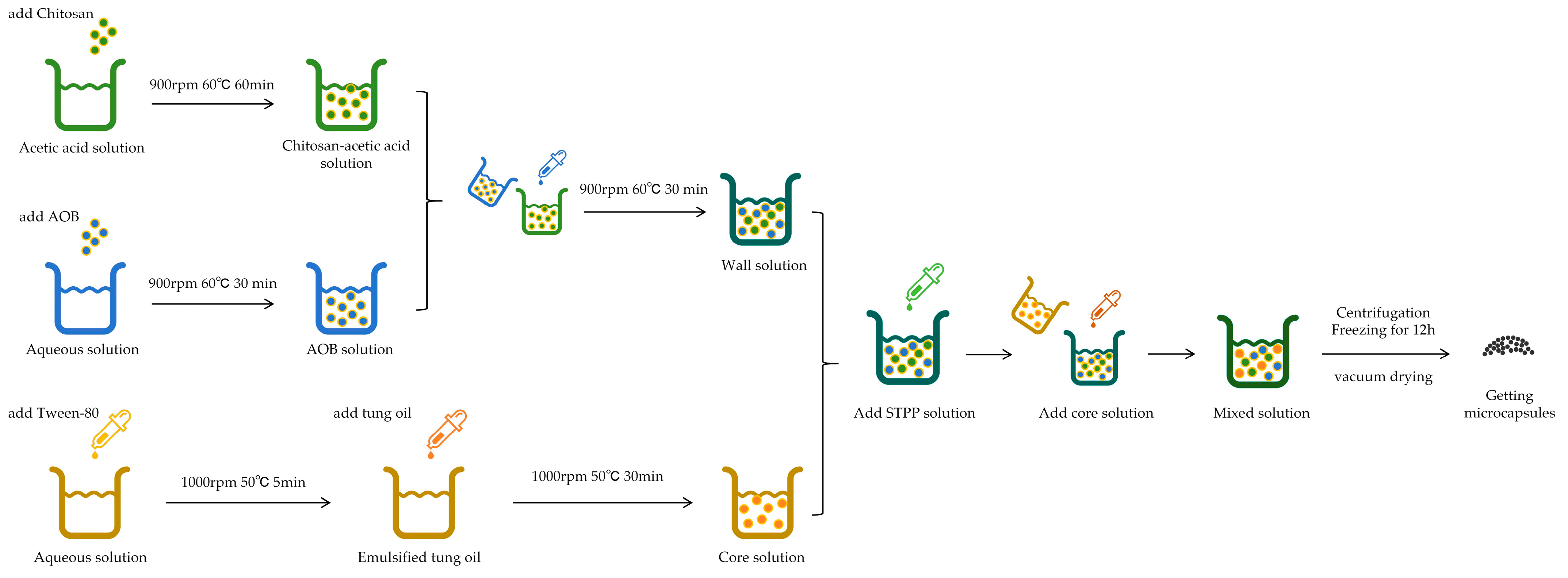
2.2. Preparation Method of Microcapsules

The core-to-wall ratio of the microcapsules was 1:2, and the chitosan and AOB content ratios in the walls were 9:1, 8:2, and 7:3, respectively. The high viscosity of tung oil has been shown to result in the formation of large droplets during the emulsification process. This phenomenon necessitates the use of additional wall material for the purpose of achieving uniform encapsulation. A core-to-wall ratio of 1:2 has been shown to provide sufficient wall material to cover the droplet surface, thereby reducing the risk of aggregation and rupture. With a core-to-wall ratio of 1:2, the minimum concentration of chitosan required for film formation can be maintained when a reasonable proportion of chitosan is present in the wall material, ensuring the film-forming ability of the compound. The addition of AOB enhances the antioxidant property but does not significantly dilute the film-forming component.
The preparation of microcapsules was conducted in three stages, with Sample 4# serving as a case study.

2.2.1. Preparation Method of Wall Materials

An acetic acid solution was prepared by the addition of 0.7 g of acetic acid to a beaker containing 69.3 g of deionised water. A weight of 0.7 g of chitosan was then added to the solution, which was subsequently placed in a thermostatic water bath and subjected to magnetic rotor stirring. The stirrer settings were configured to 900 rpm and 60 °C for a 60 min period to ensure complete dissolution. The pH was adjusted using sodium hydroxide until a pH of 4.5 was reached, thereby obtaining the desired acetic acid–chitosan solution. A solution of 0.3 g of AOB in water was prepared and placed in a water bath. It was then subjected to the following conditions: 900 rpm, 60 °C, and 30 min. The prepared AOB solution was then added to the chitosan solution, after which the agitation was performed using a magnetic rotor in a constant water bath. The conditions were set to 900 rpm, 60 °C, and 30 min. The preparation stage is thus concluded.

2.2.2. Preparation Method of the Core

In a suitable beaker, 0.4 g of Tween-80 was weighed out and added to 39.6 g of deionized water. The beaker was then placed in a constant water bath and stirred using a magnetic rotor. The experimental setup conditions were established at 1000 rpm and 50 °C. The stirring was continued until the solution was clear, at which point 0.5 g of tung oil was added. The mixture was then stirred for a period of 30 min to ensure that the core solution was obtained.
As demonstrated in Figure 1, the application of Tween-80 during emulsification results in fresh tung oil that exhibits stability under storage conditions. It is evident from the analysis that the tung oil did not delaminate after being left undisturbed for 3 d, indicating that the current emulsification process is well designed to effectively maintain the dispersion of the oil and water phases and to achieve physical stability under short-term and storage conditions.

2.2.3. Synthesis of Microcapsules

The flow chart illustrating the microencapsulation preparation process is presented in Figure 2. As illustrated in Table 2, the proportion of microencapsulated materials is specified in the following way.
A solution of 0.2 g of sodium tripolyphosphate was prepared by adding the salt to a beaker containing 19.8 g of deionised water. A magnetic spin plate was attached and placed in a water bath. The conditions were set to 50 °C and 900 rpm. We started stirring and continued stirring until the solution was completely dissolved. A solution of sodium tripolyphosphate was added to the wall material chitosan solution, and subsequently added dropwise to the core material solution, which was then stirred until a clear solution was obtained. The resulting mixed solution was then packed into four experimental test tubes, which were subjected to centrifugation at a speed of 7000 rpm for a period of 15 min. The supernatant was then removed, leaving behind the precipitate, which was subsequently transferred to a freezer for a period of 12 h. Upon removal from the freezer, the precipitate was placed in a vacuum desiccator for drying, resulting in the formation of a powder, designated as 4#. The same method was employed to prepare microcapsules of 2# and 3#.

2.3. Painting Method for Basswood

In accordance with the stipulations outlined in the “Technical Specification for Application of Waterborne Wood Coatings on Furniture Surface”, the finishing method employed is hand finishing, with the dosage of one layer of coating ranging from 60 g/m2 to 80 g/m2. The application of the finishing method for Basswood is achieved through the utilisation of a brushing technique, with a total of three layers of primer coating. The quantity of coating per layer is 80 g/m2, and the total quantity of primer coating is 240 g/m2. Furthermore, considering the losses of coating during the real application of the finish, the real amount of coating was twice the theoretical amount. Consequently, the combined coating applied to the Basswood was 1.20 g. Initially, the Basswood was meticulously sanded using 240-gauge sandpaper until the surface was rendered smooth and even. Secondly, the prepared microcapsules were mixed with Dulux waterborne coating at the specified ratio, with the 1.20 g primer containing 1% microcapsules. The experiment involved the application of a waterborne coating containing microcapsules to the Basswood. Following the initial application of primer, the samples were permitted to dry at ambient temperature for a period of 10 min prior to the application of a second coat of primer. This process was repeated on three occasions to obtain samples for the coating experiment.

2.4. Testing and Characterisation

2.4.1. Analysis of Microstructure

In order to facilitate a more comprehensive observation of the microstructure of the microcapsules, the morphology of the microcapsules was examined using a Zeiss optical microscope (OM, Carl Zeiss AG, Oberkochen, Germany). Subsequently, to observe and analyse the microstructure of the microcapsules and the prepared coatings, a scanning electron microscope (SEM, Tescan, Brno, Czech Republic) was used.

2.4.2. Analysis of Chemical Composition

The chemistry of the microcapsules and coatings was investigated by means of Fourier Transform Infrared Spectroscopy (FTIR, Brucker AG, Karlsruhe, Germany).

2.4.3. Optical Performance Testing

(1) The following test method is employed for the purpose of determining the colour deviation: In accordance with the stipulations set out in GB/T 11186.3-1989 [44], the measurement and documentation of the colour difference value of the coating was to be conducted using a colourimeter (Shenzhen San’enshi Technology Co., Ltd., Shenzhen, China). The calculation of the colour deviation was then to be performed. To ascertain the colour deviation (∆E) in different samples, the following formula was used (1), where ∆L = L1L2, ∆a = a1a2, and ∆b = b1b2. ∆L represents the difference in brightness of the coating; ∆a represents the red-green difference in the coating; ∆b represents the yellow-blue difference in the coating [45].
E = [ ( L ) 2 + ( a ) 2 + ( b ) 2 ] 1 2
(2) Glossiness of the coating: The HG268 glossmeter (Shenzhen Linshang Technology Co., Ltd., Shenzhen, China) is employed to assess and document the gloss value of the paint film at an angle of incidence of 60°. The unit of measurement is GU. It can be observed that an increase in brightness correlates with an enhanced coating quality. Samples exhibiting uniform coating distribution, as discernible to the unassisted eye, and devoid of defects in the underlying material, were selected for testing. These tests were conducted at a temperature of 23 ± 2 °C and a RH of less than 75%. A multi-angle gloss metre should be used to conduct the 60° gloss test on the paint samples in accordance with the standard procedure.

2.4.4. Test Methods for Mechanical Properties of Coatings

(1) Adhesion test of coating: The test was conducted in accordance with the provisions outlined in GB/T 4893.4-2013 [46]. It was carried out employing an adhesion tester (Quzhou Aipu Measuring Instrument Co., Ltd., Quzhou, China). The adhesion to the wood furniture is defined as the degree to which the paint is firmly bonded to the wood furniture substrate, or to the paint itself [47]. The blade was then oriented in a vertical direction at a 90° angle relative to the paint film, thereby delineating a tic-tac-toe pattern of cross-cuts. Subsequent to this, the pattern was rapidly applied with tape and then removed to observe the coating peel off. These levels are measured on a scale ranging from 0 to 5, with the results being determined by comparing the area of the taped-off surface with the national standard.
(2) Hardness test of coating: In accordance with the stipulations set out in GB/T 6739-2022 [48], the pencil utilised in this study was characterised by a hardness ranging from 9B to 9H. This instrument was employed in conjunction with a portable pencil hardness tester of the QHQ-A variety (Quzhou Aipu Measuring Instrument Co., Ltd., Quzhou, China). In order to perform the scratch test, a pencil must be held at a 45° angle to the tested sample, and a 1 kg load must be applied evenly. The resultant damage to the coating is then observed, and the pencil hardness required to scratch the coating is registered as the hardness value of the coating under test.
(3) Impact resistance performance test of coatings: In accordance with GB/T 4893.9-2013, entitled “Physical and chemical properties test of furniture surface coatings”, specifically Part 9, which pertains to the measurement of impact resistance [49], the test was performed. It was tested and recorded by means of a coating impact metre (Jiaxin Measuring Instrument Co., Ltd., Dongguan, China). The coating is impacted by releasing a 1.0 kg weight through the positioning height. The presence of cracks, wrinkles, and flaking in the coating can be determined by observing the sample under a 4× magnifying glass. Each sample is subjected to five replicates, and the integer that corresponds most closely to the arithmetic mean of the evaluation ratings is designated as the outcome of the assessment. The number of impact ratings was incremented from 5 to 1. The criteria for the evaluation of the ratings are shown in Table 3.

2.4.5. Self-Healing Performance Test of Coating

A knife was utilised to create a 15 mm long incision in the coating, after which the incised coating was positioned on the observation stage of the microscope. An external light source was utilised to illuminate the tested sample from a fixed angle. The width of the incision was measured and recorded as Wd using the provided software. The tested coatings were maintained at ambient temperature conditions for a period of 24 h. Subsequent to this, a second measurement of width was taken at the previous incision site, and the resulting data were plotted as Wc. The degree of self-healing capacity exhibited by the coatings, Wf, was calculated as in Formula (2). The differences in self-healing performance of coatings with different paint film surfaces were compared using the self-healing rate.
Wf = [(WdWc)/Wd] × 100%

2.4.6. Testing of Antimicrobial Properties of Coatings

The experimental procedure was conducted in accordance with the protocol outlined in GB/T 21866-2008 [50] for the assessment of the antimicrobial properties of paint films. Initially, Escherichia coli (ATCC25922) and Staphylococcus aureus (ACTT6538) were utilised as live bacteria. The production of plate agar media entails the preparation of 1000 mL of deionized water, to which 24 g of agar material, meticulously weighed, was added. This was then subjected to a rigorous sterilisation process. The strains were cultivated in a refrigerated slant and subsequently inoculated onto plate agar media for incubation for 18–20 h.
The subsequent stage of the process involved the preparation of the desired bacterial suspension. A precise quantity of 9 g of nutrient broth and 500 mL of distilled water was weighed out and subjected to a sterilisation process at 121 °C for a duration of 15 min. Using an inoculating loop, 1 to 2 loops of fresh bacteria were scraped from the live bacterial agar medium and added to the broth culture solution. The sterilised nutrient broth culture solution was then diluted to a concentration of 106 CFU/mL of the test bacterial suspension, after which it was set aside.
The experimental procedure was then conducted on the sample test. Approximately 0.5 mL of the bacterial suspension employed in the test was added dropwise to the area of the coated film to be tested and to the area of the blank control group. It was dispersed over the test specimen using sterilised tweezers and sterilised plastic sheeting to ensure that the test specimen was even and free of bubbles. The samples were put into clean Petri dishes after being treated. The conditions of the chamber were set at 38 °C and RH > 90%. The experimental procedure was then conducted on the sample test. Following a 24 h period, the cultured samples were removed. The eluent was prepared in advance, and the film was clamped with sterilised tweezers. The specimen was then rinsed repeatedly. The eluate was then inoculated into nutrient agar and placed in a constant chamber at 38 °C conditions for 48 h. The colony number in the culture was measured and recorded using a colony counter according to GB/T 4789.2-2022 [51]. The Formula (3) for the antimicrobial rate is represented, where R represents the antimicrobial rate in %, B represents the average number of bacteria obtained after 2 d for pure samples in CFU/piece, and C represents the average number of bacteria obtained after 2 d for antimicrobial lacquer in CFU/piece.
R = B C B × 100 %

3. Results and Discussion

3.1. Analysis of the Morphology and Chemical Composition of Microcapsules

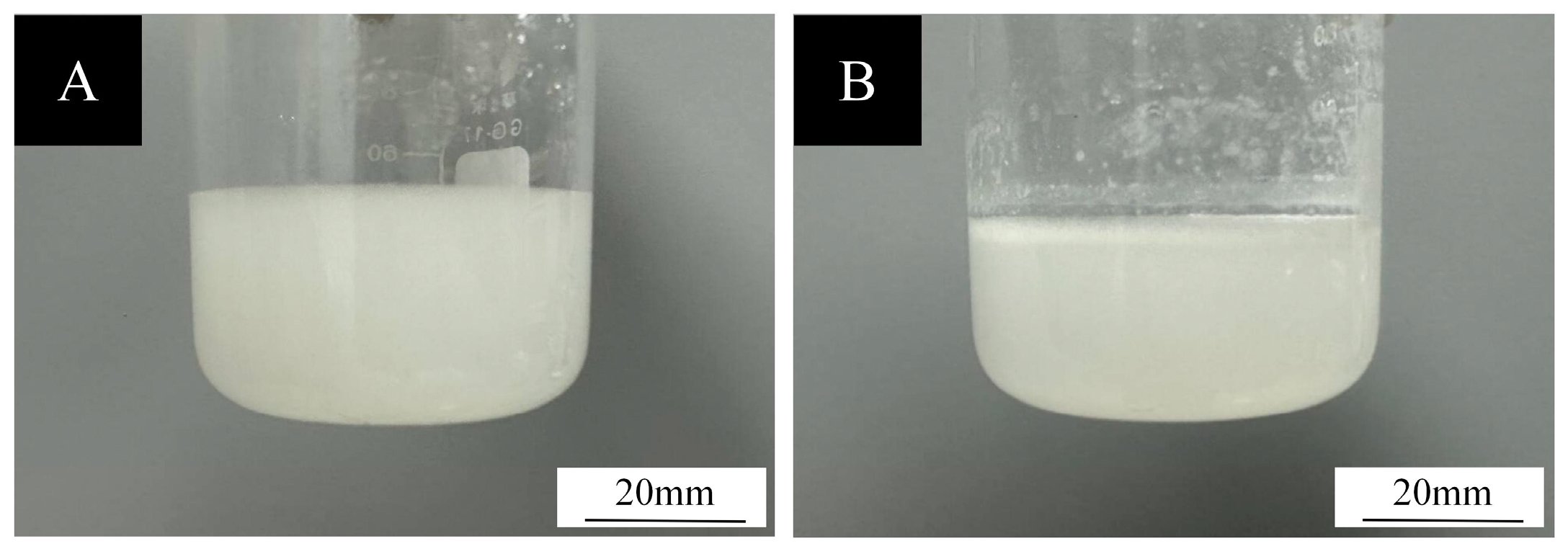
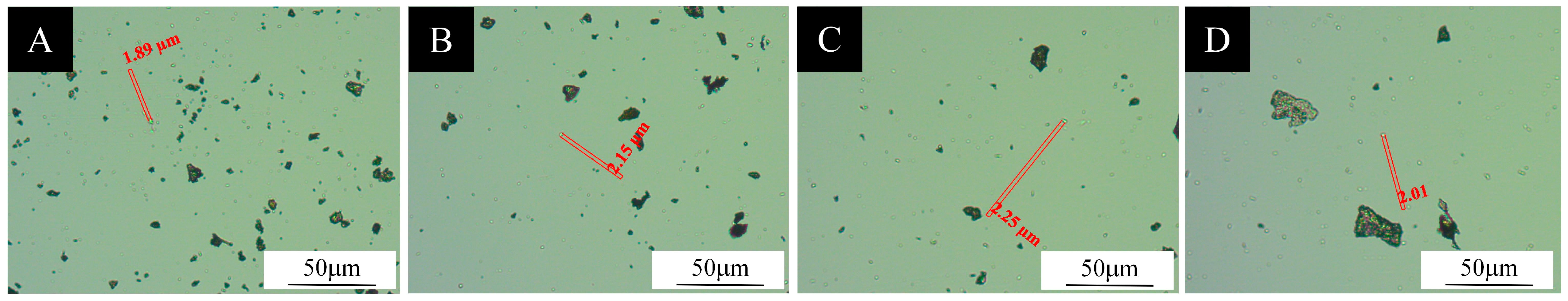
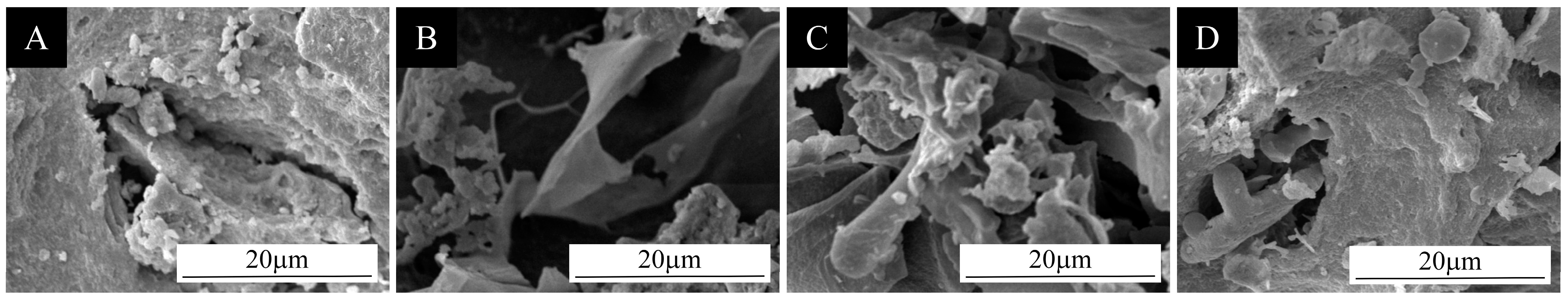
3.1.1. Micromorphometric Analysis of Microcapsules

The prepared microencapsulated powder was uniformly coated onto slides and observed for morphological characteristics. The optical microscope diagram is shown in Figure 3. It is evident that microcapsule Samples 1# to 4# possess a diminutive particle size. The shell and core structures are discernible, with the inner bright spots representing the core material and the outer black circles denoting the shell material. However, aggregates are present in Samples 1#–4#, suggesting the potential for interaction between the phenolic hydroxyl group in AOB and the amino group of chitosan, which may impact the film-forming property or charge distribution, and, consequently, the stability of the microcapsules.
SEM images of four groups of microcapsules are shown in Figure 4. There was a particle agglomeration phenomenon, and an irregular block structure was visible in the local area for Sample 1#, and these granular distributions were relatively dispersed and sparse, indicating that there was a certain amount of discrete phases in the microstructure of the sample. There was an obvious agglomeration phenomenon, and a lamellar structure was produced for Sample 2#. This phenomenon can be attributed to the potential microphase separation between the hydrophilic chain segments of chitosan and the hydrophobic groups of AOB, which occurred during the curing process with local aggregation to form a lamellar structure. Sample 3# had a more complex structure, appearing as interlaced fibrous structures accompanied by porous features, with the pores interconnected to each other, displaying a three-dimensional network structure. Sample 4# exhibited an even more complex layered microstructure, with the appearance of, in addition to the granular microcapsules, a porous structure.
The distribution of particle sizes is illustrated in Figure 5. Firstly, microcapsules demonstrate a broad particle size distribution, ranging from 1 to 12 µm, with 1–2 µm particles in addition to the predominant particle size. In contrast, the 2# microcapsules exhibit a more concentrated particle size distribution, with an average diameter of approximately 2–7 µm. The 3# microcapsules have an overall more centralised particle size, with a distribution of 2–7 µm. The distribution of particle size of 4# microcapsules exhibits a predominance within the range of 4–8 µm with a concurrent presence of smaller particle sizes.

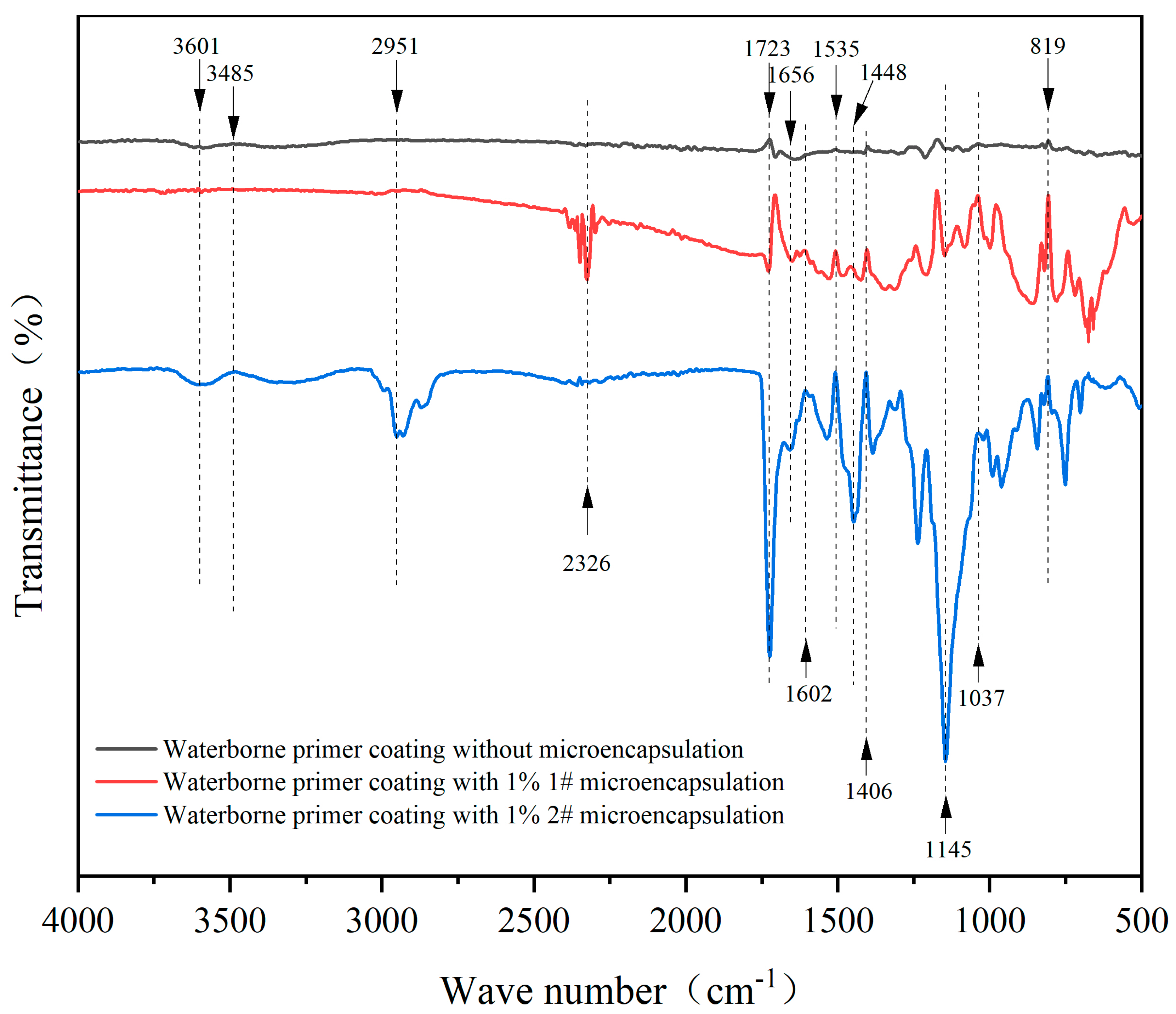
3.1.2. A Study of the Chemicals in Microcapsules

As demonstrated in Figure 6, the waterborne primer’s infrared spectra show two distinct patterns. The first pattern corresponds to the waterborne primer coatings devoid of microcapsules, while the second pattern corresponds to the waterborne primer coatings containing 1% of 1# and 1% of 2# microcapsules. The infrared spectrogram of the added 1# compound displays a C-H asymmetrical stretching vibrational absorption feature at 2326 cm−1. In addition, the infrared spectrogram displays a -NH bending vibrational absorption feature at 1602 cm−1 and a C-O stretching vibrational absorption feature at 1037 cm−1. The C-O stretching vibration peak of alcohols was observed at 1145 cm−1, the stretching vibration region of the carbonyl group was found at 1900–1500 cm−1, and the 1656 cm−1 peak corresponds to the flavonoid carbonyl peak. The skeleton vibration region of the aryl ring was identified at 1600–1450 cm−1, and the absorption peaks of the benzene ring skeleton vibration are found at 1450 cm−1, 1500 cm−1, and near 1600 cm−1. These findings aligned with the absorption peaks of flavonoids [52]. The infrared spectrograms of the waterborne primer coatings with 2# microcapsules added at 1656–1602 cm−1 may be the amino peak displacement due to the electrostatic interaction of AOB and chitosan. The change in the superposition of the hydroxyl and amino peaks at 3485 cm−1 reflects hydrogen bond formation. The presence of microcapsules did not interfere with the curing of the waterborne primer, as evidenced by the observation of the stretching maximum of the C=O functional group at 1723 cm−1 and the out-of-plane bending maximum of the C-H functional group at 819 cm−1 in all three curves. It is thus proved that the prepared microcapsules can stably exist in the waterborne primer coating.

3.2. Optical Performance Analysis of Coatings

As illustrated in Table 4, the effect of chromaticity value and colour deviation of the coating of Basswood without microcapsules and Basswood boards with 1#, 2#, 3#, and 4# microcapsules, respectively, is demonstrated. The L denotes the luminance, and a positive value means that the surface colour of the object being tested is light, and vice versa if it is dark. The a denotes the change in the colour from red to green, and a positive value means that the colour is reddish, and vice versa if it is greenish. The b denotes the change in the colour from yellow to blue, and the positive value means that the surface colour of the object under test is yellowish, and the negative value means that it is bluish. A positive value indicates a yellowish hue, while a negative value indicates a bluish hue. The ∆L (luminance difference) was calculated as L1L2, with the test data presented in the table. The colour difference was then calculated using the formula for colour difference. The data in Table 4 represent the optimal value of the colour difference after three replicates. The colour difference value of the samples exhibited no significant pattern; thus, it can be deduced that the ratio of chitosan and AOB has a substantial impact on the colour deviation value. However, it was observed that the colour deviation value with the addition of 2# was notably high, while the remaining samples exhibited similar or negligible differences.
As demonstrated in Table 5, an investigation was conducted into the gloss of the Basswood coating with different microcapsule additions. The data presented in the table are the mean of three measurements of the coating’s gloss at a 60° angle of incidence. According to the data, samples coated with only waterborne coating had a gloss level of 18.5 GU at a 60° angle. With the increase in AOB, it tends to decrease and then increase, indicating that the addition of AOB leads to an improvement in the gloss. A comparison of the experimental samples revealed that the gloss decreased when microcapsules without AOB were added, while the gloss of the coating film with microcapsules containing AOB rose as the percentage of AOB content increased continuously, and the addition of microcapsules 4# was superior to the gloss of all other linden boards, reaching 16.0 GU. This suggests that the addition of AOB enhanced the gloss. This is due to the fact that microencapsulated powders exhibit a greater degree of roughness and are less well dispersed in waterborne coatings. The presence of solid microcapsule powder in waterborne coatings has been demonstrated to reduce the specular light reflection, causing a reduction in the gloss of the coating [53,54,55,56]. The enhancement in the gloss of the coating may be ascribed to the synergistic effect of the combination of chitosan and AOB, which collectively elevate the refractive index of the coating, thus ensuring a more precise congruence with the refractive index of the Basswood substrate. This adjustment is hypothesised to reduce light loss at the interface, thereby improving the gloss of the coating.

3.3. Mechanical Properties of Coatings

3.3.1. Adhesion of Coatings on Basswood

As demonstrated in Table 6, the adhesion of surface coatings on Basswood boards was influenced by the incorporation of microcapsules. The data indicated that the adhesion of the waterborne coating without microcapsules was one, while the addition of microcapsules resulted in an increasing trend. This suggests that microcapsules may reduce the adhesion of the paint film and impact its performance. The adhesion of the coating with 1# microcapsules added and the coating of the Basswood with 2# microcapsules added were both 1, which was consistent with the blank samples. This indicated that the proportion of the ingredients in these three samples did not have a significant influence on the adhesion. The adhesion of the other two groups increased from one to two and four. This finding indicated that the incorporation of AOB caused a decrease in the adhesion. The reason may be because chitosan contains hydrophilic amino (-NH2) and hydroxyl (-OH) groups, while AOB may contain hydrophobic aromatic ring structure. After compounding, the surface chemistry of the microcapsules may be biased toward hydrophobicity, and the compatibility with polar substrates such as acrylic resins in waterborne coatings is poor, forming a weak interfacial bond.

3.3.2. Hardness of Coatings on Basswood

As demonstrated in Table 7, the hardness of Basswood was influenced by the incorporation of microcapsules. The data presented in Table 7 indicated that the hardness of the waterborne primer devoid of microcapsules was categorised as 2H, whereas the incorporation of waterborne primer without microcapsules containing AOB resulted in a decline in film hardness to 2B. However, the introduction of microcapsules comprising AOB leads to an enhancement in film hardness, with the magnitude of this effect increasing in proportion to the quantity of AOB contained within the microcapsules. It can thus be concluded that microcapsules containing only chitosan caused the hardness of the waterborne coating to decrease, and that the addition of AOB increased the hardness of the paint film. This phenomenon can be attributed to the inherent properties of chitosan, a natural polymer, which, when applied to waterborne coatings, forms a dense, continuous film layer. The hydrogen bonds formed between the molecular chains of chitosan and the phenolic hydroxyl groups of the AOB result in enhanced mechanical strength of the coating. The high cross-linking density enhances the hardness of the coating, which acts on the wood surface to form a rigid protective layer.

3.3.3. Impact Resistance of Surface Coating on Basswood

As demonstrated in Table 8, an enhancement in the impact resistance of Basswood boards was observed with increasing microcapsule addition to their surface coatings. An analysis of the data indicates a substantial variation in the impact resistance among the samples within all of the experimental groups. It is worth mentioning that the incorporation of microcapsules containing AOB led to a marked enhancement of this property in Basswood. Experiments demonstrated that as the proportion of chitosan to AOB varied, the increase in AOB produced an increase in performance. It was concluded that AOB can enhance the impact resistance of waterborne coatings. This phenomenon can be attributed to the formation of reversible hydrogen or coordination bonds between the -NH2 of chitosan and the -OH of AOB. During impact, the fracture-reorganisation of these dynamic bonds can dissipate energy, similar to the “sacrificial bond” mechanism, and enhance the toughness of the coating.
Subsequently, analysis of variance (ANOVA) was performed on the results of gloss, hardness, adhesion, and impact resistance tests. The results of the analysis are presented in Table 9. The grades of hardness are converted to numerical values for the purpose of analysis, and the conversion criteria are 4H-1, 3H-2, 2H-3, HB-4, 2B-5. Significance analysis was conducted utilising the one-way ANOVA method. The F-value and p-value were obtained, respectively. The F-value is the ratio of the between-group mean square to the within-group mean square and is used to test whether the difference between groups is significant. The p-value indicates the probability of observing the current F-value under the condition that the original hypothesis is valid. The following judgement criteria were employed to determine the significance of the observed difference: 0.01 ≤ p-value ≤ 0.05, indicating a significant difference; p-value ≤ 0.01, indicating a highly significant difference; p-value ≥ 0.05, indicating no significance. According to the aforementioned methods, the results are as follows: p < 0.01, indicating a significant difference between the measured data in terms of gloss, hardness, adhesion, and impact resistance, respectively. The experimental results are supported by these significant differences. This finding is consistent with the analysis of the results.

3.4. Self-Healing Rate of Surface Coatings on Basswood

As demonstrated in Figure 7 and Table 10, the investigation focused on the self-healing rate of Basswood with varying microcapsule additions. In waterborne coating with 1.0% addition amount, 1#, 2#, 3#, and 4# microcapsules were incorporated into the waterborne coating, and the surface coating with 1# addition exhibited the highest self-healing rate of 24.20%. It was evident that the coatings of the three groups of samples exhibit a certain degree of self-healing capability. This phenomenon can be attributed to the following mechanism: when subjected to external stresses, the wall material undergoes a rupture. Subsequent to this rupture, the core material, in this case tung oil, is dispersed into the surrounding environment, thus filling the minute fissures. Due to its material compatibility with the coating, the core material is able to gradually heal the coating. Consequently, it can be deduced that the self-healing effect of microcapsules devoid of AOB was superior. The microencapsulated wall undergoes rupture due to localised stress when the coating is damaged, thus releasing the core tung oil. Tung oil contains a high proportion of conjugated double bonds (e.g., α-tungstic acid), which, under conditions of oxygen and light, undergo an auto-oxidation reaction, thereby generating peroxides and further cross-linking to form a three-dimensional mesh structure. This process generates a dense hydrophobic film, which seals cracks and prevents the intrusion of water, oxygen, and microorganisms. The synergy between physical filling and oxidative cross-linking mechanisms facilitates the dynamic self-healing of the wood coating.
The decrease in self-healing rate after the addition of AOB could be due to larger or uneven particle size. These may have resulted in uneven dispersion in the coating, making it difficult for the restorative agent to be concentrated and released in the vicinity of the cracks. An introduction of AOB may have altered the intermolecular forces of the chitosan, which may have resulted in a denser or more brittle wall structure of the chitosan, decreasing the storage stability of the microcapsules.

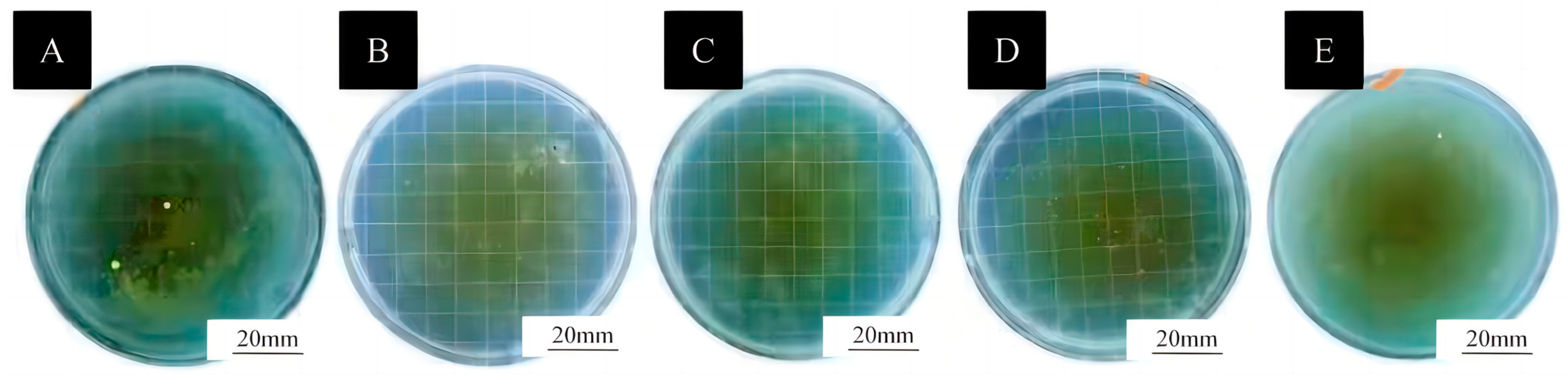
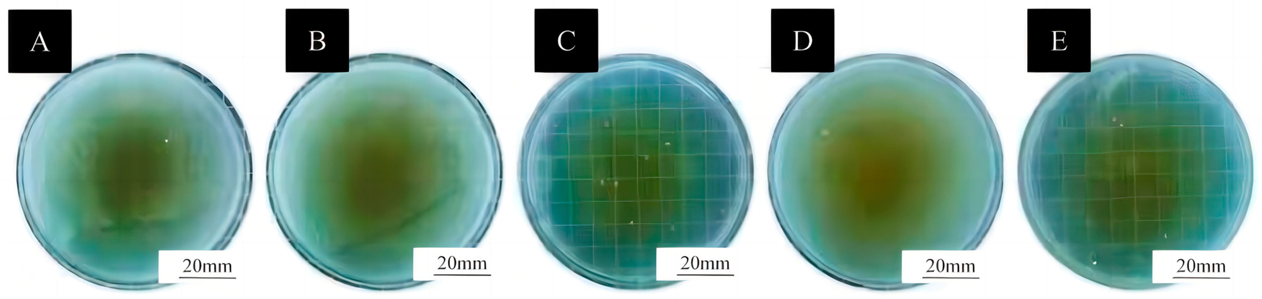
3.5. Antibacterial Properties of Surface Coatings on Basswood

The results of the antimicrobial test carried out are shown in Figure 8 and Figure 9. By observing the Petri dishes, it was found that the medium with Staphylococcus aureus did not have antimicrobial properties because it did not contain microcapsules. A large number of bacteria were produced on the surface, and a large number of bacterial colonies covered the surface of the Petri dishes. Observing the antimicrobial samples with the addition of 1# and with the addition of 4#, it can be seen from the pictures that no significant antimicrobial properties were produced, and their surfaces were covered with a layer of bacteria. It can be seen that the culture medium with the addition of 2# and with the addition of 3# microcapsules had a more pronounced antimicrobial property.
Subsequent observation of the samples containing Escherichia coli revealed that they did not demonstrate significant antimicrobial properties. Microscopic analysis of the samples without added microcapsules revealed a thin layer of bacteria covering the surface. The antimicrobial samples containing Sample 1 and Sample 3 exhibited a similar bacterial layer on their surface, though this layer did not manifest an obvious antimicrobial effect. However, the samples containing 2# and 4# demonstrated a more pronounced antimicrobial property. In conclusion, the sample containing 2# demonstrated superior antimicrobial properties.

4. Conclusions

This paper proposes the development of tung oil antimicrobial microcapsules, coated with a blend of AOB and chitosan, which have been demonstrated to possess effective antimicrobial properties. The experimental investigation involved the preparation of microcapsules through a rigorous process of experimentation, leading to the successful formulation of the microcapsules. The performance of the paint film was then tested, revealing that gloss increased with the percentage of AOB, though no significant change or effect on the colour difference value was observed. Impact resistance increased with the increase in the content of AOB, but adhesion decreased again with the increase in the percentage of AOB in the microcapsules. Overall, the hardness decreased, though it increased with the increase in the content of AOB. In terms of antimicrobial properties, it was found that microcapsules prepared with a ratio of 9:1 of chitosan-coated AOB exhibited superior antimicrobial effects. Subsequent experiments will focus on enhancing the antimicrobial properties of microcapsules by utilising essential oils with enhanced antimicrobial properties as the core material.

Author Contributions

Conceptualization and methodology, writing—review and editing, validation, resources, data management, N.Z.; formal analysis, investigation, and supervision, X.Y. All authors have read and agreed to the published version of the manuscript.

Funding

This project was partly supported by the Qing Lan Project and the Natural Science Foundation of Jiangsu Province (BK20201386).

Institutional Review Board Statement

Not applicable.

Informed Consent Statement

Not applicable.

Data Availability Statement

Data are contained within the article.

Conflicts of Interest

The authors declare that there are no conflicts of interest.

References

  1. Hisham, F.; Akmal, M.H.M.; Ahmad, F.; Ahmad, K.; Samat, N. Biopolymer Chitosan: Potential Sources, Extraction Methods, and Emerging Applications. Ain Shams Eng. J. 2024, 15, 102424. [Google Scholar] [CrossRef]
  2. Li, X.X.; Dong, W.J.; Nalin, A.P.; Wang, Y.F.; Pan, P.; Xu, B.; Zhang, Y.B.; Tun, S.; Zhang, J.Y.; Wang, L.S.; et al. The Natural Product Chitosan Enhances the Anti-Tumor Activity of Natural Killer Cells by Activating Dendritic Cells. Oncoimmunology 2018, 7, e1431085. [Google Scholar] [CrossRef]
  3. Diwu, W.L.; Dong, X.; Nasif, O.; Alharbi, S.A.; Zhao, J.; Li, W. In-vivo Investigations of Hydroxyapatite/Co-polymeric Composites Coated Titanium Plate for Bone Regeneration. Front. Cell Dev. Biol. 2020, 8, 631107. [Google Scholar] [CrossRef]
  4. Xie, L.; Chen, F.Y.; Tang, Y.B.; Hao, C.C. Tetracycline degradation by persulfate activated with novel magnetic composite ferrocene/chitosan@Fe3O4. J. Chem. Technol. Biotechnol. 2022, 97, 2766–2774. [Google Scholar] [CrossRef]
  5. Zheng, K.W.; Yang, F.; Huang, Z.H.; Zhan, Y.F.; Xiao, Z.F.; Li, W.; Wang, W.; Qin, C.Q. Preparation of Chitosan Film-Loaded Palladium Catalyst Materials and Their Application in Suzuki Coupling Reactions. J. Mater. Res. Technol. 2022, 20, 3905–3917. [Google Scholar] [CrossRef]
  6. Sun, X.X.; Yu, D.Y.; Ying, Z.Y.; Pan, C.Q.; Wang, N.; Huang, F.F.; Ling, J.H.; Ouyang, X.K. Fabrication of Ion-Crosslinking Aminochitosan Nanoparticles for Encapsulation and Slow Release of Curcumin. Pharmaceutics 2019, 11, 584. [Google Scholar] [CrossRef]
  7. Yuan, T.; Guo, X.F.; Shao, S.Y.; An, R.M.; Wang, J.; Sun, J. Characterization and identification of flavonoids from Bambusa chungii leaves extract by UPLC-ESI-Q-TOF-MS/MS. Acta Chromatogr. 2021, 33, 281–294. [Google Scholar] [CrossRef]
  8. Gong, J.Y.; Xia, D.Z.; Huang, J.; Ge, Q.; Mao, J.W.; Liu, S.W.; Zhang, Y. Functional components of bamboo shavings and bamboo leaf extracts and their antioxidant activities in vitro. J. Med. Food 2015, 18, 453–459. [Google Scholar] [CrossRef] [PubMed]
  9. Mao, J.W.; Yin, J.; Ge, Q.; Jiang, Z.L.; Gong, J.Y. In vitro antioxidant activities of polysaccharides extracted from Moso BambooLeaf. Int. J. Biol. Macromol. 2013, 55, 1–5. [Google Scholar] [CrossRef]
  10. Xie, H.K.; Li, A.; Zhao, M.T.; Zhang, M.; Liu, Z.Y.; Yin, F.W.; Liu, X.Y.; Zhou, D.Y.; Shahidi, F.; Zhu, B.W. Effects of antioxidants of bamboo leaves (AOB) on the oxidative susceptibility of glycerophosphocholine and glycerophosphoethanolamine in dried scallop (Argopecten irradians) adductor muscle during storage. LWT—Food Sci. Technol. 2020, 134, 110214. [Google Scholar] [CrossRef]
  11. Liu, L.Y.; Xia, B.N.; Jin, C.; Zhang, Y.; Zhang, Y. Chemical acylation of water-soluble antioxidant of bamboo leaves (Aob-w) and functional evaluation of oil-soluble AOB (cAob-o). J. Food Sci. 2014, 79, C1886–C1894. [Google Scholar] [CrossRef] [PubMed]
  12. Shang, Y.F.; Kim, S.M.; Um, B.H. Optimisation of pressurised liquid extraction of antioxidants from black bamboo leaves. Food Chem. 2014, 154, 164–170. [Google Scholar] [CrossRef] [PubMed]
  13. Ma, X.; Wang, E.P.; Lu, Y.Y.; Wang, Y.; Ou, S.Y.; Yan, R. Acylation of antioxidant of bamboo leaves with fatty acids by lipase and the acylated derivatives’ efficiency in the inhibition of acrylamide formation in fried potato crisps. PLoS ONE 2015, 10, e0130680. [Google Scholar] [CrossRef]
  14. Liu, L.Y.; Pang, M.R.; Zhang, Y. Lipase-catalyzed regioselective synthesis of flavone C-glucosides esters and high-efficiency oil-soluble antioxidant of bamboo leaves (eAOB-o). Eur. J. Lipid Sci. Technol. 2015, 117, 1636–1646. [Google Scholar] [CrossRef]
  15. Chaves, J.O.; de Souza, M.C.; da Silva, L.C.; Lachos-Perez, D.; Torres-Mayanga, P.C.; Machado, A.P.D.; Forster-Carneiro, T.; Vázquez-Espinosa, M.; González-de-Peredo, A.V.; Barbero, G.F.; et al. Extraction of flavonoids from natural sources using modern techniques. Front. Chem. 2020, 8, 507887. [Google Scholar] [CrossRef]
  16. Ma, E.P.; Wei, Z.H.; Lian, C.; Zhou, Y.P.; Gan, S.C.; Xu, B. Preparation of colored microcapsule phase change materials with colored SiO2 shell for thermal energy storage and their application in latex paint coating. Materials 2021, 14, 4012. [Google Scholar] [CrossRef] [PubMed]
  17. Alencar, D.D.D.; de Souza, E.L.; Almeida, E.T.D.; da Silva, A.L.; Oliveira, H.M.L.; Cavalcanti, M.T. Microencapsulation of Cymbopogon citratus D.C. Stapf Essential Oil with Spray Drying: Development, Characterization, and Antioxidant and Antibacterial Activities. Foods 2022, 11, 1111. [Google Scholar] [CrossRef]
  18. Zhao, C.; Chen, G.P.; Wang, H.; Zhao, Y.J.; Chai, R.J. Bio-inspired Intestinal Scavenger from Microfluidic Electrospray for Detoxifying Lipopolysaccharide. Bioact. Mater. 2020, 6, 1653–1662. [Google Scholar] [CrossRef]
  19. Long, M.; Feng, C.; Li, Y.J.; Wang, X.H.; Cai, H.Z.; Zhan, G. Preparation, Characterization and in Vitro Release Characteristics of Sustained-Release Chitosan Microcapsules Containing Tea Tree Oil. Food Sci. 2019, 40, 242–248. [Google Scholar]
  20. Huang, Z.X.; Wang, Y.L.; Tan, S.J.; Gao, J.M.; Du, L. Preparation, Characterization and Antibacterial Activity of Chitosan-Cinnamon Essential Oil Microcapsules. J. Cellul. Sci. Technol. 2024, 32, 14–22. [Google Scholar]
  21. Hu, W.G.; Liu, Y.; Konukcu, A.C. Study on withdrawal load resistance of screw in wood-based materials: Experimental and numerical. Wood Mater. Sci. Eng. 2023, 18, 334–343. [Google Scholar] [CrossRef]
  22. Hu, W.G.; Yu, R.Z. Mechanical and acoustic characteristics of four wood species subjected to bending load. Maderas Cienc. Tecnol. 2023, 25, 39. [Google Scholar] [CrossRef]
  23. Hu, W.G.; Luo, M.Y.; Hao, M.M.; Tang, B.; Wan, C. Study on the effects of selected factors on the diagonal tensile strength of oblique corner furniture joints constructed by wood dowel. Forests 2023, 14, 1149. [Google Scholar] [CrossRef]
  24. Liu, Y.; Hu, W.G.; Kasal, A.; Erdil, Y.Z. The State of the Art of Biomechanics Applied in Ergonomic Furniture Design. Appl. Sci. 2023, 13, 12120. [Google Scholar] [CrossRef]
  25. Hu, W.G.; Luo, M.Y.; Yu, R.Z.; Zhao, Y. Effects of the selected factors on cyclic load performance of T-shaped mortise-and-tenon furniture joints. Wood Mater. Sci. Eng. 2024, 18, 1–10. [Google Scholar] [CrossRef]
  26. Xu, R.; Wang, D.; Dou, L.H.; Cui, J.Q.; He, L.; Feng, F.Q.; Liu, F.F. Preparation and properties of self-healing polyurethane wood coatings with borate ester bonds. J. For. Eng. 2024, 9, 54–60. [Google Scholar]
  27. Xu, H.J.; Chen, C.B.; Xiao, J.H.; Pan, J.Q.; Shen, L.M.; Zhang, Y.; Sun, F.L. Effect of chitosan quaternary ammonium salt-modified waterborne epoxy resin on wood dimensional stability and anticorrosion/antimildew properties. J. For. Eng. 2024, 38, 1209–1527. [Google Scholar]
  28. Wu, Z.H.; Chang, Y.J. Research and application of microcapsule technology in the wood industry. J. For. Eng. 2024, 38, 1029–1345. [Google Scholar]
  29. Liu, Q.Q.; Gao, D.; Xu, W. Effect of Polyurethane Non-Transparent Coating Process on Paint Film Performance Applied on Modified Poplar. Coatings 2022, 12, 39. [Google Scholar] [CrossRef]
  30. Xue, J.X.; Xu, W.; Zhou, J.C.; Mao, W.G.; Wu, S.S. Effects of High-Temperature Heat Treatment Modification by Impregnation on Physical And Mechanical Properties of Poplar. Materials 2022, 15, 7334. [Google Scholar] [CrossRef]
  31. Mai, C.; Schmitt, U.; Niemz, P. A brief overview on the development of wood research. Holzforschung 2022, 75, 102–119. [Google Scholar] [CrossRef]
  32. Zhang, N.; Xu, W.; Tan, Y. Multi-attribute hierarchical clustering for product family division of customized wooden doors. BioResources 2023, 18, 7889–7904. [Google Scholar] [CrossRef]
  33. Hu, J.; Liu, Y.; Wang, J.X.; Xu, W. Study of selective modification effect of constructed structural color layers on European beech wood surfaces. Forests 2024, 15, 261. [Google Scholar] [CrossRef]
  34. Jian, H.; Liang, Y.Q.; Deng, C.; Xu, J.X.; Liu, Y.; Shi, J.Y.; Wen, M.Y.; Park, H.J. Research progress on the improvement of flame retardancy, hydrophobicity, and antibacterial properties of wood surfaces. Polymers 2023, 15, 951. [Google Scholar] [CrossRef]
  35. Gu, Y.T.; Zhang, J.L. Tensile properties of natural and synthetic rattan strips used as furniture woven materials. Forests 2020, 11, 129. [Google Scholar] [CrossRef]
  36. Wu, S.S.; Zhou, L.C.; Xu, W. A convenient approach to manufacturing lightweight and high-sound-insulation plywood using furfuryl alcohol/multilayer graphene oxide as a shielding layer. Wood Mater. Sci. Eng. 2024, 19, 1226–1233. [Google Scholar] [CrossRef]
  37. Liu, Q.Q.; Gao, D.; Xu, W. Influence of the Bottom Color Modification and Material Color Modification Process on the Performance of Modified Poplar. Coatings 2021, 11, 660. [Google Scholar] [CrossRef]
  38. Su, Y.; Liu, Y.; Hu, J. Impact of wood surface roughness on optical properties of photonic crystal and amorphous photonic crystal structural color coatings. Wood Mater. Sci. Eng. 2024, 19, 1305–1313. [Google Scholar] [CrossRef]
  39. Weng, M.Y.; Zhu, Y.T.; Mao, W.G.; Zhou, J.C.; Xu, W. Nano-Silica/Urea-Formaldehyde Resin-Modified Fast-Growing Lumber Performance Study. Forests 2023, 14, 1440. [Google Scholar] [CrossRef]
  40. Wang, C.; Zhang, C.Y.; Zhu, Y. Reverse design and additive manufacturing of furniture protective foot covers. BioResources 2024, 19, 4670–4678. [Google Scholar] [CrossRef]
  41. Wang, X.Y.; Liu, X.; Wu, S.S.; Xu, W. The influence of different impregnation factors on mechanical properties of silica sol-modified Populus tomentosa. Wood Fiber Sci. 2024, 56, 65–71. [Google Scholar]
  42. Tampucci, S.; Tofani, G.; Chetoni, P.; Di Gangi, M.; Mezzetta, A.; Paganini, V.; Burgalassi, S.; Pomelli, C.S.; Monti, D. Sporopollenin Microcapsule: Sunscreen Delivery System with Photoprotective Properties. Pharmaceutics 2022, 14, 2041. [Google Scholar] [CrossRef] [PubMed]
  43. Liu, C.; Xu, W. Effect of Coating Process on Properties of Two-Component Waterborne Polyurethane Coatings for Wood. Coatings 2022, 12, 1857. [Google Scholar] [CrossRef]
  44. GB/T 11186.3-1989; Method for Measuring the Color of Painted Film—Part 3: Calculation of Color Difference. Standardization Administration of the People’s Republic of China: Beijing, China, 1989.
  45. GB/T 4893.6-2013; Physical and Chemical Performance Tests of the Paint Film on the Surface of Furniture—Part 6: Method for Determining Gloss. Standardization Administration of the People’s Republic of China: Beijing, China, 2013.
  46. GB/T 4893.4-2013; Physical and Chemical Performance Tests of the Paint Film on the Surface of Furniture—Part 4: Determination of Adhesion by Cross-Cutting Method. Standardization Administration of the People’s Republic of China: Beijing, China, 2013.
  47. GB/T 6739-2006; Paints and Varnishes—Determination of Film Hardness by Pencil Method. Standardization Administration of the People’s Republic of China: Beijing, China, 2006.
  48. GB/T 4893.9-2013; Physical and Chemical Performance Tests of the Paint Film on the Surface of Furniture—Part 9: Determination of Impact Resistance. Standardization Administration of the People’s Republic of China: Beijing, China, 2013.
  49. GB/T 21866-2008; Test Method for Antibacterial Property and Antibacterial Effect of Antibacterial Coatings (Paint Films). Standardization Administration of the People’s Republic of China: Beijing, China, 2008.
  50. GB/T 4789.2-2022; Microbiological Examination of Food Hygiene—Aerobic Plate Count. Standardization Administration of the People’s Republic of China: Beijing, China, 2022.
  51. Shen, Y.; Shan, X.F.; Etim, I.P.; Siddiqui, M.A.; Yang, Y.; Shi, Z.W.; Su, X.P.; Chen, J.X. Comparative study of the effects of nano ZnO and CuO on the biodegradation, biocompatibility, and antibacterial properties of micro-arc oxidation coating of magnesium alloy. Acta Metall. Sin. Engl. 2024, 37, 242–254. [Google Scholar] [CrossRef]
  52. Zhao, L.R.; Zhang, W.; Wu, Q.; Fu, C.H.; Ren, X.L.; Lv, K.P.; Ma, T.C.; Chen, X.D.; Tan, L.F.; Meng, X.W. Lanthanide Europium MOF Nanocomposite as the Theranostic Nanoplatform for Microwave Thermo-Chemotherapy and Fluorescence Imaging. J. Nanobiotechnol. 2022, 20, 133. [Google Scholar] [CrossRef]
  53. Wang, C.; Yu, J.H.; Jiang, M.H.; Li, J.Y. Effect of selective enhancement on the bending performance of fused deposition methods 3D-printed PLA models. BioResources 2024, 19, 2660–2669. [Google Scholar] [CrossRef]
  54. Wang, C.; Zhou, Z.Y. Optical properties and lampshade design applications of PLA 3D printing materials. BioResources 2023, 18, 1545–1553. [Google Scholar] [CrossRef]
  55. Zhou, J.C.; Xu, W. A fast method to prepare highly isotropic and optically adjustable transparent wood-based composites based on interface optimization. Ind. Crops Prod. 2024, 218, 118898. [Google Scholar] [CrossRef]
  56. Wang, C.; Zhang, C.Y.; Ding, K.Q.; Jiang, M.H. Immersion polishing post-treatment of PLA 3D printed formed parts on its surface and mechanical performance. BioResources 2023, 18, 7995–8006. [Google Scholar] [CrossRef]
Figure 1. (A) Fresh tung oil after Tween-80 emulsification, (B) emulsified tung oil under storage conditions after 3 days.
Coatings 15 00517 g001
Figure 2. Flow chart for microencapsulation preparation.
Coatings 15 00517 g002
Figure 3. Microscopic diagram of microcapsules (A) Sample 1#, (B) Sample 2#, (C) Sample 3#, and (D) Sample 4#.
Coatings 15 00517 g003
Figure 4. Image shows SEM microcapsules: (A) Sample 1#, (B) Sample 2#, (C) Sample 3#, and (D) Sample 4#.
Coatings 15 00517 g004
Figure 5. Particle size distribution (A) Sample 1#, (B) Sample 2#, (C) Sample 3#, and (D) Sample 4#.
Coatings 15 00517 g005
Figure 6. Infrared spectra of waterborne coating without microcapsules, waterborne coating with 1# microcapsules, and waterborne coating with 2# microcapsules.
Coatings 15 00517 g006
Figure 7. Scratch images of surface coatings on Basswood with different microcapsules added: (A) coating with 1#, (B) coating with 2#, (C) coating with 3#, (D) coating with 4#, (E) coating without microcapsules, (F) coating with 1# after self-healing, (G) coating with 2# after self-healing, (H) coating with 3# after self-healing, (I) coating with 4# after self-healing, (J) coating without microcapsules after self-healing.
Coatings 15 00517 g007
Figure 8. Plot of Staphylococcus aureus colonisation on coated surfaces with different microcapsules added: (A) coatings without microcapsules, coatings with (B) 1#, (C) 2#, (D) 3#, (E) 4# microcapsules.
Coatings 15 00517 g008
Figure 9. Plot of Escherichia coli colonisation on coated surfaces with different microcapsules added (A) coatings without microcapsules, coatings with (B) 1#, (C) 2#, (D) 3#, and (E) 4# microcapsules.
Coatings 15 00517 g009
Table 1. Experimental materials list.
Test MaterialPurityManufacturer
Chitosan with a deacetylation degree of 80.0%–95.0%ARSinopharm Chemical Reagent Co., Ltd., Shanghai, China
Acetic acidARHenan Maigao Chemical Co., Ltd., Zhengzhou, China
Sodium tripolyphosphate (STPP)ARTianjin Zhonglian Chemical Reagent Co., Ltd., Tianjin, China
Tween-80ARJinan HSBC Chemical Co., Ltd., Jinan, China
NaOHARShandong Xuanhai Chemical Co., Ltd., Weifang, China
Anhydrous ethanolARJinan Hongrun Chemical Co., Ltd., Jinan, China
Nutrient agar medium-Tianjin Zhonglian Chemical Reagent Co., Ltd., Tianjin, China
Nutrient broth-Zhongshan Baiwei Biotechnology Co., Ltd., Zhongshan, China
Staphylococcus aureus-Shanghai Shifeng Biotechnology Co., Ltd., Shanghai, China
Escherichia coli-Shanghai Shifeng Biotechnology Co., Ltd., Shanghai, China
Cleaning agentARGuangdong Baiyun Cleaning Group Co., Ltd., Guangzhou, China
Citric acidARShandong Lemon Biochemical Co., Ltd., Anqiu, China
Table 2. The amount of raw material needed for the microcapsule.
Sample
(#)
Chitosan
(g)
Antioxidant of Bamboo Leaves
(g)
Ethanol
(g)
Deionized Water
(g)
Tung Oil
(g)
Tween-80
(g)
Deionized Water
(g)
STPP
(g)
Deionized Water
(g)
11.000.001.0099.900.500.4039.600.2019.80
20.900.100.9089.100.500.4039.600.2019.80
30.800.200.8079.200.500.4039.600.2019.80
40.700.300.7069.300.500.4039.600.2019.80
Table 3. Evaluation table of coating impact position grade.
LevelChanges in Coating on Wood Surface
1No visible changes (no damage).
2No cracks on the surface of the coating, but visible impact marks.
3There are mild cracks on the surface of the coating, usually 1–2 circular or arc cracks.
4There are moderate to severe cracks on the surface of the coating, usually 3–4 circular or arc cracks.
5The surface of the coating is severely damaged, usually with more than 5 cycles of ring cracks, arc cracks, or coating detachment.
Table 4. The chromaticity values and colour deviation of surface coatings on Basswood with different microcapsules added.
Sample (#)Microcapsule Content (%)Chromaticity ParameterE
Lab
No microcapsules0.069.612.620.34.78
66.716.420.3
1# microcapsules added1.067.014.627.51.51
66.215.628.3
2# microcapsules added1.066.928.258.731.03
73.510.933.8
3# microcapsules added1.072.610.921.65.76
72.610.115.9
4# microcapsules added1.082.15.5028.33.67
68.214.727.0
Table 5. Glossiness and light loss rate of surface coatings on Basswood with different microcapsules added.
Sample (#)Microcapsule Content (%)Glossiness (GU)Light Loss Rate (%)
60°
No microcapsules0.018.5-
1# microcapsules1.011.736.76
2# microcapsules1.09.747.57
3# microcapsules1.013.924.86
4# microcapsules1.016.013.51
Table 6. Adhesion grade of surface coatings on Basswood with different microcapsules added.
Sample (#)Adhesion Level (Level)
No microcapsules1
1# microcapsules1
2# microcapsules1
3# microcapsules2
4# microcapsules4
Table 7. Hardness of coatings on Basswood with different samples.
Sample (#)Hardness
No microcapsules2H
1# microcapsules2B
2# microcapsulesHB
3# microcapsules3H
4# microcapsules4H
Table 8. Impact resistance grade of surface coatings on Basswood with different microcapsules added.
Sample (#)Impact Resistance Level (cm)
No microcapsules10.0
1# microcapsules10.0
2# microcapsules12.0
3# microcapsules13.0
4# microcapsules15.0
Table 9. Significance analysis of coatings.
PropertyDifference SourceSSDFMSFp
HardnessBetween Groups21.06745.26719.750<0.001
Within Groups2.667100.267
Total23.73314
Impact ResistanceBetween Groups43.333410.83340.625<0.001
Within Groups2.667100.267
Total46.00014
AdhesionBetween Groups16.26744.06761.000<0.001
Within Groups0.667100.067
Total16.93314
GlossinessBetween Groups151.123437.78189.246<0.001
Within Groups4.233100.423
Total155.35614
Table 10. Self-healing rate of surface coatings on Basswood with different microcapsules added.
Sample (#)Microcapsule Content (%)Initial Scratch Width (μm)Scratch Width After 24 h (μm)Self-Healing Rate (%)
No microcapsules1.026.2923.50 10.61
1# microcapsules1.029.0922.05 24.20
2# microcapsules1.044.9742.20 6.16
3# microcapsules1.023.6219.11 19.09
4# microcapsules1.033.6330.97 7.91
Disclaimer/Publisher’s Note: The statements, opinions and data contained in all publications are solely those of the individual author(s) and contributor(s) and not of MDPI and/or the editor(s). MDPI and/or the editor(s) disclaim responsibility for any injury to people or property resulting from any ideas, methods, instructions or products referred to in the content.

