
Antifouling Performance and Long-Term Efficiency of a Zwitterionic Sulfobetaine-Hydroxyethyl-Containing Polymethylmethacrylate Ter-Co-Polymer Coating Against Biomass-Producing Photosynthetic Strains


 ,
,  , and
, and 
Abstract
1. Introduction
2. Materials and Methods
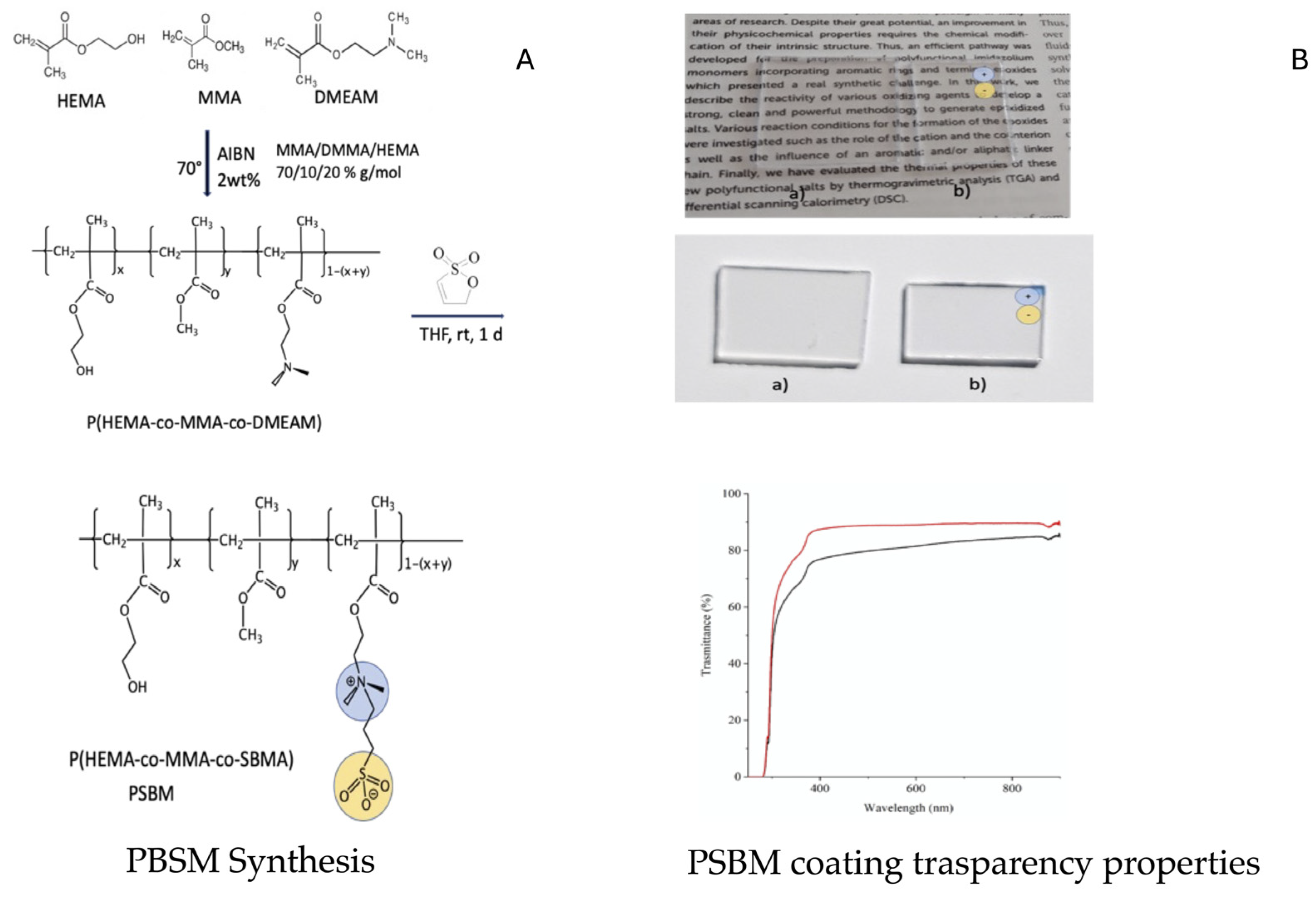
2.1. PSBM Synthesis
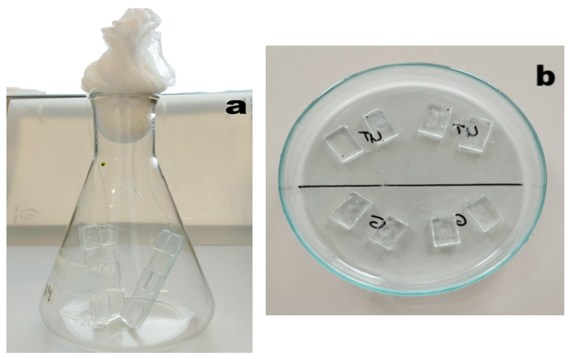
2.2. Preparation of PSBM Drop-Casting Mixture
2.3. PMMA Coupons
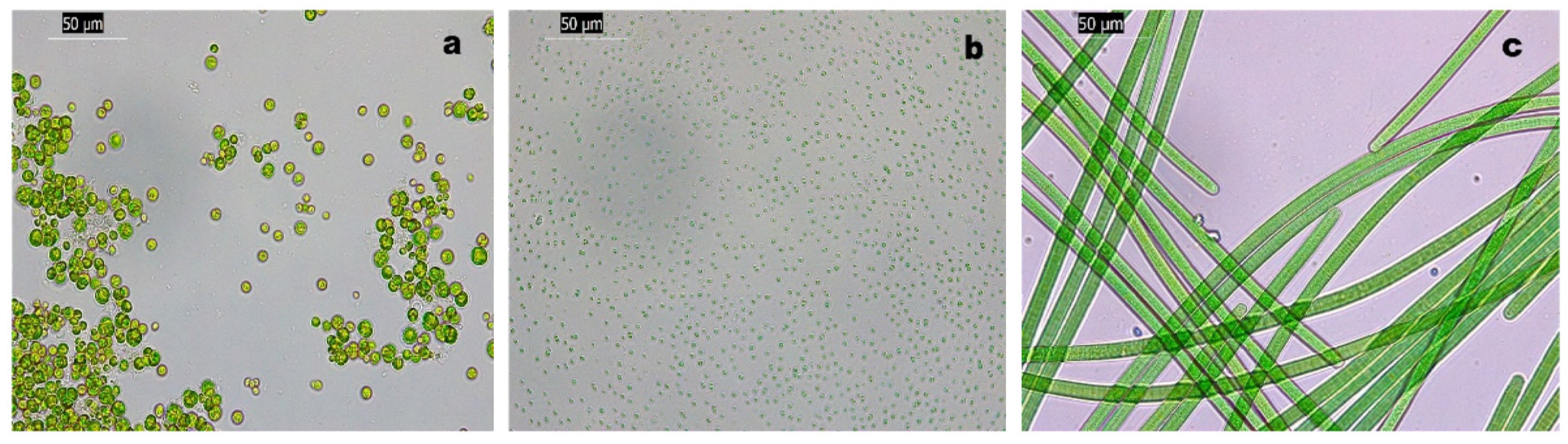
2.4. Photosynthetic Strains
2.5. Antifouling Assays
2.6. Tribological Tests
2.7. Statistical Analysis
3. Results and Discussion
3.1. Biomass Production
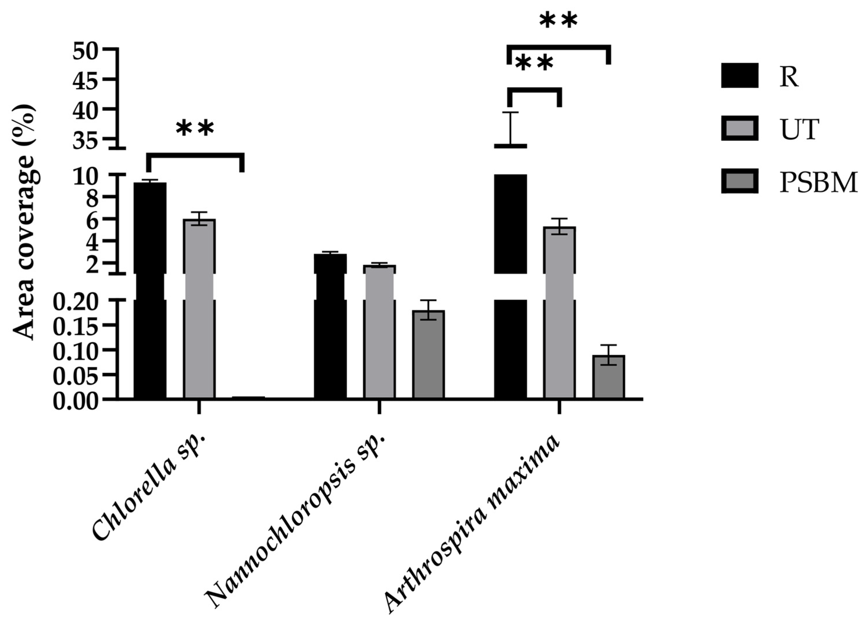
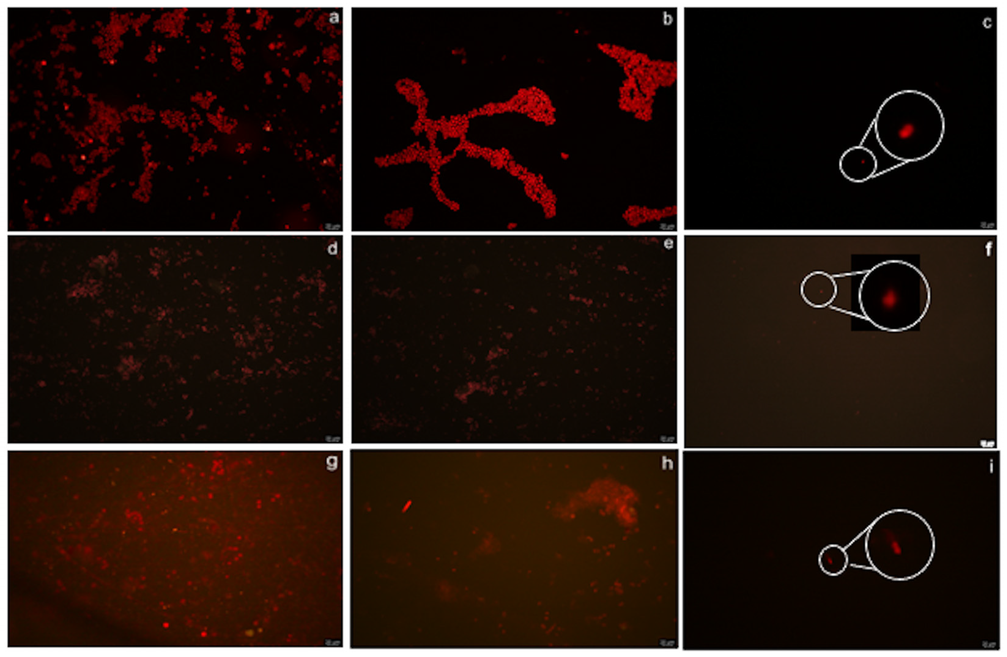
3.2. Antifouling Performance
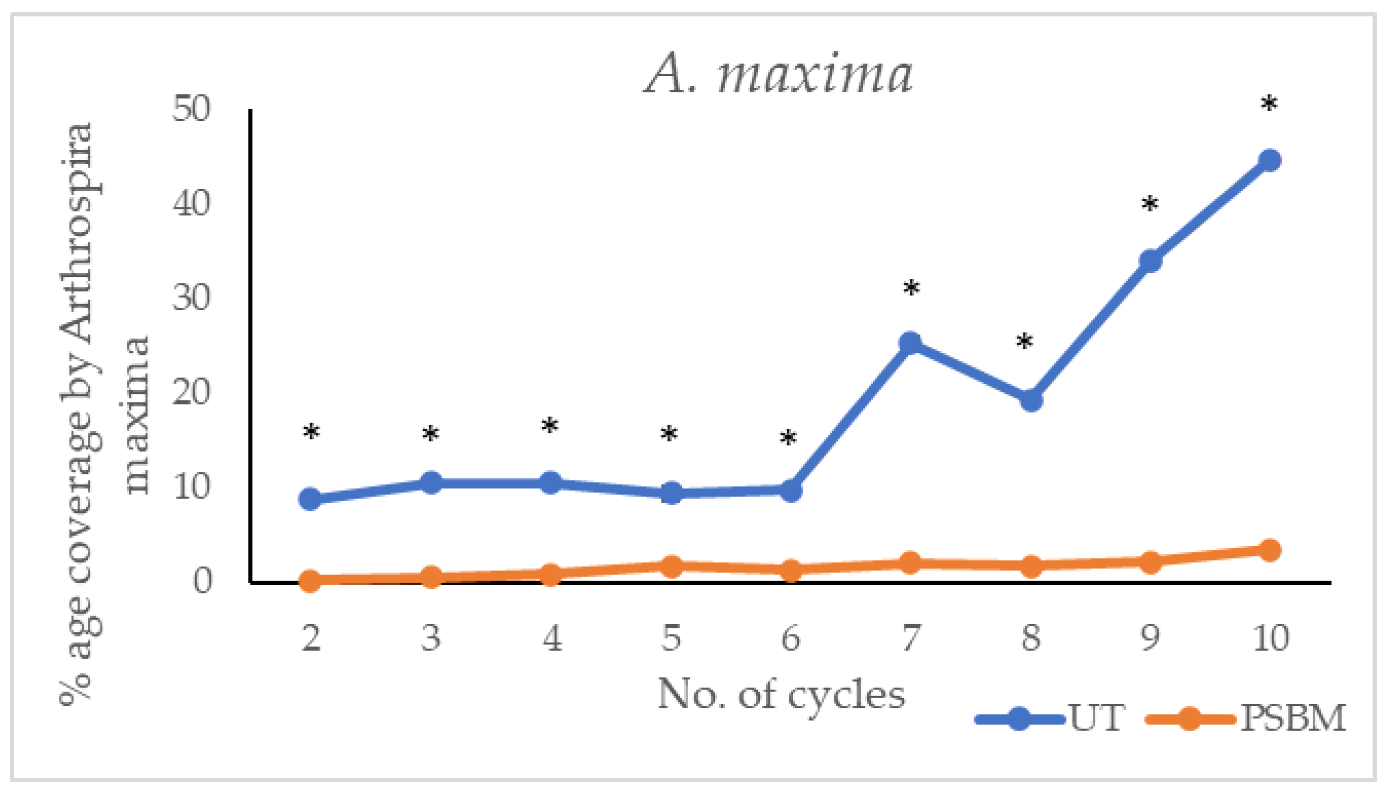
3.3. Long-Term Effectiveness of the PSBM
3.3.1. Biomass Production During Each Cycle
3.3.2. Antifouling Performance of Strains Through Tribological Approach
4. Conclusions
- mitigate the biofouling due to the three photosynthetic strains considered as models, i.e., Chlorella sp., Nannochloropsis sp., and A. maxima;
- maintenance of optical transparency;
- surface protection against wear.
5. Patents
Author Contributions
Funding
Institutional Review Board Statement
Informed Consent Statement
Data Availability Statement
Acknowledgments
Conflicts of Interest
Abbreviations
| PMMA | Polymethylmethacrylate |
| PSBM | Polysulfobetaine methacrylate/zwitterionic sulfobetaine-hydroxyethyl-containing polymethylmethacrylate ter-co-polymer |
| AF | Antifouling |
| UT | Untreated |
| R | Rough |
References
- Joy, S.R.; Anju, T.R. Microalgal Biomass: Introduction and Production Methods. In Handbook of Biomass; Thomas, S., Hosur, M., Pasquini, D., Jose Chirayil, C., Eds.; Springer: Singapore, 2023. [Google Scholar] [CrossRef]
- López-Hernández, J.-F.; Kean-Meng, T.; Asencio-Alcudia, G.-G.; Asyraf-Kassim, M.; Alvarez-González, C.-A.; Márquez-Rocha, F.-J. Sustainable Microalgae and Cyanobacteria Biotechnology. Appl. Sci. 2022, 12, 6887. [Google Scholar] [CrossRef]
- Mobin, S.M.A.; Chowdhury, H.; Alam, F. Commercially important bioproducts from microalgae and their current applications—A review. Energy Procedia 2019, 160, 752–760. [Google Scholar] [CrossRef]
- Rizwan, M.; Mujtaba, G.; Memon, S.A.; Lee, K.; Rashid, N. Exploring the potential of microalgae for new biotechnology applications and beyond: A review. Renew. Sustain. Energy Rev. 2018, 92, 394–404. [Google Scholar] [CrossRef]
- Khan, M.I.; Shin, J.H.; Kim, J.D. The promising future of microalgae: Current status, challenges, and optimization of a sustainable and renewable industry for biofuels, feed, and other products. Microb. Cell Factories 2018, 17, 36. [Google Scholar] [CrossRef]
- Eze, C.N.; Onyejiaka, C.K.; Ihim, S.A.; Ayoka, T.O.; Aduba, C.C.; Ndukwe, J.K.; Nwaiwu, O.; Onyeaka, H. Bioactive compounds by microalgae and potentials for the management of some human disease conditions. AIMS Microbiol. 2023, 7, 55–74. [Google Scholar] [CrossRef]
- Sun, Y.; Huang, Y.; Liao, Q.; Fu, Q.; Zhu, X. Enhancement of microalgae production by embedding hollow light guides to a flat-plate photobioreactor. Bioresour. Technol. 2016, 207, 31–38. [Google Scholar] [CrossRef]
- Kaur, M.; Bhatia, S.; Gupta, U.; Decker, E.; Tak, Y.; Bali, M.; Gupta, V.K.; Dar, R.A.; Bala, S. Microalgal bioactive metabolites as promising implements in nutraceuticals and pharmaceuticals: Inspiring therapy for health benefits. Phytochem. Rev. 2023, 22, 903–933. [Google Scholar] [CrossRef]
- Chanquia, S.N.; Vernet, G.; Kara, S. Photobioreactors for cultivation and synthesis: Specifications, challenges, and perspectives. Eng. Life Sci. 2022, 12, 712–724. [Google Scholar] [CrossRef]
- Razzak, S.A.; Bahar, K.; Islam, K.M.O.; Abdul Khaleel Haniffa, A.K.; Faruque, M.O.; Hossain, K.M.O.; Hossain, M.M. Microalgae cultivation in photobioreactors: Sustainable solutions for a greener future. Green Chem. Eng. 2024, 5, 418–439. [Google Scholar] [CrossRef]
- Soriano-Jerez, Y.; Gallardo-Rodríguez, J.J.; López-Rosales, L.; García-Camacho, F.; Cerón-García, M.C. Preventing biofouling in microalgal photobioreactors. Bioresour. Technol. 2024, 407, 131125. [Google Scholar] [CrossRef]
- Genin, S.N.; Aitchison, J.S.; Allen, D.G. Design of algal film photobioreactors: Material surface energy effects on algal film productivity, colonization and lipid content. Bioresour. Technol. 2014, 155, 136–143. [Google Scholar] [CrossRef]
- Soriano-Jerez, Y.; Macías-de la Rosa, A.; García-Abad, L.; López-Rosales, L.; Maza-Márquez, P.; García-Camacho, F.; Bressy, C.; Cerón-García, M.C.; Molina-Grima, E. Transparent antibiofouling coating to improve the efficiency of Nannochloropsis gaditana and Chlorella sorokiniana culture photobioreactors at the pilot-plant scale. Chemosphere 2024, 347, 140669. [Google Scholar] [CrossRef] [PubMed]
- Zeriouh, O.; Reinoso-Moreno, J.V.; López-Rosales, L.; Cerón-García, M.C.; Sánchez-Mirón, A.; García-Camacho, F.; Molina-Grima, E. Biofouling in photobioreactors for marine microalgae. Crit. Rev. Biotechnol. 2017, 37, 1006–1023. [Google Scholar] [CrossRef]
- Ouassim, Z.; Reinoso-Moreno, J.V.; López-Rosales, L.; Cerón-García, M.C.; Mirón, A.S.; García-Camacho, F.; Molina-Grima, E. Assessment of a photobioreactor-coupled modified Robbins device to compare the adhesion of Nannochloropsis gaditana on different materials. Algal Res. 2019, 37, 277–287. [Google Scholar] [CrossRef]
- Talluri, S.N.L.; Winter, R.M.; Salem, D.R. Conditioning film formation and its influence on the initial adhesion and biofilm formation by a cyanobacterium on photobioreactor materials. Biofouling 2020, 36, 183–199. [Google Scholar] [CrossRef] [PubMed]
- Damodaran, V.B.; Murthy, N.S. Bio-inspired strategies for designing antifouling biomaterials. Biomater. Res. 2016, 20, 18. [Google Scholar] [CrossRef]
- García-Abad, L.; López-Rosales, L.; Cerón-García, M.D.C.; Fernández-García, M.; García-Camacho, F.; Molina-Grima, E. Influence of abiotic conditions on the biofouling formation of flagellated microalgae culture. Biofouling 2022, 38, 507–520. [Google Scholar] [CrossRef] [PubMed]
- Maan, A.M.C.; Hofman, A.H.; de Vos, W.M.; Kamperman, M. Recent developments and practical feasibility of polymer-based antifouling coatings. Adv. Funct. Mater. 2020, 30, 2000936. [Google Scholar] [CrossRef]
- Banerjee, I.; Pangule, R.C.; Kane, R.S. Antifouling coatings: Recent developments in the design of surfaces that prevent fouling by proteins, bacteria, and marine organisms. Adv. Mater. 2011, 23, 690–718. [Google Scholar] [CrossRef]
- Soriano-Jerez, Y.; López-Rosales, L.; Cerón-García, M.C.; Sánchez-Mirón, A.; Gallardo-Rodríguez, J.J.; García-Camacho, F.; Molina-Grima, E. Long-term biofouling formation mediated by extracellular proteins in Nannochloropsis gaditana microalga cultures at different medium N/P ratios. Biotechnol. Bioeng. 2021, 118, 1152–1165. [Google Scholar] [CrossRef]
- Borucinska, E.; Zamojski, P.; Grodzki, W.; Blaszczak, U.; Zglobicka, I.; Zielinski, M.; Kurzydlowski, K.J. Degradation of polymethylmethacrylate (PMMA) bioreactors used for algal cultivation. Materials 2023, 16, 4873. [Google Scholar] [CrossRef] [PubMed]
- Tang, J.; Liu, B.; Gao, L.; Wang, W.; Liu, T.; Su, G. Impacts of surface wettability and roughness of styrene-acrylic resin films on adhesion behavior of microalgae Chlorella sp. Colloids Surf. B Biointerfaces 2021, 199, 111522. [Google Scholar] [CrossRef] [PubMed]
- Tong, C.Y.; Derek, C.J.C. Membrane surface roughness promotes rapid initial cell adhesion and long-term microalgal biofilm stability. Environ. Res. 2022, 206, 112602. [Google Scholar] [CrossRef]
- Zhang, H.; Wang, F.; Guo, Z. The antifouling mechanism and application of bio-inspired superwetting surfaces with effective antifouling performance. Adv. Colloid Interface Sci. 2024, 325, 103097. [Google Scholar] [CrossRef] [PubMed]
- Zheng, L.; Sundaram, H.S.; Wei, Z.; Li, C.; Yuan, Z. Applications of zwitterionic polymers, reactive and functional polymers. React. Funct. Polym. 2017, 118, 51–61. [Google Scholar] [CrossRef]
- Li, M.; Zhuang, B.; Yu, J. Functional zwitterionic polymers on surface: Structures and applications. Chem. Asian J. 2020, 15, 2060–2075. [Google Scholar] [CrossRef]
- Qu, K.; Yuan, Z.; Wang, Y.; Song, Z.; Gong, X.; Zhao, Y.; Mu, Q.; Zhan, Q.; Xu, W.; Wang, L. Structures, properties, and applications of zwitterionic polymers. ChemPhysMater 2022, 1, 294–309. [Google Scholar] [CrossRef]
- Xiao, S.; Ren, B.; Huang, L.; Shen, M.; Zhang, Y.; Zhong, M.; Yang, J.; Zheng, J. Salt-responsive zwitterionic polymer brushes with anti-polyelectrolyte property. Curr. Opin. Chem. Eng. 2018, 19, 86–93. [Google Scholar] [CrossRef]
- Lv, W.; Wang, Y.; Fu, H.; Liang, Z.; Huang, B.; Jiang, R.; Wu, J.; Zhao, Y. Recent advances of multifunctional zwitterionic polymers for biomedical application. Acta Biomater. 2024, 181, 19–45. [Google Scholar] [CrossRef]
- Zheng, K.; Ouyang, X.; Xie, H.; Peng, S. Responsive Zwitterionic Materials for Enhanced Drug Delivery. Langmuir 2025, 41, 3744–3756. [Google Scholar] [CrossRef]
- Higaki, Y.; Nishida, J.; Takenaka, A.; Yoshimatsu, R.; Kobayash, M.; Takahara, A.A. Versatile inhibition of marine organism settlement by zwitterionic polymer brushes. Polym. J. 2015, 47, 811–818. [Google Scholar] [CrossRef]
- Mkpuma, V.O.; Moheimani, N.R.; Fischer, K.; Schulze, A.; Ennaceri, H. Membrane surface zwitterionization for an efficient microalgal harvesting: A review. Algal Res. 2022, 66, 102797. [Google Scholar] [CrossRef]
- Zeriouh, O.; Marco-Rocamora, A.; Reinoso-Moreno, J.V.; López-Rosales, L.; García-Camacho, F.; Molina-Grima, E. New insights into developing antibiofouling surfaces for industrial photobioreactors. Biotechnol. Bioeng. 2019, 116, 2212–2222. [Google Scholar] [CrossRef]
- Wang, D.; Wu, X.; Long, L.; Yuan, X.; Zhang, Q.; Xue, S.; Wen, S.; Yan, C.; Wange, J.; Cong, W. Improved antifouling properties of photobioreactors by surface grafted sulfobetaine polymers. Biofouling 2017, 33, 970–979. [Google Scholar] [CrossRef] [PubMed]
- Wang, Y.; Chen, C.; Wu, X.; Wang, Z.; Wen, S.; Yu, J.; Yan, C.; Cong, W. Improved antibiofouling properties of photobioreactor with amphiphilic sulfobetaine copolymer coatings. Prog. Org. Coat. 2020, 144, 105666. [Google Scholar] [CrossRef]
- Akhaia, S.; Wadhwa, A.S. Recent advances in bio-tribology from joint lubrication to medical implants: A review. J. Mater. Eng. 2024, 2, 125–135. [Google Scholar] [CrossRef]
- Simson, D.; Subbu, S.K. Investigating the tribological performance of bioimplants. In Bioimplants Manufacturing; eBook; CRC Press: Boca Raton, FL, USA, 2024; pp. 258–283. ISBN 9781003509943. [Google Scholar]
- Prabhu, A.; Raghavan, S.; Kumar, S. Recent review of tribology, rheology of biodegradable and FDM compatible polymers. J. Mater. Res. Technol. 2020, 9, 12345–12367. [Google Scholar] [CrossRef]
- Chen, M.; Song, Z.; Yang, X.; Song, Z.; Luo, X. Antifouling strategies for protecting bioelectronic devices. APL Mater. 2021, 9, 020701. [Google Scholar] [CrossRef]
- Jakovljević, J.; Rakić, D.; Vuković, M. Special issue: Tribological coatings—Properties, mechanisms, and applications in surface engineering. Materials 2023, 16, 451. [Google Scholar] [CrossRef]
- Lo Schiavo, S.; Gulino, A.; Fragalà, M.E.; Mineo, P.; Nicosia, A.; Ali, R.H.; Calorenni, P.; Ferlazzo, A.; Nicolò, M.S.; De Leo, F.; et al. A sulfobetaine containing-polymethylmethacrylate surface coating as an excellent antifouling agent against Chlorella sp. Prog. Org. Coat. 2025, 199, 108940. [Google Scholar] [CrossRef]
- Stanier, R.Y.; Kunisawa, R.; Mandel, M.C.B.G.; Cohen-Bazire, G. Purification and properties of unicellular blue-green algae (order Chroococcales). Bacteriol. Rev. 1971, 35, 171–205. [Google Scholar] [CrossRef] [PubMed]
- Madkour, F.F.; Kamil, A.E.-W.; Nasr, H.S. Production and nutritive value of Spirulina platensis in reduced-cost media. Egypt. J. Aquat. Res. 2012, 38, 51–57. [Google Scholar] [CrossRef]
- Meireles, A.; Gonçalves, A.L.; Gomes, I.B.; Simões, L.C.; Simões, M. Methods to study microbial adhesion on abiotic surfaces. AIMS Bioeng. 2015, 2, 297–309. [Google Scholar] [CrossRef]
- Liao, Y.; Fatehi, P.; Liao, B. Microalgae cell adhesions on hydrophobic membrane substrates using quartz crystal microbalance with dissipation. Colloids Surf. B Biointerfaces 2023, 230, 113514. [Google Scholar] [CrossRef]
- El-Sapagh, S.; El-Shenody, R.; Pereira, L.; Elshobary, M. Unveiling the potential of algal extracts as promising antibacterial and antibiofilm agents against multidrug-resistant Pseudomonas aeruginosa: In vitro and in silico studies including molecular docking. Plants 2023, 12, 3324. [Google Scholar] [CrossRef]
- Zhang, H.; Zhu, S.; Yang, J.; Ma, A. Advancing strategies of biofouling control in water-treated polymeric membranes. Polymers 2022, 14, 1167. [Google Scholar] [CrossRef] [PubMed]
- Passos, L.F.; Berneira, L.M.; Poletti, T.; Mariotti, K.D.C.; Carreño, N.L.; Hartwig, C.A.; Pereira, C.M. Evaluation and characterization of algal biomass applied to the development of fingermarks on glass surfaces. Aust. J. Forensic Sci. 2021, 53, 337–346. [Google Scholar] [CrossRef]
- He, M.; Gao, K.; Zhou, L.; Jiao, Z.; Wu, M.; Cao, J.; You, X.; Cai, Z.; Su, Y.; Jiang, Z. Zwitterionic materials for antifouling membrane surface construction. Acta Biomater. 2016, 40, 142–152. [Google Scholar] [CrossRef]
- Murali, S.; Agirre, A.; Arrizabalaga, J.; Rafaniello, I.; Schäfer, T.; Tomovska, R. Zwitterionic stabilized water-borne polymer colloids for antifouling coatings. React. Funct. Polym. 2024, 196, 105843. [Google Scholar] [CrossRef]
- Xue, Y.J.; Zhang, Y.Z. Surface coatings in tribological and wear-resistant applications. Int. Heat Treat. Surf. Eng. 2009, 3, 17–25. [Google Scholar] [CrossRef]


| Strains | Average Cells/mm2 | % Area Coverage/mm2 | ||||
|---|---|---|---|---|---|---|
| R | UT | PSBM | R | UT | PSBM | |
| Chlorella sp. | 4780 ± 118 | 3369 ± 302 | 3 ± 0.01 | 9.3 ± 0.23 | 7 ± 0.6 | 0.006 ± 0.0001 |
| Nannochloropsis sp. | 5144 ± 374 | 3415 ± 411 | 218 ± 33 | 2.8 ± 0.2 | 2 ± 0.2 | 0.2 ± 0.02 |
| A. maxima | N.A. | N.A. | N.A. | 34.1 ± 5.28 | 5 ± 0.7 | 0.1 ± 0.02 |
| UT | PSBM | |||
|---|---|---|---|---|
| Cells/mm2 | ||||
| No. of Cycles | Chlorella sp. | Nannochloropsis sp. | Chlorella sp. | Nannochloropsis sp. |
| 2 | 763 ± 48 | 526 ± 40 | 7 ± 2 | 10 ± 2 |
| 3 | 970 ± 77 | 618 ± 300 | 8 ± 3 | 10 ± 5 |
| 4 | 1118 ± 70 | 841 ± 105 | 12 ± 2 | 9 ± 5 |
| 5 | 1307 ± 83 | 964 ± 244 | 11 ± 3 | 27 ± 8 |
| 6 | 1678 ± 83 | 2495 ± 194 | 12 ± 2 | 33 ± 3 |
| 7 | 2196 ± 209 | 2516 ± 147 | 16 ± 3 | 44 ± 7 |
| 8 | 2885 ± 77 | 2701 ± 30 | 16 ± 2 | 48 ± 8 |
| 9 | 348 ± 39 | 3255 ± 341 | 22 ± 5 | 70 ± 20 |
| 10 | 3800 ± 463 | 3769 ± 162 | 22 ± 5 | 102 ± 19 |
| Chlorella sp. | Nannochloropsis sp. | A. maxima | ||||
|---|---|---|---|---|---|---|
| No. of Cycles | UT | PSBM | UT | PSBM | UT | PSBM |
| 2 | 1.52 ± 0.10 | 0.014 ± 0.004 | 0.17 ± 0.01 | 0.0032 ± 0.0006 | 8.78 ± 0.37 | 0.218 ± 0.0261 |
| 3 | 1.94 ± 0.15 | 0.016 ± 0.007 | 0.19 ± 0.10 | 0.0032 ± 0.002 | 10.48 ± 0.03 | 0.489 ± 0.0391 |
| 4 | 2.23 ± 0.14 | 0.024 ± 0.004 | 0.26 ± 0.03 | 0.0029 ± 0.002 | 10.49 ± 0.25 | 0.88225 ± 0.010 |
| 5 | 2.74 ± 0.17 | 0.026 ± 0.006 | 0.30 ± 0.08 | 0.0084 ± 0.003 | 9.37 ± 0.69 | 1.685 ± 0.009 |
| 6 | 3.35 ± 0.17 | 0.024 ± 0.004 | 0.78 ± 0.06 | 0.0105 ± 0.001 | 9.74 ± 0.19 | 1.244 ± 0.174 |
| 7 | 4.39 ± 0.42 | 0.032 ± 0.007 | 0.79 ± 0.05 | 0.014 ± 0.002 | 25.22 ± 0.82 | 2.0255 ± 0.009 |
| 8 | 5.77 ± 0.15 | 0.032 ± 0.004 | 0.85 ± 0.01 | 0.0151 ± 0.003 | 19.26 ± 0.32 | 1.7525 ± 0.160 |
| 9 | 6.69 ± 0.08 | 0.044 ± 0.010 | 1.11 ± 0.11 | 0.0221 ± 0.006 | 34.03 ± 0.42 | 2.192 ± 0.027 |
| 10 | 7.72 ± 0.93 | 0.046 ± 0.010 | 1.18 ± 0.05 | 0.0317 ± 0.006 | 44.70 ± 0.35 | 3.481 ± 0.096 |
Disclaimer/Publisher’s Note: The statements, opinions and data contained in all publications are solely those of the individual author(s) and contributor(s) and not of MDPI and/or the editor(s). MDPI and/or the editor(s) disclaim responsibility for any injury to people or property resulting from any ideas, methods, instructions or products referred to in the content. |
© 2025 by the authors. Licensee MDPI, Basel, Switzerland. This article is an open access article distributed under the terms and conditions of the Creative Commons Attribution (CC BY) license (https://creativecommons.org/licenses/by/4.0/).
Share and Cite
Ali, R.H.; Zammuto, V.; Nicolò, M.; De Leo, F.; Lo Schiavo, S.; Urzì, C. Antifouling Performance and Long-Term Efficiency of a Zwitterionic Sulfobetaine-Hydroxyethyl-Containing Polymethylmethacrylate Ter-Co-Polymer Coating Against Biomass-Producing Photosynthetic Strains. Coatings 2025, 15, 462. https://doi.org/10.3390/coatings15040462
Ali RH, Zammuto V, Nicolò M, De Leo F, Lo Schiavo S, Urzì C. Antifouling Performance and Long-Term Efficiency of a Zwitterionic Sulfobetaine-Hydroxyethyl-Containing Polymethylmethacrylate Ter-Co-Polymer Coating Against Biomass-Producing Photosynthetic Strains. Coatings. 2025; 15(4):462. https://doi.org/10.3390/coatings15040462
Chicago/Turabian StyleAli, Rana Haider, Vincenzo Zammuto, Marco Nicolò, Filomena De Leo, Sandra Lo Schiavo, and Clara Urzì. 2025. "Antifouling Performance and Long-Term Efficiency of a Zwitterionic Sulfobetaine-Hydroxyethyl-Containing Polymethylmethacrylate Ter-Co-Polymer Coating Against Biomass-Producing Photosynthetic Strains" Coatings 15, no. 4: 462. https://doi.org/10.3390/coatings15040462
APA StyleAli, R. H., Zammuto, V., Nicolò, M., De Leo, F., Lo Schiavo, S., & Urzì, C. (2025). Antifouling Performance and Long-Term Efficiency of a Zwitterionic Sulfobetaine-Hydroxyethyl-Containing Polymethylmethacrylate Ter-Co-Polymer Coating Against Biomass-Producing Photosynthetic Strains. Coatings, 15(4), 462. https://doi.org/10.3390/coatings15040462