Review

Surface Engineering of Biodegradable Magnesium Alloys as Orthopedic Implant Materials: Recent Developments and Future Prospects

1
School of Materials and Energy, University of Electronic Science and Technology of China, Chengdu 611731, China
2
Chengdu Seekon Microwave Communications Co., Ltd., Chengdu 610213, China
3
Nuclear Power Institute of China, No. 328, 1ST Section, Changshun Avenue, Shuangliu District, Chengdu 610213, China
4
State Key Laboratory of Intelligent Construction and Healthy Operation and Maintenance of Deep Underground Engineering, School of Mechanical Engineering, Sichuan University, Chengdu 610065, China
5
University of Electronic Science and Technology of China Hospital, Chengdu 611731, China
*
Authors to whom correspondence should be addressed.
Coatings 2025, 15(2), 191; https://doi.org/10.3390/coatings15020191
Submission received: 5 January 2025 / Revised: 24 January 2025 / Accepted: 4 February 2025 / Published: 6 February 2025
(This article belongs to the Special Issue Synthesis and Applications of Bioactive Coatings)

Abstract

:
Magnesium and its alloys are widely used for bone fixation and repair due to their biodegradability. However, their application is limited due to their poor corrosion resistance and rapid degradation rate in the human body. Surface engineering represents an effective approach to modifying the degradation characteristics of magnesium alloys to meet the requirements of biological systems. Constructing bioactive coatings on the surface of substrates is essential to improve their biocompatibility and corrosion resistance, representing an effective method of surface modification. This review summarizes the current research on the construction of bioactive coatings on magnesium and its alloys, including inorganic coatings, polymer coatings, and composite coatings, which presents an analysis of their respective research statuses. Furthermore, this review discusses the future developments of biodegradable magnesium and its alloy systems, focusing on achieving satisfactory specifications for clinical trials and implementation. Its goal is to provide critical insights to materials scientists and clinical practitioners involved in the development of applicable biodegradable magnesium alloys.

1. Introduction

Metals, due to their relatively high mechanical strength and fracture toughness, are more suitable for load-bearing applications compared with ceramic or polymer materials. Currently, the commonly used metal biomaterials include stainless steel, titanium, cobalt-chromium alloys, and magnesium and its alloys [1,2]. However, metallic biomaterials, such as stainless steel and titanium, are intended to remain in the human body as permanent fixtures and require removal by a second operation; material–tissue mismatches may also occur [3,4]. In contrast, magnesium and its alloys possess mechanical properties closer to human bones and exhibit ideal biocompatibility and degradability, which prevents stress shielding and avoids secondary surgery to remove implants, thereby reducing patient costs and alleviating pain [5,6]. In recent years, magnesium and its alloys have gained wide acceptance as materials for bone fixation and repair in orthopedics [7,8]. Owing to their biodegradability in biological environments [9] and coupled with favorable mechanical properties—particularly an elastic modulus comparable to that of bone-magnesium and its alloys—it can effectively reduce the stress shielding effect [10]. Numerous studies have been conducted on magnesium and its alloys as materials for orthopedic implants, including bone nails, bone plates, and vascular stents [11].
The utilization of magnesium-based alloys is limited due to their generally high degradation rates [12]. This rapid degradation results in the expeditious release of hydrogen [13], which creates localized alkaline conditions and compromises mechanical integrity before the tissue has healed [14]. Consequently, a range of strategies, including alloying [15] and surface coatings, has been adopted to modulate the degradation rate of magnesium in physiological environments. Another critical aspect of these prospective biomedical materials, equally significant as corrosion resistance, is their bioactivity: the implant’s capacity to bond with surrounding bone tissue post-implantation [16]. Furthermore, in addition to alloying, Mg alloys enhanced with biocompatible coatings can regulate the degradation rate throughout the requisite healing period and concurrently enhance the implant/tissue interaction.
Numerous studies have explored biocompatibility coatings on the surface of magnesium and its alloys for their utility as biodegradable materials [17]. The outcomes have been promising. It is widely recognized that the characteristics of a coating are chiefly influenced by the coating’s composition and microstructure, influencing the corrosion resistance and biological activity of magnesium alloys in turn. Subsequently, surface coatings, including inorganic coatings [18], polymer coatings [19,20], and composite coatings [21], have been applied to the surface of magnesium and its alloys to enhance their corrosion resistance and reduce the degradation rate. The enhancement of the comprehensive properties of biomaterials through coatings has been one of the focal points of research in recent years [22,23]. Extending from existing research efforts, this review provides a summary of the research development regarding three principal types of biocompatibility coatings on Mg alloys (Scheme 1). Moreover, this review outlines the distinctive characteristics of these coatings. Finally, this review offers an analysis of both the prospects and challenges related to the utilization of magnesium alloy biomaterials.

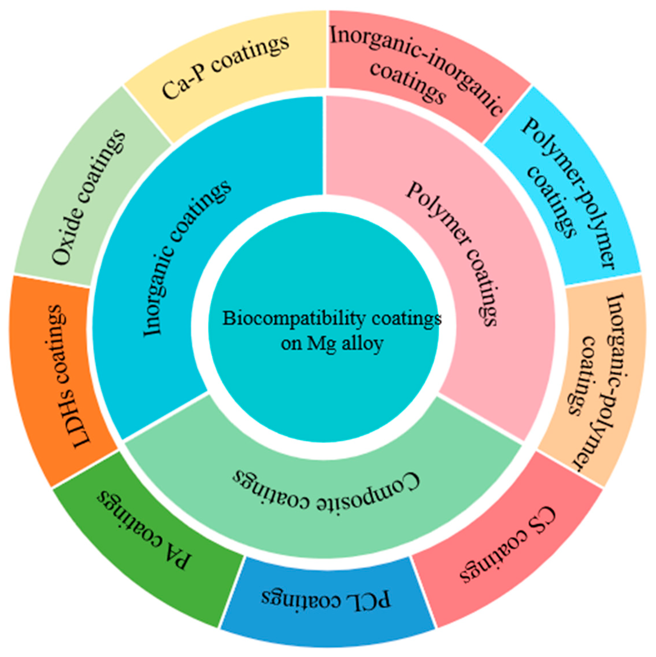
2. Inorganic Coatings

2.1. Ca-P-Based Coatings

Calcium and phosphorus are the principal elements in human bones, and they play critical roles in the processes of bone mineralization and absorption. The majority of these elements exist in the form of hydroxyapatite in bones and teeth. Calcium and phosphorus are also key components of Ca-P coatings, which have been widely used as biomaterials. Various coating processes, such as biomineralization, chemical conversion, electrophoretic, electrodeposition, and micro-arc oxidation were used to fabricate Ca-P-based coatings on Mg alloys (as shown in Figure 1).
Ca-P coatings primarily comprise hydroxyapatite (HA) and tricalcium phosphate (TCP). A Ca-P coating composed of dicalcium phosphate dihydrate (DCPD, CaHPO4·2H2O) was electrochemically deposited on an Mg-Ca alloy by M. B. Kannan and co-workers [24]. This deposited coating significantly reduced the release of Mg2+ ions from the substrate and minimized the localized degradation susceptibility of the alloy. Similarly, Cesarz-Andraczke et al. [25] synthesized a Ca-P coating including DCPD on an AM50 alloy, which effectively protected against the progression of corrosion into the substrate. The DCPD imparted biocompatibility and osteoconductive properties to the Ca-P coating. Trang et al. [26] developed several Ca-P coatings at varying pH levels. Cellular and in vivo responses were examined using osteoblastic cells and subsequent implantation in a rabbit model. The coatings remarkably enhanced the corrosion resistance of the alloy.
Y. Wang et al. [27] fabricated a Ca-P coating on the surface of an AZ31 alloy using a chemical deposition process, and the coating not only reduced the corrosion rate of the AZ31 alloy but also enhanced its biocompatibility. Wei et al. [28] synthesized a nanostructured needle-like hydroxyapatite (HA) coating on an AZ31 alloy using the sodium citrate template-assisted hydrothermal method. The needle-like morphology of HA transitioned to a flake morphology of β-tricalcium phosphate (β-TCP) as the mole ratio of Ca/sodium citrate increased. When the molar ratio of Ca/sodium citrate was 1, the HA coating exhibited significant corrosion resistance (Rt = 235.300 ± 3.584 kΩ·cm2), almost 200 times larger than that of the uncoated AZ31 alloy. M. Zaludin et al. [29] investigated the corrosion behavior of HA-coated Mg in various media, and their findings indicated that the corrosiveness of the solution media (ranging from least to most corrosive) was in the order of PBS ˂ SBF ˂ Ringers. The inhibition of corrosion by Cl- ions may be attributable to the presence of other ions such as HCO3, HPO42−, and SO42−. Yang and Gray-Munro [30] applied a corrosion-resistant Ca-P coating to the AZ31 alloy utilizing a two-step method. The coating derived from SBF solution, when supplemented with low levels of Ca(H2PO4)2, exhibited the highest corrosion resistance, demonstrating a reduction in the degradation rate of 99.8% compared with untreated samples, furthermore, it demonstrated improved uniformity and increased grain size.
Figure 1. Schematic diagram illustrating the formation process of Ca-P coatings: (a) schematic illustration of the reaction mechanism of calcium phosphate coatings on ZK60 substrate [31], (b) schematic illustration of the formation and growth mechanism of the HAp coating [32], (c) schematic illustration of the reaction mechanism of calcium phosphate coatings by electrodeposition, and (d) schematic illustration of the ion distribution on the surface of the Mg anode during MAO process [33].
Coatings 15 00191 g001
Due to the rapid dissolution rate of pure Ca-P coating in a human environment, many researchers have incorporated various elements into the Ca-P coating to improve its properties. Most of the incorporated elements are beneficial to the human body and promote bone formation, such as Nb, F, and Sr. Nb-reinforced HA (HA-Nb) coatings were fabricated using plasma spray techniques by Singh and co-workers [34]. The HA-Nb coatings proved more effective than both the pure HA and Nb coatings in inhibiting the rapid corrosion of the Mg alloy. Shen et al. [35] reported the substitution of OH in the HA lattice with F to form fluoridated hydroxyapatite (FHA). The FHA coating, in combination with the formed HA mineralized layer, offered favorable long-term protection for the magnesium alloy and significantly enhanced the osteogenic differentiation capacity. Su et al. [36] investigated the effect of fluorine content on the degradation and biomineralization behaviors of FHA coatings. Fluorine incorporation enhanced apatite crystallization and purity, which in turn could mitigate the oxidation of the magnesium alloy substrate. Furthermore, strontium-doped (Sr-doped) HA coatings were prepared on Mg-4Zn substrates by Wei and co-workers [37]. The incorporation of Sr into the HA coatings resulted in lattice distortion and reduced crystallinity, which significantly enhanced osteogenic differentiation. Sedelnikova et al. [38] conducted a comparison of Sr-TCP with Sr-HA coatings on the Mg0.8Ca alloy substrate. The Sr-HA coatings were found to be thicker, less soluble, and exhibited greater roughness along with improved corrosion resistance. Lu et al. [39] fabricated Sr-Ca-P coatings in Sr-containing electrolyte solutions using chemical deposition. Sr altered the microstructure of the coating and reduced its thickness. The coating effectively shielded the magnesium substrate from degradation.
Other elements, including Si, Mn, Fe, and Ta, have also been incorporated into the Ca-P coatings. Dehghanian et al. [40] deposited Si–HA coatings on Mg-5Zn-0.3Ca alloys, the presence of Si was found to increase the compactness of the coatings, and those prepared with an electrolyte containing 0.005 mol/L SiO32− exhibited higher corrosion resistance than the pure HA coatings. Su et al. [41] prepared an optimum coating using a solution containing 0.05 mol/L Mn2+. The coating possessed a uniform and compact microstructure and was composed of MnHPO4⋅3H2O, CaHPO4⋅2H2O, and Ca3(PO4)2. Iron-substituted tricalcium phosphate (Fe-TCP) coatings, comprising approximately 1 wt. %, were deposited on Mg-Ca alloys by Antoniac and co-workers [42]. Fe-TCP coatings deposited at 300 °C were found to have the highest protection efficiency in simulated body fluid (SBF), along with the best biocompatibility and antibacterial properties. Singh et al. [43] incorporated Ta into HA coatings, which effectively enhanced the surface hardness of the Mg-Ca alloys, and the surface roughness of the HA coatings was observed to decrease with an increase in Ta’s content.
Wang et al. [44] prepared CaHPO4⋅2H2O (DCPD) and MgHPO4⋅3H2O coatings using a one-step chemical conversion method. MgHPO4⋅3H2O could play a supportive role in maintaining structural integrity during the degradation process. The coatings demonstrated optimal long-term corrosion resistance and exhibited biomineralization capacity. Pana et al. [45] successfully applied a uniform Mg-doped hydroxyapatite coating to the AZ31B magnesium alloy via RF magnetron sputtering. This coating demonstrated excellent bonding strength and enhanced chemical reactivity. Kalaiyarasan et al. [46] fabricated a flaky-like aggregated structure of Fe/Ag doped hydroxyapatite (Fe/Ag-HAp) coating on the surface of the AZ31 Mg alloy, which exhibited improved anticorrosion properties and cellular responses. Hernández et al. [47] deposited HAp coatings on pure Mg substrates via a biomimetic method, which demonstrated higher corrosion resistance and bioactivity. Calcium phosphate (Ca-P) composite coatings were prepared using supersonic atmospheric plasma spraying and the microwave-hydrothermal method. The microwave-hydrothermal treatment can seal surface defects and improve the bioactivity and corrosion resistance of the sprayed Ca-P coatings [48].

2.2. Oxide Coatings

In addition to Ca-P coatings, other inorganic coatings commonly used on magnesium alloys include oxides such as TiO2, ZrO2, Al2O3, SiO2, and MgO. Zirconium (Zr) and zirconia (ZrO2) not only promote bone formation but also exhibit good corrosion resistance, cell compatibility, and antibacterial properties. Hydrothermal method, laser deposition method, and chemical conversion method were applied to formed oxide coatings on the surface of the Mg alloy (as shown in Figure 2).
A porous ZrO2 coating was engineered onto the surface of an AZ31B alloy as demonstrated by Gao and co-workers [49]. The ZrO2-AZ31B coating exhibited enhanced corrosion resistance compared with the AZ31B substrate when immersed in a simulated body fluid solution. Similarly, Amiri et al. [50] fabricated a nano-structured zirconia coating on the alloy utilizing the EPD technique. The zirconia coating served as a long-term barrier against corrosion, with the coating reducing the corrosion current density of the AZ91D alloy from 18.4 to 12 μA/cm2 under optimal conditions. However, the obtained coating did not significantly enhance hydroxyapatite formation on the surface.
Al2O3 ceramic coatings offer many advantages, including high hardness, excellent chemical properties, optimal stability, and adaptability. Gao et al. [51] applied an Al2O3 coating to an AZ91HP magnesium alloy using plasma spraying technology. The coating was composed primarily of stable α-Al2O3 and metastable γ-Al2O3 phases, featuring a lamellar microstructure. Ji et al. [52] developed coatings from various aluminum sources (aluminum isopropoxide, aluminum nitrate, or a mixture of these two) on AZ31 alloys. The coatings deposited from aluminum isopropoxide were composed of γ-Al2O3, while those derived from aluminum nitrate consisted of MgAl2O4. The former was noted to be more compact and uniform than the latter.
Kong et al. [53] synthesized wollastonite nanoceramic coatings using an electrophoretic technique. The incorporation of wollastonite with silica nanoparticles into a magnesium alloy significantly enhances bone formation, as evidenced by the highest level of apatite formation in the alloy containing 10% wollastonite. Kotoka et al. [54] engineered Al2O3 and ZrO2 coatings onto pure Mg; the Al2O3 thin film coatings exhibited circular grains ranging from 5 to 25 nm, whereas the ZrO2 coatings featured larger ellipsoidal grains. The Mg coated with Al2O3 demonstrated a corrosion resistance approximately threefold greater than that of the Mg coated with ZrO2.
TiO2 is also known for its good biocompatibility. Yang et al. [55] applied a nanoscale TiO2 coating using a precisely controlled temperature and thickness through atomic layer deposition (ALD) to the magnesium–zinc alloy stent material. The coating is capable of stimulating the adhesion and proliferation of coronary artery endothelial cells, and it possesses a protective barrier function.
Figure 2. Schematic diagram illustrating the formation process of oxide coatings: (a) mechanism of zinc-doped phosphate coating [56], (b) deposition mechanism of schematic illustration of the reaction mechanism of calcium phosphate coatings on ZK60 substrate, and (c) schematic of laser processing [57].
Coatings 15 00191 g002
The use of nanostructures for orthopedic coatings has increased to enhance stabilization, integration with host tissues, and to promote bone tissue regeneration. The application of nano- and microstructured oxide coatings has significantly advanced due to their favorable biological properties. Jiang et al. [58] employed a hydrothermal method to apply a coating of CaCO3 and MgSO4 onto the Mg alloy. The findings indicated that this coating not only enhanced the corrosion resistance but also improved the biological activity of the substrate. Liu et al. [57] reported that the degradation of Mg alloys could be regulated by utilizing laser surface treatments. Li et al. [59] integrated nano-ZnO into the MAO electrolyte to create micro-arc oxidation coatings that contain ZnO. The findings demonstrated that the incorporation of nano-ZnO significantly improved the corrosion resistance of the MAO-coated alloy. The alloy coated with nano-ZnO exhibited remarkable resistance to degradation, enhanced corrosion resistance, and effective antibacterial properties.

2.3. LDHs Coatings

Common inorganic coatings include calcium phosphate (Ca-P) coatings and oxide coatings. In addition, in recent years, hydrotalcite (layered double hydroxides—LDHs) coatings characterized by their unique structure have been utilized for the corrosion protection of magnesium alloys. The chemical composition of hydrotalcite is represented by the formula [M1-x2+Mx3+(OH)2]x+[(An-)x/n·mH2O]; M2+ is Mg2+, Ni2+, Co2+, Mn2+, Cu2+, and other trivalent metal cations; M3+ is Al3+, Cr3+, Fe3+, Sc3+, and other divalent metal cations; An− is CO32−, NO3, Cl, OH, SO42−, PO43−, C6H4(COO)22−, and other inorganic, organic, and complex ions. If the inorganic anions between the layers are different, the interlayer spacing of LDHs is different [60]. The structural characteristics of LDHs facilitate the exchange of interlayer anions with a diverse array of anions, including inorganic, organic, homoionic, and coordination compound anions. Owing to their high anion exchange capacity, LDHs hold significant potential in the realm of corrosion protection.
A uniform and dense zinc-aluminum layered double hydroxide (Zn-Al LDH) coating was electrochemically deposited on an AZ91D alloy substrate by Wu and co-workers [61]. The coating consisted of crystalline Zn-Al LDH with nitrate intercalated. The LDH-coated Mg alloy exhibited enhanced corrosion resistance compared with the uncoated alloy in 3.5 wt. % NaCl solution. Peng et al. [62] incorporated magnesium-aluminum layered double hydroxide (Mg-Al LDH) into an Mg(OH)2 coating via hydrothermal treatment. The Mg-Al LDH significantly enhanced cell adhesion, migration, and proliferation in-vitro and offered exceptional long-lasting corrosion protection. The mechanism was further elucidated, with the explanation that anions in the interlayer of Mg-Al LDH could be substituted by chloride ions, leading to a relatively low chloride ion concentration near the surface of the coating. Zheng et al. [63] synthesized a Zn-Fe interlayered phosphate ion-layered double hydroxide coating on an AZ31 Mg alloy via hydrothermal treatment. The results demonstrated that the AZ31 Mg alloy coated with ZnFe-PO43− could engage in interactions with Ca2+ ions, leading to the formation of Ca-P deposits. Furthermore, the coating promoted the proliferation and adhesion of MC3T3-E1 cells.
There are also studies focusing on the combination of LDH and other coatings. Yang et al. [64] synthesized an LDH-containing coating on AZ61 alloy. Compared with Zn-Al LDH and ZIF-8/LDH coatings, the ZIF-8-DMBIM/LDH composite coating exhibited a high contact angle (115.7°), low corrosion current density, high corrosion potential, and polarization resistance due to its near-smooth surface, dense two-layer structure, and hydrophobic characteristics. Wu et al. [65] deposited a magnesium-aluminum layered-double hydroxides (Mg-Al LDHs) coating onto the surface of an AZ31 alloy, followed by the electrophoretic deposition of an Al2O3 nanoparticle layer. The mechanism is discussed as shown in Figure 3. LDHs and Al2O3 exhibit a synergistic effect in improving the corrosion resistance of the alloy. The ion exchange capacity of LDHs prevents the release of chloride ions and nitrate ions, while the formation of Mg(OH)2 under alkaline conditions can inhibit the expansion of pitting corrosion. Simultaneously, the Al2O3 nanoparticle layer is deposited to block the channels between the nanosheets, further improving the corrosion resistance of the coating. Hydrothermal method, chemical conversion method, etc., are applied to fabricated LDH coatings on Mg alloys (as shown in Figure 4).
Li et al. [66] effectively developed a magnesium-aluminum layered double hydroxide coating intercalated with silicate (LDH-SiO3) using a hydrothermal technique combined with an anion exchange process. They discovered that the corrosion current density of the alloy modified by this coating was reduced by two orders of magnitude compared with the substrate. Furthermore, the LDH-SiO3 coating improved the attachment and proliferation of MC3T3-E1 pre-osteoblasts.
Figure 4. Schematic diagram illustrating the formation process and corrosion process of LDHs coatings: (a) schematic illustration of the CPCC formation process [67], (b) the schematic representation of the protection mechanism for LDHs film and Mg-LDHs-HA composite film in SBF [68], (c) schematic of the preparation of LDH-SiO3 coatings on anodized AZ [66], and (d) schematic showing the self-healing mechanism of the smart LDH-SiO3 coating on anodized AZ [66].
Coatings 15 00191 g004
The preparation processes of the magnesium alloy substrate and hydrotalcite also affect the formation and properties of the LDHs coating. The effects of the preparation process and grain size on the corrosion resistance of the LDHs coating have been studied by Chen and co-workers [69]. The corrosion protection performance of the LDHs coatings was ranked as an increasing series: CS-LDHs < AE-LDHs < SE-LDHs < RS-LDHs. The grain size of the RS-LDHs sample was finer and more uniform, providing more nucleation sites for the formation of the LDHs coating. The effect of hot extrusion on the microstructure and anti-corrosion performance of the LDHs coating on AZ91D alloy has also been investigated by Zhang and co-workers [70]. The refinement and rearrangement of the β-Mg17Al12 phase promote the micro-galvanic effect during the formation process after hot extrusion. Due to microstructural differences, the electrochemical activity of various specimens during the formation process can be ranked in an increasing series: as-cast < transverse < longitudinal. To address the issues of long preparation time and the difficulty of controlling the formation process in the traditional method of preparing hydrotalcite conversion coatings, an LDHs coating has been prepared on the surface of a magnesium alloy by the CO2 pressure method by Zhang and co-workers [71]. The CO2 pressurization has been shown to promote the formation reaction rate, achieving high formation efficiency and good stability without pollution.

3. Polymer Coatings

Mg alloys coated with biodegradable polymers demonstrate great potential due to their degradability, which is coupled with features such as biocompatibility, self-healing properties, and osteo-inductive potential. In addition, polymeric coatings are widely recognized as a feasible means to modulate cell responses, such as adhesion, proliferation, and differentiation. Numerous investigations have reported on the efficacy of forming coatings from polylactic acid (PLA) [72], poly (lactic-co-glycolic) acid (PLGA) [73], polycaprolactone (PCL) [74], polydopamine (PDA) [75], and natural polymers such as chitosan (CS) [76,77] and collagen (Co) [78].

3.1. Phytic Acid (PA) Coatings

PA (Phytic acid, C6H18O24P6) is a naturally occurring organic macromolecular compound, which is non-toxic and can be degraded and absorbed in the human body. Hernández-Alvarado et al. [79] fabricated powder metallurgy Mg and AZ31 alloy samples coated with a phytic acid layer. The test results in a saline phosphate buffer solution (PBS) at 37 °C indicated that the powder metallurgy magnesium specimens underwent complete dissolution within 250 h of immersion due to their inherent heterogeneity and porosity. In contrast, the phytic acid coating on AZ31 was shown to improve corrosion resistance in long-term tests up to 336 h, without affecting biocompatibility and biodegradability.
To enhance the biocompatibility and corrosion resistance of PA coatings, studies have focused on incorporating drugs or other substances into the PA coatings. Heparin (Hep) or bivalirudin (BVLD) were immobilized within an organic phytic acid (PA) coating on Mg surfaces by Chen and co-workers [80]. The phytic acid coating, when combined with both drugs, exhibited comparable corrosion protection on the magnesium surface to that of the direct PA coating and enhanced anticoagulation, thereby improving histocompatibility. The results were attributed to the distinct coordination mechanisms that occur between the drug biomolecules and the Mg-chelated PA molecules.
Tang et al. [81] prepared a TiO2-containing PA coating on an Mg substrate. A small amount of TiO2 was incorporated in situ into the PA coating under ultraviolet (UV) irradiation. This coating significantly enhanced the adhesion and proliferation of MC3T3-E1 pre-osteoblasts. Compared with the PA or TiO2 coatings, the PA and TiO2-UV coating was denser, thicker, and more hydrophilic. The electrochemical corrosion current density of the PA and TiO2-UV coating on the Mg substrate decreased by a factor of seven, and the in-vitro degradation rate in phosphate buffer solution was significantly lower. A series of phytic acid/3-aminopropyltrimethoxysilane (γ-APS) hybrid coatings were prepared on AZ31 alloys by immersing the magnesium alloy in a mixed solution of phytic acid and γ-APS by Li and co-workers [82]. The pH of the mixed solution considerably influenced the uniformity of the coating, which, in turn, affected the coating’s corrosion resistance. The coating also possessed self-healing properties. After prolonged immersion in SBF, the coating’s quality loss diminished, its surface cracks gradually healed, and the charge transfer resistance increased.

3.2. Polycaprolactone (PCL) Coatings

Polycaprolactone (PCL) exhibits good mechanical properties, tissue compatibility, and biodegradability. It can be completely decomposed into CO2 and H2O within 6–12 months when exposed to a soil–water environment. J. Degner et al. [83] synthesized a biodegradable polycaprolactone (PCL) coating at various concentrations using spin coating. The PCL coatings proved to be uniform and pore-free. The corrosion resistance of the coatings was enhanced, but the bonding strength to the substrate remained low. Numerous studies have addressed the issue of poor adhesion between PCL coating and substrates. Palanisamy et al. [84] introduced a two-layer coating technique, where magnesium carbonate served as the initial layer, deposited via a chemical conversion method, followed by the application of a PCL coating through dipping. The PCL layer adhered effectively to the MgCO3 surface, with penetration into the MgCO3 crystals, enhancing the double-layer coating’s adhesion to the substrate as well as its corrosion resistance and bioactivity. Niu et al. [85] bonded PCL to the Mg matrix via silane coupling agent (SCA) pretreatment. Concurrently, alkali pretreatment ensured adequate OH availability for the reaction between the Mg matrix and SCA. Owing to the coupling effect of SCA, the adhesion of the PCL coating to the Mg substrate was significantly enhanced, along with a nearly two orders of magnitude improvement in corrosion resistance. Carangelo et al. [86] utilized a polydopamine (PDOPA) layer to augment adhesion between the metallic substrate and the external organic coating. This intermediate layer can effectively bolster the corrosion resistance of the organic coating and strengthen the bond between PLA and the magnesium alloy substrate.
Research has also focused on improving the antibacterial performance of the PCL coating by incorporating antibacterial particles or agents. Yang et al. [87] introduced copper (Cu)-containing bioactive glass nanoparticles (Cu-BGNs) into polycaprolactone (PCL) coating systems to enhance the bioactivity, antibacterial properties, and corrosion resistance of susceptible magnesium matrices. Cu-BGNs were evenly distributed within the PCL coating, and the Cu2+ ions released by the Cu-BGNs coating effectively inhibited the growth of S. carnosus and E. coli. In comparison to pure magnesium, the activity and proliferation of MG-63 cells on the Cu-BGN coating were significantly enhanced; however, as the concentration of Cu-BGNs increased, a marginal decline in cell proliferation and activity was observed, alongside a reduction in corrosion resistance. Zomorodian et al. [88] fabricated a degradable polycaprolactone (PCL) coating on an AZ31 alloy. The coating was modified with nano-HA particles and cephalexin; this modification with HA particles was found to enhance the biocompatibility of the PCL coating. The PCL coating acquired an antibacterial effect through the addition of cephalexin, which promoted osseointegration and cell viability, although corrosion resistance experienced a concomitant decrease. Therefore, the enhancement of the corrosion resistance of the organic coating remains an area necessitating further investigation.

3.3. Chitosan (CS) Coatings

Chitosan is a naturally occurring alkaline polysaccharide that is non-toxic and exhibits good degradability. Studies have demonstrated that coatings made of chitosan are biocompatible and can effectively enhance corrosion resistance and reduce the formation of subcutaneous gas pockets. Consequently, chitosan coatings have been applied to magnesium and its alloy surfaces in order to improve their corrosion resistance [89].
A Mg-Zn-tricalcium phosphate composite coated with chitosan was synthesized by Zhao and co-workers [77]. The immersion corrosion rate, the pH values of the simulated body fluids, and the concentration of released metal ions in the chitosan-coated composite were all found to be lower than those associated with the uncoated composite. In vivo testing further demonstrated that the chitosan-coated composite was non-toxic to the rabbits’ vital visceral organs and promoted healing in bone tissues. Dai et al. [90] synthesized Mg-6%Zn-10%β-Ca3(PO4)2 composites with 4, 7, and 10 layers of chitosan coatings. Chitosan coatings proved to be non-toxic and were able to modulate the biodegradation rate of composites. Specimens with seven layers of chitosan coating exhibited superior corrosion resistance, characterized by the minimal volume of hydrogen evolution and the most stable pH value fluctuations in SBF.
Höhlinger et al. [91] developed surface pre-treatments for corrosion protection. The results indicated that DMEM and HF pretreatments were the most effective at protecting the magnesium alloy matrix from corrosion during the electrophoretic deposition process. Francis et al. [92] employed a straightforward one-step chemical conversion treatment to synthesize a Mg(II)-chitosan composite coating. The interaction of chitosan with the magnesium matrix led to the formation of a Mg(II)-chitosan complex, which prevented alkalization near the coating’s surface, thus constraining the pH conditions conducive to hydrogen evolution, diminishing hydrogen production, and enhancing the magnesium matrix’s corrosion resistance. Cui et al. [93] fabricated a corrosion-resistant and antimicrobial coating through the layer-by-layer assembly of chitosan (CHI) and poly-L-glutamic acid (PGA). The coating’s effective corrosion resistance and antibacterial properties were attributed to the pH buffering capacity of the weak polyelectrolyte pair. Jia et al. [94] constructed biodegradable sodium alginate and carboxymethyl chitosan (SA/CMCS) coatings on the surface of magnesium alloys using hydrothermal treatment and a dipping method. The SA/CMCS coating facilitated endothelial cell growth, proliferation, and migration, thereby promoting neovascularization.

4. Composite Coatings

Composite coatings can combine the advantages of multiple coatings while compensating for the shortcomings of a single coating. Currently, inorganic composite coatings, polymer composite coatings, and inorganic–polymer composite coatings are being actively researched. By combining the corrosion resistance, biocompatibility, and antibacterial properties of multiple coatings, the preparation of bioactive coatings with excellent comprehensive performance becomes highly significant for the surface modification of bioimplant materials.

4.1. Inorganic-Inorganic Composite Coatings

4.1.1. Ca-P-Based Composite Coatings

Owing to their excellent biocompatibility, Ca-P coatings are widely used as surface coatings on bone repair and bone fixation metal materials to enhance both their corrosion resistance and biocompatibility. Numerous studies explore the combination of Ca-P coatings with other materials to develop bioactive coatings that exhibit superior corrosion resistance, biocompatibility, and antibacterial properties. Studies also focus on depositing these elements as coatings and forming composites with Ca-P coatings on the surfaces of magnesium alloys. For instance, Bakhsheshi-Rad et al. [95] initially deposited a nano-silicon (Si) coating on an Mg-6Zn-0.8Mn-3Ca substrate, followed by a subsequent layer of nano-hydroxyapatite (HA). The resulting composite coating proved to be uniform and dense. Moreover, the Si–HA composite coating on the magnesium alloy significantly reduced the corrosion current density and the hydrogen evolution rate, while simultaneously increasing the polarization resistance (Rp).
Calcium phosphate (Ca-P) coatings are susceptible to large cracks in physiological environments. To address this issue, Peng et al. [96] developed a composite coating comprising an impermeable hydroxyapatite inner layer and a graphene oxide outer layer on an AZ31 alloy. This coating combined the impermeability of graphene oxide with the biocompatibility of hydroxyapatite, which not only inhibited the growth of coating cracks but also enhanced the corrosion resistance of the substrate. Additionally, Neupane et al. [97] applied a graphene oxide coating to shield the magnesium matrix against ion diffusion from oxidizing gases and corrosive liquid solutions. Santos et al. [98] fabricated a multifunctional biodegradable surface by incorporating hydroxyapatite nanoparticles and graphene oxide (GO), whereby the surface wettability of the coating could be modulated by varying the concentrations of hydroxyapatite nanoparticles and GO.
Cao et al. [99] affixed the arginine-glycine-aspartate-cysteine (RGDC) peptide to the surface of the AZ31 alloy, effectively promoting the nucleation and crystallization of Ca-P coatings. Compared with the uncoated alloy, the corrosion resistance and biocompatibility of the magnesium alloy coated with the RGDC-HA were significantly improved, but the adhesion of the Ca-P coating to the AZ31 substrate remained suboptimal. A composite coating of calcium phosphate/zirconia (Ca-P/ZrO2) was applied to the AZ91D alloy by Wang and co-workers [100]. The adhesion strength between the coating and the substrate increased from 12.1 MPa for a single-layer Ca-P coating to 24.4 MPa. This increase was attributed to the fact that the Ca-P/ZrO2 coating formed strong chemical bonds and facilitated interdiffusion between elements during the deposition process. The uppermost Ca-P layer also significantly enhanced cell adhesion and proliferation.
The incorporation of the Ca-P coating with an antibacterial agent to enhance the anti-inflammatory and antibacterial properties of the composite coating has been investigated. Tan et al. [101] reported a novel Ca-P/TC composite coating for Mg implants that enhances both the corrosion resistance of Mg and offers sustained drug release. Utilizing a hydrothermal method, Bai et al. [102] fabricated a cost-effective and multifunctional hydroxyapatite/pefloxacin (HA/PFLX) composite coating on the surface of AZ91 alloy. PFLX possesses broad-spectrum antibacterial properties analogous to TC, which can effectively prevent bone infection and inflammation. Additionally, Tian et al. [103] applied a Ca-P coating on the surface of AZ91 alloy combined with AMPs to decelerate the corrosion rate of the substrate. The AMP-loaded coating exhibited antibacterial activity against Staphylococcus aureus, achieving an antibacterial rate exceeding 50% after 4 days and sustaining this efficacy for 7 days. A two-step electrodeposition process was employed to synthesize a ceria/calcium-phosphate (Ce/Ca-P) composite coating on magnesium alloys [104]. The pin-like structure of the outer layer of the Ce/Ca-P coating enhances its antibacterial properties during the initial stages of implantation. The calcium-phosphate outer layer, in conjunction with the ceria interlayer, facilitates the deposition of calcium and phosphate ions, thereby aiding bone healing over the medium term.

4.1.2. MAO-Based Composite Coatings

Micro-arc oxidation (MAO) is an effective and economical method to reduce the corrosion rate due to its high hardness, enhanced adhesion to the substrate, and superior corrosion resistance achieved within a brief duration. However, the application of the coating is constrained by issues such as porosity, poor density, electric spark occurrence during the process, and a significant reduction in the protective effect over time [105]. Generally, additional coatings combined with the MAO coating can fill and seal its pores, thereby preventing the ingress of corrosive agents and enhancing the corrosion resistance of the composite coating. Seyfoori et al. [106] synthesized a nanostructured composite coating that included biphasic calcium phosphate (BCP) through the micro-arc oxidation (MAO) technique applied to an AZ31 alloy. Compared with the pure MAO coating, the composite coating exhibited better corrosion resistance, attributed to the nanoparticle-induced barriers to corrosive agents penetrating the substrate. Wang et al. [107] deposited a dense layer of needle-like fluorapatite (FHA) with a thickness of 6 μm atop the MAO layer. The MAO-FHA coating demonstrated a high mineralization capacity in simulated body fluid, which facilitated the deposition of hydroxyapatite. Compared with the alloy coated with the MAO coating, the alloy treated with the MAO-FHA coating exhibited greater surface tissue integration, enhanced bone tissue adhesion, and improved biocompatibility by mitigating the excessive release of Mg2+ ions from MAO-treated samples.
Wei et al. [108] fabricated a micro-arc oxidized/poly (L-lactide) (MAO/PLLA) composite coating on an AZ31 alloy by combining the MAO process and sealing with PLLA. The composite coating notably enhanced the corrosion resistance of the AZ31 alloy. The release of Mg2+ ions and hydrogen, along with the pH value changes in the solution due to degradation, were all significantly reduced. Bai et al. [109] prepared a chitosan/MAO composite coating for a Mg-Zn-Ca alloy. The corrosion current density of the coated alloy decreased by nearly three orders of magnitude compared with the uncoated alloy. This improvement was attributed to the fact that the composite coating sealed the pores, altered the interface bonding conditions, and provided a barrier against corrosive media. Feng et al. [110] employed a micro-arc oxidation coating incorporating HA particles (MAO-HA) as the inner layer and polydopamine (PDA) as the outer layer. The PDA layer completely covered the surface of the MAO-HA layer, resulting in the transition of the coating from hydrophobic to hydrophilic. Compared with the MAO-HA layer, the pores on the surface of the PDA/MAO-HA layer were significantly reduced, resulting in a decrease in corrosion current density by an order of magnitude, and the coating exhibited good cell compatibility.
The chemical conversion method, hydrothermal method, solvothermal method, electrodeposition method, etc., were exploited to fabricate inorganic coatings on the surface of MAO coatings (as shown in Figure 5). Zhang et al. [111] developed a double-layer coating (Ca-P/PEO) for the application on pure magnesium. The results indicated that the corrosion current density of the coated Mg was two orders of magnitude lower than that of the uncoated Mg before immersion in SBF solution, and threefold lower after 168 h of immersion. Additionally, the coating facilitated the nucleation of osteoconductive minerals. Zhang et al. [112] engineered Mg-Fe LDH films on the surface of MAO-coated Mg alloy via immersion and hydrothermal treatment. The findings demonstrated that the MAO/Mg-Fe LDH coatings presented a lower hemolysis rate (less than 5%) in comparison to the MAO coating alone. Furthermore, the MAO/Mg-Fe LDH coatings enhanced cell adhesion and proliferation more effectively than the MAO coating alone. Abolfazl Zarei et al. [113] fabricated an akermanite (Ca2MgSi2O7) and alginate (C6H9NaO7) layer on the surface of an MAO coating using the electrophoretic deposition method, which demonstrated a significant reduction in the release of Mg2+ ions and hydrogen bubbles, thereby enhancing corrosion resistance.
Figure 5. Schematic diagram of the processing steps for the intermediate transition layer of PEO coating. (a) Schematic illustrations showing the deposition mechanisms of the 5.7Ca-P coating on PEO coated Mg in a 5.7Ca-P solution [111], (b) schematic illustrations showing the deposition mechanisms of the 5.7Ca-P coating on PEO coated Mg in a 5.7Ca-P solution [111], and (c) diagram of preparation process of PEO/Mg-Fe LDH composite coating [112].
Coatings 15 00191 g005
In addition to traditional Ca-P and organic coatings, other substances, including various oxides and pharmacological agents, are compounded with MAO. Xiong et al. [114] prepared MAO coatings in alkaline electrolytes enriched with CeO2 and ZrO2 nanoparticles. The resultant MAO coating exhibited good mechanical properties, including wear resistance, enhanced hardness, and strong adhesion to the substrate. Subsequently, the bioactive HA coating was applied to the MAO coating, and the MAO-HA composite coating demonstrated improved corrosion resistance in the SBF solution. A dense Mg(OH)2 coating was developed on a MAO-coated AZ31 alloy in an alkaline electrolyte that included ethylenediamine tetra acetic acid disodium (EDTA-2Na) by Li and co-workers [115]. The intermetallic compound of the Al-Mn phase in the substrate is instrumental in the growth of the Mg(OH)2 coating. The addition of EDTA-2Na facilitates the nucleation and growth of Mg(OH)2. The Mg(OH)2 coating features a porous nano-sized structure and effectively seals the micro-pores and micro-cracks of the MAO coating, thereby promoting the formation of calcium phosphate corrosion products. Bordbar-Khiabani et al. [116] created a thin layer of magnesium oxide (MgO) on the surface of AZ91 alloy by the micro-arc oxidation (MAO) process and then deposited a betamethasone sodium phosphate (BSP) layer onto the MAO coating. The BSP layer effectively sealed the holes and cracks of the MAO coating, serving as a barrier layer against the penetration of the SBF solution. The MAO/BSP coating exhibited a higher impedance mode, which was 300 times greater than that of the MAO coating. At the same time, the coating spontaneously formed a highly active Ca-P layer on the surface of the MAO/BSP.
Integrating other technologies with MAO represents a promising approach to enhancing material properties. Xiong et al. [117] developed an LSP/MAO composite bio-coating on AZ80 alloy by combining the LSP and MAO processes. LSP is an alternative non-contact surface treatment technique that utilizes high-power, short-pulse laser shock waves to enhance the target surface’s mechanical properties, including fatigue, corrosion cracking, and wear resistance, and to induce grain refinement. In comparison with the LSP layer and MAO coating alone, the LSP/MAO composite coating not only significantly enhances the magnesium alloy substrate’s corrosion resistance but also its mechanical properties. Liu et al. [118] fabricated a chitosan and polystyrene sulfonic acid composite coating on a WE43 alloy substrate that had been treated by micro-arc oxidation (MAO) using layer-by-layer (LBL) self-assembly technology. The resulting MAO/LBL coating significantly improved the corrosion resistance of magnesium alloys in simulated body fluids.

4.2. Polymer–Polymer Composite Coatings

Composite coatings, which consist of multiple polymers, serve as protective layers utilizing an inert barrier to shield substrates from exposure to external conditions. The efficacy of these coatings in providing protection largely derives from the benefits previously highlighted. Among them, biodegradable polymeric coatings are particularly appealing because of their degradable nature. Moreover, the capacity to tailor these coatings to enhance bone integration (osseointegration) and enable precise, site-specific drug release makes biodegradable polymer coatings ideally suited for application with magnesium alloys in the context of implants, as reported in the literature. Nonetheless, for such medical applications, it is essential that these coatings not only exhibit biocompatibility but also meet the fundamental requirements of providing complete surface coverage and preserving structural integrity. Figure 6 shows several typical processes for the preparation of composite coatings.
Figure 6. Schematic representation of the preparation of polymer–polymer composite coatings: (a) the schematic diagram of PCNZ preparation [119], (b) schematic of process for coating preparation [120], (c) schematic representation of the S-C technique used for the coating production [121], and (d) schematic illustration of preparation of (CHI/DNA)5 /Mg(OH)2 composite coating on an AZ31 substrate [122].
Coatings 15 00191 g006
A composite coating of chitosan and polylactic acid was developed for the AZ31 alloy, tailored specifically for medical applications. This coating successfully sealed the pores within the chitosan layer, imparting robust protection to the AZ31 alloy and significantly reducing its corrosion current density by four orders of magnitude [120]. Furthermore, the composite’s exceptional biocompatibility was corroborated by improved cell viability assays and by observing the morphology of cells adhered to the chitosan/polylactic acid-coated sample. Zhao and colleagues [121] skillfully engineered a composite coating of polyvinylpyrrolidone (PVP) and polyacrylic acid (PAA) on an AZ31 magnesium alloy using the spin-casting method. The findings indicated that the PVP/PAA composite layer, noted for its absence of defects, density, and uniformity, was successfully applied to the magnesium alloy’s surface. Additionally, this coating exhibited superior corrosion resistance and robust adhesion characteristics. A coating comprising alternating layers of polyvinylpyrrolidone (PVP) and deoxyribonucleic acid (DNA), referred to as (PVP/DNA)n, was synthesized using a layer-by-layer (LbL) assembly dip-coating technique [122]. Cui et al. [123] described the application of a novel biomimetic polyelectrolyte multilayer template, composed of polyvinylpyrrolidone (PVP) and polyacrylic acid (PAA), which was assembled using a layer-by-layer (LbL) technique. This approach enhanced the corrosion resistance of the alloy. Both hydrogen evolution and electrochemical corrosion tests confirmed the effectiveness of the polyelectrolyte-induced Ca-P coating in protecting the AZ31 alloy from corrosion (as shown in Figure 7).
A multilayered coating of chitosan/deoxyribonucleic acid (CHI/DNA) was engineered on the AZ31 alloy using the layer-by-layer assembly dip-coating technique, with an underlying Mg(OH)2 coating acting as the protective layer [124]. The outer CHI/DNA coating serves as a catalyst for the formation of a biocompatible calcium phosphate (Ca-P) corrosion-resistant coating during immersion, offsetting its relatively thin profile. Furthermore, this composite coating promotes bone growth, owing to the biomineralization properties of the outer polyelectrolyte layers. The outcomes indicated that the (PVP/DNA)n coating displayed a smooth surface, albeit with minor scratches and several corrosion-related cracks. Despite these imperfections, the coating retained good corrosion resistance when exposed to simulated body fluid. Monfared et al. [125] developed tannic acid/poly (N-vinylpyrrolidone) composite coatings on the surface of Mg, which demonstrated that the coating significantly enhanced the corrosion resistance of Mg.
Zhao et al. [126] developed a self-healing polymer coating on the surface of Mg alloy using a spin-spray layer-by-layer assembly process. The results demonstrated that the coating supported the activity of alkaline phosphatase and accelerated the mineralization of the extracellular matrix. A novel coating comprising silk fibroin (SF) and cellulose nanocrystals (CNCs) was designed for application on the biodegradable AZ31 magnesium alloy [127]. Electrochemical corrosion assessments and in-vitro immersion experiments conclusively demonstrated that the corrosion resistance of the SF-coated alloy significantly improved with the addition of CNCs. Compared with the Mg alloy, the SF-CNC-coated AZ31 exhibited a significant improvement in cytocompatibility, as evidenced by a cell viability of 114%, along with enhanced adhesion and proliferation of human fetal osteoblast cells on the coated surface.

4.3. Inorganic–Organic Composite Coatings

A single inorganic or organic coating alone cannot meet the requirements of biomaterials. Therefore, combining the advantages of inorganic and organic coatings is essential to obtain a more suitable coating with improved properties. Due to their excellent biocompatibility, Ca-P coatings have been combined with organic coatings, such as Ca-P/PLA [128], HA/SA [129], and HA/Methylcellulose [130].
G. Perumal et al. [131] developed a polycaprolactone (PCL) and nano-hydroxyapatite (nHA) composite coating via dip-coating and electrospinning on an AZ31 alloy, subsequently implanting it in New Zealand white rabbits as a femoral repair material. After four weeks, the defect was successfully repaired, and the material’s corrosion resistance was significantly enhanced. Similarly, Tian et al. [132] applied a nHA-PLGA composite coating on pure Mg via EPD, using electrolytes containing 1 mg/mL HA and 0.2 mg/mL PLGA, with a deposition voltage of 75 V for 6 min. J. Zhang et al. [133] designed a Ca-P/chitosan/carbon nanotube (CNT) coating on AZ91D magnesium alloy. The addition of CNTs increased the gentamicin loading and cell viability, while decreasing the release rate of gentamicin compared with the Ca-P/chitosan coating. Kim et al. [134] fabricated an HA/PLLA micro-textured coating layer onto a Mg implant by treating the Mg substrate with a micro-patterned photoresist surface in an aqueous solution containing calcium and phosphate ions. The HA micro-dots were surrounded by PLLA. The HA/PLLA micro-textured coating consisted of a porous top surface and a dense layer on the Mg surface, which protected the substrate, thereby exhibiting excellent corrosion resistance and providing a favorable environment for cellular interactions. Additionally, the HA/PLLA micro-textured coating exhibited excellent stability under mechanical strain, even at an elongation of 5%. It effectively retained the coating without producing noticeable defects, while the single HA and PLLA coatings were damaged due to brittleness and weak bond strength. The micro-textured coating combined the high flexibility of PLLA with the excellent biocompatibility and bioactivity of HA, thus offering promising potential for application.
Electrophoresis, dip cast, electrostatic spinning, and hydrothermal, etc., were utilized to form composite coatings (as shown in Figure 8). Qian et al. [135] developed a composite coating system (PTMC-MAO) for Mg alloys, seamlessly integrating micro-arc oxidation (MAO) and poly (trimethylene carbonate) (PTMC) layers. The coatings achieved a distinguished protection efficiency (η) of 99.9%, accompanied by higher impedance. The ΔpH change and the released Mg2+ concentration were 0.25 and 42 μg/mL, respectively, after 21 days of immersion. Lv et al. [136] successfully synthesized a polypyrrole (PPy)-loaded polycaprolactone (PCL) organic composite layer on MAO-coated Mg alloy using the dip-casting method. They found that the formation of this organic composite layer resulted in a reduction in the corrosion current by four orders of magnitude. A combination of MAO coating and electrospun nanofibers was formed on ZK60 magnesium alloy, with curcumin-loaded mesoporous silica nanoparticles and bioactive glass incorporated into the electrospun nanofibers [137]. This composite configuration improved both corrosion resistance and antibacterial properties, making it suitable for implant applications.
In addition to the composite of Ca-P coatings and organic coatings, inorganic oxides and inorganic element particles are also combined with the organic coating. Bakhsheshi-Rad et al. [139] prepared Si/PCL composite coatings using physical vapor deposition (PVD) and dip coating on a Mg-Ca-Bi alloy. The underlayer consisted of 1.2 μm thick Si composed of spherical nanoparticles, which provided a dense barrier layer for the substrate, while the overlayer consisted of 75.2 μm thick PCL with a pore network. The corrosion-induced reduction in compressive strength and bonding strength of the Si/PCL-coated Mg-Ca-Bi alloy was greater than that of the uncoated or Si layer-coated alloys. Ren et al. [140] fabricated a coating composed of nano-amorphous magnesium phosphate (nAMP) and poly (lactic acid) (PLA) on an AZ31 alloy. The coating was smooth, crack-free, and the nAMP particles were uniformly distributed within the PLA matrix. The composite coating also exhibited excellent corrosion resistance and biomineralization ability, with massive apatite precipitation, similar to bone components, forming on the surface of the sample. Silane/Mg(OH)2 composite coating was prepared on the surface of an AZ31 alloy by Wang et al. [141] using the sol-gel method, which significantly enhanced corrosion resistance and slowed the degradation rate. The coating exhibited good bioactivity and promoted cell proliferation and differentiation. Córdoba et al. [142] deposited a silane-TiO2/biopolymer coating on AZ31 and ZE41 alloys. Silane-TiO2 served as the inner coating, while the outer layer consisted of collagen and chitosan. Carbonate phases, such as MgCO3 and CaCO3, formed as corrosion products, providing additional corrosion protection to the Mg alloys at longer immersion times when collagen or chitosan were present. The outer layer of collagen and chitosan had no adverse effect on the barrier properties of the silane-TiO2 coating and also prevented the escape of H2, thereby avoiding its release into the solution during the initial stage of immersion.
Chen et al. [143] prepared a Zr-containing micro-arc oxidation-polylactic acid (MAO-PLA) composite, where the micropores and cracks on the surface of the MAO coating were sealed by PLA, thereby minimizing the corrosion rate of the sealed sample. The PLA coating played a critical role in protecting the substrate. A uniform coating composed of polyvinyl alcohol (PVA), bioactive glass (BG), and natural chitosan (CH) was applied to AZ91D magnesium using the electrophoretic deposition method [144]. The corrosion current density of the coated sample was significantly reduced by three orders of magnitude lower than that of the untreated Mg alloy. Immersion testing revealed that the coating consistently protected the substrate. The hydrogen evolution rate of the optimally coated sample was only one-ninth that of the Mg alloy, indicating excellent corrosion resistance. Furthermore, the coating demonstrated robust biomineralization capabilities. Cell viability tests on the coated samples also indicated good biocompatibility, with cell viability rates exceeding 116.5%. Figure 9 shows several typical method to form polymer coating on Ca-P films.
A composite coating of PCL/HA was applied to a Mg-Zn-Ca alloy sample using a combination of electrodeposition and dip-coating methods [146]. This hybrid coating significantly slowed the degradation process when immersed in simulated body fluid (SBF). Furthermore, in-vitro electrochemical studies confirmed that the PCL/HA coating substantially enhanced corrosion resistance, reducing the corrosion rate by approximately tenfold compared with the HA coating alone, and by about 900 times in comparison with the untreated Mg-Zn-Ca alloy. Additionally, cytotoxicity evaluations demonstrated that the PCL/HA hybrid coating enhanced biocompatibility and bioactivity, which can be attributed to the effective use of interfacial engineering techniques. A double-layer dicalcium phosphate dihydrate (DCPD) sandwiched siloxane composite coating was successfully applied to the Mg alloy through chemical conversion, immersion treatment, and biomimetic deposition methods. Electrochemical testing revealed that the corrosion current density of the coated sample was significantly lower than that of the uncoated Mg alloy. Immersion experiments demonstrated that the coating provided continuous protective coverage to the sample. The hydrogen evolution rate for the optimal coating was only one-ninth that of the Mg alloy, and the coated sample exhibited excellent bio-mineralization capability. Additionally, cell viability assays showed that the coated samples displayed strong biocompatibility (cell viability ≥ 116.5%). This study presents a novel approach for the development of surface coatings on biomedical Mg alloys, offering potential applications in bone tissue engineering [147]. The schematic representation of vitro corrosion and cytocompatibility of MAO and polymer composite coatings are exhibited in Figure 10.
Several studies have also been conducted to enhance the biocompatibility and corrosion resistance of the substrate by depositing an inorganic–organic composite coating layer-by-layer (LBL) on the surface of the magnesium substrate. A biodegradable composite coating containing nano-hydroxyapatite (nHA) particles and polylactic acid (PLA) nanofibers was applied layer-by-layer (LBL) on a AM50 magnesium alloy by Abdal-Hay and co-workers [151]. The presence of nHA particles enhanced the bonding strength between the coating and the substrate, resulting in a more uniform coating. Compared with the AM50 matrix, the composite coating containing a small amount of nHA (3%) exhibited higher corrosion resistance and cell compatibility and effectively reduced the initial degradation rate.
Heparin was covalently immobilized onto the surface of graphene oxide (GO) to prepare heparinized graphene oxide (HGO). Following alkali heat treatment of the magnesium alloy substrate, the Chi/HGO composite coating was prepared via layer-by-layer (LBL) assembly by F. Gao and co-workers [152]. The Chi/HGO composite coating on the magnesium alloy surface degraded slowly in simulated body fluid (SBF), effectively isolating the corrosive medium from the magnesium alloy matrix over an extended period. Moreover, the electronegativity of the outermost layer of HGO inhibits the erosion of anions, significantly enhancing the corrosion resistance of the magnesium alloy. Additionally, it exhibited good biocompatibility, reducing the hemolysis rate and platelet adhesion, while significantly promoting the adhesion and proliferation of endothelial cells.

5. Conclusions

This article presents a comprehensive overview of recent advancements in the engineering of biocompatible coatings for magnesium and its alloys, with a focus on their potential use in biomedical applications. It thoroughly examines the key design criteria and advanced coating technologies for magnesium alloys, which are engineered to mitigate degradation while enhancing bioactivity. The article classifies coatings for magnesium-based implant materials into three categories: inorganic, polymer, and composite coatings. Each type of coating presents distinct benefits and limitations in addressing the rapid biodegradation of magnesium alloys, considering factors such as ease of application, cost-effectiveness, biocompatibility, and the provision of necessary biological functions.
To date, considerable progress has been made in developing coatings to protect magnesium substrates; however, substantial challenges remain in advancing coatings for biomedical magnesium alloys. The ideal coatings for clinical use on biodegradable magnesium alloys should be corrosion-resistant, capable of self-degradation, and biocompatible. However, the development of coatings that incorporate all these properties remains a challenging goal. Specifically, controlling the corrosion rate remains a significant challenge for magnesium alloys intended for degradable biomedical implants. Composite coatings, which combine inorganic and polymer layers, could be a promising approach for magnesium-based implants. These coatings exhibit strong adhesion to the magnesium substrate through chemical modification, facilitated by the formation of chemical bonds with the substrate. The polymer layer is engineered to degrade in a controlled manner, ensuring the preservation of the underlying alloy and maintaining the structural integrity and mechanical properties of the implant. Despite the potential, research in this field is still in its early stages, and further work is required to fully realize these advanced coating systems.

Funding

This research received no external funding.

Institutional Review Board Statement

Not applicable.

Informed Consent Statement

Not applicable.

Data Availability Statement

Data sharing is not applicable. No new data were created or analyzed in this study.

Conflicts of Interest

Author Hualong Chen was employed by Chengdu Seekon Microwave Communications Co., Ltd., and University of Electronic Science and Technology of China. The remaining authors declare that the research was conducted in the absence of any commercial or financial relationships that could be construed as a potential conflict of interest.

References

  1. Vishnu, J.; Praveenkumar, K.; Kumar, A.A.; Nair, A.; Arjun, R.; Pillai, V.G.; Shankar, B.; Shankar, K.V. Multifunctional zinc oxide loaded stearic acid surfaces on biodegradable magnesium WE43 alloy with hydrophobic, self-cleaning and biocompatible attributes. Appl. Surf. Sci. 2025, 680, 161455. [Google Scholar] [CrossRef]
  2. Staiger, M.P.; Pietak, A.M.; Huadmai, J.; Dias, G. Magnesium and its alloys as orthopedic biomaterials: A review. Biomaterials 2006, 27, 1728–1734. [Google Scholar] [CrossRef] [PubMed]
  3. Witte, F.; Fischer, J.; Nellesen, J.; Crostack, H.A.; Kaese, V.; Pisch, A.; Beckmann, F.; Windhagen, H. In vitro and in vivo corrosion measurements of magnesium alloys. Biomaterials 2006, 27, 1013–1018. [Google Scholar] [CrossRef] [PubMed]
  4. Taghipour, S.; Vakili-Tahami, F.; Chakherlou, T.N. Comparing the performance of a femoral shaft fracture fixation using implants with biodegradable and non-biodegradable materials. Biomed. Phys. Eng. Express 2024, 11, 015014. [Google Scholar] [CrossRef]
  5. Sousa, A.M.; Branco, R.V.; Morais, P.; Pereira, M.F.; Amaro, A.M.; Piedade, A.P. Evaluation of the interface of metallic-coated biodegradable polymeric stents with prokaryotic and eukaryotic cells. Bioact. Mater. 2025, 46, 55–81. [Google Scholar] [CrossRef]
  6. Waizy, H.; Weizbauer, A.; Maibaum, M.; Witte, F.; Windhagen, H.; Lucas, A.; Denkena, B.; Meyer-Lindenberg, A.; Thorey, F. Biomechanical characterisation of a degradable magnesium-based (MgCa0.8) screw. J. Mater. Sci. Mater. Med. 2012, 23, 649–655. [Google Scholar] [CrossRef]
  7. Wolters, L.; Besdo, S.; Angrisani, N.; Wriggers, P.; Hering, B.; Seitz, J.M.; Reifenrath, J. Degradation behaviour of LAE442-based plate-screw-systems in an in vitro bone model. Mater. Sci. Eng. C Mater. Biol. Appl. 2015, 49, 305–315. [Google Scholar] [CrossRef]
  8. On, S.W.; Cho, S.W.; Byun, S.H.; Yang, B.E. Bioabsorbable Osteofixation Materials for Maxillofacial Bone Surgery: A Review on Polymers and Magnesium-Based Materials. Biomedicines 2020, 8, 300. [Google Scholar] [CrossRef]
  9. Prasad, S.V.S.; Prasad, S.B.; Verma, K.; Mishra, R.K.; Kumar, V.; Singh, S. The role and significance of Magnesium in modern day research-A review. J. Magnes. Alloys 2022, 10, 1–61. [Google Scholar] [CrossRef]
  10. Zheng, Y.; Lu, J.; Liu, J.; Lu, Y.; Zhang, Z.; He, D.; Li, Y. Effect of glucose concentration on the corrosion behaviour of WE43 alloy in Hank’s balanced salt solution. Mater. Today Commun. 2025, 42, 111243. [Google Scholar] [CrossRef]
  11. Zhang, A.; Fan, X.; Yang, Z.; Xie, Y.; Wu, T.; Zhang, M.; Xue, Y.; Wang, Y.; Zhao, Y.; Wu, X.; et al. Optimized design and biomechanical evaluation of biodegradable magnesium alloy vascular stents. Acta Mech. Sin. 2025, 41, 624055. [Google Scholar] [CrossRef]
  12. Hashemi, T.S.; Jaiswal, S.; Celikin, M.; McCarthy, H.O.; Levingstone, T.J.; Dunne, N.J. Strategically designed bioactive dual-layer coating of octacalcium phosphate and dicalcium phosphate dihydrate for enhancement of the corrosion resistance of pure magnesium for orthopaedic applications. Surf. Coat. Technol. 2025, 495, 131556. [Google Scholar] [CrossRef]
  13. Ma, S.; Zhang, D.; Zhang, P.; Markert, B. Rapid prediction of the corrosion behaviour of coated biodegradable magnesium alloys using phase field simulation and machine learning. Comput. Mater. Sci. 2025, 247, 113546. [Google Scholar] [CrossRef]
  14. Ruggiero, R.; Marano, R.M.; Marrelli, B.; Facente, A.; Aiello, E.; Conte, R.; Serratore, G.; Ambrogio, G.; Paduano, F.; Tatullo, M. Enhancing magnesium-based materials for biomedical applications using an innovative strategy of combined single point incremental forming and bioactive coating. J. Mech. Behav. Biomed. Mater. 2025, 163, 106858. [Google Scholar] [CrossRef] [PubMed]
  15. Mao, L.; Zhou, Y.; Zheng, X.; Cai, X.; Chen, Y.; Yang, W.; Wang, J.; Zhang, J.; Song, C. Structural optimization and in vitro corrosion analysis of biodegradable Mg-Nd-Zn-Zr alloy clip. J. Mech. Behav. Biomed. Mater. 2025, 161, 106790. [Google Scholar] [CrossRef]
  16. Vignesh, C.; Chockalingam, K.; Karthic, M.; Athithyan, K.C. Effect of β-TCP on the In Vitro Biocompatibility, Mechanical Properties, and Corrosion Resistance of Centrifugally Cast Mg-2Zn-1Mn Alloy for Orthopedic Implants. Int. J. Met. 2024; early access. [Google Scholar] [CrossRef]
  17. Xing, Y.; Tan, L.; Ma, Z.; Zhu, T.; Liang, G. Evaluating the Biodegradability and Cellular Compatibility of Mg-Zn-Nd Alloy for Vascular Stent Application. Adv. Biol. 2024, 8, 2400165. [Google Scholar] [CrossRef]
  18. Zhang, J.; Hou, S.; Zhang, M.; Zhang, S.; Li, W. Corrosion resistance and biocompatibility of silica coatings on AZ31 magnesium alloy via magnetron sputtering. Mater. Today Commun. 2024, 41, 110890. [Google Scholar] [CrossRef]
  19. On, S.-W.; An, H.-W.; Lee, S.M.; Choi, Y.I.; Woo, J.; Hong, S.O.; Choi, J.-Y. Safety and efficacy of Mg-Dy membrane with poly-L-lactic acid coating for guided bone regeneration. Sci. Rep. 2024, 14, 25522. [Google Scholar] [CrossRef]
  20. Meng, J.; Zhang, Y.; Yu, X.; Jiao, J.; Tan, L.; Yu, B. In vivo biodegradation and biological properties of a Mg-Zn-Ca amorphous alloy for bone defect repair. Mater. Technol. 2024, 39, 2307846. [Google Scholar] [CrossRef]
  21. Zhang, K.; Liu, Y.; Zhao, Z.; Shi, X.; Zhang, R.; He, Y.; Zhang, H.; Wang, W. Magnesium-Doped Nano-Hydroxyapatite/Polyvinyl Alcohol/Chitosan Composite Hydrogel: Preparation and Characterization. Int. J. Nanomed. 2024, 19, 651–671. [Google Scholar] [CrossRef]
  22. Liu, Y.; Liu, H.; Xu, X.; Chen, S.; Zhu, C.; Chen, K. Self-healing property and corrosion resistance of MgF2/polylactic acid/ amorphous calcium phosphate coating on biodegradable Mg alloy for medical implant. Ceram. Int. 2024, 50, 54835–54847. [Google Scholar] [CrossRef]
  23. Liu, J.; Yin, B.; Song, F.; Liu, B.; Peng, B.; Wen, P.; Tian, Y.; Zheng, Y.; Ma, X.; Wang, C. Improving corrosion resistance of additively manufactured WE43 magnesium alloy by high temperature oxidation for biodegradable applications. J. Magnes. Alloys 2024, 12, 940–953. [Google Scholar] [CrossRef]
  24. Kannan, M.B.; Walter, R.; Yamamoto, A. Biocompatibility and in Vitro Degradation Behavior of Magnesium-Calcium Alloy Coated with Calcium Phosphate Using an Unconventional Electrolyte. ACS Biomater. Sci. Eng. 2016, 2, 56–64. [Google Scholar] [CrossRef] [PubMed]
  25. Cesarz-Andraczke, K.; Nowosielski, R.; Basiaga, M.; Babilas, R. Study of the Morphology and Properties of Biocompatible Ca-P Coatings on Mg Alloy. Materials 2019, 13, 2. [Google Scholar] [CrossRef]
  26. Trang, L.T.; Le, H.V.; Hiromoto, S.; Minho, O.; Kobayashi, E.; Nguyen, N.V.; Cao, N.Q. In vitro cellular biocompatibility and in vivo degradation behavior of calcium phosphate-coated ZK60 magnesium alloy. Biomed. Mater. 2023, 18, 035003. [Google Scholar] [CrossRef]
  27. Wang, Y.; Zhu, Z.; Xu, X.; He, Y.; Zhang, B. Improved corrosion resistance and biocompatibility of a calcium phosphate coating on a magnesium alloy for orthopedic applications. Eur. J. Inflamm. 2016, 14, 169–183. [Google Scholar] [CrossRef]
  28. Wei, J.; Cai, S.; Li, Q.; Xie, Y.; Bao, X.; Xu, G. Controllable Synthesis of Nanostructured Ca-P Coating on Magnesium Alloys via Sodium Citrate Template-Assisted Hydrothermal Method and Its Corrosion Resistance. Coatings 2020, 10, 1232. [Google Scholar] [CrossRef]
  29. Zaludin, M.A.F.; Jamal, Z.A.Z.; Derman, M.N.; Kasmuin, M.Z. Corrosion analysis of hydroxyapatite coated magnesium in simulated body fluid (SBF), phosphate buffered saline (PBS), and Ringer’s solution. Mater. Today Proc. 2019, 16, 1686–1691. [Google Scholar] [CrossRef]
  30. Yang, D.Y.; Gray-Munro, J.E. Influence of coating bath chemistry on biomimetic calcium phosphate coatings. Emerg. Mater. Res. 2013, 2, 263–273. [Google Scholar] [CrossRef]
  31. Yang, H.; Xia, K.; Wang, T.; Niu, J.; Song, Y.; Xiong, Z.; Zheng, K.; Wei, S.; Lu, W. Growth, in vitro biodegradation and cytocompatibility properties of nano-hydroxyapatite coatings on biodegradable magnesium alloys. J. Alloys Compd. 2016, 672, 366–373. [Google Scholar] [CrossRef]
  32. Tomozawa, M.; Hiromoto, S. Growth mechanism of hydroxyapatite-coatings formed on pure magnesium and corrosion behavior of the coated magnesium. Appl. Surf. Sci. 2011, 257, 8253–8257. [Google Scholar] [CrossRef]
  33. Tang, H.; Han, Y.; Wu, T.; Tao, W.; Jian, X.; Wu, Y.; Xu, F. Synthesis and properties of hydroxyapatite-containing coating on AZ31 magnesium alloy by micro-arc oxidation. Appl. Surf. Sci. 2017, 400, 391–404. [Google Scholar] [CrossRef]
  34. Singh, B.; Singh, G.; Sidhu, B.S.; Bhatia, N. In-vitro assessment of HA-Nb coating on Mg alloy ZK60 for biomedical applications. Mater. Chem. Phys. 2019, 231, 138–149. [Google Scholar] [CrossRef]
  35. Shen, S.; Cai, S.; Bao, X.; Xu, P.; Li, Y.; Jiang, S.; Xu, G. Biomimetic fluoridated hydroxyapatite coating with micron/nano-topography on magnesium alloy for orthopaedic application. Chem. Eng. J. 2018, 339, 7–13. [Google Scholar] [CrossRef]
  36. Su, Y.; Lu, C.; Hu, X.; Guo, Y.; Xun, X.; Zhang, Z.; Li, G.; Lian, J.; Ren, L. Improving the Degradation Resistance and Surface Biomineralization Ability of Calcium Phosphate Coatings on a Biodegradable Magnesium Alloy via a Sol-Gel Spin Coating Method. J. Electrochem. Soc. 2018, 165, C155–C161. [Google Scholar] [CrossRef]
  37. Wei, S.; Dapeng, Z.; Peng, S.; Hemin, N.; Yuan, Z.; Jincheng, T. Strontium-doped Hydroxyapatite Coatings Deposited on Mg-4Zn Alloy: Physical-chemical Properties and in vitro Cell Response. Rare Met. Mater. Eng. 2018, 47, 2371–2380. [Google Scholar] [CrossRef]
  38. Sedelnikova, M.B.; Sharkeev, Y.P.; Tolkacheva, T.V.; Khimich, M.A.; Bakina, O.V.; Fomenko, A.N.; Kazakbaeva, A.A.; Fadeeva, I.V.; Egorkin, V.S.; Gnedenkov, S.V.; et al. Comparative Study of the Structure, Properties, and Corrosion Behavior of Sr-Containing Biocoatings on Mg0.8Ca. Materials 2020, 13, 1942. [Google Scholar] [CrossRef]
  39. Lu, Y.; Tan, L.; Zhang, B.; Lin, J.; Yang, K. Synthesis and characterization of Ca–Sr–P coating on pure magnesium for biomedical application. Ceram. Int. 2014, 40, 4559–4565. [Google Scholar] [CrossRef]
  40. Dehghanian, C.; Aboudzadeh, N.; Shokrgozar, M.A. Characterization of silicon- substituted nano hydroxyapatite coating on magnesium alloy for biomaterial application. Mater. Chem. Phys. 2018, 203, 27–33. [Google Scholar] [CrossRef]
  41. Su, Y.; Su, Y.; Zai, W.; Li, G.; Wen, C. In Vitro Degradation Behaviors of Manganese-Calcium Phosphate Coatings on an Mg-Ca-Zn Alloy. Scanning 2018, 2018, 6268579. [Google Scholar] [CrossRef]
  42. Antoniac, I.V.; Filipescu, M.; Barbaro, K.; Bonciu, A.; Birjega, R.; Cotrut, C.M.; Galvano, E.; Fosca, M.; Fadeeva, I.V.; Vadalà, G.; et al. Iron Ion-Doped Tricalcium Phosphate Coatings Improve the Properties of Biodegradable Magnesium Alloys for Biomedical Implant Application. Adv. Mater. Interfaces 2020, 7, 2000531. [Google Scholar] [CrossRef]
  43. Singh, B.; Singh, G.; Sidhu, B.S. Analysis of corrosion behaviour and surface properties of plasma-sprayed composite coating of hydroxyapatite–tantalum on biodegradable Mg alloy ZK60. J. Compos. Mater. 2019, 53, 2661–2673. [Google Scholar] [CrossRef]
  44. Wang, X.; Guo, L.; Liu, X.; Dai, Y.; She, J.; Zhang, D.; Qi, F.; Wei, W.; Ouyang, X. Enhanced corrosion resistance and cell activity of magnesium alloy by DCPD/MgHPO4.3H2O coating via one-step chemical conversion. Surf. Coat. Technol. 2024, 476, 130228. [Google Scholar] [CrossRef]
  45. Pana, I.; Parau, A.C.; Cotrut, C.M.; Dinu, M.; Vranceanu, D.M.; Kiss, A.E.; Serratore, G.; Boehner, D.A.; Vitelaru, C.; Ambrogio, G.; et al. Influence of deposition temperature on the structure and functional properties of Mg doped hydroxyapatite coatings deposited on manufactured AZ31B alloy substrates by RF magnetron sputtering. Ceram. Int. 2023, 49, 22340–22354. [Google Scholar] [CrossRef]
  46. Kalaiyarasan, M.; Bharathi, S.M.; Rajendran, N.; Bargavi, P.; Ramya, R.; Saranya, K.; Kumar, B.S.; Pratibha, R. Synergetic effect of Fe/Ag ions incorporated HAp on AZ31 Mg alloy to improve corrosion resistance and osteogenic activity. J. Alloys Compd. 2023, 960, 170711. [Google Scholar] [CrossRef]
  47. Hernandez, L.; Gonzalez, J.E.; Barranco, V.; Veranes-Pantoja, Y.; Galvan, J.C.; Rodriguez Gattorno, G. Biomimetic hydroxyapatite (HAp) coatings on pure Mg and their physiological corrosion behavior. Ceram. Int. 2022, 48, 1208–1222. [Google Scholar] [CrossRef]
  48. Su, Y.; Li, K.; Zhu, X.; Wang, C.; Zhang, Y. Microwave-hydrothermal method post-treatment of sprayed Ca-P coating. Ceram. Int. 2019, 45, 874–884. [Google Scholar] [CrossRef]
  49. Gao, Y.E.; Zhao, L.F.; Yao, X.H.; Hang, R.Q.; Zhang, X.Y.; Tang, B. Corrosion behavior of porous ZrO2 ceramic coating on AZ31B magnesium alloy. Surf. Coat. Technol. 2018, 349, 434–441. [Google Scholar] [CrossRef]
  50. Amiri, H.; Mohammadi, I.; Afshar, A. Electrophoretic deposition of nano-zirconia coating on AZ91D magnesium alloy for bio-corrosion control purposes. Surf. Coat. Technol. 2017, 311, 182–190. [Google Scholar] [CrossRef]
  51. Gao, Y.L.; Jie, M.; Liu, Y. Mechanical properties of Al2O3 ceramic coatings prepared by plasma spraying on magnesium alloy. Surf. Coat. Technol. 2017, 315, 214–219. [Google Scholar] [CrossRef]
  52. Ji, R.N.; Ma, M.Y.; He, Y.T.; Liu, C.X.; Fang, T.; Zhang, Z.; Wang, Y.; He, Y.D.; Wu, J.S. Improved corrosion resistance of Al2O3 ceramic coatings on AZ31 magnesium alloy fabricated through cathode plasma electrolytic deposition combined with surface pore-sealing treatment. Ceram. Int. 2018, 44, 15192–15199. [Google Scholar] [CrossRef]
  53. Kong, J.; Kolooshani, A.; Kolahdouz, A.; Nejad, M.G.; Toghraie, D. Fabrication and characterization of magnesium implants coated with magnetic nanoparticles-wollastonite-hydroxyapatite for medical and sports injury applications: Finite element analysis. Ceram. Int. 2024, 50, 5755–5765. [Google Scholar] [CrossRef]
  54. Kotoka, R.; Yarmolenko, S.; Pai, D.; Sankar, J. Corrosion Behavior of Reactive Sputtered Al2O3 and ZrO2 Thin Films on Mg Disk Immersed in Saline Solution. J. Mater. Sci. Technol. 2015, 31, 873–880. [Google Scholar] [CrossRef]
  55. Yang, F.; Chang, R.; Webster, T.J. Atomic Layer Deposition Coating of TiO2 Nano-Thin Films on Magnesium-Zinc Alloys to Enhance Cytocompatibility for Bioresorbable Vascular Stents. Int. J. Nanomed. 2019, 14, 9955–9970. [Google Scholar] [CrossRef]
  56. Mujahid, K.; Iqbal, F.; Ali, A.; Butt, M.; Bukhari, N.; Nosheen, S.; Sharif, F.; Abbas, Z. Zinc-doped phosphate coatings for enhanced corrosion resistance, antibacterial properties, and biocompatibility of AZ91D Mg alloy. J. Alloys Compd. 2024, 1005, 176025. [Google Scholar] [CrossRef]
  57. Liu, S.; Chen, P.; Yang, T.; Xia, C.; Liang, C.; Liu, N. Laser-zoned treatment of magnesium surfaces with predictable degradation applications. Surf. Coat. Technol. 2024, 494, 131300. [Google Scholar] [CrossRef]
  58. Jiang, P.; Zeng, Z.; Hou, R.; Mei, D.; Zhu, S.; Wang, L.; Guan, S. Tunable corrosion protection of calcium carbonate (CaCO3) coating on biomedical Mg2Zn0.2Ca alloy. Mater. Des. 2022, 222, 111073. [Google Scholar] [CrossRef]
  59. Li, J.X.; Chen, J.X.; Siddiqui, M.A.; Kolawole, S.K.; Yang, Y.; Shen, Y.; Yang, J.P.; Wang, J.H.; Su, X.P. Enhancing Corrosion Resistance and Antibacterial Properties of ZK60 Magnesium Alloy Using Micro-Arc Oxidation Coating Containing Nano-Zinc Oxide. Acta Metall. Sin.-Engl. Lett. 2024; early access. [Google Scholar] [CrossRef]
  60. Gu, Z.; Atherton, J.J.; Xu, Z.P. Hierarchical layered double hydroxide nanocomposites: Structure, synthesis and applications. Chem. Commun. 2015, 51, 3024–3036. [Google Scholar] [CrossRef]
  61. Wu, F.X.; Liang, J.; Peng, Z.J.; Liu, B.X. Electrochemical deposition and characterization of Zn-Al layered double hydroxides (LDHs) films on magnesium alloy. Appl. Surf. Sci. 2014, 313, 834–840. [Google Scholar] [CrossRef]
  62. Peng, F.; Li, H.; Wang, D.; Tian, P.; Tian, Y.; Yuan, G.; Xu, D.; Liu, X. Enhanced Corrosion Resistance and Biocompatibility of Magnesium Alloy by Mg-Al-Layered Double Hydroxide. ACS Appl. Mater. Interfaces 2016, 8, 35033–35044. [Google Scholar] [CrossRef]
  63. Zheng, B.; Ou, J.; Li, H.; Zhou, Z.; Qin, A.; Zhang, K.; Wang, X. Preparation of phosphate ion-doped Zn-Fe-layered double hydroxide with corrosion resistance and inducing Ca-P deposition on AZ31 Mg alloy. J. Mater. Res. 2022, 37, 763–772. [Google Scholar] [CrossRef]
  64. Yang, H.M.; Liu, X.; Wang, J.H.; Hu, W.B. Preparation and Corrosion Resistance of ZIF-8-(5, 6-dimethylbenzimidazole)/LDHs Composite Film on Magnesium Alloy. Int. J. Electrochem. Sci. 2020, 15, 12203–12219. [Google Scholar] [CrossRef]
  65. Wu, L.; Ding, X.; Zheng, Z.C.; Ma, Y.L.; Atrens, A.; Chen, X.B.; Xie, Z.H.; Sun, D.E.; Pan, F.S. Fabrication and characterization of an actively protective Mg-Al LDHs/Al2O3 composite coating on magnesium alloy AZ31. Appl. Surf. Sci. 2019, 487, 558–568. [Google Scholar] [CrossRef]
  66. Li, J.; He, N.; Li, J.; Fu, Q.; Feng, M.; Jin, W.; Li, W.; Xiao, Y.; Yu, Z.; Chu, P.K. A silicate-loaded MgAl LDH self-healing coating on biomedical Mg alloys for corrosion retardation and cytocompatibility enhancement. Surf. Coat. Technol. 2022, 439, 128442. [Google Scholar] [CrossRef]
  67. Su, Y.; Su, Y.; Lu, Y.; Lian, J.; Li, G. Composite Microstructure and Formation Mechanism of Calcium Phosphate Conversion Coating on Magnesium Alloy. J. Electrochem. Soc. 2016, 163, G138–G143. [Google Scholar] [CrossRef]
  68. Zhu, D.; Wang, J.; Hu, W. Hydroxyapatite film prepared by hydrothermal method on layered double hydroxides coated Mg Alloy and its corrosion resistance. Colloids Surf. A Physicochem. Eng. Asp. 2022, 647, 129075. [Google Scholar] [CrossRef]
  69. Chen, J.; Wu, L.; Ding, X.X.; Liu, Q.; Dai, X.; Song, J.F.; Jiang, B.; Atrens, A.; Pan, F.S. Effects of deformation processes on morphology, microstructure and corrosion resistance of LDHs films on magnesium alloy AZ31. J. Mater. Sci. Technol. 2021, 64, 10–20. [Google Scholar] [CrossRef]
  70. Zhang, X.C.; Zhong, F.; Li, X.P.; Liu, B.; Zhang, C.Y.; Buhe, B.; Zhang, T.; Meng, G.Z.; Wang, F.H. The effect of hot extrusion on the microstructure and anti-corrosion performance of LDHs conversion coating on AZ91D magnesium alloy. J. Alloys Compd. 2019, 788, 756–767. [Google Scholar] [CrossRef]
  71. Zhang, X.C.; Wang, J.X.; Zhang, C.Y.; Liu, B.; Jiang, P.; Zhao, Y.; Buhe, B.; Zhang, T.; Meng, G.Z.; Wang, F.H. Formation Process of an LDHs Coating on Magnesium Alloy by a CO2 Pressurization Method. Coatings 2019, 9, 47. [Google Scholar] [CrossRef]
  72. Liu, Y.; Liu, H.; Yuan, D.; Chen, S.; Zhu, C.; Chen, K. The effect of polylactic acid ordering on the long-term corrosion protection capacity of biodegradable magnesium alloys. Int. J. Biol. Macromol. 2024, 282, 135549. [Google Scholar] [CrossRef]
  73. Amani, S.; Faraji, G. Processing and Properties of Biodegradable Magnesium Microtubes for Using as Vascular Stents: A Brief Review. Met. Mater. Int. 2019, 25, 1341–1359. [Google Scholar] [CrossRef]
  74. Tabia, Z.; Akhtach, S.; Bricha, M.; El Mabrouk, K. Tailoring the biodegradability and bioactivity of green-electrospun polycaprolactone fibers by incorporation of bioactive glass nanoparticles for guided bone regeneration. Eur. Polym. J. 2021, 161, 110841. [Google Scholar] [CrossRef]
  75. Jiang, Y.; Wang, B.; Jia, Z.; Lu, X.; Fang, L.; Wang, K.; Ren, F. Polydopamine mediated assembly of hydroxyapatite nanoparticles and bone morphogenetic protein-2 on magnesium alloys for enhanced corrosion resistance and bone regeneration. J. Biomed. Mater. Res. Part A 2017, 105, 2750–2761. [Google Scholar] [CrossRef]
  76. He, P.; Zhao, Y.; Wang, B.; Liu, G.; Zhang, L.; Li, M.; Xu, B.; Cai, W.; Chu, C.; Cong, Y. A biodegradable magnesium phosphate cement incorporating chitosan and rhBMP-2 designed for bone defect repair. J. Orthop. Transl. 2024, 49, 167–180. [Google Scholar] [CrossRef]
  77. Zhao, J.; Chen, L.; Yu, K.; Chen, C.; Dai, Y.; Qiao, X.; Yan, Y. Biodegradation performance of a chitosan coated magnesium-zinc-tricalcium phosphate composite as an implant. Biointerphases 2014, 9, 031004. [Google Scholar] [CrossRef]
  78. Kikuchi, M.; Ikoma, T.; Itoh, S.; Matsumoto, H.N.; Koyama, Y.; Takakuda, K.; Shinomiya, K.; Tanaka, J. Biomimetic synthesis of bone-like nanocomposites using the self-organization mechanism of hydroxyapatite and collagen. Compos. Sci. Technol. 2004, 64, 819–825. [Google Scholar] [CrossRef]
  79. Hernández-Alvarado, L.A.; Hernández, L.S.; Lomelí, M.A.; Miranda, J.M.; Narváez, L.; Diaz, I.; Escudero, M.L. Phytic acid coating on Mg-based materials for biodegradable temporary endoprosthetic applications. J. Alloys Compd. 2016, 664, 609–618. [Google Scholar] [CrossRef]
  80. Chen, Y.; Zhang, X.; Zhao, S.; Maitz, M.F.; Zhang, W.; Yang, S.; Mao, J.; Huang, N.; Wan, G. In situ incorporation of heparin/bivalirudin into a phytic acid coating on biodegradable magnesium with improved anticorrosion and biocompatible properties. J. Mater. Chem. B 2017, 5, 4162–4176. [Google Scholar] [CrossRef]
  81. Tang, X.; Zhang, X.; Chen, Y.Q.; Zhang, W.T.; Qian, J.Y.; Soliman, H.; Qu, A.; Liu, Q.J.; Pu, S.M.; Huang, N.; et al. Ultraviolet irradiation assisted liquid phase deposited titanium dioxide (TiO2)-incorporated into phytic acid coating on magnesium for slowing-down biodegradation and improving osteo-compatibility. Mat. Sci. Eng. C-Mater. 2020, 108, 110487. [Google Scholar] [CrossRef]
  82. Li, Y.; Cai, S.; Shen, S.B.; Xu, G.H.; Zhang, F.Y.; Wang, F.W. Self-healing hybrid coating of phytic acid/silane for improving the corrosion resistance of magnesium alloy. J. Coat. Technol. Res. 2018, 15, 571–581. [Google Scholar] [CrossRef]
  83. Degner, J.; Singer, F.; Cordero, L.; Boccaccini, A.R.; Virtanen, S. Electrochemical investigations of magnesium in DMEM with biodegradable polycaprolactone coating as corrosion barrier. Appl. Surf. Sci. 2013, 282, 264–270. [Google Scholar] [CrossRef]
  84. Palanisamy, M.S.; Kulandaivelu, R.; Nellaiappan, S.N.T.S. Improving the corrosion resistance and bioactivity of magnesium by a carbonate conversion-polycaprolactone duplex coating approach. New J. Chem 2020, 44, 4772–4785. [Google Scholar] [CrossRef]
  85. Niu, J.L.; Liu, H.Y.; Ping, X.; Xun, X.C.; Li, G.Y. Silane coupling agent (SCA) pretreatment and polycaprolactone (PCL) coating for enhanced corrosion resistance for magnesium. J. Coat. Technol. Res. 2019, 16, 125–133. [Google Scholar] [CrossRef]
  86. Carangelo, A.; Acquesta, A.; Monetta, T. In-vitro corrosion of AZ31 magnesium alloys by using a polydopamine coating. Bioact. Mater. 2019, 4, 71–78. [Google Scholar] [CrossRef]
  87. Yang, Y.; Zheng, K.; Liang, R.; Mainka, A.; Taccardi, N.; Roether, J.A.; Detsch, R.; Goldmann, W.H.; Virtanen, S.; Boccaccini, A.R. Cu-releasing bioactive glass/polycaprolactone coating on Mg with antibacterial and anticorrosive properties for bone tissue engineering. Biomed. Mater. 2017, 13, 015001. [Google Scholar] [CrossRef]
  88. Zomorodian, A.; Santos, C.; Carmezim, M.J.; Silva, T.M.E.; Fernandes, J.C.S.; Montemor, M.F. “In-vitro” corrosion behaviour of the magnesium alloy with Al and Zn (AZ31) protected with a biodegradable polycaprolactone coating loaded with hydroxyapatite and cephalexin. Electrochim. Acta 2015, 179, 431–440. [Google Scholar] [CrossRef]
  89. Liangjian, C.; Jun, Z.; Kun, Y.; Chang, C.; Yilong, D.; Xueyan, Q.; Zhiming, Y. Improving of in vitro Biodegradation Resistance in a Chitosan Coated Magnesium Bio-composite. Rare Met. Mater. Eng. 2015, 44, 1862–1865. [Google Scholar] [CrossRef]
  90. Dai, Y.L.; Yu, K.; Chen, L.J.; Chen, C.; Qiao, X.Y.; Yan, Y. In vitro corrosion behavior and cytotoxicity property of magnesium matrix composite with chitosan coating. J. Cent. South Univ. 2015, 22, 829–834. [Google Scholar] [CrossRef]
  91. Höhlinger, M.; Heise, S.; Wagener, V.; Boccaccini, A.R.; Virtanen, S. Developing surface pre-treatments for electrophoretic deposition of biofunctional chitosan-bioactive glass coatings on a WE43 magnesium alloy. Appl. Surf. Sci. 2017, 405, 441–448. [Google Scholar] [CrossRef]
  92. Francis, A.; Yang, Y.; Boccaccini, A.R. A new strategy for developing chitosan conversion coating on magnesium substrates for orthopedic implants. Appl. Surf. Sci. 2019, 466, 854–862. [Google Scholar] [CrossRef]
  93. Cui, L.Y.; Xu, J.; Lu, N.; Zeng, R.C.; Zou, Y.H.; Li, S.Q.; Zhang, F. In vitro corrosion resistance and antibacterial properties of layer-by-layer assembled chitosan/poly-L-glutamic acid coating on AZ31 magnesium alloys. Trans. Nonferrous Met. Soc. 2017, 27, 1081–1086. [Google Scholar] [CrossRef]
  94. Jia, R.; He, Y.; Liang, J.; Duan, L.; Ma, C.; Lu, T.; Liu, W.; Li, S.; Wu, H.; Cao, H.; et al. Preparation of biocompatibility coating on magnesium alloy surface by sodium alginate and carboxymethyl chitosan hydrogel. iScience 2024, 27, 109197. [Google Scholar] [CrossRef] [PubMed]
  95. Bakhsheshi-Rad, H.R.; Hamzah, E.; Daroonparvar, M.; Yajid, M.A.M.; Medraj, M. Fabrication and corrosion behavior of Si/HA nano-composite coatings on biodegradable Mg–Zn–Mn–Ca alloy. Surf. Coat. Technol. 2014, 258, 1090–1099. [Google Scholar] [CrossRef]
  96. Peng, F.; Zhang, D.; Wang, D.; Liu, L.; Zhang, Y.; Liu, X. Enhanced corrosion resistance and biocompatibility of magnesium alloy by hydroxyapatite/graphene oxide bilayer coating. Mater. Lett. 2020, 264, 127322. [Google Scholar] [CrossRef]
  97. Neupane, M.P.; Lee, S.J.; Kang, J.Y.; Park, I.S.; Bae, T.S.; Lee, M.H. Surface characterization and corrosion behavior of silanized magnesium coated with graphene for biomedical application. Mater. Chem. Phys. 2015, 163, 229–235. [Google Scholar] [CrossRef]
  98. Santos, C.; Piedade, C.; Uggowitzer, P.J.; Montemor, M.F.; Carmezim, M.J. Parallel nano-assembling of a multifunctional GO/HapNP coating on ultrahigh-purity magnesium for biodegradable implants. Appl. Surf. Sci. 2015, 345, 387–393. [Google Scholar] [CrossRef]
  99. Cao, L.; Wang, L.; Fan, L.; Xiao, W.; Lin, B.; Xu, Y.; Liang, J.; Cao, B. RGDC Peptide-Induced Biomimetic Calcium Phosphate Coating Formed on AZ31 Magnesium Alloy. Materials 2017, 10, 358. [Google Scholar] [CrossRef]
  100. Wang, M.-J.; Chao, S.-C.; Yen, S.-K. Electrolytic calcium phosphate/zirconia composite coating on AZ91D magnesium alloy for enhancing corrosion resistance and bioactivity. Corros. Sci. 2016, 104, 47–60. [Google Scholar] [CrossRef]
  101. Tan, C.; Zhang, X.; Li, Q. Fabrication of multifunctional CaP-TC composite coatings and the corrosion protection they provide for magnesium alloys. Biomed. Tech. 2017, 62, 375–381. [Google Scholar] [CrossRef]
  102. Bai, N.; Tan, C.; Li, Q.; Xi, Z. Study on the corrosion resistance and anti-infection of modified magnesium alloy. Biomed. Mater. Eng. 2017, 28, 339–345. [Google Scholar] [CrossRef]
  103. Tian, J.; Shen, S.; Zhou, C.; Dang, X.; Jiao, Y.; Li, L.; Ding, S.; Li, H. Investigation of the antimicrobial activity and biocompatibility of magnesium alloy coated with HA and antimicrobial peptide. J. Mater. Sci. Mater. Med. 2015, 26, 66. [Google Scholar] [CrossRef]
  104. Chen, D.; Mei, D.; Chen, L.; Wang, C.; Bai, J.; Xue, F.; Chu, C.; Wang, L.; Zhu, S.; Guan, S. A ceria/calcium-phosphate functional composite coating on magnesium alloy for enhanced adhesion strength, corrosion resistance, and biocompatibility. Appl. Surf. Sci. 2024, 672, 160790. [Google Scholar] [CrossRef]
  105. Zhang, L.; Zhang, J.Q.; Chen, C.F.; Gu, Y.H. Advances in microarc oxidation coated AZ31 Mg alloys for biomedical applications. Corros. Sci. 2015, 91, 7–28. [Google Scholar] [CrossRef]
  106. Seyfoori, A.; Mirdamadi, S.; Seyedraoufi, Z.S.; Khavandi, A.; Aliofkhazraei, M. Synthesis of biphasic calcium phosphate containing nanostructured films by micro arc oxidation on magnesium alloy. Mater. Chem. Phys. 2013, 142, 87–94. [Google Scholar] [CrossRef]
  107. Wang, Y.; Li, X.; Chen, M.; Zhao, Y.; You, C.; Li, Y.; Chen, G. In Vitro and in Vivo Degradation Behavior and Biocompatibility Evaluation of Microarc Oxidation-Fluoridated Hydroxyapatite-Coated Mg-Zn-Zr-Sr Alloy for Bone Application. ACS Biomater. Sci. Eng. 2019, 5, 2858–2876. [Google Scholar] [CrossRef]
  108. Wei, Z.; Tian, P.; Liu, X.; Zhou, B. In vitro degradation, hemolysis, and cytocompatibility of PEO/PLLA composite coating on biodegradable AZ31 alloy. J. Biomed. Mater. Res. Part B Appl. Biomater. 2015, 103, 342–354. [Google Scholar] [CrossRef]
  109. Bai, K.; Zhang, Y.; Fu, Z.; Zhang, C.; Cui, X.; Meng, E.; Guan, S.; Hu, J. Fabrication of chitosan/magnesium phosphate composite coating and the in vitro degradation properties of coated magnesium alloy. Mater. Lett. 2012, 73, 59–61. [Google Scholar] [CrossRef]
  110. Feng, Y.; Ma, X.; Chang, L.; Zhu, S.; Guan, S. Characterization and cytocompatibility of polydopamine on MAO-HA coating supported on Mg-Zn-Ca alloy. Surf. Interface Anal. 2017, 49, 1115–1123. [Google Scholar] [CrossRef]
  111. Zhang, X.; Cui, S.-D.; Zhou, L.; Lian, J.-B.; He, J.; Li, X.-W. Preparation and characterization of calcium phosphate containing coating on plasma electrolytic oxidized magnesium and its corrosion behavior in simulated body fluids. J. Alloys Compd. 2022, 896, 163042. [Google Scholar] [CrossRef]
  112. Zhang, D.; Zhou, J.; Peng, F.; Tan, J.; Zhang, X.; Qian, S.; Qiao, Y.; Zhang, Y.; Liu, X. Mg-Fe LDH sealed PEO coating on magnesium for biodegradation control, antibacteria and osteogenesis. J. Mater. Sci. Technol. 2022, 105, 57–67. [Google Scholar] [CrossRef]
  113. Zarei, A.; Dehghanian, C.; Farhangi, H.; Jafari, Z. Improvement of Mg alloy corrosion resistance and apatite formation ability via PEO/Alginate/Akermanite hybrid coatings for biomedical purposes. Ceram. Int. 2024, 50, 20994–21007. [Google Scholar] [CrossRef]
  114. Xiong, Y.; Hu, X.; Song, R. Characteristics of CeO2/ZrO2-HA composite coating on ZK60 magnesium alloy. J. Mater. Res. 2017, 32, 1073–1082. [Google Scholar] [CrossRef]
  115. Li, C.Y.; Fan, X.L.; Zeng, R.C.; Cui, L.-Y.; Li, S.Q.; Zhang, F.; He, Q.K.; Kannan, M.B.; Jiang, H.W.; Chen, D.C.; et al. Corrosion resistance of in-situ growth of nano-sized Mg(OH)2 on micro-arc oxidized magnesium alloy AZ31-Influence of EDTA. J. Mater. Sci. Technol. 2019, 35, 1088–1098. [Google Scholar] [CrossRef]
  116. Bordbar-Khiabani, A.; Yarmand, B.; Mozafari, M. Functional PEO layers on magnesium alloys: Innovative polymer-free drug-eluting stents. Surf. Innov. 2018, 6, 237–243. [Google Scholar] [CrossRef]
  117. Xiong, Y.; Hu, Q.; Song, R.; Hu, X. LSP/MAO composite bio-coating on AZ80 magnesium alloy for biomedical application. Mater Sci. Eng. C Mater. Biol. Appl. 2017, 75, 1299–1304. [Google Scholar] [CrossRef]
  118. Liu, P.; Pan, X.; Yang, W.; Cai, K.; Chen, Y. Improved anticorrosion of magnesium alloy via layer-by-layer self-assembly technique combined with micro-arc oxidation. Mater. Lett. 2012, 75, 118–121. [Google Scholar] [CrossRef]
  119. Zhang, Y.; Liao, B.; Xu, L.; Wan, S.; Guo, X. A novel anti-corrosion and antibacterial integrated MAO/PCNZ composite coating on AZ31B Mg alloy. Surf. Coat. Technol. 2024, 483, 130794. [Google Scholar] [CrossRef]
  120. Wang, T.; Xu, Y.; Liu, Z.; Li, G.; Guo, Y.; Lian, J.; Zhang, Z.; Ren, L. A chitosan/polylactic acid composite coating enhancing the corrosion resistance of the bio-degradable magnesium alloy. Prog. Org. Coat. 2023, 178, 107469. [Google Scholar] [CrossRef]
  121. Zhao, Y.B.; Liu, H.P.; Li, C.Y.; Chen, Y.; Li, S.Q.; Zeng, R.C.; Wang, Z.L. Corrosion resistance and adhesion strength of a spin-assisted layer-by-layer assembled coating on AZ31 magnesium alloy. Appl. Surf. Sci. 2018, 434, 787–795. [Google Scholar] [CrossRef]
  122. Cui, L.Y.; Gao, L.; Zhang, J.C.; Tang, Z.; Fan, X.L.; Liu, J.C.; Chen, D.C.; Zeng, R.C.; Li, S.Q.; Zhi, K.Q. In vitro corrosion resistance, antibacterial activity and cytocompatibility of a layer-by-layer assembled DNA coating on magnesium alloy. J. Magnes. Alloys 2021, 9, 266–280. [Google Scholar] [CrossRef]
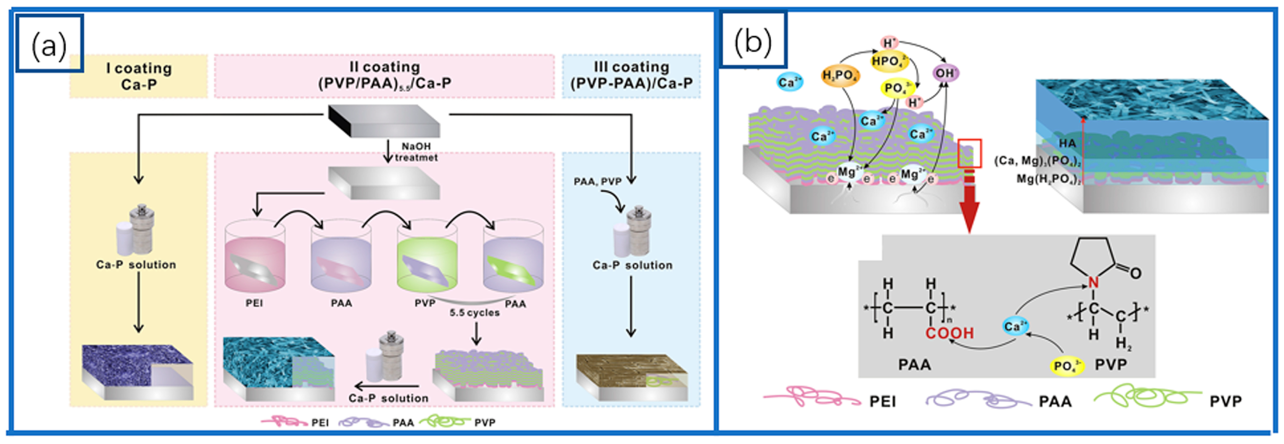
  123. Cui, L.Y.; Cheng, S.C.; Liang, L.X.; Zhang, J.C.; Li, S.Q.; Wang, Z.L.; Zeng, R.C. In vitro corrosion resistance of layer-by-layer assembled polyacrylic acid multilayers induced Ca–P coating on magnesium alloy AZ31. Bioact. Mater. 2020, 5, 153–163. [Google Scholar] [CrossRef] [PubMed]
  124. Cui, L.Y.; Fang, X.H.; Cao, W.; Zeng, R.C.; Li, S.Q.; Chen, X.B.; Zou, Y.H.; Guan, S.K.; Han, E.H. In vitro corrosion resistance of a layer-by-layer assembled DNA coating on magnesium alloy. Appl. Surf. Sci. 2018, 457, 49–58. [Google Scholar] [CrossRef]
  125. Monfared, A.; Ghaee, A.; Ebrahimi-Barough, S. Fabrication of tannic acid/poly(N-vinylpyrrolidone) layer-by-layer coating on Mg-based metallic glass for nerve tissue regeneration application. Colloids Surf. B-Biointerfaces 2018, 170, 617–626. [Google Scholar] [CrossRef] [PubMed]
  126. Zhao, Y.; He, P.; Yao, J.; Li, M.; Wang, B.; Han, L.; Huang, Z.; Guo, C.; Bai, J.; Xue, F.; et al. pH/NIR-responsive and self-healing coatings with bacteria killing, osteogenesis, and angiogenesis performances on magnesium alloy. Biomaterials 2023, 301, 122237. [Google Scholar] [CrossRef]
  127. Asadi, H.; Ghalei, S.; Handa, H.; Ramasamy, R.P. Cellulose nanocrystal reinforced silk fibroin coating for enhanced corrosion protection and biocompatibility of Mg-based alloys for orthopedic implant applications. Prog. Org. Coat. 2021, 161, 106525. [Google Scholar] [CrossRef]
  128. Kannan, M.B.; Liyanaarachchi, S. Hybrid coating on a magnesium alloy for minimizing the localized degradation for load-bearing biodegradable mini-implant applications. Mater. Chem. Phys. 2013, 142, 350–354. [Google Scholar] [CrossRef]
  129. Zhang, X.; Li, Q.; Li, L.; Zhang, P.; Wang, Z.; Chen, F. Fabrication of hydroxyapatite/stearic acid composite coating and corrosion behavior of coated magnesium alloy. Mater. Lett. 2012, 88, 76–78. [Google Scholar] [CrossRef]
  130. Fan, W.; Du, H.; An, Y.; Guo, C.; Wei, Y.; Hou, L. Fabrication and characterization of a hydroxyapatite–methylcellulose composite coating on the surface of AZ31 magnesium alloy. Mater. Lett. 2015, 152, 32–35. [Google Scholar] [CrossRef]
  131. Perumal, G.; Ramasamy, B.; Nandkumar, A.M.; Dhanasekaran, S.; Ramasamy, S.; Doble, M. Bilayer nanostructure coated AZ31 magnesium alloy implants: In vivo reconstruction of critical-sized rabbit femoral segmental bone defect. Nanomedicine 2020, 29, 102232. [Google Scholar] [CrossRef]
  132. Tian, Q.; Rivera-Castaneda, L.; Liu, H. Optimization of nano-hydroxyapatite/poly(lactic-co-glycolic acid) coatings on magnesium substrates using one-step electrophoretic deposition. Mater. Lett. 2017, 186, 12–16. [Google Scholar] [CrossRef]
  133. Zhang, J.; Wen, Z.; Zhao, M.; Li, G.; Dai, C. Effect of the addition CNTs on performance of CaP/chitosan/coating deposited on magnesium alloy by electrophoretic deposition. Mater. Sci. Eng. C Mater. Biol. Appl. 2016, 58, 992–1000. [Google Scholar] [CrossRef] [PubMed]
  134. Kim, S.M.; Kang, M.H.; Kim, H.E.; Lim, H.K.; Byun, S.H.; Lee, J.H.; Lee, S.M. Innovative micro-textured hydroxyapatite and poly(l-lactic)-acid polymer composite film as a flexible, corrosion resistant, biocompatible, and bioactive coating for Mg implants. Mater. Sci. Eng. C Mater. Biol. Appl. 2017, 81, 97–103. [Google Scholar] [CrossRef] [PubMed]
  135. Qiu, L.; Sun, M.; Huang, N.; Yang, P.; Jing, F.; Zhao, A.; Akhavan, B. Biodegradable PTMC-MAO composite coatings on AZ31 Mg-alloys for enhanced corrosion-resistance. J. Alloys Compd. 2024, 998, 175017–175025. [Google Scholar] [CrossRef]
  136. Lv, Y.; Zhang, Y.; Zheng, M.; Liu, H.; Dong, Z.; Zhang, X. NIR-induced corrosion regulation strategy for biodegradable Mg-based implants. Prog. Org. Coat. 2023, 185, 107921. [Google Scholar] [CrossRef]
  137. Monfared, M.M.; Aghdam, R.M.; Pishbin, F.; Rahmani, M. Enhancement of PEO-coated ZK60 Mg alloy: Curcumin-enriched mesoporous silica and PLA/bioglass for antibacterial properties, bioactivity and biocorrosion resistance. Surf. Coat. Technol. 2024, 493, 131237. [Google Scholar] [CrossRef]
  138. Zhao, Y.B.; Shi, L.Q.; Cui, C.Y.; Zhang, C.L.; Li, S.Q.; Zeng, R.C. Corrosion Resistance of Silane-Modified Hydroxyapatite Films on Degradable Magnesium Alloys. Acta Metall. Sin. -Engl. Lett. 2018, 31, 180–188. [Google Scholar] [CrossRef]
  139. Bakhsheshi-Rad, H.R.; Hamzah, E.; Daroonparvar, M.; Abdul Kadir, M.R.; Kasiri-Asgarani, M.; Staiger, M.P. Enhancement of corrosion resistance and mechanical properties of Mg–1.2Ca–2Bi via a hybrid silicon-biopolymer coating system. Surf. Coat. Technol. 2016, 301, 133–139. [Google Scholar] [CrossRef]
  140. Ren, Y.; Babaie, E.; Bhaduri, S.B. Nanostructured amorphous magnesium phosphate/poly (lactic acid) composite coating for enhanced corrosion resistance and bioactivity of biodegradable AZ31 magnesium alloy. Prog. Org. Coat. 2018, 118, 1–8. [Google Scholar] [CrossRef]
  141. Wang, C.; Shen, J.; Zhang, X.; Duan, B.; Sang, J. In vitro degradation and cytocompatibility of a silane/Mg(OH)2 composite coating on AZ31 alloy by spin coating. J. Alloys Compd. 2017, 714, 186–193. [Google Scholar] [CrossRef]
  142. Córdoba, L.C.; Marques, A.; Taryba, M.; Coradin, T.; Montemor, F. Hybrid coatings with collagen and chitosan for improved bioactivity of Mg alloys. Surf. Coat. Technol. 2018, 341, 103–113. [Google Scholar] [CrossRef]
  143. Chen, X.W.; Cai, L.P.; Zhang, D.F.; Ran, Y.; Ping, W. In Vitro Corrosion Behavior of Zr-Containing MAO-PLA Composite Coating on Magnesium Alloy. Trans. Indian Inst. Met. 2023, 76, 2123–2130. [Google Scholar] [CrossRef]
  144. Vafa, E.; Bazargan-lari, R.; Bahrololoom, M.E.; Amani, A.M. Effect of polyvinyl alcohol concentration on biomedical application of chitosan/bioactive glass composite coated on AZ91D magnesium alloy. Mater. Chem. Phys. 2022, 291, 126650. [Google Scholar] [CrossRef]
  145. Roshan, S.; Eivaz Mohammadloo, H.; Sarabi, A.A.; Afshari, M. Biocompatible hybrid chitosan/hydroxyapatite coating applied on the AZ31 Mg alloy substrate: In-vitro corrosion, surface and structure studies. Mater. Today Commun. 2022, 30, 103153–103168. [Google Scholar] [CrossRef]
  146. Rahman, M.; Chowdhury, M.A.; Mia, M.S.; Ali, M.R.; Rahman, A.; Ali, M.O.; Mahmud, S. Fabrication and characterization of hybrid coating on Mg-Zn-Ca Mg alloy for enhanced corrosion and degradation resistance as medical implant. Ceram. Int. 2022, 48, 23314–23324. [Google Scholar] [CrossRef]
  147. Liu, Z.; Wang, T.; Xu, Y.; Liang, C.; Li, G.; Guo, Y.; Zhang, Z.; Lian, J.; Ren, L. Double-layer calcium phosphate sandwiched siloxane composite coating to enhance corrosion resistance and biocompatibility of magnesium alloys for bone tissue engineering. Prog. Org. Coat. 2023, 177, 107417. [Google Scholar] [CrossRef]
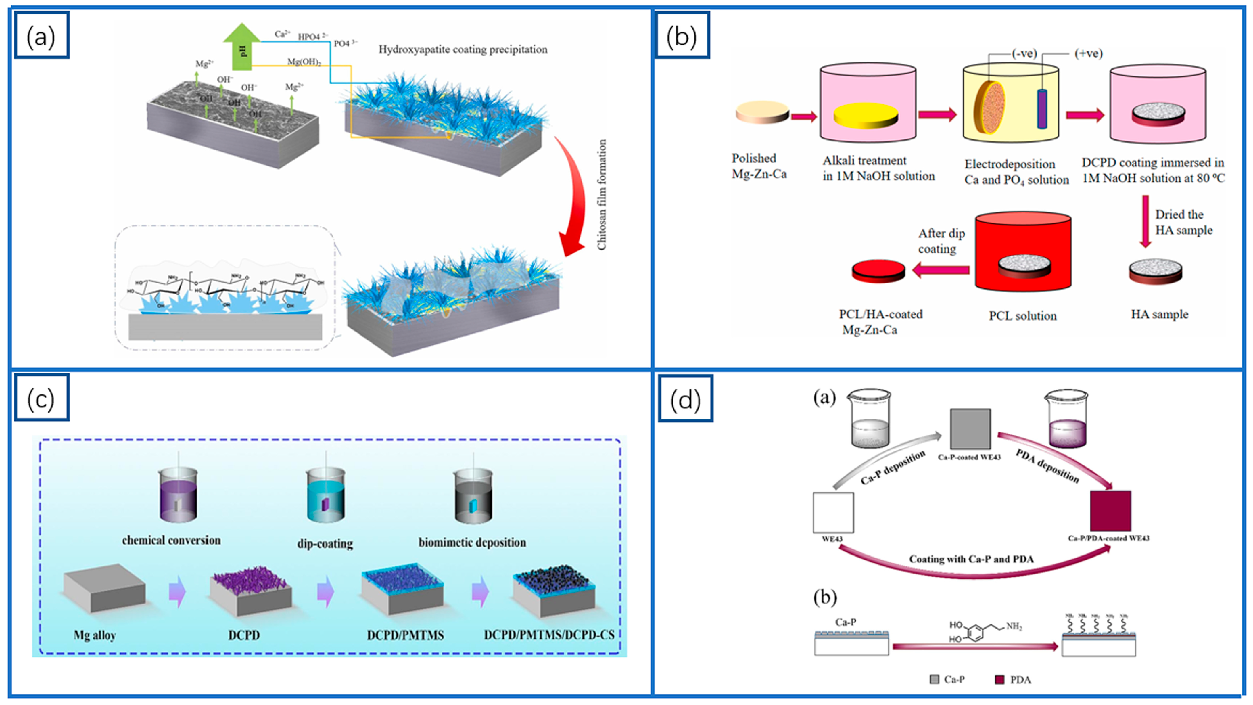
  148. Wang, X.; Liu, X.; Dai, Y.; She, J.; Zhang, D.; Qi, F.; Wei, W.; Ouyang, X. A novel Ca-Mg-P/PDA composite coating of Mg alloys to improve corrosion resistance for orthopedic implant materials. Surf. Coat. Technol. 2023, 471, 129920. [Google Scholar] [CrossRef]
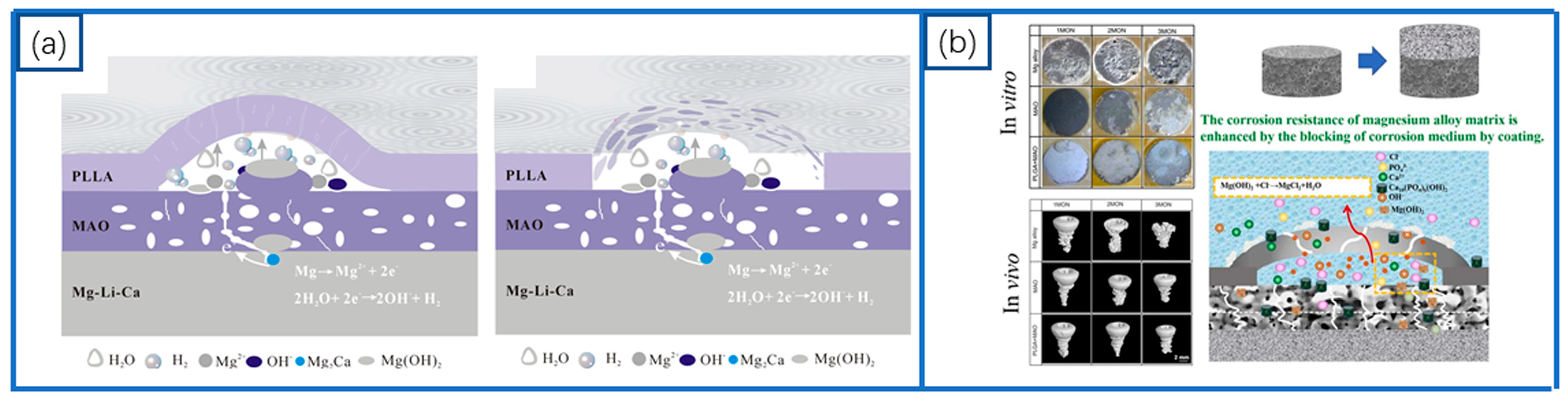
  149. Zeng, R.C.; Cui, L.Y.; Jiang, K.; Liu, R.; Zhao, B.D.; Zheng, Y.F. In Vitro Corrosion and Cytocompatibility of a Microarc Oxidation Coating and Poly(L-lactic acid) Composite Coating on Mg-1Li-1Ca Alloy for Orthopedic Implants. ACS Appl. Mater. Interfaces 2016, 8, 10014–10028. [Google Scholar] [CrossRef]
  150. Li, X.; Hu, J.; Liu, M.; Xiao, X.; Yang, L.; Qin, G.; Zhang, E. Effect of PLGA+MAO composite coating on the degradation of magnesium alloy in vivo and in vitro. Mater. Today Commun. 2023, 34, 105197. [Google Scholar] [CrossRef]
  151. Abdal-Hay, A.; Hasan, A.; Kim, Y.K.; Lee, M.H.; Hamdy, A.S.; Khalil, K.A. Biocorrosion behavior of biodegradable nanocomposite fibers coated layer-by-layer on AM50 magnesium implant. Mater. Sci. Eng. C Mater. Biol. Appl. 2016, 58, 1232–1241. [Google Scholar] [CrossRef]
  152. Gao, F.; Hu, Y.; Gong, Z.; Liu, T.; Gong, T.; Liu, S.; Zhang, C.; Quan, L.; Kaveendran, B.; Pan, C. Fabrication of chitosan/heparinized graphene oxide multilayer coating to improve corrosion resistance and biocompatibility of magnesium alloys. Mater. Sci. Eng. C Mater. Biol. Appl. 2019, 104, 109947. [Google Scholar] [CrossRef]
Scheme 1. Schematic diagram of different composed coatings on Mg surface.
Coatings 15 00191 sch001
Figure 3. Schematic diagram illustrating the corrosion process of oxide coatings: (a) schematic illustration for the effect of MgSO4 on the formation and anti-corrosion performance of CaCO3 coatings on Mg alloy. Red arrow indicates the penetration of aggressive ions into coating for Mg corrosion [58], and (b) corrosion schematic [57].
Coatings 15 00191 g003
Figure 7. Schematic representation of the preparation and corrosion process of polyacrylic acid multilayers [123]: (a) schematic construction of the Ca-P coatings via hydrothermal treatment, and (b) schematic coating formation mechanism of the II coating via hydrothermal treatment with LbL assembled templates.
Coatings 15 00191 g007
Figure 8. Schematic representation of the preparation of polymer coatings on MAO layer: (a) schematic diagram illustrating the reparation of PTMC-MAO composite coatings [135], (b) schematic diagram of the fabrication process of Mg/MAO/PCL-PPy composite coating [136], (c) schematic diagram illustrating the fabrication process of Mg/MAO/mesoporous silica-PLA composite coatings [137], and (d) schematic representation of the preparation of PMTMS/HA coating [138].
Coatings 15 00191 g008
Figure 9. Schematic representation of the preparation of Ca-P coating and polymer composite coatings: (a) the proposed mechanism of the post-treated HA coating by chitosan film [145], (b) schematic illustration of PCL/HA hybrid coating process [146], (c) schematic diagram of the preparation process of composite coated samples [147], and (d) experimental flow and film layer formation flow [148].
Coatings 15 00191 g009
Figure 10. Schematic representation of in vitro corrosion and cytocompatibility of MAO and polymer composite coatings: (a) a microarc oxidation coating and poly (l-lactic acid) composite coating on Mg-1Li-1Ca alloy for orthopedic implants [149], and (b) micro-CT images and corrosion mechanism diagram of PLGA + MAO screws [150].
Coatings 15 00191 g010
Disclaimer/Publisher’s Note: The statements, opinions and data contained in all publications are solely those of the individual author(s) and contributor(s) and not of MDPI and/or the editor(s). MDPI and/or the editor(s) disclaim responsibility for any injury to people or property resulting from any ideas, methods, instructions or products referred to in the content.

