Article

DC Sputtered Ultra-Thin Au Films and the Effect of Their Morphologies on Au-Catalyzed CIGS Films

1
Department of Physics, Niğde Ömer Halisdemir University, 51240 Nigde, Türkiye
2
Nanotechnology Application & Research Center, Niğde Ömer Halisdemir University, 51240 Nigde, Türkiye
Coatings 2025, 15(11), 1274; https://doi.org/10.3390/coatings15111274
Submission received: 5 September 2025 / Revised: 7 October 2025 / Accepted: 20 October 2025 / Published: 3 November 2025

Abstract

Gold (Au) is one of the noble metals most used as a catalyst in the growth of one-dimensional nanostructures. Usually, an ultra-thin Au film is coated followed by thermal annealing to obtain Au nanoclusters. Although annealing temperature, duration and film thickness parameters have been heavily studied, there are no studies on the sputter working gas pressure, which also greatly affects the film microstructure. In this study, low (5 mTorr) and high (15 mTorr) working gas pressures were examined in addition to Au film thicknesses of 2 nm, 5 nm and 8 nm. Additionally, copper indium gallium selenide (CIGS) films were deposited on Au films with different thicknesses and argon (Ar) gas pressures. It was confirmed from SEM and AFM images that the Au films undergo drastic morphology change from smooth to extremely porous film surfaces with increasing thickness regardless of gas pressure. However, the porosity of films is increased at higher growth pressure for each thickness. Specifically, the most porous film was obtained at a 5 nm thickness with 15 mTorr, and it was filled with nanomounds. Not surprisingly, the only apparent columnar-type formation was observed for CIGS deposition, which was carried out on the most porous film. It can be interpreted that Au nanomounds behave like catalysts on which the CIGS nanocolumns grow.

1. Introduction

Noble metal nanomaterials have been of immense interest in a wide range of applications including photovoltaics [1,2], chemical and biological sensing [3,4,5,6], optoelectronic devices [7], and growth of one-dimensional nanostructures [8,9] due to their enormous electrical and structural properties. Among all, gold (Au) is one of the most preferred noble metals since it can utilize the electromagnetic field enhanced by localized surface plasmons that leads to increased light absorption which is crucial for photocurrent harvesting devices [10,11,12]. Additionally, Au has a high chemical stability that makes it very suitable for use in one-dimensional growth [13,14,15].
For one-dimensional nanostructure fabrication, an ultra-thin Au layer is coated on a substrate, and then the thermal annealing process is carried out to promote the formation of Au nanostructures which behave as nucleation sites for the growth of nanowires and nanorods by the vapor–liquid–solid (VLS) growth mechanism [16]. When annealing is carried out at a high annealing temperature, usually higher than 200 °C [17,18,19], the ultra-thin Au film undergoes a morphological change due to the thermal dewetting process, with dewetting at the beginning followed by melting and agglomeration into mostly spherical nanoparticles [20,21]. Although Au film deposition prior to Au nanostructure formation is carried out by many growth methods, with thermal evaporation mostly dominating [22,23], sputtering has arisen as a very appropriate technique due to its wide-area application facility, clean growth process in a high-vacuum atmosphere, and controllable film growth [17,18,20,24]. Thus, sputtering was chosen for Au film growth and post-annealing in this study. In the literature, film thickness, annealing temperature, and annealing duration are the most studied parameters of Au film formation [22,23,24,25,26,27]. However, the effect of working gas pressure on the morphology of Au films and the resulting Au nanostructures has not been investigated yet. Additionally and more importantly, although CIGS is one of the most important materials used in optoelectronic applications, the usage of CIGS nanoporous films is usually disregarded despite their superior optical advantages due to the challenging quaternary material properties of CIGS.
In this study, we studied the effect of both film thickness and Ar pressure on Au film formation. After thermal treatment, CIGS films were simultaneously coated on Au films. The growth of CIGS nanocolumns on the most porous films by Au catalysts resulting from the dewetting of Au thin films was analyzed. Since this is the first study of both Ar gas pressure and CIGS columnar-type film formation by Au catalysts, to the best of our knowledge, it is expected that the results revealed in this study will make a significant contribution to the literature.

2. Materials and Methods

Glass was used as a substrate for all Au thin film growths. Firstly, glass substrates were ultrasonically cleaned for 15 min in acetone, isopropanol, and ethanol, respectively, to remove the probable dust as well as organic and inorganic contaminants. Then, the glass substrates were rinsed with deionized water and dried using nitrogen gas followed by storing in a sealed container to keep them clean until the deposition process. All Au thin films were direct current (DC)-sputtered by using an Au target with a high purity of 99.99%. The sputter chamber base pressure was lowered to 10−6 Torr to prepare the system for the actual growth. To ensure the high quality of the grown films, 5N-Argon (Ar-99.999%) gas was used during depositions and pre-cleaning of the target was carried out to avoid the possible contamination of substrates. The rotation of the substrate was fixed at 8 rpm for all depositions to promote homogeneous and uniform film growth. The Au films were grown with varying thicknesses of 2 nm, 5 nm and 8 nm under 5 mTorr and 15 mTorr Ar gas pressures at room temperature. All six Au films were then thermally annealed at 300 °C for 1 h under Ar gas. After the thermal treatment, the samples were left in the vacuum environment to cool down to room temperature.
CIGS thin films were then coated on these six Au films by RF (13.6 MHz Radio Frequency) magnetron sputtering at room temperature until the thickness reached 500 nm. Since the CIGS film deposition was carried out at the same time on Au films grown with different thicknesses and gas pressures, the effect of Au film morphology on the formation of CIGS films could be safely studied by comparison.
The surface of Au films was characterized by both SEM (Quanta 250 FEG Scanning Electron Microscope, FEI, Zaragoza, Spain ) and AFM (Bruker/Innova, Billerica, MA, USA) systems. The rms values of Au films were obtained by AFM imaging. The structural information of Au films was investigated by Raman spectroscopy (Renishaw/Invia). Additionally, the morphology of the final CIGS films was monitored by cross-sectional SEM (Quanta 250 FEG Scanning Electron Microscope, FEI, Zaragoza, Spain) measurement.

3. Results

Top-view SEM images of Au thin films with different thicknesses and growth pressures are given in Figure 1. The set thickness of films for both columns follows the order of 2 nm, 5 nm, and 8 nm from top to bottom. Meanwhile, the growth pressure was set as 5 mTorr and 15 mTorr for left and right images, respectively, for each thickness. The Au film experienced a drastic microstructural change from a smooth film to a porous surface with nanomounds and again, the formation of a film partially covered with pinholes with increasing thickness. Interestingly, the morphological change of Au films demonstrated by our results does not follow the general assumption that the very tiny Au islands form firstly at a 2 nm thickness, and these islands start to coalesce and compose bigger clusters with increasing thickness [19,20,21,22,23]. However, this type of evolution is usually observed at high annealing temperatures, whereas the Au films in our study annealed at 300 °C only. In addition, the characteristics of Au film can change according to the substrate on that the growth is carried out [28]. Therefore, we can claim that the Au films grown in our case conformed to typical metal film growth by sputtering as a uniform and homogenous coating was observed [29]. Upon thermal annealing, the film with 5 nm thickness seems to be extremely porous with lots of nanomounds due to the thermal dewetting-induced morphological change caused by the tendency of Au film to shrink its surface area in order to minimize the total energy [27]. Meanwhile, it can be concluded that other two films are not affected in the same way, as 2 nm is too thin and 8 nm is too thick to create nanospherical structures by heat treatment.
On the other hand, the working gas pressure has an impact on the elevation of surface properties for each film thickness as the number of nanomounds and pinholes is apparently increased by higher pressure. For 2 nm thickness, a very smooth surface is visible at 5 mTorr of Ar gas, while some circular formations are observed at the high (15 mTorr) pressure, as shown in Figure 1(a-1,a-2). For the 5 nm and 8 nm thicknesses, the surface properties show a higher number of structures at higher pressure. When higher-pressure Ar gas is applied during the film growth, the flux of incoming atoms is be more spread out due to higher angular directions of atoms pulled off from the target due to more collisions between Ar+ ions and target atoms. Therefore, the target atoms reach the substrate at a wide variety of angular directions and as a result, a more porous film is formed [30,31], which was directly observed at 5 nm and 8 nm thicknesses. In the 8 nm case, the pinholes became less numerous and smaller due to the plasma damage at higher pressure.
The AFM images of Au films are given in Figure 2. AFM analysis allows an in-depth investigation of the surface properties of ultra-thin films [19,25,26,27,28,29,30,31,32]. The homogeneity and porosity of each film based on the growth conditions demonstrated by AFM images is supported by the results concluded from SEM imaging. This is counter-intuitive and perhaps related to the quantum size effect, significantly lower melting point temperatures, and too-high annealing temperature.
Figure 3 shows the Raman spectra of Au thin films with different thicknesses and working gas pressures, which are labeled in the graph for each sample. The peaks for all Au films are not so sharp; indeed, the peaks are barely visible except those on the Au film with 5 nm and 5 mTorr in Figure 3a, due to the morphology of the film. The continuous Au film typically does not present any Raman peaks since the surface plasmons cannot be excited due to smoothness [33]. Meanwhile, the Au film grown at 5 nm thickness and under 5 mTorr gas pressure has the most significant peaks as a result of its nanoporous morphology, as can be clearly seen from SEM and AFM images. When the incident laser light meets the nanopores or nanogaps on the metallic film, the nanostructures provide plasmonic hotspots that amplify the electromagnetic signal and thus enhance the Raman peak intensity [18,34].
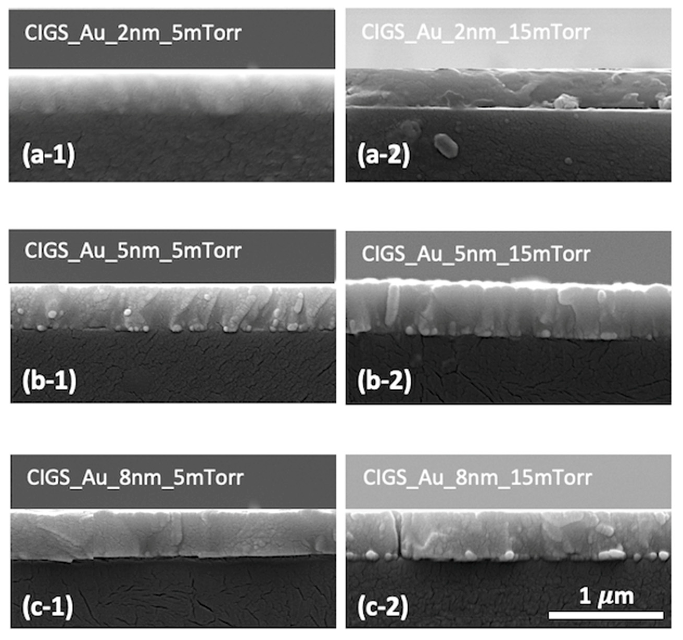
The morphology of CIGS films grown on Au thin-film-coated glass substrates is demonstrated by cross-sectional SEM images in Figure 4. It can be interpreted that the thickness of all CIGS films is around 500 nm, which is the set value, regardless of Au film thickness as it is too small to affect the resulting film formation. However, the thickness of CIGS films grown on the most porous surface with nanomounds and nanopits as concluded from AFM images seems a little higher (Figure 4(b-1,b-2,c-1)) compared to their counterparts which were grown on more smooth Au films. Specifically, CIGS films fabricated on Au films with 5 nm thickness grown under both 5 mTorr and 15 mTorr present a more columnar type of formation rather than smooth films along with a slightly higher thickness (Figure 4(b-1,b-2)). As the Au films with 5 nm thickness present as extremely porous films filled with abundant nanomounds, it is believed that these nanoscale hillocks behave as metallic seeds, namely “catalysts” on which the nanocolumns grow. In this growth mechanism called VLS [16], firstly, the metallic atoms nucleate on the substrate and form metallic clusters—catalysts—which act as energetically favored nucleation sites for the incoming target material atoms. Since the catalysts behave as sinking centers, the vaporized material atoms can be collected more effectively, and thus, the film tends to grow in a vertical direction rather than horizontal. Additionally, due to more atoms contributing, the growth occurs faster vertically and results in nanorod formation. That is why CIGS films grown on Au films with 5 nm thickness and 15 mTorr (Figure 4(b-2)) present a clear nanocolumnar formation along with a slightly higher thickness since the Au-5 nm-15 mTorr film has the most abundant nanomounds, which promote VLS growth.

4. Conclusions

In conclusion, the effect of working gas pressure in addition to film thickness on the morphology of DC-sputtered Au thin films was investigated in this study. The morphology change according to the film thickness is dramatic. A smooth film is observed for a 2 nm thickness, whereas the film surface is filled with nanomounds and pinholes for 5 nm and 8 nm thicknesses, respectively. Meanwhile, the surface properties do not change drastically but become more apparent at higher pressure compared to lower growth pressure. Specifically, the most porous film was obtained for 5 nm thickness under 15 mTorr Ar gas pressure as the film is formed by numerous nanomounds which behave as catalysts for the further growth of CIGS columnar-type films.

Funding

This research received no external funding.

Data Availability Statement

Data are contained within the article.

Acknowledgments

The author would like to thank Center Research Laboratory at Niğde Ömer Halisdemir University for AFM and Raman measurements, and Scientific and Technological Application and Research Center at Aksaray University for SEM analysis.

Conflicts of Interest

The author declares no conflicts of interest.

References

  1. Pillai, S.; Green, M.A. Plasmonics for photovoltaic applications. Sol. Energy Mater. Sol. Cells 2010, 94, 1481–1486. [Google Scholar] [CrossRef]
  2. Kac, H.; Baltakesmez, A.; Çaldıran, Z.; Aydogan, S.; Yılmaz, M.; Sevim, M. Optical and electrical characterization of organic solar cells obtained using gold and silver metal nanoparticles. Mater. Today Proc. 2021, 46, 6986–6990. [Google Scholar]
  3. Kim, S.; Kim, T.G.; Lee, S.H.; Kim, W.; Bang, A.; Moon, S.W.; Song, J.; Shin, J.-H.; Yu, J.S.; Choi, S. Label-free surface-enhanced Raman spectroscopy biosensor for on-site breast cancer detection using human tears. ACS Appl. Mater. Interfaces 2020, 12, 7897–7904. [Google Scholar]
  4. Siampour, H.; Abbasian, S.; Moshaii, A.; Amirsoleimani, A.R. Stable, reproducible, and binder-free gold/copper core–shell nanostructures for high-sensitive non-enzymatic glucose detection. Sci. Rep. 2022, 12, 18945. [Google Scholar]
  5. Mo, Y.; Zhang, X.; Zou, K.; Xing, W.; Hou, X.; Zeng, Y.; Cai, Y.; Xu, R.; Zhang, H.; Cai, W. Au ordered array substrate for rapid detection and precise identification of etomidate in E-liquid through surface-enhanced Raman spectroscopy. Nanomaterials 2024, 14, 1958. [Google Scholar]
  6. Kamakshi, K.; Silva, J.P.B.; Sekhar, K.C.; Marslin, G.; Moreira, J.A.; Conde, O.; Almeida, A.; Pereira, M.; Gomes, M.J.M. Influence of substrate temperature on the properties of pulsed laser deposited silver nanoparticle thin films and their application in SERS detection of bovine serum albumin. Appl. Phys. B 2016, 122, 108. [Google Scholar]
  7. Yun, J. Ultrathin Metal films for Transparent Electrodes of Flexible Optoelectronic Devices. Adv. Funct. Mater. 2017, 27, 1606641. [Google Scholar] [CrossRef]
  8. Wang, Y.; Schmidt, V.; Senz, S.; Gosele, U. Epitaxial growth of silicon nanowires using an aluminium catalyst. Nat. Nanotech 2016, 1, 186–189. [Google Scholar]
  9. Ly, L.Q.; Bonvicini, S.N.; Shi, Y. Platinum nanoparticle formation by pulsed laser-induced dewetting and its application as catalyst in silicon nanowire growth. J. Phys. Chem. C 2025, 129, 4553–4564. [Google Scholar] [CrossRef]
  10. Gao, Y.; Jin, F.; Su, Z.; Zhao, H.; Luo, Y.; Chu, B.; Li, W. All thermal-evaporated surface plasmon enhanced organic solar cells by Au nanoparticles. Org. Electron. 2016, 39, 71–76. [Google Scholar] [CrossRef]
  11. Chen, S.-C.; Chen, Y.-J.; Chen, W.T.; Yen, Y.-T.; Kao, T.S.; Chuang, T.-Y.; Liao, Y.-K.; Wu, K.-H.; Yabushita, A.; Hsieh, T.-P.; et al. Toward omnidirectional light absorption by plasmonic effect for high-efficiency flexible nonvacuum Cu(In,Ga)Se2 thin film solar cells. ACS Nano 2014, 8, 9341–9348. [Google Scholar] [CrossRef]
  12. Lie, S.; Brun, A.; Wong, L.H.; Etgar, L. Semitransparent perovskite solar cells with >13% efficiency and 27% transperancy using plasmonic Au nanorods. ACS Appl. Mater. Interfaces 2022, 14, 11339–11349. [Google Scholar] [CrossRef]
  13. Dorsey, G.; Moore, A.; Heffner, C.; Ueda, A.; Mu, R.; Collins, W.E. The growth and characterization of Au-catalyzed gallium oxide nanowires. MRS Adv. 2024, 9, 1318–1323. [Google Scholar] [CrossRef]
  14. Yu, Z.; Yang, Z.; Wang, S.; Jin, Y.; Liu, J.G.; Gong, M.; Sun, X. Growth of GaN nanorods via Au catalyst-assisted CVD. Chem. Vap. Depos. 2005, 11, 433–436. [Google Scholar] [CrossRef]
  15. Chou, Y.-C.; Wen, C.-Y.; Reuter, M.C.; Su, D.; Stach, E.A.; Ross, F.M. Controlling the growth of Si/Ge nanowires and heterojunctions using silver-gold alloy catalysts. ACS Nano 2012, 6, 6407–6415. [Google Scholar] [CrossRef]
  16. Givargizov, E.I. Fundamental aspects of VLS growth. J. Cryst. Growth 1975, 31, 20–30. [Google Scholar] [CrossRef]
  17. Garg, V.; Sengar, B.S.; Awasthi, V.; Aaryashree; Sharma, P.; Mukherjee, C.; Kumar, S.; Mukherjee, S. Localized surface plasmon resonance on Au nanoparticles: Tuning and exploitation for performance enhancement in ultrathin photovoltaics. RSC Adv. 2016, 6, 26216. [Google Scholar] [CrossRef]
  18. Nguyen, T.H.T.; Nguyen, T.M.A.; Sai, C.D.; Le, T.H.Y.; Tran, T.N.A.; Bach, T.C.; Le, V.V.; Pham, N.H.; Ngac, A.B.; Nguyen, V.T.; et al. Efficient surface enhanced Raman scattering substrates based on complex gold nanostructures formed by annealing sputtered gold thin films. Opt. Mater. 2021, 121, 111488. [Google Scholar] [CrossRef]
  19. Hedl, E.; Bregović, V.B.; Rakić, I.Š.; Mandić, Š.; Samec, Ž.; Bergmann, A.; Sancho-Parramon, J. Optical properties of annealed nearly percolated Au thin films. Opt. Mater. 2023, 135, 113237. [Google Scholar] [CrossRef]
  20. Xia, C.; Wang, R.; Zhu, P.; Wang, F.; Dong, L.; Wang, H.; Wang, Y. Fabrication of gold nanoparticle decorated surfaces for controlled nucleation of plasmonic microbubbles. Surf. Interfaces 2023, 36, 102591. [Google Scholar] [CrossRef]
  21. Farmer, G.; Shymkiv, D.; Krokhin, A.; Littler, C.; Syllaios, A.J.; Philipose, U. Plasmonic properties of gold nanoparticle arrays fabricated using a sequential dewetting process. Appl. Phys. Lett. 2025, 126, 081103. [Google Scholar] [CrossRef]
  22. Yang, M.-C.; Chien, T.-Y.; Cheng, Y.-W.; Hsieh, C.-K.; Syu, W.-L.; Wang, K.-S.; Chen, Y.-C.; Chen, J.-S.; Chen, C.-C.; Liu, T.-Y. Reproducible SERS substrates manipulated by interparticle spacing and particle diameter of gold nano-island array using in-situ thermal evaporation. Spectrochim. Acta Part A Mol. Biomol. Spectrosc. 2023, 303, 123190. [Google Scholar] [CrossRef]
  23. Gaspar, D.; Pimentel, A.C.; Mateus, T.; Leita, J.P.; Soares, J.; Falca, B.P.; Arau, A.; Vicente, A.; Filonovich, S.A.; Águas, H.; et al. Influence of the layer thickness in plasmonic gold nanoparticles produced by thermal evaporation. Sci. Rep. 2013, 3, 1469. [Google Scholar] [CrossRef]
  24. Ruffino, F.; Grimaldi, M.G. Atomic force microscopy study of the growth mechanisms of nanostructured sputtered Au film on Si(111): Evolution with film thickness and annealing time. J. Appl. Phys. 2010, 107, 104321. [Google Scholar] [CrossRef]
  25. Mishra, Y.K.; Mohapatra, S.; Kabiraj, D.; Tripathi, A.; Pivin, J.C.; Avasthi, D.K. Growth of Au nanostructures by annealing electron beam evaporated thin films. J. Opt. A Pure Appl. Opt. 2007, 9, S410–S414. [Google Scholar] [CrossRef]
  26. Li, M.-Y.; Sui, M.; Pandey, P.; Zhang, Q.; Kim, E.-S.; Lee, J. Systematic control of self-assembled Au nanoparticles and nanostructures through the variation of deposition amount, annealing duration, and temperature on Si (111). Nanoscale Res. Lett. 2015, 10, 380. [Google Scholar] [CrossRef] [PubMed]
  27. Sun, H.; Yu, M.; Wang, G.; Sun, X.; Lian, J. Temperature-dependent morphology evolution and surface plasmon absorption of ultrathin gold island films. J. Phys. Chem. C 2012, 116, 9000–9008. [Google Scholar] [CrossRef]
  28. Malinský, P.; Slepička, P.; Hnatowicz, V.; Švorčík, V. Early stages of growth of gold layers sputter deposited on glass and silicon substrates. Nanoscale Res. Lett. 2012, 7, 241. [Google Scholar] [CrossRef] [PubMed]
  29. Keles, F.; Atasoy, Y.; Seyhan, A. Sputtered Mo-bilayer thin films with reduced thickness and improved electrical resistivity. Mater. Res. Express 2019, 6, 126455. [Google Scholar] [CrossRef]
  30. Keles, F.; Badradeen, E.; Karabacak, T. Self-anti-reflective density-modulated thin films by HIPS technique. Nanotechnology 2017, 28, 335703. [Google Scholar] [CrossRef]
  31. Keles, F.; Cansizoglu, H.; Badraddin, E.O.; Brozak, M.P.; Watanabe, F.; Karabacak, T. HIPS-GLAD core shell nanorod array photodetectors with enhanced photocurrent and reduced dark current. Mater. Res. Express 2016, 3, 105028. [Google Scholar] [CrossRef]
  32. Li, M.-Y.; Sui, M.; Pandey, P.; Zhang, Q.-Z.; Kunwar, S.; Salamo, G.J.; Lee, J. Precise control of configuration, size and density of self-assembled Au nanostructures on 4H-SiC (0001) by systematic variation of deposition amount, annealing temperature and duration. Cryst. Eng. Comm. 2016, 18, 3347. [Google Scholar] [CrossRef]
  33. Yu, M.; Huang, Z.; Liu, Z.; Chen, J.; Liu, Y.; Tang, L.; Liu, G. Annealed gold nanoshells with highly-dense hotspots for large-area efficient Raman scattering substrates. Sens. Actuators B 2018, 262, 845–851. [Google Scholar] [CrossRef]
  34. Tang, L.; Liu, Y.; Liu, G.; Chen, Q.; Li, Y.; Shi, L.; Liu, Z.; Liu, X. A novel SERS substrate platform: Spatially stacking plasmonic hotspots films. Nanoscale Res. Lett. 2019, 14, 94. [Google Scholar] [CrossRef]
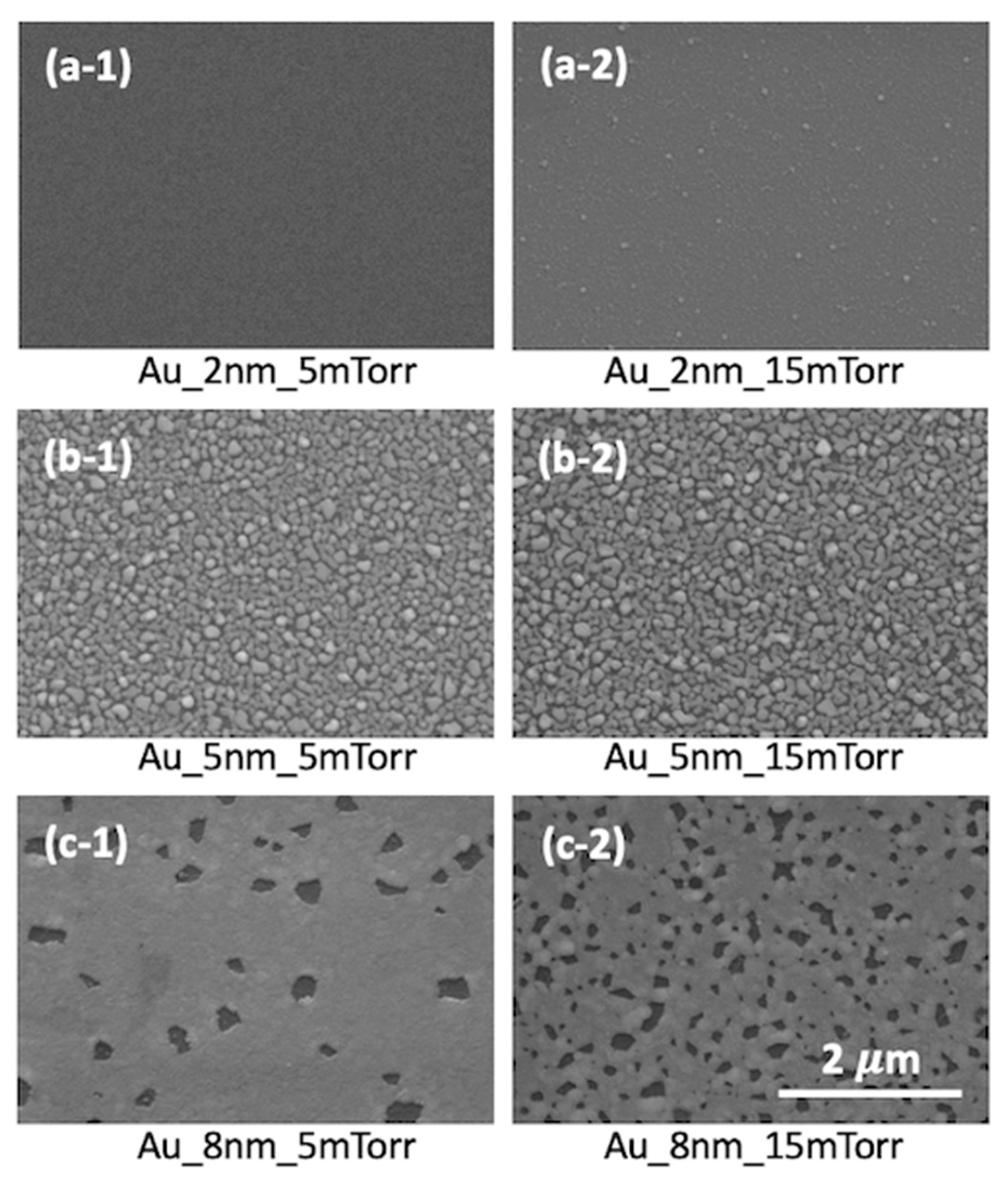
Figure 1. Top-view SEM images of Au thin films with various thicknesses of 2 nm (a-1,a-2), 5 nm (b-1,b-2) and 8 nm (c-1,c-2). Left- and right-hand-side films grown under 5 mTorr and 15 mTorr Ar gas pressure, respectively.
Coatings 15 01274 g001
Figure 2. Slightly tilted 3D AFM images of Au thin films with various thicknesses of 2 nm (a-1,a-2), 5 nm (b-1,b-2) and 8 nm (c-1,c-2). Left- and right-hand-side films grown under 5 mTorr and 15 mTorr Ar gas pressure, respectively.
Coatings 15 01274 g002
Figure 3. Raman spectra of Au thin films with different thickness at 5 mTorr (a) and 15 mTorr (b) sputtering pressure.
Coatings 15 01274 g003
Figure 4. Cross-sectional SEM images of CIGS thin films grown on Au thin films with; (a-1) 2 nm thikcness & 5 mTorr Ar pressure; (a-2) 2 nm thikcness & 15 mTorr Ar pressure; (b-1) 5 nm thikcness & 5 mTorr Ar pressure; (b-2) 5 nm thikcness & 15 mTorr Ar pressure; (c-1) 8 nm thikcness & 5 mTorr Ar pressure; (c-2) 8 nm thikcness & 15 mTorr Ar pressure.
Coatings 15 01274 g004
Disclaimer/Publisher’s Note: The statements, opinions and data contained in all publications are solely those of the individual author(s) and contributor(s) and not of MDPI and/or the editor(s). MDPI and/or the editor(s) disclaim responsibility for any injury to people or property resulting from any ideas, methods, instructions or products referred to in the content.