Article

Substrate Temperature-Induced Crystalline Phase Evolution and Surface Morphology in Zirconium Thin Films Deposited by Pulsed Laser Ablation

1
Department of Physics & Astronomy, Western Kentucky University, Bowling Green, KY 42101, USA
2
Department of Mathematics, Western Kentucky University, Bowling Green, KY 42101, USA
3
Universities Space Research Association, NASA Marshall Space Center, Huntsville, AL 35805, USA
*
Author to whom correspondence should be addressed.
Coatings 2025, 15(10), 1198; https://doi.org/10.3390/coatings15101198
Submission received: 1 September 2025 / Revised: 2 October 2025 / Accepted: 5 October 2025 / Published: 11 October 2025
(This article belongs to the Collection Collection of Papers on Thin Film Deposition)

Abstract

Zirconium (Zr) thin films were deposited on silicon (Si) substrates via pulsed laser deposition (PLD) using a 248 nm excimer laser. The effects of substrate temperature on film morphology and crystallinity were systematically investigated. X-ray diffraction (XRD) revealed that the Zr(100) plane exhibited the strongest orientation at 400 °C while Zr (002) was maximum at 500 °C. Scanning electron microscopy (SEM) and atomic force microscopy (AFM) analyses demonstrated an increase in surface roughness with temperature, with the smoothest surface observed at lower temperatures and significant island formation at 500 °C due to the transition to 3D growth. At 500 °C, interdiffusion effects led to the formation of zirconium silicide at the Zr/Si interface. To further interpret the experimental findings, computational modeling was employed to analyze the transition from 2D layer-by-layer growth to 3D island formation at elevated temperatures. Using a multi-parameter kinetics-free model based on free energy minimization, the critical film thickness for this transition was determined to be ~1–2 nm, aligning well with experimental observations. A separate kinetic model of island nucleation and growth predicts that this shift is driven by the kinetics of adatom surface diffusion. Additionally, the kinetic simulations revealed that, at 400 °C, adatom diffusivity optimally balances crystallization and surface energy minimization, yielding the highest film quality. At 500 °C, the rapid increase in diffusivity leads to the proliferation of 3D islands, consistent with the roughness trends observed in SEM and AFM data. These findings underscore the critical role of deposition parameters in tailoring Zr thin films for applications in advanced coatings and electronic devices.

1. Introduction

Zirconium-based materials are highly valued for their exceptional properties, including high-temperature ionic conductivity, corrosion and heat resistance, wear resistance, fracture toughness, and mechanical strength, making them indispensable for a wide range of applications [1]. These attributes position zirconium as a critical material for protective coatings in mechanical, chemical, and biomedical fields, as well as for its use in multilayer technology, microelectronics, nuclear fusion devices, fuel cells, and oxygen sensors. Its versatility in forms such as multilayers, thin films, metallic glasses, oxide layers, and bulk crystals further broadens its applications across various fields [2,3,4,5,6,7,8].
A promising application of zirconium lies in its use in multilayer coatings for aluminum mirrors operating in the extreme ultraviolet (EUV) spectrum, particularly around the 17–19 nm wavelength range. This is due to its optimal properties at the 17 nm edge. Despite challenges such as uneven crystallization, layer mixing, and contamination, Al/Zr multilayer systems have emerged as promising candidates for high-performance mirrors in EUV applications. These systems offer enhanced optical performance, structural stability, and environmental resistance, expanding their potential in high-precision optical applications [9]. Achieving these properties requires advanced deposition techniques, such as pulsed laser deposition (PLD), chemical vapor deposition (CVD), magnetron sputtering, and additive manufacturing, alongside a thorough understanding of fabrication and characterization parameters, to ensure precise control over material properties [10,11,12,13,14].
Substrate temperature is widely recognized as one of the most influential parameters in pulsed laser deposition (PLD) and laser material interactions [15,16,17,18,19,20]. It governs the mobility of adatoms on the substrate surface, which in turn controls nucleation density, grain coalescence, and the development of long-range crystalline order. At low substrate temperatures, insufficient adatom mobility often results in amorphous or nanocrystalline films with high defect densities and poor textures. As the substrate temperature increases, surface diffusion is enhanced, enabling adatoms to migrate into energetically favorable lattice sites. This process promotes grain growth, reduces defect density, and leads to sharper diffraction peaks that signify improved crystallinity. Furthermore, intermediate substrate temperatures often favor the establishment of preferential orientations, where specific crystallographic planes dominate film growth due to minimized surface and interface energies. At excessively high temperatures, however, re-evaporation of volatile elements and thermally induced stress can degrade film quality, reduce stoichiometric control, or even cause delamination. These well-established trends have been reported for a wide range of metallic and oxide systems [21,22,23,24,25,26,27,28,29]. For zirconium thin films in particular, substrate heating has been shown to strongly influence phase stability, preferred orientation, and grain size, thereby impacting mechanical strength and corrosion resistance. In our previous study, we systematically investigated Zr thin films deposited on Si(100) substrates over a substrate temperature range from room temperature to 500 °C, confirming that substrate heating leads to significant enhancements in crystallinity and texture evolution [30]. In this work, substrate temperature was varied while fluence was kept constant. This approach isolates the influence of plume energetics on film nucleation and growth, thereby providing a complementary perspective to our earlier temperature-dependent studies.
Singh et al. studied the effect of substrate temperature on zirconium (Zr) films deposited by pulsed DC magnetron sputtering in an argon (Ar) atmosphere [12]. Their findings revealed the formation of the α-phase of Zr between 27–600 °C, with X-ray diffraction (XRD) confirming a [001] texture. Higher substrate temperatures led to increased crystallite size, and, at 500 °C, hexagonal crystallites grew along the normal surface of the substrate. The films exhibited hardness between 6–10 GPa, while scratch tests showed excellent adhesion, with no significant failure observed under loads up to 20 N.
Further, Liu et al. investigated zirconium thin films deposited on silicon (Si) substrates using a KrF excimer laser (248 nm) and varying pulse repetition rates. Their study demonstrated that the deposition process produced polycrystalline Zr films, with surface morphology, droplet formation, and roughness significantly influenced by the repetition rate. Subsequently, Liu et al. examined the effects of laser fluence, pulse repetition rate, substrate temperature and deposition time, revealing that increased fluence resulted in larger nanoparticle sizes, higher root mean square (RMS) roughness, and a more compact crystalline structure. Additionally, zirconium films exposed to deuterium atmospheres formed Zr0.38D0.62 at elevated temperatures (650 °C), with surface morphology and optical properties affected by deuterium-induced defects, further supporting their utility in fusion devices [31,32,33].
Molecular dynamics simulations of Zr thin films on Si offered valuable insights into reactive interfacial diffusion and alloy formation. The studies demonstrated significant inter-diffusion of Zr and Si atoms at temperatures above the melting point, leading to the formation of ZrSi crystal alloys. This alloy formation was found to reduce diffusion rates and elevate experimental barriers, providing essential knowledge for optimizing Zr-Si interfaces in thin-film applications [32].
We have previously investigated zirconium thin films deposited on silicon substrates using a Nd:YAG laser at wavelengths of 1064 nm and 532 nm, exploring various substrate temperatures and fluence conditions. Our study highlights that optimal crystalline Zr films were obtained at a substrate temperature of 300 °C, with smoother films observed using a 1064 nm wavelength. The continuum model applied in this study provides valuable insights into the growth dynamics and morphological evolution of Zr thin films, emphasizing the importance of parameter optimization for EUV solar imaging and other optical applications [33].
In this work, we investigate the effects of various deposition parameters on zirconium coatings on Si (100) substrates using a 248 nm excimer laser, with a primary focus on substrate temperature. The selection of the 248 nm wavelength was based on its higher photon energy compared to longer wavelengths such as 1064 nm, enabling more efficient material ablation and improved stoichiometric transfer. Ultraviolet laser ablation also generates a more energetic plasma plume, which can enhance surface mobility of adatoms and influence film crystallinity and morphology. In contrast, 1064 nm lasers tend to produce lower energy plumes, which result in smoother surfaces, but may limit crystallinity due to reduced surface activation. By comparing our results with those reported by Khuzhakulov et al. [33], who employed a 1064 nm laser, we demonstrate that excimer-based PLD can yield highly crystalline zirconium films under a range of substrate temperature conditions. These findings offer wavelength-dependent insights into film growth mechanisms, supporting the use of UV laser sources for applications requiring enhanced crystallographic orientation and surface control [34].
By comparing our results to those reported by Khuzhakulov et al. [33], we confirmed that PLD effectively produces crystalline zirconium films under varying substrate temperature conditions using an excimer laser. It should be noted that, in this study, we used different laser fluence and target to substrate distance compared to our previous work. In addition, the computational model was employed to analyze the transition from 2D layer-by-layer growth to 3D island formation at elevated temperatures. Using a multi-parameter kinetic free model based on free energy minimization, the critical film thickness for this transition was determined.
These findings contribute to a deeper understanding of the influence of substrate temperature on film growth, aiding in the optimization of deposition parameters for high-performance zirconium thin films.

2. Experimental Setup

A pulsed laser deposition (PLD) system was utilized to deposit zirconium thin films onto Si(100) substrates under various temperatures (300 °C, 400 °C and 500 °C), with a laser energy of 75 mJ for a duration of 1 h. The setup included a 248 nm KrF excimer laser (Coherent COMPex 4.0, Gottingen, Germany) which is directed and focused to a stainless-steel ultrahigh vacuum (UHV) chamber with a substrate holder and a rotating target holder. The laser has a fundamental wavelength of 248 nm, a pulse duration of 10 nanoseconds, a repetition rate of 10 Hz, and produced a rectangular-shaped pulse with dimensions of 5 mm by 14 mm to ablate particles from the Zr target. The estimated spot size on the target is measured as 1.65 mm2.
These parameters were selected based on prior studies demonstrating their relevance in optimizing crystallinity and surface morphology in zirconium films deposited by PLD and magnetron sputtering [12,31,33,34]. The temperature range was chosen to capture both the 2D-to-3D growth transition and the temperature-dependent crystalline orientation evolution. The laser fluence and deposition time were constrained to ensure measurable film thickness and minimal target damage while preserving consistent ablation conditions across all experiments.
A 99.95% pure Zr target was used, and the UHV chamber was evacuated to a pressure of around 10−7 Torr using a combination of mechanical rotary, turbomolecular, and ion-sputtering pumps. The target was rotated at a speed of 8 rpm to reduce particulate formation as much as possible. Silicon (100) substrates (MTI Corporation, Richmond, CA, USA) were cleaned ultrasonically for 5 min after being treated in methanol and stored in deionized water to ensure a minimally contaminated surface. The target to substrate distance was fixed at 4 cm. X-ray diffraction (XRD) measurements were conducted using a BRUKER 2nd generation D2 PHASER (Karlsruhe, Germany) to observe crystallinity. The thin film surface morphology and cross-sectional thickness were examined using scanning electron microscopy (SEM, Jeol 6510LV, Tokyo, Japan) and atomic force microscopy (AFM, Nanosurf FlexAFM, Liestal, Switzerland) with contact mode. Modeling, computations, and visualizations were performed in Mathematica on a Windows PC with an octa-core 1.7 GHz Intel CPU.

3. Results and Discussions

3.1. Effect of Substrate Temperature on Crystallinity (XRD)

Zirconium exists in different crystalline phases depending on temperature and pressure. At room temperature and atmospheric pressure, Zr is in the α-phase (hexagonal close-packed structure). When heated above 862 °C in atmospheric pressure, it transitions to the β-phase (body-centered cubic structure). At room temperature and high pressures (above 2–8 GPa), Zr adopts the ω-phase (hexagonal structure).
Zirconium thin films were deposited on silicon substrates at 300, 400, and 500 °C with a constant laser energy of 75 mJ. The XRD patterns reveal a strong dependence of film crystallinity and texture on substrate temperature. At 300 °C, distinct Zr reflections are already evident (with Zr(100) prominent), and the peak widths are only modestly broader than at higher temperatures, indicating crystalline Zr with finite grain size rather than incomplete structural development. Limited surface diffusion at this temperature is lower than at 400–500 °C, but it is still sufficient to establish crystallinity.
When the substrate temperature is raised to 400 °C, the diffraction peaks sharpen and intensify, with well-defined Zr(100), Zr(002), and Zr(101) peaks. The Zr(100) peak emerges as the dominant reflection, accompanied by well-defined (002) and (101) peaks. The sharpness of the peaks, and therefore a decrease in their full width at half maximum (FWHM), indicates an increase in crystallite size together with a reduction in microstrain and lattice defects. The sharpness and strength of the (100) peak therefore demonstrate a strong preferred orientation along this plane, consistent with enhanced surface diffusion and reduced defect density. This behavior reflects the optimal balance at intermediate substrate temperatures, where mobility is sufficient to promote long-range ordering without significant re-evaporation of adatoms.
In our previous work using a Nd:YAG laser (1064 nm and 532 nm), zirconium films deposited at ~300 °C showed the best surface morphology and crystalline quality [33]. In the present study, however, employing a KrF excimer laser at 248 nm, we find that the strongest (100) orientation develops at ~400 °C. This difference can be attributed to the significantly higher photon energy of the 248 nm laser, which results in a more energetic ablation plume, enhanced ionization, and modified plasma dynamics compared to Nd:YAG irradiation. These effects increase the kinetic energy of arriving species and alter their surface mobility, shifting the optimal substrate temperature required to promote oriented growth. While both studies reveal a strong dependence of film quality on substrate temperature, the trends observed here are shifted to higher temperatures due to the different laser–matter interaction mechanisms. At elevated temperatures (>500 °C), roughening occurs in both cases, but, under 248 nm irradiation, it is more pronounced due to the combined effects of grain coarsening and stress relaxation.
At 500 °C, the Zr peaks become even more intense and narrower, and the (102) reflection appears, evidencing continued crystallite growth. The additional narrowing of the peaks indicates further improvement in crystallinity, with larger grain size and reduced lattice disorder. However, the relative intensities show contributions from multiple planes ((100), (002), (101), (102)), with (002) becoming dominant, confirming a reorientation of texture and increased polycrystallinity. In other words, while overall crystallinity improves, the films lose the single-orientation character observed at 400 °C, reflecting the activation of multiple low-energy growth planes at higher temperatures. At this higher temperature, interfacial reactions such as Zr–Si interdiffusion or silicide formation may also occur. While we do not have direct experimental evidence (such as new XRD peaks or chemical analysis) for silicide phases in our present films, this interpretation is consistent with prior literature reporting ZrSi2 formation near these temperature ranges [35,36,37,38,39].
These phase transformations show the critical influence of thermodynamic and kinetic factors on the microstructure of Zr film [11,40]. The XRD patterns reveal hexagonal Zr structures with peaks corresponding to the (100), (002), (101), and (102) planes, in addition to the Si substrate peak observed under 248 nm excimer laser pulses. As shown in Figure 1, the Zr(002) peak intensity increases significantly with temperature, demonstrating enhanced crystallinity and preferred orientation along this plane [41]. At low temperatures, reduced peak intensity reflects insufficient atomic mobility and the resulting defect-prone structure, while at 400 °C, sharper peaks confirm optimal crystallization and orientation [42]. Secondary peaks, such as Zr(100), Zr(101), and Zr(102), are also present but less intense, confirming a polycrystalline nature with a preference for the Zr(002) plane. These results highlight the critical role of substrate temperature in optimizing the structural properties of zirconium thin films, with the observed trends reflecting the interplay between adatom mobility, crystallization, and defect formation in laser-deposited films [43,44].
The XRD patterns of Zr thin films deposited on Si substrates using a 248 nm excimer laser align well with the zirconium XRD database (COD 1512554) and indicate characteristic peak intensities at specific two-theta angles corresponding to various Zr crystal planes. The change in the preferred orientation of the films with increasing substrate temperature, as validated by texture coefficient and crystallite size calculations, shows an increase in the intensity of the (101) and (002) peak intensities [11,30,33,34,35].
Overall, these results demonstrate that substrate temperature critically governs the microstructure of Zr thin films. At 400 °C, Zr films exhibit the highest degree of preferred orientation, dominated by the (100) plane, while at 500 °C the films become highly crystalline but more polycrystalline in nature, with an increased likelihood of interdiffusion at the Zr/Si interface. Here, interdiffusion refers to the mutual diffusion of Zr and Si atoms across the interface, leading to the formation of mixed or compound phases (e.g., ZrSi2) that compromise interface sharpness and adversely affect the electrical, optical, and mechanical performance of the thin film [35,36,45,46].
Auger/EXAFS studies reported a marked structural rearrangement and Si out-diffusion around 350–375 °C with crystalline Zr/Si appearing near ~425 °C, and PLD work shows that higher substrate temperature and plume energy promote interfacial Zr2Si formation [35,47,48]. This finding highlights the importance of carefully balancing thermodynamic and kinetic factors during deposition to achieve films with both high crystallinity and interfacial stability.

3.2. Effect of Substrate Temperature on Morphology

The SEM images in Figure 2a–c show the surface morphology of Zr thin films deposited using a 248 nm excimer laser, highlighting an increase in surface roughness with rising substrate temperature. Results from Abed et al. align with our results that increasing substrate temperature leads to an increase in RMS roughness, partly due to droplet formation [49].
The SEM images show significant changes in the surface morphology of Zr thin films at 75 mJ with increasing substrate temperature. At 300 °C, the films appear smooth and dense, with a uniform fine-grained texture that reflects stable two-dimensional growth. When the substrate temperature is increased to 400 °C, the surface becomes rougher and more granular, with the development of larger clusters due to enhanced adatom mobility and grain coalescence. At 500 °C, the films show a porous microstructure with distinct squared defects and coalesced grains, indicating a transition from compact 2D growth to 3D island formation.
The cross-sectional SEM images of the zirconium thin films reveal a continuous coating with an average thickness on the order of 300 nm as a result of 1 h deposition, consistent across the different deposition conditions, Figure 3. While the films appear generally uniform, the surface shows clear evidence of particulates and droplet-like features, characteristic of pulsed laser deposition due to molten target ejection. These droplets vary in size but remain relatively sparse, suggesting they are localized rather than pervasive across the film surface. The images also highlight differences in surface roughness, with some samples exhibiting a smoother top interface compared to others with a more corrugated morphology. At higher temperature deposition (500 °C), the film surface exhibits a wavy morphology not observed in 300 and 400 °C. This effect is likely associated with stress accumulation during growth and subsequent relaxation mechanisms, which can induce buckling or corrugation in the film. In addition, the evolution of a columnar microstructure and increased incorporation of droplets at longer deposition times may contribute to the observed roughness. Such features are consistent with thickness-dependent morphological changes commonly reported in PLD-grown films.
PLD is known to produce unwanted droplets. It was shown by Liu et al. that the average size of the 100 largest droplets decreases significantly up to 350 °C and then stabilizes, with the total number of droplets reducing as temperature increases, likely due to the migration and coalescence of smaller droplets into larger ones [31]. This trend is consistent with our observations, where moderate heating improves surface uniformity, but further temperature increase promotes roughening.
Contaminants remaining from the cleaning process may also contribute to higher roughness at low temperature, whereas heating can “anneal out” such imperfections and improve film quality [50,51]. Furthermore, if the silicon substrate cleaning process (e.g., methanol and ultrasonic cleaning) was not entirely effective, residual surface contaminants could contribute to a higher initial roughness at low temperature. This could be partly due to the lack of sufficient adatom thermal energy at room temperature to migrate and settle into low-energy, smooth configurations on the substrate. However, elevated temperatures during deposition can promote surface reactions that “anneal out” these initial imperfections, leading to smoother films. This results in the formation of a more disordered surface with small clusters and random nucleation, which increases roughness compared to films deposited at slightly elevated temperatures where mobility is enhanced [50,51].
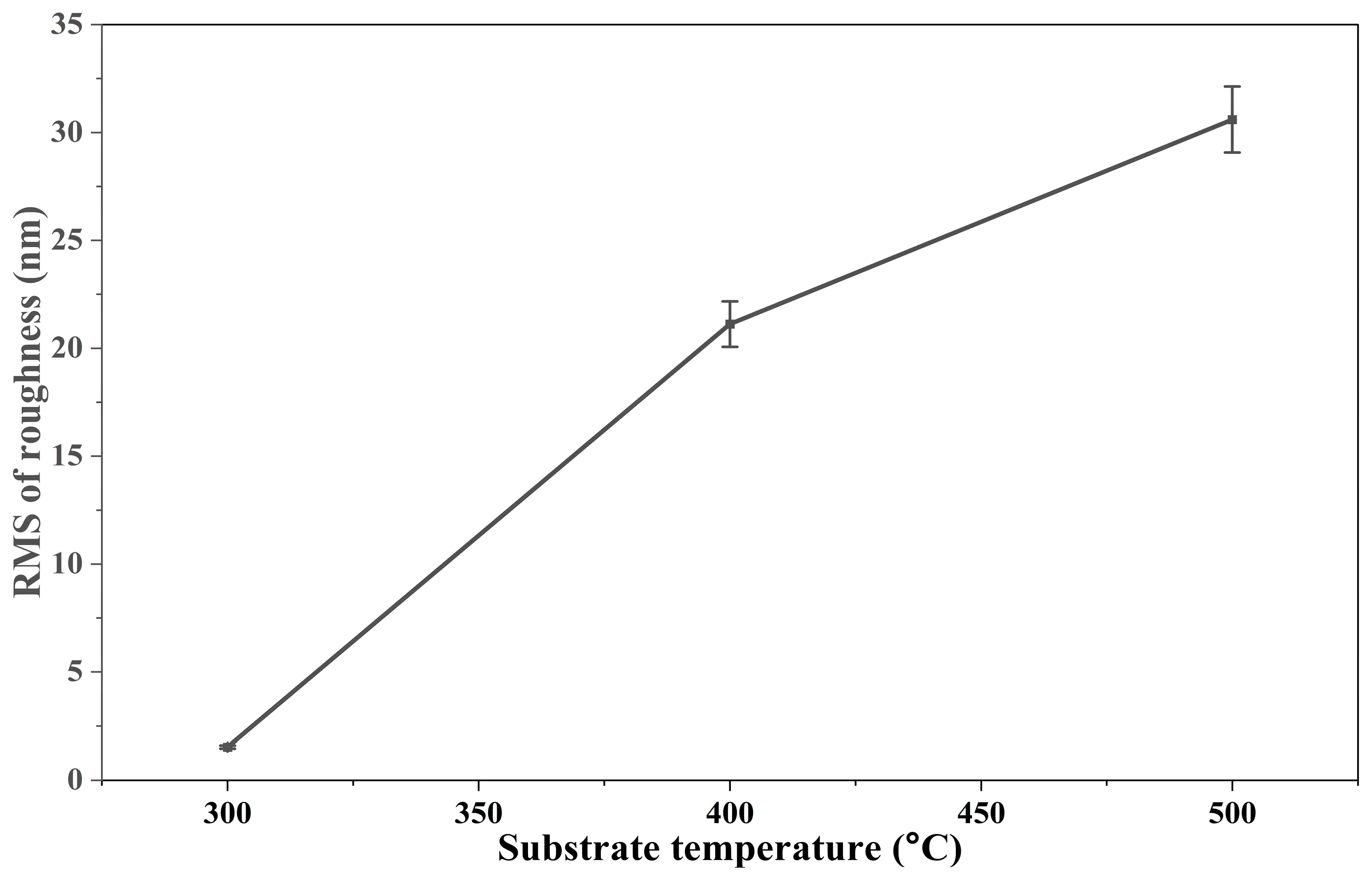
The surface roughness graph shows that the roughness of Zr thin films increases as the substrate temperature rises as in Figure 4. At 300 °C, the film has a very smooth surface with roughness around 1.5 nm. When the temperature is raised to 400 °C, the roughness increases sharply to about 21 nm, indicating the development of a more irregular surface. At 500 °C, the roughness continues to grow and reaches about 30 nm, showing that higher temperatures lead to coarser and less uniform surfaces. This trend suggests that increasing substrate temperature enhances atomic mobility, which promotes grain growth and results in rougher film morphology.
We have previously shown that the surface morphology and crystallinity of Zr films are influenced by laser wavelength, substrate temperature, and fluence with 1064 nm Nd:YAG laser pulses [33]. Films deposited using the 1064 nm laser exhibited smoother surfaces compared to those deposited using the 532 nm laser, likely due to differences in laser-material interaction and deposition dynamics. The longer wavelength of the 1064 nm laser produces less energetic plasma and more controlled material ejection, resulting in a stable deposition process. In contrast, the higher photon energy of the 532 nm laser leads to more intense ablation, turbulent plasma, and irregular deposition. Additionally, the lower kinetic energy of particles from the 1064 nm laser allows for more uniform settling on the substrate, contributing to smoother film surfaces [52]. Furthermore, higher laser fluence increased surface roughness, while elevated substrate temperatures enhanced surface diffusion, promoting grain growth and a transition to a denser crystalline structure [53].
These effects were mostly pronounced at higher temperatures, with significant roughness observed at 500 °C due to pit and rectangular structure formation. The investigation of ZrCuAlSi thin-film metallic glasses, deposited on glass substrate at substrate temperatures ranging from room temperature (RT) to 430 °C using PLD, parallels with our result that RMS roughness increases with rising substrate temperature [7]. As mentioned before, we selected a 248 nm KrF excimer laser in this study to explore how a shorter UV wavelength affects zirconium thin film growth compared to our previous work using 1064 nm and 532 nm Nd:YAG lasers [30]. It should be noted that the use of a UV excimer laser provides higher photon energy and stronger absorption in most materials, which enhances ablation efficiency, reduces thermal effects, and promotes better control over film stoichiometry and microstructure.
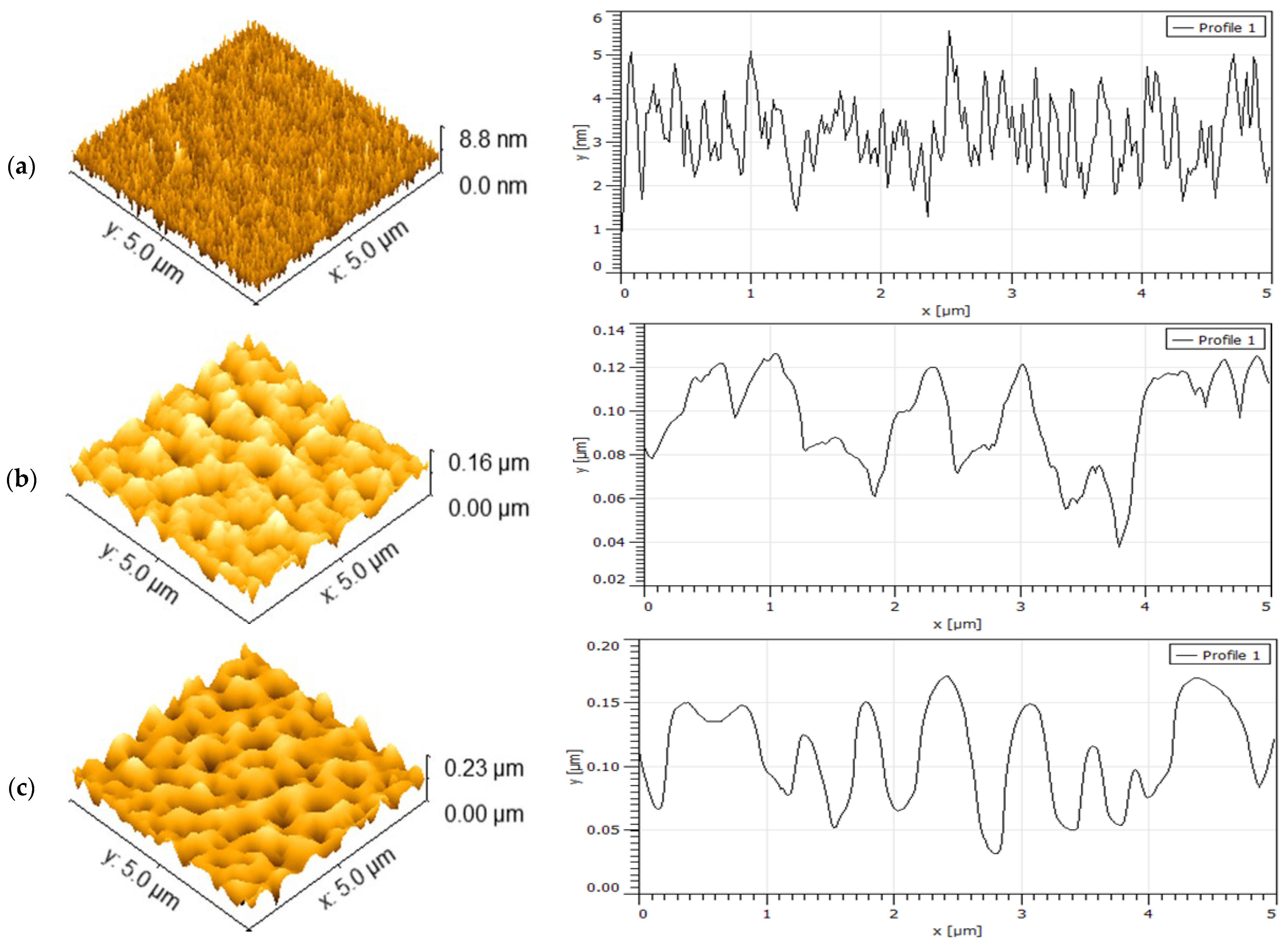
Based on these findings, we hypothesized that the higher photon energy of 248 nm would lead to more energetic ablation and potentially increased surface roughness. Our results suggests that, although UV lasers like 248 nm offer higher ablation efficiency, achieving smooth films depends strongly on optimizing substrate temperature to equalize the energetic plasma effects [54]. Figure 5a–c AFM images of Zr films deposited using a 248 nm excimer laser reveal structural features and surface patterns consistent with SEM observations [46].
As the substrate temperature increases, enhanced atomic mobility initially promotes surface diffusion and local reorganization, which can contribute to improved crystallinity. The AFM images show the change in surface topography of Zr thin films at different substrate temperatures. At lower temperature (a), the film surface appears relatively smooth with small height variations, reaching a maximum of about 8.8 nm. As the temperature increases (b), the surface develops larger grains and 3D islands, giving a rougher appearance and a maximum height variation of ~0.16 µm. At the highest temperature (c), the film surface shows even larger and more distinct 3D islands, with height variations reaching ~0.23 µm. The line profiles on the right confirm this trend, showing that the height changes become progressively larger and more periodic as the temperature increases. Overall, the AFM results indicate that higher substrate temperatures promote surface roughening and the formation of 3D island-like features.
However, at higher temperatures (above 400 °C), excessive atomic diffusion leads to increased surface roughness due to the transition from 2D layer-by-layer growth to 3D island formation. Additionally, at 500 °C, interdiffusion between the zirconium film and the silicon substrate further contributes to surface non-uniformity, supporting the observed trend of increasing roughness with temperature.
At 400 °C, the films show slightly increased surface roughness due to the development of more pronounced surface irregularities. This trend highlights the impact of higher substrate temperatures on atomic diffusion and surface morphology [7,55].
Modeling is focused on the observed transition from 2D layer-by-layer growth at T < 400 °C to 3D island growth (Volmer-Weber mode) at T > 400 °C. The most probable cause of this transition is the lattice-mismatch or thermal strain (due to spatially uneven heating of the film), although the kinetics of adatom surface diffusion may also be responsible [42,45]. Results of the strain-free kinetic model in the Section 3.4 support this conclusion. Further investigation into the role of strain on 2D->3D growth transition and subsequent island nucleation and coarsening will be the subject of future publications.
At lower substrate temperatures (e.g., 300 °C), the films form continuous nanocrystalline layers with relatively low roughness, as indicated by AFM and SEM analysis. Although the lattice mismatch between Zr and Si favors island nucleation in the initial stages, the high island density and rapid coalescence at this temperature lead to a smooth, continuous film surface rather than a rough, 3D morphology.

3.3. Computation of a Critical Film Thickness and the Island Size

We used a kinetic-free model of Lozovoy et al. [56,57] to compute a critical film thickness hc of a transition from 2D growth to 3D growth, as well as an island size lc immediately after island nucleation, as a function of three parameters: a wetting energy Ψ0, a lattice mismatch ε0, and T. This model accounts for lattice mismatch strain, but it does not account for thermal strain.
The model of Lozovoy et al. [56,57] is based on a minimization of a free energy change F(h) upon the nucleation of an island. The change in free energy has the contributions from the island’s surface energy, elastic energy, and wetting energy. The minimization of F(h) results in the following transcendental equation for the determination of hc, which is solved numerically:
2 3 π h e q d 0 ζ c a ζ c + 1 F ζ c F ζ c ζ c 2 V h e q 3 l 0 2 ν π α B D 5 / 2 exp F ζ c = 1
Here,
ζ c = h c h e q 1 ,       h e q = k 0 d 0 ln Ψ 0 d 0 ( 1 Z ) λ ε 0 2 ,       a = 3 B 4 π A ,
A = π 4 γ 4 κ 2 + 1 γ 0 α 2 l 0 2 k B T ,       α = 24 d 0 π κ l 0 4 κ 2 + 3 1 / 3 ,
B = ( 1 Z ) λ ε 0 2 l 0 2 k B T h e q k 0 ,       F ζ c = 4 A 3 27 B 2 ζ c 2 .
And the island size is given by
l c = 2 α l 0 A 3 B ζ c
The parameters are defined as: k0 = 0.8, the relaxation coefficient; d0 = 0.372 nm, a monolayer height of Zr; Z = 0.7, the elastic strain relaxation coefficient; λ = 94.5 GPa, the elastic modulus of Zr; γ = 2 J/m2, the surface energy of Zr island; γ0 = 2 J/m2, the energy of a base of Zr island; κ = 0.1, the aspect ratio of an island (height to diameter); l0 = 0.325 nm, lattice spacing of atoms comprising Zr island; V = 10 nm/h, the deposition rate; ν = 10, the cutoff parameter of the elastic strain field; D = 3.3 × 10⁻²exp(−1.2 eV/kBT) m2/s, the diffusivity of Zr atoms on the surface of Zr island; kB = 1.38 × 10⁻²³ J, the Boltzmann’s constant; T the temperature.
Note that Ψ0 and ε0 are not available in the literature for Zr on Si. To gain insight into Zr on Si system, we varied these parameters in a wide interval. For the well-studied system of Ge on Si(100) these values are: Ψ0 = 1.07 J/m2, ε0 =0.04(4%). For Zr on Si(100) Ψ0 is unknown, and the nominal ε0 = 0.4 (40%), since the lattice constant of Si(100) is 0.543 nm (recall, the lattice constant of Zr is 0.325 nm). The above values of the lattice constants are cited at room temperature; at elevated temperatures, these values slightly increase at different rates due to thermal expansion, thus a lattice mismatch ε0 deviates from a nominal 40% value.
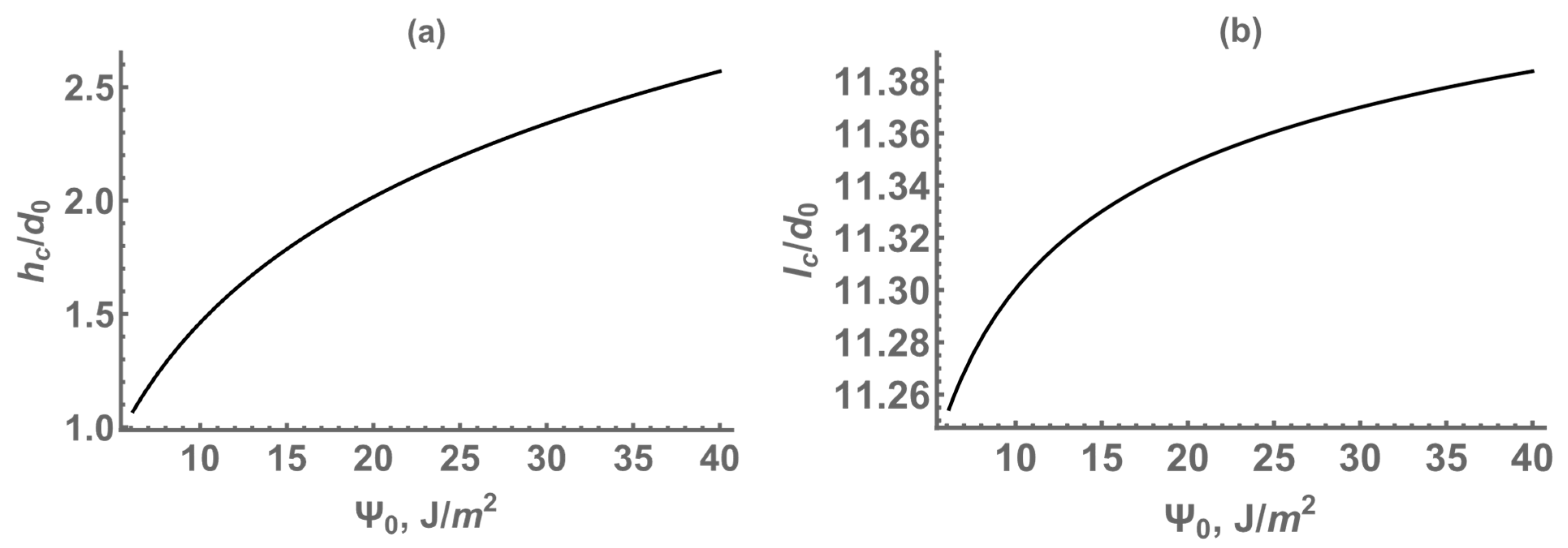
Figure 6 shows that as Ψ0 increases, the critical thickness hc of 2D->3D transition increases from a value that is slightly larger than the thickness of one Zr monolayer to around 2.5 Zr monolayers. This shows that stronger adhesion leads to larger transition thickness (subplot (a)). However, the island diameter lc is constant around 11.3 Zr monolayers regardless of Ψ0 (subplot (b)). Since the aspect ratio of an island (height to diameter) is chosen constant and equal to 0.1, this means that the island height immediately after nucleation is around 1.13 monolayers. Overall, the results indicate that changes in Ψ0 mainly affect the transition thickness, while the island diameter and height immediately after nucleation remain unchanged.
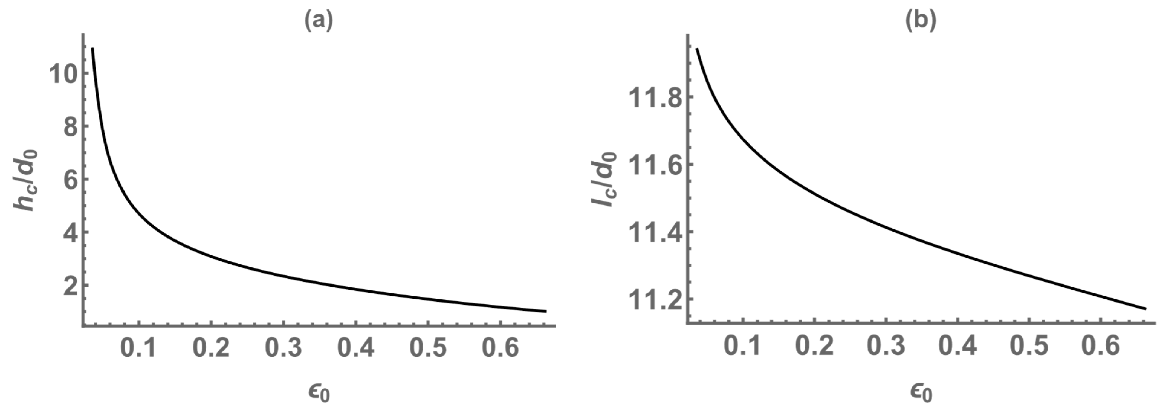
Figure 7 shows that the transition thickness decreases from a value of around ten Zr monolayers at ε0 = 0.01(1%) to around 1.1 Zr monolayers at ε0 = 0.65(65%); the island diameter changes insignificantly, staying around a constant 11.5 Zr monolayers. Subplot (a) shows that the transition thickness decreases with increasing ε0, from approximately 10 Zr monolayers at ε0 = 0.01 (1%) to about 1.1 monolayers at ε0 = 0.65 (65%). This inverse relationship indicates that greater lattice mismatch leads to 2D->3D transition at smaller film thickness. Subplot (b) demonstrates that the island diameter exhibits only minor variation across the same range of ε0, remaining nearly constant at approximately 11.5 monolayers, and therefore the island height remains nearly constant at 1.15 monolayers. These results suggest that while strain accelerates 2D->3D transition, the island height and diameter immediately after nucleation are relatively unaffected under the assumed fixed aspect ratio.

3.4. Computation of Kinetics of Island Nucleation and Growth

We used the strain-free kinetic model developed by our group in [33] and the physical parameters from the preceding section to compute Zr film growth. Ψ0 = 10 J/m2 and ε0 = 0 were adopted. Imperfect 3D islands start to form at 400 °C (Figure 8), and at 500 °C they proliferate over the entire substrate and assume a distinct circular shape in agreement with the experiment (Figure 9). Note that the model is stress-free, but it incorporates the roughening transition of a growing surface via spinodal instability [33,58]. Using this model, we computed a 2D film growth at T < 400 °C, a transitional 2D->3D growth at T = 400 °C and 3D island growth at T = 500 °C. The only parameter that varies in these computations is the Zr atoms diffusivity. Due to the Arrhenius form, the diffusivity increases when the temperature increases. For instance, when T increases from 400 °C to 500 °C the diffusivity increases by one order of magnitude. Interestingly, note that in Figure 8a and Figure 9a the transition from 2D growth to 3D island growth occurs around 1–2 nm film thickness, which is of the order of the critical thickness hc value computed in the previous section at Ψ0 = 10 J/m2 and ε0 > 0.5 (50%) using the model of references [56,57]. The islands are a few nanometers in diameter immediately after 2D-to-3D growth transition (Figure 8a and Figure 9a capture them after the nucleus had already grown in size), confirming the island diameter lc immediately after nucleation from free energy minimization-based computation in the previous section, but reach the diameter of around twenty micrometers at later times via coarsening (Figure 9c) [56,57].
The simulation results further validate the experimental findings, offering a mechanistic understanding of the observed 2D-to-3D growth transition in zirconium thin films at elevated substrate temperatures. The computed critical film thickness of a transition from 2D growth to 3D growth (~1–2 nm) and island size after coarsening align closely with the experimental observation of 3D island formation beginning at 400 °C.
This transition, attributed to increased lattice mismatch and atomic diffusivity, correlates well with the SEM and AFM data, which show significant morphological changes and increased roughness at higher temperatures. Additionally, the simulation points out the role of enhanced adatom mobility in optimizing crystallinity at 400 °C, where a balance between diffusivity and lattice mismatch effects results in the highest crystalline quality.
At 500 °C, the proliferation of islands and interfacial interdiffusion observed experimentally is supported by the model’s prediction of increased atomic mobility and the associated energy changes. These insights provide a cohesive explanation for the interplay between growth dynamics and substrate temperature, further strengthening the conclusions drawn from experimental data [46,59].

4. Summary

Zirconium (Zr) thin films were successfully deposited on Si(100) substrates via pulsed laser deposition using a 248 nm excimer laser, with a focus on understanding the effects of substrate temperature on film morphology, crystallinity, and thickness. X-ray diffraction (XRD) analysis revealed a strong dependence of crystalline quality on deposition temperature, with the Zr(100) orientation reaching optimal crystallinity at 400 °C and Zr(002) at 500 °C. Surface morphology, examined via scanning electron microscopy (SEM) and atomic force microscopy (AFM), demonstrated a progressive increase in roughness with temperature, attributed to the transition from 2D layer-by-layer growth to 3D island formation. At 500 °C, interdiffusion between the Zr and the Si substrate was observed, suggesting the formation of zirconium silicide, as supported by XRD and morphological changes.
To complement the experimental findings, computational modeling was performed to elucidate the 2D-to-3D growth transition and the role of substrate temperature in structural evolution. The simulations predicted a critical thickness of ~1–2 nm for this transition, aligning with experimental observations. The model also revealed that increased adatom diffusivity at elevated temperatures promotes island nucleation, explaining the AFM-observed roughness increase. Moreover, thermal expansion mismatches at higher temperatures contributed to enhanced surface stress, further affecting growth dynamics. These results provide a comprehensive framework for optimizing Zr thin films via PLD, offering valuable insights into the interplay of deposition parameters in tailoring surface morphology and crystallinity for applications in protective coatings, microelectronics, and advanced materials.

Author Contributions

Methodology, B.A., Z.K., J.T., M.K. and A.O.E.; validation, A.O.E. and Z.K.; formal analysis, B.A., M.K. and A.O.E.; investigation, B.A., Z.K., J.T., M.K., D.G. and A.O.E.; data curation, A.O.E.; writing—original draft preparation, B.A., M.K. and A.O.E.; writing—review and editing, B.A., M.K., D.G. and A.O.E.; visualization, B.A. and M.K.; supervision, A.O.E.; project administration, A.O.E.; funding acquisition, A.O.E. All authors have read and agreed to the published version of the manuscript.

Funding

This work was supported by NASA award number 80NSSC21M0362, the NASA Kentucky EPSCoR Program and the Kentucky Cabinet for Economic Development.

Institutional Review Board Statement

Not applicable.

Informed Consent Statement

Not applicable.

Data Availability Statement

Data is contained within the article.

Acknowledgments

We thank John Andersland for his help and expertise on SEM measurements.

Conflicts of Interest

The authors declare no conflict of interest.

References

  1. Ul-Hamid, A. The Effect of Deposition Conditions on the Properties of Zr-Carbide, Zr-Nitride and Zr-Carbonitride Coatings—A Review. Mater. Adv. 2020, 1, 988–1011. [Google Scholar] [CrossRef]
  2. Singh, D.P.; Gupta, S.K.; Manohar, R.; Varia, M.C.; Kumar, S.; Kumar, A. Effect of Cadmium Selenide Quantum Dots on the Dielectric and Physical Parameters of Ferroelectric Liquid Crystal. J. Appl. Phys. 2014, 116, 034106. [Google Scholar] [CrossRef]
  3. Almyras, G.A.; Matenoglou, G.M.; Komninou, P.; Kosmidis, C.; Patsalas, P.; Evangelakis, G.A. On the Deposition Mechanisms and the Formation of Glassy Cu–Zr Thin Films. J. Appl. Phys. 2010, 107, 084313. [Google Scholar] [CrossRef]
  4. Zhu, S.; Song, Y.; Zhao, X.; Shao, J.; Zhang, J.; Yang, B. The Photoluminescence Mechanism in Carbon Dots (Graphene Quantum Dots, Carbon Nanodots, and Polymer Dots): Current State and Future Perspective. Nano Res. 2015, 8, 355–381. [Google Scholar] [CrossRef]
  5. Balakrishnan, G.; Kuppusami, P.; Sastikumar, D.; Song, J.I. Growth of Nanolaminate Structure of Tetragonal Zirconia by Pulsed Laser Deposition. Nanoscale Res. Lett. 2013, 8, 82. [Google Scholar] [CrossRef] [PubMed]
  6. Dubbink, D.; Koster, G.; Rijnders, G. Growth Mechanism of Epitaxial YSZ on Si by Pulsed Laser Deposition. Sci. Rep. 2018, 8, 5774. [Google Scholar] [CrossRef]
  7. Zhang, B.; Yang, X.; Cao, G.; Liu, G.; Wei, Z.; Zong, H.; Yin, Y.; Bala, H. Characteristic Studies on the ZrCuAlSi Multi-Component Thin-Film Metallic Glasses Fabricated by Pulsed Laser Deposition. Appl. Phys. A 2020, 126, 453. [Google Scholar] [CrossRef]
  8. Wan, X.; Fang, S.; Xu, S.; Yu, L.; Zhou, J.; Qian, S.; Huang, F.; Ma, C. Enhanced Anti-Corrosion and Biological Performance of Plasma-Sprayed Nb/ZrO2/HA Coatings on ZK60 Mg Alloy. Coatings 2024, 14, 1282. [Google Scholar] [CrossRef]
  9. Zhong, Q.; Zhang, Z.; Qi, R.; Li, J.; Wang, Z.; Le Guen, K.; André, J.-M.; Jonnard, P. Enhancement of the Reflectivity of Al/Zr Multilayers by a Novel Structure. Opt. Express 2013, 21, 14399–14408. [Google Scholar] [CrossRef]
  10. Hettiarachchi, B.S.; Takaoka, Y.; Uetake, Y.; Yakiyama, Y.; Yoshikawa, H.Y.; Maruyama, M.; Sakurai, H. Mechanistic Study in Gold Nanoparticle Synthesis through Microchip Laser Ablation in Organic Solvents. Metals 2024, 14, 155. [Google Scholar] [CrossRef]
  11. Axente, E.; Socol, G. Special Issue “Pulsed Laser Deposition of Thin Films: Recent Advances and Challenge”. Coatings 2022, 12, 368. [Google Scholar] [CrossRef]
  12. Singh, A.; Kuppusami, P.; Thirumurugesan, R.; Ramaseshan, R.; Kamruddin, M.; Dash, S.; Ganesan, V.; Mohandas, E. Study of Microstructure and Nanomechanical Properties of Zr Films Prepared by Pulsed Magnetron Sputtering. Appl. Surf. Sci. 2011, 257, 9909–9914. [Google Scholar] [CrossRef]
  13. Bastianini, A.; Battiston, G.A.; Gerbasi, R.; Porchia, M.; Daolio, S. Chemical Vapor Deposition of ZrO2 Thin Films Using Zr(NEt2)4 as Precursor. J. Phys. IV Fr. 1995, 5, C5–C525. [Google Scholar] [CrossRef]
  14. Sirjita, E.N.; Rusen, L.; Brajnicov, S.; Craciun, C.; Ion, V.; Filipescu, M.; Dinescu, M. Properties of Hafnium and Aluminium Silicates Coatings Obtained by PLD. Coatings 2021, 11, 753. [Google Scholar] [CrossRef]
  15. Allamyradov, Y.; Yosef, J.B.; Kylychbekov, S.; Majidov, I.; Khuzhakulov, Z.; Er, A.Y.; Kitchens, C.; Banga, S.; Er, A.O. The Role of Efflux Pump Inhibitor in Enhancing Antimicrobial Efficiency of Ag NPs and MB as an Effective Photodynamic Therapy Agent. Photodiagnosis Photodyn. Ther. 2024, 47, 104212. [Google Scholar] [CrossRef]
  16. Khuzhakulov, Z.; Kylychbekov, S.; Allamyradov, Y.; Majidov, I.; Ben Yosef, J.; Er, A.Y.; Kitchens, C.; Banga, S.; Badarudeen, S.; Er, A.O. Formation of Picosecond Laser-Induced Periodic Surface Structures on Steel for Knee Arthroplasty Prosthetics. Front. Met. Alloys 2023, 1, 1090104. [Google Scholar] [CrossRef]
  17. Kylychbekov, S.; Allamyradov, Y.; Khuzhakulov, Z.; Majidov, I.; Banga, S.; Ben Yosef, J.; Duta, L.; Er, A.O. Bioactivity and Mechanical Properties of Hydroxyapatite on Ti6Al4V and Si(100) Surfaces by Pulsed Laser Deposition. Coatings 2023, 13, 1681. [Google Scholar] [CrossRef]
  18. Majidov, I.; Allamyradov, Y.; Kylychbekov, S.; Khuzhakulov, Z.; Er, A.O. Phase Transition and Controlled Zirconia Implant Patterning Using Laser-Induced Shockwaves. Appl. Sci. 2025, 15, 362. [Google Scholar] [CrossRef]
  19. Hakimov, S.; Kylychbekov, S.; Harness, B.; Neupane, S.; Hurley, J.; Brooks, A.; Banga, S.; Er, A.O. Evaluation of Silver Nanoparticles Attached to Methylene Blue as an Antimicrobial Agent and Its Cytotoxicity. Photodiagnosis Photodyn. Ther. 2022, 39, 102904. [Google Scholar] [CrossRef]
  20. Belekov, E. Improved Antimicrobial Properties of Methylene Blue Attached to Silver Nanoparticles. Photodiagn. Photodyn. Ther. 2020, 32, 102012. [Google Scholar] [CrossRef]
  21. Ohring, M. Materials Science of Thin Films; Academic Press: Cambridge, MA, USA, 2002. [Google Scholar]
  22. Iljinas, A.; Stankus, V.; Virbukas, D.; Kaliasas, R. Synthesis of (Bi2O3)1-x(PbO)x Thin Films by Plasma-Assisted Reactive Evaporation. Coatings 2025, 15, 748. [Google Scholar] [CrossRef]
  23. Wang, Y.; He, T.; Du, X.; Vereschaka, A.; Sotova, C.; Ding, Y.; Chen, K.; Li, J.; He, P. Influence of Aluminum Alloy Substrate Temperature on Microstructure and Corrosion Resistance of Cr/Ti Bilayer Coatings. Coatings 2025, 15, 891. [Google Scholar] [CrossRef]
  24. Zou, H.; Zhao, X.; Fu, B.; Yang, H.; Sun, C. Numerical and Experimental Study on Deposition Mechanism of Laser-Assisted Plasma-Sprayed Y2O3 Coating. Coatings 2025, 15, 904. [Google Scholar] [CrossRef]
  25. Eason, R. Pulsed Laser Deposition of Thin Films: Applications; John Wiley & Sons, Inc.: Hoboken, NJ, USA, 2007. [Google Scholar]
  26. Chrisey, D.B.; Hubler, G.K. Pulsed Laser Deposition of Thin Films; Wiley Inc.: New York, NY, USA, 1994. [Google Scholar]
  27. Miao, J.; Yang, H.; Hao, W.; Yuan, J.; Xu, B.; Qiu, X.Q.; Cao, L.X.; Zhao, B.R. Temperature Dependence of the Ferroelectric and Dielectric Properties of the Ba0.5Sr0.5TiO3/La0.67Sr0.33MnO3 Heterostructure. J. Phys. D Appl. Phys. 2004, 38, 5. [Google Scholar] [CrossRef]
  28. Mazurek, P.; Daniluk, A.; Paprocki, K. Substrate Temperature Control from RHEED Intensity Measurements. Vacuum 2004, 72, 363–367. [Google Scholar] [CrossRef]
  29. Farha, A.H.; Er, A.O.; Ufuktepe, Y.; Myneni, G.; Elsayed-Ali, H.E. Effects of Substrate Temperature on Properties of NbNx Films Grown on Nb by Pulsed Laser Deposition. Appl. Surf. Sci. 2011, 258, 1613–1618. [Google Scholar] [CrossRef]
  30. Khuzhakulov, Z.; Annamuradov, B.; Kylychbekov, S.; Allamyradov, Y.; Majidov, I.; Khenner, M.; Terzic, J.; Gurgew, D.; Er, A.O. Fabrication and Analysis of Zirconium Thin Films on Silicon (Si) by Pulsed Laser Deposition. In Proceedings of the Nanoscale and Quantum Materials: From Synthesis and Laser Processing to Applications, San Francisco, CA, USA, 12 March 2024; Volume 12874, p. 128740C. [Google Scholar]
  31. Liu, W.; Wen, C.; Zhang, X.; Liu, J.; Liang, J.; Long, X. Investigation of Deposition Rate, Liquid Droplets and Surface Morphology of Zirconium Films Deposited by Pulsed Laser Ablation. Phys. Scr. 2015, 90, 035402. [Google Scholar] [CrossRef]
  32. Chen, H.-L.; Ju, S.-P.; Wu, T.-Y.; Hsieh, J.-Y.; Liu, S.-H. Investigation of Zr and Si Diffusion Behaviors during Reactive Diffusion—A Molecular Dynamics Study. RSC Adv. 2015, 5, 26316–26320. [Google Scholar] [CrossRef]
  33. Khuzhakulov, Z.; Kylychbekov, S.; Allamyradov, Y.; Majidov, I.; Khenner, M.; Terzic, J.; Gurgew, D.; Er, A.O. Synthesis and Characterization of a Zirconium (Zr) Thin Film on Si(100) via Pulsed Laser Deposition. Coatings 2023, 13, 1748. [Google Scholar] [CrossRef]
  34. Annamuradov, B.; Khuzhakulov, Z.; Terzic, J.; Khenner, M.; Er, A.O. Surface Morphologies and Crystallinities of a Zirconium (Zr) Thin Film on Si(100) by Pulsed Laser Deposition. In Proceedings of the Nanoscale and Quantum Materials: From Synthesis and Laser Processing to Applications 2025, San Francisco, CA, USA, 19 March 2025; Volume 13352, p. 133520A. [Google Scholar]
  35. Liu, W.; Liang, J.; Zhou, X.; Long, X. Formation of Zirconium Silicide between Silicon Substrate and Zirconium Films. Mater. Lett. 2014, 122, 220–222. [Google Scholar] [CrossRef]
  36. Troglia, A.; van Vliet, S.; Yetik, G.; El Wakil, I.; Momand, J.; Kooi, B.J.; Bliem, R. Free-Standing Nanolayers Based on Ru Silicide Formation on Si(100). Phys. Rev. Mater. 2022, 6, 043402. [Google Scholar] [CrossRef]
  37. Vasco, E.; Sacedón, J.L. Role of Cluster Transient Mobility in Pulsed Laser Deposition-Type Growth Kinetics. Phys. Rev. Lett. 2007, 98, 036104. [Google Scholar] [CrossRef]
  38. Shepelin, N.A.; Tehrani, Z.P.; Ohannessian, N.; Schneider, C.W.; Pergolesi, D.; Lippert, T. A Practical Guide to Pulsed Laser Deposition. Chem. Soc. Rev. 2023, 52, 2294–2321. [Google Scholar] [CrossRef]
  39. Tarasenka, N.; Butsen, A.; Pankov, V.; Tarasenko, N. Structural Defects and Magnetic Properties of Gadolinium Silicide Nanoparticles Synthesized by Laser Ablation Technique in Liquid. Phys. Status Solidi (b) 2013, 250, 809–814. [Google Scholar] [CrossRef]
  40. Goldak, J.; Lloyd, L.T.; Barrett, C.S. Lattice Parameters, Thermal Expansions, and Grüneisen Coefficients of Zirconium, 4.2 to 1130°K. Phys. Rev. 1966, 144, 478–484. [Google Scholar] [CrossRef]
  41. Cho, H.W.; Pujar, P.; Choi, M.; Kang, S.; Hong, S.; Park, J.; Baek, S.; Kim, Y.; Lee, J.; Kim, S. Direct Growth of Orthorhombic Hf0.5Zr0.5O2 Thin Films for Hysteresis-Free MoS2 Negative Capacitance Field-Effect Transistors. npj 2D Mater. Appl. 2021, 5, 46. [Google Scholar] [CrossRef]
  42. Kang, S.J.; Joung, Y.H.; Shin, H.H.; Yoon, Y.S. Effect of Substrate Temperature on Structural, Optical and Electrical Properties of ZnO Thin Films Deposited by Pulsed Laser Deposition. J. Mater. Sci. Mater. Electron. 2008, 19, 1073–1078. [Google Scholar] [CrossRef]
  43. Rabo, J.R.; Takayanagi, M.; Tsuchiya, T.; Nakajima, H.; Terabe, K.; Cervera, R.B.M. Effects of Oxygen Partial Pressure and Substrate Temperature on the Structure and Morphology of Sc and Y Co-Doped ZrO2 Solid Electrolyte Thin Films Prepared via Pulsed Laser Deposition. Materials 2022, 15, 410. [Google Scholar] [CrossRef]
  44. Vogt, S.; Petersen, C.; von Wenckstern, H.; Grundmann, M.; Schultz, T.; Koch, N. Zr doping in pulsed-laser-deposited α-Ga2O3 for device applications. Phys. Rev. Appl. 2024, 21, 064016. [Google Scholar] [CrossRef]
  45. Sisodia, V.; Bolse, W.; Avasthi, D.K.; Kabiraj, D.; Jain, I.P. Silicide Formation Study at the Interface of Zr/Si System Using High-Energy Swift Heavy Ion. Radiat. Meas. 2005, 40, 762–764. [Google Scholar] [CrossRef]
  46. Wong, Y.H.; Cheong, K.Y. ZrO2 Thin Films on Si Substrate. J. Mater. Sci. Mater. Electron. 2010, 21, 980–993. [Google Scholar] [CrossRef]
  47. Edwards, A.M.; Dao, Y.; Nemanich, R.J.; Sayers, D.E.; Kemner, K.M. Structural Investigation of the Initial Interface Region Formed by Thin Zirconium Films on Silicon (111). J. Appl. Phys. 1994, 76, 4630–4635. [Google Scholar] [CrossRef]
  48. Cheng, J.Y.; Chen, L.J. Formation of Amorphous Interlayers by a Solid-state Diffusion in Zr and Hf Thin Films on Silicon. Appl. Phys. Lett. 1990, 56, 457–459. [Google Scholar] [CrossRef]
  49. Abed, N.Z.; Ismail, R.A.; Shaker, S.S. Role of Substrate Temperature on the Performance of BaTiO3/Si Photodetector Prepared by Pulsed Laser Deposition. Sci. Rep. 2024, 14, 4531. [Google Scholar] [CrossRef]
  50. Zhou, G.; Qu, P.; Huo, X.; Jin, P.; Wu, J.; Wang, Z. The Deposition of Ir/YSZ Double-Layer Thin Films on Silicon by PLD and Magnetron Sputtering: Growth Kinetics and the Effects of Oxygen. Results Phys. 2023, 47, 106357. [Google Scholar] [CrossRef]
  51. Elofsson, V.; Lü, B.; Magnfält, D.; Münger, E.P.; Sarakinos, K. Unravelling the Physical Mechanisms That Determine Microstructural Evolution of Ultrathin Volmer-Weber Films. J. Appl. Phys. 2014, 116, 044302. [Google Scholar] [CrossRef]
  52. Ichino, Y.; Yoshida, Y.; Yoshimura, T.; Ono, I.; Takai, Y.; Yoshizumi, M.; Izumi, T.; Shiohara, Y. Possibility of Nd:YAG-PLD Method for Fabricating REBCO Coated Conductors. IEEE Trans. Appl. Supercond. 2011, 21, 2949–2952. [Google Scholar] [CrossRef]
  53. Kao, Q.; Xiao, Y.; Jia, Y.; Wang, J.; Wang, C. Infrared Photoelectric Detection of Polycrystalline Bi2Te3 Thin Films Prepared by Pulsed Laser Deposition. J. Vac. Sci. Technol. A 2024, 42, 023421. [Google Scholar] [CrossRef]
  54. Chaluvadi, S.K.; Punathum Chalil, S.; Mazzola, F.; Dolabella, S.; Rajak, P.; Ferrara, M.; Ciancio, R.; Fujii, J.; Panaccione, G.; Rossi, G.; et al. Nd:YAG Infrared Laser as a Viable Alternative to Excimer Laser: YBCO Case Study. Sci. Rep. 2023, 13, 3882. [Google Scholar] [CrossRef]
  55. To, T.B.T.; Almeida, R.; Ferreira, S.O.; Aarão Reis, F.D.A. Roughness and Correlations in the Transition from Island to Film Growth: Simulations and Application to CdTe Deposition. Appl. Surf. Sci. 2021, 560, 149946. [Google Scholar] [CrossRef]
  56. Lozovoy, K.A.; Korotaev, A.G.; Kokhanenko, A.P.; Dirko, V.V.; Voitsekhovskii, A.V. Kinetics of Epitaxial Formation of Nanostructures by Frank–van Der Merwe, Volmer–Weber and Stranski–Krastanow Growth Modes. Surf. Coat. Technol. 2020, 384, 125289. [Google Scholar] [CrossRef]
  57. Lozovoy, K.A.; Kokhanenko, A.P.; Dirko, V.V.; Akimenko, N.Y.; Voitsekhovskii, A.V. Evolution of Epitaxial Quantum Dots Formed by Volmer–Weber Growth Mechanism. Cryst. Growth Des. 2019, 19, 7015–7021. [Google Scholar] [CrossRef]
  58. Ortiz, M.; Repetto, E.A.; Si, H. A Continuum Model of Kinetic Roughening and Coarsening in Thin Films. J. Mech. Phys. Solids 1999, 47, 697–730. [Google Scholar] [CrossRef]
  59. Modabberasl, A.; Sharifi, M.; Shahbazi, F.; Kameli, P. Multifractal Analysis of DLC Thin Films Deposited by Pulsed Laser Deposition. Appl. Surf. Sci. 2019, 479, 639–645. [Google Scholar] [CrossRef]
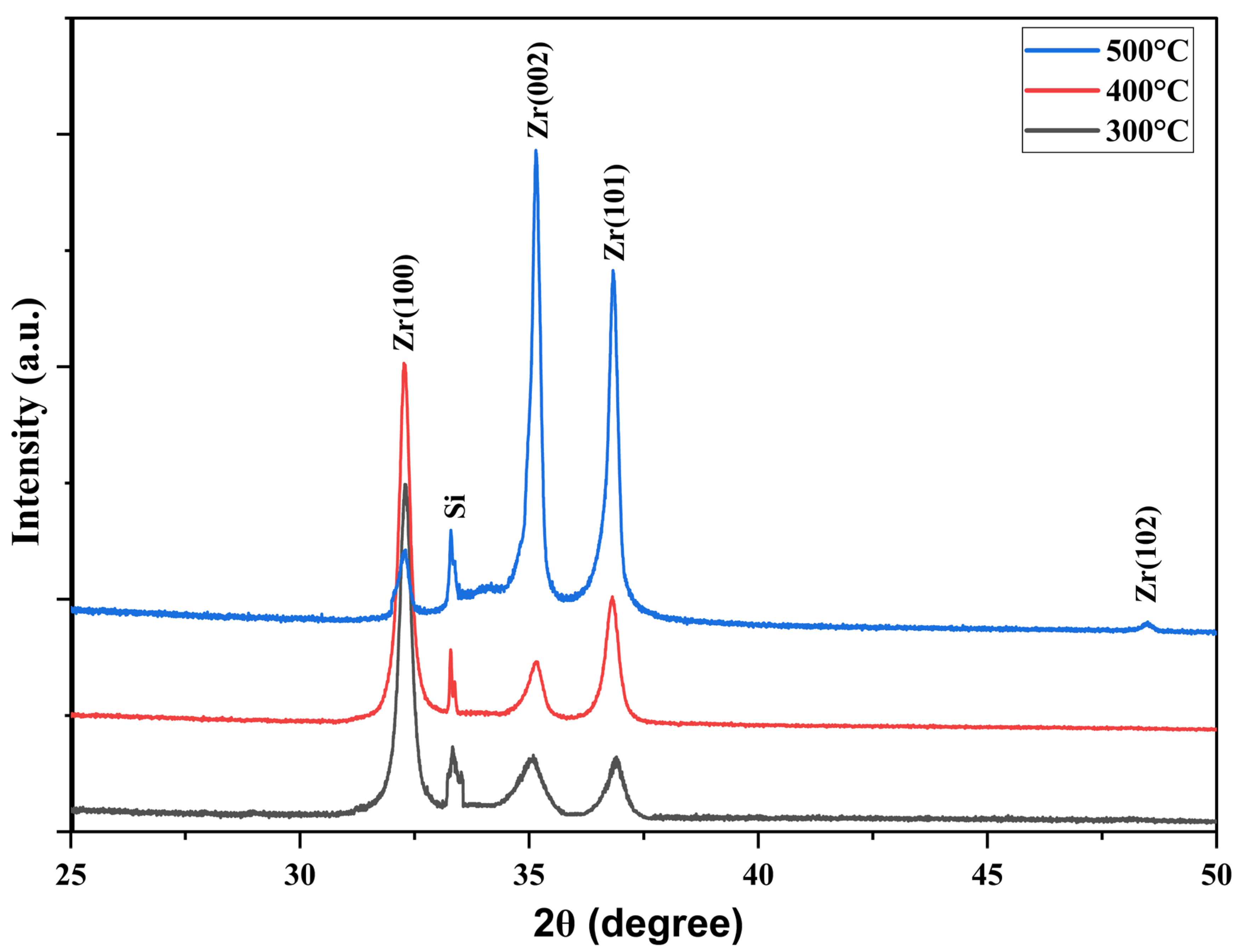
Figure 1. XRD data for Zr thin films deposited on Si substrate by 248 nm excimer laser at 75 mJ, for different substrate temperatures: 300 °C, 400 °C and 500 °C.
Coatings 15 01198 g001
Figure 2. SEM data for Zr thin films deposited on Si substrate by 248 nm excimer laser at 75 mJ, for different substrate temperatures: (a) 300 °C, (b) 400 °C and (c) 500 °C.
Coatings 15 01198 g002
Figure 3. Cross-sectional SEM images of zirconium thin films deposited by PLD at (a) 300 °C, (b) 400 °C and (c) 500 °C for 1 h deposition.
Coatings 15 01198 g003
Figure 4. RMS of surface roughness of the thin films at various substrate temperatures obtained from a 5 × 5 μm surface area by AFM analysis.
Coatings 15 01198 g004
Figure 5. AFM images for Zr thin films deposited on Si substrate by 248 nm excimer laser at 75 mJ, for different substrate temperatures: (a) 300 °C, (b) 400 °C and (c) 500 °C.
Coatings 15 01198 g005
Figure 6. (a) Critical transition thickness and (b) island diameter vs. Ψ0 at T = 500 °C.
Coatings 15 01198 g006
Figure 7. (a) Critical transition thickness and (b) island diameter vs. ε0. Larger values of ε0 are expected at higher temperatures.
Coatings 15 01198 g007
Figure 8. Simulation of 3D growth after (a) t = 6 min, (b) t = 7.5 min, (c) t = 9 min at T = 400 °C. 2D-to-3D growth transition is seen in (a).
Coatings 15 01198 g008
Figure 9. Simulation of 3D growth after (a) t = 6 min, (b) t = 7.5 min, (c) t = 9 min at T = 500 °C. 2D-to-3D growth transition is seen in (a).
Coatings 15 01198 g009
Disclaimer/Publisher’s Note: The statements, opinions and data contained in all publications are solely those of the individual author(s) and contributor(s) and not of MDPI and/or the editor(s). MDPI and/or the editor(s) disclaim responsibility for any injury to people or property resulting from any ideas, methods, instructions or products referred to in the content.