Article

Silver Nanoparticles–Chitosan Nanocomposites as Protective Coatings for Dental Remineralization Treatment: An In Vitro Study

by
Katleen A. Aguirre de Rodríguez
1,2,
Wendy Y. Escobar de González
1,2,
Vianney Castañeda Monroy
2,
Sean Murphy
3,
Gabriel-Alejandro Martínez-Castañón
4,*,
Horacio Bach
5 and
Nereyda Niño-Martínez
6,*
1
Facultad de Estomatología, Zona Universitaria, Av. Dr. Manuel Nava No. 2, San Luis Potosí CP 78290, Mexico
2
Laboratorio de Biología Molecular, CENSALUD, Universidad de El Salvador (UES), San Salvador CP 1101, El Salvador
3
Faculty of Dentistry, Department of Oral Biological and Medical Sciences, University of British Columbia, Vancouver, BC V6H 3Z6, Canada
4
Facultad de Estomatología, Universidad Autónoma de San Luis Potosi, Zona Universitaria, Av. Dr. Manuel Nava No. 2, San Luis Potosí CP 78290, Mexico
5
Faculty of Medicine, Division of Infectious Diseases, University of British Columbia, Vancouver, BC V6H 3Z6, Canada
6
Facultad de Ciencias, Universidad Autónoma de San Luis Potosi, Av. Parque Chapultepec No. 1570, Privadas del Pedregal, San Luis Potosí CP 78295, Mexico
*
Authors to whom correspondence should be addressed.
Coatings 2025, 15(1), 40; https://doi.org/10.3390/coatings15010040
Submission received: 9 December 2024 / Revised: 31 December 2024 / Accepted: 2 January 2025 / Published: 3 January 2025

Abstract

:
Research with nanoparticles for the treatment and prevention of dental caries is of special interest given the high prevalence of the disease worldwide. Several studies support the use of nanoparticles associated with materials given their antimicrobial properties and potential demineralization reduction. This study aimed to evaluate the impact of the application of silver nanoparticles (AgNPs) and chitosan gel in combination with commercial fluoride varnish on the remineralization of dental enamel. Ninety-six tooth blocks were macroscopically evaluated via stereomicroscopy, ICDAS II, and laser fluorescence. Enamel blocks were subjected to artificial demineralization and divided into four exposure groups (24, 48, 120, and 168 h), and five different remineralizing agents were applied, namely, FV (fluoride varnish), FV + CG (fluoride varnish + chitosan gel), FV + AgNPs (fluoride varnish + AgNPs), FV + AgNPs + CG (fluoride varnish + AgNPs + chitosan gel), and AgNPs + CG (AgNPs + chitosan gel). Enamel surface changes were evaluated via laser fluorescence, X-ray diffraction, scanning electron microscopy, and energy-dispersive X-ray spectroscopy. Laser fluorescence results obtained from demineralized blocks and subsequently exposed to remineralizing treatment indicate significant differences. After exposure to remineralizing agents, hydroxyapatite and modified apatite phases were identified mainly in the samples treated with FV + AgNPs + CG in the groups exposed for 24, 48, and 120 h. The FV + AgNPs + CG and AgNPs + CG indicate good performance in terms of the Ca/P ratio in in vitro demineralization compared to the group treated with fluorine varnish.

1. Introduction

Dental caries is a highly prevalent disease in the world population. Its progression from the enamel surface to the innermost structures of the tooth depends on multiple factors such as cariogenic diet, hygiene habits, time, and salivary pH variations [1]. The onset of the lesion is related to the acidification of the environment, causing the demineralization of the teeth, which implies the loss of minerals in the initial stages. This process can be reversed naturally by repair through remineralization, which has the purpose of restoring the lost minerals. This process occurs at near-neutral physiological pH [2,3].
The timely identification of incipient lesions in enamel is of great importance in establishing preventive treatments and immediate intervention to stop their progression. The use of materials such as varnish fluoride for the management of the remineralization of dental tissues has been widely studied. Kim et al. (2021) compared the demineralization resistance and remineralization effects of various fluoride-releasing restorative commercial materials under clinical conditions and found that alkasite restorative material is a superior fluoride-releasing restorative material in stimulating demineralization and remineralization in an enamel [3]. However, in a different study, Chhatwani et al. (2021) found that the effectiveness of fluoride varnishes is compromised and limited due to its permanence in the oral environment, which stresses the materials under different temperature and pH conditions [4]. It has also been reported that the use of fluoride varnish (FV) could produce sensitivity and a burning sensation in some patients and, in regions where the levels of fluoride in water are high, the use of this varnish is limited; Parisay et al. (2024) explain that excessive fluoride consumption can result in symptoms like nausea, vomiting, diarrhea, escalated salivation, abdominal discomfort, muscle frailty, and spasms. Fluoride varnish is not recommended for patients with ulcerative gingivitis, gingivitis stomatitis, or asthma [5]. Silver diamine fluoride has also been reported as a material which inhibits demineralization, promoting the remineralization of both enamel and dentin, but staining on the areas where it is applied limits its use [6,7].
On the other hand, nanotechnology and its application in dentistry have allowed for the identification of some characteristics, properties, and benefits in the management of oral diseases such as dental caries [7]; silver nanoparticles (AgNPs) have been extensively used due to their antibacterial properties with sustained activity [8]. Pichaiaukrit et al. (2019) and Ghafar et al. (2020) studied combinations of chitosan with calcium fluoride and sodium fluoride nanoparticles, trying to obtain a material with a sustained release of fluoride for the treatment of incipient lesions of dental caries [9,10]. Hydroxyapatite NPs have been used in toothpaste [11] and other dentifrices to promote remineralization, but their abrasive effect on enamel still causes concerns [12]. In another in vitro study, Surija et al. (2018) found satisfactory results on the effects of chitosan; it inhibits the enamel demineralization process in vitro by increasing the environmental pH and reducing the solubility of the enamel [13].
This study aimed to evaluate the impact of the application of AgNPs and chitosan gel (CG) in combination with a commercial FV on the remineralization of dental enamel through macroscopic evaluation, ICDAS II, laser fluorescence (DIAGNOdent pen), scanning electron microscopy (SEM), and X-ray diffraction (XRD). The following null hypothesis is proposed: There are no differences in the remineralizing effect between the proposed FV + AgNPs + CG treatment and the conventional protocol with FV for initial lesions on smooth surfaces.

2. Materials and Methods

2.1. Participants

The protocol of this study was approved by the Research Ethics Committee of the Faculty of Dentistry of the University of El Salvador with the following document number: CEI-FOUES/2022/003. Participants provided verbal consent, and the recruitment of the patients ranged from 1 April 2022 until 3 March 2023. The 61 healthy teeth used in this study were extracted for orthodontic reasons without any relation to this protocol. The patients were informed, and they donated their pieces for this study with verbal consent.

2.2. Preparation and Characterization of Chitosan Gel with AgNPs

For the preparation of CG, a published protocol was used [14]. In brief, 2 mL of acetic acid was added to 47.8 mL of a dispersion of AgNPs (0.1% by weight concentration). To this mixture, 3.5 wt.% chitosan was added, together with 0.2 mL of glycerol. The mixture was stirred under the same conditions mentioned above until the gel was formed.

2.3. Sample Preparation

The sample consisted of 122 blocks of 61 healthy teeth extracted for orthodontic reasons, preserved in chloramine-T at −17 °C in the Biobank of Dental Organs (BBOD) of the Research Center of the School of Dentistry, University of El Salvador. Previously, soft tissue and calculus residues were removed from the teeth and cleaned with a low-speed micromotor Lynx Classic motor MTI Dental, USA, and pumice stone. A coronal cut of the teeth was made with a diamond disc at low speed, and tooth blocks of vestibular and/or lingual surface were obtained with a proportion of 4 mm long × 3 mm wide, corresponding to the middle third of the oral surface. Previous studies on in vitro remineralization were considered to determine the dimensions of the samples [15,16,17]. The thickness of the blocks was approximately 2 mm. This was considered to specifically evaluate the middle third of the teeth given the structural characteristics and the thickness of the enamel identified in this location. The surface of the sample blocks was not polished. The blocks were stored in 5 mL beakers with deionized water.

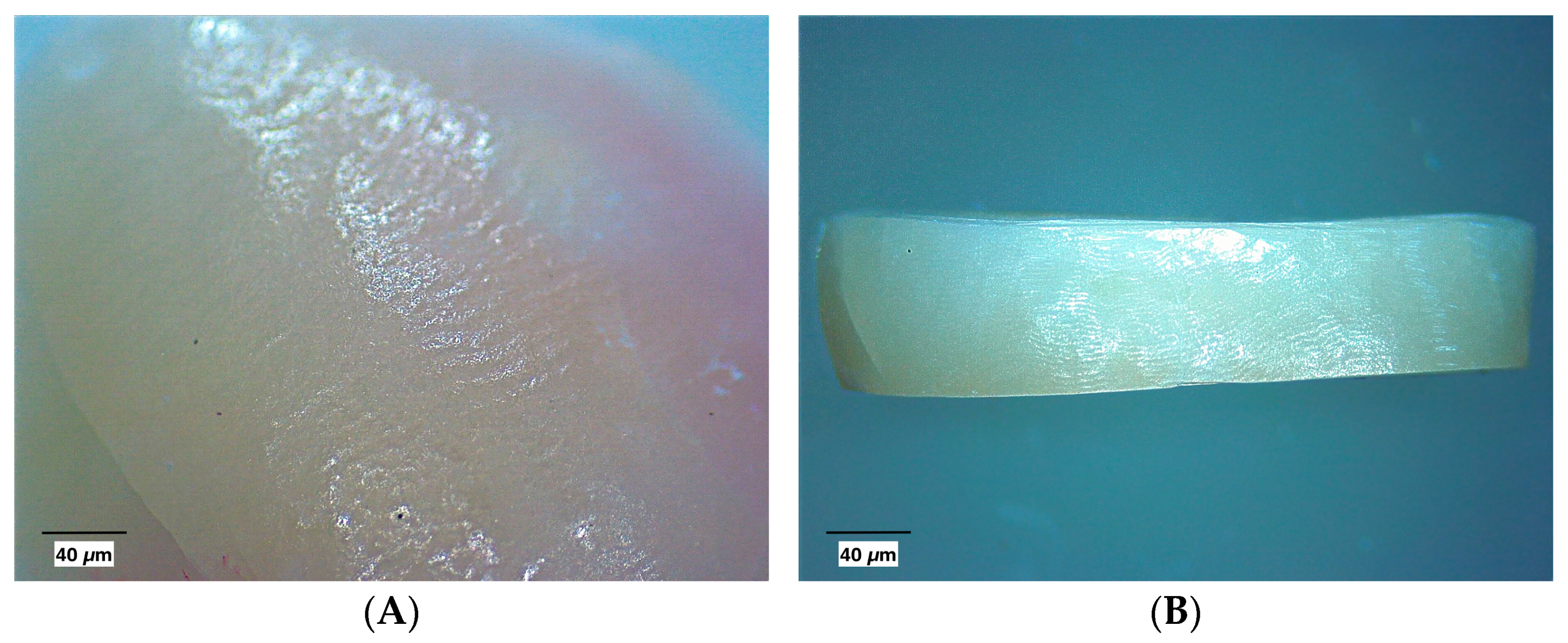
2.4. Macroscopic Evaluation

The blocks were macroscopically evaluated using a stereomicroscope (Leica EZ4E, Leica Microsystems, Balgach, Switzerland), and a vestibular and/or lingual surface photographic record was/were taken. The visual examination was performed according to the criteria of the International Caries Detection and Assessment System ICDAS II and coded as follows: 0—healthy tooth surface; 1—caries lesion in the outer half of the enamel; and 2—caries lesion in the inner half of the enamel. Two previously calibrated researchers performed macroscopic evaluations. Weighted Kappa was calculated to evaluate agreement between examiners. High intra-examiner agreement was obtained for macroscopic evaluation/ICDAS II (Kappa 1.00) and macroscopic evaluation/DIAGNOdent pen (Kappa 0.967). The investigators individually evaluated each of the blocks while the surface was wet. Ninety-six enamel blocks, which met the characteristics of healthy enamel according to published evaluations, were selected [18].

2.5. Laser Fluorescence Measurement

The DIAGNOdent pen 2190 (DIAGNOdent pen, Kavo, Biberach, Germany) was used according to the manufacturer’s recommendations; this is a device which measures the depth of the caries lesion and gives a unitless numerical value. A cylindrical sapphire tip was fitted for smooth surfaces and calibrated using a ceramic reference block. For each measurement, the device was zeroed; the sapphire tip was moved over the selected evaluation sites at a 90-degree angle, as indicated by the manufacturer. Evaluations were completed by two examiners. Each examiner made two measurements for each test site, and the mean values were recorded (reported as initial evaluation in Table 1). The manufacturer’s suggested cut-off points were used to interpret the measurements and were coded as follows: 0–12 healthy tooth surfaces; 13–24 superficial caries in enamel; >25 lesions in the inner half of enamel [18].

2.6. Artificial Demineralization of the Sample Surfaces

The vestibular and/or lingual surfaces of the blocks were demineralized following the application of 35% phosphoric acid 3M, ESPE, St. Paul, MN, USA, for 1 min (Figure 1B,D). The samples were washed with deionized water for acid removal and evaluated again using the DIAGNOdent pen (Table 1) to establish differences between measurements and an adequate demineralization process [19].

2.7. Application of Remineralizing Agents

The samples were divided into six different groups (n = 16 for each group) according to the remineralizing agent: (1) FV (Clinpro, 3M ESPE, St. Paul, MN, USA); (2) FV + CG; (3) FV + AgNPs; (4) FV + AgNPs + CG; (5) AgNPs + CG; and (6) artificial saliva (Kim Hidrat, Spain) composed of Xylitol 1%, potassium thiocynate, potassium chloride, sodium chloride, calcium chloride, magnesium chloride, potassium dihydrogen phosphate, sodium saccharin, hydrogenated castor oil, sodium methylparaben, sodium propylparaben, bonoprol, menthol, aroma, citric acid, and water. For the preparation of the different treatment combinations, a ratio of 900 μL:20 μL FV–composite was used. The application volume was 1000 microliters, with an FV ratio of 90% and 2% compound. This ratio was considered based on a previous study on the bacteriological efficacy of chitosan gel and silver nanoparticles [14,20].
A thin layer of remineralizing agent was applied using a microbrush on the demineralized enamel surfaces. The remineralizing agent applications were completed according to the manufacturer’s directions. After application, the samples were allowed to stand for 5 min and then immersed in a commercial artificial saliva solution with a pH of 7.2 and kept in an incubator at 37 °C for the exposure time. Four exposure times were considered (24, 48, 120, and 168 h), and treatment was applied every 24 h. At the end of the exposure time, the varnish was carefully removed, and the samples were washed with deionized water. After the application of remineralizing agents, the samples were rinsed with deionized water, removing excess material with a scalpel blade. Group 6 was immersed in the artificial saliva solution without any treatment. The pH value of the artificial saliva solution for the groups was 7.2, and the solution was renewed daily. Evaluations with a DIAGNOdent pen 2190 were performed at the end of each exposure time to record the surface changes.

2.8. X-Ray Diffraction

For the analysis of the crystal structure of tooth enamel, a Rigaku miniFlex 300/600 System X-ray diffractometer (Rigaku Corporation, Tokyo, Japan) using CuKα radiation was used. The operating conditions were as follows: 600 W X-ray tube; wavelength of λ = 0.154056 nm at 40 kV. Diffraction spectra were acquired in the range of 10–80 degrees. Thirty-two tooth blocks were evaluated which, were pulverized with mortar, obtaining 30 mg; this content was dehydrated using ethanol and placed in the sample holder for the respective analysis in the X-ray diffractor.

2.9. Scanning Electron Microscopy and Energy-Dispersive X-Ray Analyses

Scanning electron microscopy (SEM) and energy dispersive X-ray analysis (EDX) were performed at the Centre for High-Throughput Phenogenomics (University of British Columbia) on an FEI Helios Nanolab 650 SEM/FIB equipped with an EDAX Octane Pro EDX detector (FEI Company, Hillsboro, OR, USA). SEM was used to characterize morphological changes in enamel surfaces after the remineralization procedures. Surface morphology images of the enamel samples were acquired using a 1 kV electron beam with accelerating voltage. Compositional information was obtained via EDX analysis to determine the presence of calcium and phosphorous and their relative concentrations in the sample surfaces. The EDX analyses were performed using a 10 kV electron beam with accelerating voltage.

2.10. Statistical Analysis

The data obtained were subjected to statistical analysis using SPSS V25 software. Kolmogorov–Smirnov was used to identify the normality of the data. Data analysis was performed using the Student’s T and Bonferroni correction test. The Bonferroni correction test was used to analyze the comparison of means between laser fluorescence assessments. The significance level was set at p < 0.05, with a 95% confidence interval.

3. Results

3.1. Macroscopic Evaluation

A total of 122 blocks of vestibular/lingual surfaces were examined in this study, and 96 of them were included in this study after the microscope examination. Selected images of the blocks were evaluated for healthy enamel characteristics (smooth and continuous surface without white spots) via stereomicroscopy and ICDAS II system assessments (Figure 1A,B). In contrast, images of enamel after the demineralization process are shown in Figure 1C,D. The demineralization process was confirmed using fluorescence.

3.2. Laser Fluorescence

After different times of each treatment, the assessments obtained by DIAGNOdent pen (demineralized/post-treatment) were compared, and we observed that the laser fluorescence values decreased significantly after 168 h of applying the remineralization process in all treatment groups. Table 1 indicates the average values obtained with laser fluorescence before and after demineralization and after the application of remineralizing agents. Significant differences (p < 0.05) were obtained between the initial evaluations and after artificial demineralization. Likewise, the comparison of means between the exposure groups was established, being significant for the FV groups (24, 48, 120, and 168 h), FV+ CG (24, 120, and 168 h), FV + AgNPs (24, 48, and 168 h), and FV + AgNPs + CG (24 and 168 h). The lowest value was achieved using the FV + AgNPs + CG treatment, with a reduction from 72.5 to 4.75. The means values obtained with laser fluorescence before and after demineralization were recorded (Table 1). The individual values obtained for the group at 48 and 120 h are scattered around the mean, which represents the variation in the data obtained.

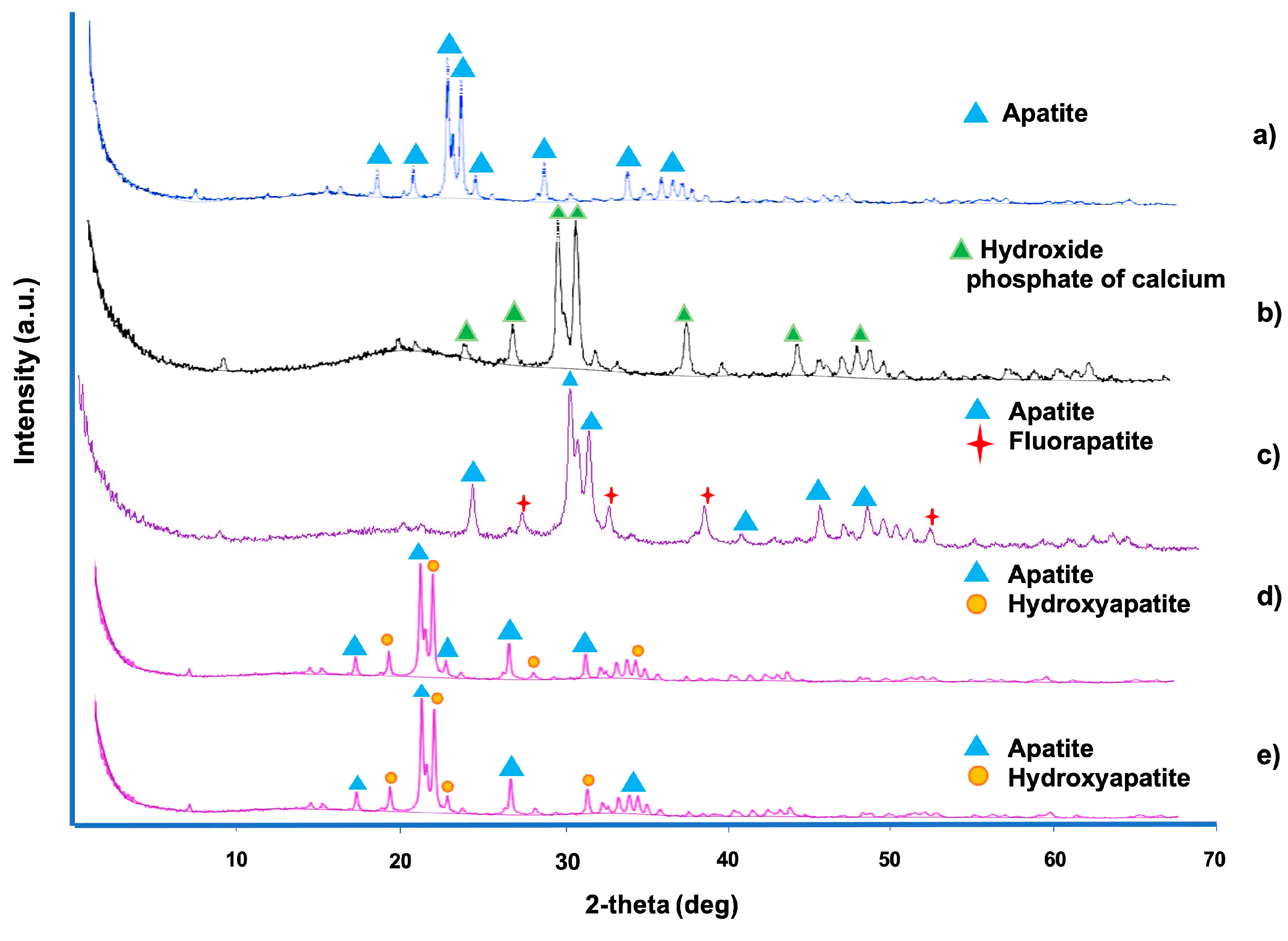
3.3. X-Ray Diffraction

For X-ray diffraction (XRD) evaluation, the results were analyzed in SmartLab Studio II and OriginPro software, Version 2023b (OriginLab Corporation, Northampton, MA, USA). Refinement was performed via the Rietveld method. Lattice parameters indicating the presence of hydroxyapatite and fluorapatite were observed in the samples exposed to FV + CG + AgNPs at 24, 48, and 120 h. The phases identified in the diffractogram (Figure 2) indicate the presence of apatite in healthy enamel and calcium hydroxide phosphate in demineralized enamel. Table 2 indicates the crystalline phases and crystallographic parameters identified in the samples through refinement via the Rietvelt method. The phases of apatite and modified hydroxyapatite are mainly identified, highlighting the identification of fluorapatite in the FV + AgNPs + CG group at 24 h of exposure. The crystallographic axes (a, b, and c) are also indicated, which indicate that the structures evaluated are anisotropic in the “c” direction, which means that the properties and arrangement of the atoms varies in this direction with a hexagonal configuration.

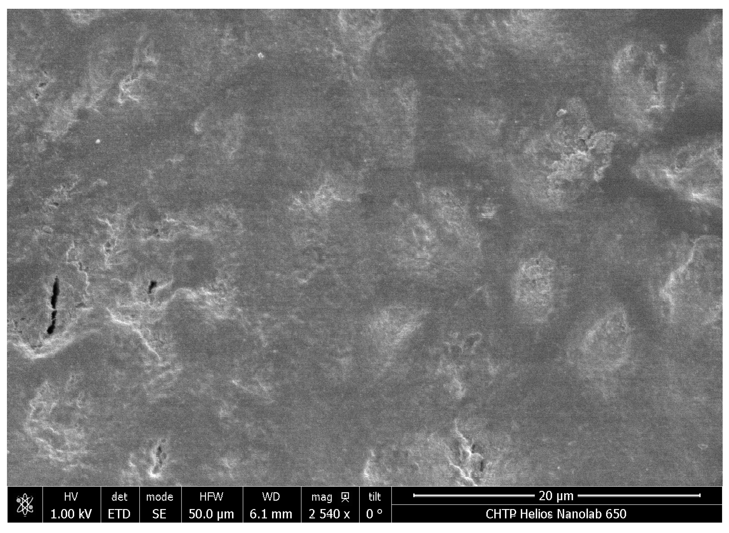
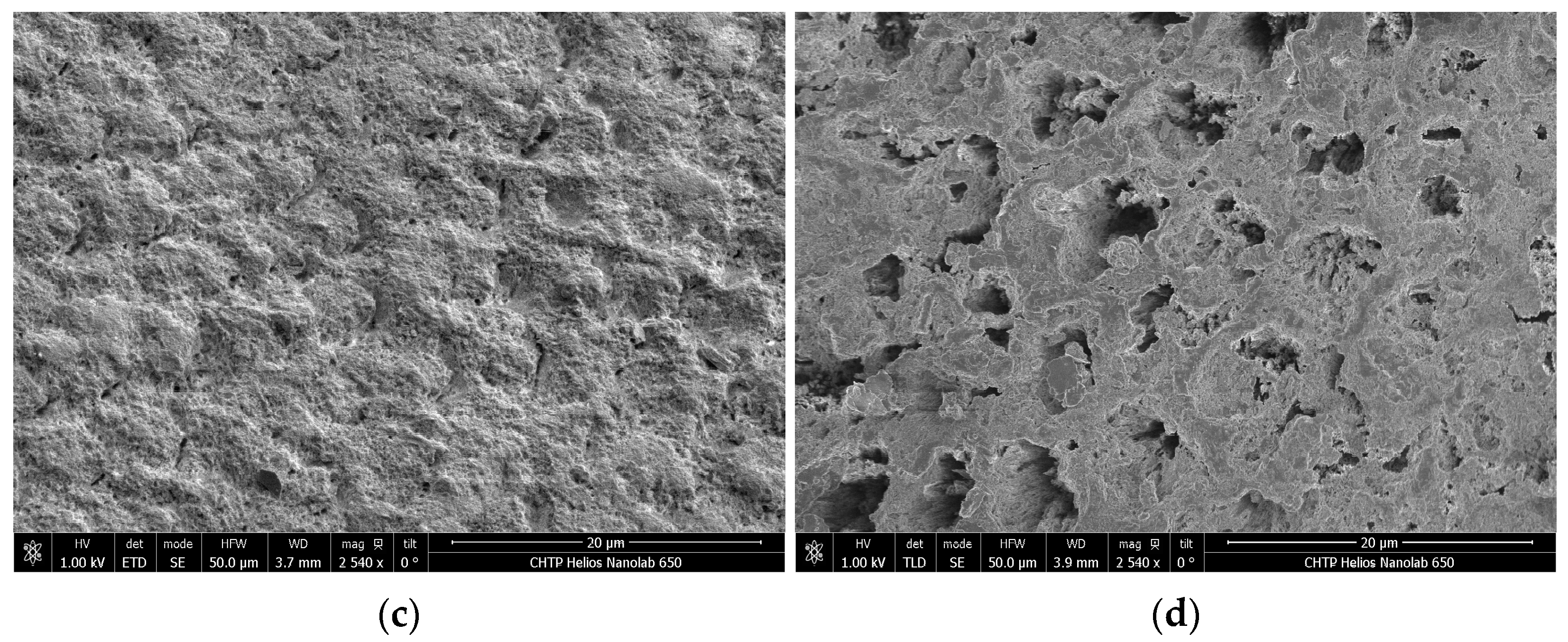
3.4. Scanning Electron Microscopy and EDX Analysis

Surface analysis using scanning electron microscopy showed a characteristic smooth surface in healthy enamel (Figure 3) but not in the artificial saliva group (Figure 4), where eroded prismatic enamel and interprismatic enamel protrusions are identified as a result of the demineralization process. The concentration of the crystalline structure of the enamel shows variations in all groups with respect to normal enamel, with an increase concerning the eroded enamel of the control group. In the groups to which the remineralizing agent was applied at 120 h and 168 h (Figure 4 and Figure 5), morphological changes were observed on the surface where the presence of small crystals was identified in all the groups; however, they do not have a homogeneous distribution.
The EDX elemental analysis (Figure S1 in Supplementary File) confirmed that the surface consisted of calcium, phosphorus, carbon, and oxygen. Within the comparison of the groups exposed at 120 h and 168 h, the results of the present study show that after remineralization, the carbon and oxygen content increased in all exposed groups, except in the control group. F content was identified in the FV group (0.92 weight %) and the combination of FV + CG + AgNPs group (0.91) at 120 h and in the groups exposed to 168 h FV + CG + AgNPs and CG gel + AgNPs at 0.19 and 3.5, respectively, with higher F content in the samples combined with CG + AgNPs for the 120 h and 168 h groups. The detection limit of the technique could be the reason why there were groups where FV was used but F was not detected. The Ca/P ratio was higher in the samples treated with FV + CG + AgNPs, CG, and AgNPs in both exposure groups. The values closer to normal enamel indicate that remineralizing treatment achieves a great percentage recovery of enamel mineralization.

4. Discussion

Remineralization is a natural process by which lost minerals are redeposited in the enamel. However, this process is often not sufficient to counteract the continuous loss of minerals, as conditions may vary according to oral conditions. In this regard, biomaterials such as AgNPs and CG play an important role in the remineralization process due to their characteristics and properties [12,13,14,18,19]. AgNPs have a broad spectrum of antibacterial properties which can inactivate enzymes and prevent DNA replication in bacteria, thereby reducing the risk of caries [20,21]. Moreover, several studies indicate that chitosan products enhance remineralization in artificial lesions in vitro [22,23,24]. Changes in the surface of demineralized enamel indicate a negative charge due to the leaching of Ca++, and the positive charge left from the chitosan favors adhesion to the demineralized surface [25].
Diagnostic evaluation using the DIAGOdent pen laser is a complementary method for the diagnosis of lesions on the smooth surface of the enamel. Likewise, it is capable of detecting in vitro changes in the surface of dental enamel after exposure to a demineralizing agent. DIAGNOdent pen titrations obtained pre- and post-artificial demineralization indicate significant differences. This is congruent with the XRD analysis and SEM analysis showing important variations among the samples evaluated.
X-ray diffraction results indicate changes related to significant mineral loss and surface changes in the artificially demineralized samples, indicating the presence of calcium hydroxide phosphate. This denotes variations in the composition of the enamel surface in an in vitro context. This allows us to establish that laser fluorescence is capable of detecting in vitro changes in the surface of dental enamel after exposure to a demineralizing agent. The identified differences between initial and post-demineralization values were measured with the DIAGNOdent pen on the smooth surface of permanent teeth [25]. However, slight variations were measured with the DIAGNOdent pen on the enamel surface after exposure to different acid solutions [26].
The mechanism of remineralization under in vitro conditions has been studied elsewhere, but there is still no agreement among researchers. According to Enax et al. (2024), “…It has been conclusively shown by such in vitro crystallization studies under well-defined conditions that additives can influence nucleation and crystal growth of calcium phosphate even at low concentrations…” [27]. In our research, we found that the presence of silver nanoparticles promotes a different sort of compound in the crystallization products (see DRX results) due to a heterogeneous precipitation process, and also, the great affinity of silver to fluoride is well known, and the presence of silver fluoride in low concentrations—not detectable via XRD—can influence the remineralization process. Research in this area is also needed.
For this present study, the comparison between combinations of remineralizing treatments, considering measurements of demineralized surface and post-treatment with the DIAGNOdent pen, indicate significant differences. Additionally, an increase in means is identified for the FV, FV + AgNPs, FV + GC, and FV + CG + AgNPs groups at 24 and 68h post-exposure, compared to values recorded post-desmineralization. The evaluations stand out for groups containing CG (FV + CG and FV + CG + AgNPs) compared to the conventional FV treatment group; this could be related to CG’s adhesion capability on the enamel surface. Other studies identified differences in the means obtained in the groups where chitosan was incorporated, showing significantly lower levels compared to other remineralizing agents [16,28]. Similar conclusions were identified with the same system in groups of enamel blocks exposed to remineralizing treatment [29].
The results related to the exposure time show significant differences in the four exposed groups. It is noteworthy that the XRD results identify crystalline phases of hydroxyapatite in the FV + CG + AgNPs treatment for 120 h groups, congruent with remineralization. The control group, in which only artificial saliva was used, shows remineralizing potential, indicating a decrease in the measurements obtained in this group.
The results of the commercial FV show a predictable behavior concerning its remineralizing effects; however, the means obtained after the application of the treatment groups that included chitosan indicate significant variations with respect to the measurements in demineralized samples.
The samples processed in XRD indicate important variations in the composition and phases identified in the samples submitted to artificial demineralization compared to healthy samples. This allows us to establish that the variations identified by the post-remineralization evaluations allow us to identify phases that coincide with a remineralization process.
Other studies reported similar characteristics in the diffraction spectra obtained in healthy enamel and enamel exposed to F [30,31,32,33]. During the evaluation, a loss of crystallinity in demineralized enamel was recorded. Although the samples exposed to F denote characteristic apatite peaks, they indicate that the apatite crystal lattice was recovered to some extent. However, samples exposed to nanocomplexes of phosphorylated chitosan and amorphous calcium phosphate (Pchi-ACP) showed narrower and sharper peaks in the diffractogram. Similar results were reported with the use of mesoporous bioactive glasses loaded with amorphous calcium phosphate to treat white spot lesions [34]. Diffraction analysis indicated that remineralized deposits in all four groups showed a diffraction pattern similar to that of normal enamel and shared a crystal structure similar to hydroxyapatite. Likewise, as a result of the refinement through the Rietveld method, variations in the crystallographic parameters were identified, registering the presence of fluorapatite and hydroxyapatite in the group of blocks exposed to FV + CG + AgNPs at 48 h and 120 h of exposure, which is congruent with remineralization on the enamel surface.
On the other hand, previous studies have used SEM-EDX analysis to study the changes associated with the demineralization–remineralization of dental enamel [3,28,35,36,37]. In the current study, enamel remineralization included the evaluation of samples on EDX, identifying the weight percentage of different minerals such as calcium and phosphate because they represent the main composition of hydroxyapatite. Ca/P titrations are important indicators of the effects of agents on enamel remineralization [38]. For example, changes in the Ca/P ratio were monitored for samples exposed at 120 h and 168 h. The results indicate that remineralizing agents such as FV, combined with CG and AgNPs, improved enamel remineralization. It is considered that the remineralization process is related to the replacement of minerals, mainly through the redeposition of calcium and phosphate in the enamel [3]. Research on the release of F and its applications in the remineralization process indicates the capacity of F release after an initial application followed by continuous diffusion of F, which allows us to determine that it is one of the treatments with better results for the management of white spot lesions in enamel; however, its effect decreases after seven days of treatment [4,38]. On the other hand, other minerals or compounds besides Ca and P could have a great influence on the remineralization process; for example, chitosan, which, due to its high nitrogen content, represents an excellent vehicle for transporting ions such as Ca and P for biomineralization. Likewise, chitosan adheres to negatively charged surfaces such as those of demineralized enamel [30,39,40,41]. The results of this study indicate a higher Ca/P ratio in the groups in which commercial FV was combined with CG and AgNPs.
Table 3 shows the results of some studies concerning remineralization using nanoparticles and chitosan in recent years. As we can see, there is active research seeking a coating material with sustained activity without the complications of the use of fluoride, as we stated in the introduction to this paper.
Finally, some questions about the biocompatibility of silver nanoparticles in dentistry have been raised by researchers; in previous studies, we have evaluated the biocompatibility of these gallic acid stabilized-silver nanoparticles, finding cell viability percentages higher than 95% when tested against human dermal fibroblast, and we have also reported that AgNPs concentrations less than 4 μg/mL showed a cytotoxic effect that resulted in a death rate of 13.8% or less. We also found that AgNPs at 500 ppm results in a viability above 75% in human fibroblasts. In an in vivo study, it was found that silver nanoparticles suspensions administrated to Wistar rats did not generate alterations among clinical characteristics, and clinical chemistry and hematology values showed slight alterations; however, at the end of the treatment, most were inside the normal concentration [48].
Considering that the present study was carried out in a laboratory environment, some of the limitations can be attributed to the difficulty of imitating the in vivo characteristics of saliva in the oral cavity. It is necessary to continue the evaluations assessing the microhardness of the enamel after exposure to remineralizing agents. Likewise, based on the results, in vivo evaluations could be considered.

5. Conclusions

The results of the present in vitro study suggest that the combination of FV + CG + AgNPs and CG + AgNPs could induce changes in the surface of the dental enamel related to remineralization, extending its effect up to 168 h compared to commercial FV. Although all the tested materials produce a remineralization effect, it is concluded that they do it at different proportions, producing different remineralization products as observed in X-ray diffraction analysis. Future research could include atomic force microscopy and microhardness analysis to confirm these results. The main contributions of this work are as follows:
  • The changes evaluated on the enamel surface after applying FV + CG + AgNPs are related to the remineralization process, extending its effect up to 168 h compared to FV.
  • The absence of pigmentation on the enamel surface is also highlighted.
Although these results are remarkable, further investigation into the variations in the microhardness of the enamel surface after exposure to the treatment is needed. Additionally, its clinical evaluation should be considered, considering the variations in salivary pH in vivo.

Supplementary Materials

The following supporting information can be downloaded at: https://www.mdpi.com/article/10.3390/coatings15010040/s1, Figure S1. EDX results for all groups at 120 h and 168 h of treatment.

Author Contributions

Conceptualization, N.N.-M. and G.-A.M.-C.; formal analysis, K.A.A.d.R.; funding acquisition, N.N.-M.; investigation, K.A.A.d.R., W.Y.E.d.G., V.C.M., S.M., N.N.-M. and G.-A.M.-C.; methodology, K.A.A.d.R., W.Y.E.d.G., V.C.M., N.N.-M. and G.-A.M.-C.; writing—original draft preparation, K.A.A.d.R., N.N.-M. and G.-A.M.-C.; writing—review and editing, H.B. and G.-A.M.-C. All authors have read and agreed to the published version of the manuscript.

Funding

This research received no external funding.

Institutional Review Board Statement

This study was conducted in accordance with the Declaration of Helsinki and approved by Research Ethics Committee of the Faculty of Dentistry of the University of El Salvador with the following document number: CEI-FOUES/2022/003.

Informed Consent Statement

Informed consent was obtained from all subjects involved in this study. Participants provided verbal consent, and the recruitment of the patients ranged from 1 April 2022 until 3 March 2023.

Data Availability Statement

Data are contained within the article.

Conflicts of Interest

The authors declare no conflicts of interest.

References

  1. John, H.; Franklin, G.-G.; Catherine, F. Biological factors in dental caries: Role of saliva and dental plaque in the dynamic process of demineralization and remineralization (part 1). J. Clin. Pediatr. Dent. 2003, 28, 47–52. [Google Scholar]
  2. Chen, H.; Gu, L.; Liao, B.; Zhou, X.; Cheng, L.; Ren, B. Advances of Anti-Caries Nanomaterials. Molecules 2020, 25, 5047. [Google Scholar] [CrossRef]
  3. Kim, M.-J.; Li, M.-J.; Kim, K.-M.; Yang, S.-Y.; Seo, Y.-J.; Chul, C.S.; Ren, B. Enamel Demineralization Resistance and Remineralization by Various Fluoride-Releasing Dental Restorative Materials. Materials 2021, 14, 4554. [Google Scholar] [CrossRef]
  4. Chhatwani, S.; Hoppe, J.; Naumova, E.A.; Arnold, W.H.; Möhlhenrich, S.C.; Bizhang, M.; Danesh, G. Fluoride ion release characteristics of fluoride-containing varnishes-an in vitro study. Appl. Sci. 2021, 11, 1452. [Google Scholar] [CrossRef]
  5. Parisay, I.; Boskabady, M.; Bagheri, H.; Babazadeh, S.; Hoseinzadeh, M.; Esmaeilzadeh, F. Investigating the efficacy of a varnish containing gallic acid on remineralization of enamel lesions: An in vitro study. BMC Oral Health 2024, 24, 175. [Google Scholar] [CrossRef] [PubMed]
  6. Oliveira, B.H.; Rajendra, A.; Veitz-Keenan, A.; Niederman, R. The effect of silver diamine fluoride in preventing caries in the primary dentition: A systematic review and meta-analysis. Caries Res. 2019, 53, 24–32. [Google Scholar] [CrossRef]
  7. Tejaswi, B.; Gopal Sree, V.; Sivapriya, E.; Archana, D.; PradeepKumar, A.R. Nanoparticles in caries prevention: A review. J. Glob. Oral Health 2021, 4, 56–66. [Google Scholar] [CrossRef]
  8. Salas-López, E.K.; Pierdant-Pérez, M.; Hernández-Sierra, J.F.; Ruíz, F.; Mandeville, P.; Pozos-Guillén, A.J. Effect of silver nanoparticle-added pit and fissure sealant in the prevention of dental caries in children. J. Clin. Pediatr. Dent. 2017, 41, 48–52. [Google Scholar] [CrossRef]
  9. Pichaiaukrit, W.; Thamrongananskul, N.; Siralertmukul, K.; Swasdison, S. Fluoride varnish containing chitosan demonstrated sustained fluoride release. Dent. Mater. J. 2019, 38, 1036–1042. [Google Scholar] [CrossRef]
  10. Ghafar, H.; Khan, M.I.; Sarwar, H.S.; Yaqoob, S.; Hussain, S.Z.; Tariq, I.; Madni, A.U.; Shahnaz, G.; Sohail, M.F. Development and Characterization of Bioadhesive Film Embedded with Lignocaine and Calcium Fluoride Nanoparticles. AAPS PharmSciTech 2020, 21, 60. [Google Scholar] [CrossRef]
  11. Memarpour, M.; Shafiei, F.; Rafiee, A.; Soltani, M.; Dashti, M.H. Effect of hydroxyapatite nanoparticles on enamel remineralization and estimation of fissure sealant bond strength to remineralized tooth surfaces: An in vitro study. BMC Oral. Health 2019, 19, 92. [Google Scholar] [CrossRef] [PubMed]
  12. Abedi, M.; Ghasemi, Y.; Nemati, M.M. Nanotechnology in toothpaste: Fundamentals, trends, and safety. Heliyon 2024, 10, e24949. [Google Scholar] [CrossRef] [PubMed]
  13. Surija, I.; Gunawan, H.A.; Amir, L.R. Effect of chitosan on the enamel demineralization process in vitro: An enamel solubility test. J. Phys. Conf. Ser. 2018, 1073, 052005. [Google Scholar] [CrossRef]
  14. Sámano-Valencia, C.; Martinez-Castanon, G.A.; Martínez-Gutiérrez, F.; Ruiz, F.; Toro-Vázquez, J.F.; Morales-Rueda, J.A.; Espinosa-Cristóbal, L.F.; Alonso, N.V.Z.; Martínez, N.N. Characterization and biocompatibility of chitosan gels with silver and gold nanoparticles. J. Nanomater. 2014, 2014, 543419. [Google Scholar] [CrossRef]
  15. Rajan, R.; Krishnan, R.; Bhaskaran, B.; Kumar, S.V. A Polarized Light Microscopic Study to Comparatively evaluate Four Remineralizing Agents on Enamel viz CPP-ACPF, ReminPro, SHY-NM and Colgate Strong Teeth. Int. J. Clin. Pediatr. Dent. 2015, 8, 42–47. [Google Scholar] [CrossRef] [PubMed]
  16. Yamakami, S.A.; Faraoni, J.J.; Lia, N.S.N.D.; Regula, F.B.; Ohyama, H.; Palma-Dibb, R.G. Effect of an experimental chitosan/casein gel on demineralized enamel under a cariogenic challenge. Dent. Med. Probl. 2022, 59, 531–538. [Google Scholar] [CrossRef]
  17. Seredin, P.; Goloshchapov, D.; Kashkarov, V.; Emelyanova, A.; Buylov, N.; Barkov, K.; Ippolitov, Y.; Khmelevskaia, T.; Mahdy, I.A.; Mahdy, M.A.; et al. Biomimetic Mineralization of Tooth Enamel Using Nanocrystalline Hydroxyapatite under Various Dental Surface Pretreatment Conditions. Biomimetics 2022, 7, 111. [Google Scholar] [CrossRef]
  18. Shaalan, O.O. DIAGNOdent versus International Caries Detection and Assessment System in detection of incipient carious lesions: A diagnostic accuracy study. J. Conserv. Dent. 2023, 26, 199–206. [Google Scholar] [CrossRef] [PubMed]
  19. El Moshy, S.; Abbass, M.M.S.; El-Motayam, A.M. Biomimetic remineralization of acid etched enamel using agarose hydrogel model. F1000Res 2018, 7, 1476. [Google Scholar] [CrossRef]
  20. Sámano-Valencia, C.; Martinez-Castanon, G.A.; Martínez-Martínez, R.E.; Loyola-Rodríguez, J.P.; Reyes-Macías, J.F.; Ortega-Zarzosa, G.; Niño-Martínez, N. Bactericide efficiency of a combination of chitosan gel with silver nanoparticles. Mater. Lett. 2013, 106, 413–416. [Google Scholar] [CrossRef]
  21. Noronha, V.T.; Paula, A.J.; Durán, G.; Galembeck, A.; Cogo-Müller, K.; Franz-Montan, M.; Durán, N. Silver nanoparticles in dentistry. Dent. Mater. 2017, 33, 1110–1126. [Google Scholar] [CrossRef]
  22. Ruan, Q.; Zhang, Y.; Yang, X.; Nutt, S.; Moradian-Oldak, J. An amelogenin-chitosan matrix promotes assembly of an enamel-like layer with a dense interface. Acta Biomater. 2013, 9, 7289–7297. [Google Scholar] [CrossRef] [PubMed]
  23. Ruan, Q.; Liberman, D.; Bapat, R.; Chandrababu, K.B.; Phark, J.H.; Moradian-Oldak, J. Efficacy of amelogenin-chitosan hydrogel in biomimetic repair of human enamel in pH-cycling systems. J. Biomed. Eng. Inform. 2015, 2, 119. [Google Scholar] [CrossRef] [PubMed]
  24. Nimbeni, S.B.; Nimbeni, B.S.; Divakar, D.D. Role of chitosan in remineralization of enamel and dentin: A systematic review. Int. J. Clin. Pediatr. Dent. 2021, 14, 562–568. [Google Scholar] [CrossRef] [PubMed]
  25. NouhzadehMalekshah, S.; Fekrazad, R.; Bargrizan, M.; Kalhori, K.A. Evaluation of laser fluorescence in combination with photosensitizers for detection of demineralized lesions. Photodiag. Photodyn. Ther. 2019, 26, 300–305. [Google Scholar] [CrossRef]
  26. Kolumban, A.; Moldovan, M.; Țig, I.A.; Chifor, I.; Cuc, S.; Bud, M.; Badea, M.E. An evaluation of the demineralizing effects of various acidic solutions. Appl. Sci. 2021, 11, 8270. [Google Scholar] [CrossRef]
  27. Enax, J.; Fandrich, P.; Schulze zur Wiesche, E.; Epple, M. The Remineralization of Enamel from Saliva: A Chemical Perspective. Dent. J. 2024, 12, 339. [Google Scholar] [CrossRef] [PubMed]
  28. Zhang, J.; Lynch, R.J.M.; Watson, T.F.; Banerjee, A. Remineralisation of enamel white spot lesions pre-treated with chitosan in the presence of salivary pellicle. J. Dent. 2018, 72, 21–28. [Google Scholar] [CrossRef]
  29. Üstün, N.; Aktören, O. Analysis of efficacy of the self-assembling peptide-based remineralization agent on artificial enamel lesions. Microsc. Res. Tech. 2019, 82, 1065–1072. [Google Scholar] [CrossRef]
  30. Zhang, X.; Li, Y.; Sun, X.; Kishen, A.; Deng, X.; Yang, X.; Wang, H.; Cong, C.; Wang, Y.; Wu, M. Biomimetic remineralization of demineralized enamel with nano-complexes of phosphorylated chitosan and amorphous calcium phosphate. J. Mater. Sci. Mater. Med. 2014, 25, 2619–2628. [Google Scholar] [CrossRef]
  31. Bijle, M.N.; Abdalla, M.M.; Ashraf, U.; Siu, K.W.; Tsoi, J.; Yiu, C.K.Y. The acid-resistance potential of arginine-fluoride varnish treated enamel. J. Mech. Behav. Biomed. Mater. 2022, 125, 104763. [Google Scholar] [CrossRef]
  32. Zhao, I.S.; Yin, I.X.; Mei, M.L.; Lo, E.C.M.; Tang, J.; Li, Q.; So, L.Y.; Chu, C.H. Remineralising dentine caries using sodium fluoride with silver nanoparticles: An in vitro study. Int. J. Nanomed. 2020, 15, 2829–2839. [Google Scholar] [CrossRef]
  33. Dai, L.L.; Mei, M.L.; Chu, C.H.; Lo, E.C.M. Remineralizing effect of a new strontium-doped bioactive glass and fluoride on demineralized enamel and dentine. J. Dent. 2021, 108, 103633. [Google Scholar] [CrossRef]
  34. Ren, J.; Rao, J.; Wang, H.; He, W.; Feng, J.; Wei, D.; Zhao, B.; Wang, X.; Bian, W. Synergistic remineralization of enamel white spot lesions using mesoporous bioactive glasses loaded with amorphous calcium phosphate. Front. Bioeng. Biotechnol. 2023, 11, 1109195. [Google Scholar] [CrossRef] [PubMed]
  35. Bijle, M.N.; Abdalla, M.M.; Ashraf, U.; Ekambaram, M.; Yiu, C.K.Y. Enamel remineralization potential of arginine-fluoride varnish in a multi-species bacterial pH-cycling model. J. Dent. 2021, 104, 103528. [Google Scholar] [CrossRef] [PubMed]
  36. Zhang, Y.Y.; Wong, H.M.; McGrath, C.P.J.; Li, Q.L. In vitro and in vivo evaluation of electrophoresis-aided casein phosphopeptide-amorphous calcium phosphate remineralisation system on pH-cycling and acid-etching demineralised enamel. Sci. Rep. 2018, 8, 8904. [Google Scholar] [CrossRef] [PubMed]
  37. Wassel, M.O.; Sherief, D.I. Ion release and enamel remineralizing potential of miswak, propolis and chitosan nano-particles based dental varnishes. Pediatr. Dent. J. 2019, 29, 1–10. [Google Scholar] [CrossRef]
  38. Vitiello, F.; Tosco, V.; Monterubbianesi, R.; Orilisi, G.; Gatto, M.L.; Sparabombe, S.; Memé, L.; Mengucci, P.; Putignano, A.; Orsini, G. Remineralization Efficacy of Four Remineralizing Agents on Artificial Enamel Lesions: SEM-EDS Investigation. Materials 2022, 15, 4398. [Google Scholar] [CrossRef] [PubMed]
  39. Zhang, J.; Boyes, V.; Festy, F.; Lynch, R.J.M.; Watson, T.F.; Banerjee, A. In-vitro subsurface remineralisation of artificial enamel white spot lesions pre-treated with chitosan. Dent. Mater. 2018, 34, 1154–1167. [Google Scholar] [CrossRef]
  40. Muşat, V.; Anghel, E.M.; Zaharia, A.; Atkinson, I.; Mocioiu, O.C.; Buşilă, M.; Alexandru, P. A chitosan–agarose polysaccharide-based hydrogel for biomimetic remineralization of dental enamel. Biomolecules 2021, 11, 1137. [Google Scholar] [CrossRef] [PubMed]
  41. Chu, S.; Wang, J.; Gao, F. The application of chitosan nanostructures in stomatology. Molecules 2021, 26, 6315. [Google Scholar] [CrossRef] [PubMed]
  42. Deokar, K.K.; Shashikiran, N.; Maurya, A.; Gaonkar, N.; Gugwad, S.; Hadakar, S.; Taur, S. Comparative Evaluation of Chitosan Nanoparticles, Silver Diamine Fluoride and Acidulated Phosphate Fluoride Gel on Microhardness of Artificial Carious Lesions Created on Extracted Teeth. J. Clin. Diagn. Res. 2020, 14, 20. [Google Scholar] [CrossRef]
  43. Hanafy, R.A.; El-Fattah, A.A.; Mostafa, D.; Kandil, S. Biomimetic chitosan against bioinspired nanohydroxyapatite for repairing enamel surfaces. Bioinspired Biomim. Nanobiomater. 2020, 9, 85–94. [Google Scholar] [CrossRef]
  44. Magalhães, T.C.; Teixeira, N.M.; França, R.S.; Denadai, Â.M.L.; Dos Santos, R.L.; Carlo, H.L.; Munchow, E.A.; de Carvalho, F.G. Synthesis of a Chitosan nanoparticle suspension and its protective effects against enamel demineralization after an in vitro Cariogenic challenge. J. Appl. Oral Sci. 2021, 29, e20210120. [Google Scholar] [CrossRef]
  45. Xu, V.W.; Nizami, M.Z.I.; Yin, I.X.; Lung, C.Y.K.; Yu, O.Y.; Chu, C.H. Caries Management with Non-Metallic Nanomaterials: A Systematic Review. Int. J. Nanomed. 2022, 17, 5809–5824. [Google Scholar] [CrossRef] [PubMed]
  46. Wahied, D.M.; Ezzeldin, N.; Abdelnabi, A.; Othman, M.S.; El Rahman, M.H.A. Evaluation of Surface Properties of Two Remineralizing Agents after Modification by Chitosan Nano Particles: An In vitro Study. Contemp. Clin. Dent. 2023, 14, 265–271. [Google Scholar] [CrossRef] [PubMed]
  47. Zhang, O.L.; Niu, J.Y.; Yin, I.X.; Yu, O.Y.; Mei, M.L.; Chu, C.H. Bioactive Materials for Caries Management: A Literature Review. Dent. J. 2023, 11, 59. [Google Scholar] [CrossRef]
  48. Tristán-López, J.-D.; Niño-Martínez, N.; Kolosovas-Machuca, E.-S.; Patiño-Marín, N.; De Alba-Montero, I.; Bach, H.; Martínez-Castañón, G.-A. Application of Silver Nanoparticles to Improve the Antibacterial Activity of Orthodontic Adhesives: An In Vitro Study. Int. J. Mol. Sci. 2023, 24, 1401. [Google Scholar] [CrossRef] [PubMed]
Figure 1. Macroscopic evaluation of vestibular/lingual surfaces by stereomicroscopy: (A) smooth and (B) continuous surfaces without white spots in healthy enamel; (C) smooth and (D) continuous surfaces showing porous opaque demineralized enamel.
Coatings 15 00040 g001aCoatings 15 00040 g001b
Figure 2. X-ray diffraction spectra: (a) healthy enamel; (b) enamel demineralized for 1 min with 35% phosphoric acid; (c) FV + CG + AgNPs-24 h; (d) FV + CG + AgNPs-48 h; (e) FV + CG + AgNPs-120 h.
Coatings 15 00040 g002
Figure 3. Scanning electron microscopy images of healthy enamel.
Coatings 15 00040 g003
Figure 4. Scanning electron microscopy images of the enamel surface. Exposure to (a) FV, (b) FV + CG + AgNPs, (c) CG + AgNPs, and (d) artificial saliva after 120 h of exposure.
Coatings 15 00040 g004aCoatings 15 00040 g004b
Figure 5. Scanning electron microscopy images of enamel surface. Exposure to (a) FV, (b) FV + CG + AgNPs, (c) CG + AgNPs, and (d) artificial saliva after 168 h of exposure.
Coatings 15 00040 g005
Table 1. DIAGNOdent pen scores were obtained at different exposure times and different remineralization treatments (n = 16/group), expressed as mean ± SD.
Initial aAfter b Exposition Time
Group 24 h48 h120 h168 h
nMeanSDMeanSDp *MeanSDMeanSDMeanSDMeanSDp **
1166.441.7966.0029.49<0.0561.75 **16.03 **81.5 **21.76 **61.25 **32.36 **36.50 **11.21 **<0.05
2166.942.4164.5632.7746.75 **12.04 **48.2535.3880.5 **37.00 **34.75 **11.35 **
3167.252.4465.0630.5547.00 **6.22 **77.50 **28.44 **12.54.4337.00 **22.14 **
4168.312.5576.7521.9281.75 **8.06 **36.2511.1230.513.7733.50 **1.91 **
5167.062.2970.5029.3514.256.9510.503.7027.7512.0412.255.68
6166.001.4161.4429.8328.2512.0413.501.737.51.734.751.50
a Evaluation with DIAGNOdent pen; b after demineralization; * associations evaluated via Student’s t-test p < 0.05. ** Bonferroni correction p < 0.05. Group 1, FV; fluoride varnish; 2, fluoride varnish + chitosan gel; 3, fluoride varnish + AgNPs; 4, fluoride varnish + AgNPs + chitosan gel; 5, AgNPs + chitosan gel; 6, artificial saliva.
Table 2. Identified crystalline phase.
Exposition Time TreatmentPhasea/Å *b/Å *c/Å *
Healthy enamelApatite9.439.436.87
Demineralized enamelCalcium phosphate hydroxide9.459.456.89
24 hFluoride varnishCarbonate Hidroxiapatite9.449.446.88
Fluoride varnish + chitosan gelApatite9.439.436.88
Fluoride varnish + NpAg + chitosan gelApatite
Fluorapatite
9.45
9.45
9.45
9.45
6.89
6.88
NpAg + chitosan gelApatite9.439.436.87
48 hFluoride varnishApatite9.459.456.88
Fluoride varnish + chitosan gelApatite9.459.456.88
Fluoride varnish + NpAg + chitosan gelHidroxiapatite9.439.456.87
NpAg + chitosan gelApatite9.439.436.87
120 hFluoride varnishApatite9.429.426.86
Fluoride varnish + chitosan gelApatite9.429.426.87
Fluoride varnish + NpAg + chitosan gelApatite
Hidroxiapatite
9.42
9.42
9.42
9.42
6.86
6.85
NpAg + chitosan gelApatite
Potasium, calcium
9.47
3.88
9.47
3.88
6.89
16.63
168 hFluoride varnishCarbonate Hidroxiapatite9.449.446.87
Fluoride varnish + chitosan gelApatite9.439.436.86
Fluoride varnish + NpAg + chitosan gel Apatite9.429.426.86
NpAg + chitosan gelApatite9.459.456.88
* lattice parameters.
Table 3. Comparison of findings on remineralization using nanoparticles and chitosan in selected articles.
Author(s)AimBioactive MaterialsMain ResultsRef.
Deokar et al.To evaluate and compare the efficacy of the remineralizing potential of acidulated phosphate fluoride gel, chitosan nanoparticles, and silver diamine fluoride on the microhardness of artificial carious lesions created on extracted teeth.Chitosan nanoparticles, silver diamine fluoride, and acidulated phosphate fluoride gel
  • Chitosan nanoparticles showed the highest increase in remineralization followed by SDF and APF gel
[42]
Hanafy et al.Chitosan was employed as a novel biomimetic mineralization model to repair damaged enamel to compare its performance with that of bioinspired zinc-doped nanohydroxyapatite.Chitosan–hydrogel and a zinc-doped nanohydroxyapatite
  • Marked reduction in the Ca/P ratio and the lack of the characteristic hydroxyapatite diffraction peaks were detected for demineralized enamel.
  • The remineralization stage revealed evident recovery of the mineral contents with apparent distinctive XRD patterns of hydroxyapatite in both groups.
  • SEM analysis showed the absence of etched enamel porosity with the formation of a newly formed rod-like apatite layer.
[43]
Muşat et al.To induce the biomimetic remineralization of acid-etched human enamel in artificial saliva containing fluoride under agarose and novel chitosan–agarose hydrogels action without a toxic crosslinking agent for chitosan.Chitosan (CS) and agarose (A) in a biopolymer-based hydrogel
  • The values for the CS-A series are closer to the theoretical value of the carbonated hydroxyapatite of natural enamel; those for the series-A indicate the formation of calcium deficient phosphates.
[40]
Magalhães et al.Synthesize, characterize, and determine the effects of a ChNPs suspension on human enamel after cariogenic challenge via pH cycling.Chitosan nanoparticles
  • ChNPs suspension minimized human enamel demineralization after a cariogenic challenge.
[44]
Yamakami et al.Evaluate the effects of an experimental chitosan/casein gel on enamel demineralization/remineralization in an environment with a high cariogenic challenge.Chitosan/casein gel
  • Chitosan seems to be the major component responsible for reducing the volume loss of demineralized enamel.
[16]
Xu et al.Review the types, properties, and potential uses of non-metallic nanomaterials systematically for managing dental caries.Biological organic nanomaterials, synthetic organic nanomaterials, carbon-based nanomaterials, and selenium nanomaterials
  • Chitosan nanoparticles have shown a remineralization ability on demineralized enamel.
[45]
Wahied et al.Evaluate the effect of chitosan nanoparticles on the remineralization
of the demineralized enamel surface after being added to nano-hydroxyapatite and nano-calcium
phosphate materials.
Nano-hydroxyapatite and nano-calcium phosphate
  • Addition of chitosan nanoparticles had a significant effect on the remineralization of the demineralized enamel surface.
  • The nano NHA + chitosan group had the highest significant value, while the Nβ-TCP group exhibited the lowest significant value.
[46]
Zhang et al.The purpose of this literature review is to provide an overview of current bioactive materials for caries management.Bioactive materials for caries management
  • Combination of AgNPs and NaF or nano-silver fluoride does not cause black staining and metallic taste, unlike SDF.
  • AgNPs’ properties suggest their potential as an effective anticaries agent without black staining.
[47]
Disclaimer/Publisher’s Note: The statements, opinions and data contained in all publications are solely those of the individual author(s) and contributor(s) and not of MDPI and/or the editor(s). MDPI and/or the editor(s) disclaim responsibility for any injury to people or property resulting from any ideas, methods, instructions or products referred to in the content.

